

Novitates - American Museum of Natural History
Novitates - American Museum of Natural History
Novitates - American Museum of Natural History

You also want an ePaper? Increase the reach of your titles
YUMPU automatically turns print PDFs into web optimized ePapers that Google loves.
AMERICAN MUSEUM<br />
<strong>Novitates</strong><br />
PUBLISHED BY THE AMERICAN MUSEUM OF NATURAL HISTORY<br />
CENTRAL PARK WEST AT 79TH STREET, NEW YORK, N.Y. 10024<br />
Number 2931, 42 pp., 34 figs., 2 tables February 6, 1 989<br />
Hamiltonichthys mapesi, g. & sp. nov.<br />
(Chondrichthyes; Elasmobranchii), from the<br />
Upper Pennsylvanian <strong>of</strong> Kansas<br />
JOHN G. MAISEY'<br />
A new genus and species <strong>of</strong> hybodontiform elasmobranch,<br />
Hamiltonichthys mapesi, is described<br />
on the basis <strong>of</strong> six specimens from the Upper<br />
Pennsylvanian (Middle Virgilian) Hartford Limestone<br />
<strong>of</strong> Hamilton Quarry, Kansas. The new genus<br />
ABSTRACT<br />
INTRODUCTION<br />
Hybodus and its allies (e.g., Acrodus, Asteracanthus)<br />
were common elasmobranchs<br />
during most <strong>of</strong> the Mesozoic era, and apparently<br />
occupied a wide range <strong>of</strong> aquatic habitats.<br />
Despite the paucity <strong>of</strong> well-preserved<br />
fossil specimens, it is now known that the<br />
cranial anatomy <strong>of</strong> these hybodontoid sharks<br />
was specialized (Maisey, 1982, 1983). Morphological<br />
peculiarities <strong>of</strong> the neurocranium,<br />
mandibular suspensorium, and dermal skeleton<br />
suggest that these hybodontoids collectively<br />
form a monophyletic group (Maisey,<br />
is related to "typical" Mesozoic hybodontoids such<br />
as Hybodus and Acrodus. Hamiltonichthys is the<br />
first articulated hybodontoid to be described from<br />
North America, and is the oldest complete hybodontoid<br />
known thus far.<br />
1987). The previously popular notion that<br />
some or all Recent elasmobranchs are descended<br />
from a Hybodus-like ancestor does<br />
not withstand critical investigation and cannot<br />
be substantiated on anatomical grounds<br />
(Maisey, 1982), although it is still maintained<br />
in some noncladistic studies (e.g., Thies, 1983;<br />
Thies and Reif, 1985; Reif, 1985). Supposedly<br />
"transitional" fossils (e.g., Palaeospinax,<br />
Synechodus) certainly share some apomorphic<br />
skeletal characters with Recent<br />
elasmobranchs, but possess none <strong>of</strong> the derived<br />
attributes <strong>of</strong> Mesozoic hybodontoids<br />
(Maisey, 1977, 1984a, 1984b, 1985). Recent<br />
Heterodontidae (Port Jackson sharks) and<br />
Mesozoic hybodontoids share only plesio-<br />
' Associate Curator, Department <strong>of</strong> Vertebrate Paleontology, <strong>American</strong> <strong>Museum</strong> <strong>of</strong> <strong>Natural</strong> <strong>History</strong>.<br />
Copyright © <strong>American</strong> <strong>Museum</strong> <strong>of</strong> <strong>Natural</strong> <strong>History</strong> 1989 ISSN 0003-0082 / Price $4.20
2 AMERICAN MUSEUM NOVITATES<br />
NO. 2931<br />
Fig. 1. Hamiltonichthys mapesi, g. and sp. nov., holotype; A, part, KUVP 86308; B, counterpart,<br />
AMNH 10807. Entire female individual, UV fluorescent image.
1989<br />
MAISEY: HAMILTONICHTHYS MAPESI<br />
3<br />
morphic characters (e.g., the presence <strong>of</strong> dorsal<br />
fin-spines and a large basal plate in the<br />
anal fin) and convergent characters (e.g., lowcrowned<br />
teeth, heterodonty, and jaws with a<br />
strong ethmoidal support). A close relationship<br />
between these hybodontoids and Heterodontus<br />
is no longer feasible (Maisey, 1982),<br />
although some other fossils previously regarded<br />
as "hybodonts" (e.g., Synechodus, Orthacodus)<br />
may have affinity with Recent galeomorphs<br />
or Heterodontus (Maisey, 1985;<br />
Cappetta, 1987).<br />
The cranial anatomy is now known in some<br />
Mesozoic hybodontoids, including Hybodus<br />
reticulatus (considered to be the type species<br />
<strong>of</strong>Hybodus; Koken, 1907; Woodward, 1916;<br />
Maisey, 1987). The postcranial skeleton is<br />
known in a few cases, where it is anatomically<br />
conservative (Maisey, 1982). One <strong>of</strong>the most<br />
striking peculiarities is the presence <strong>of</strong> elongate<br />
pleural ribs, although the systematic value<br />
<strong>of</strong> this feature is unsettled (Dick, 1978;<br />
Dick and Maisey, 1980; Maisey, 1982).<br />
Hybodontoids primitively possess paired<br />
cephalic spines and distinctive fin-spine morphology<br />
(Maisey, 1978, 1982). Various morphological<br />
and histological criteria <strong>of</strong>the teeth<br />
have been used in phylogenetic studies <strong>of</strong> hybodontoids,<br />
but the systematic value <strong>of</strong> such<br />
data has proven highly controversial (Glikman,<br />
1964, 1967; Patterson, 1966; Reif, 1973,<br />
1978a; Maisey, 1982, 1987). Dermal denticles<br />
have also been investigated (e.g., Reif,<br />
1978b) but their value in systematic studies<br />
seems limited (Reif, 1985).<br />
During the 19th and early 20th centuries<br />
it was customary to regard a great many isolated<br />
Upper Paleozoic chondrichthyan teeth,<br />
fin-spines, and other dermal structures as evidence<br />
<strong>of</strong> early "hybodonts," without much<br />
regard to the possibility <strong>of</strong> greater taxic diversity.<br />
Determining the systematic position<br />
and status <strong>of</strong> most taxa founded upon such<br />
disparate fragments is problematical, but with<br />
the discovery <strong>of</strong> articulated Late Paleozoic<br />
chondrichthyans at Bear Gulch (Montana),<br />
Bearsden (Scotland), and elsewhere, it is finally<br />
becoming evident that chondrichthyans<br />
underwent pr<strong>of</strong>ound taxic diversification<br />
early in their evolutionary history and that<br />
many lineages developed bizarre and specialized<br />
dermal defenses. Although this early<br />
radiation is poorly resolved, two <strong>of</strong> the putative<br />
lineages involved are <strong>of</strong> particular interest<br />
here. One <strong>of</strong> these includes sharks possessing<br />
teeth, cephalic spines, and fin-spines<br />
like those <strong>of</strong> the Mesozoic Hybodontoidei<br />
(Hay, 1899; Hussak<strong>of</strong>, 1911; Woodward,<br />
1934; Romer, 1942; Zidek, 1969; Lund, 1970;<br />
Berman, 1970; Bendix-Almgreen, 1975;<br />
Johnson, 1981; Maisey, 1982), while the other<br />
includes form-taxa (presently known only<br />
from fragmentary dermal remains) with tooth,<br />
denticle, and fin-spine morphologies resembling<br />
those <strong>of</strong> Recent elasmobranchs (Reif,<br />
1977; Reif and Goto, 1979; Duffin, 1980,<br />
1982a, 1982b; Maisey, 1982, 1984b; Thies,<br />
1983; Duffin and Ward, 1983; Turner, 1982,<br />
1985).<br />
Historically, a large number <strong>of</strong> other Paleozoic<br />
form-taxa have also been considered<br />
"hybodonts." Space precludes further discussion<br />
here, but these assignations are generally<br />
ill-founded (see Maisey, 1982, for--a<br />
review). Until recently, Tristychius and Onychoselache<br />
were the only articulated Paleozoic<br />
elasmobranchs which seemed even remotely<br />
related to Mesozoic hybodontoids<br />
(Dick, 1978; Dick and Maisey, 1980).<br />
An abandoned quarry in an Upper Pennsylvanian<br />
sequence near Hamilton, eastern<br />
Kansas, has yielded a small sample <strong>of</strong> wellpreserved<br />
hybodontoid elasmobranchs. These<br />
specimens provide an insight into the earlier<br />
history <strong>of</strong> "hybodont" sharks, and may<br />
eventually help to place the enigmatic taxa<br />
Tristychius and Onychoselache within a less<br />
chaotic phylogenetic perspective. To some<br />
extent, the Hamilton "hybodonts" also contribute<br />
to a better understanding <strong>of</strong> the putative<br />
relationship between Recent elasmobranchs<br />
and the hybodontoids. This new<br />
material represents the first articulated hybodontoid<br />
to be documented from North<br />
America, as well as the oldest articulated hybodontoid<br />
so far recorded.<br />
MATERIALS AND METHODS<br />
The Hamilton hybodont is described here<br />
on the basis <strong>of</strong> eight specimens (see below,<br />
systematic section). Six <strong>of</strong> these represent
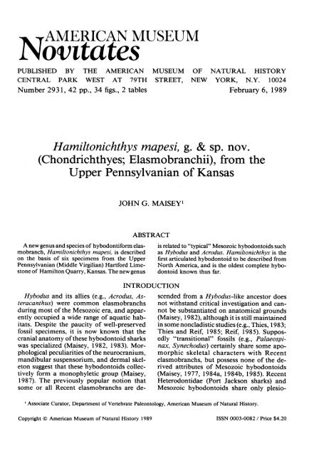
4 AMERICAN MUSEUM NOVITATES<br />
NO. 2931<br />
Fig. 2. Hamiltonichthys mapesi, KUVP 65016, part (A) and counterpart (B) <strong>of</strong> complete female<br />
individual. UV fluorescent image.
1 989 MAISEY: HAMILTONICHTHYS MAPESI<br />
5<br />
complete individuals. The most complete is<br />
designated the holotype <strong>of</strong> a new genus and<br />
species. The other two specimens are fragmentary;<br />
one is a caudal fin and the other<br />
consists <strong>of</strong> four associated lateral teeth within<br />
a coprolite. All the specimens are preserved<br />
in a thin-bedded argillaceous limestone containing<br />
a moderate amount <strong>of</strong> organic debris,<br />
especially carbonized plant fragments. The<br />
bedding planes on which the fishes are preserved<br />
are uneven, although it remains to be<br />
determined whether this is a result <strong>of</strong> diagenesis<br />
or <strong>of</strong> postdiagenetic disturbance. The<br />
specimens described here are all compressed,<br />
but are otherwise well preserved. The calcified<br />
endoskeleton and dermal skeleton are<br />
particularly clear, and the squamation gives<br />
us a good idea <strong>of</strong> body shape, fin position,<br />
and size. So much is preserved that interpretation<br />
becomes difficult where structures have<br />
become crushed on top <strong>of</strong> one another, for<br />
example in the pharyngeal region.<br />
In spite <strong>of</strong> the excellent state <strong>of</strong> preservation,<br />
much <strong>of</strong> the finer detail only becomes<br />
apparent using various fairly sophisticated<br />
techniques. Virtually every photograph in this<br />
work was taken with ultraviolet fluorescence<br />
or reflected-UV microphotography, a scanning<br />
electron microscope, or x-ray photography.<br />
Thus many <strong>of</strong> the features illustrated<br />
would be poorly visible, or even invisible, if<br />
conventional microscopical techniques had<br />
been used. This point is stressed in order to<br />
encourage other investigators <strong>of</strong> the important<br />
Hamilton Quarry biota to explore as<br />
broad a range <strong>of</strong> preparatory and illustrative<br />
techniques as possible. A brief note <strong>of</strong> the<br />
various methods employed is given here.<br />
The specimens required little mechanical<br />
preparation, and we chose not to make acid<br />
preparations by the transfer method. Some<br />
denticles were removed using dilute acetic<br />
acid. Where it was found necessary to clean<br />
small areas <strong>of</strong>matrix (e.g., around some teeth),<br />
excellent results were obtained by first immersing<br />
the fossil in Waller's Solution (for<br />
technique, see Waller, 1980; King, 1983),<br />
which contains sodium citrate (to sequestrate<br />
ferrous ions), sodium dithionite (a reducing<br />
agent), and sodium bicarbonate (to maintain<br />
neutrality). This immersion reduces ferrous<br />
ions in the matrix, rendering them water-soluble.<br />
The limestone matrix becomes slightly<br />
s<strong>of</strong>ter and hence more amenable to preparation<br />
using mounted needles. Teeth were<br />
cleaned from the matrix better using this<br />
technique than by using acid.<br />
Detailed investigation and microphotography<br />
<strong>of</strong> the specimens were mainly conducted<br />
under long-wave (3660 A) ultraviolet<br />
light (for details, see Rolfe, 1965). The skeletal<br />
material fluoresces a pale lemon yellow<br />
(calcified cartilage) or deep yellow to orange<br />
(teeth, fin-spines, squamation). Small areas<br />
were illuminated by mercury-vapor spot<br />
sources, but for views <strong>of</strong> entire specimens<br />
(e.g., figs. 1-5) it was necessary to cluster several<br />
small 15W blacklight tubes over the<br />
whole area. Exposures varied in duration up<br />
to approx. 20 minutes, depending on relief,<br />
degree <strong>of</strong> fluorescence, and aperture setting.<br />
Scanning electron microscopy <strong>of</strong> teeth and<br />
scales was conducted using a Cambridge<br />
Stereoscan-250 system operating at 10 kV.<br />
Specimens were gold spatter-coated. Denticles<br />
and a broken tooth were examined after<br />
being freed from the matrix by the Waller<br />
technique (see above). Sections <strong>of</strong> the dentitions<br />
<strong>of</strong> two specimens were also investigated<br />
by SEM, by making high-density epoxy<br />
casts from "Silastic" molds, and using the<br />
casts in place <strong>of</strong> the actual specimens (which<br />
were too large for the vacuum chamber). As<br />
the published figures show, there is no appreciable<br />
loss <strong>of</strong> detail at the levels <strong>of</strong> magnification<br />
used, and this technique provides<br />
views at otherwise unobtainable orientations.<br />
Reflected-fluorescence microphotography<br />
<strong>of</strong> epoxy tooth casts (sometimes in conjunction<br />
with visible light) was also utilized. By<br />
taking advantage <strong>of</strong> the shorter reflected<br />
wavelengths, this technique virtually doubles<br />
the resolution <strong>of</strong> fine detail in small objects.<br />
In general, the limestone matrix did not<br />
provide clear x-ray images, probably because<br />
ferruginous matter within the limestone, as<br />
well as the calcium itself, was blocking some<br />
<strong>of</strong> the radiation. Some details <strong>of</strong> the pharyngeal<br />
region were revealed in KUVP 86307,<br />
in which the matrix is extremely thin (fig. 7).<br />
Comparisons are made throughout the text<br />
with many Recent and fossil elasmobranchs.<br />
It was originally intended to illustrate many
6 AMERICAN MUSEUM NOVITATES<br />
NO. 2931<br />
Fig. 3. Hamiltonichthys mapesi, KUVP<br />
6501 7A, complete male individual with plant frond<br />
obscuring part <strong>of</strong> head. UV fluorescent image.<br />
Fig. 4. Hamiltonichthys mapesi, KUVP 86304,<br />
complete female individual. UV fluorescent image.
1989 MAISEY: HAMILTONICHTHYS MAPESI<br />
7<br />
<strong>of</strong> the comparative features by means <strong>of</strong> line<br />
drawings and/or photographs, but these plans<br />
were abandoned in order to publish the descriptive<br />
account in a timely manner. It is<br />
hoped that sufficient references are given in<br />
the text to enable the interested reader to seek<br />
out pertinent literature.<br />
ON THE NAMING OF ICHTHYOLITHS<br />
Following the formal systematic diagnosis<br />
below, I have appended a second ichthyolithic<br />
diagnosis, utilizing the terminology and<br />
enumeration recently developed by Tway and<br />
Zidek (1982, 1983a, 1983b) for describing<br />
isolated microscopic fish skeletal remains<br />
from the Late Pennsylvanian <strong>of</strong> North<br />
America. My purpose in doing this is not to<br />
assign particular taxonomic handles to their<br />
un-named categories. Indeed, to do so would<br />
in my opinion be a retrograde step to the dark<br />
ages <strong>of</strong> paleontological investigation, when<br />
every ichthyolithic scrap was assigned a binomial<br />
albatross to wear around its neck. Jaekel<br />
(1889) long ago discovered that the proliferation<br />
<strong>of</strong>genera and species founded upon<br />
scraps <strong>of</strong> fossil vertebrates can create enormous<br />
nomenclatural problems. Even where<br />
certain categories <strong>of</strong> ichthyolithic remains can<br />
be shown to occur in a fossil species, it would<br />
be foolhardy indeed to attempt to synonymize<br />
all such similar ichthyoliths, given the<br />
taxonomically unstructured descriptor system<br />
devised by Tway and Zidek (1982, 1 983a,<br />
1983b).<br />
Although their system certainly avoids the<br />
pitfalls <strong>of</strong> traditional nomenclatural practice,<br />
it can nevertheless be criticized for its lack<br />
<strong>of</strong> internal cohesiveness. Teeth and denticles<br />
that may be parts <strong>of</strong> morphological transformation<br />
series have different descriptors. Furthermore,<br />
it is obvious from the present investigation<br />
that a broad range <strong>of</strong> pr<strong>of</strong>oundly<br />
different ichthyoliths can pertain to a single<br />
fossil species, and that more than one descriptor<br />
(e.g., 018, 202) may be applicable to<br />
the same dermal elements.<br />
It may ultimately be possible to develop a<br />
comprehensive multielement taxonomic system<br />
for vertebrate ichthyoliths that approaches<br />
the one developed by conodont<br />
workers in stratigraphic and biological utility.<br />
Progress in this direction has already been<br />
made by Hansen (1986), who has analyzed<br />
microscopic chondrichthyan remains from<br />
106 localities in marine Pennsylvanian sequences<br />
<strong>of</strong> Ohio and adjacent regions.<br />
Drawing upon the multielement approach<br />
utilized by conodont investigators, Hansen<br />
(1986) recognized 34 chondrichthyan species,<br />
18 <strong>of</strong> which are founded on teeth and 16 on<br />
denticles. A degree <strong>of</strong> taxonomic redundancy<br />
is doubtless inherent in his procedure, since<br />
some <strong>of</strong> the species founded on different elements<br />
(teeth, scales) are probably synonymous.<br />
Hansen's (1986) methodology, being<br />
based on multielement analysis, nevertheless<br />
<strong>of</strong>fers a considerable taxonomic and biostratigraphic<br />
advantage over the earlier nomenclatural<br />
practice <strong>of</strong> naming every scrap,<br />
as well as over the relatively unstructured<br />
practice <strong>of</strong> assigning a descriptor code.<br />
In giving an ichthyolithic diagnosis, it is<br />
simply my intention to indicate which categories<br />
<strong>of</strong> ichthyolith have been identified in<br />
the material under investigation. It is readily<br />
apparent from my diagnosis that a broad range<br />
<strong>of</strong> ichthyolith morphologies occur in this<br />
species.<br />
If future investigations <strong>of</strong> other articulated<br />
fossil remains follow the present example, it<br />
may be possible to generate a multielement<br />
ichthyolithic data base for many fossil chondrichthyan<br />
species. Such a data base would<br />
undoubtedly be informative to stratigraphers,<br />
paleoecologists, and systematists, and<br />
would permit even closer collaboration<br />
among their disciplines.<br />
ACKNOWLEDGMENTS<br />
I cordially thank Dr. H.-P. Schultze (<strong>Museum</strong><br />
<strong>of</strong> <strong>Natural</strong> <strong>History</strong>, University <strong>of</strong> Kansas)<br />
for permitting me to study the specimens<br />
in his care, and to Dr. J. Zidek (New Mexico<br />
Bureau <strong>of</strong> Mines and Mineral Resources, Socorro)<br />
who first brought this material (collected<br />
in large part by Alan Grafham, Geological<br />
Enterprises, Ardmore, Okla.) to my<br />
attention and who graciously yielded the<br />
privilege <strong>of</strong> working on it. I also thank him<br />
for reading an earlier draft <strong>of</strong> this paper and<br />
for providing many helpful comments. My<br />
thanks are extended to R. Mapes (Dept. Geo-
8<br />
AMERICAN MUSEUM NOVITATES<br />
NO. 2931<br />
Fig. 5. Hamiltonichthys mapesi, KUVP 86305, part (A) and counterpart (B), male individual lacking<br />
part <strong>of</strong> head and caudal extremity. UV fluorescent images.<br />
logical Sciences, Ohio University, Athens),<br />
who collected some <strong>of</strong> the specimens and in<br />
whose honor the species is named. My investigation<br />
has benefited from valuable discussions<br />
with Drs. R. Lund (Adelphi College,<br />
N.Y.), R. Zangerl (Field <strong>Museum</strong> <strong>of</strong> <strong>Natural</strong><br />
<strong>History</strong>, Chicago), B. Schaeffer, and the late<br />
D. Rosen (<strong>American</strong> <strong>Museum</strong> <strong>of</strong> <strong>Natural</strong>
1 989 MAISEY: HAMILTONICHTHYS MAPESI<br />
9<br />
<strong>History</strong>). Dr. M. Hansen (Ohio Geological<br />
Survey) has provided valuable criticism <strong>of</strong><br />
the manuscript, and has patiently explained<br />
the relative merits <strong>of</strong> multielement, descriptor,<br />
and "traditional" analysis <strong>of</strong> vertebrate<br />
ichthyoliths to me. Dr. B. Stahl (St. Anselm<br />
College, Manchester, N.H.) also reviewed the<br />
manuscript and <strong>of</strong>fered many helpful improvements,<br />
all <strong>of</strong> which were incorporated.<br />
I thank 0. Rieppel (University <strong>of</strong> Zurich) for<br />
giving me access to Acronemus, Palaeobates,<br />
and Asteracanthus material, and K. S. Thomson<br />
(then at Yale Peabody <strong>Museum</strong>; now at<br />
Philadelphia Academy <strong>of</strong> Sciences) for access<br />
to the Madagascar "Acrodus." This research<br />
was supported in part by the National Science<br />
Foundation (award no. BSR83-08419). The<br />
complex illustrations were prepared by Ms.<br />
Lorraine Meeker and Mr. Chester Tarka.<br />
Versions <strong>of</strong>the manuscript were typed by Ms.<br />
Alejandra Lora and Cathy Szymanski, and<br />
the submitted version was edited by Ms.<br />
Brenda Jones.<br />
ABBREVIATIONS<br />
Institutional<br />
AMNH <strong>American</strong> <strong>Museum</strong> <strong>of</strong> <strong>Natural</strong> <strong>History</strong><br />
KUVP Kansas University, Vertebrate Paleontology<br />
Collection<br />
Anatomical<br />
b<br />
bbr<br />
bh<br />
bpt<br />
cbr<br />
ch<br />
cik<br />
cor<br />
df<br />
end f<br />
f hyp<br />
gf<br />
hbr<br />
hym<br />
i<br />
jc<br />
Mc<br />
mes<br />
met<br />
mpt<br />
oc cot<br />
ot cap<br />
pdbc<br />
pdsp<br />
pnw<br />
basal<br />
basibranchial<br />
basihyal<br />
basipterygium<br />
ceratobranchial<br />
ceratohyal<br />
caudal internasal keel<br />
coracoid part <strong>of</strong> scapulocoracoid<br />
diazonal foramen<br />
endolymphatic fossa<br />
hypophyseal fenestra<br />
glenoid fossa<br />
hypobranchial<br />
hyomandibula<br />
intermediate cartilage<br />
jugular canal<br />
Meckel's cartilage<br />
mesopterygium<br />
metapterygium<br />
mixipterygium<br />
occipital cotylus<br />
otic capsule<br />
posterior dorsal basal cartilage<br />
posterior dorsal fin-spine<br />
postnasal wall<br />
popr<br />
pq prcf<br />
pro<br />
r<br />
scap<br />
subs<br />
supcr<br />
postorbital process<br />
palatoquadrate<br />
precerebral fontanelle<br />
propterygium<br />
radial<br />
scapular part <strong>of</strong> scapulocoracoid<br />
suborbital shelf<br />
supraorbital crest<br />
GEOLOGIC OCCURRENCE<br />
The history <strong>of</strong> collecting at Hamilton<br />
Quarry is briefly reviewed by Zidek (1976),<br />
who recorded a varied vertebrate fauna including<br />
a dissorophid amphibian, a lungfish,<br />
paleoniscoid fishes, numerous Acanthodes<br />
(particularly immature specimens), a xenacanth<br />
shark, and the hybodonts. Also collected<br />
at Hamilton were eurypterids (Andersen,<br />
1974), arachnids, insects (Hanson, 1973),<br />
myriapods, ostracods, and an abundant flora,<br />
said by Zidek (1976) to bear a close resemblance<br />
to that known from Garnett, Kans.<br />
The age <strong>of</strong> the Hamilton flora and fauna is<br />
not yet firmly established, although the Pre-<br />
Atokan (Lower Pennsylvanian) date postulated<br />
by Andersen (1974) now seems too old.<br />
Rocks <strong>of</strong> Morrowan age (Lower Pennsylvanian)<br />
are thought to be restricted to the Hugoton<br />
Embayment (western Kansas). The<br />
succeeding Atokan stage is mainly restricted<br />
to the south-west, although some Atokan<br />
shales occur beneath the Demoinesian in parts<br />
<strong>of</strong> eastern Kansas (Ebanks et al., 1979). Zidek<br />
(1976) reported previously unpublished findings<br />
by Pr<strong>of</strong>essor Thomas E. Bridge (Emporia<br />
State College), placing the beds either within<br />
the Hartford Limestone Member <strong>of</strong> the Topeka<br />
Limestone Formation (i.e., uppermost<br />
Shawnee Group) or else as a post-Topekan<br />
(i.e., Wabaunsee Group) erosional channel<br />
deposit within the Hartford Limestone and<br />
the underlying Calhoun Shale formations. In<br />
either case, Bridge asserted that the fossiliferous<br />
clastic limestones <strong>of</strong> Hamilton Quarry<br />
are <strong>of</strong> Middle Virgilian age, and represent<br />
infilling <strong>of</strong>an erosional channel. Zidek (1976)<br />
further suggested that the depositional environment<br />
was a very low-energy regime, allowing<br />
the preservation <strong>of</strong> extremely delicate<br />
plants, invertebrates, and juvenile acanthodian<br />
fishes. This scenario for the age and paleoenvironment<br />
<strong>of</strong>the Hamilton biota is provisionally<br />
accepted with one reservation:
10 AMERICAN MUSEUM NOVITATES<br />
NO. 2931<br />
Fig. 6. Hamiltonichthys mapesi, KUVP 86307, anterior part <strong>of</strong> individual, sex not determined,<br />
showing basicranium and palatoquadrates. UV fluorescent image.<br />
according to Bridge there ought to be a post-<br />
Topekan erosional surface with which the<br />
channel would be associated, but Ebanks et<br />
al. (1979) stated that the Wabaunsee Group<br />
"conformably overlies the Shawnee Group<br />
and caps the Virgilian Stage and Pennsylvanian<br />
System in Kansas." Besides the discrepancy<br />
over conformity between the Wabaunsee<br />
and Shawnee groups, Ebanks et al.<br />
(1979) leave some doubt as to whether the<br />
Wabaunsee Group is itself Pennsylvanian or<br />
Permian in age. According to them the Wabaunsee<br />
Group is conformably overlain by<br />
Lower Permian rocks.
1989 MAISEY: HAMILTONICHTHYS MAPESI<br />
I1I<br />
A<br />
ci k<br />
B<br />
.. .A.<br />
+E .*_ '_<br />
*4ii<br />
# -e<br />
to<br />
aNf'<br />
ssX<br />
*^ss'<br />
calcite crystals<br />
Fig. 7. Principal features <strong>of</strong> head in KUVP 86307; A, outline <strong>of</strong> salient morphology, rendered from<br />
part (fig. 6) and counterpart; B, the counterpart (positive print from x-ray). Note that views in figures 6<br />
and 7 are to the same scale and have the same orientation to facilitate comparison.<br />
SYSTEMATICS<br />
CHONDRICHTHYES<br />
ELASMOBRANCHII<br />
PLESION HYBODONTIFORMES<br />
SUBORDER HYBODONTOIDEI<br />
Hamiltonichthys, g. nov.<br />
DIAGNOSIS: Hybodontoid sharks <strong>of</strong> approximately<br />
300 mm total length, with<br />
low-crowned teeth, each with a single asymmetrical<br />
peak; teeth traversed by numerous<br />
cristae, with a lingual swelling and continuous<br />
shoulder; lower dentition comprising a<br />
symphyseal series, six anterior lateral series,<br />
a single elongate lateral series, and a single<br />
short posterior series; upper dentition comprising<br />
parasymphyseal series, six anterior<br />
lateral series, single long lateral series, and<br />
single short posterior series; jaws broad, lacking<br />
postorbital articulation; four simple multicuspid<br />
cephalic spines in males, each cusp<br />
with an open pulp cavity; pharyngeal dentition<br />
consisting <strong>of</strong> several multicuspid toothwhorls;<br />
pelvic girdle a continuous puboischiadic<br />
bar only in males; posterior dorsal fin<br />
with metapterygial-like axis; anal fin supported<br />
by series <strong>of</strong> small cartilage plates.<br />
ETYMOLOGY: After the type locality.<br />
TYPE SPECIES: Hamiltonichthys mapesi, g.<br />
nov., sp. nov.<br />
Hamiltonichthys mapesi, g. nov., sp. nov.<br />
DIAGNOSIS: As for genus.<br />
ETYMoLoGY: In recognition <strong>of</strong> Dr. Royal<br />
Mapes, whose collecting at Hamilton Quarry<br />
has done much to further the interests <strong>of</strong> paleontology,<br />
including the discovery <strong>of</strong> the<br />
specimen designated here as the holotype.<br />
HOLOTYPE: KUVP 86308 (part) and<br />
AMNH 10807 (counterpart); female (fig. 1).<br />
Other Referred Material:<br />
KUVP 86304 (formerly HQ 106); female (fig. 4).<br />
KUVP 86305 A & B (formerly HQ 266,268); male<br />
(fig. 5).<br />
KUVP 86307 A & B (formerly HQ 270), sex indeterminate<br />
(figs. 6, 7).<br />
KUVP 65016 A & B; female (fig. 2).
12 AMERICAN MUSEUM NOVITATES<br />
NO. 2931<br />
Fig. 8. Hamiltonichthys mapesi, KUVP 86307. Enlarged views <strong>of</strong> head, both to same scale; A, white<br />
light (tungsten) illumination; B, UV fluorescent image. Different aspects <strong>of</strong> the morphology can be seen<br />
by varying the source <strong>of</strong> illumination. Note well-developed ethmoidal articulation. Specimen has split<br />
open obliquely, so that the ethmoidal region is exposed in ventral view, but the neurocranial walls are<br />
split through behind the orbits, and the otico-occipital region is essentially an internal mold <strong>of</strong> the dorsal<br />
surface.
1 989<br />
MAISEY: HAMILTONICHTHYS MAPESI<br />
1 3<br />
KUVP 65017 A & B; male (fig. 3).<br />
KUVP 88641; sex indeterminate (not figured: caudal<br />
fin only).<br />
KUVP 87870; four lateral teeth in coprolite (not<br />
figured).<br />
Unit Horizon and Locality: Upper Pennsylvanian,<br />
Middle Virgilian, Upper Shawnee<br />
Group or Wabaunsee Group, Hartford Limestone,<br />
Hamilton Quarry, Sec. 8, T24S, RI 2E,<br />
Greenwood County, Kansas.<br />
Ichthyolithic Diagnosis (after Tway and Zidek,<br />
1982, 1983a, 1983b): See table 1.<br />
COMMENTARY<br />
In the following description <strong>of</strong> Hamiltonichthys,<br />
I compare H. mapesi with several<br />
species <strong>of</strong> Mesozoic Hybodontoidei and various<br />
Paleozoic elasmobranchs, particularly<br />
Tristychius and Onychoselache. In the case <strong>of</strong><br />
these two genera, my taxonomic usage follows<br />
that <strong>of</strong> Dick (1978; see also Dick and<br />
Maisey, 1980), which is rather different from<br />
that <strong>of</strong> earlier investigators (e.g., Traquair,<br />
1888; Woodward, 1924; Moy-Thomas,<br />
1936), whose "Tristychius arcuatus" is the<br />
equivalent <strong>of</strong> Onychoselache traquairi Dick.<br />
Among the Hybodontoidei, many nominal<br />
species (mostly founded upon isolated teeth<br />
and fin-spines) have been assigned to a few<br />
genera (e.g., Hybodus, Acrodus, Polyacrodus,<br />
Asteracanthus, Lonchidion). Some progress is<br />
being made toward assigning certain species<br />
to new genera (e.g., Egertonodus, containing<br />
Hybodus basanus and H. fraasi; Maisey,<br />
1987). Other taxa have been removed altogether<br />
from the Hybodontoidei, and have<br />
been shown to be related to Recent elasmobranchs<br />
(e.g., Palaeospinax, Synechodus;<br />
Maisey, 1977, 1982, 1985).<br />
I have been able to examine the two articulated<br />
specimens <strong>of</strong> "Hybodus" cassangensis<br />
from the Triassic <strong>of</strong> Angola described by<br />
Teixeira (1954, 1956, 1978; see also Maisey,<br />
1982). From the impressions <strong>of</strong> teeth preserved<br />
in one <strong>of</strong> these specimens, I tentatively<br />
assign this species to Lissodus pending a revised<br />
description, and it is referred to below<br />
as L. cassangensis.<br />
DESCRIPTION<br />
BRAINCASE. No single specimen <strong>of</strong><br />
Hamiltonichthys mapesi has a complete<br />
braincase. A composite partial reconstruction<br />
<strong>of</strong> the neurocranium is shown in figure<br />
10<br />
Ṫhe cranium is broadest at the postorbital<br />
processes, between which are located the otic<br />
capsules and a small, ovoid endolymphatic<br />
(parietal) fossa. Anteriorly there is a broad<br />
precerebral fontanelle. There is no indication<br />
<strong>of</strong> basal communicating canals. On its ventral<br />
side, the ethmoid region forms a median<br />
intemasal keel (fig. 7), with which the palatoquadrates<br />
are articulated just anterior to<br />
the point at which the keel broadens into the<br />
suborbital shelf. It has not been determined<br />
by direct observation whether Hamiltonichthys<br />
possessed a paired ethmopalatine process<br />
as in Egertonodus basanus. The postnasal<br />
wall was well developed, but it is<br />
uncertain whether an ectethmoid process was<br />
present. The olfactory region is broader than<br />
in Egertonodus, but less so than in many Recent<br />
elasmobranchs (e.g., Heterodontus).<br />
In the orbitotemporal region the supraorbital<br />
shelves are well developed. The interorbital<br />
wall cannot be studied in the available<br />
material, and the positions <strong>of</strong> various cranial<br />
Table 1<br />
Section Subtype Colloquial Structure/<br />
no. no. name position<br />
I 062 Three peaks, three keels<br />
Denticle/trunk, fins<br />
I 089 Three peaks, cruciform platform<br />
Denticle/trunk, fins<br />
III<br />
199 Three peaks, subcircular platform<br />
Denticle/trunk, fins<br />
224 Three peaks, triangular platform<br />
Denticle/trunk, fins<br />
229 Three peaks, lobed platform<br />
Denticle/trunk, fins<br />
VIII 018 Many cusps striated & curved<br />
Tooth whorl/pharynx<br />
VIII 202 Round curved cusps<br />
Tooth whorl/pharynx<br />
x 140 Flat surface radiating lines<br />
Denticle/snout
14 AMERICAN MUSEUM NOVITATES<br />
NO. 2931<br />
Fig. 9. Hamiltonichthys mapesi, KUVP 65016, details <strong>of</strong> head, part (A) and counterpart (B) to same<br />
scale. Boxed areas indicate location <strong>of</strong> pharyngeal tooth-whorls. UV fluorescent image.
1989 MAISEY: HAMILTONICHTHYS MAPESI<br />
1 5<br />
Fig. 10. Hamiltonichthys mapesi, holotype, details <strong>of</strong> head. A, part (KUVP 86308); B, counterpart<br />
(AMNH 10802) to same scale. UV fluorescent image. Boxed areas contain pharyngeal tooth-whorls<br />
(discussed in text).
16 AMERICAN MUSEUM NOVITATES<br />
NO. 2931<br />
A<br />
pnw<br />
B<br />
cik<br />
prcf<br />
subs<br />
supcr<br />
endf<br />
thyp?<br />
popr<br />
f M?<br />
otcap<br />
oc<br />
occo t<br />
occot<br />
Fig. 11. Reconstruction <strong>of</strong> neurocranium in H. mapesi. A, dorsal view, with otic region between<br />
postorbital processes; B, ventral view showing folds in anterior part <strong>of</strong> suborbital shelf, forming ethmoidal<br />
articulation. Pencil rendering.<br />
foramina have not been determined. The cranial<br />
ro<strong>of</strong>between the endolymphatic fossa and<br />
the precerebral fontanelle is completely covered<br />
by prismatic calcified tissue (fig. 9). In<br />
Recent elasmobranchs this region (formed in<br />
part by the tectum antoticum; de Beer, 1931,<br />
1937) is one <strong>of</strong> the last to chondrify; its chondrification<br />
occurs particularly late in orectoloboids,<br />
and the braincase ro<strong>of</strong>may remain<br />
partially open (forming the "posterior fontanelle")<br />
in adult batoids (Holmgren, 1940,<br />
1941).<br />
The basicranium <strong>of</strong> KUVP 86307 is also<br />
fully chondrified (fig. 8). A median pitlike<br />
area may represent the internal carotid foramen<br />
and/or hypophyseal fossa (hyp?; fig.<br />
10). If this interpretation is correct, the pit<br />
denotes the approximate level <strong>of</strong> fusion between<br />
parachordal and trabecular moieties <strong>of</strong><br />
the developing braincase. These regions were<br />
apparently <strong>of</strong> subequal length in Hamiltonichthys,<br />
as in Egertonodus basanus and Hybodus<br />
reticulatus (Maisey, 1983, 1987).<br />
In Hamiltonichthys the occiput, otic capsules,<br />
and postorbital process are arranged as<br />
in Egertonodus basanus. The occipital arch<br />
extends anteriorly between the posterior parts<br />
<strong>of</strong> the otic capsules, which are located mesial<br />
to the postorbital processes (fig. 10). The occiput<br />
also extends behind the otic capsules,<br />
as in Egertonodus basanus and many Paleozoic<br />
elasmobranchs. The lateral otic process<br />
has not been located in Hamiltonichthys, but<br />
its probable position (suggested in KUVP<br />
65016 and 86307) is anterolateral to large<br />
paired openings. The latter may represent<br />
either the posterior end <strong>of</strong> the jugular canal,<br />
or else the vagus-glossopharyngeal fossa (suggested<br />
by comparison with Egertonodus<br />
basanus).<br />
Although the glossopharyngeal canal has<br />
not been located with certainty in Hamiltonichthys,<br />
the absence <strong>of</strong> basicranial foramina<br />
in the otic region suggests that the hypotic<br />
lamina was well developed. By contrast, where<br />
the hypotic lamina is absent (e.g., in holocephalans),<br />
the glossopharyngeal and vagus<br />
nerves emerge ventrally.<br />
VISCERAL SKELETON. The jaws are exposed<br />
to KUVP 86307, 65016, and 65017<br />
(figs. 3, 6, 8, 11). In 86307 and 65016, the<br />
head is flattened dorsoventrally, and the pal-
1 989 MAISEY: HAMILTONICHTHYS MAPESI<br />
17<br />
atoquadrates are splayed out from their ethmopalatine<br />
articulation with the braincase<br />
(fig. 7A). In KUVP 65017 the braincase is<br />
again exposed in ventral aspect, but the jaws<br />
are displaced and the lateral surfaces <strong>of</strong> the<br />
right palatoquadrate and mandible are seen<br />
(fig. 3).<br />
The broad mandibular arcade is sigmoidally<br />
curved, with the mandibular joint located<br />
lateral to the otic capsules. There is a welldeveloped<br />
articular surface on the anteromesial<br />
surface <strong>of</strong> the palatine ramus <strong>of</strong> the palatoquadrate<br />
(figs. 7, 8), in approximately the<br />
same position as the ethmoidal articulation<br />
in Hybodus, Egertonodus, Xenacanthus, and<br />
Synechodus (Hotton, 1952; Maisey, 1982,<br />
1983, 1985). In Hybodus and Egertonodus,<br />
however, there is an additional articular surface<br />
anterodorsally. The palatoquadrate is<br />
sandwiched between the suborbital shelf and<br />
an ethmopalatine process. Hamiltonichthys<br />
appears to lack the more dorsal palatoquadrate<br />
articulation. Its presence is regarded here<br />
as a derived character within hybodontoids.<br />
Anterior to the ethmoidal articulation, the<br />
palatine ramus curves beneath the orbitonasal<br />
lamina toward its antimere. The palatoquadrates<br />
apparently did not have a cartilaginous<br />
symphysis, and were probably<br />
separated by the ethmoidal intemasal keel,<br />
as in Hybodus, Egertonodus, and Xenacanthus<br />
(the "rostral articulation" <strong>of</strong> Schaeffer,<br />
1981: 56). The teeth lie within a distinct but<br />
shallow dental groove, extending from the<br />
symphyseal area to a point approximately<br />
three-quarters <strong>of</strong> the length along the palatoquadrate.<br />
The adductor fossa begins just<br />
behind this point. The mandibular joint has<br />
a double arrangement, as in elasmobranchs<br />
generally, but is otherwise unremarkable.<br />
There is no evidence <strong>of</strong> a postorbital articulation.<br />
The quadrate moiety <strong>of</strong> the upper jaw is<br />
curved away from the braincase, and there<br />
cannot have been a sliding contact with the<br />
floor <strong>of</strong> the otic region as occurs in Egertonodus<br />
and Hybodus (Maisey, 1982, 1983,<br />
1987).<br />
A hyomandibula has been identified in<br />
KUVP 86307, passing from the mandibular<br />
joint toward the posterior end <strong>of</strong> the otic region<br />
(figs. 6, 7A). This orientation <strong>of</strong> the hyomandibula<br />
in Hamiltonichthys is probably<br />
hbr .<br />
~4 cbr5<br />
Fig. 12. Outline rendering (A) and reconstruction<br />
(B) <strong>of</strong> the ventral branchial skeleton in H.<br />
mapesi, based on KUVP 65016.<br />
related to the relatively shortened gape as in<br />
some Recent elasmobranchs (e.g., Squalus,<br />
orectoloboids, Heterodontus), and is different<br />
from that described in Hybodus and Egertonodus<br />
(Maisey, 1982, 1983). The ceratohyal<br />
has not been positively identified, although<br />
parts <strong>of</strong> it may be present in the<br />
counterpart <strong>of</strong> KUVP 86307 (seen in x-ray;<br />
fig. 7B).<br />
Some parts <strong>of</strong> the branchial skeleton are<br />
preserved in KUVP 65016 and the holotype.<br />
As far as can be determined, five branchial<br />
arches are present, with characteristic elasmobranch<br />
architecture, as in Lissodus cassangensis<br />
and Egertonodus fraasi (Maisey,
18 AMERICAN MUSEUM NOVITATES<br />
NO. 2931<br />
Fig. 13. Hamiltonichthys mapesi, holotype, pectoral girdle and fins; A, KUVP 86308; B, AMNH<br />
10807. UV fluorescent image (see fig. 14 for explanation).<br />
1982: fig. 9A, B, D). Parts <strong>of</strong> the branchial<br />
skeleton are also faintly discernible in x-rays<br />
<strong>of</strong> the counterpart <strong>of</strong> KUVP 83607 (fig. 7B).<br />
In KUVP 65016, an en echelon series <strong>of</strong>paired<br />
elements is present (fig. 11). Smaller paired<br />
central elements are flanked by larger overlapping<br />
ones. From the overlap, shape, and<br />
position <strong>of</strong> these larger elements, I conclude<br />
that they are ceratobranchials, and that the<br />
smaller series represent hypobranchials. Be-
1 989<br />
MAISEY: HAMILTONICHTHYS MAPESI<br />
19<br />
df<br />
gf -<br />
Fig. 14. Outline reconstruction <strong>of</strong> scapulocoracoid<br />
and pectoral fin skeleton in H. mapesi, based<br />
mainly on the holotype (see fig. 13).<br />
hind one <strong>of</strong> the endoskeletal shoulder girdles<br />
is an elongated, posteriorly directed cartilage<br />
which may be the basibranchial copula. An<br />
outline restoration <strong>of</strong> the ventral gill-arch<br />
morphology is given in figure 12.<br />
AXIAL SKELETON. Six specimens <strong>of</strong><br />
Hamiltonichthys are almost complete apart<br />
from their caudal extremities, and the number<br />
and arrangement <strong>of</strong> elements in the axial<br />
skeleton can be fairly accurately determined<br />
(figs. 1-5).<br />
There are no calcified vertebral centra, and<br />
the axial skeleton consists for the most part<br />
<strong>of</strong> neural and hemal elements, as in Hybodus<br />
(Maisey, 1982: fig. 10). In the vicinity <strong>of</strong> the<br />
branchial arches the precise number and arrangement<br />
<strong>of</strong> neural arches are uncertain, but<br />
there were probably about 10 between the<br />
occiput and base <strong>of</strong> the first dorsal fin-spine.<br />
There are between 30 and 32 neural arches<br />
between the first and second fin-spines, followed<br />
by 16 as far as the first epichordal radials<br />
<strong>of</strong> the caudal fin. Within the tail there<br />
are at least 50 additional dorsal elements, but<br />
the tip <strong>of</strong> the tail is generally missing and the<br />
actual number <strong>of</strong> caudal elements may have<br />
been more like 55 or 60.<br />
Each neural element has a corresponding<br />
ventral component. Anteriorly there is a ribcage<br />
comprising 13 or 14 elongate pairs <strong>of</strong><br />
ribs, <strong>of</strong> which the sixth is the longest. The<br />
ribs become progressively shorter again posteriorly,<br />
and merge with the series <strong>of</strong> hemal<br />
arches below the first dorsal fin. From here<br />
to the anal fin the hemal elements become<br />
shorter and squatter, but they rapidly lengthen<br />
again at the base <strong>of</strong>the caudal fin, reaching<br />
a maximum length opposite the start <strong>of</strong> the<br />
epichordal radials.<br />
Spinal nerve foramina have not been located<br />
in the dorsal arcualia <strong>of</strong> Hamiltonichthys.<br />
These elements are nevertheless arranged<br />
one-to-one with the ribs, suggesting<br />
homology with Recent gnathostome interdorsals<br />
which occupy a primary (neural) position.<br />
This was also postulated for Mesozoic<br />
hybodontoid dorsal arcualia, using the same<br />
topographic criteria (Maisey, 1982: fig. 1 1).<br />
FINS. The size and shape <strong>of</strong> the various<br />
fins can be determined from the extent <strong>of</strong><br />
their squamation. No traces <strong>of</strong> ceratotrichia<br />
now remain. Toward the fin margins, however,<br />
dermal denticles are arranged in rows<br />
parallel to where the underlying ceratotrichia<br />
would be expected. The fin endoskeletons are<br />
generally calcified and well preserved.<br />
PAIRED FINS: The pectoral fins are nearly<br />
triangular, but their leading edge is somewhat<br />
convex (figs. 13, 15). The attachment<br />
area to the scapulocoracoid is narrow, and<br />
the glenoid fossa is correspondingly small (fig.<br />
14). Separate suprascapulars have not been<br />
found. In comparison with Mesozoic Hybodontoidei,<br />
the scapulocoracoid <strong>of</strong> Hamiltonichthys<br />
is slender. The coracoid region is only<br />
one-third as long as the scapular moiety,<br />
whereas in Mesozoic Hybodontoidei the two<br />
regions are <strong>of</strong> almost equal length. In the type<br />
specimen <strong>of</strong> H. mapesi there is evidence <strong>of</strong><br />
at least one and perhaps two openings for the<br />
diazonal innervation <strong>of</strong> the pectoral fins, adjacent<br />
to the glenoid fossa (fig. 13).<br />
The pectoral fin <strong>of</strong> H. mapesi is tribasal.<br />
As in Lissodus cassangensis, Hybodus hauffianus,<br />
and most Recent elasmobranchs, articulation<br />
with the scapulocoracoid is mainly<br />
propterygial, although the mesopterygium and<br />
metapterygium may also have met the shoulder<br />
girdle (Maisey, 1982: fig. 12). Such an<br />
arrangement suggests a high degree <strong>of</strong> rotational<br />
mobility for the pectoral fin.<br />
The propterygium <strong>of</strong> Hamiltonichthys apparently<br />
lacks calcified radials. The mesopterygium<br />
carries at most three radial series,
20 AMERICAN MUSEUM NOVITATES<br />
NO. 2931<br />
B<br />
C<br />
A<br />
Fig. 15. Full-body reconstruction <strong>of</strong> H. mapesi, in ventral (A, B), lateral (C), and dorsal (D) views.<br />
Female pelvic skeleton is depicted in A; male individual is shown in B, C, and D (note claspers and<br />
cephalic spines).
1989 MAISEY: HAMILTONICHTHYS MAPESI<br />
21<br />
D<br />
Fig.<br />
15-Continued.<br />
and there are seven or eight metapterygial<br />
series (fig. 14). All the mesopterygial and<br />
metapterygial radials are jointed at least once<br />
and possibly twice, and the distal radials are<br />
pointed. The proximal end <strong>of</strong> the first<br />
metapterygial radial is sandwiched between<br />
the metapterygium and propterygium, as in<br />
Lissodus cassangensis (Maisey, 1982: fig. 12).<br />
The pelvic girdle and fins are well preserved<br />
in KUVP 65016, 65017, and the holotype<br />
(figs. 1-3, 16-19). In addition, parts<br />
<strong>of</strong> the pelvic clasper complex are preserved<br />
in KUVP 86305 (figs. 5, 16B). Claspers are<br />
also present in KUVP 65017 (fig. 16A), but<br />
these are lacking in the other specimens (presumed<br />
to be female).<br />
In Hamiltonichthys, the female pelvic girdle<br />
clearly comprises two separate antimeres<br />
(e.g., KUVP 86308). Each half-girdle articulates<br />
with three anterior radials (fig. 19) and<br />
a basipterygial element which bears several<br />
more radials. Most <strong>of</strong> these details are confirmed<br />
by KUVP 65016, although the girdle<br />
is less clearly displayed (fig. 18B). The radials<br />
are unjointed.<br />
In one <strong>of</strong> the male specimens (KUVP<br />
65017), the pelvic girdle forms a continuous<br />
transverse bar, as in Hybodus (fig. 16A). The<br />
girdle articulates with three anterior radials<br />
and a pelvic basipterygium which bears an<br />
additional five radials (fig. 17). the basipterygium<br />
also articulates with a clasper complex,<br />
which consists <strong>of</strong> five or six radial-bearing<br />
segments, followed by three or four<br />
segments lacking radials. The first two <strong>of</strong>these<br />
are quite short, and may be regarded as the<br />
intermediate elements. The next element is<br />
slightly longer and more pointed than the<br />
others, possibly representing a submature<br />
mixipterygium (see below). Finally there is<br />
an indeterminate piece <strong>of</strong> cartilage, which<br />
could represent part <strong>of</strong> a terminal clasper<br />
complex. Most <strong>of</strong> the radials are joined in the<br />
male specimens.<br />
UNPAIRED FINS: In the caudal fin (figs.<br />
16, 18, 20), the neural arches are small and<br />
closely spaced, rendering the individual elements<br />
indistinct. Above this band <strong>of</strong> neural<br />
arches there are at least 50 short, unjointed<br />
epichordal fin supports. It cannot be determined<br />
whether a 1:1 ratio <strong>of</strong> supports and<br />
neural arches is maintained throughout the<br />
caudal fin, although this pattern occurs at the
22 AMERICAN MUSEUM NOVITATES<br />
NO. 2931<br />
Fig. 16. Pelvic fins, second dorsal and caudal skeleton <strong>of</strong> two male H. mapesi specimens. A, KUVP<br />
65017; B, KUVP 86305. UV fluorescent image. See figure 17 for explanation <strong>of</strong> pelvic fin morphology.<br />
peduncle. Ventrally, the hypochordal endoskeleton<br />
involves some eight or nine segments<br />
anterior to the first epichordal fin support<br />
(i.e., almost 60 hypochordal arcualia are<br />
present), and the longest hypochordal fin supports<br />
occur opposite the first in the epichordal<br />
series. None <strong>of</strong> the hypochordal fin supports<br />
is jointed. As in the case <strong>of</strong> the<br />
epichordal series, the anteriormost hypochordals<br />
are arranged in a 1:1 ratio with separate<br />
hemal elements. The latter are uncalcified<br />
at the caudal extremity, however, so<br />
their segmental arrangement cannot be followed<br />
throughout the tail.<br />
In the present work, the term "fin-support"<br />
is used in preference to "basal" or "radial"<br />
<strong>of</strong> other authors, in referring to the caudal<br />
endoskeleton, so as to avoid implied homology<br />
or homonomy <strong>of</strong> these and other fin<br />
elements. According to Zangerl (1981: 33):<br />
"the neural arch elements may be fringed by<br />
cartilage rodlets that are homonomous with<br />
the basal rods <strong>of</strong> the dorsal; rarely, elements<br />
homonomous with the radials <strong>of</strong> the dorsal<br />
fins are also present, for example, in Cobelodus<br />
where three rows <strong>of</strong> cartilaginous rods<br />
are seen dorsal to the notochordal space."<br />
In Falcatus, a Paleozoic chondrichthyan<br />
morphologically very similar to Cobelodus,<br />
Lund (1985: 1 1, fig. 16) recognized discrete
1989 MAISEY: HAMILTONICHTHYS MAPESI<br />
23<br />
Fig. 17. Outline rendering <strong>of</strong> pelvic skeleton<br />
in KUVP 65017 (above), and reconstruction <strong>of</strong><br />
male fin and clasper complex (below), based on<br />
KUVP 65017 and 86305 (see fig. 16). Note that<br />
pelvis forms a continuous transverse bar, and that<br />
the clasper axis is short (possibly indicating immaturity).<br />
Note that figures 16 and 17 are not to<br />
scale.<br />
hypochordal "basals" between the hemal<br />
arches and ventral "radials" only in the caudal<br />
peduncle. Farther posteriorly, the hemal<br />
elements articulate directly with the "radials."<br />
Dorsally, the neural arches are said to<br />
support both "basals" and "radials," although<br />
the arrangement is indistinct posteriorly.<br />
The distinction between caudal "basals"<br />
and "radials" in fossil chondrichthyans<br />
thus seems to be somewhat arbitrary.<br />
The anal fin <strong>of</strong> Hamiltonichthys is supported<br />
by a series <strong>of</strong> large basal plates, plus<br />
a posterior "axial" series <strong>of</strong> two or three elements<br />
(fig. 21). A complex basal series <strong>of</strong><br />
large elements also occurs in the anal fin <strong>of</strong><br />
Hybodus hauffianus (Koken, 1907; Maisey,<br />
1982), Diplodoselache (Dick, 1981), Onychoselache,<br />
and Tristychius (Dick, 1978; Dick<br />
and Maisey, 1980), as well as in Recent Heterodontus<br />
(Koken, 1907; Smith, 1942).<br />
There are two dorsal fins in Hamiltonichthys,<br />
each with a fin-spine (figs. 22, 23).<br />
The endoskeleton is extensively calcified in<br />
the posterior dorsal fin, but calcification <strong>of</strong><br />
the anterior dorsal endoskeleton is confined<br />
to a small area just behind the fin-spine. The<br />
following description therefore refers to the<br />
posterior dorsal fin.<br />
There is a single triangular basal cartilage.<br />
At its deepest point this element almost meets<br />
the dorsal arcualia <strong>of</strong> the vertebral column.<br />
There is no evidence <strong>of</strong> calcified cartilaginous<br />
plates anterior to the basal cartilage as in<br />
Squalus, Squatina, or Pristiophorus (see<br />
Holmgren, 1941: 89 et seq., for references).<br />
Extending posteriorly from the basal cartilage<br />
is an axis <strong>of</strong> four elements, very reminiscent<br />
<strong>of</strong>a pectoral metapterygial axis (fig. 23). Each<br />
axial element usually articulates with a single<br />
preaxial radial, as well as the next axial piece.<br />
In addition there are at least three preaxial<br />
radials which articulate directly with the triangular<br />
basal cartilage. In KU 86308 (fig.<br />
22A), however, the first axial element differs<br />
slightly in that it apparently articulates with<br />
two radials, the anteriormost <strong>of</strong> which meets<br />
the basal in other specimens (e.g., KUVP<br />
65016; fig. 22B, C).<br />
The anterior fin-spine is inserted at approximately<br />
40-45° to the axis <strong>of</strong> the vertebral<br />
column, whereas the posterior one is<br />
more erect (approx. 700 in KUVP 86304,<br />
86305, 86308, 65016, and 65017), an apparently<br />
plesiomorphic condition among<br />
elasmobranchs (Maisey, 1984a).<br />
TEETH. In KUVP 65016A the left upper<br />
dentition is best represented (figs. 26, 27).<br />
The most obvious dentitional landmark is a<br />
single family <strong>of</strong> elongate teeth, followed by a<br />
single series <strong>of</strong> shorter posterior lateral teeth.<br />
In KUVP 86307 an elongate lateral tooth approximately<br />
6 mm long is exposed, behind<br />
which is a single smaller tooth (fig. 8). Anteriorly<br />
there are six more tooth families in<br />
the upper dentition. The anteriormost family<br />
is parasymphyseal, with a corresponding<br />
family positioned on the opposite palatoquadrate.<br />
Between the parasymphyseal families<br />
is a single lower tooth, presumed to occupy<br />
a symphyseal position. In KUVP 86304<br />
and the holotype (figs. 9,24,25, 30), the elongate<br />
lateral tooth family is again present.<br />
Three upper and lower tooth families are seen<br />
in their occlusal relationship on a broken surface<br />
in the holotype (fig. 9). Only lower tooth<br />
families are exposed in KUVP 86304, but<br />
this specimen confirms the presence <strong>of</strong> a single<br />
tooth family behind the longest lateral<br />
teeth (fig. 30).
24<br />
AMERICAN MUSEUM NOVITATES<br />
NO. 2931<br />
Fig. 18. Pelvic fins, second dorsal and caudal skeleton in two female H. mapesi specimens. A, the<br />
holotype, KUVP 86308; B, KUVP 65016. UV fluorescent image. See figure 19 for explanation <strong>of</strong> pelvic<br />
fin morphology.<br />
The upper dentition <strong>of</strong> Hamiltonichthys<br />
therefore consists <strong>of</strong> a parasymphyseal series,<br />
followed by six families <strong>of</strong> progressively larger<br />
anterior or anterolateral teeth, a single series<br />
<strong>of</strong> very elongated teeth (approximately<br />
three times larger than in the preceding family),<br />
and a single series <strong>of</strong> upper posterior<br />
teeth. The lower dentition includes a symphyseal<br />
series, six anterior or anterolateral<br />
series, an elongate lateral series, and a single<br />
row <strong>of</strong> posterior teeth. It is possible that additional<br />
series <strong>of</strong> small posterior teeth were<br />
also present, but none have been reliably<br />
identified. There is some overlap <strong>of</strong> successional<br />
teeth in adjacent tooth families (figs.<br />
26, 27), as in Lissodus africanus (discussed<br />
further below).<br />
The teeth <strong>of</strong> Hamiltonichthys resemble<br />
those <strong>of</strong> Lissodus and Lonchidion in certain<br />
respeets, and up to a point the descriptive<br />
terminology <strong>of</strong> Duffin (1985: 107) may conveniently<br />
be used here. The tooth crown is<br />
covered by fine vertical cristae which extend<br />
as far as the occlusal crest (figs. 24-27). This<br />
crest is straight, or else slightly angled at the<br />
highest point <strong>of</strong> the crown. Several cristae<br />
converge at the highest point, defining a cusp<br />
which is moderately elevated in the anterior<br />
teeth. This cusp is generally central, except<br />
in the elongate lateral teeth where it is located
1989 MAISEY: HAMILTONICHTHYS MAPESI<br />
25<br />
2t7L,<br />
Fig. 21. Outline reconstruction <strong>of</strong> anal fin endoskeleton<br />
in H. mapesi, based on the holotype.<br />
9 r<br />
pelvg<br />
Fig. 19. Outline rendering <strong>of</strong> pelvic skeleton<br />
in KUVP 86308 (above), and reconstruction <strong>of</strong><br />
female fin (below), based on KUVP 86308 and<br />
65016 (see fig. 18). Note that pelvis consists <strong>of</strong> two<br />
separate antimeres (contrast with male specimens,<br />
fig. 17). Note that figures 18 and 19 are not to scale<br />
with each other or with figures 16 and 17.<br />
anteriorly (fig. 25B). No lateral cusps or cusplets<br />
are present. The occlusal crest breaks up<br />
and becomes indistinct toward the posterior<br />
end <strong>of</strong>the longest lateral teeth (fig. 25C). Cristae<br />
are largely confined to the upper labial<br />
and lingual faces <strong>of</strong> the crown. These faces<br />
meet corresponding lower labial and lingual<br />
faces at a distinct, angular crown shoulder,<br />
which can be traced around most <strong>of</strong>the crown<br />
(fig. 25C, D).<br />
The crown is swollen lingually, below the<br />
principal cusp (figs. 25B, 27A). The crown<br />
shoulder is continuous, but there may be a<br />
small labial "pressure scar" opposite the lin-<br />
Fig. 20. Outline reconstruction <strong>of</strong> caudal endoskeleton<br />
in H. mapesi, based on various specimens.<br />
gual swelling <strong>of</strong>the preceding tooth (fig. 25C).<br />
This arrangement is apparently the reverse<br />
<strong>of</strong> that described in Lonchidion and Lissodus<br />
(Patterson, 1966; Duffin, 1985), where there<br />
is a supposedly labial peg and a corresponding<br />
lingual "pressure scar."<br />
Most <strong>of</strong> the teeth discussed by Patterson<br />
(1966) and Duffin (1985) are isolated examples.<br />
Only in Lissodus africanus is the dentition<br />
reasonably well documented. Even here,<br />
however, there is some uncertainty as to tooth<br />
orientation. In the example figured by Brough<br />
(1935: pl. 2), in which upper and lower teeth<br />
are present, it cannot be determined whether<br />
the left or right teeth are exposed. In the first<br />
case, the swollen coronal peg is lingual (as in<br />
Hamiltonichthys). In the alternative case, the<br />
peg is labial (as generally supposed). In<br />
another specimen <strong>of</strong> Lissodus africanus<br />
(AMNH 8854), parts <strong>of</strong> the upper and lower<br />
dentition are also preserved. The lower tooth<br />
families lie within a shallow depression, which<br />
may represent the dental groove <strong>of</strong> Meckel's<br />
cartilage. In this case, the mesial surface <strong>of</strong><br />
the right Meckel's cartilage is exposed, and<br />
the coronal peg <strong>of</strong> the contained teeth is labial.<br />
These observations tend to confirm the<br />
presence <strong>of</strong> a labial peg on the crown <strong>of</strong> Lissodus<br />
(and probably Lonchidion spp.), but<br />
Hamiltonichthys demonstrates that deceptively<br />
similar teeth may in fact have an orientation<br />
opposite to that expected. Considerable<br />
care should therefore be exercised when<br />
describing the morphologies <strong>of</strong> isolated teeth<br />
where orientation is not firmly established.<br />
In Acronemus "the main cusp forms a bulbous<br />
lingual projection which overlaps the<br />
next inner tooth <strong>of</strong> the same tooth family"<br />
(Rieppel, 1982: 401), a possibly primitive<br />
similarity with Hamiltonichthys.<br />
The tooth root is multiforaminate, but the<br />
configuration <strong>of</strong> the foramina is uncertain.<br />
There appears to be a row <strong>of</strong> small foramina
26 AMERICAN MUSEUM NOVITATES<br />
NO. 2931<br />
ramina has recently been reviewed elsewhere<br />
(Maisey, 1987).<br />
SEM analysis <strong>of</strong> the teeth reveals an orthodont<br />
crown overlying the vascularized root<br />
(fig. 28), an apparently primitive arrangement<br />
among Hybodontoidei (Maisey, 1987).<br />
The enameled layer is extremely thin, and<br />
apparently consists <strong>of</strong> single-crystallite enameloid<br />
(sensu Reif, 1973).<br />
Four associated lateral teeth, each approximately<br />
3.5 mm long, were found inside a<br />
small phosphatized coprolite at Hamilton<br />
pdsp<br />
Fig. 23. Outline reconstruction <strong>of</strong> second dorsal<br />
fin and fin-spine in H. mapesi, based primarily<br />
on KUVP 65016 (fig. 22B, C). Arrow marks level<br />
<strong>of</strong>posterior closure in fin-spine. To scale with views<br />
in figure 22.<br />
Fig. 22. Second dorsal fin endoskeleton and<br />
fin-spine in H. mapesi. A, the holotype (from<br />
KUVP 86308); B, C, part and counterpart <strong>of</strong>KUVP<br />
65016. UV fluorescent image. See figure 23 for<br />
explanation.<br />
immediately below the crown, possibly corresponding<br />
to the "foramens individualisees"<br />
(Casier, 1947a, 1947b, 1947c) <strong>of</strong> various<br />
Mesozoic hybodontoid teeth. The<br />
possible phylogenetic significance <strong>of</strong> such fo-<br />
Quarry (KUVP 87870). Their surface ornamentation<br />
pattern is identical to that <strong>of</strong>Hamiltonichthys.<br />
The teeth in the coprolite evidently<br />
came from a smaller individual than<br />
is represented by complete skeletons, perhaps<br />
less than 150 mm in body length.<br />
PHARYNGEAL DENTITION: What was<br />
initially thought to be a displaced symphyseal<br />
(mandibular) tooth-whorl in KUVP 86305<br />
(labeled e in fig. 3 1A; also see fig. 33) has,<br />
following preparation <strong>of</strong> other specimens,<br />
turned out to be one <strong>of</strong> a series <strong>of</strong> presumed<br />
pharyngeal tooth-whorls, located toward the<br />
rear <strong>of</strong> the pharynx in close proximity to the<br />
scapulocoracoids (shown within boxes in figs.<br />
9, 1 1; see also fig. 30). Several whorls have<br />
been found in KUVP 86304, aligned transversely<br />
across the pharyngeal region (figs. 29,<br />
30). All the whorls are arranged with their<br />
cusps directed posteriorly, and seem to have<br />
lain in the ro<strong>of</strong> <strong>of</strong> the pharynx.
1989 MAISEY: HAMILTONICHTHYS MAPESI<br />
27<br />
These pharyngeal tooth-whorls are generally<br />
under 2 mm in diameter. Each possesses<br />
a series <strong>of</strong> cusps, ranging in number from<br />
three (fig. 29) to seven or eight (fig. 33), with<br />
the smallest at the presumed anterior end.<br />
Many <strong>of</strong> the whorls have acuminate, recurved<br />
cusps, each ornamented laterally by<br />
a single vertical ridge. Some whorls (possibly<br />
located toward the middle <strong>of</strong> the series) have<br />
short, bladelike cusps. There is considerable<br />
variation in cusp shape between adjacent<br />
whorls, although no pattern has been determined.<br />
Cusp shape is relatively consistent<br />
within each whorl, despite progressive increase<br />
in cusp size posteriorly.<br />
It was not possible to section one <strong>of</strong> the<br />
Hamiltonichthys tooth-whorls, and it is<br />
therefore uncertain whether these are continuously<br />
growing structures. Certainly this is<br />
suggested by their linear cusp alignment and<br />
by the progressive increase in cusp height,<br />
but without histological evidence this cannot<br />
be confirmed. Such a growth pattern would<br />
be remarkable, since it implies the presence<br />
<strong>of</strong>an additional toothlike dental lamina within<br />
the oropharynx, posterior to the mandibular<br />
dental lamina, and capable <strong>of</strong> regular<br />
generation <strong>of</strong>new dermal papillae within presumed<br />
pharyngeal endoderm. This in turn<br />
suggests a somewhat more sophisticated inductive<br />
mechanism between pharyngeal endoderm<br />
and ectomesenchyme than has been<br />
reported in Recent elasmobranchs. An equally<br />
peculiar pattern <strong>of</strong> skeletal genome expression<br />
in early chondrichthyans is hinted at by<br />
the "extragnathal" (continously growing?)<br />
tooth-whorls <strong>of</strong> Falcatus (Lund, 1985), although<br />
their rather superficial location external<br />
to the jaws suggests that they were <strong>of</strong><br />
ectodermal rather than endodermal origin.<br />
CEPHALIC SPINES: Cephalic spines are<br />
present in KUVP 86305 and 65017 (both <strong>of</strong><br />
which are male, according to their pelvic fin<br />
morphology). The remains <strong>of</strong> four cephalic<br />
spines have been exposed in KUVP 86305,<br />
one <strong>of</strong> which is almost complete (labeled<br />
a-d in fig. 31; corresponding spines a-c are<br />
shown in fig. 32). Only one poorly preserved<br />
and morphologically uninformative spine has<br />
been identified in KUVP 65017.<br />
Two pairs <strong>of</strong> cephalic spines were present<br />
in male Hamiltonichthys, as in the majority<br />
<strong>of</strong> Mesozoic hybodontoids (Woodward,<br />
1889a, 1889b; Brown, 1900; Brough, 1935;<br />
Rieppel, 1982; Maisey, 1982). The arrangement<br />
<strong>of</strong> these spines upon the head may also<br />
have been similar. Two occur together on<br />
KUVP 86305A, toward the front <strong>of</strong>the head,<br />
while two others are situated farther posteriorly.<br />
Possibly this pair is homologous with<br />
the supraotic pair in Hybodus whereas the<br />
anterior pair was borne above the postorbital<br />
process (Maisey, 1982). The anteriorly located<br />
spines are both approximately 3 mm<br />
tall (measured from the basal plate to the<br />
upper/anterior face <strong>of</strong> the recurved principal<br />
cusp), and one is 4 mm wide (the width <strong>of</strong><br />
the other cannot be determined). The most<br />
complete posterior spine is slightly larger (at<br />
least 3.5 mm high and 4.25 mm wide). The<br />
size differences agree with what is known <strong>of</strong><br />
Mesozoic Hybodus cephalic spines, in which<br />
the posterior (supraotic) pair is the larger (e.g.,<br />
Woodward, 1889a: pl. 8, fig. 1; Maisey 1982,<br />
1987).<br />
The cephalic spines <strong>of</strong>Hamiltonichthys are<br />
multicuspid, with a median principal cusp<br />
and at least three pairs <strong>of</strong> accessory or lateral<br />
cusps (fig. 32). All the cusps are acuminate<br />
and slender, but are apparently devoid <strong>of</strong> ornamentation.<br />
In section the cusps are round.<br />
The principal cusp is strongly recurved (presumably<br />
posteriorly) over the basal plate; the<br />
lateral cusps are less curved, and are progressively<br />
shorter farther laterally.<br />
The basal plate is broad and flat, with a<br />
slightly thickened central area. There are no<br />
marginal indentations or distinct lobes. In<br />
this respect, and in cusp arrangement, Hamiltonichthys<br />
cephalic spines differ from most<br />
Mesozoic examples, but resemble isolated<br />
Pennsylvanian and Permian specimens (e.g.,<br />
Maisey, 1982: fig. 16G, H, J, K).<br />
Despite the presence <strong>of</strong> cephalic spines in<br />
male Hamiltonichthys, the clasper morphology<br />
in the two available specimens suggests<br />
a submature condition. Paradoxically, the size<br />
<strong>of</strong> the cephalic spines relative to the head is<br />
much greater in Hamiltonichthys than in<br />
Hybodus.<br />
A few histological details can be inferred,<br />
although none <strong>of</strong> the Hamiltonichthys cephalic<br />
spines was sectioned. The basal plate<br />
is composed <strong>of</strong> spongy tissue. The principal<br />
cusp (and at least the first pair <strong>of</strong> lateral cusps)<br />
has an open pulp cavity, surrounded by an
28 AMERICAN MUSEUM NOVITATES<br />
NO. 2931<br />
Fig. 24. Hamiltonichthys mapesi, lateral teeth<br />
in KUVP 86304. Above; stereo images <strong>of</strong> epoxy<br />
cast, reflected UV illumination to provide higher<br />
definition. Below, single image <strong>of</strong> same teeth from<br />
actual specimen, reflected UV illumination. All<br />
images to same scale.<br />
unvascularized layer (<strong>of</strong> orthodentine?). The<br />
cusp surfaces are dull, and may have lacked<br />
an enameled outer layer.<br />
DENTICLES: The body and fins <strong>of</strong> Hamiltonichthys<br />
are covered by a squamation <strong>of</strong><br />
unicuspid denticles. Compound fusions <strong>of</strong><br />
scales have not been observed. The squamation<br />
is best seen under ultraviolet light<br />
(e.g., figs. 1-6, 13, 18). Individual denticles<br />
were removed using dilute acetic acid and the<br />
Waller method, and were studied under a<br />
scanning electron microscope (fig. 34).<br />
Most <strong>of</strong> the denticles have a laterally compressed<br />
crown with a distinct median crest<br />
extending to its tip. The median crest may<br />
bifurcate proximally (fig. 34C); additionally<br />
there may be a pair <strong>of</strong> lateral ridges (fig. 34A).<br />
Denticle ornamentation is considerably simpler<br />
than in some Mesozoic hybodontoids<br />
(e.g., Maisey, 1983: fig. 23; 1986: figs. 6, 7).<br />
In the snout region, the denticles are rounded,<br />
with a low cusp bearing several weak radial<br />
ridges (fig. 34D). Neck canals are present at<br />
the base <strong>of</strong> the crown (fig.<br />
34A). The basal<br />
plate (usually broken in acid-prepared denticles)<br />
is circular or weakly stellate in outline.<br />
The position <strong>of</strong> the lateral line is sometimes<br />
indicated by a series <strong>of</strong> closely spaced<br />
denticles along each side <strong>of</strong> the presumed canal<br />
(e.g., figs. 1B, 2B, 13A). These denticles<br />
are somewhat smaller than elsewhere on the<br />
trunk, but are otherwise apparently unmodified.<br />
DORSAL FIN-SPINES: Hamiltonichthys<br />
possesses two dorsal fin-spines as in Hybodus<br />
and many other fossil and Recent elasmo-
1 989<br />
MAISEY: HAMILTONICHTHYS MAPESI<br />
29<br />
k'W<br />
Fig. 25. Hamiltonichthys mapesi, lateral teeth in KUVP 86304 (compare with fig. 24). Scanning<br />
electron microscope images <strong>of</strong> gold-coated epoxy cast. Scale is for A only.<br />
branchs (figs. 1-5, 15, 16, 18, 22, 23). Morphologically<br />
these fin-spines closely resemble<br />
those referred to Hybodus, Acrodus, Palaeobates,<br />
Lissodus, and Lonchidion. The anterior<br />
fin-spine is slightly the longer, as the following<br />
measurements (in mm) indicate:<br />
KUVP no.<br />
65017<br />
65016<br />
86304<br />
86304<br />
Ant.<br />
29.5<br />
43<br />
37<br />
52.5<br />
Post.<br />
23<br />
39.5<br />
30<br />
Terminology used in the present account is<br />
after Maisey (1978). The fin-spines have an<br />
open pulp cavity extending almost to their<br />
tip (e.g., KUVP 86304B). The posterior wall<br />
is open proximally for about a third <strong>of</strong> the<br />
spine length (fig. 23, arrow). Above the level<br />
<strong>of</strong> posterior closure the spine bears a series<br />
<strong>of</strong> retrorse denticles, arranged alternately on<br />
either side <strong>of</strong> the midline. These denticles are<br />
poorly preserved in most cases. The anterior<br />
fin-spine <strong>of</strong> KUVP 86304 has about a dozen<br />
denticles. About half this number are preserved<br />
on the incomplete posterior fin-spine<br />
<strong>of</strong> KUVP 65016A.<br />
The anterior and lateral surfaces <strong>of</strong> the finspines<br />
are ornamented by continuous smooth<br />
ribbing. In addition to a single rib along the<br />
leading edge (e.g., KUVP 86305B, 65016B,<br />
65017), there are no more than two or three<br />
lateral ribs. Each rib has a shiny surface, but
30 AMERICAN MUSEUM NOVITATES<br />
NO. 2931<br />
Fig. 26. Hamiltonichthys mapesi, lateral teeth in KUVP 65016. Stereo images <strong>of</strong> actual specimen<br />
taken with combined tungsten (white light) and UV fluorescence illumination.<br />
lacks an obvious enameled layer (i.e., as in<br />
Hybodus and Acrodus; Maisey, 1978). The<br />
shape in cross section is ovoid, and the posterior<br />
wall is convex. There is a distinct anterior<br />
"saddle," or change in pr<strong>of</strong>ile, at the<br />
level <strong>of</strong> insertion <strong>of</strong> the posterior fin-spine<br />
(fig. 22, 23), but the anterior fin-spine lacks<br />
a saddle, confirming the view that hybodontid<br />
posterior fin-spines may be identified by<br />
the presence <strong>of</strong>a saddle (Maisey, 1974, 1978).<br />
DISCUSSION<br />
PALEOBIOLOGY OF HAMILTONICHTHYS<br />
Certain aspects <strong>of</strong>Hamiltonichthys skeletal<br />
anatomy collectively suggest that only submature<br />
individuals are represented at the<br />
Hamilton Quarry locality. When this is considered<br />
in the light <strong>of</strong> the supposed paleoenvironment<br />
in which these sediments were deposited,<br />
some tentative statements may be<br />
made concerning the mode <strong>of</strong> life <strong>of</strong> Hamiltonichthys.<br />
It must be stressed, however, that<br />
these remarks are highly speculative and difficult<br />
or impossible to confirm (see also Maisey,<br />
1988).<br />
All <strong>of</strong> the complete specimens <strong>of</strong> Hamiltonichthys<br />
mapesi are small, none exceeding<br />
300 mm total length. Only the isolated caudal<br />
fin (KUVP 88641) may have come from a<br />
somewhat larger individual. Male specimens<br />
possess cephalic spines and a well-developed<br />
clasper complex, although the axial mixipterygial<br />
cartilage is relatively shorter than in<br />
Lissodus cassangensis or Hybodus hauffianus<br />
(see Maisey, 1982: fig. 13), and a terminal<br />
clasper complex is not preserved. This raises<br />
the possibility that the Hamiltonichthys specimens<br />
are immature or submature, like the<br />
majority <strong>of</strong> Acanthodes from Hamilton (Zidek,<br />
1976). Differences in male and female<br />
pelvic girdle morphology (cf. figs. 17, 19)<br />
might then be <strong>of</strong> ontogenetic significance.<br />
Although adult clasper morphology <strong>of</strong> Recent<br />
chondrichthyans is well documented,<br />
ontogenetic data are sparse. Thorson's (1983)<br />
investigation <strong>of</strong> a euryhaline stingray, Dasyatis<br />
guttata, has revealed that there is a burst<br />
<strong>of</strong> rapid clasper growth during sexual maturation,<br />
around 500 mm disc width. Clasper<br />
length increases dramatically, from between<br />
5 and 10 percent <strong>of</strong> disc width. to over 20
1989 MAISEY: HAMILTONICHTHYS MAPESI<br />
31<br />
A<br />
Fig. 27. Hamiltonichthys mapesi, lateral teeth<br />
in KUVP 65016 (compare with fig. 26). Scanning<br />
electron microscope images <strong>of</strong> gold-coated epoxy<br />
cast. Scale is for B only.<br />
Fig. 28. Hamiltonichthys mapesi, holotype,<br />
AMNH 10708. Scanning electron microscope images<br />
<strong>of</strong> incomplete lateral tooth. A, oblique labial<br />
view, showing coronal wrinkles; B, view <strong>of</strong> broken<br />
surface, showing orthodentine crown and vascularized<br />
root. Tooth prepared using Waller method.<br />
percent. Capape (198 5) has documented similar<br />
clasper growth patterns in several genera<br />
<strong>of</strong> Recent sharks, skates, and rays.<br />
My own anatomical comparisons <strong>of</strong>cleared<br />
and stained juvenile and adult Chlamydoselachus,<br />
Squalus, Squatina, Pristiophorus,<br />
and Mustelus suggest that the axial, Beta, and<br />
intercalary cartilages are chondrified at an<br />
early stage, but that the terminal clasper complex<br />
chondrifies somewhat later. Furthermore,<br />
the axial cartilage is generally thin and<br />
spindly in juveniles, and is relatively shorter<br />
than in the mature adult. In none <strong>of</strong>the Ham-<br />
iltonichthys material where the claspers are<br />
preserved can a terminal clasper complex be<br />
discerned, unlike in Lissodus cassangensis and<br />
Hybodus hauffianus (Maisey, 1982). The<br />
shortness <strong>of</strong> the axial cartilage, together with<br />
the absence <strong>of</strong> a terminal clasper complex in<br />
the Kansas hybodonts, suggests that these are<br />
submature individuals, but the prominent cephalic<br />
spines <strong>of</strong>males suggest impending sexual<br />
maturity.<br />
Calcification <strong>of</strong> the braincase floor around<br />
the hypophyseal fossa, and <strong>of</strong> the tectum antoticum,<br />
suggests that cranial development
32<br />
AMERICAN MUSEUM NOVITATES<br />
NO. 2931<br />
was essentially complete by the 300 mm stage.<br />
These areas are among the last to chondrify<br />
in Recent elasmobranch embryos (de Beer,<br />
1931, 1937; Holmgren, 1940). By the time<br />
Hamiltonichthys reached a length <strong>of</strong>300 mm,<br />
it possessed essentially adult dentition, squamation,<br />
and fin-spines (plus cephalic spines<br />
in males).<br />
Among Recent elasmobranchs, few taxa<br />
attain sexual maturity at such a small size<br />
(e.g., Etmopterus spp.; Bigelow and Schroeder,<br />
1948). Lissodus cassangensis nevertheless<br />
apparently reached maturity at approximately<br />
150 mm total length (Teixeira, 1954,<br />
1956; Maisey, 1982). Most Lissodus africanus<br />
specimens are not longer than 200-300<br />
mm (Broom, 1909; Brough, 1935). It is quite<br />
possible that, because <strong>of</strong> some physiological,<br />
ecological, or preservational factors, only<br />
small individuals have been preserved in the<br />
fossil examples cited above (as seems to be<br />
the case for Hamilton Acanthodes; Zidek,<br />
1976).<br />
In this regard, Thorson's (1983) study <strong>of</strong><br />
Dasyatis guttata is again <strong>of</strong> interest. Parturition<br />
in this species typically occurs in inland<br />
waters with very low salinity, between 0 and<br />
5 parts per thousand (ppt). The neonate stingrays<br />
soon migrate downstream to waters <strong>of</strong><br />
higher salinity (20 ppt). During maturation<br />
the males can tolerate very high salinities,<br />
even full-strength seawater, but after maturation<br />
they migrate back upstream and reenter<br />
freshwater, along with the maturing females.<br />
The latter become confined to these<br />
waters and can only tolerate salinities <strong>of</strong> up<br />
to 4 ppt, where they may attain great size (up<br />
to 30 kg weight and 1 m disc width).<br />
Extending this analogy to Hamiltonichthys,<br />
the available specimens could represent<br />
a submature population which was in<br />
the process <strong>of</strong> migrating downstream, from<br />
a low-salinity parturition site (in which larger<br />
adults were perhaps confined), to a more saline<br />
environment (where the new generation<br />
might reach maturity). This is an admittedly<br />
elaborate scenario (Maisey, 1988), but is<br />
worth exploring since there is a modem precedent,<br />
and some Mesozoic hybodontoids may<br />
also have been euryhaline (e.g., Egertonodus<br />
basanus; Maisey, 1983). According to Duffin<br />
(1985), Lissodus includes many marine and<br />
freshwater species, although it is not known<br />
whether particular taxa were euryhaline.<br />
Whatever the case, there is a distinct possibility<br />
that many hybodontoid species were<br />
partly or fully euryhaline.<br />
It is noteworthy that the Hamilton Quarry<br />
locality has yielded other supposedly juvenile<br />
fossil vertebrates, including many small specimens<br />
<strong>of</strong> Acanthodes bridgei (Zidek, 1976,<br />
1988), palaeoniscoids (Gottfried, 1988), dissorophoid<br />
amphibians (Daly, 1988), and a<br />
VA-<br />
Fig. 29. Hamiltonichthys mapesi, KUVP<br />
86304, pharyngeal tooth-whorl complex. Pencil<br />
rendering viewed from same orientation as photo<br />
in figure 30.<br />
juvenile diapsid reptile (Reisz, 1988). Larger,<br />
presumably more mature vertebrates are<br />
somewhat rare, and are represented by xenacanths<br />
(including one or two large, isolated<br />
teeth; Schultze and Chom, 1988), the dipnoan<br />
Sagenodus (Chom and Schultze, 1988),<br />
an osteolepidid (Schultze, 1988), and a diapsid<br />
tetrapod (Reisz, 1988). The Hamilton<br />
biota thus appears to be biased toward juvenile<br />
vertebrates, which could be accounted
1 989 MAISEY: HAMILTONICHTHYS MAPESI<br />
33<br />
4<br />
j, ,\<br />
Ie<br />
I<br />
i AW<br />
IS ,<br />
- I<br />
-1<br />
.<br />
2<br />
-0 Ir<br />
-., i..4K<br />
-W<br />
11 .- -<br />
..-4 4<br />
K '. '!'-<br />
I^, 'A + ;<br />
K 4. k<br />
A 1 4RI<br />
Iffi. 16-<br />
w,<br />
1164 -Ir-I..<br />
", 4. ..I I<br />
-,. , j<br />
Fig. 30. Hamiltonichthys mapesi, KUVP 86304, head region showing dentitional teeth and pharyngeal<br />
tooth-whorl complex (compare with fig. 29). UV fluorescent image.
34 AMERICAN MUSEUM NOVITATES<br />
NO. 2931<br />
ci,<br />
;~.-H<br />
o 0<br />
oA<br />
cm<br />
C1 00<br />
o- .~-'-4<br />
;<br />
0 cn 0<br />
C<br />
CE<br />
0<br />
ooY_m<br />
0<br />
00<br />
0<br />
0 T<br />
Cd C- 0<br />
> - 4<br />
0<br />
r-.<br />
>,><br />
6 g oto<br />
CU3<br />
_.¢ ><br />
o C<br />
14 C
1 989 MAISEY: HAMILTONICHTHYS MAPESI<br />
35<br />
Fig. 32. Hamiltonichthys mapesi, KUVP<br />
86305, three <strong>of</strong> four cephalic spines from male<br />
individual, designated a-c as in figure 31. Spine<br />
(a) is broken through transversely, revealing a large<br />
pulp cavity in each cusp. Spine (b) is sliced tangentially,<br />
but the principal cusp (viewed from left<br />
side) is intact. Cephalic spine (c) has been excavated<br />
and is seen in posterior view; the tip <strong>of</strong> the<br />
principal cusp is missing, revealing a pulp cavity.<br />
Views are to scale. Combined tungsten (white light)<br />
and UV fluorescent images.<br />
for by any number <strong>of</strong> ecological and taphonomic<br />
factors. It is hoped that future sedimentological<br />
and paleoecological investigations<br />
at this important site will shed further light<br />
upon the causes <strong>of</strong> this remarkable occurrence.<br />
PHYLOGENETIC RELATIONSHIPS<br />
A detailed investigation <strong>of</strong> all the morphological<br />
data now available for hybodontiform<br />
elasmobranchs is being prepared for publication,<br />
and consequently only a few remarks<br />
concerning the phylogenetic position <strong>of</strong><br />
Hamiltonichthys will be made here.<br />
Elsewhere I have advanced the hypothesis<br />
that certain well-known Mesozoic hybodontids<br />
(Hybodus, Acrodus, Egertonodus, Asteracanthus,<br />
Palaeobates, and perhaps Bdellodus)<br />
form a monophyletic group. Within that<br />
Fig. 33. Hamiltonichthys mapesi, KUVP<br />
86305. Single pharyngeal tooth-whorl (designated<br />
as e in fig. 3 1); tungsten (white light) image (above)<br />
and UV fluorescent image (below), to same scale.<br />
Note: scale differs from that used in figure 32.<br />
group (Maisey, 1987), Palaeobates was considered<br />
to be the most plesiomorphic member.<br />
I also suggested that Hybodus and Egertonodus<br />
formed a monophyletic group<br />
characterized by high-crowned, multicuspid,<br />
osteodont teeth. Taxa with low-crowned,<br />
tumid, osteodont teeth may form another<br />
monophyletic group, although the data presented<br />
were equivocal.<br />
Cappetta (1987) subdivided these taxa<br />
similarly, but on a phenetic basis. He also<br />
recognized a third group (his Polyacrodontidae)<br />
to include Polyacrodus, Lissodus, Lonchidion,<br />
and (tentatively) Palaeobates. The<br />
validity <strong>of</strong> the Polyacrodontidae will not be<br />
debated here, but as defined by Cappetta<br />
(1987) the group is probably founded upon<br />
plesiomorphic characters (Patterson, 1966;<br />
Maisey, 1987).<br />
These Mesozoic taxa are not all known in<br />
equivalent detail, and the distribution <strong>of</strong> certain<br />
critical characters needs to be better determined.<br />
Nevertheless, it appears that Hamiltonichthys<br />
differs from many, if not all, <strong>of</strong><br />
them in several features, including:
36 AMERICAN MUSEUM NOVITATES<br />
NO. 2931<br />
Fig. 34. Hamiltonichthys mapesi, scanning electron microscope images <strong>of</strong> isolated dermal denticles,<br />
removed from matrix using the Waller method, and then gold-coated. A-C from trunk region <strong>of</strong>KUVP<br />
65016; D from snout <strong>of</strong> holotype, KUVP 86308. Note that A-C are different denticles, not different<br />
views <strong>of</strong> same denticle. Using Tway and Zidek designations, denticles A and B are subtype 229; C is<br />
subtype 062; D is subtype 140.<br />
(a) no puboischiadic bar in females<br />
(b) palatoquadrate has only an anteromesial<br />
ethmoidal articulation with the braincase,<br />
lacking a more dorsal (ethmopalatine)<br />
one, and the quadrate moiety is<br />
curved laterally away from the braincase,<br />
not sliding beneath the postorbital process<br />
(c) hyomandibular orientation<br />
(d) "hybodontid-type" growing dermal denticles<br />
absent<br />
(e) cephalic spines lack triradiate base morphology,<br />
retain large lateral cusps<br />
(f) coracoid region shorter than scapular one<br />
Hamiltonichthys is thus a cladistically primitive<br />
genus in comparison with most Mesozoic<br />
hybodontids, although it shares the following<br />
characters with them:<br />
1. Pectoral basal and radial configuration<br />
2. Low number (10-12) paired pleural ribs<br />
3. Convex fin-spine posterior wall with retrorse,<br />
alternating denticle rows<br />
4. Cephalic spines<br />
5. Puboischiadic bar<br />
6. Large, downturned postorbital process<br />
7. Inflated, elongate jugular canal<br />
8. Postorbital articulation absent<br />
9. Otic capsules between postorbital processes<br />
This list provides compelling evidence that<br />
Hamiltonichthys is a hybodontoid, albeit a<br />
cladistically primitive one. It is therefore regarded<br />
here as a sister-taxon to the betterknown<br />
(mainly Mesozoic) hybodontoids.<br />
Taking all these taxa and comparing them<br />
with other putative Paleozoic "hybodonts"
1989 MAISEY: HAMILTONICHTHYS MAPESI<br />
37<br />
HYBODONTIFORMES<br />
Fig. 35. Hypothesis <strong>of</strong> phylogenetic relationships among the better-known fossil Hybodontiformes.<br />
Nodes on the cladogram are defended by the following characters: 1, shoulder-joint with strong propterygial<br />
support; 2, pectoral articulation rotated; 3, enlarged pelvic "propterygium"; 4, glossopharyngeal<br />
canal; 5, calcified pleural ribs; 6, fin-spines possess large retrorse denticles and smooth ribs; 7, lowcrowned<br />
teeth; 8, otic capsules located between postorbital processes; 9, elongate lateral teeth; 10,<br />
postorbital articulation absent; 11, inflated and elongate jugular canal; 12, large, downturned postorbital<br />
process; 13, puboischiadic bar in males; 14, cephalic spines present; 15, fin-spines have convex posterior<br />
wall, denticles adjacent to posterior midline; 16, low number (10-12) <strong>of</strong> pleural ribs; 17, pattern <strong>of</strong><br />
pectoral basals and radials; 18, coracoid and scapular regions <strong>of</strong> subequal length; 19, puboischiadic bar<br />
in females; 20, palatoquadrate with separate ethmopalatine and ethmoidal articular surfaces anteriorly;<br />
21, erect hyomandibula with broad upper end passing dorsal to palatoquadrate; 22, "hybodontid-type"<br />
growing dermal denticles (sensu Reif); 23, "Sphenonchus" cephalic spines; 24, "convict arrow"-shaped<br />
cephalic spines; 25, osteodont teeth with anaulacorhize base; 26, cephalic spines with secondarily closed<br />
pulp cavity; 27, large labial apparatus; 28, teeth with columnar osteodentine; 29, adjacent tooth-rows;<br />
30, fin-spines with stellate tubercles; 31, cephalic spines lack accessory cusps; 32, elaborate durophagous<br />
dentition and specially braced mandibular cartilages; 33, high-crowned, multicuspid teeth; 34, all dermal<br />
denticles nongrowing; 35, single pair <strong>of</strong> cephalic spines.<br />
such as Tristychius and Onychoselache leads<br />
to the conclusion that all <strong>of</strong> them share elongate<br />
calcified pleural ribs and fin-spines with<br />
large retrorse denticles posteriorly and smooth<br />
costal ornament anteriorly, which may represent<br />
synapomorphies (fig. 35). Additionally,<br />
Onychoselache apparently possesses<br />
small tooth-whorls like those <strong>of</strong> Hamilton-
38 AMERICAN MUSEUM NOVITATES<br />
NO. 2931<br />
ichthys, although their arrangement and location<br />
within Onychoselache has not been determined.<br />
The evidence that Tristychius or<br />
Onychoselache are related to hybodontoids is<br />
therefore weak, but some contrary evidence<br />
(e.g., from their cladistically primitive unfused<br />
pelvic half-girdles) is shown by Hamiltonichthys<br />
to be inadmissible. Hamiltonichthys<br />
seems to refute the hypothesis that<br />
the fused puboischiadic bar is a synapomorphy<br />
<strong>of</strong> hybodontoids and Recent elasmobranchs<br />
(Compagno, 1973). Further data are<br />
required in this regard from cladistically<br />
primitive neoselachians such as Hopleacanthus<br />
(see Schaumberg, 1982; Maisey, 1 984a).<br />
I conclude that the ichthy<strong>of</strong>auna <strong>of</strong> the<br />
Pennsylvanian Hamilton Quarry, Kansas,<br />
includes a very early representative <strong>of</strong> the<br />
Hybodontoidei, a monophyletic group <strong>of</strong><br />
elasmobranchs which rose to dominance during<br />
the Mesozoic. This new species is considered<br />
generically distinct, and is named<br />
Hamiltonichthys mapesi. The description is<br />
based on a hypodigm <strong>of</strong> eight specimens, all<br />
<strong>of</strong> which may be submature, according to<br />
clasper morphology in male individuals.<br />
Comparison <strong>of</strong> Hamiltonichthys with other<br />
fossils allows us to develop a hypothesis<br />
<strong>of</strong> relationships among the Hybodontiformes<br />
that is open to outgroup comparison with<br />
Recent elasmobranchs. Available data have<br />
not refuted my hypothesis that those hybodontids<br />
possessing high-crowned osteodont<br />
teeth are phylogenetically advanced (Maisey,<br />
1987). Among plesiomorphic Hybodontoidei<br />
with low-crowned orthodont teeth, Palaeobates<br />
is perhaps closest to the osteodont<br />
group, with which it shares specialized<br />
"Sphenonchus" cephalic spines and some<br />
cranial characters. Resolution <strong>of</strong> relationships<br />
among putative primitive members <strong>of</strong><br />
the Hybodontiformes (e.g., Onychoselache,<br />
Tristychius) is made harder by their phylogenetic<br />
proximity to a major divergence <strong>of</strong><br />
the group from neoselachians (a monophyletic<br />
group comprising all Recent elasmobranchs,<br />
plus fossils such as Palaeospinax and<br />
Hopleacanthus). The phylogenetic hypothesis<br />
shown in figure 35 is based in part upon<br />
the suite <strong>of</strong> characters listed in the figure caption.<br />
Some <strong>of</strong> these characters involve as yet<br />
unpublished data for certain taxa that have<br />
not been included in this paper. Further discussion<br />
<strong>of</strong> the hypothesis and characters will<br />
appear in due course.<br />
REFERENCES<br />
Anderson, R. E.<br />
1974. The taxonomy, biostratigraphy, and paleoecology<br />
<strong>of</strong> a group <strong>of</strong> Pennsylvanian<br />
eurypterids from Kansas. Unpubl. M.S.<br />
thesis, Wichita State University, Wichita,<br />
Kans., 85 pp.<br />
Beer, G. R. de<br />
1931. The development <strong>of</strong> the skull <strong>of</strong> Scyllium<br />
(Scyliorhinus) canicula L. Q. J. Microsc.<br />
Sci. 74: 591-645.<br />
1937. The development <strong>of</strong>the vertebrate skull.<br />
Oxford: Oxford Univ. Press.<br />
Bendix-Almgreen, S. E.<br />
1975. The palaeovertebrate faunas <strong>of</strong> Greenland.<br />
In A. Escher and W. S. Watt (eds.),<br />
Geology <strong>of</strong> Greenland, pp. 536-5 73.<br />
Copenhagen: Geol. Surv. Greenland.<br />
Berman, D. S.<br />
1970. Vertebrate fossils from the Lueders Formation,<br />
lower Permian <strong>of</strong> north-central<br />
Texas. California Univ. Publ. Geol. Sci.<br />
86: 39 pp.<br />
Bigelow, H. B., and W. C. Schroeder<br />
1948. Fishes <strong>of</strong> the western North Atlantic:<br />
no. 1 Sharks. Memoir Sears Foundation<br />
for Marine Research. New Haven,<br />
Conn.: Yale University.<br />
Broom, R.<br />
1909. The fossil fishes <strong>of</strong> the upper Karroo<br />
beds <strong>of</strong> South Africa. Ann. South African<br />
Mus. 7: 251-269.<br />
Brough, J.<br />
1935. On the structure and relationships <strong>of</strong>the<br />
hybodont sharks. Mem. Machester Lit.<br />
Phil. Soc. 79: 35-49.<br />
Brown, C.<br />
1900. Uber das Genus Hybodus und seine systematische<br />
Stellung. Palaeontographica<br />
46: 149-174.<br />
Capape, C.<br />
1985. Propos sur la sexualite des poissons Selachiens.<br />
Arch. Inst. Pasteur Tunis 62(4):<br />
429-464.<br />
Cappetta, H.<br />
1987. Chondrichthyes II. In H.-P. Schultze<br />
(ed.), Handbook <strong>of</strong> paleoichthyology 36:<br />
193 pp. New York: Gustav Fischer.<br />
Casier, E.<br />
1947a. Constitution et evolution de la racine<br />
dentaire des Euselachii. I. Note preliminaire.<br />
Bull. Mus. Hist. Nat. Belgique<br />
23(13): 1-15.<br />
1947b. Constitution et evolution de la racine
1989<br />
MAISEY: HAMILTONICHTHYS MAPESI<br />
39<br />
dentaire des Euselachii. II. Etude comparative<br />
des types. Bull. Mus. Hist. Nat.<br />
Belgique 23(14): 1-32.<br />
1947c. Constitution et evolution de la racine<br />
dentaire des Euselachii. III. Evolution<br />
des principaux caracteres morphologiques<br />
et conclusions. Bull. Mus. Hist.<br />
Nat. Belgique 23(15): 1-45.<br />
Compagno, L. J. V.<br />
1973. Interrelationships <strong>of</strong> living elasmobranchs.<br />
In P. H. Greenwood, R. S.<br />
Miles, and C. Patterson (eds.), Interrelationships<br />
<strong>of</strong> fishes. Zool. J. Linnean<br />
Soc., Suppl. 1, 53: 15-61. London: Academic<br />
Press.<br />
Daly, E.<br />
1988. A note on the dissorophoid amphibian<br />
<strong>of</strong> Hamilton Quarry. In G. Mapes and<br />
R. H. Mapes (eds.), Regional geology<br />
and paleontology <strong>of</strong> Upper Paleozoic<br />
Hamilton Quarry area in southeastern<br />
Kansas. 22nd Annual Meeting, South-<br />
Central Section, Geol. Soc. Am. 1988:<br />
185-187.<br />
Dick, J. R. F.<br />
1978. On the Carboniferous shark Tristychius<br />
arcuatus Agassiz from Scotland. Trans.<br />
R. Soc. Edinburgh 70: 63-109.<br />
1981. Diplodoselache woodi gen. et sp. nov.,<br />
an early Carboniferous shark from the<br />
Midland Valley <strong>of</strong> Scotland. Trans. R.<br />
Soc. Edinburgh 72: 99-113.<br />
Dick, J. R. F., and J. G. Maisey<br />
1980. The Carboniferous shark Onychoselache<br />
traquairi. Palaeontology 23(2):<br />
363-374.<br />
Duffin, C. J.<br />
1980. A new euselachian shark from the Upper<br />
Triassic <strong>of</strong> Germany. Neues Jahrb.<br />
Geol. Palaeontol. 1: 1-16.<br />
1982a. A palaeospinacid shark from the Upper<br />
Triassic <strong>of</strong> south-west England. Zool. J.<br />
Linnean Soc. 74(1); 1-7.<br />
1982b. Teeth <strong>of</strong> a new selachian from the Upper<br />
Triassic <strong>of</strong> England. Neues Jahrb.<br />
Geol. Palaeontol. Monatsh. 3: 156-166.<br />
1985. Hybodont selachian genus Lissodus<br />
Brough (1935). Palaeontogr. Abt. A 188:<br />
105-152.<br />
Duffin, C. J., and D. J. Ward<br />
1983. Neoselachian sharks' teeth from the<br />
Lower Carboniferous <strong>of</strong> Britain and the<br />
Lower Permian <strong>of</strong> the U.S.A. Palaeontology<br />
26: 93-110.<br />
Ebanks, W. J., L. L. Brady, P. H. Heckel, H. G.<br />
O'Connor, G. A. Sanderson, R. R. West, and F.<br />
W. Wilson<br />
1979. The Mississippian and Pennsylvanian<br />
(Carboniferous) systems in the United<br />
States-Kansas. In Geological Survey<br />
Pr<strong>of</strong>essional Paper 1110-M-DD, pp.<br />
Q 1-30.<br />
Glikman, L. S.<br />
1964. Akuly paleogene i ikh stratigraficheskoe<br />
znachenie. Moskva, Akad. Nauk. SSSR,<br />
229 pp.<br />
1967. Subclass Elasmobranchii (sharks). In Y.<br />
A. Orlov and D. V. Obruchev (eds.),<br />
Fundamentals <strong>of</strong> paleontology, pp. 292-<br />
352. Jerusalem: Israel Program for Scientific<br />
Translations.<br />
Gottfried, M. D.<br />
1988. Palaeoniscoid fishes from the Upper<br />
Pennsylvanian Hamilton Quarry,<br />
Greenwood County, Kansas. In G.<br />
Mapes and R. H. Mapes (eds.), Regional<br />
geology and paleontology <strong>of</strong> Upper Paleozoic<br />
Hamilton Quarry area in southeastern<br />
Kansas. 22nd Annual Meeting,<br />
South-Central Section, Geol. Soc. Amer.<br />
1988: 161-166.<br />
Hansen, M. C.<br />
1986. Microscopic chondrichthyan remains<br />
from Pennsylvanian marine rocks <strong>of</strong><br />
Ohio and adjacent areas. Unpubl. Ph.D.<br />
thesis, Ohio State University, Columbus,<br />
2 vols., 514 pp.<br />
Hanson, J.<br />
1973. Upper Pennsylvanian insect fossils from<br />
Hamilton Quarry. Kansas State Teachers<br />
College, Emporia, Kans., 26 pp.<br />
Hay, 0. P.<br />
1899. On some changes in the names, generic<br />
and specific, <strong>of</strong> certain fossil fishes. Am.<br />
Nat. 33: 783-792.<br />
Holmgren, N.<br />
1940. Studies on the head in fishes: Part 1.<br />
Development <strong>of</strong> the skull in sharks and<br />
rays. Acta Zool. Stockholm 21: 51-257.<br />
1941. Studies on the head in fishes: Part 2.<br />
Comparative anatomy <strong>of</strong> the adult selachian<br />
skull with remarks on the dorsal<br />
fins in sharks. Acta Zool. Stockholm 22:<br />
1-100.<br />
Hotton, N.<br />
1952. Jaws and teeth <strong>of</strong> <strong>American</strong> xenacanth<br />
sharks. J. Paleontol. 26: 489-500.<br />
Hussak<strong>of</strong>, L.<br />
1911. The Permian fishes <strong>of</strong> North America.<br />
Carnegie Inst. Washington Publ. 146:<br />
153-178.<br />
Jaekel, 0.<br />
1889. Die Selachier aus dem oberen Muschelkalk<br />
Lothringens. Abh. zur Geol.<br />
Spez. Karte von Elsass-Lothringen 3(4):<br />
273-332.
40<br />
AMERICAN MUSEUM NOVITATES<br />
NO. 2931<br />
Johnson, Gary D.<br />
1981. Hybodontoidei (Chondrichthyes) from<br />
the Wichita-Albany Group (Early<br />
Permian) <strong>of</strong> Texas. J. Vert. Paleontol.<br />
1(1): 1-41.<br />
King, R. J.<br />
1983. The care <strong>of</strong>minerals. J. Russell Soc. 1(2):<br />
54-77.<br />
Koken, E.<br />
1907. Ueber Hybodus. Geol. Palaontol. Abh.<br />
5(4): 261-276.<br />
Lund, R.<br />
1970. Fossil fishes from southwestern Pennsylvania,<br />
Part 1. Fishes from the Duquesne<br />
Limestones (Conemaugh, Pennsylvanian).<br />
Ann. Carnegie Mus. 41(8):<br />
231-261.<br />
1985. Stethacanthid elasmobranch remains<br />
from the Bear Gulch Limestone (Namurian<br />
E2b) <strong>of</strong> Montana. Am. Mus.<br />
<strong>Novitates</strong> 2828: 24 pp.<br />
Maisey, J. G.<br />
1974. Chondrichthyan dorsal spines and the<br />
relationships <strong>of</strong> spinate chondrichthyans.<br />
Unpubl. Ph.D. thesis, University<br />
<strong>of</strong> London, London, 592 pp.<br />
1977. The fossil selachian fishes Palaeospinax<br />
Egerton 1872 and Nemacanthus Agassiz<br />
1837. Zool. J. Linnean Soc. 60: 259-<br />
273.<br />
1978. Growth and form <strong>of</strong> finspines in hybodont<br />
sharks. Palaeontology 21(3): 657-<br />
666.<br />
1982. The anatomy and interrelationships <strong>of</strong><br />
Mesozoic hybodont sharks. Am. Mus.<br />
<strong>Novitates</strong> 2724: 48 pp.<br />
1983. Cranial anatomy <strong>of</strong> Hybodus basanus<br />
Egerton from the Lower Cretaceous <strong>of</strong><br />
England. Am. Mus. <strong>Novitates</strong> 2758: 64<br />
PP.<br />
1984a. Chondrichthyan phylogeny: a look at the<br />
evidence. J. Vert. Paleontol. 4(3): 359-<br />
371.<br />
1984b. Higher elasmobranch phylogeny and<br />
biostratigraphy. Zool. J. Linnean Soc.<br />
82: 33-54.<br />
1985. Cranial morphology <strong>of</strong> the fossil elasmobranch<br />
Synechodus dubrisiensis. Am.<br />
Mus. <strong>Novitates</strong> 2804: 28 pp.<br />
1986. Anatomical revision <strong>of</strong> the fossil shark<br />
Hybodus fraasi (Chondrichthyes: Elasmobranchii).<br />
Am. Mus. <strong>Novitates</strong> 2857:<br />
16 pp.<br />
1987. Cranial anatomy <strong>of</strong> the Lower Jurassic<br />
shark Hybodus reticulatus (Chondrichthyes:<br />
Elasmobranchii), with comments<br />
on hybodontid systematics. Am. Mus.<br />
<strong>Novitates</strong> 2878: 39 pp.<br />
1988. The hybodont shark from Hamilton<br />
Quarry. In G. Mapes and R. H. Mapes<br />
(eds.), Regional geology and paleontology<br />
<strong>of</strong> Upper Paleozoic Hamilton<br />
Quarry area in southeastern Kansas.<br />
22nd Annual Meeting, South-Central<br />
Section, Geol. Soc. Am. 1988: 177-180.<br />
Moy-Thomas, J. A.<br />
1936. The structure and affinities <strong>of</strong> the fossil<br />
elasmobranch fishes from the Lower<br />
Carboniferous rocks <strong>of</strong> Glencartholme,<br />
Eskdale. Proc. Zool. Soc. London (1936):<br />
761-788.<br />
Patterson, C.<br />
1966. British Wealden sharks. Bull. Br. Mus.<br />
(Nat. Hist.) Geology 11(7): 28 1-350.<br />
Reif, W. E.<br />
1973. Morphologie und ultrastruktur des Hai-<br />
"Schmelzes." Zool. Scr. 2: 231-250.<br />
1977. Tooth enameloid as a taxonomic criterion:<br />
1. A new euselachian shark from<br />
the Rhaetic-Liassic boundary. Neues<br />
Jahrb. Geol. Palaeontol. Monatsh. 9:<br />
565-576.<br />
1978a. Tooth enameloid as a taxonomic criterion:<br />
2. Is "Dalatias" barnstonensis<br />
Sykes, 1971 (Triassic, England) a squalomorphic<br />
shark? Neues Jahrb. Geol.<br />
Palaeontol. Monatsh. 1: 42-58.<br />
1978b. Types <strong>of</strong> morphogenesis <strong>of</strong> the dermal<br />
skeleton in fossil sharks. Palaeontol. Z.<br />
51: 110-128.<br />
1985. Squamation and ecology <strong>of</strong>sharks. Cour.<br />
Forschungsinst. Senckenberg 78: 1-255.<br />
Reif, W. E., and M. Goto<br />
1979. Placoid scales from the Permian <strong>of</strong> Japan.<br />
Neues Jahrb. Geol. Palaeont. Monatsh.<br />
4: 201-207.<br />
Reisz, R. R.<br />
1988. Two small reptiles from a Late Pennsylvanian<br />
quarry near Hamilton, Kansas.<br />
In G. Mapes and R. H. Mapes (eds.),<br />
Regional geology and paleontology <strong>of</strong><br />
Upper Paleozoic Hamilton Quarry area<br />
in southeastern Kansas. 22nd Annual<br />
Meeting, South-Central section,<br />
Geol.<br />
Soc. Am. 1988: 189-194.<br />
Rieppel, 0.<br />
1982. A new genus <strong>of</strong> shark from the Middle<br />
Triassic <strong>of</strong> Monte San Giorgio, Switzerland.<br />
Palaeontology 25(2): 399-412.<br />
Rolfe, W. D.<br />
1965. Uses <strong>of</strong> ultraviolet rays. In B. Kummel<br />
and D. Raup (eds.), Handbook <strong>of</strong> paleontological<br />
techniques, pp. 350-360.<br />
San Francisco: W. H. Freeman.<br />
Romer, A. S.<br />
1942. Notes on certain <strong>American</strong> Paleozoic<br />
fishes. Am. J. Sci. 240: 216-228.
1989<br />
MAISEY: HAMILTONICHTHYS MAPESI<br />
41<br />
Schaeffer, B.<br />
1981. The Xenacanth shark neurocranium,<br />
with comments on elasmobranch<br />
monophyly. Bull. Am. Mus. Nat. Hist.<br />
169(1): 3-66.<br />
Schaumberg, G.<br />
1982. Hopleacanthus richelsdorfensis n. g. n.<br />
sp., ein Euselachier aus dem permischen<br />
Kupferschiefer von Hessen<br />
(W-Deutschland). Palaeontol. 56: 235-<br />
257.<br />
Schultze, H.-P.<br />
1988. An osteolepidid rhipidistian in Upper<br />
Pennsylvanian deposits <strong>of</strong> Hamilton<br />
Quarry, Kansas. In G. Mapes and R. H.<br />
Mapes (eds.), Regional geology and paleontology<br />
<strong>of</strong> Upper Paleozoic Hamilton<br />
Quarry area in southeastern Kansas.<br />
22nd Annual Meeting, South-Central<br />
section, Geol. Soc. Am. 1988, pp. 181-<br />
183.<br />
Schultze, H.-P., and J. Chorn<br />
1988. The Upper Pennsylvanian vertebrate<br />
fauna <strong>of</strong> Hamilton, Kansas. Ibid., pp.<br />
147-154.<br />
Smith, B. G.<br />
1942. The heterodontid sharks: their natural<br />
history, and the external development<br />
<strong>of</strong> Heterodontus japonicus based on<br />
notes and drawings by Bashford Dean.<br />
Amer. Mus. Nat. Hist., Bashford Dean<br />
Memorial volume: Archaic Fishes, part<br />
II, 8: 649-784.<br />
Teixeira, C.<br />
1954. Sur un hybodontide du Karroo de l'Angola.<br />
Garcia de Orta 2(2): 205-207.<br />
1956. Sur un hybodontide du Karroo de l'Angola.<br />
Rev. Fac. Cien. de Lisboa, ser. C,<br />
5(1): 135-136.<br />
1978. Les poissons fossiles du Karroo du Lota<br />
et de Iongo en Angola. No. II Centenario<br />
de Academia das Ciencas de Lisboa,<br />
pp.275-300.<br />
Thies, D.<br />
1983. Jurazeitliche Neoselachier aus Deutschland<br />
und S.-England. Cour. Forschungsinst.<br />
Senckenberg 58: 1-116.<br />
Thies, D., and W. E. Reif<br />
1985. Phylogeny and evolutionary ecology <strong>of</strong><br />
Mesozoic Neoselachii. Neues Jahrb.<br />
Geol. Palaeontol. Abh. 169(3): 333-361.<br />
Thorson, T. B.<br />
1983. Observations on the morphology, ecology<br />
and life history <strong>of</strong> the euryhaline<br />
stingray, Dasyatis guttata (Bloch and<br />
Schneider) 1801. Acta Biol. Venezuelica<br />
11(4): 95-125.<br />
Traquair, R. H.<br />
1888. Notes on Carboniferous Selachii. Geol.<br />
Mag. 5(3): 81.<br />
Turner, S.<br />
1982. Middle Palaeozoic elasmobranch remains<br />
from Australia. J. Vert. Paleontol.<br />
2(2): 117-131.<br />
1985. Remarks on the early history <strong>of</strong> Chondrichthyans,<br />
Thelodonts, and some<br />
"Higher Elasmobranchs." New Zealand<br />
Geol. Surv. Record 9 Hornibrook Symposium<br />
Extended Abstracts 1985: 93-<br />
95.<br />
Tway, L. E., and J. Zidek<br />
1982. Catalog <strong>of</strong> Late Pennsylvanian ichthyoliths,<br />
part I. J. Vert. Paleontol. 2(3):<br />
328-361.<br />
1983a. Catalog <strong>of</strong> Late Pennsylvanian ichthyoliths,<br />
part II. J. Vert. Paleontol. 2(4):<br />
414-438.<br />
1983b. Catalog <strong>of</strong> Late Pennsylvanian ichthyoliths,<br />
addendum. J. Vert. Paleontol.<br />
3(2): 67-88.<br />
Waller, R. R.<br />
1980. A rust removal method for mineral<br />
specimens. Miner. Rec. 11: 109-110.<br />
Woodward, A. S.<br />
1889a. Catalogue <strong>of</strong> the fossil fishes in the British<br />
<strong>Museum</strong> (<strong>Natural</strong> <strong>History</strong>); Part I.<br />
British Mus. (Nat. Hist.) Publ., London,<br />
xlvii and 474 pp.<br />
1889b. Palaeoichthyological notes. 1. On the socalled<br />
Hybodus keuperinus, Murch. and<br />
Strickl. 2. On DiplodusMoorei, sp. nov.,<br />
from the Keuper <strong>of</strong> Somersetshire. 3.<br />
On a symmetrical Hybodont tooth from<br />
the Oxford Clay <strong>of</strong> Peterborough. 4. On<br />
a maxilla <strong>of</strong> Saurichthys from the<br />
Rhaetic <strong>of</strong> Aust Cliff, near Bristol. Ann.<br />
Mag. Nat. Hist. 3(6): 297-302.<br />
1916. The fossil fishes <strong>of</strong> the English Wealden<br />
and Purbeck Formations. Part I. Monogr.<br />
Palaeontogr. Soc. London 1916: 1-<br />
48.<br />
1924. On a hybodont shark (Tristychius) from<br />
the Calciferous Sandstone Series <strong>of</strong> Eskdale<br />
(Dumfriesshire). Q. J. Geol. Soc.<br />
80: 338-342.<br />
1934. Notes on some recently discovered Palaeozoic<br />
fishes. Ann. Mag. Nat. Hist.<br />
13(10): 526-528.<br />
Zangerl, R.<br />
1981. Chondrichthyes I. Paleozoic Elasmobranchii.<br />
In H. P. Schultze (ed.), Handbook<br />
<strong>of</strong> paleoichthyology 3A: 115 pp.<br />
New York: Gustav Fischer.<br />
Zidek, J.<br />
1969. Upper Carboniferous Hybodontoid remains<br />
and problematic ichthyodoru-
42 AMERICAN MUSEUM NOVITATES<br />
NO. 2931<br />
lites <strong>of</strong> Bohemia. Part 1: Hybodus<br />
vestitus. Cas. Nar. Muz., Oddil. Prirodovedny<br />
137(1/2): 70-76.<br />
1976. Kansas Hamilton Quarry (Upper Pennsylvanian)<br />
Acanthodes, with remarks on<br />
the previously reported North <strong>American</strong><br />
occurrences <strong>of</strong> the genus. Univ.<br />
Kansas Paleontol. Contrib. 83: 41 pp.<br />
1988. Hamilton Quarry Acanthodes (Acanthodii;<br />
Kansas, Late Pennsylvanian. In<br />
G. Mapes and R. H. Mapes (eds.), Regional<br />
Geology and Paleontology <strong>of</strong><br />
Upper Paleozoic Hamilton Quarry area<br />
in southeastern Kansas. 22nd Annual<br />
Meeting, South-Central section, Geol.<br />
Soc. Am. 1988: 155-159.
Recent issues <strong>of</strong> the <strong>Novitates</strong> may be purchased from the <strong>Museum</strong>. Lists <strong>of</strong> back issues <strong>of</strong> the<br />
<strong>Novitates</strong>, Bulletin, and Anthropological Papers published during the last five years are available<br />
free <strong>of</strong> charge. Address orders to: <strong>American</strong> <strong>Museum</strong> <strong>of</strong> <strong>Natural</strong> <strong>History</strong> Library, Department D,<br />
Central Park West at 79th St., New York, N.Y. 10024.<br />
1 THIS PUBLICATION IS PRINTED ON ACID-FREE PAPER. I


