Nr 1 - ITME
Nr 1 - ITME
Nr 1 - ITME

Create successful ePaper yourself
Turn your PDF publications into a flip-book with our unique Google optimized e-Paper software.
W. Mielcarek, K. Prociów, J. Warycha<br />
2. EXPERIMENT<br />
Bi 2<br />
O 3<br />
modifications of the (Bi 0.85<br />
Me 0.15<br />
) 2<br />
O 3<br />
type were made and tested for crystal-<br />
-phase composition. Next, the action of the modified Bi 2<br />
O 3<br />
forms was checked for the<br />
crystal-phase composition and microstructure development. Finally, from the same<br />
point of view, i.e., the crystal-phase composition and microstructure development,<br />
the modified forms of Bi 2<br />
O 3<br />
were verified in varistor compositions.<br />
2.1. Preparation of the samples<br />
To make the (Bi 0.85<br />
Me 0.15<br />
) 2<br />
O 3<br />
samples, the mixtures of 85 mol% Bi 2<br />
O 3<br />
and<br />
15 mol% of other metal oxide were mixed and milled in water for 18 hours and<br />
dried. After adding 7 wt % of binder, the mixtures were sieved using a nylon mesh,<br />
pressed in the form of discs, sintered at 770 o C for 1 hour, cooled and grinded.<br />
The composition of the varistor samples was as proposed by Matsuoka [2], with<br />
exception of Bi 2<br />
O 3<br />
that was in modified form. The details are given in Tab. 1.<br />
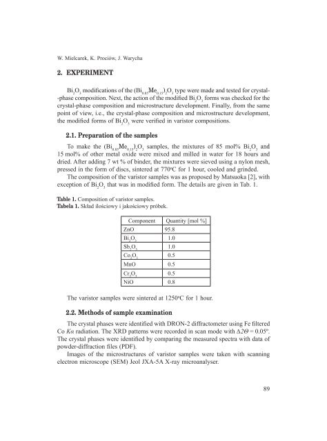
Table 1. Composition of varistor samples.<br />
Tabela 1. Skład ilościowy i jakościowy próbek.<br />
Component Quantity [mol %]<br />
ZnO 95.8<br />
Bi 2<br />
O 3<br />
1.0<br />
Sb 2<br />
O 3<br />
1.0<br />
Co 2<br />
O 3<br />
0.5<br />
MnO 0.5<br />
Cr 2<br />
O 3<br />
0.5<br />
NiO 0.8<br />
The varistor samples were sintered at 1250 o C for 1 hour.<br />
2.2. Methods of sample examination<br />
The crystal phases were identified with DRON-2 diffractometer using Fe filtered<br />
Co Kα radiation. The XRD patterns were recorded in scan mode with ∆2Θ = 0.05º.<br />
The crystal phases were identified by comparing the measured spectra with data of<br />
powder-diffraction files (PDF).<br />
Images of the microstructures of varistor samples were taken with scanning<br />
electron microscope (SEM) Jeol JXA-5A X-ray microanalyser.<br />
89