

CHAPTER 1 - IARC
CHAPTER 1 - IARC
CHAPTER 1 - IARC

Create successful ePaper yourself
Turn your PDF publications into a flip-book with our unique Google optimized e-Paper software.
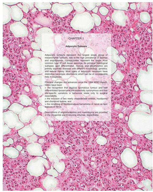
<strong>CHAPTER</strong> 1<br />
Adipocytic Tumours<br />
Adipocytic tumours represent the largest single group of<br />
mesenchymal tumours, due to the high prevalence of lipomas<br />
and angiolipomas. Liposarcomas represent the single most<br />
common type of soft tissue sarcoma. Its principal histological<br />
subtypes (well differentiated, myxoid, and pleomorphic) are<br />
entirely separate diseases with different morphology, genetics,<br />
and natural history. Most types of adipocytic neoplasm have<br />
distinctive karyotypic aberrations which can be of considerable<br />
help in diagnosis.<br />
Principal changes and advances since the 1994 WHO classification<br />
have been<br />
> the recognition that atypical lipomatous tumour and well<br />
differentiated liposarcoma are essentially synonymous and that<br />
site-specific variations in behaviour relate only to surgical<br />
resectability,<br />
> the inclusion of two newly characterized entities, myolipoma<br />
and chondroid lipoma, and<br />
> the renaming of fibrolipomatous hamartoma of nerve as lipomatosis<br />
of nerve.<br />
Descriptions of angiomyolipoma and myelolipoma are provided<br />
in the Urogenital and Endocrine volumes, respectively.
Lipoma<br />
G.P. Nielsen<br />
N. Mandahl<br />
Definition<br />
Lipoma is a benign tumour composed of<br />
mature white adipocytes and is the most<br />
common soft tissue mesenchymal neoplasm<br />
in adults.<br />
ICD-O code 8850/0<br />
Epidemiology<br />
Conventional lipoma occurs over a wide<br />
age range but is most common between<br />
the ages of 40 and 60 years and is more<br />
frequent in obese individuals {601}.<br />
Lipomas are rare in children.<br />
Approximately 5% of patients have multiple<br />
lipomas.<br />
Sites of involvement<br />
Conventional lipoma can arise within<br />
subcutaneous tissue (superficial lipoma)<br />
or within deep soft tissues (deep lipoma)<br />
or even on the surfaces of bone<br />
(parosteal lipoma) {1079,1800}. Deep<br />
seated lipomas that arise within or<br />
between skeletal muscle fibres are<br />
called intramuscular or intermuscular<br />
lipomas, respectively {685,1113}.<br />
Intramuscular lipoma arises during mid<br />
to late adulthood and involves skeletal<br />
muscle in a variety of locations including<br />
the trunk, head and neck region, upper<br />
and lower extremities {685,1113}.<br />
Intermuscular lipoma arises between<br />
muscles most frequently in the anterior<br />
abdominal wall, and involves a similar<br />
age group as the intramuscular lipoma.<br />
So-called lipoma arborescens (villous<br />
lipomatous proliferation of synovial membrane)<br />
is characterized by fatty infiltration<br />
of the subsynovial connective tissue<br />
and may represent a reactive process.<br />
Clinical features<br />
Lipomas usually present as a painless<br />
soft tissue mass, except for larger ones<br />
that can be painful when they compress<br />
peripheral nerves. Superficial lipomas<br />
are generally smaller (5cm). Patients with<br />
lipoma arborescens are usually adult<br />
men that complain of gradual swelling of<br />
the affected joint {324,837,875,1343,<br />
1982}. Imaging studies show a homogeneous<br />
soft tissue mass that is isodense<br />
to the subcutaneous tissue and demonstrates<br />
fat saturation. Attenuated fibrous<br />
strands can be seen but they are not as<br />
prominent as seen in the atypical lipomas.<br />
Intramuscular lipomas are more<br />
variably circumscribed, and lipoma<br />
arborescens shows diffuse fatty infiltration<br />
of the synovium.<br />
Aetiology<br />
Unknown. Lipomas are more common in<br />
obese individuals.<br />
Macroscopy<br />
Grossly, lipomas are well circumscribed<br />
and have a yellow, greasy cut surface.<br />
Different types are basically similar in<br />
appearance, however bone formation<br />
can be seen in osteolipoma and grey<br />
glistening nodules may be seen in chondrolipoma.<br />
Intramuscular and intermuscular<br />
lipoma do not show any specific<br />
gross features except that a portion of<br />
skeletal muscle is often attached to the<br />
periphery of the tumour. In lipoma arborescens<br />
the entire synovium assumes a<br />
nodular and papillary appearance and<br />
has a bright yellow cut surface.<br />
Histopathology<br />
Conventional lipoma is composed of lobules<br />
of mature adipocytes. The cells are<br />
identical to the surrounding adipose tissue<br />
except for slight variation in the size<br />
and shape of the cells in lipomas.<br />
Lipomas can occasionally have areas of<br />
bone formation (osteolipoma), cartilage<br />
(chondrolipoma), abundant fibrous tissue<br />
(fibrolipoma), or extensive myxoid<br />
change (myxolipoma). Intramuscular<br />
lipoma may be either well demarcated<br />
from the surrounding skeletal muscle or,<br />
more often, shows an infiltrative growth<br />
pattern with mature adipocytes infiltrating<br />
and encasing skeletal muscle fibres<br />
that often show evidence of atrophy. In<br />
lipoma arbor-escens the subsynovial<br />
connective tissue is infiltrated by mature<br />
adipocytes; scattered inflammatory cells<br />
are also usually present.<br />
Immunophenotype<br />
Mature adipocytes stain for vimentin,<br />
S100 protein and leptin {1610}.<br />
Ultrastructure<br />
Lipoma is composed of cells that have a<br />
large, single lipid droplet compressing a<br />
peripherally situated nucleus.<br />
Fig. 1.01 Image of a deep seated conventional lipoma<br />
showing a well circumscribed, homogenous<br />
tumour with the same characteristics as the adjacent<br />
subcutaneous fat.<br />
Fig. 1.02 Synovial lipoma (lipoma arborescens)<br />
demonstrating a fatty infiltration of the synovium<br />
that assumes papillary appearance.<br />
20 Adipocytic tumours
A<br />
Fig. 1.03 Conventional lipoma. A Grossly, the tumour is well circumscribed and has a homogenous yellow cut<br />
surface. B The mature adipocytes vary only slightly in size and shape and have small eccentric nuclei.<br />
A<br />
Fig. 1.04 Intramuscular lipoma. A This intramuscular lipoma appears well circumscribed from the adjacent<br />
skeletal muscle (right). B Mature adipocytes infiltrate and encase skeletal muscle fibres.<br />
Pinocytotic vesicles are present and<br />
external lamina is seen surrounding the<br />
cells {1110}.<br />
Genetics<br />
Cytogenetics<br />
Lipomas have been analysed extensively<br />
by chromosome banding. In larger<br />
cytogenetically investigated series, chromosome<br />
aberrations have been found in<br />
55-75% of the cases {1320,2020,2271}.<br />
Among the abnormal tumours, about<br />
75% show seemingly balanced karyotypes<br />
and in more than 50% there is a<br />
single abnormality in at least one clone<br />
{1477}. On average, signs of clonal evolution<br />
is found in every sixth tumour.<br />
Numerical chromosome changes are<br />
rare and randomly distributed, and chromosome<br />
numbers deviating from 46 are<br />
exceedingly rare. The pattern of cytogenetic<br />
aberrations is quite heterogeneous,<br />
but three cytogenetically defined subgroups<br />
have been distinguished: 1) the<br />
major subgroup consisting of tumours<br />
with aberrations involving 12q13-15, 2)<br />
tumours with aberrations involving 6p21-<br />
23, and 3) tumours with loss of material<br />
from 13q. Patients with and without aberrations<br />
of 12q13-15 show no differences<br />
B<br />
B<br />
with respect to age distribution and gender.<br />
The frequency of abnormal karyotypes<br />
seems to be higher among older<br />
patients {2020,2271}. Otherwise, no<br />
clear, consistent correlations between<br />
clinical and cytogenetic data have been<br />
identified.<br />
Tumours with 12q13-15 aberrations<br />
About two-thirds of tumours with abnormal<br />
karyotypes show aberrations of<br />
12q13-15, which has been found to<br />
recombine with a large number of bands<br />
in all chromosomes except 16 and Y. The<br />
preferred rearrangement, seen in more<br />
than 20% of tumours with 12q13-15 aberrations,<br />
is t(3;12)(q27-28;q13-15). Other<br />
recurrent recombination partner regions,<br />
present in 3-7% of these tumours, are<br />
1p36, 1p32-34, 2p22-24, 2q35-37, 5q33,<br />
11q13, 12p11-12, 12q24, 13q12-14,<br />
17q23-25, and 21q21-22. The majority of<br />
these aberrations originate through<br />
translocations or insertions. One in six of<br />
these tumours show more or less complex<br />
intrachromosomal rearrangements -<br />
including primarily inversions, but also<br />
deletions and duplications - leading to<br />
recombination between 12q13-15 and<br />
other segments of chromosome 12, primarily<br />
12p11-12 and 12q24.<br />
Tumours without 12q13-15 aberrations<br />
Among these tumours, constituting onethird<br />
of lipomas with acquired chromosome<br />
aberrations, all chromosomes<br />
except 20 have been involved, but the<br />
only distinct clustering of breakpoints<br />
seen is to 6p21-23, 13q11-22, and, less<br />
often, 12q22-24, together constituting<br />
about half of this group of tumours.<br />
Involvement of 6p21-23, mostly in the<br />
form of seemingly balanced translocations,<br />
has been found in more than 20%<br />
of these tumours. The only recurrent<br />
translocation partner has been 3q27-28<br />
in two cases. Aberrations affecting the<br />
long arm of chromosome 13 are dominated<br />
by deletions, which have been<br />
found in slightly less than 20% of the<br />
cases. Most aberrations are interstitial<br />
deletions with breakpoints in 13q12-14<br />
and 13q22, respectively. There is an<br />
overlap between 6p21-23 rearrangements<br />
and 13q deletions, with some<br />
tumours showing both aberrations, but<br />
more often these aberrations occur as<br />
sole anomalies.<br />
Simultaneous involvement of 6p21-23<br />
and 12q13-15 is uncommon, in contrast<br />
to the coexistence of 12q13-15 aberrations<br />
and 13q losses. In tumours with<br />
combinations of 6p, 12q, and 13q aberrations,<br />
13q is mostly involved in bal-<br />
A<br />
B<br />
Fig. 1.05 A Synovial lipoma (lipoma arborescens).<br />
The entire synovium is bright yellow and has a<br />
nodular or papillary appearance. B Synovial lipoma<br />
(lipoma arborescens). The subsynovial connective<br />
tissue has been replaced by mature adipocytes.<br />
Note also scattered chronic inflammatory cells.<br />
Lipoma<br />
21
A<br />
Fig. 1.06 A Karyotype from a lipoma showing the most common structural rearrangement, a translocation t(3;12)(q27;q15). B Lipoma with t(12;21)(q15;q22) as the sole chromosomal<br />
aberration. Arrowheads indicate breakpoints.<br />
B<br />
anced translocations when recombining<br />
with 6p21-23 or 12q13-15, whereas deletions<br />
in 13q are predominating when<br />
aberrations of 6p21-23 or 12q13-15 are<br />
present but recombine with other chromosome<br />
segments.<br />
Among tumours without rearrangements<br />
of 12q13-15 or 6p21-23 or loss of 13q<br />
sequences, one-fifth of the breakpoints<br />
coincide with those recurrently recombining<br />
with 12q13-15.<br />
Molecular genetics<br />
The HMGIC (a.k.a. HMGA2) gene,<br />
encoding a family member of the high<br />
mobility group of proteins, located in<br />
12q15 is affected in at least some lipomas<br />
with rearrangements of 12q13-15<br />
{90,1890}. In tumours with t(3;12)(q27-<br />
28;q13-15), the consequence at the<br />
molecular level is the formation of a<br />
fusion gene involving HMGIC and LPP in<br />
3q27-28, a member of the LIM protein<br />
gene family {1696}. In addition, this<br />
fusion gene has been observed in a few<br />
cases with complex karyotypic changes<br />
including 12q13-15 but not 3q27-28 and<br />
in cases with normal karyotypes, indicating<br />
that cytogenetic analysis underestimates<br />
the frequency of tumours with<br />
recombination between these two chromosome<br />
segments {1696}. In all cases,<br />
the chimeric HMGIC/LPP transcript is<br />
expressed, whereas the reciprocal<br />
LPP/HMGIC transcript is expressed only<br />
occasionally. Alternative fusion transcripts,<br />
encoding the three DNA binding<br />
AT-hook domains of HMGIC and two or<br />
three LIM domains of LPP have been<br />
reported, thus excluding the 3´ acidic,<br />
protein-binding domain and the N-terminal<br />
leucine-zipper motif, respectively.<br />
The preferred breakpoints are in the<br />
large intron 3 of HMGIC and LPP intron 8.<br />
The chimeric transcript is not unique for<br />
lipomas of the soft tissues but has also<br />
been detected in parosteal lipoma and<br />
pulmonary chondroid hamartoma<br />
{1698,1803}.<br />
Rearrangement of HMGIC has been<br />
detected also in tumours with changes<br />
involving 12q13-15 and other chromosome<br />
segments. In a single case of lipoma<br />
with t(12;13)(q13-15;q12), an<br />
HMGIC/LHFP fusion transcript has been<br />
reported {1697}. Also in this case, the<br />
breakpoint was in HMGIC intron 3. In<br />
lipomas with recombination between<br />
12q13-15 and 12p11, due to inversion,<br />
fusion of putative but yet unidentified<br />
gene sequences in 12p11 with HMGIC<br />
was found {1081}, and ectopic<br />
sequences mapping to chromosome 15<br />
have been implicated {90}. Possibly, the<br />
related HMGIY (HMGA1B) gene is the<br />
target, directly or indirectly, in lipomas<br />
with 6p21-23 aberrations; split FISH signals,<br />
using probes covering HMGIY,<br />
have been reported in cases with translocations<br />
involving 6p {1082,2083}.<br />
Transcriptional activation of HMGIC or<br />
HMGIY is indicated by immunohistochemical<br />
studies, and correlates well<br />
with cytogenetic findings of breakpoints<br />
in the regions where these two gene loci<br />
are located {2083}.<br />
Prognostic factors<br />
The subclassification of conventional<br />
lipoma does not have any prognostic significance<br />
except for the infiltrating intramuscular<br />
lipoma that has a higher local<br />
recurrence rate, therefore total removal of<br />
the involved muscle or a compartmental<br />
resection has been suggested for these<br />
infiltrating tumours in order to minimize<br />
local recurrence {206}.<br />
22 Adipocytic tumours
Lipomatosis<br />
G.P Nielsen<br />
A.E. Rosenberg<br />
Definition<br />
Lipomatosis is a diffuse overgrowth of<br />
mature adipose tissue. It occurs in a variety<br />
of clinical settings and can affect different<br />
anatomic regions of the body.<br />
ICD-O code 8850/0<br />
Synonyms<br />
Madelung disease, Launois-Bensaude<br />
syndrome.<br />
Fig. 1.07 Lipomatosis presenting as diffuse<br />
enlargement of the lower leg in an infant<br />
Fig. 1.08 Patient showing typically symmetrical,<br />
massive expansion of the neck.<br />
Epidemiology<br />
Diffuse lipomatosis usually occurs in<br />
individuals under 2 years of age but it<br />
may also arise in adults {1574}. Pelvic<br />
lipomatosis most frequently affects black<br />
males who range in age from 9 to 80<br />
{839,944,1135}. Symmetric lipomatosis<br />
develops in middle aged men of<br />
Mediterranean origin. Many patients<br />
have a history of liver disease or excessive<br />
alcohol consumption. Steroid lipomatosis<br />
manifests in patients on hormonal<br />
therapy or have increased endogenous<br />
production of adrenocortical<br />
steroids. HIV lipodystrophy is frequently<br />
seen in AIDS patients treated with protease<br />
inhibitors but is also seen in<br />
patients receiving other forms of antiretroviral<br />
therapy {234,1175}.<br />
Sites of involvement<br />
Diffuse lipomatosis involves the trunk,<br />
large portion of an extremity, head and<br />
neck, abdomen, pelvis or intestinal tract.<br />
It may be associated with macrodactyly<br />
or gigantism of a digit {836,1365,1616}.<br />
Symmetric lipomatosis manifests as<br />
symmetric deposition of fat in the upper<br />
part of the body particularly the neck. In<br />
pelvic lipomatosis there is diffuse overgrowth<br />
of fat in the perivesical and<br />
perirectal areas. Steroid lipomatosis is<br />
characterized by the accumulation of fat<br />
in the face, sternal region or the upper<br />
middle back (buffalo hump). HIV-lipodystrophy<br />
typically shows the accumulation<br />
of visceral fat, breast adiposity, cervical<br />
fat pads, hyperlipidemia, insulin resistance<br />
as well as fat wasting in the face<br />
and limbs {400,1461}.<br />
Clinical features<br />
In most forms of lipomatosis the patients<br />
present with massive accumulation of fat<br />
in the affected areas that may mimic a<br />
neoplasm. Additionally patients with<br />
symmetric lipomatosis can have neuropathy<br />
and central nervous system<br />
involvement {1541,1712}. Accumulation<br />
of fat in the lower neck areas in these<br />
patients can also cause laryngeal<br />
obstruction, and compression of the<br />
vena cava. Patients with pelvic lipomatosis<br />
frequently complain of urinary frequency,<br />
perineal pain, constipation, and<br />
abdominal and back pain. Bowel<br />
obstruction and hydronephrosis may<br />
eventually develop. Imaging studies in<br />
all forms of lipomatosis show accumulation<br />
of fat and are only helpful in determining<br />
the extent of its accumulation and<br />
excluding other processes.<br />
Aetiology<br />
The basic mechanism underlying lipomatosis<br />
is not well understood. In symmetric<br />
lipomatosis point mutations in<br />
mitochondrial genes have been implicated<br />
in its pathogenesis {1140}. The similarity<br />
between HIV lipodystrophy and<br />
benign symmetric lipomatosis suggests<br />
a similar pathogenesis in that mitochondrial<br />
DNA damage may be induced by<br />
the drugs being used to treat HIV<br />
{153,400}.<br />
Macroscopy<br />
The gross appearance of lipomatosis is<br />
the same for all of the different subtypes.<br />
The lesions consist of poorly circumscribed<br />
aggregates of soft yellow fat that<br />
is identical in appearance to normal fat.<br />
The only differences are the site of<br />
involvement and the distribution of the<br />
fat.<br />
Histopathology<br />
All of the different types of lipomatosis<br />
have identical morphologic features,<br />
consisting of lobules and sheets of<br />
mature adipocytes that may infiltrate<br />
Fig. 1.09 Diffuse lipomatosis showing extensive<br />
skeletal muscle infiltration of mature adipocytes.<br />
Lipomatosis 23
other structures such as skeletal muscle.<br />
Immunophenotype<br />
The adipose tissue stains for vimentin<br />
and S-100, similar to normal fat.<br />
Ultrastructure<br />
The adipocytes have the features of<br />
white fat.<br />
Genetics<br />
An association with several genetic disorders<br />
has been reported, and an autosomal<br />
dominant inheritance is suggested<br />
{1377}.<br />
Prognostic factors<br />
All idiopathic forms of lipomatoses have<br />
a tendency to recur locally after surgery.<br />
The treatment is palliative surgical<br />
removal of excess fat. Massive accumulation<br />
of fat in the neck region may cause<br />
death due to laryngeal obstruction. The<br />
fat in steroid lipomatosis regresses after<br />
steroid levels have been lowered.<br />
Experimental drugs such as recombinant<br />
growth hormones have been used to<br />
treat HIV-lipodystrophy.<br />
Lipomatosis of nerve<br />
G.P. Nielsen<br />
Definition<br />
Lipomatosis of nerve is characterized by<br />
infiltration of the epineurium by adipose<br />
and fibrous tissue. The tissue grows between<br />
and around nerve bundles thereby<br />
causing enlargment of the affected nerve.<br />
ICD-O code 8850/0<br />
Synonyms and historical annotations<br />
Fibrolipomatous hamartoma, lipofibroma,<br />
fibrolipomatosis, intraneural lipoma<br />
of the median nerve, perineural lipoma,<br />
median nerve lipoma, macrodystrophia<br />
lipomatosa, neural fibrolipoma.<br />
Epidemiology<br />
Lipomatosis of nerve is frequently first<br />
noted at birth or in early childhood, but<br />
patients may not present for treatment<br />
until early or mid adulthood. In the<br />
largest reported series the patients<br />
ranged in age from 11 to 39 years.<br />
Because the constituent tissues are<br />
normal components of the epineurium,<br />
some have considered this lesion to be<br />
a hamartoma of the nerve sheath {445,<br />
2103}. In some cases it is associated<br />
with macrodactyly of the digits inervated<br />
by the affected nerve.<br />
Associated macrodactyly was present<br />
in approximately 1/3 of patients,<br />
including 5 females and 2 males<br />
{1952}. Females predominate when<br />
lipofibroma is accompanied by macrodactyly,<br />
whereas males are more commonly<br />
affected when macrodactyly is<br />
absent.<br />
Sites of involvement<br />
The median nerve and its digital branches<br />
are most commonly affected followed<br />
by the ulnar nerve {189,1952}. The<br />
process has also been reported to<br />
involve unusual sites such as the cranial<br />
nerves and the brachial plexus<br />
{176,1726}.<br />
Clinical features<br />
Patients present with a gradually enlarging<br />
mass in the affected area that may be<br />
asymptomatic or associated with motor<br />
or sensory deficits. Patients with macrodactyly<br />
have symmetrical or asymmetrical<br />
enlargement of the affected finger(s)<br />
with enlargement of the involved bones.<br />
Imaging studies show fusiform enlargement<br />
of the nerve with fatty infiltration<br />
A<br />
B<br />
Fig. 1.10 Lipomatosis of nerve. A A clinical picture showing macrodactyly of the second and third fingers. B An intraoperative view of lipomatosis of nerve showing<br />
a transition between the normal nerve (left) and the affected area (right). C Cross section reveals nerve bundles entrapped within fibroadipose tissue.<br />
C<br />
24 Adipocytic tumours
{474} and MRI findings are virtually<br />
pathognomonic {1336}.<br />
Aetiology<br />
The aetiology is unknown. Lipomatosis of<br />
nerve is not associated with any syndrome<br />
nor is there any known hereditary<br />
predisposition.<br />
Macroscopy<br />
Grossly there is fusiform enlargement of<br />
the nerve by yellow fibrofatty tissue,<br />
which is generally confined within the<br />
epineurial sheath.<br />
Histopathology<br />
The epineurial and perineurial compartments<br />
of the enlarged nerve are infiltrated<br />
by mature adipose tissue admixed<br />
with fibrous tissue which dissects<br />
between and separates individual nerve<br />
bundles {1952}. Concentric perineurial<br />
fibrous tissue is a prominent feature. The<br />
affected nerve may also show other<br />
changes such as perineural septation,<br />
microfascicle formation and pseudoonion<br />
bulb formation mimicking an intraneural<br />
perineurioma {1882}. Metaplastic<br />
bone formation is rarely present {551}.<br />
A<br />
Immunophenotype<br />
Immunohistochemical studies are not<br />
helpful in diagnosing this lesion as all of<br />
its components are seen in normal<br />
nerves.<br />
Ultrastructure<br />
There are no characteristic ultrastructural<br />
findings. The nerve bundles demonstrate<br />
onion bulblike formations with one<br />
or two nerve fibres and peripheral perineural<br />
cells {99}.<br />
Prognostic factors<br />
Lipomatosis of nerve is a benign lesion<br />
with no effective therapy. Surgical excision<br />
usually causes severe damage of<br />
the involved nerve. Division of the transverse<br />
carpal ligament may relieve neurological<br />
symptoms.<br />
B<br />
Fig. 1.11 A Epineural infiltration of fibroadipose tissue separating nerve bundles. B The nerves show pseudoonion<br />
bulb formation and perineural fibrosis.<br />
Lipomatosis of nerve<br />
25
Lipoblastoma / Lipoblastomatosis<br />
R. Sciot<br />
N. Mandahl<br />
Definition<br />
A lobulated, localized (lipoblastoma) or<br />
diffuse (lipoblastomatosis) tumour,<br />
resembling fetal adipose tissue.<br />
ICD-O code 8881/0<br />
Synonyms<br />
Foetal lipoma, embryonic lipoma, infantile<br />
lipoma.<br />
Epidemiology<br />
Both tumours are most commonly found<br />
in the first three years of life. They may<br />
occasionally be present at birth or in<br />
older children. There is a male predilection<br />
{348,391,1410,2196}.<br />
Sites of involvement<br />
The extremities are most commonly<br />
involved, but locations in the mediastinum,<br />
retroperitoneum, trunk, head &<br />
neck, and various organs (lung, heart,<br />
parotid gland) have been described<br />
{273,500,525,1002,1010,1177,1192,<br />
1352,1654,1713,1720,1762,2134,2149}.<br />
Clinical features<br />
Most patients present with a slowly growing<br />
soft tissue nodule/mass, well circumscribed<br />
and confined to the subcutis in case of<br />
lipoblastoma, infiltrating the deeper muscle<br />
in case of lipoblastomatosis. Depending on<br />
the location, the tumour may compress<br />
adjacent structures, such as the trachea.<br />
Imaging reveals a mass with adipose tissue<br />
density, but does not allow distinction<br />
from lipoma and liposarcoma {1777}.<br />
A<br />
Macroscopy<br />
Notwithstanding exceptions, lipoblastomas<br />
are relatively small lesions (2-5<br />
cm), showing fatty looking tissue with<br />
gelatinous areas.<br />
Fig. 1.12 Lipoblastoma. A Grossly, the tumour shows vague lobularity and fibrous / gelatinous areas.<br />
B Low power view. Note the prominent lobulation.<br />
B<br />
Histopathology<br />
Lipoblastoma shows a lobulated appearance<br />
with an admixture of mature and<br />
immature adipocytes, the latter corresponding<br />
to lipoblasts in various stages<br />
of development. Depending on the age<br />
of the patient, lipoblasts may be very<br />
scarce. Connective tissue septa separate<br />
the lobules. The lobulation is less<br />
prominent in lipoblastomatosis, in which<br />
entrapped muscle fibres frequently<br />
occur. The matrix can be quite myxoid,<br />
with a plexiform vascular pattern, thus<br />
mimicking myxoid liposarcoma. The latter<br />
tumour, which is exceptionally rare<br />
under the age of 10, usually shows<br />
nuclear atypia and does not show the<br />
pronounced lobulated pattern of<br />
lipoblastoma {223}. However, in rare<br />
cases molecular genetic analysis may<br />
be required for definitive distinction.<br />
Occasionally, lipoblastoma(tosis) may<br />
show extramedullary haematopoiesis or<br />
cells resembling brown fat. Cellular maturation<br />
has been described, leading to a<br />
lipoma-like picture. When fascicles of<br />
primitive mesenchymal cells are present<br />
in the septa, lipoblastoma resembles<br />
infantile lipofibromatosis or infantile fibromatosis<br />
{658}. The lobulated aspect, the<br />
at least focal myxoid stroma and plexiform<br />
capillaries, as well as the overwhelming<br />
fat component with lipoblasts,<br />
help to separate lipoblatoma(tosis) from<br />
these lesions.<br />
Ultrastructure<br />
Lipoblastoma(tosis) strongly resembles<br />
normal developing fat, with a spectrum<br />
ranging from primitive mesenchymal<br />
cells to multivacuolated lipoblasts and<br />
mature lipocytes {223}.<br />
Genetics<br />
Typically, lipoblastomas have simple,<br />
pseudodiploid karyotypes with structural<br />
chromosome aberrations. The characteristic<br />
cytogenetic feature is rearrangement<br />
of 8q11-13, which has been found<br />
in the vast majority of cases. The only<br />
chromosome segments that, so far, have<br />
been found to be involved in recurrent<br />
recombinations with 8q11-13 are 3q12-<br />
13, 7p22, and 8q24, but several other<br />
chromosome segments have been the<br />
translocation partners in single cases.<br />
Numerical changes are rare, but gain of<br />
chromosome 8 has been found in cases<br />
with or without simultaneous rearrangement<br />
of 8q11-13.<br />
To date, two different fusion genes have<br />
been reported to result from the chromosomal<br />
rearrangements, HAS2/PLAG1 in<br />
three cases and COL1A2/PLAG1 in a<br />
single case {945}. The PLAG1 gene is<br />
located in 8q12, HAS2 in 8q24 and<br />
COL1A2 in 7q22. The genomic breakpoint<br />
of PLAG1 seems to be in intron1,<br />
resulting in loss of exon 1. The entire<br />
HAS2 5´ untranslated region is involved<br />
in the fusion gene, which is probably<br />
under control of the HAS2 promoter,<br />
leading to transcriptional up-regulation<br />
of PLAG1 and production of a full-length<br />
PLAG1 protein. The COL1A2-PLAG1<br />
fusion gene encodes a chimeric protein<br />
containing the first amino acids of<br />
COL1A2 and full-length PLAG1. These<br />
fusion genes seem to act through a promoter-swapping<br />
mechanism {105,945}.<br />
An alternative mechanism associated<br />
with lipoblastoma tumourigenesis may<br />
act through excess copies of chromosome<br />
8 {792}. Since +8 may be present<br />
26 Adipocytic tumours
A<br />
Fig. 1.13 A Admixture of multivacuolated lipoblasts and mature adipocytes. B 315 Delicate plexiform vascular pattern and myxoid changes in lipoblastoma.<br />
B<br />
in tumours both with and without<br />
changes of 8q12, the effect of PLAG1<br />
rearrangement might be reinforced by<br />
gain of chromosome 8 in some cases.<br />
Whether the extra copies of the PLAG1<br />
gene are normal or have point mutations<br />
is not known. By in situ hybridization it<br />
has been shown that split PLAG1 signals<br />
are present in both classical, myxoid,<br />
and lipoma-like lipoblastomas as well as<br />
in a variety of mesenchymal cell components,<br />
indicating the mutation to occur in<br />
a progenitor cell that then differentiates<br />
{792}.<br />
Prognostic factors<br />
Lipoblastoma(tosis) is fully benign and<br />
malignant transformation or metastasis<br />
does not occur. Recurrences are<br />
described in 9% to 22% of cases, mainly<br />
in lipoblastomatosis. Therefore wide total<br />
excision of diffuse lesions is advised<br />
{348,391,1410,2196}.<br />
Lipoblastoma / Lipoblastomatosis<br />
27
Angiolipoma<br />
R. Sciot<br />
N. Mandahl<br />
Definition<br />
A subcutaneous nodule consisting of<br />
mature fat cells, intermingled with small<br />
and thin-walled vessels, a number of<br />
which contain fibrin thrombi.<br />
ICD-O code 8861/0<br />
Epidemiology<br />
Angiolipomas are relatively common and<br />
usually appear in the late teens or early<br />
twenties. Children and patients older<br />
than 50 years are rarely involved. There<br />
is a male predominance and an<br />
increased familial incidence has been<br />
described (5% of all cases) {230,357,<br />
942,977,1062,1232}. The mode of inheritance<br />
is not clear.<br />
Sites of involvement<br />
The forearm is the most common site, followed<br />
by the trunk and upper arm.<br />
Spinal angiolipomas and intramuscular<br />
haemangiomas, previously also called<br />
‘infiltrating angiolipomas’, are different<br />
lesions {878,2148}.<br />
Clinical features<br />
Angiolipomas most frequently present as<br />
multiple subcutaneous small nodules,<br />
usually tender to painful. There is no correlation<br />
between the intensity/occurrence<br />
of pain and the degree of vascularity<br />
{527}.<br />
Macroscopy<br />
Angiolipomas appear as encapsulated<br />
yellowish to reddish nodules, most often<br />
less than 2 cm in diameter.<br />
Histopathology<br />
Angiolipomas typically consist of two<br />
mesenchymal elements: mature<br />
adipocytes and branching capillary<br />
sized vessels, which usually contain fibrin<br />
thrombi. The vascularity is more<br />
prominent in the subcapsular area {527}.<br />
The relative proportion of adipocytes and<br />
vessels varies and some lesions are<br />
almost completely composed of vascular<br />
channels. These ‘cellular’ angiolipomas<br />
should be distinguished from angiosarcoma<br />
and Kaposi sarcoma {983}.<br />
Interstitial mast cells may be prominent<br />
and in older lesions, increased fibrosis is<br />
present.<br />
A<br />
Genetics<br />
With a single exception, all cytogenetically<br />
investigated tumours have had a<br />
normal karyotype {1905}.<br />
Prognostic factors<br />
Angiolipomas are always benign and<br />
show no tendency to recur. Malignant<br />
transformation does not occur.<br />
B<br />
Fig. 1.14 Angiolipoma. A The tumour consists of mature adipocytes and capillaries, some of which contain<br />
microthrombi. B Cellular angiolipoma, in which the vessels predominate.<br />
28 Adipocytic tumours
Myolipoma of soft tissue<br />
J.M. Meis-Kindblom<br />
L.G. Kindblom<br />
Definition<br />
Myolipoma of soft tissue is a benign<br />
tumour exhibiting features of mature<br />
smooth muscle and mature adipose tissue.<br />
ICD-O code 8890/0<br />
Synonym<br />
Extrauterine lipoleiomyoma.<br />
Epidemiology<br />
Myolipoma of soft tissue is an extremely<br />
rare lesion occurring in adults, with a<br />
male to female ratio of 1:2 {1393}.<br />
Sites of involvement<br />
The majority of cases are deeply located<br />
and involve the abdominal cavity,<br />
retroperitoneum, and inguinal areas. The<br />
trunk wall and extremities may also be<br />
involved; such cases are subcutaneous<br />
and may grow deeply to involve the<br />
superficial muscular fascia {1393}.<br />
Clinical features<br />
Most lesions present as a palpable<br />
mass; the remainder are incidental<br />
findings.<br />
Macroscopy<br />
Deep-seated myolipomas of soft tissue<br />
range between 10 and 25 cm in size; the<br />
average size is 15 cm. Smaller lesions<br />
are seen in the subcutis. A completely or<br />
partially encapsulated lipomatous<br />
tumour intermingles with strands and<br />
nodules of firm white-tan, fibrillary to<br />
whorled areas corresponding to smooth<br />
muscle.<br />
Histopathology<br />
The smooth muscle component usually<br />
dominates with a muscle to fat ratio of<br />
2:1. Smooth muscle tends to be evenly<br />
distributed and arranged in short fascicles,<br />
resulting in a sieve-like pattern as it<br />
traverses the fat. Individual smooth muscle<br />
fibres have deeply acidophilic fibrillary<br />
cytoplasm that becomes<br />
fuchsinophilic with the Masson trichrome<br />
stain. Nuclear chromatin is evenly dispersed,<br />
nucleoli are inconspicuous and<br />
no appreciable mitotic activity is seen.<br />
Equally important is the absence of any<br />
atypia in the mature lipomatous component<br />
of myolipoma. Floret cells and<br />
lipoblasts are not seen, nor are medium<br />
calibre thick-walled blood vessels as<br />
Fig. 1.15 An encapsulated myolipoma of the pelvis with<br />
clear fatty and smooth muscle components.<br />
seen in angiomyolipoma. Sclerosis and<br />
focal inflammation may be present in<br />
the fat.<br />
Immunophenotype<br />
Diffusely and strongly positive smooth<br />
muscle actin and desmin immunostaining<br />
confirm the presence of smooth muscle<br />
in myolipoma.<br />
Prognostic factors<br />
Myolipoma does not recur. Complete<br />
surgical resection is curative.<br />
A<br />
Fig. 1.16 A,B Mature adipose tissue and mature smooth muscle arranged in short fascicles are seen in a myolipoma of the distal extremity.<br />
B<br />
Myolipoma of soft tissue<br />
29
Chondroid lipoma<br />
L.G. Kindblom<br />
J.M. Meis-Kindblom<br />
N. Mandahl<br />
Definition<br />
Chondroid lipoma is a unique and<br />
recently recognized benign adipose tissue<br />
tumour containing lipoblasts, mature<br />
fat, and a chondroid matrix. It bears a<br />
strikingly close resemblance to myxoid<br />
liposarcoma and extraskeletal myxoid<br />
chondrosarcoma.<br />
ICD-O code 8862/0<br />
Epidemiology<br />
Chondroid lipoma is rare and affects primarily<br />
adults with a male:female ratio of<br />
1:4 {1396} without racial predilection.<br />
Sites of involvement<br />
This tumour occurs most commonly in<br />
the proximal extremities and limb girdles.<br />
However, the trunk and head and neck<br />
areas may also be affected. Chondroid<br />
lipoma is often deep-seated, involving<br />
skeletal muscle or deep fibrous connective<br />
tissues. Those cases involving the<br />
subcutis tend to impinge on the superficial<br />
muscular fascia.<br />
Clinical features<br />
The majority of patients present with a<br />
painless mass of variable duration. There<br />
is a recent history of enlargement in<br />
roughly one-half of cases.<br />
Reports of imaging studies of this lesion<br />
are exceedingly sparse {1277,2320}.<br />
Macroscopy<br />
Most chondroid lipomas are 2–7 cm in<br />
size, although cases with haemorrhage<br />
may be significantly larger {1396}.<br />
Tumours are typically well circumscribed<br />
and yellowish, suggesting fatty differentiation.<br />
Histopathology<br />
Chondroid lipoma is often encapsulated<br />
and occasionally multilobular. Its histologic<br />
hallmarks are nests and cords of<br />
abundant uni- and multivacuolated<br />
lipoblasts embedded in a prominent<br />
myxoid to hyalinized chondroid matrix<br />
admixed with a variable amount of<br />
mature adipose tissue. The lipoblast<br />
nuclei are small and uniform, ranging<br />
from oval, reniform to multilobated in<br />
shape, with evenly dispersed chromatin<br />
and small nucleoli. The cytoplasm is finely<br />
vacuolated, containing small lipid<br />
droplets and PAS positive glycogen.<br />
Cells may have granular eosinophilic<br />
cytoplasm. Chondroid lipoma is highly<br />
vascular and not infrequently contains<br />
haemorrhage and fibrosis.<br />
Toluidine blue and alcian blue stains at<br />
controlled pHs confirm the typical presence<br />
of chondroitin sulfates in the matrix<br />
{1116}.<br />
Immunophenotype<br />
Lipoblasts are weakly S100 protein positive<br />
whereas stronger staining is seen<br />
with increasing adipocytic maturation<br />
{1116}. Vimentin is uniformly positive in<br />
all cells; cytokeratins are detected in rare<br />
cases, corresponding ultrastructurally to<br />
tonofilaments. EMA is uniformly negative.<br />
Proliferative index with MIB1 is
Spindle cell lipoma /<br />
Pleomorphic lipoma<br />
M.M. Miettinen<br />
N. Mandahl<br />
Definition<br />
Spindle cell and pleomorphic lipoma,<br />
ends of a common histological spectrum,<br />
are circumscribed subcutaneous<br />
lesions occurring typically on the neck<br />
and back usually of males and composed<br />
of a variable admixture of bland<br />
spindled cells, hyperchromatic rounded<br />
cells, and multinucleate giant cells associated<br />
with ropey collagen.<br />
ICD-O codes<br />
Spindle cell lipoma 8857/0<br />
Pleomorphic lipoma 8854/0<br />
Sites of involvement<br />
Spindle cell / pleomorphic lipomas occur<br />
predominantly in the posterior neck and<br />
shoulder area. Face, forehead, scalp,<br />
buccal-perioral area and upper arm are<br />
less common sites, and occurrence in<br />
the lower extremity is distinctly rare.<br />
Clinical features<br />
Spindle cell / pleomorphic lipomas typically<br />
present in older men with a median<br />
age of over 55 years, and only 10% of<br />
patients are women {60,102,595,684,<br />
1944}. The tumour forms an asymptomatic,<br />
mobile dermal or subcutaneous<br />
mass, and there is often a long history.<br />
Rare patients have multiple lesions, and<br />
familial occurrence has been reported,<br />
mostly in men {633}. Spindle cell / pleomorphic<br />
lipomas have benign behaviour<br />
and conservative local excision is considered<br />
sufficient.<br />
A<br />
Macroscopy<br />
Grossly spindle cell lipoma / pleomorphic<br />
lipoma forms an oval or discoid yellowish<br />
to greyish-white mass depending<br />
on the relative extent of the fatty and<br />
spindle cell components. The tumour<br />
often has a firmer texture than ordinary<br />
lipoma, but some examples have a<br />
gelatinous texture.<br />
Histopathology<br />
Histologically, at one end at the histological<br />
spectrum, spindle cell lipoma is<br />
composed of bland mitotically inactive<br />
B<br />
Fig. 1.19 Spindle cell lipoma. A The relative proportions of the adipocytic and spindle cell components are variable.<br />
B Some lesions are almost devoid of adipocytes and show vague nuclear palisading. Note the typically<br />
ropey collagen bundles.<br />
Fig. 1.20 Spindle cell lipoma. Typical case with bland<br />
spindle cells in a background with thick collagen<br />
fibres and a small number of adipocytes.<br />
Spindle cell lipoma / Pleomorphic lipoma<br />
31
Fig. 1.22 Immunopositivity for CD34 is a consistent<br />
feature of the spindle cell component.<br />
A<br />
B<br />
C<br />
Fig. 1.21 Pleomorphic lipoma. A Prominent myxoid change of the stroma is not an uncommon feature.<br />
B Classical example showing numerous floret-like multinucleate cells. C Some pleomorphic lipomas consist<br />
almost entirely of mature adipocytes with admixed multinucleated stroma cells, often having floret-like nuclei.<br />
vascular slits ("pseudoangiomatoid variant")<br />
{911}.<br />
At the opposite end of the spectrum,<br />
pleomorphic lipoma is characterized by<br />
small spindled and rounded hyperchromatic<br />
cells and multinucleated giant cells<br />
with radially arranged nuclei in a"floretlike"<br />
pattern, like petals of flowers. Cases<br />
with features intermediate between classic<br />
spindle cell lipoma and pleomorphic<br />
lipoma quite often occur.<br />
Immunophenotype<br />
The spindle cells in both spindle cell and<br />
pleomorphic lipomas are strongly positive<br />
for CD34 and may rarely be positive<br />
for S100 protein {626,2059,2102}.<br />
Cytogenetics<br />
Spindle cell lipomas and pleomorphic<br />
lipomas show similar cytogenetic aberrations.<br />
The karyotypes are, on average,<br />
more complex than those found in ordinary<br />
lipomas and are frequently<br />
hypodiploid, often with multiple partial<br />
losses, no gain of sequences, and few<br />
balanced rearrangements. Monosomy or<br />
partial loss of chromosomes 13 and/or 16<br />
have been found in seven to eight out of<br />
ten cases. Half of the tumours with<br />
involvement of chromosome 16 have had<br />
a breakpoint in 16q13, and all of them<br />
have had loss of 16q13-qter. The most<br />
frequently lost segments of chromosome<br />
13 include 13q12 and 13q14-q22. Other<br />
chromosome segments lost in two to<br />
three of the ten cases are 6pter-p23,<br />
6q15-q21, 10pter-p15, 10q23-qter, and<br />
17pter-p13 {442}.<br />
spindled cells arranged in parallel registers<br />
between the fat cells and associated<br />
with thick rope-like collagen bundles.<br />
{60,595,684,1944}. Large numbers of<br />
mast cells are often seen in between the<br />
spindle cells, and lymphocytes and plasma<br />
cells may occur, especially in pleomorphic<br />
lipoma. Some spindle cell lipomas<br />
show myxoid stromal change or display<br />
slit-like cleavage spaces resembling<br />
Prognostic factors<br />
These are benign lesions which only<br />
rarely recur locally.<br />
32 Adipocytic tumours
Hibernoma<br />
M.M. Miettinen<br />
J.C. Fanburg-Smith<br />
N. Mandahl<br />
Definition<br />
Hibernoma is a rare benign adipose<br />
tumour composed at least in part of<br />
brown fat cells with granular, multivacuolated<br />
cytoplasm. This brown fat component<br />
is admixed in variable proportion<br />
with white adipose tissue. Residual<br />
brown fat, mostly seen around cervical<br />
and axillary lymph nodes, should not be<br />
classified as hibernoma.<br />
ICD-O codes 8880/0<br />
Epidemiology<br />
Recognized since around the turn of the<br />
century {1424}, hibernoma comprises<br />
1.6% of benign lipomatous tumours and<br />
approximately 1.1% of all adipocytic<br />
tumours in AFIP files. Based on AFIP<br />
data on 170 cases {747}, hibernoma<br />
occurs predominantly in young adults,<br />
with a mean age of 38 years. 60% occur<br />
in the third and fourth decades, only 5%<br />
occur in children 2-18 years, and 7% in<br />
patients over 60 years. There is a slight<br />
male predominance {747}.<br />
Sites of involvement<br />
Hibernoma occurs in a wide variety of<br />
locations. The most common site is the<br />
thigh, followed by the trunk, upper<br />
extremity, and head and neck. The myxoid<br />
and spindle cell variants tend to be<br />
located in the posterior neck and shoulders,<br />
similar to spindle cell lipoma {747}.<br />
Less than 10% occur in the intra-abdominal<br />
or thoracic cavities {19}.<br />
Clinical features<br />
Hibernoma is a relatively slow growing<br />
tumour of the subcutis. At least 10% of<br />
cases are intramuscular. Hibernomas are<br />
usually painless. MRI reveals non-fat<br />
septations in hibernoma, not found in<br />
lipoma. By CT scan, hibernoma has a tissue<br />
attenuation intermediate between fat<br />
and skeletal muscle and enhances with<br />
contrast {1172}.<br />
Aetiology<br />
The aetiology of hibernoma is unknown,<br />
although many lesions arise at the sites<br />
where brown fat is normally found in<br />
hibernating animals and human fetuses/newborns<br />
{754}.<br />
Macroscopy<br />
The median size for hibernoma is 9.3<br />
centimeters, range 1-24 centimeters<br />
{747}. Hibernomas are lobular, welldemarcated,<br />
and vary in colour from yellow<br />
to brown. They have a greasy, soft,<br />
and spongy cut surface {747,1113}.<br />
Histopathology<br />
Histologically, hibernomas vary in the<br />
content and appearance of the polygonal<br />
brown fat cells, the associated small<br />
capillary proliferation, and the stromal<br />
background, resulting in six variants.<br />
Most tumours contain large numbers of<br />
multivacuolated brown fat cells with<br />
abundant, granular cytoplasm and a<br />
Fig. 1.23 Hibernoma. The eosinophilic variant is<br />
composed mostly of granular-appearing, multivacuolated<br />
brown fat cells with prominent nucleoli.<br />
Fig. 1.24 Hibernoma. Detail of the eosinophilic variant<br />
with granular, multivacuolated brown fat cells<br />
and prominent nucleoli.<br />
Fig. 1.25 Hibernoma. The pale cell variant has a pale<br />
tinctorial quality of the multivacuolated brown fat<br />
cells.<br />
Hibernoma<br />
33
del(11) der(11) der(17) 17<br />
Fig. 1.28 Hibernoma. Partial G-banded karyotype<br />
showing a translocation t(11;17)(q13;p13).<br />
Fig. 1.26 Hibernoma. The myxoid variant has a myxoid background with floating brown fat cells.<br />
Fig. 1.27 Hibernoma. The spindle cell variant, a hybrid tumour between hibernoma and spindle cell lipoma,<br />
shows brown fat cells, mature white fat cells, scattered mast cells, bland spindled cells.<br />
small, central nucleus, the granular or<br />
eosinophilic variant. The brown fat cells<br />
vary from pale staining to variably<br />
eosinophilic, and some cases have a<br />
mixture of pale and eosinophilic cells, the<br />
mixed variant, while other cases have<br />
pure pale brown fat cells, the pale variant.<br />
Some hibernomas contain small<br />
clusters of brown fat amidst ordinary<br />
white fat, the "lipoma-like" variant.<br />
Multivacuolated lipoblast-like cells are<br />
often seen. Rare variants with myxoid<br />
stroma (myxoid variant), or a spindle cell<br />
component, with thick bundles of collagen<br />
fibres, scattered mast cells, and<br />
mature adipose tissue (spindle cell variant),<br />
a hybrid between hibernoma and<br />
spindle cell lipoma, have been<br />
described. Mitoses are exceptional and<br />
cytological atypia is unusual. Such features<br />
should not be equated with malignancy<br />
as the biologic behaviour of hibernoma<br />
is invariably benign. However,<br />
scattered normal brown fat cells may be<br />
found in an otherwise classic myxoid or<br />
well differentiated liposarcoma.<br />
Immunophenotype<br />
Hibernoma cells are variably, sometimes<br />
strongly, positive for S100 protein. The<br />
spindle cell variant has a CD34 positive<br />
spindle cell component, similar to spindle<br />
cell lipoma, whereas the other hibernoma<br />
variants are negative for CD34<br />
{747}.<br />
Genetics<br />
Although hibernomas frequently show<br />
somewhat more complex chromosome<br />
changes than ordinary lipomas and<br />
lipoblastomas, the karyotypes are nearor<br />
pseudodiploid. The only recurrent<br />
aberration is the involvement of 11q13-<br />
21, most often 11q13, in structural<br />
rearrangements, which in the majority of<br />
cases affect three or more chromosomes.<br />
No chromosome band has been<br />
involved more than once as a translocation<br />
partner.<br />
Metaphase FISH analyses have demonstrated<br />
that the chromosomal rearrangements<br />
are more complex than can be<br />
detected by chromosome banding<br />
analysis {793}. The aberrations not only<br />
affect the obviously rearranged chromosome<br />
11, but also the seemingly normal<br />
homologue. Both heterozygous and<br />
homozygous deletions have been<br />
detected, with deletions comprising segments<br />
up to 4 Mb. Homozygous deletion<br />
of the multiple endocrine neoplasia type<br />
I tumour suppressor gene MEN1 has<br />
been found in four of five tumours,<br />
whereas all five hibernomas investigated<br />
showed heterozygous loss of PPP1A<br />
{793}. Yet, no conclusive evidence of the<br />
pathogenetically important event is available.<br />
Prognostic factors<br />
Hibernoma is a benign tumour that does<br />
not recur with complete local excision<br />
{747}. All morphologic variants have the<br />
same good prognosis.<br />
34 Adipocytic tumours
Atypical lipomatous tumour /<br />
Well differentiated liposarcoma<br />
A.P. Dei Tos<br />
F. Pedeutour<br />
Definition<br />
Atypical lipomatous tumour (ALT) / welldifferentiated<br />
(WD) liposarcoma is an<br />
intermediate (locally aggressive) malignant<br />
mesenchymal neoplasm composed<br />
either entirely or in part of a mature<br />
adipocytic proliferation showing significant<br />
variation in cell size and at least<br />
focal nuclear atypia in both adipocytes<br />
and stromal cells. The presence of scattered<br />
hyperchromatic, often multinucleate<br />
stromal cells and a varying number<br />
of monovacuolated or multivacuolated<br />
lipoblasts (defined by the presence of<br />
single or multiple sharply marginated<br />
cytoplasmic vacuoles scalloping an<br />
enlarged hyperchromatic nucleus) may<br />
contribute to the morphologic diagnosis.<br />
Use of the term ‘atypical lipomatous<br />
tumour’ is determined principally by<br />
tumour location and resectability.<br />
ICD-O code 8851/3<br />
Fig. 1.29 Atypical lipomatous tumour / Well differentiated<br />
liposarcoma. Surgical specimen showing<br />
a well circumscribed, lobulated mass.<br />
Terminology in clinical practice<br />
The fact that WD liposarcoma shows no<br />
potential for metastasis unless it undergoes<br />
dedifferentiation led, in the late<br />
1970s, to the introduction of terms such<br />
as atypical lipoma or atypical lipomatous<br />
tumour {626}, particularly for lesions arising<br />
at surgically amenable locations in<br />
the limbs and on the trunk since, at these<br />
sites, wide excision should usually be<br />
curative and hence the designation ‘sarcoma’<br />
is not warranted. However, the<br />
variable, sometimes controversial application<br />
of this new terminology has represented<br />
a source of potential diagnostic<br />
confusion {620, 1112, 2246}. Atypical<br />
lipomatous tumour and WD liposarcoma<br />
are synonyms describing lesions which<br />
are identical both morphologically and<br />
karyotypically (see below) and in terms<br />
of biologic potential. The choice of terminology<br />
is therefore best determined by<br />
the degree of reciprocal comprehension<br />
between the surgeon and the pathologist<br />
to prevent either inadequate or excessive<br />
treatment {486}. However, in sites<br />
such as the retroperitoneum and mediastinum<br />
it is commonly impossible to<br />
obtain a wide surgical excision margin<br />
and, in such cases, local recurrence<br />
(often repeated and ultimately uncontrolled)<br />
is almost inevitable and often<br />
leads to death, even in the absence of<br />
dedifferentiation and metastasis – hence,<br />
at these sites, retention of the term WD<br />
liposarcoma can readily be justified.<br />
Spindle cell/pleomorphic lipoma must be<br />
kept separated from the atypical lipoma<br />
category as it is morphologically as well<br />
as cytogenetically distinct, rarely recurs<br />
and has no potential to dedifferentiate<br />
(see page 31).<br />
Synonyms<br />
Atypical lipoma, adipocytic liposarcoma,<br />
lipoma-like liposarcoma, sclerosing<br />
liposarcoma, spindle cell liposarcoma,<br />
inflammatory liposarcoma.<br />
Epidemiology<br />
ALT/WD liposarcoma accounts for about<br />
40-45% of all liposarcomas and therefore<br />
represents the largest subgroup of<br />
aggressive adipocytic neoplasms. These<br />
lesions mostly occur in middle aged<br />
adults with a peak incidence in the 6th<br />
decade. Convincing examples in childhood<br />
are extremely rare. Males and<br />
females are equally affected with the<br />
obvious exception of those lesions affecting<br />
the spermatic cord {588,678, 2242}.<br />
Sites of involvement<br />
ALT/WD liposarcoma occurs most frequently<br />
in deep soft tissue of the limbs,<br />
especially the thigh, followed by the<br />
retroperitoneum, the paratesticular area<br />
and the mediastinum {588, 678, 2242}.<br />
These lesions may also arise in subcutaneous<br />
tissue and, very rarely, in skin.<br />
Clinical features<br />
ALT/WD liposarcoma usually presents as<br />
a deep-seated, painless enlarging mass<br />
A<br />
B<br />
Fig. 1.30 Atypical lipomatous tumour / Well differentiated liposarcoma. A Marked variation in adipocytic size is one of the most important diagnostic clues for the diagnosis.<br />
B The presence of atypical, hyperchromatic stromal cells represents a common finding. C A varying number of lipoblasts can be seen in well-differentiated liposarcoma<br />
but their presence does not make (nor is required for) a diagnosis of liposarcoma.<br />
C<br />
Atypical lipomatous tumour / Well differentiated liposarcoma<br />
35
A<br />
B<br />
Fig. 1.31 Atypical lipomatous tumour / Well differentiated liposarcoma. A The presence of scattered bizarre stromal cells, exhibiting marked nuclear hyperchromasia<br />
set in a fibrillary collagenous background represent the most important diagnostic feature of sclerosing variant. B Neural-like spindle cell proliferation in a fibrous and /<br />
or myxoid background, associated with an atypical lipomatous component that usually includes lipoblasts, characterize the spindle cell variant. C Bizarre, often multinucleate<br />
cells in the stroma are an important diagnostic clue in the inflammatory variant. Note the accompanying inflammatory component.<br />
C<br />
that can slowly attain a very large size,<br />
particularly when arising in the retroperitoneum.<br />
Retroperitoneal lesions are often<br />
asymptomatic until the tumour has<br />
exceeded 20 cm in diameter and may be<br />
found by chance.<br />
Macroscopy<br />
ALT/WD liposarcoma consists usually of<br />
a large, usually well-circumscribed, lobulated<br />
mass. In the retroperitoneum there<br />
may be muliple discontiguous masses.<br />
Rarely an infiltrative growth pattern may<br />
be encountered. Colour varies from yellow<br />
to white (and firm) depending on the<br />
proportion of adipocytic, fibrous and/or<br />
myxoid areas. Areas of fat necrosis are<br />
common in larger lesions.<br />
Histopathology<br />
ALT/WD liposarcoma can be subdivided<br />
morphologically into four main subtypes:<br />
adipocytic (lipoma-like), sclerosing,<br />
inflammatory {2234} and spindle cell<br />
{490}. The presence of more than one<br />
morphological pattern in the same lesion<br />
is common, particularly in retroperitoneal<br />
tumors.<br />
Microscopically, ALT/WD liposarcoma is<br />
composed of a relatively mature<br />
adipocytic proliferation in which, in contrast<br />
to benign lipoma, significant variation<br />
in cell size is easily appreciable.<br />
Focal adipocytic nuclear atypia as well<br />
as hyperchromasia also contributes to<br />
the usual morphologic picture and scattered<br />
hyperchromatic as well as multinucleate<br />
stromal cells are often identified.<br />
Hyperchromatic stromal cells tend to be<br />
more numerous within fibrous septa. A<br />
varying number (from many to none) of<br />
monovacuolated or multivacuolated<br />
lipoblasts may be found. It is commonly<br />
believed that lipoblasts represent the<br />
hallmark of any liposarcoma subtype;<br />
however, it is important to emphasise that<br />
the mere presence of lipoblasts does not<br />
make (nor is required for) a diagnosis of<br />
liposarcoma.<br />
Sclerosing liposarcoma ranks second in<br />
frequency among the group of ALT/WD<br />
liposarcoma. This pattern is most often<br />
seen in retroperitoneal or paratesticular<br />
lesions. Microscopically, the main histological<br />
finding is the presence of scattered<br />
bizarre stromal cells, exhibiting<br />
marked nuclear hyperchromasia and<br />
associated with rare multivacuolated<br />
lipoblasts set in an extensive fibrillary<br />
collagenous stroma. As occasionally the<br />
fibrous component may represent the<br />
majority of the neoplasm, lipogenic areas<br />
(which are often limited in extent) can be<br />
easily overlooked or even missed in a<br />
small tissue sample. Extensive sampling<br />
of the surgical specimen is therefore<br />
mandatory, and blocks should be taken<br />
from any area showing variation in gross<br />
appearance.<br />
Inflammatory liposarcoma represents a<br />
rare variant of ALT/WD liposarcoma,<br />
occurring most often in the retroperitoneum,<br />
in which a chronic inflammatory<br />
infiltrate predominates to the extent that<br />
the adipocytic nature of the neoplasm<br />
can be obscured. In such instances, the<br />
differential diagnosis is mainly with non<br />
adipocytic lesions such as inflammatory<br />
myofibroblastic tumour, Castleman disease<br />
and Hodgkin as well as non-<br />
Hodgkin lymphomas {78, 1174}. The<br />
inflammatory infiltrate is usually composed<br />
of polyphenotypic lymphoplasmacytic<br />
aggregates in which a B-cell phenotype<br />
tends to predominate. Cases<br />
exist in which a polyclonal T-cell population<br />
represents the main inflammatory<br />
component. When dealing with cases in<br />
which the adipocytic component is<br />
scarce the presence of bizarre multinucleate<br />
stromal cells represents a useful<br />
diagnostic clue and should raise the suspicion<br />
of inflammatory liposarcoma.<br />
The spindle cell variant of ALT/WD<br />
liposarcoma {490} is composed morphologically<br />
of a fairly bland neural-like spindle<br />
cell proliferation set in a fibrous<br />
and/or myxoid background and is associated<br />
with an atypical lipomatous component<br />
which usually includes lipoblasts.<br />
An interesting albeit rare finding in<br />
ALT/WD liposarcoma, is the presence of<br />
heterologous differentiation. In addition<br />
to metaplastic bone formation, a well differentiated<br />
smooth or striated muscle<br />
component can rarely be seen and<br />
should be distinguished from heterologous<br />
differentiation arising in the context<br />
of dedifferentiated liposarcoma (see<br />
page 38) {2063}.<br />
Immunophenotype<br />
Immunohistochemistry plays a very<br />
minor role in the differential diagnosis of<br />
ALT / WD liposarcoma. Adipocytic cells<br />
usually exhibit S-100 protein immunoreactivity<br />
that may be helpful in highlighting<br />
the presence of lipoblasts {493}. HMB-45<br />
immunonegativity has proved useful in<br />
the differential diagnosis with angiomyolipoma<br />
that occasionally may mimic<br />
liposarcoma.<br />
Genetics<br />
The defining genetic features of ALT/WD<br />
liposarcoma cells are supernumerary circular<br />
("ring") and giant rod chromosomes.<br />
These rings and giant markers<br />
contain amplification of the 12q14-15<br />
region, including the MDM2 gene, associated<br />
with co-amplification of various<br />
other chromosomal regions; they most<br />
36<br />
Adipocytic tumours
often lack alpha-satellite centromeric<br />
sequences.<br />
Cytogenetics<br />
The supernumerary ring and giant marker<br />
chromosomes have been observed as<br />
the sole change or concomitant with a<br />
few other numerical or structural abnormalities<br />
{1477}. Metaphase cells are usually<br />
near-diploid but often near-tetraploid.<br />
Random and non-random telomeric<br />
associations are frequently observed<br />
and may give a false impression of complexity<br />
to ALT/WD liposarcoma karyotypes<br />
{1322}. Cells containing either<br />
rings or giant markers or both can be<br />
observed in the same tumour sample.<br />
Varying stages of complexity are<br />
observed, from the simple, classical picture<br />
of a supernumerary ring or giant<br />
marker in addition to 46 apparently normal<br />
chromosomes up to more complex<br />
patterns showing several copies of rings<br />
and giant markers, telomeric associations,<br />
and other structural alterations.<br />
Molecular cytogenetics and genetics<br />
The combination of fluorescence in-situ<br />
hybridisation (FISH) using whole chromosome<br />
painting probes and comparative<br />
genomic hybridisation indicates that<br />
both supernumerary rings and giant<br />
markers are composed of interspersed<br />
amplified sequences consistently originating<br />
from the 12q14-15 region. A variety<br />
of other chromosomal regions, the<br />
most frequent of which are 12q21-22 and<br />
1q21-25, have been shown to be coamplified<br />
with 12q14-15 {434, 1678,<br />
1680, 2053, 2072}. Investigations using<br />
FISH with unique probes and Southern<br />
blotting showed that MDM2, located in<br />
12q14-15, was consistently amplified,<br />
usually accompanied by amplification of<br />
neighbouring genes, such as SAS,<br />
CDK4, and HMGIC. This 12q14-15<br />
amplification is not observed in lipomas<br />
and its detection may therefore serve to<br />
distinguish ALT/WD liposarcoma from<br />
benign adipose tumours. More centromeric<br />
genes, located in 12q13, such<br />
as GLI or DDIT3 (CHOP), have not been<br />
shown to be amplified. Nuclear blebs,<br />
anaphase bridges, and strings or<br />
micronuclei containing the amplified<br />
regions are frequently observed. The<br />
TP53 gene is usually not subject to mutations<br />
in ALT/WD liposarcoma {1706,<br />
1889}. Another striking feature of<br />
ALT/WDLPS supernumerary chromosomes<br />
is that they have a functional centromere,<br />
as indicated by positive labeling<br />
with anti-CENPC antibodies that bind to<br />
the kinetochore, but they do not contain<br />
alpha-satellite sequences, and C-banding<br />
is often negative {1962}.<br />
Prognostic factors<br />
The most important prognostic factor for<br />
ALT/WD liposarcoma is anatomic location.<br />
Lesions located in surgically<br />
amenable soft tissue do not recur following<br />
complete (preferably wide) excision<br />
with a clear margin. Tumours occurring in<br />
deep anatomic sites such as retroperitoneum,<br />
spermatic cord or mediastinum<br />
tend to recur repeatedly to the extent that<br />
they may cause the patient’s death as a<br />
result of uncontrolled local effects or they<br />
may dedifferentiate and metastasise. The<br />
ultimate risk of dedifferentiation varies<br />
according to site and lesional duration<br />
and is probably >20% in the retroperitoneum<br />
but < 2% in the limbs. Overall<br />
mortality ranges from essentially 0% for<br />
ALT of the extremities to more than 80%<br />
for WD liposarcomas occurring in the<br />
retroperitoneum if the patients are followed<br />
up for 10-20 years. Median time to<br />
death ranges between 6 and 11 years<br />
{1290, 2246}.<br />
Fig. 1.33 Metaphase spread from an atypical lipomatous tumour, showing characteristic ring chromosome.<br />
A<br />
B<br />
Fig. 1.32 Atypical lipomatous tumour / Well differentiated<br />
liposarcoma. A Lipoma-like subtype. B In<br />
the inflammatory subtype, the inflammatory infiltrate<br />
often predominates and may obscure the<br />
adipocytic nature of the neoplasm.<br />
Atypical lipomatous tumour / Well differentiated liposarcoma<br />
37
Dedifferentiated liposarcoma<br />
A.P. Dei Tos<br />
F. Pedeutour<br />
Definition<br />
Malignant adipocytic neoplasm showing<br />
transition, either in the primary or in a<br />
recurrence, from atypical lipomatous<br />
tumour/well differentiated liposarcoma to<br />
non-lipogenic sarcoma of variable histological<br />
grade, usually at least several<br />
milimeters in diameter.<br />
ICD-O code 8858/3<br />
Epidemiology<br />
Dedifferentiation occurs in up to 10% of<br />
well differentiated (WD) liposarcomas of<br />
any subtype, although the risk of dedifferentiation<br />
appears to be higher when<br />
dealing with deep seated (particularly<br />
retroperitoneal) lesions and is significantly<br />
less in the limbs.<br />
This most probably represents a timedependent<br />
more than a site-dependent<br />
phenomenon. Dedifferentiated liposarcoma<br />
affects basically the same patient<br />
population as WD liposarcoma (see<br />
page 35).<br />
No sex predilection is observed. About<br />
90% of dedifferentiated liposarcomas<br />
arise "de novo" while 10% occur in recurrences<br />
{678, 2242}.<br />
Sites of involvement<br />
The retroperitoneum represents the most<br />
common anatomic location, outnumbering<br />
the soft tissue of the extremities by at<br />
least 3:1. Other locations include the<br />
spermatic cord and, more rarely, the<br />
head and neck and trunk. Occurrence in<br />
subcutaneous tissue is extremely rare<br />
{678, 2242}.<br />
Clinical features<br />
Dedifferentiated liposarcoma usually<br />
presents as a large painless mass, which<br />
may be found by chance (particularly in<br />
the retroperitoneum).<br />
In the limbs, the history of a long-standing<br />
mass exhibiting recent increase in<br />
size often indicates dedifferentiation.<br />
Radiological imaging shows coexistence<br />
of both fatty and non-fatty solid components<br />
which, in the retroperitoneum, may<br />
be discontiguous.<br />
Macroscopy<br />
Dedifferentiated liposarcoma usually<br />
consists of large multinodular yellow<br />
masses containing discrete, solid, often<br />
tan-grey non-lipomatous (dedifferentiated)<br />
areas. Dedifferentiated areas often<br />
show necrosis. The transition between<br />
the lipomatous and the dedifferentiated<br />
areas sometimes may be gradual.<br />
Histopathology<br />
The histological hallmark of dedifferentiated<br />
liposarcoma is represented by the<br />
transition from ALT/WD liposarcoma of<br />
any type to non-lipogenic sarcoma<br />
which, in most cases, is high grade. The<br />
extent of dedifferentiation is variable but<br />
most often this component is evident to<br />
the naked eye. The prognostic significance<br />
of microscopic foci of dedifferentation<br />
is uncertain. The transition<br />
usually occurs abruptly. However in<br />
some cases this can be more gradual<br />
and, exceptionally, low grade and high<br />
grade areas appear to be intermingled.<br />
Dedifferentiated areas exhibit a variable<br />
histological picture but most frequently<br />
they resemble unclassified ‘MFH’-like<br />
pleomorphic sarcoma or intermediate to<br />
high grade myxofibrosarcoma {1374,<br />
2246}.<br />
Although, originally, dedifferentiation was<br />
characterized definitionally by high<br />
grade morphology {617}, the concept of<br />
low grade dedifferentiation has increasingly<br />
been recognized {578,937}. Low<br />
grade dedifferentiation is characterized<br />
most often by the presence of uniform<br />
fibroblastic spindle cells with mild<br />
nuclear atypia, often organized in a fascicular<br />
pattern and exhibiting cellularity<br />
intermediate between WD sclerosing<br />
liposarcoma and usual high grade areas.<br />
Low grade dedifferentiation should not<br />
be confused with WD spindle cell<br />
liposarcoma which is invariably a<br />
lipogenic lesion (i.e. it contains atypical<br />
adipocytes or lipoblasts), whereas dedifferentiated<br />
areas, both low and high<br />
grade are generally non lipogenic.<br />
Dedifferentiated liposarcoma may exhibit<br />
heterologous differentiation in about 5-<br />
10% of cases which apparently does not<br />
affect the clinical outcome. Most often<br />
the line of heterologous differentiation is<br />
myogenic or osteo/chondrosarcomatous<br />
but angiosarcomatous elements have<br />
also been reported. A peculiar "neurallike"<br />
or "meningothelial-like" whorling pattern<br />
of dedifferentiation has recently<br />
been described {636, 1538}. This pattern<br />
is often associated with ossification.<br />
Dedifferentiated liposarcoma appears to<br />
exhibit less aggressive clinical behaviour<br />
when compared with other high grade<br />
pleomorphic sarcomas. Careful and<br />
extensive sampling is therefore mandatory,<br />
particularly in large retroperitoneal<br />
lesions, as the well differentiated component<br />
may be overlooked. Additionally, it<br />
should be noted that local recurrences of<br />
dedifferentiated liposarcoma may be<br />
entirely well differentiated {1374, 2246}.<br />
Immunophenotype<br />
Immunohistochemistry plays its main<br />
role in permitting the recognition of divergent<br />
differentiation and in excluding<br />
other tumour types.<br />
Genetics<br />
Cytogenetics<br />
Similar to ALT/WD liposarcoma, dedifferentiated<br />
liposarcoma most often has ring<br />
or giant marker chromosomes {680,794,<br />
1389,1425,1706,1962}. However, the<br />
number of karyotyped cases is presently<br />
too small to establish whether significant<br />
Fig. 1.34 Dedifferentiated liposarcoma. Note the<br />
solid, fleshy areas with haemorrhage, indicating the<br />
presence of a high grade component in this otherwise<br />
well differentiated retroperitoneal liposarcoma.<br />
38 Adipocytic tumours
A<br />
Fig. 1.35 Dedifferentiated liposarcoma. A Abrupt transition between well differentiated liposarcoma and high grade non lipogenic area is seen. B The morphology of the<br />
dedifferentiated component usually overlaps with so called storiform and pleomorphic MFH.<br />
B<br />
differences between the well differentiated<br />
and dedifferentiated types exist. A<br />
peculiarity of dedifferentiated liposarcoma<br />
might be the presence of multiple<br />
abnormal clones, with one or more containing<br />
supernumerary rings or large<br />
markers {1389,1425}.<br />
Molecular cytogenetics and genetics<br />
Comparative genomic hybridization and<br />
fluorescence in situ hybridisation analyses<br />
revealed amplification of the 12q13-<br />
21 region associated with the co-amplification<br />
of other regions, as also observed<br />
in WD liposarcomas {794, 1389, 1706,<br />
1962, 2072}. Southern blot studies<br />
showed MDM2 amplification in 5/5<br />
retroperitoneal, but not in 4/4 nonretroperitoneal<br />
dedifferentiated liposarcoma<br />
cases {1706,1889}. These 4 nonretroperitoneal<br />
cases negative for MDM2<br />
amplification were found to have TP53<br />
mutations, whereas in another series of<br />
14 dedifferentiated liposarcoma, a majority<br />
of which expressed MDM2, TP53<br />
mutation was detected only in the dedifferentiated<br />
component of a single case<br />
{487}.<br />
Prognostic factors<br />
Dedifferentiated liposarcoma is characterized<br />
by a tendency to recur locally in<br />
at least 40% of cases. However, almost<br />
all retroperitoneal examples seem to<br />
recur locally if the patients are followed<br />
for 10-20 years. Distant metastases are<br />
observed in 15-20% of cases with an<br />
overall mortality ranging between 28 and<br />
30% at 5 years follow-up {937, 1374,<br />
2246}, although this figure is undoubtedly<br />
much higher at 10-20 years. The most<br />
important prognostic factor is represented<br />
by anatomic location, with retroperitoneal<br />
lesions exhibiting the worst clinical<br />
behaviour. The extent of dedifferentiated<br />
areas does not seem to predict the outcome.<br />
Interestingly, dedifferentiated<br />
liposarcoma, despite its high grade morphology,<br />
exhibits a less aggressive clinical<br />
course than other types of high grade<br />
pleomorphic sarcoma, although the<br />
basis for this difference is unknown {937,<br />
1374, 2246}. Relative absence of complex<br />
karyotypic aberrations as well as<br />
integrity of the TP53 gene in most cases<br />
(at variance with what is observed in high<br />
grade pleomorphic sarcomas) may at<br />
least in part explain the discrepancy<br />
between morphology and clinical outcome<br />
{399,487}.<br />
A<br />
B<br />
Fig. 1.36 Dedifferentiated liposarcoma. A Often the dedifferentiated component exhibits morphologic features indistinguishable from myxofibrosarcoma. B Rarely, dedifferentiated<br />
liposarcoma features a peculiar whorling growth pattern reminiscent of neural or meningothelial structures. C Approximately 5% of cases exhibit heterologous<br />
differentiation, most often myogenic. This example shows rhabdomyosarcomatous differentiation.<br />
C<br />
Atypical lipomatous tumour / Well differentiated liposarcoma<br />
39
Myxoid liposarcoma<br />
C. Antonescu<br />
M. Ladanyi<br />
Definition<br />
A malignant tumour composed of uniform<br />
round to oval shaped primitive nonlipogenic<br />
mesenchymal cells and a variable<br />
number of small signet-ring<br />
lipoblasts in a prominent myxoid stroma<br />
with a characteristic branching vascular<br />
pattern. Included in this category are<br />
lesions formerly known as round cell<br />
liposarcoma.<br />
ICD-O codes<br />
Myxoid liposarcoma 8852/3<br />
Round cell liposarcoma 8853/3<br />
Synonyms<br />
Myxoid / round cell (RC) liposarcoma,<br />
round cell liposarcoma.<br />
Epidemiology<br />
Myxoid liposarcoma (MLS) is the second<br />
most common subtype of liposarcoma,<br />
accounting for more than one third of<br />
liposarcomas and representing about<br />
10% of all adult soft tissue sarcomas.<br />
Sites of involvement<br />
MLS occurs with predilection in the deep<br />
soft tissues of the extremities, and in<br />
more than two-thirds of cases arises<br />
within the musculature of the thigh. MLS<br />
rarely arises primarily in the retroperitoneum<br />
or in subcutaneous tissue.<br />
Clinical features<br />
MLS typically occurs as a large painless<br />
mass within the deep soft tissues of the<br />
limbs. MLS is a disease of young adults,<br />
with the age at presentation on average<br />
a decade younger than with other histological<br />
subtypes of liposarcoma. It has a<br />
peak incidence in the 4th and 5th<br />
decades of life and, although very rare, it<br />
is the commonest form of liposarcoma in<br />
patients younger than 20 years old.<br />
There is no gender predilection. MLS is<br />
prone to recur locally and one-third of<br />
patients develop distant metastases, but<br />
this is dependent on the histological<br />
grade. In contrast to other types of<br />
liposarcoma or other myxoid sarcomas<br />
of the extremities, MLS tends to metastasise<br />
to unusual soft tissue (such as<br />
retroperitoneum, opposite extremity, axilla,<br />
etc) or bone (with predilection to<br />
spine) locations, even before spread to<br />
lung. In a significant number of cases,<br />
MLS patients present clinically with synchronous<br />
or metachronous multifocal<br />
disease {73}. This unusual clinical phenomenon<br />
most likely represents a pattern<br />
of haematogenous metastases to<br />
other sites by tumour cells seemingly<br />
incompetent to seed the lungs.<br />
Macroscopy<br />
Grossly, MLS are well-circumscribed,<br />
multinodular intramuscular tumours,<br />
showing a tan, gelatinous cut surface in<br />
predominantly low-grade tumours. In<br />
contrast, areas of RC component, representing<br />
high-grade sarcoma, have a<br />
white fleshy appearance. Gross evidence<br />
of tumour necrosis is uncommon.<br />
Histopathology<br />
At low-power MLS has a nodular growth<br />
pattern, with enhanced cellularity at the<br />
A<br />
A<br />
B<br />
Fig. 1.37 A,B Myxoid liposarcoma. CT images of multifocal myxoid liposarcoma, showing synchronous soft<br />
tissue masses, likely representing multiple soft tissue metastases.<br />
B<br />
Fig. 1.38 Myxoid liposarcoma. Gross appearance of<br />
MLS with (A) a gelatinous cut surface in low grade<br />
myxoid areas and (B) a yellow-white appearance in<br />
the high grade round cell component.<br />
40 Adipocytic tumours
A<br />
B<br />
Fig. 1.39 Histological spectrum of myxoid liposarcoma (MLS). A Uniform round to oval shaped primitive nonlipogenic mesenchymal cells and a variable number of<br />
small lipoblasts in a prominent myxoid stroma. B Signet-ring lipoblasts with multivacuolated cytoplasm. C Delicate arborizing vasculature.<br />
C<br />
periphery of the lobules. There is a mixture<br />
of uniform round to oval shaped<br />
primitive nonlipogenic mesenchymal<br />
cells and small signet-ring lipoblasts in a<br />
prominent myxoid stroma, rich in a delicate,<br />
arborising, "chicken-wire" capillary<br />
vasculature. Frequently the extracellular<br />
mucin forms large confluent pools, creating<br />
a microcystic lymphangioma-like or<br />
so-called "pulmonary oedema" growth<br />
pattern. Interstitial haemorrhage is common.<br />
Typically, MLS lacks nuclear pleomorphism,<br />
giant tumour cells, prominent<br />
areas of spindling, or significant mitotic<br />
activity. A subset of MLS shows histological<br />
progression to hypercellular or RC<br />
morphology, which is associated with a<br />
significantly poorer prognosis. The RC<br />
areas are characterized by solid sheets<br />
of back-to-back primitive round cells with<br />
a high nuclear/cytoplasmic ratio and<br />
conspicuous nucleoli, with no intervening<br />
myxoid stroma {677}. The RC (hypercellular)<br />
areas may be composed of<br />
close-packed relatively small cells similar<br />
to those in the myxoid areas or may<br />
less often consist of larger rounded cells<br />
A<br />
B<br />
C<br />
Fig. 1.40 Histological spectrum of myxoid liposarcoma (MLS). A Characteristic "pulmonary oedema" growth pattern due to pools of stromal mucin. B Low power view of<br />
a low grade MLS showing focal areas of increased cellularity. C High power view of a "transitional area", showing increased cellularity. Tumour cells are not closely<br />
packed, retaining a small amount of intercellular myxoid stroma. D Round cell MLS characterized by solid sheets of back-to-back primitive round cells with a high nuclear<br />
/ cytoplasmic ratio and conspicuous nucleoli, with no intervening myxoid stroma.<br />
D<br />
Myxoid liposarcoma<br />
41
Immunophenotype<br />
Although, for most MLS cases, immunohistochemical<br />
studies are not needed for<br />
establishing a correct diagnosis, it can<br />
be useful in cases showing predominantly<br />
round cell morphology. In the majority<br />
of cases this shows a diffuse staining for<br />
S100 protein.<br />
Fig. 1.41 Myxoid liposarcoma. Ultrastructural<br />
appearance of signet ring lipoblast, with<br />
microvesicular fat droplets.<br />
with variable amounts of eosinophilic<br />
cytoplasm. These two morphologic patterns<br />
show no clear difference in prognosis<br />
but have been responsible for some<br />
of the confusion regarding definition of<br />
the round cell variant. The presence of<br />
gradual transition from myxoid to hypercellular/<br />
RC areas, commonly observed<br />
in MLS, provides strong evidence that<br />
myxoid and RC liposarcoma represent a<br />
histological continuum of MLS. The socalled<br />
areas of transition are defined as<br />
areas of increased cellularity, not reaching<br />
the level of RC component and still<br />
retaining small amount of intercellular<br />
myxoid stroma. The existence of a morphologic<br />
spectrum, in which purely myxoid<br />
and RC liposarcoma represent the<br />
well and poorly differentiated components<br />
is supported by the same recurrent<br />
genetic alteration in both.<br />
Ultrastructure<br />
Ultrastructurally the proportion of undifferentiated<br />
cells, devoid of lipid droplets<br />
and rich in clusters of vimentin-type intermediate<br />
filaments, and signet-ring<br />
lipoblasts vary from case to case.<br />
Lipoblasts in variable stages of adipocytic<br />
maturation can be identified, containing<br />
either relatively few small lipid<br />
droplets, or large confluent lipid droplets<br />
that displace the nucleus to the periphery.<br />
Flocculent mucoid stromal material<br />
coating the cells and extracellular<br />
spaces is common.<br />
Genetics<br />
The karyotypic hallmark of myxoid and<br />
round cell liposarcoma is the<br />
t(12;16)(q13;p11) present cytogenetically<br />
in more than 90% of cases {2018,<br />
2145}. The translocation leads to the<br />
fusion of the DDIT3 (a.k.a. CHOP) and<br />
FUS (a.k.a. TLS) genes at 12q13 and<br />
16p11, respectively, and the generation<br />
of FUS/DDIT3 hybrid protein {104, 410,<br />
1687, 1741}. In rare cases of MLS a<br />
variant chromosomal translocation<br />
Fig. 1.43 Myxoid liposarcoma. Karyotype showing the characteristic translocation t(12;16)(q13;p11) in a<br />
myxoid liposarcoma. Arrowheads indicate breakpoints.<br />
Fig. 1.42 Myxoid liposarcoma. Schematic illustration<br />
of the breakpoints involved in the specific<br />
translocations of myxoid/round cell liposarcoma,<br />
t(12;16)(q13;p11) and t(12;22)(q13;q12).<br />
t(12;22)(q13;q12) has been described,<br />
in which DDIT3 fuses instead with EWS,<br />
a gene highly related to FUS {1641}.<br />
FUS/DDIT3 fusion transcripts occur as<br />
different recurrent structural variants<br />
based on the presence or absence of<br />
FUS exons 6 to 8 in the fusion product.<br />
Of the possible FUS genomic breakpoints,<br />
only breaks in FUS introns 5, 7,<br />
and 8 give rise to in-frame fusion transcripts<br />
joining FUS exons 5, 7, and 8,<br />
respectively, to exon 2 of DDIT3 {1061,<br />
1642}. Thus, three major recurrent fusion<br />
transcript types have been reported:<br />
type 7-2 (a.k.a. type I), seen in about<br />
20% of cases, type 5-2 (a.k.a. type II),<br />
seen in approximately two-thirds of<br />
cases, and type 8-2 (a.k.a. type III), seen<br />
in about 10% {73, 1143, 1642}.<br />
Sequence analysis of the genomic<br />
t(12;16) breakpoints in FUS and DDIT3<br />
and associated functional studies suggest<br />
the involvement of translin and<br />
topoisomerase II in the process of<br />
translocation {971,1061}.<br />
The monoclonal origin of the synchronous<br />
and/or metachronous multifocal<br />
MLS has been confirmed by comparing<br />
FUS/DDIT3 or EWS/DDIT3 genomic<br />
rearrangement structure in tumours from<br />
different sites {66}.<br />
The presence of the FUS/DDIT3 fusion is<br />
highly sensitive and specific for the MLS<br />
entity, and is absent in other morphologic<br />
mimics, such as the predominantly<br />
myxoid well differentiated liposarcomas<br />
of the retroperitoneum and myxofibrosarcomas<br />
{67}. No convincing genetic evidence<br />
has been provided to date to<br />
42 Adipocytic tumours
support the concept of mixed type<br />
liposarcoma composed of MLS and dedifferentiated<br />
liposarcoma.<br />
Prognostic factors<br />
High histological grade, often defined as<br />
5%RC areas, presence of necrosis, and<br />
TP53 overexpression are predictors of<br />
unfavourable outcome in localized MLS<br />
{73, 1103, 1976}. The prognostic significance<br />
of more limited hypercellularity<br />
(transitional areas) is less certain. The<br />
clinical outcome of multifocal MLS is<br />
poor, regardless of its often bland or "low<br />
grade" histological appearance. In contrast<br />
with some other translocation-associated<br />
sarcomas, the molecular variability<br />
of fusion transcripts in MLS does not<br />
appear to have a significant impact on<br />
histological grade or clinical outcome<br />
{73}. Fig. 1.44 Myxoid liposarcoma. Kaplan-Meier curve showing a correlation between high histological grade<br />
(5%RC) and disease specific survival in patients with localized MLS (From C.R. Antonescu et al. {73}).<br />
Myxoid liposarcoma<br />
43
Pleomorphic liposarcoma<br />
T. Mentzel<br />
F. Pedeutour<br />
Definition<br />
Pleomorphic liposarcoma is a pleomorphic,<br />
high grade sarcoma containing a<br />
variable number of pleomorphic<br />
lipoblasts. No areas of atypical lipomatous<br />
tumour (well differentiated liposarcoma)<br />
or another line of differentiation<br />
(malignant mesenchymoma) are evident.<br />
ICD-O code 8854/3<br />
Epidemiology<br />
Pleomorphic liposarcoma represents the<br />
rarest subtype of liposarcoma, accounting<br />
for approximately 5% of all liposarcomas<br />
{101} and 20% of pleomorphic sarcomas<br />
{675}. The majority of neoplasms<br />
arise in elderly patients (>50 years) with<br />
an equal sex distribution.<br />
Sites of involvement<br />
Pleomorphic liposarcoma tends to occur<br />
on the extremities (lower>upper limbs),<br />
whereas the trunk and the retroperitoneum<br />
are less frequently affected; rare<br />
sites of involvement include the mediastinum,<br />
the paratesticular region, the<br />
scalp, the abdominal/pelvic cavities, and<br />
the orbit {290, 489, 548, 1139, 1445,<br />
1609}. Although most cases arise in<br />
deep soft tissues, examples in subcutis<br />
or rare purely dermal pleomorphic<br />
liposarcomas have been reported {489,<br />
548, 774}.<br />
Clinical features<br />
As in other deep seated sarcomas, most<br />
patients complain of a firm, enlarging<br />
mass; many cases have a notably short<br />
preoperative history. In general, pleomorphic<br />
liposarcoma is an aggressive<br />
mesenchymal neoplasm showing a 30%<br />
to 50% metastasis rate and an overall<br />
tumour associated mortality of 40% to<br />
50% {548, 1445, 2332}. Many patients<br />
die within a short period of time {1445},<br />
and the lung represents the preferred<br />
site of metastases {548}. In contrast,<br />
dedifferentiated liposarcomas and highgrade<br />
myxofibrosarcomas have a prolonged<br />
clinical course, whereas pleomorphic<br />
myogenic sarcomas of deep<br />
soft tissues show an even more aggressive<br />
clinical course emphasising the<br />
need for subclassification of pleomorphic<br />
sarcomas.<br />
Macroscopy<br />
Grossly, the neoplasms are typically<br />
described as firm, often multinodular<br />
lesions with white to yellow cut surfaces.<br />
In many cases myxoid areas and areas<br />
of necrosis are noted. The majority of<br />
neoplasms are large with a median<br />
greatest diameter of more than 10 cm.<br />
Histopathology<br />
Histologically, well circumscribed, nonencapsulated<br />
cases as well as ill defined<br />
and infiltrative neoplasms composed of a<br />
varying number of pleomorphic<br />
lipoblasts in a background of a high<br />
grade, pleomorphic sarcoma are seen.<br />
The majority of neoplasms consist of<br />
pleomorphic spindle shaped tumour<br />
cells and fascicles of spindled and<br />
smaller, round cells admixed with multinucleated<br />
giant cells (similar to so called<br />
malignant fibrous histiocytoma), as well<br />
as pleomorphic, multivacuolated<br />
lipoblasts, with bizarre, hyperchromatic<br />
and scalloped nuclei. In some cases<br />
only scattered pleomorphic lipoblasts<br />
are found, whereas sheets of pleomorphic<br />
lipoblasts are evident in other<br />
examples. Frequently, intra- and extracellular<br />
eosinophilic hyaline droplets or<br />
globules are noted, that most likely represent<br />
lysosomal structures. Rarely a<br />
prominent inflammatory infiltrate is evident.<br />
In a number of cases, areas with<br />
morphological features of pleomorphic<br />
sarcoma resembling intermediate to high<br />
Fig. 1.45 Pleomorphic liposarcoma. Deep<br />
seated tumour with grey-white cut surface.<br />
Fig. 1.46 Pleomorphic liposarcoma. Pleomorphic spindle and giant cells as well as pleomorphic lipoblasts<br />
which contain enlarged and hyperchromatic nuclei scalloped by cytoplasmic vacuoles.<br />
44 Adipocytic tumours
grade myxofibrosarcoma associated<br />
with pleomorphic lipoblasts are noted.<br />
The recently described epithelioid variant<br />
of pleomorphic liposarcoma {1445} is<br />
composed predominantly of solid, cohesive<br />
sheets of epithelioid tumour cells<br />
with distinct cell borders, eosinophilic<br />
cytoplasm and round to oval nuclei with<br />
prominent nucleoli separated by narrow<br />
fibrous septa with thin-walled<br />
capillaries; at least focally, lipogenic differentiation<br />
with pleomorphic lipoblasts is<br />
noted also in these neoplasms. The<br />
mitotic rate is higher in the epithelioid<br />
variant, but areas of tumour necrosis are<br />
seen in the majority of cases irrespective<br />
of the morphological subtype. Most<br />
recently a small round cell variant containing<br />
pleomorphic lipoblasts and small<br />
round cells virtually indistinguishable<br />
from round cell liposarcoma has been<br />
proposed {1389}.<br />
Immunophenotype<br />
The tumour cells stain positively for<br />
vimentin, but despite unequivocal<br />
lipogenic differentiation S-100 protein is<br />
seen in less than half of the cases. Some<br />
cases of the epithelioid variant of pleomorphic<br />
liposarcoma show focal expression<br />
of epithelial markers, an important<br />
finding in the differential diagnosis of<br />
these lesions {774, 1445}.<br />
Ultrastructure<br />
Neoplastic cells of pleomorphic liposarcoma<br />
contain abundant and coalescing<br />
lipid droplets, numerous cytoplasmic<br />
organelles and surrounding plasma<br />
membranes {2231}.<br />
Genetics<br />
Cytogenetics<br />
All 11 pleomorphic liposarcomas from<br />
which karyotypic data exist have shown<br />
high chromosome counts and complex<br />
structural rearrangements {1425,2018}.<br />
This complexity, represented by numerous<br />
unidentifiable marker chromosomes,<br />
non-clonal alterations, polyploidy and<br />
intercellular heterogeneity has made the<br />
detection of specific rearrangements difficult.<br />
The presence of ring, large marker,<br />
or double minute chromosomes has<br />
A<br />
Fig. 1.47 Pleomorphic liposarcoma. In some neoplasms sheets of pleomorphic lipoblasts are seen (right part,<br />
A), whereas only scattered lipoblasts are present in other cases (B).<br />
A<br />
Fig. 1.48 This retroperitoneal neoplasm shows features of a myxoid sarcoma resembling myxofibrosarcoma (A),<br />
however, focally, lipoblasts with hyperchromatic and scalloped nuclei were noted (B)<br />
been reported in 6 of the 11 cases. The<br />
cytogenetic profile of pleomorphic<br />
liposarcoma appears therefore to be<br />
closer to other pleomorphic sarcomas<br />
than to well differentiated liposarcoma.<br />
Molecular genetics<br />
In contrast to well differentiated liposarcomas,<br />
amplification of the 12q14-15<br />
region and the MDM2 gene does not<br />
occur consistently in pleomorphic<br />
liposarcomas. A number of varied chromosomal<br />
gains and losses but no amplification<br />
of the 12q14-15 region were<br />
found in two cases studied by comparative<br />
genomic hybridisation {2072}. The<br />
amplification of MDM2 was observed in<br />
approximately one third of the cases,<br />
and could be associated with the presence<br />
of ring chromosomes {1568, 1889}.<br />
TP53 alterations, such as mutations in<br />
exons 7 or 8 or loss of heterozygosity,<br />
have been observed in 4/9 studied<br />
cases; all these 4 cases were negative<br />
for MDM2 amplification {1889}.<br />
B<br />
B<br />
Fig. 1.49 Epithelioid variant of pleomorphic liposarcoma,<br />
characterized by sheets and clusters of<br />
atypical epithelioid tumour cells associated with<br />
pleomorphic lipoblasts (upper part).<br />
Prognostic factors<br />
Although no single morphological factor<br />
predicts the clinical prognosis reliably,<br />
tumour depth and size, more than 20<br />
mitoses in 10 HPFs, and areas of tumour<br />
necrosis are associated with a worse<br />
clinical prognosis {548,1408,1445}.<br />
Pleomorphic liposarcoma<br />
45
Mixed-type liposarcoma<br />
T. Mentzel<br />
F. Pedeutour<br />
Definition<br />
Liposarcomas showing features of combined<br />
myxoid/round cell liposarcoma<br />
and atypical lipomatous tumour (well differentiated<br />
liposarcoma)/dedifferentiated<br />
liposarcoma or of myxoid/round cell<br />
liposarcoma and pleomorphic liposarcoma.<br />
ICO-O codes<br />
Mixed type liposarcoma 8855/3<br />
Liposarcoma, NOS 8850/3<br />
Epidemiology<br />
True mixed-type liposarcomas are<br />
exteremely rare and occur predominantly<br />
in elderly patients {1416}.<br />
Sites of involvement<br />
Most cases of mixed-type liposarcoma<br />
appear to arise in retroperitoneal or intraabdominal<br />
locations. More rarely, examples<br />
in the mediastinum and in deep soft<br />
tissue of the extremities have been<br />
reported {1114, 1139, 1389, 1416}.<br />
Clinical features<br />
The patients usually present with a large<br />
painless tumour mass, that is noted<br />
sometimes incidentally.<br />
Macroscopy<br />
Given the location, most cases of mixedtype<br />
liposarcoma are large, and often<br />
present as multinodular masses with<br />
cystic and solid areas and grey-yellow<br />
cut surfaces.<br />
Histopathology<br />
The occurrence of myxoid areas in the<br />
group of atypical lipomatous tumour<br />
(well differentiated liposarcoma)/dedifferentiated<br />
liposarcoma is well recognized<br />
and especially in retroperitoneal<br />
and intraabdominal location quite common.<br />
However, in most cases, this<br />
reflects either myxoid degeneration or<br />
dedifferentiation with myxofibrosarcomalike<br />
features in atypical lipomatous<br />
tumour (well differentiated liposarcoma)<br />
instead of a true mixed-type liposarcoma<br />
{67, 955, 1389}. Rare mixed-type liposarcomas<br />
show a combination of morphological<br />
features of myxoid/round cell<br />
liposarcoma (small undifferentiated mesenchymal<br />
cells, often univacuolated<br />
lipoblasts, and round cells set in a myxoid<br />
matrix with mucin pooling and a<br />
prominent plexiform capillary pattern),<br />
pleomorphic liposarcoma (features of<br />
pleomorphic sarcoma with a variable<br />
number of pleomorphic lipoblasts),<br />
and/or atypical lipomatous tumour (well<br />
differentiated liposarcoma) (adipocytes<br />
with marked variation in size and shape,<br />
nuclear atypia). Cases of so called dedifferentiated<br />
myxoid liposarcoma may<br />
represent mixed-type liposarcomas<br />
showing a combination of myxoid/round<br />
cell liposarcoma and dedifferentiated<br />
liposarcoma.<br />
Genetics<br />
In the three karyotyped cases of mixedtype<br />
liposarcoma, the presence of ring or<br />
giant marker chromosomes was<br />
observed either as the sole abnormality<br />
{680} or in association with complex<br />
rearrangements {794, 1389}.<br />
Amplification of the 12q14-15 region<br />
and, more specifically, of the MDM2<br />
gene has been found, but not TP53<br />
mutations {794, 1389, 1889}.<br />
A<br />
Fig. 1.50 This case of a mixed type liposarcoma shows morphological features of lipoma-like atypical lipomatous tumour (well differentiated liposarcoma) (right) and myxoid<br />
liposarcoma (left) (A). High power view reveals small undifferentiated mesenchymal cells and lipoblasts set in a myxoid matrix with a plexiform vascular pattern in the<br />
myxoid liposarcoma areas (B).<br />
B<br />
46<br />
Adipocytic tumours


