Zemes un vides zinātnes Earth and Environment Sciences - Latvijas ...
Zemes un vides zinātnes Earth and Environment Sciences - Latvijas ... Zemes un vides zinātnes Earth and Environment Sciences - Latvijas ...
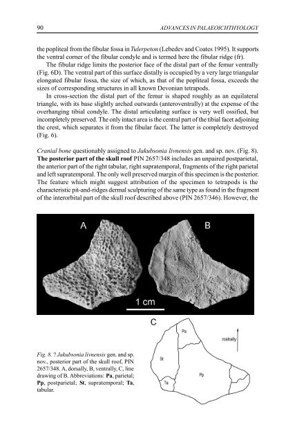
90 ADVANCES IN PALAEOICHTHYOLOGY the popliteal from the fibular fossa in Tulerpeton (Lebedev and Coates 1995). It supports the ventral corner of the fibular condyle and is termed here the fibular ridge (fr). The fibular ridge limits the posterior face of the distal part of the femur ventrally (Fig. 6D). The ventral part of this surface distally is occupied by a very large triangular elongated fibular fossa, the size of which, as that of the popliteal fossa, exceeds the sizes of corresponding structures in all known Devonian tetrapods. In cross-section the distal part of the femur is shaped roughly as an equilateral triangle, with its base slightly arched outwards (anteroventrally) at the expense of the overhanging tibial condyle. The distal articulating surface is very well ossified, but incompletely preserved. The only intact area is the central part of the tibial facet adjoining the crest, which separates it from the fibular facet. The latter is completely destroyed (Fig. 6). Cranial bone questionably assigned to Jakubsonia livnensis gen. and sp. nov. (Fig. 8). The posterior part of the skull roof PIN 2657/348 includes an unpaired postparietal, the anterior part of the right tabular, right supratemporal, fragments of the right parietal and left supratemporal. The only well preserved margin of this specimen is the posterior. The feature which might suggest attribution of the specimen to tetrapods is the characteristic pit-and-ridges dermal sculpturing of the same type as found in the fragment of the interorbital part of the skull roof described above (PIN 2657/346). However, the Fig. 8. ? Jakubsonia livnensis gen. and sp. nov., posterior part of the skull roof, PIN 2657/348. A, dorsally, B, ventrally, C, line drawing of B. Abbreviations: Pa, parietal; Pp, postparietal; St, supratemporal; Ta, tabular.
O.A. Lebedev. A new tetrapod from Russia 91 roofing bone pattern is unusual, and among Devonian tetrapods may be compared only to that in Ichthyostega, in which the postparietal is unpaired. In contrast to the condition in that animal, the posterior margin in PIN 2657/348 is convex rather than concave. More precisely, its shape is angular and the most conspicuous medial point is formed by the posteromesial process of the postparietal as in Ventastega (Lukševics, pers. comm.). The postparietal is hexagonal and occupies about half of the skull table width in this area. The lateral side contacts the tabular and the supratemporal, the anterolateral one the parietal. Given the skull roof pattern of this specimen, which is characteristic of dipnoans, one could be skeptical of its attribution to a tetrapod. However, no Devonian dipnoan is known to possess pit-and-ridge dermal sculpturing. Discussion Jakubsonia, an aquatic tetrapod In order to support the hypothesis of the primary aquatic life mode of Acanthostega and Ichthyostega, Clack and Coates (1995) put forward a number of morphological arguments relating to limb structure, such as the absence of olecranon on the ulna, correspondence of radius and ulna length and polydactyly. The femoral structure was never discussed from this point of view, as most of the previously known early tetrapod femora are more or less similarly built. In this respect the femur in Jakubsonia demonstrates probably the most unusual morphology in that there is no intercondylar fossa on its dorsal (extensor) surface distally and, respectively the condyles themselves are not expressed as well (Fig. 8). That means that the extensor muscles were not capable of pulling the epipodium forwards and the knee joint movements were more than limited. It is very probable that natural mobility in the joint was passive (that is, the muscles did not really act to deflect the knee effectively enough to reach significant limb bending) and instead of muscles, impeding of the surrounding matter (most likely, water!) was compensated only by exceptionally strong ligaments originating at the popliteal and fibular fossae. Thus, this animal was not capable of walking at all and paddling was the only function of its hind limb. This implies that the earlier the tetrapod is found in the Devonian, the more features support its primarily aquatic mode of life. Palaeoecological remarks Earlier attempts to study palaeoecology of Devonian tetrapods were undertaken by Spjeldnaes (1982) who focused on Ichthyostega, Bendix-Almgreen et al. (1990), Coates and Clack (1995) on Acanthostega, and Lebedev (1985, 1992) who considered Tulerpeton in the Andreyevka biota. Since those times several new localities have been discovered, which yielded previously unknown tetrapod remains (see Introduction). The palaeoecology and taphonomy of the Gornostayevka locality from where Jakubsonia livnensis gen. and sp. nov. originates were recently described by Moloshnikov (2001). This contributed much to our knowledge of the subject. In order to evaluate palaeoecological data the primary taphonomical, environmental and assemblage structure information was assembled in the table (Table I). An attempt to synthesize this accumulated information is made here.
- Page 39 and 40: D.K. Elliott, E. Mark-Kurik, E.B. D
- Page 41 and 42: D.K. Elliott, E. Mark-Kurik, E.B. D
- Page 43 and 44: D.K. Elliott, E. Mark-Kurik, E.B. D
- Page 45 and 46: D.K. Elliott, E. Mark-Kurik, E.B. D
- Page 47 and 48: C.G.Miller, T. Marss, H. Blom. New
- Page 49 and 50: C.G.Miller, T. Marss, H. Blom. New
- Page 51 and 52: C.G.Miller, T. Marss, H. Blom. New
- Page 53 and 54: C.G.Miller, T. Marss, H. Blom. New
- Page 55 and 56: C.G.Miller, T. Marss, H. Blom. New
- Page 57 and 58: ACTA UNIVERSITATIS LATVIENSIS, 2004
- Page 59 and 60: H.-P. Schultze, T. Marss. Revisitin
- Page 61 and 62: H.-P. Schultze, T. Marss. Revisitin
- Page 63 and 64: H.-P. Schultze, T. Marss. Revisitin
- Page 65 and 66: H.-P. Schultze, T. Marss. Revisitin
- Page 67 and 68: H.-P. Schultze, T. Marss. Revisitin
- Page 69 and 70: H.-P. Schultze, T. Marss. Revisitin
- Page 71 and 72: H.-P. Schultze, T. Marss. Revisitin
- Page 73 and 74: H.-P. Schultze, T. Marss. Revisitin
- Page 75 and 76: H.-P. Schultze, T. Marss. Revisitin
- Page 77 and 78: H.-P. Schultze, T. Marss. Revisitin
- Page 79 and 80: ACTA UNIVERSITATIS LATVIENSIS, 2004
- Page 81 and 82: O.A. Lebedev. A new tetrapod from R
- Page 83 and 84: O.A. Lebedev. A new tetrapod from R
- Page 85 and 86: O.A. Lebedev. A new tetrapod from R
- Page 87 and 88: O.A. Lebedev. A new tetrapod from R
- Page 89: O.A. Lebedev. A new tetrapod from R
- Page 93 and 94: O.A. Lebedev. A new tetrapod from R
- Page 95 and 96: O.A. Lebedev. A new tetrapod from R
- Page 97 and 98: O.A. Lebedev. A new tetrapod from R
- Page 99 and 100: ACTA UNIVERSITATIS LATVIENSIS, 2004
- Page 101 and 102: E. Lukševičs, I. Zupiņš. Sedime
- Page 103 and 104: E. Lukševičs, I. Zupiņš. Sedime
- Page 105 and 106: E. Lukševičs, I. Zupiņš. Sedime
- Page 107 and 108: E. Lukševičs, I. Zupiņš. Sedime
- Page 109 and 110: E. Lukševičs, I. Zupiņš. Sedime
- Page 111 and 112: E. Lukševičs, I. Zupiņš. Sedime
- Page 113 and 114: E. Lukševičs, I. Zupiņš. Sedime
- Page 115 and 116: E. Lukševičs, I. Zupiņš. Sedime
- Page 117 and 118: E. Lukševičs, I. Zupiņš. Sedime
- Page 119 and 120: E. Lukševičs, I. Zupiņš. Sedime
- Page 121 and 122: J. Valiukevičius. Silurian acantho
- Page 123 and 124: J. Valiukevičius. Silurian acantho
- Page 125 and 126: J. Valiukevičius. Silurian acantho
- Page 127 and 128: J. Valiukevičius. Silurian acantho
- Page 129 and 130: J. Valiukevičius. Silurian acantho
- Page 131 and 132: J. Valiukevičius. Silurian acantho
- Page 133 and 134: J. Valiukevičius. Silurian acantho
- Page 135 and 136: J. Valiukevičius. Silurian acantho
- Page 137 and 138: J. Valiukevičius. Silurian acantho
- Page 139 and 140: J. Valiukevičius. Silurian acantho
90 ADVANCES IN PALAEOICHTHYOLOGY<br />
the popliteal from the fibular fossa in Tulerpeton (Lebedev <strong>and</strong> Coates 1995). It supports<br />
the ventral corner of the fibular condyle <strong>and</strong> is termed here the fibular ridge (fr).<br />
The fibular ridge limits the posterior face of the distal part of the femur ventrally<br />
(Fig. 6D). The ventral part of this surface distally is occupied by a very large triangular<br />
elongated fibular fossa, the size of which, as that of the popliteal fossa, exceeds the<br />
sizes of corresponding structures in all known Devonian tetrapods.<br />
In cross-section the distal part of the femur is shaped roughly as an equilateral<br />
triangle, with its base slightly arched outwards (anteroventrally) at the expense of the<br />
overhanging tibial condyle. The distal articulating surface is very well ossified, but<br />
incompletely preserved. The only intact area is the central part of the tibial facet adjoining<br />
the crest, which separates it from the fibular facet. The latter is completely destroyed<br />
(Fig. 6).<br />
Cranial bone questionably assigned to Jakubsonia livnensis gen. <strong>and</strong> sp. nov. (Fig. 8).<br />
The posterior part of the skull roof PIN 2657/348 includes an <strong>un</strong>paired postparietal,<br />
the anterior part of the right tabular, right supratemporal, fragments of the right parietal<br />
<strong>and</strong> left supratemporal. The only well preserved margin of this specimen is the posterior.<br />
The feature which might suggest attribution of the specimen to tetrapods is the<br />
characteristic pit-<strong>and</strong>-ridges dermal sculpturing of the same type as fo<strong>un</strong>d in the fragment<br />
of the interorbital part of the skull roof described above (PIN 2657/346). However, the<br />
Fig. 8. ? Jakubsonia livnensis gen. <strong>and</strong> sp.<br />
nov., posterior part of the skull roof, PIN<br />
2657/348. A, dorsally, B, ventrally, C, line<br />
drawing of B. Abbreviations: Pa, parietal;<br />
Pp, postparietal; St, supratemporal; Ta,<br />
tabular.


