Zemes un vides zinātnes Earth and Environment Sciences - Latvijas ...
Zemes un vides zinātnes Earth and Environment Sciences - Latvijas ...
Zemes un vides zinātnes Earth and Environment Sciences - Latvijas ...

Create successful ePaper yourself
Turn your PDF publications into a flip-book with our unique Google optimized e-Paper software.
38 ADVANCES IN PALAEOICHTHYOLOGY<br />
visible at the broken edge. It seems probable that this embayment is the same as that<br />
seen on the postero-lateral part of the dorsal disc in Obruchevia, possibly indicating<br />
the position of the branchial duct.<br />
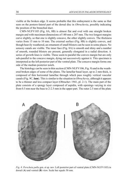
CMN-NUFV105 (Fig. 8A, 8B) is almost flat <strong>and</strong> oval with one straight broken<br />
margin <strong>and</strong> with maximum dimensions of 140 mm x 207 mm. The two longest margins<br />
curve slightly, so that one is slightly concave, the other slightly convex. The thickness<br />
varies from 15 mm to 10 mm. The external surface (Fig. 8B) is slightly convex, <strong>and</strong><br />
though heavily weathered, an ornament of small blisters can be seen in some places. No<br />
sensory canals are visible. The inner face (Fig. 8A) is smooth <strong>and</strong> shiny <strong>and</strong> a number<br />
of smooth, ro<strong>un</strong>ded blisters are present, generally elongated in a radial direction. A<br />
series of growth lines is visible. These seem to parallel the convex margin but are only<br />
sub-parallel to the concave margin, dying out successively against it. This specimen is<br />
interpreted as the left posterior part of the ventral plate. The concave margin forms one<br />
side of the median posterior notch.<br />
The histology can be seen in thin section (CMN-NUFV106, Fig. 9) <strong>and</strong> in the weathered<br />
broken edges of some of the plates. The lamellar basal layer, up to 2 mm thick, is<br />
composed of thin horizontal lamellae through which pass roughly vertical vascular<br />
canals (Fig. 9C, lam). This is similar to the situation in Obruchevia, although it appears<br />
to be a thinner <strong>and</strong> less compact layer (Obruchev 1941, pl. 2-1). The main part of the<br />
plate consists of a spongy layer composed of aspidin, with openings varying in size<br />
from 0.5 mm near the base to 2-2.5 mm in the upper part. The outer 2.5 mm of the plate<br />
Fig. 8. Perscheia pulla gen. et sp. nov. Left posterior part of ventral plate (CMN-NUFV105) in<br />
dorsal (A) <strong>and</strong> ventral (B) view. Scale bar equals 50 mm.