Zemes un vides zinātnes Earth and Environment Sciences - Latvijas ...
Zemes un vides zinātnes Earth and Environment Sciences - Latvijas ... Zemes un vides zinātnes Earth and Environment Sciences - Latvijas ...
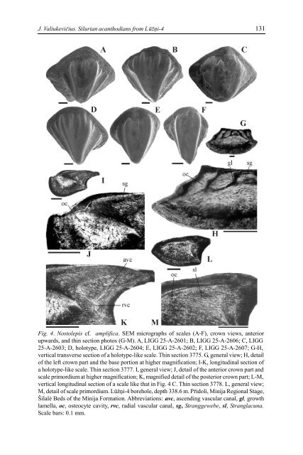
130 ADVANCES IN PALAEOICHTHYOLOGY Nostolepis cf. amplifica Fig. 4 A-M 2003 b Nostolepis amplifica; Valiukevicius, figs 2 A-H, 3 A-I. Range. The lower part of the Šilale Beds of the Minija Formation, Pridoli, Upper Silurian. Material. 72 flank scales. Remarks. N. amplifica was originally defined on large trunk scales having a high robust median crown area ornamented with two to six short parallel ridges fading out at a quarter of crown length, and narrow lateral crown steps delimited by clear oblique neck ridges. Most of the Lužni scales are within the morphologic range of N. amplifica, differing only in having more pronounced, wider bases protruding beyond the anterolateral edges of crowns, and scales generally being smaller (crowns 0.35-0.6 mm long and 0.3-0.85 mm wide). Two reasons preclude definite assignment of the scales to N. amplifica. First, the long stratigraphic gap that separates the isolated occurrence (338.6 m) of N. cf. amplifica from the range of definite N. amplifica (202.4-172.2 m); and second, histologic differences. The Stranggewebe in crowns of N. amplifica is covered by a mantle of highly cellular simple mesodentine and is distinguished by short and narrow Stranglacunae; osteocytes are present in the primordial lamella only; large principal vascular canals develop in each growth lamella (Valiukevicius 2003 b, fig. 3 A-I). These characters are almost opposite in the Lužni scales. The enveloping Stranggewebe mantle of the networked mesodentine is absent here or is very thin (Fig. 4 J-K), Stranglacunae are particularly long and osteocytes are present, though rare, in all growth lamellae (Fig. 4 H, M). The system of main vascular canals is less developed in N. cf. amplifica, and only narrow ascending and radial canals were observed. Nostolepis elegans (Brotzen) 1934 Fig. 5 A-M 1934 Diplacanthoides elegans; Brotzen, pl. 1, fig. 11 a-c. Diplacanthoides insignis; Brotzen, pl. 2, fig. 1 a-b. ? Diplacanthoides crassus; Brotzen, pl. 1, fig. 16 a-b. 1937 ? Diplacanthoides unguiparatus; Lehman, pl. 2, fig. 30. ? Diplacanthoides trilobatus; Lehman, pl. 2, figs 31; pl. 4, figs 64. Diplacanthoides elegans; Lehman, pl. 3, fig. 57. non 1980 Nostolepis arctica; Vieth, pl. 5, figs 1-9. 1998 Nostolepis striata; Valiukevicius, pl. 1, fig. 2 a-b. 1999 a Nostolepis arctica; Vergoossen, figs 2-4. 1999 b Nostolepis striata, ‘elegans’ form group; Vergoossen, pl. 2, figs 15-17. 2002 a Nostolepis striata; Vergoossen, fig. 59. 2002 b Nostolepis striata; Vergoossen, fig. 36. 2002 c Nostolepis striata, ‘elegans’ form group; Vergoossen, figs 93-99. Holotype. Brotzen 1934 a: pl. 1, fig. 11 a-c; a flank scale. Type horizon. The Lower Devonian erratics of the Baltic basin. Range. From Upper Silurian, Ludlow Series to Lower Devonian, Lochkovian.
J. Valiukevičius. Silurian acanthodians from Lūžņi-4 131 Fig. 4. Nostolepis cf. amplifica. SEM micrographs of scales (A-F), crown views, anterior upwards, and thin section photos (G-M). A, LIGG 25-A-2601; B, LIGG 25-A-2606; C, LIGG 25-A-2603; D, holotype, LIGG 25-A-2604; E, LIGG 25-A-2602; F, LIGG 25-A-2607; G-H, vertical transverse section of a holotype-like scale. Thin section 3775. G, general view; H, detail of the left crown part and the base portion at higher magnification; I-K, longitudinal section of a holotype-like scale. Thin section 3777. I, general view; J, detail of the anterior crown part and scale primordium at higher magnification; K, magnified detail of the posterior crown part; L-M, vertical longitudinal section of a scale like that in Fig. 4 C. Thin section 3778. L, general view; M, detail of scale primordium. Lūžņi-4 borehole, depth 338.6 m. Přidoli, Minija Regional Stage, Šilalė Beds of the Minija Formation. Abbreviations: avc, ascending vascular canal, gl, growth lamella, oc, osteocyte cavity, rvc, radial vascular canal, sg, Stranggewebe, sl, Stranglacuna. Scale bars: 0.1 mm.
- Page 79 and 80: ACTA UNIVERSITATIS LATVIENSIS, 2004
- Page 81 and 82: O.A. Lebedev. A new tetrapod from R
- Page 83 and 84: O.A. Lebedev. A new tetrapod from R
- Page 85 and 86: O.A. Lebedev. A new tetrapod from R
- Page 87 and 88: O.A. Lebedev. A new tetrapod from R
- Page 89 and 90: O.A. Lebedev. A new tetrapod from R
- Page 91 and 92: O.A. Lebedev. A new tetrapod from R
- Page 93 and 94: O.A. Lebedev. A new tetrapod from R
- Page 95 and 96: O.A. Lebedev. A new tetrapod from R
- Page 97 and 98: O.A. Lebedev. A new tetrapod from R
- Page 99 and 100: ACTA UNIVERSITATIS LATVIENSIS, 2004
- Page 101 and 102: E. Lukševičs, I. Zupiņš. Sedime
- Page 103 and 104: E. Lukševičs, I. Zupiņš. Sedime
- Page 105 and 106: E. Lukševičs, I. Zupiņš. Sedime
- Page 107 and 108: E. Lukševičs, I. Zupiņš. Sedime
- Page 109 and 110: E. Lukševičs, I. Zupiņš. Sedime
- Page 111 and 112: E. Lukševičs, I. Zupiņš. Sedime
- Page 113 and 114: E. Lukševičs, I. Zupiņš. Sedime
- Page 115 and 116: E. Lukševičs, I. Zupiņš. Sedime
- Page 117 and 118: E. Lukševičs, I. Zupiņš. Sedime
- Page 119 and 120: E. Lukševičs, I. Zupiņš. Sedime
- Page 121 and 122: J. Valiukevičius. Silurian acantho
- Page 123 and 124: J. Valiukevičius. Silurian acantho
- Page 125 and 126: J. Valiukevičius. Silurian acantho
- Page 127 and 128: J. Valiukevičius. Silurian acantho
- Page 129: J. Valiukevičius. Silurian acantho
- Page 133 and 134: J. Valiukevičius. Silurian acantho
- Page 135 and 136: J. Valiukevičius. Silurian acantho
- Page 137 and 138: J. Valiukevičius. Silurian acantho
- Page 139 and 140: J. Valiukevičius. Silurian acantho
- Page 141 and 142: J. Valiukevičius. Silurian acantho
- Page 143 and 144: J. Valiukevičius. Silurian acantho
- Page 145 and 146: J. Valiukevičius. Silurian acantho
- Page 147 and 148: J. Valiukevičius. Silurian acantho
- Page 149 and 150: V. Pernegre, V. Dupret. Biostratigr
- Page 151 and 152: V. Pernegre, V. Dupret. Biostratigr
- Page 153 and 154: V. Pernegre, V. Dupret. Biostratigr
- Page 155 and 156: V. Pernegre, V. Dupret. Biostratigr
- Page 157 and 158: V. Pernegre, V. Dupret. Biostratigr
- Page 159 and 160: Ž. Žigaite. New telodont from Tuv
- Page 161 and 162: Ž. Žigaite. New telodont from Tuv
- Page 163 and 164: Ž. Žigaite. New telodont from Tuv
- Page 165 and 166: Ž. Žigaite. New telodont from Tuv
J. Valiukevičius. Silurian acanthodians from Lūžņi-4<br />
131<br />
Fig. 4. Nostolepis cf. amplifica. SEM micrographs of scales (A-F), crown views, anterior<br />
upwards, <strong>and</strong> thin section photos (G-M). A, LIGG 25-A-2601; B, LIGG 25-A-2606; C, LIGG<br />
25-A-2603; D, holotype, LIGG 25-A-2604; E, LIGG 25-A-2602; F, LIGG 25-A-2607; G-H,<br />
vertical transverse section of a holotype-like scale. Thin section 3775. G, general view; H, detail<br />
of the left crown part <strong>and</strong> the base portion at higher magnification; I-K, longitudinal section of<br />
a holotype-like scale. Thin section 3777. I, general view; J, detail of the anterior crown part <strong>and</strong><br />
scale primordium at higher magnification; K, magnified detail of the posterior crown part; L-M,<br />
vertical longitudinal section of a scale like that in Fig. 4 C. Thin section 3778. L, general view;<br />
M, detail of scale primordium. Lūžņi-4 borehole, depth 338.6 m. Přidoli, Minija Regional Stage,<br />
Šilalė Beds of the Minija Formation. Abbreviations: avc, ascending vascular canal, gl, growth<br />
lamella, oc, osteocyte cavity, rvc, radial vascular canal, sg, Stranggewebe, sl, Stranglac<strong>un</strong>a.<br />
Scale bars: 0.1 mm.


