Zemes un vides zinātnes Earth and Environment Sciences - Latvijas ...
Zemes un vides zinātnes Earth and Environment Sciences - Latvijas ... Zemes un vides zinātnes Earth and Environment Sciences - Latvijas ...
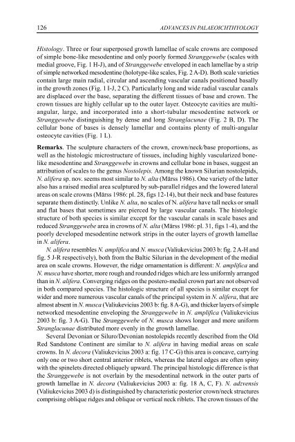
126 ADVANCES IN PALAEOICHTHYOLOGY Histology. Three or four superposed growth lamellae of scale crowns are composed of simple bone-like mesodentine and only poorly formed Stranggewebe (scales with medial groove, Fig. 1 H-J), and of Stranggewebe enveloped in each lamellae by a strip of simple networked mesodentine (holotype-like scales, Fig. 2 A-D). Both scale varieties contain large main radial, circular and ascending vascular canals positioned basally in the growth zones (Fig. 1 I-J, 2 C). Particularly long and wide radial vascular canals are displaced over the base, separating the different tissues of base and crown. The crown tissues are highly cellular up to the outer layer. Osteocyte cavities are multiangular, large, and incorporated into a short-tubular mesodentine network or Stranggewebe distinguishing by dense and long Stranglacunae (Fig. 2 B, D). The cellular bone of bases is densely lamellar and contains plenty of multi-angular osteocyte cavities (Fig. 1 L). Remarks. The sculpture characters of the crown, crown/neck/base proportions, as well as the histologic microstructure of tissues, including highly vascularized bonelike mesodentine and Stranggewebe in crowns and cellular bone in bases, suggest an attribution of scales to the genus Nostolepis. Among the known Silurian nostolepids, N. alifera sp. nov. seems most similar to N. alta (Märss 1986). One variety of the latter also has a raised medial area sculptured by sub-parallel ridges and the lowered lateral areas on scale crowns (Märss 1986: pl. 28, figs 12-14), but their neck and base features separate them distinctly. Unlike N. alta, no scales of N. alifera have tall necks or small and flat bases that sometimes are pierced by large vascular canals. The histologic structure of both species is similar except for the vascular canals in scale bases and reduced Stranggewebe area in crowns of N. alta (Märss 1986: pl. 31, figs 1-4), and the poorly developed mesodentine network strips in the outer layers of growth lamellae in N. alifera. N. alifera resembles N. amplifica and N. musca (Valiukevicius 2003 b: fig. 2 A-H and fig. 5 J-R respectively), both from the Baltic Silurian in the development of the medial area on scale crowns. However, the ridge ornamentation is different: N. amplifica and N. musca have shorter, more rough and rounded ridges which are less uniformly arranged than in N. alifera. Converging ridges on the postero-medial crown part are not observed in both compared species. The histologic structure of all species is similar except for wider and more numerous vascular canals of the principal system in N. alifera, that are almost absent in N. musca (Valiukevicius 2003 b: fig. 8 A-G), and thicker layers of simple networked mesodentine enveloping the Stranggewebe in N. amplifica (Valiukevicius 2003 b: fig. 3 A-G). The Stranggewebe of N. musca shows longer and more uniform Stranglacunae distributed more evenly in the growth lamellae. Several Devonian or Siluro/Devonian nostolepids recently described from the Old Red Sandstone Continent are similar to N. alifera in having medial areas on scale crowns. In N. decora (Valiukevicius 2003 a: fig. 17 C-G) this area is concave, carrying only one or two short central anterior riblets, whereas the lateral edges are often spiny with the spinelets directed obliquely upward. The principal histologic difference is that the Stranggewebe is not overlain by the mesodentinal network in the outer parts of growth lamellae in N. decora (Valiukevicius 2003 a: fig. 18 A, C, F). N. adzvensis (Valiukevicius 2003 d) is distinguished by characteristic posterior crown/neck structures comprising oblique ridges and oblique or vertical neck riblets. The crown tissues of the
J. Valiukevičius. Silurian acanthodians from Lūžņi-4 127 latter contain no enlarged vascular canals, and the networked mesodentine closely resembles bone in the density of osteocyte spaces and the short osteocyte processes. N. terraborea (Valiukevicius 2003 d) typically has four uniform, short and basally widened ridges with sharp crests on the raised medial area and unornamented lateral slopes. N. valentinae (Valiukevicius 2003 d) is distinguished by a highly raised elipsoidal to triangular medial area which is smooth or bears one to five very short sub-parallel rounded anterior riblets that never bifurcate. The lateral areas sometimes contain obliquely-upward, posteriorly directed riblets like in N. alifera. The Stranggewebe and simple mesodentine do not differ from those of N. alifera apart from absence of networked strips enveloping the Stranggewebe and the weakened system of principal vascular canals, particularly the radial canals, in crowns of both N. terraborea and N. valentinae. Occurrence. See Table. Nostolepis latvica sp. nov. Fig. 3 A-M 2003 c Nostolepis “prestriata”; Valiukevicius, p. 51. Etymology. The species is named from Latvia, the country of origin for the type material. Holotype. LIGG 25-A-2574, flank scale (Fig. 3 A). Type horizon. The lower part of the Venzava Beds of the Targale Formation, Pridoli, Upper Silurian. Range. Šilale and Varniai Beds of the Minija Formation and the lower part of the Venzava Beds of the Targale Formation, Pridoli, Upper Silurian. Material. About 850 scales. Diagnosis. Nostolepis having scales with rhomboidal, posteriorly tapered and elongated crowns overhanging bases. Crowns are sculptured by four to eight uniform, wide anterior ridges extending a half of crown length or have a slightly raised medial area carrying few riblets of different lengths. Lateral crown areas are narrow, outlined by oblique, short neck ridges. Scale crowns composed of Stranggewebe and networked mesodentine. The Stranggewebe area of the posterior crown is reduced, replaced by simple bone-like mesodentine in the lower (neck) part; its strips are not covered by the mesodentinal network. Simple mesodentine of the anterior crown part is densely networked and highly cellular. The main vascular system is poorly developed. Description. Morphology. The scales are mostly of medium size and only rarely reach 0.9-1.1 mm in crown length. Crowns are slightly downsloped anteriorly, flat, rarely medially concave, of rhomboidal to elipsoidal form, tapered and clearly elongated posteriorly. The posterior crown part (up to a half of total length) overhangs the base. Crowns are ornamented by four to eight uniform, low, basally widened ridges extending half of their length (Fig. 3 A-B, D). The lateral step-like lowered areas are frequently present (Fig. 3 C, E-F), but are narrow and sometimes very short. The medial area, occupying the main part of the crown, is only slightly raised, flat or side to side concave, and bears two to
- Page 75 and 76: H.-P. Schultze, T. Marss. Revisitin
- Page 77 and 78: H.-P. Schultze, T. Marss. Revisitin
- Page 79 and 80: ACTA UNIVERSITATIS LATVIENSIS, 2004
- Page 81 and 82: O.A. Lebedev. A new tetrapod from R
- Page 83 and 84: O.A. Lebedev. A new tetrapod from R
- Page 85 and 86: O.A. Lebedev. A new tetrapod from R
- Page 87 and 88: O.A. Lebedev. A new tetrapod from R
- Page 89 and 90: O.A. Lebedev. A new tetrapod from R
- Page 91 and 92: O.A. Lebedev. A new tetrapod from R
- Page 93 and 94: O.A. Lebedev. A new tetrapod from R
- Page 95 and 96: O.A. Lebedev. A new tetrapod from R
- Page 97 and 98: O.A. Lebedev. A new tetrapod from R
- Page 99 and 100: ACTA UNIVERSITATIS LATVIENSIS, 2004
- Page 101 and 102: E. Lukševičs, I. Zupiņš. Sedime
- Page 103 and 104: E. Lukševičs, I. Zupiņš. Sedime
- Page 105 and 106: E. Lukševičs, I. Zupiņš. Sedime
- Page 107 and 108: E. Lukševičs, I. Zupiņš. Sedime
- Page 109 and 110: E. Lukševičs, I. Zupiņš. Sedime
- Page 111 and 112: E. Lukševičs, I. Zupiņš. Sedime
- Page 113 and 114: E. Lukševičs, I. Zupiņš. Sedime
- Page 115 and 116: E. Lukševičs, I. Zupiņš. Sedime
- Page 117 and 118: E. Lukševičs, I. Zupiņš. Sedime
- Page 119 and 120: E. Lukševičs, I. Zupiņš. Sedime
- Page 121 and 122: J. Valiukevičius. Silurian acantho
- Page 123 and 124: J. Valiukevičius. Silurian acantho
- Page 125: J. Valiukevičius. Silurian acantho
- Page 129 and 130: J. Valiukevičius. Silurian acantho
- Page 131 and 132: J. Valiukevičius. Silurian acantho
- Page 133 and 134: J. Valiukevičius. Silurian acantho
- Page 135 and 136: J. Valiukevičius. Silurian acantho
- Page 137 and 138: J. Valiukevičius. Silurian acantho
- Page 139 and 140: J. Valiukevičius. Silurian acantho
- Page 141 and 142: J. Valiukevičius. Silurian acantho
- Page 143 and 144: J. Valiukevičius. Silurian acantho
- Page 145 and 146: J. Valiukevičius. Silurian acantho
- Page 147 and 148: J. Valiukevičius. Silurian acantho
- Page 149 and 150: V. Pernegre, V. Dupret. Biostratigr
- Page 151 and 152: V. Pernegre, V. Dupret. Biostratigr
- Page 153 and 154: V. Pernegre, V. Dupret. Biostratigr
- Page 155 and 156: V. Pernegre, V. Dupret. Biostratigr
- Page 157 and 158: V. Pernegre, V. Dupret. Biostratigr
- Page 159 and 160: Ž. Žigaite. New telodont from Tuv
- Page 161 and 162: Ž. Žigaite. New telodont from Tuv
- Page 163 and 164: Ž. Žigaite. New telodont from Tuv
- Page 165 and 166: Ž. Žigaite. New telodont from Tuv
126<br />
ADVANCES IN PALAEOICHTHYOLOGY<br />
Histology. Three or four superposed growth lamellae of scale crowns are composed<br />
of simple bone-like mesodentine <strong>and</strong> only poorly formed Stranggewebe (scales with<br />
medial groove, Fig. 1 H-J), <strong>and</strong> of Stranggewebe enveloped in each lamellae by a strip<br />
of simple networked mesodentine (holotype-like scales, Fig. 2 A-D). Both scale varieties<br />
contain large main radial, circular <strong>and</strong> ascending vascular canals positioned basally<br />
in the growth zones (Fig. 1 I-J, 2 C). Particularly long <strong>and</strong> wide radial vascular canals<br />
are displaced over the base, separating the different tissues of base <strong>and</strong> crown. The<br />
crown tissues are highly cellular up to the outer layer. Osteocyte cavities are multiangular,<br />
large, <strong>and</strong> incorporated into a short-tubular mesodentine network or<br />
Stranggewebe distinguishing by dense <strong>and</strong> long Stranglac<strong>un</strong>ae (Fig. 2 B, D). The<br />
cellular bone of bases is densely lamellar <strong>and</strong> contains plenty of multi-angular<br />
osteocyte cavities (Fig. 1 L).<br />
Remarks. The sculpture characters of the crown, crown/neck/base proportions, as<br />
well as the histologic microstructure of tissues, including highly vascularized bonelike<br />
mesodentine <strong>and</strong> Stranggewebe in crowns <strong>and</strong> cellular bone in bases, suggest an<br />
attribution of scales to the genus Nostolepis. Among the known Silurian nostolepids,<br />
N. alifera sp. nov. seems most similar to N. alta (Märss 1986). One variety of the latter<br />
also has a raised medial area sculptured by sub-parallel ridges <strong>and</strong> the lowered lateral<br />
areas on scale crowns (Märss 1986: pl. 28, figs 12-14), but their neck <strong>and</strong> base features<br />
separate them distinctly. Unlike N. alta, no scales of N. alifera have tall necks or small<br />
<strong>and</strong> flat bases that sometimes are pierced by large vascular canals. The histologic<br />
structure of both species is similar except for the vascular canals in scale bases <strong>and</strong><br />
reduced Stranggewebe area in crowns of N. alta (Märss 1986: pl. 31, figs 1-4), <strong>and</strong> the<br />
poorly developed mesodentine network strips in the outer layers of growth lamellae<br />
in N. alifera.<br />
N. alifera resembles N. amplifica <strong>and</strong> N. musca (Valiukevicius 2003 b: fig. 2 A-H <strong>and</strong><br />
fig. 5 J-R respectively), both from the Baltic Silurian in the development of the medial<br />
area on scale crowns. However, the ridge ornamentation is different: N. amplifica <strong>and</strong><br />
N. musca have shorter, more rough <strong>and</strong> ro<strong>un</strong>ded ridges which are less <strong>un</strong>iformly arranged<br />
than in N. alifera. Converging ridges on the postero-medial crown part are not observed<br />
in both compared species. The histologic structure of all species is similar except for<br />
wider <strong>and</strong> more numerous vascular canals of the principal system in N. alifera, that are<br />
almost absent in N. musca (Valiukevicius 2003 b: fig. 8 A-G), <strong>and</strong> thicker layers of simple<br />
networked mesodentine enveloping the Stranggewebe in N. amplifica (Valiukevicius<br />
2003 b: fig. 3 A-G). The Stranggewebe of N. musca shows longer <strong>and</strong> more <strong>un</strong>iform<br />
Stranglac<strong>un</strong>ae distributed more evenly in the growth lamellae.<br />
Several Devonian or Siluro/Devonian nostolepids recently described from the Old<br />
Red S<strong>and</strong>stone Continent are similar to N. alifera in having medial areas on scale<br />
crowns. In N. decora (Valiukevicius 2003 a: fig. 17 C-G) this area is concave, carrying<br />
only one or two short central anterior riblets, whereas the lateral edges are often spiny<br />
with the spinelets directed obliquely upward. The principal histologic difference is that<br />
the Stranggewebe is not overlain by the mesodentinal network in the outer parts of<br />
growth lamellae in N. decora (Valiukevicius 2003 a: fig. 18 A, C, F). N. adzvensis<br />
(Valiukevicius 2003 d) is distinguished by characteristic posterior crown/neck structures<br />
comprising oblique ridges <strong>and</strong> oblique or vertical neck riblets. The crown tissues of the


