Electron Microscopy: TEM, Immunogold Labeling, SEM, Correlative ...
Electron Microscopy: TEM, Immunogold Labeling, SEM, Correlative ...
Electron Microscopy: TEM, Immunogold Labeling, SEM, Correlative ...

Create successful ePaper yourself
Turn your PDF publications into a flip-book with our unique Google optimized e-Paper software.
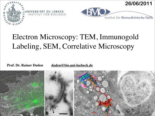
26/06/2011<br />
<strong>Electron</strong> <strong>Microscopy</strong>: <strong>TEM</strong>, <strong>Immunogold</strong><br />
<strong>Labeling</strong>, <strong>SEM</strong>, <strong>Correlative</strong> <strong>Microscopy</strong><br />
Prof. Dr. Rainer Duden<br />
duden@bio.uni-luebeck.de<br />
1
Resolution<br />
Comparison<br />
Light vs <strong>Electron</strong><br />
<strong>Microscopy</strong>
Microscope Resolution<br />
• ability of a lens to separate or distinguish small<br />
objects that are close together<br />
• wavelength of light used is major factor in<br />
resolution<br />
shorter wavelength ⇒ greater resolution<br />
3
<strong>Electron</strong> <strong>Microscopy</strong><br />
• beams of electrons<br />
are used to produce<br />
images<br />
• wavelength of<br />
electron beam is<br />
much shorter than<br />
light, resulting in<br />
much higher<br />
resolution<br />
4
•Light microscopy<br />
• Glass lenses<br />
• Source of illumination is usually light of visible<br />
wavelengths<br />
•<strong>Electron</strong> microscopy<br />
• Electromagnetic lenses<br />
• Source of illumination is electrons<br />
• Hairpin tungsten filament (thermionic<br />
emission)<br />
• Pointed tungsten crystal (cold cathode field<br />
emission)
The Transmission <strong>Electron</strong><br />
Microscope<br />
• electrons scatter when they pass through thin<br />
sections of a specimen<br />
• transmitted electrons (those that do not scatter)<br />
are used to produce image<br />
• denser regions in specimen, scatter more electrons<br />
and appear darker
Comparison of LM and <strong>TEM</strong>
Specimen Preparation<br />
• analogous to procedures used for light microscopy<br />
• for transmission electron microscopy, specimens<br />
must be cut very thin<br />
• specimens are chemically fixed and stained with<br />
electron dense material
Transmission <strong>Electron</strong> <strong>Microscopy</strong> (<strong>TEM</strong>)<br />
Zeiss 10/A conventional<br />
<strong>TEM</strong><br />
Excellent for training<br />
Film only
Negative Staining<br />
Viruses, small<br />
particles, proteins,<br />
molecules<br />
No sectioning<br />
Same day results
negative staining<br />
particles<br />
<strong>Electron</strong> dense<br />
negative stain
negative staining<br />
• requires minimal interaction between particle & ‘stain’<br />
• to avoid binding, heavy metal ion should be of same<br />
charge +/- as the particle<br />
• positive staining usually destructive of bio-particles<br />
• biological material usually -ve charge at neutral pH<br />
• widely used negative contrast media include:<br />
anionic cationic<br />
phosphotungstate uranyl actetate/formate<br />
molybdate (ammonium) (@ pH ~ 4)
Negative Stain<br />
Ebola
Double <strong>Immunogold</strong> <strong>Labeling</strong> of<br />
Negatively Stained Specimens<br />
Bacterial pili<br />
serotypes dried onto<br />
grid and sequentially<br />
labeled with primary<br />
antibody, then<br />
Protein-A-5nm-gold<br />
and Protein-A-15-<br />
nm-gold before<br />
negative staining
metal shadowing - rotary
metal shadowing - rotary<br />
• Contrast usually inverted to give dark shadows<br />
> resolution 2 - 3 nm - single DNA strand detectable<br />
- historic use for ‘molecular biology’<br />
(e.g. heteroduplex mapping)<br />
> good preservation of shape, but enlargement of<br />
apparent dimensions<br />
> in very recent modification (MCD -<br />
microcrystallite<br />
decoration), resolution ~1.1 nm
Clathrin:<br />
a major and evolutionarily<br />
conserved coat protein<br />
200<br />
116<br />
97<br />
Crude membranes<br />
Purified CCVs<br />
clathrin<br />
heavy chain<br />
~100kD proteins<br />
66<br />
45<br />
31<br />
~50kD proteins<br />
clathrin<br />
light chains<br />
21.5<br />
~20kD proteins
Rotary shadowing<br />
EM images of purified Clathrin triskelia
Rotary shadowing<br />
EM images of purified Clathrin triskelia<br />
Kirchhausen and Harrison (1981). Cell 23, 755-761:<br />
see EM image above<br />
Ungewickell and Branton (1981). Nature 289, 420-422:<br />
- reversible dissassembly of Clathrin triskelions into clathrin - coats in vitro
Clathrin triskelions<br />
3 heavy chains<br />
3 light chains
Adaptors:<br />
essential for cargo sorting<br />
200<br />
116<br />
97<br />
Crude membranes<br />
Purified CCVs<br />
clathrin<br />
heavy chain<br />
~100kD proteins<br />
66<br />
45<br />
31<br />
~50kD proteins<br />
clathrin<br />
light chains<br />
21.5<br />
~20kD proteins
Protein pattern of<br />
Adaptor Complexes<br />
extracted from<br />
purified brain CCVs<br />
after SDS-PAGE<br />
β1<br />
γ<br />
AP-1 AP-2<br />
αA<br />
β2<br />
αC<br />
µ1<br />
µ2<br />
σ1<br />
σ2
Rotary shadowing<br />
EM images of purified AP-2 complex
Adaptor proteins mediate sorting of specific cargo<br />
from different compartments<br />
α<br />
µ2<br />
σ2<br />
β2<br />
γ<br />
µ1<br />
σ1<br />
β1<br />
δ<br />
µ3<br />
σ3<br />
β3<br />
ε<br />
µ4<br />
σ4<br />
β4<br />
AP-1 AP-2 AP-3 AP-4<br />
Margaret Robinson, Univ. Cambridge
Overview of Biological Specimen Preparation<br />
Killing & Fixation<br />
- Death; Molecular stabilization<br />
Dehydration<br />
- Chemical removal of H 2 O<br />
Infiltration<br />
- Replace liquid phase with resin<br />
Embedding & Polymerization<br />
- Make solid, sectionable block<br />
Sectioning<br />
- Ultramicrotome, mount, stain
Preparing for cutting sections for <strong>TEM</strong><br />
27
Estimating Section Thickness<br />
Interference reflection angle from Sjöstrand (1967)
Serial section 3-D reconstruction
The Freeze Fracture Technique
Gap Junctions in negative stain, freeze fracture & <strong>TEM</strong>
Tight Junction<br />
structure in<br />
<strong>TEM</strong>, freeze fracture, and<br />
live fluorescence<br />
microscopy
Cryotechniques<br />
Ultrarapid cryofixation<br />
Metal mirror impact<br />
Liquid propane plunge<br />
Freeze fracture with<br />
Balzers 400T<br />
Cryosubstitution<br />
Cryo-ultramicrotomy –<br />
Ultrathin frozen<br />
sections (primarily for<br />
antibody labeling)
Clathrin - coated vesicles - the minimal machinery<br />
Clathrin triskelions<br />
3 heavy chains<br />
3 light chains<br />
Adaptor (AP2)<br />
α<br />
- four adaptins<br />
β2<br />
σ2 µ2<br />
Tom Kirchhausen, Harvard Medical School
John Heuser’s Quick Freeze Deep Etch Technique
Quick freeze - deep etch<br />
technique<br />
John Heuser<br />
Washington<br />
University<br />
School of Medicine,<br />
USA
Clathrin - coated Vesicles<br />
J. Heuser<br />
Inner layer : membrane containing cargo<br />
Middle layer : adaptors and accessory proteins<br />
Outer layer : mechanical scaffold
Now please put on<br />
the 3-D glasses…
John Heuser, Washington Univ. School of Medicine
Immunolabeling for Transmission<br />
<strong>Electron</strong> <strong>Microscopy</strong><br />
Normally do Two-Step<br />
Method<br />
Primary antibody<br />
applied followed by<br />
colloidal gold-labeled<br />
secondary antibody<br />
May also be enhanced<br />
with silver<br />
Can also do for LM
Preparation of Biological Specimens for<br />
Immunolabeling<br />
The goal is to preserve tissue as closely as<br />
possible to its natural state while at the same time<br />
maintaining the ability of the antigen to react with<br />
the antibody<br />
Chemical fixation of whole mounts prior to labeling<br />
for LM<br />
Chemical fixation, dehydration, and embedment in<br />
paraffin or resin for sectioning for LM or <strong>TEM</strong><br />
Chemical fixation for cryosections for LM<br />
Cryofixation for LM or <strong>TEM</strong>
Chemical Fixation<br />
Antigenic sites are easily denatured or masked during<br />
chemical fixation<br />
Glutaraldehyde gives good fixation but may mask<br />
antigens, plus it is fluorescent<br />
Paraformaldehyde often better choice, but results in poor<br />
morphology , especially for electron microscopy<br />
May use e.g., 4% paraformaldehyde with 0.5%<br />
glutaraldehyde as a good compromise
Specimen Preparation for <strong>TEM</strong><br />
Chemical fixation with buffered glutaraldehyde<br />
Or 4% paraformaldehyde with >1% glutaraldehyde<br />
Postfixation with osmium tetroxide<br />
Or not, or with subsequent removal from sections<br />
Dehydration and infiltration with liquid epoxy or<br />
acrylic resin<br />
Polymerization of hard blocks by heat or UV<br />
Ultramicrotomy – 60-80nm sections<br />
<strong>Labeling</strong> and/or staining<br />
View with <strong>TEM</strong>
Approaches to Immunolabeling<br />
Direct Method: Primary antibody contains<br />
label<br />
Indirect Method: Primary antibody followed<br />
by labeled secondary antibody<br />
Amplified Method: Methods to add more<br />
reporter to labeled site<br />
Protein A Method: May be used as secondary<br />
reagent instead of antibody
• Colloidal gold of defined sizes, e.g., 5 nm,<br />
10 nm, 20 nm, easily conjugated to<br />
antibodies<br />
• Results in small, round, electron-dense<br />
label easily detected with EM<br />
• Can be enhanced after labeling to enlarge<br />
size for LM or EM
Immunolocalization<br />
LM<br />
Fluor/confocal<br />
<strong>TEM</strong><br />
<strong>SEM</strong> with<br />
backscatter<br />
detector
Preembedding or Postembedding<br />
<strong>Labeling</strong><br />
May use preembedding labeling for surface<br />
antigens or for permeabilized cells<br />
The advantage is that antigenicity is more likely<br />
preserved<br />
Postembedding labeling is performed on sectioned<br />
tissue, on grids, allowing access to internal<br />
antigens<br />
Antigenicity probably partially compromised by<br />
embedding
Steps in <strong>Labeling</strong> of Sections<br />
Chemical fixation<br />
Dehydration, infiltration, embedding and sectioning<br />
Optional etching of embedment, permeabilization<br />
Blocking<br />
Incubation with primary antibody<br />
Washing<br />
Incubation with secondary antibody congugated<br />
with reporter (fluorescent probe, colloidal gold)<br />
Washing, optional counterstaining<br />
Mount and view
Controls! Controls! Controls!<br />
Omit primary antibody<br />
Irrelevant primary antibody<br />
Pre-immune serum<br />
Perform positive control<br />
Check for autofluorescence<br />
Check for non-specific labeling<br />
Dilution series
Desmosomes and IFs in primary mouse keratinocytes<br />
Duden & Franke,<br />
988 (J. Cell Biol.)
Pre-embedding labelling of desmosomal vesicles<br />
in primary mouse keratinocytes<br />
Duden and Franke, 1988 (J. Cell Biol.)
Visualization of desmosomal vesicles in A431 cells<br />
grown on glass coverslips<br />
Duden and Franke, 1988 (J. Cell Biol.)
Pre-embedding labelling of desmosomal vesicles<br />
in A431 cells<br />
Duden &<br />
Franke, 1988<br />
(J. Cell Biol.)
Double-labeling Method<br />
Use primary antibodies<br />
derived from different<br />
animals (e.g., one<br />
mouse antibody and one<br />
rabbit antibody)<br />
Then use two secondary<br />
antibodies conjugated<br />
with reporters that can<br />
be distinguished from<br />
one another
George Palade<br />
1974 Nobel Prize for<br />
Physiology or Medicine<br />
Analysis of the secretory pathway<br />
by a combination of EM and<br />
autoradiography<br />
ER --> Golgi --> Vesicles --> PM
The Scanning <strong>Electron</strong><br />
Microscope<br />
• uses electrons reflected from the surface of a<br />
specimen to create image<br />
• produces a 3-dimensional image of specimen’s<br />
surface features
<strong>TEM</strong><br />
vs<br />
<strong>SEM</strong>
Scanning <strong>Electron</strong> <strong>Microscopy</strong>
<strong>SEM</strong>
<strong>Correlative</strong> Light/EM microscopy - <strong>SEM</strong>:<br />
visualization of virus particles on a cell surface
<strong>Correlative</strong> Light/EM microscopy<br />
& electron tomography<br />
Diaminobenzidine (DAB) photooxidation by GFP (GalT-GFP)<br />
Grabenbaur<br />
et al., 2005.<br />
Nat. Meth. 2.<br />
857-862
<strong>Correlative</strong><br />
Light/EM<br />
microscopy<br />
& electron<br />
tomography<br />
Grabenbaur<br />
et al., 2005.<br />
Nat. Meth. 2.<br />
857-862
miniSOG