Marine Ecosystems Research Department - jamstec japan agency ...
Marine Ecosystems Research Department - jamstec japan agency ...
Marine Ecosystems Research Department - jamstec japan agency ...

You also want an ePaper? Increase the reach of your titles
YUMPU automatically turns print PDFs into web optimized ePapers that Google loves.
JAMSTEC 2002 Annual Report<br />
Frontier <strong>Research</strong> System for Extremophiles<br />
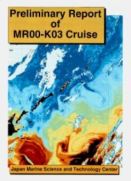
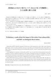
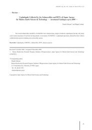
Fig. 6 Alvinocaris longirostris in the DEEP AQUARIUM. Eggs can<br />
be confirmed in the abdomen.<br />
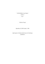
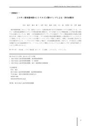
Fig. 7 Tissue culture cell (KMHA-1) of Simenchelys parasiticus.<br />
pression and decompression while observing the<br />
organism's condition. At atmospheric pressure,<br />
Alvinocaris longirostris spawned after one week.<br />
(iii) Establishment of tissue culture techniques<br />
Tissue culture techniques are necessary for observing<br />
deep-sea organisms at the tissue cell level. Primary<br />
culture was carried out on a cell from the tissue of the<br />
captured benthic multicellular organisms Simenchelys<br />
parasiticus. The captured Simenchelys parasiticus was<br />
gradually decompressed to atmospheric pressure.<br />
After slicing the fin tissue, swarmer cell was observed<br />
in the medium culture. The fibroblast cell of<br />
Simenchelys parasiticus was cultivated by using L-<br />
medium with % FBS (Fig.). Optimal temperature<br />
was ˚C, and doubling time of cell was .h - . We<br />
observed cell growth under the high-pressure environment,<br />
and pressure tolerance. New tissue culture cell<br />
(KMHA-) is cultivated and kept in frozen storage in<br />
the laboratory.<br />
3.2. Behaviors of biological substances and colloidal<br />
dispersions in supercritical water<br />
(a) Microscopic observations of biological substances<br />
in supercritical water<br />
We have developed an optical microscope equipped<br />
with a high-temperature and pressure cell, and studied<br />
behaviors of various biological substances in nearcritical<br />
and supercritical water. The samples studied so far<br />
include polysaccharides, proteins, microorganisms,<br />
inorganic materials, and synthetic polymers. Although<br />
optical microscopy helps to grasp the behavior of the<br />
system quickly, it is rather difficult to perform quantitative<br />
study by microscopic observations alone. This<br />
year, we attempted to extract quantitative information<br />
from the images by applying computer-based image<br />
analysis.<br />
Analysis was performed on the images obtained for<br />
several cellulose samples. These samples have the same<br />
molecular weight, but differ in crystallinity and crystalline<br />
form. In addition, regenerated cellulose is highly<br />
porous and has significantly larger surface area than the<br />
crystalline samples. We attempted to evaluate the effect<br />
of these factors on the dissolution temperature. The<br />
sample was dispersed in water at the concentration of<br />
. wt%, introduced into the high-temperature and<br />
pressure cell, and pressurized to MPa at room temperature.<br />
The sample was then heated to ˚C, and the<br />
behavior of the sample during heating was observed<br />
and video-taped. Images were then transferred to a<br />
computer, and subjected to image analysis.<br />
Dissolution of cellulose led to an increase of transmittance.<br />
By calculating the relative transmittance<br />
of the images taken at different temperatures, semiquantitative<br />
comparison of the dissolution temperature<br />
of different cellulose samples could be made.<br />
Comparison revealed that cellulose with higher crys-<br />
78