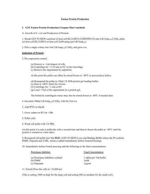
GST Fusion Protein Production
GST Fusion Protein Production
GST Fusion Protein Production

Create successful ePaper yourself
Turn your PDF publications into a flip-book with our unique Google optimized e-Paper software.
<strong>Fusion</strong> <strong>Protein</strong> <strong>Production</strong><br />
I. <strong>GST</strong> <strong>Fusion</strong> <strong>Protein</strong> <strong>Production</strong> (Yaopan Mao's method)<br />
A. Growth of E. coli and <strong>Production</strong> of <strong>Protein</strong>:<br />
1. Streak <strong>GST</strong>-FUSION construct in host cell BL21(DE3) CODONPLUS onto LB/Amp 100 /CAM 40 plate.<br />
(or host cell BL21(DE3) or host cell Xa90 using just LB/Amp 100 )<br />
2. Pick a single colony into 5ml LB/Amp 100 /CAM 40 and grow o/n.<br />
Induction of <strong>Protein</strong>:<br />
3. Pre-expression control.<br />
(a) Remove a ~1ml aliquot of cells.<br />
(b) Centrifuge for ~5-10 min at 4 o C in the microfuge.<br />
(c) Remove the supernatant by aspiration.<br />
At this point the pellet can either be stored frozen at –80 o C or processed as below.<br />
(d) Resuspend the pellet in 100µl 1X SDS protein gel loading buffer.<br />
(e) Heat to 100 o C (boil) for 10 min.<br />
(f) Centrifuge for ~1 min at RT.<br />
(g) Load ~15µl of the supernatant on a protein gel.<br />
The boiled & centrifuged extract may also be stored frozen at –80 o C if needed later.<br />
4. Inoculate 500ml LB/Amp 100 /CAM 40 with the 5ml o/n.<br />
5. Add IPTG to 60µM.<br />
7. Grow culture at RT for ~20h.<br />
6. Pellet cells.<br />
8. Wash cell pellet with 1X PBS.<br />
(At this point it is safe to pellet the cells a second time and then to freeze the pellet at –80 o C until the<br />
protein is isolated at a later date).<br />
9. Resuspend cell pellet into 9ml PGE- (<strong>GST</strong>-FUSION Lysis and Binding Buffer minus the PI cocktail,<br />
PMSF, Pepstain and 2-ME, which is added immediately before French Pressing).<br />
10. Immediately before French pressing add the following to the final concentrations:<br />
<strong>Protein</strong>ase Inhibitor<br />
(a) <strong>Protein</strong>ase Inhibitor cocktail<br />
(b) PMSF<br />
(c) Pepstatin<br />
Final Concentration<br />
1 tablet per 7ml buffer<br />
1mM<br />
1µg/ml<br />
11. French Press the cells at ~16,000 psi:<br />
(This is setting 1000 on high for the large cell and setting 800 on medium for the small cell.)
12. Add DTT to 1mM and Triton X-100 to 1%.<br />
13. Incubate on ice 40min.<br />
14. Spin cell debris down at 20,000 X g (13,000 rpm in the Sorval SS34 rotor) for 20min.<br />
15. Collect the s/n and aliquot it away for storage at -80 o C.<br />
16. Determine the protein concentration by Bradford assay.<br />
17. Load 50-100µg of the crude E. coli protein extract onto a Laemmli protein gel.<br />
B. <strong>Protein</strong> Purification<br />
1. Prepare the glutathione agarose:<br />
(a) Weigh out the required quantity of glutathione agarose (Sigma cat# 4510).<br />
From a 500ml culture of <strong>GST</strong>-FUSION resuspended in 10 ml and French pressed, approximately 2mg of<br />
r<strong>GST</strong>-FUSION can be expected. Approximately 10mg of <strong>GST</strong> protein will bind to 1ml of swollen<br />
glutathione agarose. The required glutathione agarose bed volume is then only 250µL (Mao used 1.0 ml<br />
of the French Press lysate with 60µL of glutathione agarose beads). The small polypropylene columns<br />
hold approximately 2.5ml of settled bed resin, which can therefore be used to purify up to 25mg of target<br />
protein.<br />
(b) Add 1.0 ml sterile 1X PBS per 88mg of glutathione agarose. Alternatively add 10 ml of sterile 1X<br />
PBS to the entire vial of glutathione agarose (which if the vial states 10ml, that means there is 880mg in<br />
that vial).<br />
(c) Incubate at 4 o C overnight to swell the glutathione agarose. Unswollen glutathione agarose is stored at<br />
–20 o C but once it is hydrated do not freeze it. It is stable at 4 o C for ~ 1month. If longer storage is required<br />
add EtOH to 20%. Glutathione agarose is rather expensive so don’t waste it.<br />
Column Method:<br />
2. Thaw the E. coli protein extract on ice.<br />
3. Transfer the volume of swollen glutathione agarose to be used to a sterile polypropylene screw cap tube<br />
and equilibrate the glutathione agarose with 1X PGE- by adding 10X volumes of 1X PGE-. (If working<br />
with relatively small volumes of glutathione agarose and proteins extracts, then it may be easier to perform<br />
all of these steps in a microfuge tube.)<br />
4. Decant off the liquid fraction.<br />
5. Add the E. coli protein extract to the 1X PGE- equilibrated glutathione agarose. Incubate at 4 0 C with<br />
gentle shaking or rotation for ~90 min. Once the glutathione agarose is added to the column, allow it to<br />
settle by gravity. Open the column and allow the buffer to drain from the column. A flow rate of ~ 10<br />
column volumes per hour is supposedly optimal for efficient protein purification; too fast a flow rate will<br />
result in increased impurities in the eluted fraction.<br />
6. Wash the glutathione agarose resin with 10 volumes of 1X PGE+.<br />
7. Wash the glutathione agarose resin with 10 volumes of 1X PGE Wash Buffer. Repeat the wash for a<br />
total of three washes (1 wash with 1X PGE+ and 2 washes with 1X PGE Wash Buffer).<br />
Stockinger Lab; Version 02/16/01
Note that if the objective were simply a purification of recombinant protein then the <strong>GST</strong> fusion protein<br />
could now be eluted off of the glutathione agarose resin by using a very small volume (~0.5ml) of 5mM<br />
reduced glutathione (Sigma cat # G 6529) in 50mM Tris pH 8.0 (reduced glutathione competes with the<br />
r<strong>GST</strong>-FUSION fusion protein for binding to the glutathione agarose) or by cleavage at the fusion protein<br />
junction with the thrombin cleavage protease. If the objective is to capture other proteins that “stick” to the<br />
r<strong>GST</strong>-FUSION then the r<strong>GST</strong>-FUSION should be left attached to the column matrix and stored at 4 o C (for<br />
up to several days before use).<br />
Batch Method (alternative to and much easier than a column):<br />
2. Obtain a sterile 50ml (or 15ml) disposable polypropylene tube. (If working with relatively small volumes<br />
of glutathione agarose and proteins extracts, then it may be easier to perform all of these steps in a<br />
microfuge tube.)<br />
3. Transfer the required quantity of glutathione agarose to the sterile 50ml (or 15ml) disposable<br />
polypropylene tube. Allow the resin to settle by gravity. Decant off the supernatant.<br />
4. Wash the glutathione agarose with 10 volumes of sterile 1X PGE- (1X volume = settled bed resin<br />
volume). Gently invert the resin in buffer several times, allow the resin to settle by gravity and then decant<br />
off the supernatant (if desired the resin may be spun at very low (~ 500 X g; e.g., setting #1 in a clinical<br />
centrifuge).<br />
5. Add the crude E. coli protein extract to the glutathione agarose. Gently rock the protein lysate at 4 0 C for<br />
~90 min (e.g., lay the tube on its side on a bed of ice on a shaker). Allow the resin to settle by gravity and<br />
then decant off the supernatant.<br />
6. Add 10X volumes of 1X PGE+. Gently invert to mix. Allow the resin to settle by gravity and then<br />
decant off the supernatant.<br />
7. Add 10X volumes of 1X PGE Wash Buffer. Gently invert to mix. Allow the resin to settle by gravity<br />
and then decant off the supernatant. Repeat with a second 10X volume of 1X PGE Wash Buffer.<br />
A relatively pure preparation of r<strong>GST</strong>-FUSION should now be bound to the glutathione agarose. Verify<br />
this by removing a small aliquot (5- 10µL) of the glutathione agarose to which r<strong>GST</strong>-FUSION is supposed<br />
bound add 1/3 volume SDS loading dye and incubate at 68 o C for 10 minutes. Load this sample onto a<br />
protein gel. Store the <strong>GST</strong>-FUSION bound to glutathione agarose at 4 o C.<br />
Stockinger Lab; Version 02/16/01
II. HIS <strong>Fusion</strong> <strong>Protein</strong> <strong>Production</strong> (Eric’s method for making HIS-FUSIONs)<br />
A. Growth of E. coli and <strong>Production</strong> of <strong>Protein</strong>:<br />
1. Streak HIS-FUSION construct in host cell BL21(DE3) CODONPLUS onto LB/Kan 100 /CAM 40 plate.<br />
(or host cell BL21(DE3) using just LB/Kan 50 ).<br />
2. First thing in the morning pick a single colony into 5ml LB/Kan 50 /CAM 40 and grow for several hours at<br />
37 o C until visible growth. Or pick the o/n the night before.<br />
3. Inoculate 500ml LB/Kan 50 /CAM 40 with 0.5ml of the o/n. (If the 500 ml of LB was prewarmed to 37 o C<br />
then the E. coli will take off faster.)<br />
4. Shake at 37 o C until the OD reaches 1.0 (~ 3h). (If the o/n was picked the night before it was probably in<br />
stationary phase when inoculated into the 500 ml culture and as a consequence will likely take longer than<br />
the 3 hours to come up to an OD of 1.0.)<br />
I have also prepared protein from 30 o C and RT grown cultures as in Mao’s <strong>GST</strong> method.<br />
Induction of <strong>Protein</strong>:<br />
5. Pre-expression control.<br />
(a) Remove a ~1ml aliquot of cells.<br />
(b) Centrifuge for ~5-10 min at 4 o C in the microfuge.<br />
(c) Remove the supernatant by aspiration.<br />
At this point the pellet can either be stored frozen at –80 o C or processed as below.<br />
(d) Resuspend the pellet in 100µl 1X SDS protein gel loading buffer.<br />
(e) Heat to 100 o C (boil) for 10 min.<br />
(f) Centrifuge for ~1 min at RT.<br />
(g) Load ~15µl of the supernatant on a protein gel.<br />
The boiled & centrifuged extract may also be stored frozen at –80 o C if needed later.<br />
6. Add IPTG so that the final concentration in 1.0 mM. This induces gene & protein expression. However<br />
before adding the IPTG, remove an aliquot of cells (~1-5 ml) spin them down for later use as a pre IPTG<br />
control on a protein gel.<br />
7. Shake at 37 o C for 3h.<br />
8. Pellet cells.<br />
9. Wash cell pellet with 1X PBS.<br />
(At this point it is safe to pellet the cells a second time and then to freeze the pellet at –80 o C until protein<br />
isolations at a later date)<br />
10. Resuspend cell pellet into 9ml 1X HIS-tag binding buffer.<br />
11. Add 2 tablets Protease Inhibitor cocktail, PMSF to 1mM and Pepstatin to 1µg/ml.<br />
12. French Press the cells by passing the resuspended cells through the French press three times at 16,000<br />
PSI (1000 PSIG on the Aminco French Press using the large, (40K) cell).<br />
Stockinger Lab; Version 02/16/01
Alternatively I have washed, resuspended and French pressed the cells using 10 ml T 50 E 2 throughout. The<br />
EDTA chelates Mg and therefore proteases requiring Mg are inactivated. However the problem with EDTA<br />
is that if you are planning to purify the protein by passing it over a nickel column the EDTA needs to be<br />
dialyzed away because it also chelates Ni. Reducing agents such as 2ME and DTT must also be avoided<br />
when using Ni-affinity chromatography.<br />
13. Add Triton X-100 to 1%.<br />
14. Incubate on ice 40min.<br />
15. Spin cell debris down at 20,000g for 20min.<br />
16. Collect the s/n and aliquot it away for storage at -80 o C.<br />
Note: Often a precipitate forms once the HIS-FUSION preparations are French pressed. This seems to<br />
occur after storage of the protein extracts at 4 o C but most notably after the protein extracts have gone<br />
through a -80 o C freeze-thaw cycle. The reasons for this are not yet clear. It may be that these are merely<br />
inclusion bodies that precipitate out of solution. However this same phenomenon has been observed to<br />
occur after HIS-FUSION was purified by Ni-agarose affinity chromatography which suggests that it is not<br />
inclusion body precipitating and rather it is a solubility problem of the protein. (Mao observed similar a<br />
phenomenon when working with <strong>GST</strong>-AtGCN5.) When these precipitates were analyzed by protein<br />
electrophoresis it was found that they consisted mainly of a protein migrating to a position to where the<br />
rHIS-FUSION migrates. There may be clue in the Amersham and or Novagen recombinant protein<br />
overexpression manuals that will help resolve this problem.<br />
17. Determine the protein concentration by Bradford assay.<br />
18. Load 50-100µg of the crude E. coli protein extract onto a Laemmli protein gel.<br />
B. <strong>Protein</strong> Purification (column chromatography)<br />
1. Dilute the stock solutions of His-tag charge buffer, His-tag binding buffer and His-tag wash buffer to<br />
1X with sterile dH 2 0 before use.<br />
Column Method:<br />
2. Thaw the E. coli protein extract on ice.<br />
3. Gently mix the bottle containing His•Bind resin by inversion until it is completely suspended (stored at<br />
4 o C). The resin is 50% (v/v) in storage buffer (20% EtOH). Transfer the mixture to a small polypropylene<br />
column (BioRad cat# 731-1550). Allow the His•Bind resin to settle in the column. Open the column and<br />
allow the buffer to drain from the column. (The small polypropylene columns hold approximately 2.5ml of<br />
settled bed resin, which can be used to purify up to 20mg of target protein.)<br />
4. Once the level of storage buffer drops to the top of the column bed, use the following sequence of<br />
washes and buffer additions to charge and to equilibrate the column (where 1 volume is equivalent to the<br />
settled bed volume):<br />
(a) 3 volumes of sterile dH 2 0 (to remove EtOH).<br />
(b) 5 volumes of 1X His-tag charge buffer.<br />
(c) 3 volumes of 1X His-tag binding buffer.<br />
5. Allow the binding buffer to drain to the top of the column bed and then load the column with the<br />
prepared extract. (A flow rate of ~ 10 column volumes per hour is supposedly optimal for efficient protein<br />
purification; too fast a flow rate will result in increased impurities in the eluted fraction.)<br />
Stockinger Lab; Version 02/16/01
6. Wash the column with 10 volumes of 1X His-tag binding buffer.<br />
7. Wash the column with 6 volumes of His-tag wash buffer.<br />
Note that if the objective were simply a purification of recombinant protein then the protein could now be<br />
eluted off of the Ni-agarose resin by using 6 volumes of 1X His-tag elution buffer or by cleavage at the<br />
fusion protein junction with the thrombin cleavage protease. However if the objective is to capture other<br />
proteins that “stick” to the rFUSION then the rFUSION should be left attached to the column matrix.<br />
Batch Method (alternative to and much easier than a column)<br />
2. Obtain a sterile 50ml (or 15ml) disposable polypropylene tube.<br />
3. Transfer the required quantity of His•Bind resin to the sterile 50ml (or 15ml) disposable polypropylene<br />
tube. Allow the resin to settle by gravity. Decant off the supernatant. (If working with relatively small<br />
volumes of glutathione agarose and proteins extracts, then it may be easier to perform all of these steps in a<br />
microfuge tube.)<br />
4. Using the same volumes as for the column method above follow the same sequence of washes and<br />
equilibrations to prepare the column (again where 1 volume is equivalent to the settled bed volume). Gently<br />
invert the resin and buffer several times, allow the resin to settle by gravity, decant off the supernatant and<br />
then repeat using the next buffer (if desired the resin may be spun at 500 X g; e.g., ~ setting #1 in a clinical<br />
centrifuge.<br />
(a) 3 volumes of sterile dH 2 0 (to remove EtOH).<br />
(b) 5 volumes of 1X His-tag charge buffer.<br />
(c) 3 volumes of 1X His-tag binding buffer.<br />
5. Add the crude E. coli protein extract to the prepared resin. Gently rock the protein lysate at 4 0 C for ~90<br />
min (e.g., lay the tube on its side on a bed of ice on a shaker). Allow the resin to settle by gravity and then<br />
decant off the supernatant.<br />
6. Add 5X volumes of 1X His-tag binding buffer. Gently rock the protein lysate at 4 0 C for ~5 min. Allow<br />
the resin to settle by gravity and then decant off the supernatant. Repeat with a second 5X volumes of 1X<br />
His-tag binding buffer.<br />
7. Add 3X volumes of 1X His-tag wash buffer. Gently rock the protein lysate at 4 0 C for ~5 min. Allow the<br />
resin to settle by gravity and then decant off the supernatant. Repeat with a second 3X volumes of 1X Histag<br />
binding buffer.<br />
A relatively pure preparation of rHIS-FUSION should now be bound to the Ni-agarose. Verify this by<br />
removing a small aliquot (5-10µL) of the Ni-agarose to which rHIS-FUSION is supposed bound add 1/3<br />
volume SDS loading dye and incubate at 68 o C for 10 minutes. Load this sample onto a protein gel.<br />
Stockinger Lab; Version 02/16/01
III. Solutions and stuff:<br />
A Common solutions:<br />
1. LB Medium (500 ml)<br />
(a) Bacto Tryptone 5.0g<br />
(b) Yeast Extract 2.5g<br />
(c) NaCl 2.5g<br />
(d) pH with 10N NaOH to ~7 (I add one pellet of NaOH per 500 ml which adjusts the pH to ~7)<br />
(e) dH 2 O<br />
to 500ml<br />
(f) Agar<br />
15g<br />
(g) A/C<br />
20-30 min<br />
Cool media to ~50 o C. Add antibiotic and pour plates (it helps if a stir bar was included in the flask when<br />
autoclaved). Let plates cool. Invert plates after media solidifies. Incubate on bench o/n (helps plates to dry).<br />
Store plates in 4 o C. Amp plates only last 2-4 weeks. Kan will stay good for ~ year. The others?<br />
2. Antibiotics<br />
Prepare antibiotic by dissolving it in the appropriate solvent. Filter-sterilize each antibiotic by passing the<br />
solution through a syringe with attached 0.2µm filter sterilization unit. Collect the supernatant into sterile<br />
microfuge tubes or a 15ml sterile plastic conical tube. Antibiotics dissolved in MeOH or EtOH do not<br />
require filter sterilization. Store all antibiotics at –20 o C. The final concentration of antibiotic in the media is<br />
actually dependant upon the plasmid and the host. The following are general guidelines based on frequently<br />
used plasmids and the hosts, when in doubt ask first.<br />
Antibiotic Solvent Stock conc For 10ml of stock Final conc in media<br />
(a) Ampicillin H 2 O 50mg/ml 0.5g 100µg/ml<br />
(b) Chloramphenicol EtOH 34mg/ml 0.34g 34µg/ml<br />
(c) Gentamycin H 2 O 50mg/ml 0.5g 50µg/ml (Agro)<br />
(d) Kanamycin H 2 O 50mg/ml 0.5g 50µg/ml<br />
(e) Rifampicin MeOH 50mg/ml 0.5g<br />
(f) Streptomycin H 2 O 300mg/ml 3.0g 10µg/ml E. coli<br />
300µg/ml Agro<br />
(g) Tetracycline H 2 O/EtOH 12.5mg/ml 0.125g 15µg/ml<br />
(50/50)<br />
(h) Timenton H 2 O 300mg/ml 3.0g 300µg/ml Agro<br />
3. IPTG (Isopropylthio-β-D-galactoside) (2g/10ml = 0.84M)<br />
Stock<br />
IPTG<br />
dH 2 O<br />
2g<br />
8ml<br />
Bring final volume to 10 ml.<br />
Filter-sterilize using 0.2µm filter. Aliquot into m/f tubes & store at –20 o C.<br />
4. PBS (Phosphate Buffered Saline)<br />
Stock 10X Stock sol’n Final 1X []<br />
(1000ml)<br />
Stockinger Lab; Version 02/16/01
NaCl 80g 137mM<br />
KCl 2g 2.7mM<br />
Na 2 HPO 4 11.5g 4.3mM<br />
KH 2 PO 4 2g 1.4mM<br />
PH to 7.5<br />
F, a/c<br />
5. STE (Sodium chloride, Tris, EDTA)<br />
Stock Sol'n Final [ ]<br />
(a) 5M NaCl<br />
150mM<br />
(b) 1M Tris (7.9)<br />
10mM<br />
(c) 0.5M EDTA<br />
1.0mM<br />
(d) Glycerol 10%<br />
6. PMSF (Phenylmethylsulfonyl fluoride) and AEBSF (4-(2-amino-ethyl)- benzenesulfonyl fluoride<br />
hydrochloride), which is a non-toxic alternative to PMSF)<br />
Stock For 1 ml Final []<br />
PMSF (100mM) 17.4mg 1mM<br />
2-propanol (iso) 1.0 ml<br />
Store at –20 o C<br />
or<br />
AEBSF (100mM) 23.9mg 1mM<br />
dH 2 O<br />
1.0 ml<br />
Store at –20 o C<br />
7. Pepstatin A For 1 ml Final []<br />
Pepsatin A 1mg 1µg/ml<br />
Ethanol<br />
1ml<br />
8. DTT (Dithiolthreitol) 1M<br />
Stock For 10 ml Final []<br />
DTT 1.545g 1mM<br />
0.01M NaOAc 10ml<br />
(pH 5.2)<br />
Filter sterile. Store in 1.0 ml aliquots at –80 o C.<br />
(0.01M NaOAc is 33µl of 3M NaOAc in 9.67ml dH2O)<br />
9. Complete Mini, EDTA-free Protease inhibitor cocktail tablets (Roche Diagnostics Corp. cat # 1836170).<br />
B. Solutions for <strong>GST</strong> affinity chromatography:<br />
1. <strong>GST</strong>-CBF1 Lysis and Binding Buffer (Minus <strong>Protein</strong>ase inhibitor cocktail); PGE-<br />
Stock Sol'n Final [ ]<br />
Stockinger Lab; Version 02/16/01
(a) 10X PBS 1X<br />
(b) Glycerol 10%<br />
(c) EDTA<br />
1mM<br />
2. <strong>GST</strong>-CBF1 Lysis and Binding Buffer (Plus <strong>Protein</strong>ase inhibitor cocktail); PGE+<br />
Stock Sol'n Final [ ]<br />
(a) 10X PBS<br />
1X<br />
(b) Glycerol 10%<br />
(c) EDTA<br />
1mM<br />
Always add immediately before using.<br />
(d) <strong>Protein</strong>ase Inhibitor cocktail<br />
(e) PMSF<br />
(f) Pepstatin<br />
(g) DTT<br />
1 tablet per 7ml buffer<br />
1mM<br />
1µg/ml<br />
1mM<br />
3. <strong>GST</strong>-CBF1 Wash Buffer (1X PGE Wash Buffer)<br />
Stock Sol'n Final [ ]<br />
(a) 10X PBS<br />
1X<br />
(b) Glycerol 10%<br />
(c) EDTA<br />
1mM<br />
Add immediately before using (i.e., use fresh):<br />
(d) PMSF<br />
(f) Pepstatin<br />
(g) DTT<br />
1mM<br />
1µg/ml<br />
1mM<br />
3. <strong>GST</strong> <strong>Protein</strong>-<strong>Protein</strong> Interaction Binding Buffer (PPIBB)<br />
Stock Sol'n Final [ ]<br />
(a) 1M HEPES (7.5)<br />
40mM<br />
(b) 3M NaOAc<br />
100mM<br />
(c) 1M MgCl 2<br />
5mM<br />
(d) 1M KCl<br />
5mM<br />
(e) 0.5M EDTA<br />
1mM<br />
(f) 80% Glycerol 10%<br />
(g) Triton X-100 0.2%<br />
(h) BSA 1%<br />
(i) dH 2 O<br />
Add immediately before using (i.e., use fresh):<br />
(j) <strong>Protein</strong>ase Inhibitor cocktail<br />
(k) PMSF<br />
(l) Pepstatin<br />
(m) DTT<br />
1 tablet per 7 ml<br />
1mM<br />
1µg/ml<br />
1mM<br />
4. <strong>GST</strong> <strong>Protein</strong>-<strong>Protein</strong> Interaction Wash Buffer = PPIWB (PPIBB plus KCl to 100mM)<br />
Stockinger Lab; Version 02/16/01
Stock Sol'n Final [ ]<br />
(a) 1M HEPES (7.5)<br />
40mM<br />
(b) 3M NaOAc<br />
100mM<br />
(c) 1M MgCl 2<br />
5mM<br />
(d) 1M KCl 100mM *<br />
(e) 0.5M EDTA<br />
1mM<br />
(f) 80% Glycerol 10%<br />
(g) Triton X-100 0.2%<br />
(h) BSA 1%<br />
(i) dH 2 O<br />
Add immediately before using (i.e., use fresh):<br />
(j) <strong>Protein</strong>ase Inhibitor cocktail<br />
(k) PMSF<br />
(l) Pepstatin<br />
(m) DTT<br />
1 tablet per 7 ml<br />
1mM<br />
1µg/ml<br />
1mM<br />
C. Solutions for Ni-affinity chromatography:<br />
1. 8X His-Tag Charge Buffer:<br />
Stock 8X [] For 100ml 1X []<br />
NiSO 4 0.4M 10.48g 50mM<br />
dH 2 O<br />
to 100ml<br />
F, a/c<br />
2. 8X His-Tag Binding Buffer<br />
Stock 8X [] For 100ml 1X []<br />
Imidazole 0.04M 0.272g 50mM<br />
NaCl 4.0M 23.4g 0.5M<br />
Tris (pH 7.9) 160mM 16ml 1M (pH 7.9) 20mM<br />
dH 2 O<br />
to 100ml<br />
F, a/c<br />
3. 8X His-Tag Wash Buffer<br />
Stock 8X [] For 100ml 1X []<br />
Imidazole 0.48M 3.26g 60mM<br />
NaCl 4.0M 23.4g 0.5M<br />
Tris (pH 7.9) 160mM 16ml 1M (pH 7.9) 20mM<br />
dH 2 O<br />
to 100ml<br />
F, a/c<br />
4. 4X His-Tag Elution Buffer<br />
Stockinger Lab; Version 02/16/01
Stock 4X [] For 100ml 1X []<br />
Imidazole 4.0M 27.23g 1.0M<br />
NaCl 2.0M 11.7g 0.5M<br />
Tris (pH 7.9) 80mM 8ml 1M Tris (pH 7.9) 20mM<br />
dH 2 O<br />
to 100ml<br />
F, a/c<br />
5. 4X His-Tag Strip Buffer<br />
Stock 4X [] For 100ml 1X []<br />
EDTA 0.4M 80ml 0.5M EDTA 0.1M<br />
NaCl 2.0M 11.7g 0.5M<br />
dH 2 O<br />
to 100ml<br />
F, a/c<br />
6. 1X buffers:<br />
Dilute the stock solutions (either 8X or 4X) into the appropriate volume of sterile dH 2 O (or non sterile<br />
dH 2 O but then autoclave):<br />
For 100ml:<br />
Stock solution concentration<br />
8X<br />
4X<br />
Stock solution 12.5ml 25ml<br />
dH 2 O 87.5ml 75ml<br />
Notes:<br />
Read the Amersham Recombinant Handbook and pET Expression System Manual for ideas and help<br />
trouble shooting experiments.<br />
After French pressing the cells the fusion protein should be in the supernatant but per chance that it is either<br />
in an inclusion body or that the French press step did not work it may be in the pellet so don’t discard this<br />
immediately!<br />
BL21(DE3) E. coli are deficient in the ion protease and the ompT protease genes that may cause instability<br />
and degradation of recombinant proteins. The bacteriophage T7 RNA polymerase gene is harbored on the<br />
bacteriophage λ DE3 which is integrated into the bacterial chromosome of BL21. Expression of the T7<br />
RNA polymerase gene is repressed until the addition of IPTG. IPTG derepresses expression of the T7<br />
polymerase gene, which then transcribes the HIS-tagged gene fusion proteins. Note that the promoter in the<br />
<strong>GST</strong> fusion vectors is the tac promoter and which is under direct control of IPTG derepression; i.e., no T7<br />
RNA polymerase gene is required. The BL21 (DE3) CODONPLUS cells have an additional plasmid (under<br />
chloramphenicol selection) which overexpresses the E. coli argU, ileY and leuW tRNAs. These codons are<br />
used at a much higher frequency in other organisms than they are in E. coli and thus their overexpression<br />
helps to eliminate problems associated with difficult to express proteins resulting from these codon biases.<br />
Mao used the lower temperatures and the overnight IPTG induction to obtain maximal r<strong>GST</strong>-FUSION<br />
protein expression (Stockinger et al., 2001). Eric until 37 o C but waited until the cell OD reached 1.0 (or<br />
slightly higher) to obtain maximal rHIS-FUSION overexpression (Stockinger et al., 1997). Note that the<br />
<strong>GST</strong> vector uses ampicillin and the HIS (pET) vector uses kanamycin. β-lactamase (the product of the<br />
Stockinger Lab; Version 02/16/01
Amp r gene) is secreted into the medium and thus can confer amp resistance to a cell that does not have the<br />
amp r plasmid. In contrast, kan r gene product remains in the cell and cannot confer Kan r to neighboring<br />
cells.<br />
Any proteinase inhibitor should be handled with care. PMSF is extremely destructive to the mucus<br />
membranes of the respiratory tract, the eyes, and the skin. It may be fatal if inhaled, swallowed, or absorbed<br />
through the skin. In case of contact, immediately flush eyes or skin with copious amounts of water and<br />
discard contaminated clothing. PMSF is requires alcohol (isopropanol) for solubility while AEBSF is<br />
water-soluble and is supposedly non-toxic.<br />
DTT and/or 2ME and any metal chelating agents (EDTA) must not be used when using Ni-Agarose affinity<br />
chromatography (i.e., the HIS fusion system).<br />
Stockinger Lab; Version 02/16/01