Characteristics of Breath Sounds
Characteristics of Breath Sounds
Characteristics of Breath Sounds

Create successful ePaper yourself
Turn your PDF publications into a flip-book with our unique Google optimized e-Paper software.
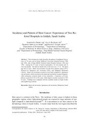
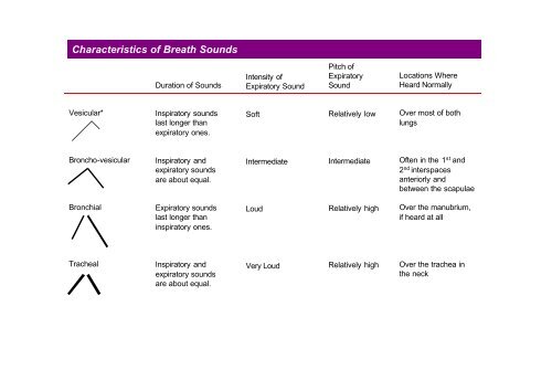
<strong>Characteristics</strong> <strong>of</strong> <strong>Breath</strong> <strong>Sounds</strong><br />
Vesicular*<br />
Broncho-vesicular<br />
Bronchial<br />
Tracheal<br />
Duration <strong>of</strong> <strong>Sounds</strong><br />
Inspiratory sounds<br />
last longer than<br />
expiratory ones.<br />
Inspiratory and<br />
expiratory sounds<br />
are about equal.<br />
Expiratory sounds<br />
last longer than<br />
inspiratory ones.<br />
Inspiratory and<br />
expiratory sounds<br />
are about equal.<br />
Intensity <strong>of</strong><br />
Expiratory Sound<br />
S<strong>of</strong>t<br />
Intermediate<br />
Loud<br />
Very Loud<br />
Pitch <strong>of</strong><br />
Expiratory<br />
Sound<br />
Relatively low<br />
Intermediate<br />
Relatively high<br />
Relatively high<br />
Locations Where<br />
Heard Normally<br />
Over most <strong>of</strong> both<br />
lungs<br />
Often in the 1 st and<br />
2 nd interspaces<br />
anteriorly and<br />
between the scapulae<br />
Over the manubrium,<br />
if heard at all<br />
Over the trachea in<br />
the neck
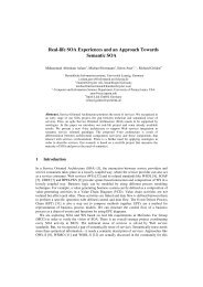
Adventitious Lung <strong>Sounds</strong><br />
DISCONTINUOUS SOUNDS (CRACKLES OR RALES) are intermittent, nonmusical, and brief –<br />
like dots in time<br />
Fine crackles (. . . . . ) are s<strong>of</strong>t, high pitched, and very brief (5 – 10 msec).<br />
Coarse crackles (• • • • • ) are somewhat louder, lower in pitch, and not quite so brief<br />
(20-30 msec).<br />
CONTINUOUS SOUNDS are > 250 msec, notably longer than crackles – like dashes in time – but<br />
do not necessarily persist throughout the respiratory cycle. Unlike crackles, they are musical.<br />
Wheezes ( ) are relatively high pitched (around 400 Hz or higher) and have a<br />
hissing or shrill quality.<br />
Rhounchi ( ) are relatively low pitched (around 200 Hz or lower and have a<br />
snoring quality.
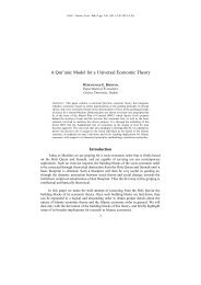
Physical Findings in Selected Chest Disorders<br />
The black boxes in this table suggest a framework for clinical sessment. Start with the three boxes under Percussion Note: resonant, dull and<br />
hyperresonant. Then move from each <strong>of</strong> these to other boxes that emphasize some <strong>of</strong> the key differences among various conditions. The<br />
changes described vary with the extent and severity <strong>of</strong> the disorders. Abnormalities deep in the chest usually produce fewer signs at all. Use<br />
the table for the direction <strong>of</strong> atypical changes, not for absolute distinctions.<br />
Condition<br />
Normal<br />
The tracheobronchial tree and<br />
alveoli are clear; pleurae are<br />
thin and close together;<br />
mobility <strong>of</strong> the chest wall in<br />
unimpaired.<br />
Chronic Bronchitis<br />
The bronchi are chronically<br />
inflamed and a productive<br />
cough is present. Airway<br />
obstruction may develop.<br />
Left-Sided Heart Failure<br />
(Early)<br />
Increased pressure in the<br />
pulmonary veins causes<br />
congestion and interstitial<br />
edema (around the alveolu);<br />
bronchial mucosa may become<br />
edematous.<br />
Consolidation<br />
Alveoli fill with fluid or blood<br />
cells, as in penumonia,<br />
pulmonary edema or pulmonary<br />
hemorrhage<br />
Percussion<br />
Note<br />
Resonant<br />
Resonant<br />
Resonant<br />
Dull over the<br />
airless area<br />
Trachea<br />
Midline<br />
Midline<br />
Midline<br />
Midline<br />
<strong>Breath</strong> <strong>Sounds</strong><br />
Vesicular, except perhaps<br />
bronchovesicular and<br />
bronchial sounds over the<br />
large bronchi and trachea<br />
respectively<br />
Vesicular (normal)<br />
Vesicular<br />
Adventitious<br />
<strong>Sounds</strong><br />
None, except<br />
perhaps a few<br />
transient inspiratory<br />
crackles at the<br />
bases <strong>of</strong> the lungs<br />
None; or scattered<br />
coarse crackles in<br />
early inspiration<br />
and perhaps<br />
expiration or<br />
wheezes or rhouchi<br />
Late inspiratory<br />
crackles in the<br />
dependent portions<br />
<strong>of</strong> the lungs;<br />
possibly iwheezes<br />
Tactile Fremitus<br />
and Transmitted<br />
Voice <strong>Sounds</strong><br />
Normal<br />
Normal<br />
Normal<br />
Bronchical over Late inspiratory Increased over the<br />
the involved area crackles over the involved area, with<br />
involved area bronchopony,<br />
egophony, and<br />
whispered<br />
pectoriloquy
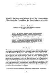
Physical Findings in Selected Chest Disorders (cont’d)<br />
Condition<br />
Atelectasis<br />
(Lobar Obstruction)<br />
When a plug in a<br />
mainstem bronchus (as<br />
from mucus or a foreign<br />
object) obstructs air<br />
flow, affected lung<br />
tissue collapses into an<br />
airless state.<br />
Pleural Effusion<br />
Fluid accumulates in the<br />
pleural space, separates<br />
airfilled lung from the<br />
chest wall, blocking the<br />
transmission <strong>of</strong> sound.<br />
Pneumothorax<br />
When air leaks into the<br />
pleural space, usually<br />
unilaterally, the lung<br />
recoils from the chest<br />
wall. Pleural blocks<br />
transmission <strong>of</strong> sound.<br />
Percussion<br />
Note<br />
Dull over the<br />
airless area<br />
Dull to flat over<br />
the fluid<br />
Hyperresonanat<br />
Or tympanitic<br />
over the pleural<br />
air<br />
Trachea<br />
May be shifted<br />
toward<br />
involved side<br />
Shifted toward<br />
opposite side in<br />
a large effusion<br />
Shifted toward<br />
opposite side if<br />
much air<br />
<strong>Breath</strong> <strong>Sounds</strong><br />
Usually absent when<br />
bronchial plug persists.<br />
Exceptions include right<br />
upper lobe atelectasis,<br />
where adjacent tracheal<br />
sounds may be<br />
transmitted.<br />
Decreased to absent, but<br />
bronchial breath sounds<br />
may be heard near top <strong>of</strong><br />
large effusion<br />
Decreased to absent over<br />
the pleural air<br />
Adventitious<br />
<strong>Sounds</strong><br />
None<br />
None, except a<br />
possible pleural rub<br />
None, except a<br />
possible pleural rub<br />
Tactile Fremitus<br />
and Transmitted<br />
Voice <strong>Sounds</strong><br />
Usually absent<br />
when the bronchial<br />
plug persists. In<br />
exceptions, e.g.,<br />
right upper lobe<br />
atelectasis, may be<br />
increased<br />
Decreased to<br />
absent, but may be<br />
increased towards<br />
the top <strong>of</strong> a large<br />
effusion.<br />
Decreased to<br />
absent over the<br />
pleural air
Physical Findings in Selected Chest Disorders (cont’d)<br />
Condition<br />
Chronic Obstructive<br />
Pulmonary Disease<br />
(COPD)<br />
Slowly progressive<br />
disorder in which the<br />
distal air spaces enlarge<br />
and lungs become<br />
hyperinflated. Chronic<br />
bronchitis is <strong>of</strong>ten<br />
associated.<br />
Asthma<br />
Widespread narrowing<br />
<strong>of</strong> the tracheobronchial<br />
tree diminishes airflow<br />
to a fluctuating degree.<br />
During attacks, airflow<br />
decreases further and<br />
lungs hyperinflate.<br />
Percussion<br />
Note<br />
Diffusely<br />
Hyperresonant<br />
Resonant to<br />
diffusely<br />
hyperresonant<br />
Trachea<br />
Midline<br />
Midline<br />
<strong>Breath</strong> <strong>Sounds</strong><br />
Decreased to absent<br />
Often obscured by<br />
wheezes<br />
Adventitious<br />
<strong>Sounds</strong><br />
None, or the<br />
crackles, wheezes,<br />
and ronchi <strong>of</strong><br />
associated chronic<br />
bronchitis<br />
Wheezes, possibly<br />
crackles<br />
Tactile Fremitus<br />
and Transmitted<br />
Voice <strong>Sounds</strong><br />
Decreased<br />
Decreased
Normal and Altered <strong>Breath</strong> and Voice Sound<br />
The origins <strong>of</strong> breath sounds are still unclear. According to leading theories, turbulent air flow in the central airways produces the tracheal<br />
and bronchial breath sounds. As these sounds pass through the lungs to the periphery, lung tissue filters out their higher-pitched components<br />
and only the s<strong>of</strong>t and lower-pitched components reach the chest wall, where they are heard as vesicular breath sounds. Noramlly, tracheal and<br />
bronchial sounds may be heard over the trachea and mainstem bronchi; vesicular breath sounds predominate throughout most <strong>of</strong> the lungs.<br />
When lung tissue loses its air, it transmits high-pitched sounds much better. If the tracheobronchial tree is open, bronchial breath sounds may<br />
replace the normal vesicular sounds over airless areas <strong>of</strong> the lung. This change is seen in lober pneumonia when the alveoli fill with fluid, red<br />
cells, and white cells – a process calles consolidation. Other causes include pulmonary edema or hemorrhage. Bronchial breath sounds<br />
usually correlate with an increase in tactile fremitus and transmitted voice sounds. These findings are summarized below.<br />
<strong>Breath</strong> <strong>Sounds</strong><br />
Transmitted Voice <strong>Sounds</strong><br />
Tactile Fremitus<br />
Normal Air-Filled Lung Airless Lung, as in Lobar Pneumonia<br />
Predominantly vesicular<br />
Spoken words muffled and indistinct<br />
Spoken “ee” heards as “ee”<br />
Whispered words faint and indistinct, if heard at<br />
all<br />
Normal<br />
Bronchial or bronchovesicular over the involved area<br />
Spoken words louder, clearer (bronchophony)<br />
Spoken “ee” heard as “ay” (egophony)<br />
Whispered words louder, clearer (whispered pectoriloquy)<br />
Increased
Abnormalities in Rate and Rhythm <strong>of</strong> <strong>Breath</strong>ing<br />
When observing respiratory patterns, think in terms <strong>of</strong> rate, depth, and regularity <strong>of</strong> the patient’s breathing. Describe what you see in these<br />
terms. Traditional terms, such as tachypnea, are given below so that you will understand them, but simple descriptions are recommended for<br />
use.<br />
Normal<br />
The respiratory rate is about<br />
14-20 per min in normal adults<br />
and up to 44 per min in<br />
infants.<br />
Cheyne-Strokes <strong>Breath</strong>ing<br />
Periods <strong>of</strong> deep breathing<br />
alternate with periods <strong>of</strong> apnea<br />
(no breathing). Children and<br />
aging people normally may<br />
show this pattern in sleep.<br />
Other causes include heart<br />
failure, uremia, drug-induced<br />
respiratory depression, and<br />
brain damage (typically on<br />
both sides <strong>of</strong> the cerebral<br />
hemispheres or diencephalon.<br />
Rapid Shallow <strong>Breath</strong>ing<br />
(Tachypnea)<br />
Rapid shallow breathing has a<br />
number <strong>of</strong> causes, including<br />
restrictive lung disease, pleuritic<br />
chest pain, and an elevated<br />
diaphragm.<br />
Rapid Deep <strong>Breath</strong>ing<br />
(Hyperpnea, Hypeventilation) Slow <strong>Breath</strong>ing (Bradypnea)<br />
Rapid deep breathing has several<br />
causes, including exercise, anxiety,<br />
and metabolic acidoses. In the<br />
comatose patient, consider infarction,<br />
hypoxia, or phypoglycemia affecting<br />
the midbrain or pons. Kussmaul<br />
breathing is deep breathing due to<br />
metabolic acidosis. It may be fast,<br />
normal in rate, or slow.<br />
Slow breathing may be secondary<br />
to such causes as diabetic coma,<br />
drug induced respiratory<br />
depression, and increased<br />
intracranial pressure.<br />
Ataxic <strong>Breath</strong>ing<br />
(Biot’s <strong>Breath</strong>ing) Sighing Respiration Obstructive <strong>Breath</strong>ing<br />
Ataxic breathing is<br />
characterized by unpredicted<br />
irregularity. <strong>Breath</strong>s may be<br />
shallow or deep, and stop for<br />
short periods. Causes include<br />
respiratory depression and brain<br />
damage, typically at the<br />
medullary level.<br />
<strong>Breath</strong>ing punctuated by frequent<br />
sighs should alert you to the<br />
possibility <strong>of</strong> hyperventilation<br />
syndrome – a common cause <strong>of</strong><br />
dyspnea and dizziness. Occasional<br />
sighs are normal.<br />
In obstructive lung disease,<br />
expiration is prolonged because<br />
narrowed airways increase the<br />
resistance to airflow. Causes<br />
include asthma, chronic<br />
bronchitis, and COPD.