Structure of the SAM-II riboswitch bound to S-adenosylmethionine
Structure of the SAM-II riboswitch bound to S-adenosylmethionine
Structure of the SAM-II riboswitch bound to S-adenosylmethionine

You also want an ePaper? Increase the reach of your titles
YUMPU automatically turns print PDFs into web optimized ePapers that Google loves.
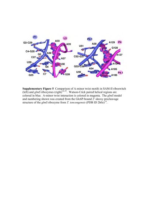
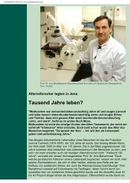
Supplementary Figure 5 Comparison <strong>of</strong> A-minor twist motifs in <strong>SAM</strong>-<strong>II</strong> <strong>riboswitch</strong><br />
(left) and glmS ribozymes (right) 18,19 . Watson-Crick paired helical regions are<br />
colored in blue. A-minor twist interaction is colored in magenta. The glmS model<br />
and numbering shown was created from <strong>the</strong> Glc6P-<strong>bound</strong> 2’-deoxy precleavage<br />
structure <strong>of</strong> <strong>the</strong> glmS ribozyme from T. tencongensis (PDB ID 2h0z) 19 .


![Programm [pdf]](https://img.yumpu.com/20944039/1/184x260/programm-pdf.jpg?quality=85)