
You also want an ePaper? Increase the reach of your titles
YUMPU automatically turns print PDFs into web optimized ePapers that Google loves.
PROJECT SYNOPSES<br />
CANCER RESEARCH<br />
PROJECTS FUNDED UNDER THE SIXTH FRAMEWORK PROGRAMME<br />
2002-2006
Interested in European research?<br />
<strong>Research</strong>*eu is our monthly magazine keeping you in touch with main developments<br />
(results, programmes, events, etc.). It is available in English, French, German and Spanish.<br />
A free sample copy or free subscription can be obtained from:<br />
European Commission<br />
Directorate-General for <strong>Research</strong><br />
Communication Unit<br />
B-1049 Brussels<br />
Fax (32-2) 29-58220<br />
E-mail: research-eu@ec.europa.eu<br />
Internet: http://ec.europa.eu/research/research-eu<br />
European Commisssion<br />
Directorate-General for <strong>Research</strong><br />
Directorate F – Health<br />
Unit F.2 – Medical and Public Health <strong>Research</strong><br />
Area: <strong>Cancer</strong><br />
Contact: Dominika Trzaska<br />
E-mail: Dominika.Trzaska@ec.europa.eu<br />
European Commission<br />
Offi ce CDMA 2/46<br />
B – 1049 Brussels
CANCER RESEARCH<br />
PROJECTS FUNDED UNDER THE SIXTH FRAMEWORK PROGRAMME (2002-2006)<br />
Edited by ELENGO MANOUSSAKI<br />
2008 Directorate-General for <strong>Research</strong> – Medical and Public Health <strong>Research</strong> EUR 23471 EN
Acknowledgements<br />
The catalogue has been produced thanks to the input of the coordinators involved in FP6 research, to the<br />
colleagues from Genomics and System Biology, Health Biotechnology and Horizontal aspects units of Health<br />
Directory. To the continuous encouragement of Manuel Hallen and dedication of Maria Vidal-Ragout. However,<br />
it would not have been possible to conceive and produce the catalogue without the input of colleagues<br />
who left, and new colleagues who arrived, especially: Olaf Kelm, Stefan Junglbluth, Christel Jaubert, Joana<br />
Namorado, Cornelius Schmaltz, Jessica Tengelidou, Dominika Trzaska, Alfredo Cesario and Jan van de Loo.<br />
LEGAL NOTICE<br />
Neither the European Commission nor any person acting on behalf of the Commission<br />
is responsible for the use which might be made of the following information.<br />
The views expressed in this publication are the sole responsibility of the author and do<br />
not necessarily refl ect the views of the European Commission.<br />
A great deal of additional information on the European Union is available on the Internet.<br />
It can be accessed through the <strong>Europa</strong> server (http://europa.eu.int).<br />
Cataloguing data can be found at the end of this publication.<br />
Luxembourg: Offi ce for Offi cial Publications of the European Communities, 2008<br />
ISBN 978-92-79-09352-4<br />
© European Communities, 2008<br />
Reproduction is authorised provided the source is acknowledged.<br />
Printed in Belgium<br />
PRINTED ON WHITE CHLORINE-FREE PAPER<br />
Europe Direct is a service to help you fi nd answers<br />
to your questions about the European Union<br />
Freephone number (*):<br />
00 800 6 7 8 9 10 11<br />
Elengo Manoussaki<br />
(*) Certain mobile telephone operators do not allow access to 00 800 numbers or these calls may be billed.
TABLE OF CONTENTS<br />
FOREWORD – CANCER: COMBATING A COMPLEX DISEASE 7<br />
Biology 9<br />
Aetiology 85<br />
Prevention 97<br />
Early detection, Diagnosis and prognosis 105<br />
Treatment 161<br />
<strong>Cancer</strong> control, Survivorship, and outcomes research 231<br />
Scientifi c model systems 243<br />
Indices – Keywords 250<br />
Indices – Partners 254<br />
Indices – Organisations 264<br />
Contact details for activities related to cancer research<br />
European Commission<br />
Directorate General <strong>Research</strong><br />
Directorate Health – F<br />
Unit Medical and Public <strong>Research</strong>-F2<br />
MANUEL HALLEN, Acting Director, Health Directorate (Manuel.HALLEN@ec.europa.eu)<br />
MARIA JOSE VIDAL-RAGOUT, PhD, Deputy Head of unit (Maria-Jose.VIDAL-RAGOUT@ec.europa.eu)<br />
ALFREDO CESARIO, MD, Scientifi c Offi cer, SNE (Alfredo.CESARIO@ec.europa.eu)<br />
ELENGO MANOUSSAKI, PhD, Scientifi c Offi cer (Elengo.MANOUSSAKI@ec.europa.eu)<br />
DOMINIKA TRZASKA, PhD, Scientifi c Offi cer (Dominika.TRZASKA@ec.europa.eu)<br />
JAN-WILLEM VAN DE LOO, PhD, Scientifi c Offi cer (Jan-Willem.VAN-DE-LOO@ec.europa.eu)<br />
3
4<br />
TABLE OF CONTENTS<br />
Biology 9<br />
Active p53 10<br />
ANGIOTARGETING 1 3<br />
Anti-tumour targeting 1 5<br />
BRECOSM 1 7<br />
CAPPELLA 19<br />
COMBIO 2 1<br />
CONTICANET 23<br />
DIAMONDS 26<br />
DNA Repair 28<br />
ENLIGHT 30<br />
EPISTEM 32<br />
EPITRON 34<br />
EuroBoNeT 36<br />
EuroCSC 38<br />
GROWTHSTOP 40<br />
INTACT 42<br />
Kids<strong>Cancer</strong>Kinome 45<br />
Lymphangiogenomics 47<br />
MetaBre 49<br />
MitoCheck 5 1<br />
MOL CANCER MED 53<br />
MSCNET 55<br />
Mutp53 57<br />
Nano4Drugs 60<br />
ONCASYM 62<br />
ONCODEATH 64<br />
PRIMA 66<br />
Regulatory Genomics 68<br />
SENECA 70<br />
SIROCCO 72<br />
SMARTER 75<br />
TCAC in <strong>Cancer</strong> 77<br />
THOVLEN 79<br />
TransDeath 8 1<br />
TRK<strong>Cancer</strong> 82<br />
Tumour-Host Genomics 83<br />
Aetiology 85<br />
AIDIT 86<br />
CARCINOGENOMICS 88<br />
GenoMEL 91<br />
INCA 93<br />
POLYGENE 95<br />
CANCER RESEARCH PROJECTS FUNDED UNDER THE SIXTH FRAMEWORK PROGRAMME
Prevention 97<br />
EPIC 98<br />
EUROCADET 100<br />
MAMMI 102<br />
Early Detection, Diagnosis and Prognosis 105<br />
BAMOD 106<br />
BioCare 108<br />
BMC 1 1 1<br />
CANCERDEGRADOME 1 1 3<br />
COBRED 1 1 6<br />
DASIM 1 1 8<br />
DISMAL 1 1 9<br />
DNA METHYLATION 1 2 1<br />
Drop-Top 123<br />
E.E.T.-Pipeline 127<br />
eTUMOUR 129<br />
GLYFDIS 1 3 1<br />
HI-CAM 133<br />
HighReX 135<br />
MCSCs 137<br />
MMR-related cancer 139<br />
MolDiag-Paca 140<br />
NEMO 142<br />
OVCAD 144<br />
P-MARK 146<br />
POC4life 148<br />
PROMET 150<br />
PROTHETS 153<br />
STROMA 155<br />
TRANSFOG 157<br />
Treatment 161<br />
ANGIOSKIN 162<br />
ANGIOSTOP 164<br />
Apotherapy 166<br />
ATTACK 168<br />
BACULOGENES 170<br />
<strong>Cancer</strong>Grid 1 7 2<br />
CANCERIMMUNOTHERAPY 1 7 5<br />
CHEMORES 1 7 7<br />
CHILDHOPE 1 7 9<br />
DC-THERA 182<br />
DC-VACC 1 8 4<br />
Dendritophages 186<br />
DEPPICT 188<br />
TABLE OF CONTENTS<br />
5
EMIL 190<br />
ENACT 194<br />
European LeukaemiaNet 196<br />
European MCL Network 201<br />
EUROXY 203<br />
FIRST 205<br />
GIANT 207<br />
HERMIONE 209<br />
Immuno-PDT 2 1 1<br />
LIGHTS 2 1 3<br />
MAESTRO 2 1 5<br />
TEMPO 2 1 7<br />
THERADPOX 219<br />
TRANSBIG 222<br />
TRIDENT 225<br />
VITAL 227<br />
<strong>Cancer</strong> control, Survivorship and outcomes research 231<br />
CCPRB 232<br />
EPCRC 234<br />
EUROCAN+PLUS 236<br />
EUSTIR 238<br />
NORMOLIFE 240<br />
Scientifi c Model Systems 243<br />
ESBIC-D 244<br />
ZF-TOOLS 246<br />
6<br />
CANCER RESEARCH PROJECTS FUNDED UNDER THE SIXTH FRAMEWORK PROGRAMME
CANCER: COMBATING A COMPLEX DISEASE<br />
Human cancer is one of the oldest documented diseases. The oldest written evidence of the<br />
disease is found in Egyptian papyri from the period 3 000 to 1 500 BC which describe ulcers and<br />
breast cancer. The oldest specimen is a bronze age (1900-1600 BC) female skull suggesting head<br />
and neck cancer, whereas skeletal remains of a Peruvian – INCA – woman suggest melanoma.<br />
Breast cancer, melanoma, as well as head and neck cancer and other solid cancers, are still<br />
present across the world today. <strong>Cancer</strong> is an increasingly important health burden, both for<br />
patients and public health systems with signifi cant societal and psychological consequences.<br />
In recognition of this, a global eff ort to fi ght and control cancer has been set. Recently, the<br />
European Parliament published a resolution on Combating cancer in the enlarged Europe ( 1)<br />
and the council of the European Union in its conclusions on reducing the burden on cancer ( 2),<br />
invited the European Commission to extend knowledge of cancer epidemiology and cancer risk<br />
factors, early detection, diagnosis, treatment, survival and palliative care, as well as to include<br />
translational research under the Seventh Framework Programme.<br />
In addition, the World cancer declaration 2008, calls for a concerted strategic action to reduce<br />
the global cancer burden and outlines the steps needed to begin to reverse the global cancer<br />
crisis by 2020. Moreover, it sets specifi c priority actions on health policy, prevention and early<br />
detection, cancer treatment, and cancer research ( 3).<br />
The European Union’s strong research community is well placed to advance the understanding<br />
and treatment of cancer. Traditional approaches such as surgery, radiotherapy and<br />
chemotherapy are constantly improving, whilst genomic and immunological research off er<br />
innovative prospects for future treatment. In addition, new screening and detection methods<br />
are making it possible to confront the disease at an increasingly early stage. Furthermore,<br />
palliative care is increasingly recognised as important in ensuring cancer patients and their<br />
families to receive the best possible care and quality of life.<br />
The European Union supports cancer research in order to prevent cancer and to improve<br />
patients’ survival chances and their quality of life. The ‘Combating <strong>Cancer</strong>’ initiative was part<br />
of the EU’s Combating Major Diseases research. Within the ‘Life Sciences, Genomics and Biotechnology<br />
for Health’ thematic priority of the Sixth Framework Programme, 2002-2006<br />
some € 480 million of funding for cancer research has been made available to the European<br />
research community through the programme.<br />
It aimed to combat cancer by developing improved patient-oriented strategies, from prevention<br />
to earlier and more eff ective diagnosis, to better treatment with minimal side eff ects. It supported<br />
initiatives aimed at translating knowledge of basic research into applications to improve clinical<br />
practice and public health actions.<br />
This distinctive patient-oriented approach included four interlinked components, focusing on:<br />
• Establishing facilities and developing initiatives for the exploitation of cancer research in<br />
Europe, encouraging the development of evidence-based guidelines for good clinical<br />
practice and improved public health strategies by accelerating the translation of existing<br />
research results into applications.<br />
(1) http://www.europarl.europa.eu/sides/getDoc.do?pubRef=-//EP//NONSGML+TA+P6-TA-2008-0121+0+DOC+WORD+V0//EN<br />
(2) Adopted 10/06/08, www.consilium.europa.eu/Newsroom<br />
(3) http://www.uicc.org/wcd/wcd2008.pdf<br />
FOREWORD<br />
7
• Supporting clinical research, particularly early stage (phase I and II) clinical trials, targeted<br />
at validating new and improved interventions.<br />
• Funding translational research that takes basic knowledge through to applications in clinical<br />
practice and public health.<br />
• Other cancer related issues, such as ageing and cancer, gender related cancer; regional<br />
diff erences, psycho-social aspects, palliative care, and guidance for support groups.<br />
During the lifetime of FP6, many research groups applied and 108 projects were selected for funding.<br />
Among the funded ones are 37 large Integrated Projects (IP) or Networks of Excellence (NoE);<br />
64 Specifi c Targeted <strong>Research</strong> Projects (STREP) and 7 Coordination or Support Actions (CSA).<br />
The EU is committed to funding excellence in European biomedical research. All submitted<br />
proposals were subject to a rigorous, transparent and equitable peer-reviewed evaluation carried<br />
out by independent international panels of high-level experts from academia, public and private<br />
research organisations, and industry.<br />
Connecting European academic excellence with innovative research-based industry is the key to<br />
the success of translational research. Therefore, the Commission particularly encouraged small and<br />
medium-sized enterprises (SME) to participate in its research programmes.<br />
The present catalogue includes cancer projects on a variety of topics. No less than 1 500 research<br />
teams are collaborating in European Union Member States and other European countries associated<br />
with the Framework Programme, as well as selected third countries, such as USA, Chile,<br />
Australia, Russian Federation etc., thus consolidating an important critical mass to fi ght cancer.<br />
In fact, virtually all major European cancer research centres are taking part in one or several of<br />
the Commission-funded support schemes.<br />
The presentation of the catalogue follows the Common Scientifi c Outline (CSO) fi rst presented as<br />
a classifi cation system of scientifi c interest in cancer research, in the framework of the <strong>Cancer</strong><br />
<strong>Research</strong> Portfolio (CRP) of the National Institute of Health, USA.<br />
They include 7 broadly defi ned research areas:<br />
• Biology<br />
• Aetiology (causes of cancer)<br />
• Prevention<br />
• Early detection, diagnosis and prognosis<br />
• Treatment<br />
• <strong>Cancer</strong> control, survivorship and outcomes research<br />
• Scientifi c model systems<br />
The division into the diff erent groups is not always straightforward; therefore some of the<br />
projects could be classifi ed in one or more of the above areas. The essential fact is that these<br />
projects are all present in the catalogue and represent the enormous eff ort and constant<br />
devotion of the European research society to fi ght cancer.<br />
Manuel Hallen Maria José Vidal Ragout<br />
Acting Director Health <strong>Research</strong> Deputy Head of Unit<br />
DG <strong>Research</strong>, European Commission Medical and Public Health <strong>Research</strong><br />
8<br />
DG <strong>Research</strong>, European Commission<br />
CANCER RESEARCH PROJECTS FUNDED UNDER THE SIXTH FRAMEWORK PROGRAMME
Biology<br />
<strong>Cancer</strong> is a disease ethat that t is chara characterized aracterized by unc uncontrolled ncont ontrolled growth and spread of abnormal cells. TransTranssformation of a nor nnormal mal al ce cell<br />
ll int in into o a cancer cell is a com comple complex p x multistep process in which whic hich hcell cells ce<br />
s acquire ire<br />
capabilities to t to divid divide vide e iinde<br />
indefi nd fi nitely nnitely<br />
independently indepe epende nd ntly of growth gro rowth signals, sig signal nals, s, evade programmed pro rogra gramme med d cell ccell<br />
ell death, de d ath ath,<br />
invade neighbouring neighbou ourin ring g tissues ues and metastasise meta tastasise to other parts part arts of the body. bod body. y. During Dur During ing this transformation<br />
transformati ation on o<br />
numerous changes cha hang nges s occur ooccu<br />
ccur r at the molecular, biochemica<br />
biochemical ical l aand<br />
and cel cellul cellular lular ar level. lev le el. All cancers canc n ers arise due e to tto<br />
accumulation accumu m lation on oof<br />
of genet genetic netic ic and epige epigenetic g netic changes. Som So Some e mmuta<br />
mutations utatio ti t ns may be<br />
present<br />
t in the germl germline mlin ine<br />
thus s iincreas<br />
increasing asing ing the e rrisk<br />
risk of cance ca cancer ncer r ddevelopment,<br />
evelopment, but b but many<br />
ny occ ooccur ur during the individual’s life in n tthe<br />
the h<br />
somatic som so ati a c cell cells. ells. s. The There here re are many mma<br />
many aet aaetiological iological factors<br />
s involved<br />
d in<br />
n cancer, including includi d ng infection, infection o , exposure e to tto<br />
o<br />
chemicals che chemic m als an and othe other the ther r eenv<br />
environmental nvi nviron ron ronmen me m tal risk ri risk s factors and<br />
nd heredity.<br />
y. y Th TThe e past years hha have ve witnessed a dramat dramatic matic ic<br />
progress pro progre gre gr ss in understanding und u ers ersta tandin din ing g the tthe<br />
he mechanisms mec e han h isms of transformation tran r sformation of a normal cell into a cancer cell cel cell l that tthat<br />
h<br />
is s exp expec expected ect e ed to lea lead<br />
d tto<br />
to o mmajo<br />
major ajor impr improvements mprove vemen men m ts in the therap therapy. ra y. Yet Y Yet, , our<br />
ur kno kn knowledge wle wledge d is still far r ffrom<br />
from r compl complete mplete e<br />
and and much mmuc<br />
remains rem emain ains s to t<br />
to o be e discovered.<br />
disc discovered<br />
ed.<br />
Thirty Thi Thirty rty six projects proj oject ects presented pres res r ent en ed in this thi this s section ssect<br />
ection ion investigate in inves vestig tigate ate mechanisms me mecha chani nisms ms that tha that t underlie uunde<br />
nderlie e cancer ccanc<br />
ncer initiation, initia tiatio tion, n<br />
progression pr progre gressi ssion and<br />
nd me metas metastasis tas ta tas tasis is an and compa compare mpare re the them m wwith<br />
with ith th their<br />
eir no norma normal rmal l ccoun<br />
counterparts. ounter terpar parts. ts A num numbe number ber of th them<br />
em foc focus<br />
us<br />
on mol mo molecular ecu ecular lar me mecha mechanisms, chanis nisms, ms, s in inclu including cluding g DDNA<br />
DNA NA rep repair repair, a , onco oncogenes, ncogen genes, es, tu tumor<br />
mor su suppr suppressor ppress es or r gen ggenes, es, ch chrom chromosome<br />
romos osome e<br />
alterations, alt altera eratio tions, ns telom te telomere lomere ere ma maint maintenance, ntena enance nce ce, epig epigenetics, pigene enetic tics, s, and progr programmed ogrammed e cel cell l ddeat<br />
death. eath. h.<br />
Due tto<br />
to availability of the<br />
he com comple complete ple l te hum human<br />
an gen genome<br />
ome and d rrapid<br />
apid incr increase ncreas ease e iin<br />
in n pprot<br />
proteomic roteom eomic c and a and tr trans transcriptomic<br />
an cri ripto pto p mic ic<br />
data dat data a aan<br />
n integrative appr approach pp oac oachh iis<br />
is s n nneeded.<br />
eed ee ed. A gro group<br />
up of pro projec projects jects ts pre presen presented sented ted in th this<br />
is sec sectio section tio tion n eempl<br />
employ mploy oy sys sy systems tem tems<br />
and computation<br />
computational ion o al bio bbiology logyy aappr<br />
approaches pproac oac oaches hes an and d hhigh<br />
high-throughput igh-th -throu roughp ghput ut ana analys analyses lyses es to inv investiga investigate iga gate te e com comple complex ple plex network<br />
ork<br />
of f int iinteractions eracti tions ons in th the cell cell. ell.<br />
Moreover, Mor oreov eover, er, FP FP6 6 ssupp<br />
supports pport orts srese rresearch<br />
esearc arch h oon<br />
on n nthe t tthe<br />
he h dev develo development elopme pment nt of sop sophis sophisticated histic ticate ated d ttool<br />
tools ool ools s a aand<br />
and nd nov novel<br />
el tec techno technologies,<br />
hno hn log lo ies,<br />
for example ex eexamp ample analyses analys lyses es and imaging im imagi aging ng in n situ, sit situ, u, that tha that t wwill<br />
will ill en enabl enable able e rrese<br />
researchers esearc archer hers s to tto<br />
o investigate inve inv<br />
nvesti stigat gate e nnorm<br />
normal ormal al fun fu functioning cti ctioni oning<br />
of f the cell ccell and the e changes cchanges<br />
that lea lead ead d tto<br />
to o c ccanc<br />
cancer anc ancer e ini initia initiation tiatio tion n aand<br />
and nd pro progre progression. gre gressi ssi ss on.<br />
‘Know ‘Kn Know your enemy’<br />
y’ – ssaid<br />
aid Sun n Tzu TTzu<br />
zu in n the ‘The Art of War’. Wa War’. r. <strong>Research</strong> Re Resea search rch on cancer ca cance n r biology biol iology og is crucial cr crucial in our<br />
fi fight gght<br />
with cancer.<br />
r. Ex Exploring g tthe<br />
the he key ggenetic,<br />
genetic, biochemical and cellular cha changes hanges ges involved<br />
ved in cancer ca cance ncer initiationtio<br />
ion and progression progressi sion on n will wil will l yield yyiel<br />
iel e dn d new disease biomarkers and targets for treatment treatme tm nt and ultimately ultimate ately l pav pave the<br />
way ay to new therapies. therapi pies es.<br />
9<br />
Dominika Trzaska Trz Tr ask as a
Active p53<br />
Manipulating tumour suppression:<br />
a key to improve cancer treatment<br />
Summary<br />
The prevention of human cancer development depends on<br />
the integrity of a complex network of defence mechanisms<br />
that help cells to respond to various stress conditions. A key<br />
player in this network is the p53 tumour suppressor protein.<br />
By inducing effi cient growth inhibition, p53 eliminates cancer<br />
cells thereby preventing the development of human<br />
malignancies. These functions of p53 often determine the<br />
effi cacy of anti-cancer therapies. Although p53 is frequently<br />
mutated in some cancers, in about 50 % of all human cancers<br />
p53 is non-mutated and could, in principle, be activated<br />
to prevent tumour progression. This situation is prevalent<br />
among a wide range of cancers, notably breast carcinoma.<br />
However, p53 activity is hampered by malfunction of its<br />
many modulators, such as Mdm2 or p73, which govern p53<br />
tumour suppressive activity by acting upstream and/or<br />
downstream of p53. There is therefore a crucial need to<br />
understand how p53 modulators contribute to human<br />
malignancies. Based on this information, we propose to<br />
develop rational therapeutic approaches to manipulate<br />
p53 modulators, thereby wakening the sleeping tumour<br />
suppression activities of p53, allowing it to eliminate cancer<br />
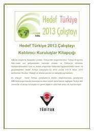
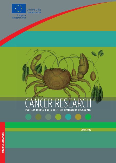
cells. This carefully structured consortium comprising<br />
20 academic research centres and SMEs (see diagram) will<br />
interactively build a technology platform to comparatively<br />
identify, characterise and evaluate the regulatory roles of<br />
p53 modulators and defi ne the mechanisms of their action.<br />
Large-scale gene functional analyses will be conducted to<br />
identify relevant signalling pathways that impair or mediate<br />
tumour suppression by p53. These analyses will include p53<br />
activators and inhibitors, p53 homologues p73/p63, and<br />
dissection of p53 target genes mediating apoptosis and<br />
growth arrest. Our links with highly profi led clinical partners<br />
and our access to large, well-characterised and clinically<br />
documented sample collections will enable the evaluation<br />
of diagnostic expression profi les, and their potential prognosis<br />
value in cancer. Particular emphasis will be directed<br />
towards translating the information on p53 regulation into<br />
the development of new anti-cancer therapies. p53 regulatory<br />
proteins will be used for the identifi cation of new<br />
molecular targets for drug discovery.<br />
10<br />
Keywords | Tumour suppression | p53 | p73 | p63 | inhibitors | activators |<br />
© C. AND M. KAGE<br />
Problem<br />
<strong>Cancer</strong> is the second leading cause of death in European<br />
countries, and one of the most imminent health problems in<br />
the developed world. The p53 protein is generally recognised<br />
as the key determinant of tumour suppression. It has<br />
been declared by the European Union that ‘a large cooperative<br />
eff ort is needed to ensure that every European<br />
citizen will rapidly profi t from the revolution of knowledge in<br />
cancer management’ (Philippe Busquin). The presence of<br />
wild type p53 is particularly prevalent in breast cancer, the<br />
type of cancer that stands at the centre of the European<br />
cancer policy. Since breast cancer aff ects mostly (though<br />
not exclusively) women, breast cancer research is also an<br />
important task to implement the gender dimension into<br />
basic research. For these reasons, we will choose breast<br />
cancer as one of our focuses in this block of work. Moreover,<br />
a non-mutated but inactive p53 is also found in a high percentage<br />
of the most frequent intracranial tumour of children,<br />
neuroblastoma. Since paediatric tumours are particularly<br />
dramatic events for patients and their families, it appears<br />
appropriate to put another focus on this tumour species.<br />
Activators<br />
WB1<br />
similarities to p53<br />
direct interference<br />
Homologues<br />
WB3<br />
p53<br />
p53 activity<br />
and<br />
technology<br />
WB4<br />
similarities to p53<br />
direct interference<br />
Activators<br />
WB2<br />
The four blocks are linked as outlined. These links are formed according to the biological<br />
activities governing p53 and, therefore, the scheme simultaneously depicts biological<br />
dependencies as well as the mode of collaboration within the consortium. Activators of p53<br />
frequently act by antagonising p53 inhibitors, and vice versa; this will be taken into account by<br />
networking accordingly between the blocks 1 and 2. Activators and inhibitors of p53 may act on<br />
p73 and p63 as well and this was shown to be true in a number of cases. Therefore, each<br />
regulator of p53 will be assessed regarding its impact on p53-homologues as well by<br />
collaborative eff orts between block of work 3 with blocks 1 and 2. Finally, the assessment of p53<br />
downstream activities, and the development of cutting-edge technologies to analyse them,<br />
will be used throughout the consortium. Therefore, block of work 4 forms a basis not only for<br />
reaching excellence on its own, but also to eff ectively advance the progress of blocks 1, 2 and 3.<br />
CANCER RESEARCH PROJECTS FUNDED UNDER THE SIXTH FRAMEWORK PROGRAMME
p53 activators<br />
ASPP Partner 5<br />
NFkappaB Partner 6<br />
Pin1 Partner 14<br />
HIPK2 Partner 14<br />
Chk1/Chk2 Partner 10<br />
JMY/Strap/p300 Partner 8<br />
Integrin avb3 Partner 13<br />
Aim<br />
The principal aim of this proposal is to ease both diagnosis<br />
and prognostic classifi cation, as well as the eff orts towards<br />
novel therapy regimens to treat patients suff ering from<br />
breast cancer and neuroblastoma. Overall, the integrated<br />
action of our consortium is aiming at re-establishing tumour<br />
suppressor activity in cancer, thereby translating basic<br />
knowledge of functional oncogenomics into cancer diagnoses<br />
and treatment, and contributing to leadership in<br />
European health technology.<br />
Expected results<br />
p53-homologues<br />
deltaNp73/p63 Partner 2<br />
p73/p63 ligands Partner 9<br />
p73, c-Abl, p300 Partner 11<br />
p73TA and YAP Partner 15<br />
Biotech SMEs<br />
genomics<br />
proteomics<br />
drug leads<br />
Clinical<br />
units<br />
patient data<br />
specimens<br />
therapy<br />
p53 activity and technology<br />
intrabodies Partner 15<br />
apoptosis Partner 4<br />
senescence Partner 7 V.R.<br />
DNA structure Partner 16<br />
DNA binding in vivo Partner 3, 15<br />
targets/regulators Partner 18, 1<br />
The overall goals of this integrated eff ort are to understand:<br />
• which modulators determine the tumour-suppressive<br />
activities of the p53 family members;<br />
• by what mechanisms these modulators aff ect the tumour<br />
suppression activities;<br />
• how the expression and activity of p53 modulators is<br />
regulated;<br />
• whether p53 modulators aff ect the biological characteristics<br />
of tumour cells;<br />
• whether the status of p53 modulators correlates with<br />
the clinical outcome and can be used to determine the<br />
individual prognosis;<br />
p53 inhibitors<br />
Mdm2 and p14Arf Partner 7<br />
Mdm2 and c-Abl Partner 3<br />
Mdmx Partner 12<br />
Mdm2 drug traget Partner 17,2<br />
Outline of the consortium.<br />
• whether and how p53 modulators can be targeted by<br />
therapeutic strategies, and be manipulated towards<br />
regaining tumour suppression;<br />
• disseminate the knowledge that will be produced to<br />
practically all the interested parties including medical<br />
doctors, and managerial staff in the industries;<br />
• familiarise SMEs with scientifi c research work and stateof-the<br />
art technology that will provide the necessary<br />
know-how for the improvement of their services and<br />
competitiveness.<br />
Potential applications<br />
The ultimate general objective of this research proposal is to<br />
provide a basis for the re-activation of tumour suppression<br />
and the design of novel therapeutic approaches to combat<br />
cancer. In particular, we are aiming at modulating p53 family<br />
activities to decrease resistance of tumour cells to anti-cancer<br />
treatments. Thus, the ultimate goal of this research proposal<br />
is the identifi cation of novel drug targets and strategies for<br />
induction of p53-mediated apoptosis in therapy-resistant<br />
cancer cells. The participation of the SMEs is expected to<br />
play a key role to the practical application of the knowledge<br />
that will be produced.<br />
BIOLOGY 11
Coordinator<br />
Giovanni Blandino<br />
Translational Oncogenomics Unit<br />
Molecular Medicine Department<br />
Regina Elena <strong>Cancer</strong> Institute<br />
Via Elio Chianesi, 53<br />
Rome, Italy<br />
blandino@ifo.it<br />
Partners<br />
Matthias Dobbelstein<br />
Georg-August-Universität Göttingen<br />
Göttingen, Germany<br />
Ygal Haupt<br />
The Lautenberg Center for General<br />
and Tumour Immunology<br />
The Hebrew University – Hadassah<br />
Medical School<br />
Jeruslem, Israel<br />
Guido Kroemer<br />
INSERM Institut National de la Santé<br />
et de la Recherche Médicale<br />
Paris, France<br />
Xin Lu<br />
Tumour Suppressor Group<br />
Ludwig Institute for <strong>Cancer</strong> <strong>Research</strong><br />
London, United Kingdom<br />
Karen Voudsen<br />
The Beatson Institute for <strong>Cancer</strong> <strong>Research</strong><br />
Tumour Suppressor Laboratory<br />
Glasgow, United Kingdom<br />
12<br />
Varda Rotter/Moshe Oren<br />
Weizmann Institute of Science<br />
Molecular Cell Biology/Biology<br />
Rehovot, Israel<br />
Nicholas B. La Thangue<br />
University of Oxford<br />
Oxford, United Kingdom<br />
Gerry Melino<br />
Medical <strong>Research</strong> Council<br />
Leicester, United Kingdom<br />
Jiri Bartèk<br />
Danish <strong>Cancer</strong> Society<br />
Dept. of Cell Cycle and <strong>Cancer</strong><br />
Institute of <strong>Cancer</strong> Biology<br />
Danish <strong>Cancer</strong> Society<br />
Copenhagen, Denmark<br />
Massimo Levrero<br />
Fondazione Andrea Cesalpino<br />
Laboratory of Gene Expression<br />
Rome, Italy<br />
Aart Gerrit Jochemsen<br />
Dept. Molecular and Cell Biology<br />
Tumour Suppressor Group<br />
Leiden University Medical Center<br />
Leiden, The Netherlands<br />
Galina Selivanova<br />
Karolinska Institute<br />
Dept. of Laboratory Medicine<br />
Stockholm, Sweden<br />
Gianni Del Sal<br />
Università degli Studi di Trieste<br />
Dipartimento di Biochimica<br />
Biofisica e Chimica delle Macromolecole<br />
Trieste, Italy<br />
Richard Iggo<br />
University of St. Andrews<br />
St. Andrews, United Kingdom<br />
Wolfgang Deppert<br />
Heinrich-Pette-Institut für Experimentelle<br />
Virologie und Immunologie an der<br />
Universität Hamburg<br />
Dept. of Tumour Virology<br />
Hamburg, Germany<br />
David Lane/Sonia Lain<br />
University of Dundee<br />
Dept. of Surgery and Molecular Oncology<br />
Nethergate<br />
Dundee, United Kingdom<br />
Simona Greco<br />
Biotecgen s.r.l.<br />
Dept. of Biological Sciences<br />
Institute of Physiology<br />
Lecce, Italy<br />
Cristina Fregonese<br />
INNOVA spa<br />
Tecnopolo Tiburtino<br />
Rome, Italy<br />
Roberto Mantovani<br />
GeneSpin srl SME<br />
Milan, Italy<br />
Project number<br />
LSHC-CT-2004-503576<br />
EC contribution<br />
€ 6 000 000<br />
Duration<br />
60 months<br />
Starting date<br />
01/12/2004<br />
Instrument<br />
IP<br />
Project website<br />
www.europeire.it/<br />
Activep53/intro.htm<br />
CANCER RESEARCH PROJECTS FUNDED UNDER THE SIXTH FRAMEWORK PROGRAMME
Keywords | Angiogenesis | vasculogenesis | blood vessels | tumour | endothelial cells | clinical trials |<br />
microarrays | proteomics molecular targets |<br />
ANGIOTARGETING<br />
Multidisciplinary research to explore<br />
and validate molecular targets<br />
for innovative treatments<br />
Summary<br />
Solid tumour growth depends on the recruitment of new<br />
blood vessels that will provide the cancer cells with nutrients<br />
and oxygen. The ANGIOTARGETING project intends to<br />
fi nd new targets on the tumour vasculature. The project will<br />
then defi ne these targets and develop new therapeutic<br />
strategies towards them.<br />
The apparent limited success in translating angiogenesis<br />
research into the clinic is, in our view, based on the fact that<br />
the research fi eld has been fragmented, and no standardised<br />
tools and models have been identifi ed that reliably<br />
refl ect the complexity of tumour angiogenesis mechanisms<br />
in humans.<br />
ANGIOTARGETING will identify and validate new therapeutic<br />
targets directed towards tumour vascular-matrix<br />
interactions, develop new therapeutic strategies and implement<br />
such strategies in pre- and clinical trials. The project<br />
represents a virtual research institute in Europe and consists<br />
of 14 highly competent research centres within the<br />
fi eld of angiogenesis research. To defi ne and validate new<br />
targets related to the tumour vascular transcriptome and<br />
proteome, the consortium will establish high throughput<br />
functional screening technologies for the identifi cation of<br />
novel secreted factors that regulate endothelial cell growth<br />
and survival. This includes the use of robotic platforms that<br />
will be used to identify cDNAs with specifi c cellular functions.<br />
In this project, basic science, translational research<br />
and clinical activities are strongly integrated, in order to<br />
validate defi ned targets and to develop new therapeutic<br />
principles for clinical implementation.<br />
Problem<br />
The signifi cant role of neovessel formation in health and disease<br />
has been clearly demonstrated during the last decade.<br />
The molecular mechanisms that lead to the establishment of<br />
functional blood vessels are also key factors that regulate<br />
tumour progression. Considerable eff orts have been devoted<br />
to research identifying the mechanisms that regulate the<br />
recruitment of blood vessels to tumours. Under normal<br />
conditions, the establishment of blood vessels is a highly<br />
complex and coordinated biological process.<br />
Targeting non-cancer cells that feed and drain the tumour and<br />
form channels through which tumour cells can disseminate,<br />
rather than targeting the neoplastic cells themselves, represents<br />
an approach to cancer therapy that holds particular<br />
promise because these cells are genetically stable, and therefore<br />
less likely to accumulate mutations that allow them to<br />
develop drug resistance. The fi rst meaningful clinical eff ects<br />
of antiangiogenic therapy in human cancer have recently been<br />
demonstrated. However, in spite of some highly encouraging<br />
results, most angiogenesis inhibitors, reported to suppress<br />
tumour growth in animal models, have thus far failed in human<br />
clinical trials. In our view, this refl ects that no standardised<br />
tools and models have been identifi ed that reliably refl ect the<br />
complexity of tumour angiogenesis mechanisms in humans.<br />
The successful translation of potential angiogenesis inhibitors<br />
to clinical application depends partly on the transfer of expertise<br />
from scientists who are familiar with the biology of<br />
angiogenesis to clinicians, as well as an active feedback from<br />
the clinicians to the scientists.<br />
ANGIOTARGETING aims at the identifi cation and validation<br />
of new therapeutic targets in tumour vasculature, develops<br />
new therapeutic strategies and implement such strategies<br />
in pre- and clinical trials. A strong focus on translational<br />
research and clinical implementation will convert R&D results<br />
into direct public and economic benefi ts.<br />
BIOLOGY 13<br />
Aim<br />
The objective is the identifi cation and validation of new<br />
therapeutic targets directed towards tumour vascularmatrix<br />
interactions.<br />
Expected results<br />
• The project will increase our understanding of how<br />
tumours generate a vascular supply.<br />
• The project will develop new technologies to defi ne<br />
and validate key molecular targets that control tumour<br />
vascularisation and invasion.<br />
• The project will identify potential therapeutic targets<br />
towards the tumour vascular and invasive transcriptome<br />
and proteome.<br />
• The project will develop comprehensive bioinformatics<br />
tools for the analysis of high throughput gene and protein<br />
data from defi ned cell populations within tumours.<br />
• The project will develop state-of-the-art platforms for<br />
preclinical and clinical assessment of newly developed<br />
compounds.<br />
• The project will provide new information on how potential<br />
antivascular therapies shall be evaluated in the clinic.<br />
This includes the development of surrogate markers to<br />
evaluate therapeutic effi cacy.
14<br />
Potential applications<br />
The project will lead to the identifi cation of a number of<br />
potential targets towards the tumour vasculature. Within the<br />
ANGIOTARGETING consortium there are a number of activities<br />
aiming at developing novel therapeutic strategies towards<br />
identifi ed and validated targets. Some of these strategies will<br />
be applied and assessed in preclinical and clinical trials.<br />
Coordinator<br />
Rolf Bjerkvig<br />
University of Bergen<br />
Bergen, Norway<br />
rolf.bjerkvig@biomed.uib.no<br />
Partners<br />
Helge Wiig, Karl-Henning Kalland<br />
and Inge Jonassen<br />
University of Bergen<br />
Bergen, Norway<br />
helge.wiig@biomed.uib.no<br />
kalland@gades.uib.no<br />
inge.jonassen@ii.uib.no<br />
Francesco Bertolini<br />
European Institute of Oncology<br />
Milan, Italy<br />
francesco.bertolini@ieo.it<br />
Lena Claesson-Welsh<br />
University of Uppsala<br />
Uppsala, Sweden<br />
lena.welsh@genpat.uu.se<br />
Peter ten Dijke<br />
Leiden Medical University Center<br />
Leiden, The Netherlands<br />
p.ten_dijke@lumc.nl<br />
Michael Kubbutat<br />
Proqinase/KTB Tumorforschungs GmbH<br />
Freiburg, Germany<br />
m.kubbutat@proqinase.com<br />
Arjan W. Griffioen<br />
and Johannes Waltenberger<br />
University Hospital Maastricht<br />
Maastricht, The Netherlands<br />
aw.griffioen@path.unimaas.nl<br />
waltenberger@cardio.azm.nl<br />
Adrian Harris<br />
University of Oxford<br />
United Kingdom<br />
aharris.lab@cancer.org.uk<br />
Henk Broxterman<br />
Vrije Universiteit Medical Center Amsterdam<br />
Amsterdam, The Netherlands<br />
h.broxterman@vumc.nl<br />
Taina Pihlajaniemi<br />
University of Oulu<br />
Oulu, Finland<br />
taina.pihlajaniemi@oulu.fi<br />
Eva Sykova<br />
Institute of Experimental Medicine<br />
Prague, Czech Republic<br />
sykova@biomed.cas.cz<br />
Simone Niclou<br />
Centre de la recherche public de la santé<br />
Luxembourg<br />
simone.niclou@crp-sante.healthnet.lu<br />
Karl Tryggvason and Yihai Cao<br />
The Karolinska Institute<br />
Stockholm, Sweden<br />
karl.tryggvason@mbb.ki.se<br />
yihai.cao@mtc.ki.se<br />
Peter Vajkozcy<br />
Charité-Universitatsmedizin<br />
Berlin, Germany<br />
peter.vajkoczy@charite.de<br />
Leo Hofstra<br />
PharmaTarget BV<br />
Maastricht, The Netherlands<br />
l.hofstra@cardio.azm.nl<br />
Project number<br />
LSHC-CT-2004-504743<br />
EC contribution<br />
€ 6 000 000<br />
Duration<br />
54 months<br />
Starting date<br />
01/11/2004<br />
Instrument<br />
IP<br />
Project website<br />
www.uib.no/med/<br />
angiotargeting/<br />
CANCER RESEARCH PROJECTS FUNDED UNDER THE SIXTH FRAMEWORK PROGRAMME
Keywords | Tumour-host interactions | tumour targeting |<br />
Anti-tumour targeting<br />
Modulation of the Recruitment of<br />
the Vessels and Immune Cells by<br />
Malignant Tumours: Targeting of Tumour<br />
Vessels and Triggering of Anti-Tumour<br />
Defence Mechanisms<br />
Summary<br />
All malignant tumours acquire the capacity for efficient<br />
recruitment of blood vessels, which are absolutely necessary<br />
for tumour growth beyond a certain size. They also<br />
frequently stimulate lymphangiogenesis supporting dissemination<br />
of tumour cells, not only via the blood vasculature<br />
but also via the lymphatic system, leading to metastasis.<br />
Vessel growth is promoted by sprouting angiogenesis and<br />
the homing of bone marrow progenitor cells into the<br />
tumours and tumour vessels. The extensive vascularisation<br />
facilitates the invasion of cells of the innate and adaptive<br />
immune system, which stay largely functionally suppressed<br />
by the tumour environment, and even contribute to angiogenesis<br />
and tumour growth by cytokine and growth factor<br />
secretion.<br />
We propose in this application to:<br />
• further investigate key regulatory pathways by which<br />
tumour-secreted molecules promote vascularisation<br />
and inhibit immune cell function;<br />
• develop methods to inhibit tumour growth and metastasis<br />
by blocking vessel and tumour cell growth;<br />
• achieve tumour clearance by additionally promoting<br />
activation and homing of functional immune cells to the<br />
tumours.<br />
The project will comprise the collaboration of laboratories<br />
with complementary expertise. It will include experts in blood<br />
vessel and lymph vessel angiogenesis, metastasis formation,<br />
progenitor cell incorporation into tumours and tumour<br />
vessels, anti-tumour defence mechanisms of the immune<br />
system and viral transduction techniques. The final goal will<br />
be the preclinical evaluation of strategies in murine models<br />
of three of the most prevalent forms of human cancer,<br />
i.e. carcinomas of the breast, colon and prostate.<br />
The strategies to target the tumour will be based on gene, cell<br />
and immune therapy methods. They will include the use of:<br />
• adenoviruses for the expression of angiogenesis inhibitors<br />
following targeted delivery of the viruses to the tumour<br />
vasculature;<br />
• the genetic modification of murine embryonic and human<br />
umbilical cord/bone marrow progenitor cells and their<br />
directed homing into the tumour;<br />
• the use of genetically-engineered immune cell products or<br />
the transduction of immune cells to activate targeting of<br />
the tumours by innate and adaptive anti-tumour defence<br />
mechanisms. We expect that this project will contribute to<br />
innovation on three levels. Firstly, we will gain basic additional<br />
novel knowledge on important pathways and<br />
regulatory molecules for the recruitment of host cells to<br />
the tumours and their functional interaction with the<br />
tumour. Secondly, we will use this knowledge to test novel<br />
ways of targeting viruses and (transduced) cells to the<br />
tumours. Finally, we will evaluate whether, by a combination<br />
of anti-angiogenesis therapy with directed anti-tumour<br />
immunotherapy, it would be possible not only to inhibit<br />
tumour growth, but also to eradicate residual disease.<br />
Problem<br />
Despite significant improvements in diagnosis, surgical techniques,<br />
general patient care, and local and systemic adjuvant<br />
therapies, many solid tumours remain a major cause of death.<br />
Among the most prevalent and fatal forms are carcinomas of<br />
the breast, colon and prostate. Most deaths from cancer are<br />
due to metastases, seeded from the primary tumour via the<br />
blood and lymph vasculature, which are resistant to conventional<br />
therapies. The main barrier to the non-surgical treatment<br />
of the primary neoplasm and its metastases is the genetic<br />
instability and biological heterogeneity of cancer cells leading<br />
to the rapid development and growth of resistant cells. Since<br />
the expansion of solid tumours and their metastases beyond<br />
a minimal size is absolutely dependent on the formation of<br />
new blood vessels, anti-angiogenesis therapies constitute<br />
a promising alternative. In this case the genetically normal<br />
endothelial cells of the tumour vessels are targeted, avoiding<br />
the problem of resistance development. Furthermore, immune<br />
therapies are based on the ability of the immune system,<br />
evolved over evolutionary times, to cope with an almost unlimited<br />
number of antigens, thus opening the possibility to find<br />
a mechanism to target any tumour variant as long as the<br />
inhibitory milieu of the tumour environment can be overcome.<br />
Therefore angiogenesis and immune therapies remain among<br />
the most promising fields of cancer therapy.<br />
BIOLOGY 15
Aim<br />
The general aim of the project is to design and evaluate strategies<br />
of anti-tumour angiogenesis and anti-tumour immune<br />
therapies and their combination in murine models of some of<br />
the most prevalent forms of human solid tumours. It will<br />
include the identification, modification and use of key regulatory<br />
molecules of vessel growth and immune defence and the<br />
development of methods to specifically and efficiently target<br />
tumours and their metastasis.<br />
Expected results<br />
We will undertake a concentrated eff ort to identify targets<br />
and develop methods for interference with vessel growth.<br />
Furthermore, we will develop techniques to target tumour<br />
endothelium by viruses and progenitor cells,methodsthat<br />
could be usedto also reachdistant metastases via the blood<br />
stream and not only the primary tumour. In addition, we will<br />
explore the use of innate receptors of NK cells to detect malignant<br />
cells and to boost the T-cell response of the immune<br />
system with the help of dendritic cells. Weanticipate thatasingle<br />
method,depending on the individual tumour, may not<br />
be sufficient, but the development of several techniques<br />
based on diff erent principles and their tumour-specific or<br />
combined application may be successful.<br />
For this purpose we combine laboratories with diff erential<br />
expertise, each having either identified a specific target molecule<br />
or developed a specific technique for targeting tumours.<br />
We will combine the expertise, molecules and techniques and<br />
comparatively evaluate diff erent strategies in corresponding<br />
models of human carcinomas.<br />
Potential applications<br />
The results of this project will be disseminated and exploited<br />
on three levels:<br />
• basic molecular medicine research level: all expected<br />
findings with the angiogenesis inhibitors and immune<br />
stimulators will be important to improve understanding<br />
of the role of vessel formation and of anti-tumour<br />
immune responses for cancer. These basic findings will<br />
be published in quality scientific journals;<br />
• clinical level: it is anticipated that our findings and developed<br />
preclinical methods will have impact on the design<br />
and further development of clinical protocols for the<br />
treatment of cancer;<br />
• company level: key novel molecules, findings and techniques<br />
will be patented and, together with patents<br />
available, used to develop reagents and protocol for<br />
gene-therapy of solid tumours.<br />
16<br />
Coordinator<br />
Erhard Hofer<br />
Dept. of Vascular Biology<br />
and Thrombosis <strong>Research</strong><br />
Centre for Biomolecular Medicine<br />
and Pharmacology<br />
Medical University Vienna<br />
Vienna, Austria<br />
erhard.hofer@meduniwien.ac.at<br />
Partners<br />
Seyedhossein Aharinejad<br />
University of Vienna<br />
Vienna, Austria<br />
seyedhossein.aharinejad@meduniwien.ac.at<br />
Michael Detmar<br />
Institute of Pharmaceutical Sciences<br />
Swiss Federal Institute of Technology<br />
Zurich, Switzerland<br />
michael.detmar@pharma.ethz.ch<br />
Hidde Haisma<br />
University of Groningen<br />
Groningen, The Netherlands<br />
h.j.haisma@rug.nl<br />
Peter Vajkoczy<br />
Charité Universitätsmedizin<br />
Berlin, Germany<br />
vajkoczy@yahoo.de<br />
Ofer Mandelboim<br />
Hebrew University<br />
Hadassah Medical School<br />
Jerusalem, Israel<br />
oferman@md2.huji.ac.il<br />
Karsten Mahnke<br />
University of Heidelberg<br />
Heidelberg, Germany<br />
Karsten.Mahnke@med.uni-heidelberg.de<br />
Melvyn Little<br />
Affimed Therapeutics AG<br />
Heidelberg, Germany<br />
m.little@affimed.com<br />
Karl-Heinz Preisegger<br />
EccoCell Biotechnology and<br />
Stem Cell Therapy GmbH<br />
Graz, Austria<br />
karl-hein.preisegger@eccocell.com<br />
Project number<br />
Project number<br />
LSHC-CT-2005-518178<br />
EC contribution<br />
€ 3 005 000<br />
Duration<br />
36 months<br />
Starting date<br />
01/11/2005<br />
Instrument<br />
STREP<br />
Project website<br />
www.tumortargeting.eu<br />
CANCER RESEARCH PROJECTS FUNDED UNDER THE SIXTH FRAMEWORK PROGRAMME
Keywords | Breast | metastasis | gene expression profi ling |<br />
BRECOSM<br />
Identifi cation of molecular pathways<br />
that regulate the organ-specifi c<br />
metastasis of breast cancer<br />
Summary<br />
The objectives of this project are to identify genes, proteins<br />
and molecular pathways involved in regulating the metastasis<br />
of breast cancer to specifi c organs. To achieve these<br />
objectives we will use a combination of gene expression<br />
profi ling, bioinformatic analysis, histology of human female<br />
breast cancer samples, genetic manipulation of transplantable<br />
tumor cells and transgenic mouse technology. In addition to<br />
fi nding new genes, we aim to analyse to what extent genes<br />
already known to play a role in breast cancer metastasis<br />
specify which organs breast tumors metastasise to. We will<br />
also establish how the currently known genes that are<br />
associated with breast cancer dissemination and the new<br />
ones we identify fi t together into pathways that regulate<br />
organ-specifi c metastasis. These fi ndings will be coupled<br />
with the analysis of clinical trials in which participants in this<br />
consortium are involved. Further deliverables include the<br />
development of improved animal models for the study of<br />
breast cancer metastasis, and the development of diagnostic<br />
methods for determining whether primary tumours already<br />
have metastatic potential. Together, the work packages in<br />
this project will establish a pipeline of activities that unite<br />
basic research into the organspecifi c metastasis of breast<br />
cancer with target validation and clinical application.<br />
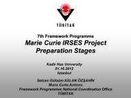
Wholemount staining of the epithelial ductal structure in a mouse mammary gland.<br />
The lymph nodes are also visible as densely-stained spheroidal structures.<br />
Problem<br />
Breast cancer is a major health issue and is highly gender relevant.<br />
It is the most often diagnosed female cancer, and the<br />
majority of cases are already invasive at diagnosis. More than<br />
17 % of cancer deaths result from breast tumours, making<br />
breast cancer a major societal problem. Treatment involves<br />
radical and disfi guring surgery, often with long-term side<br />
eff ects such as the development of lymphedema of the arm,<br />
and radiotherapy and chemotherapy, again associated with<br />
severe side eff ects. The eff ects of metastatic spread of<br />
the tumour cells and the formation of secondary deposits in<br />
a wide variety of organs are the cause of death due to breast<br />
cancer. Metastases to organs such as bone and brain are<br />
major causes of suff ering in terminally ill patients.<br />
The incidence of breast cancer increases sharply between<br />
the ages of 30 and 50 meaning that many women in the<br />
prime of life are aff ected by this disease. Not only does this<br />
mean that many families are traumatised, but it also has<br />
severe economic consequences, removing economically<br />
active women from society. Further economic consequences<br />
arise as a result of the high health care costs associated<br />
with treating breast cancer patients.<br />
Clearly improvements in the treatment and management of<br />
breast cancer would have impact on both health and the<br />
economy. By analysing molecular mechanisms that regulate<br />
organ-specifi c metastasis in breast cancer, the BRECOSM<br />
project will identify tools that will contribute to improved<br />
clinical decision-making, prognostic evaluation and therapy<br />
in breast cancer.<br />
BIOLOGY 17<br />
Aims<br />
• To identify genes that are specifi cally up- or downregulated<br />
in breast cancer metastases in specifi c organs.<br />
• To identify gene expression signatures in primary breast<br />
tumours that predict metastasis to specifi c organs or<br />
predict the prognosis of ductal carcinoma in situ (DCIS).<br />
• To determine whether genes already associated with<br />
breast cancer invasiveness and metastasis are expressed<br />
in metastases in all or only a subset of organs.<br />
• To demonstrate whether genes found to be specifi cally<br />
expressed in breast cancer metastases to given organs<br />
play a functional role in organ-specifi c metastasis.<br />
• To elucidate molecular pathways that regulate breast<br />
cancer metastasis to specifi c organs.<br />
• To develop improved animal models for studying organspecifi<br />
c metastasis of breast cancer.<br />
• To produce a prototype microarray chip for diagnostic/<br />
prognostic evaluation.<br />
• To apply the fi ndings on organ-specifi c metastasis in the<br />
clinical setting.
Expected results<br />
• The results of this project will begin to explain the molecular<br />
basis for organ-specifi c metastasis in breast cancer.<br />
• This project will identify regulatory pathways and cellular<br />
events that coordinate organ-specifi c metastasis of<br />
breast cancers. Novel targets for therapy will thereby be<br />
identifi ed.<br />
• This project will identify gene expression signatures in<br />
tumours associated with metastasis to particular organs.<br />
This will be an important advance in understanding the<br />
underlying genetic changes that regulate organ-specifi c<br />
metastasis in breast cancer.<br />
• This project will bring together European experts working<br />
on diff erent aspects of the molecular basis of tumour<br />
metastasis. As a result of coordinated eff orts, pathways<br />
that regulate metastasis to specifi c organs will be determined,<br />
and genes that play a functional role in organ-specifi c<br />
metastasis will be identifi ed.<br />
• This project will generate improved animal models for<br />
the further study of breast cancer metastasis to specifi c<br />
organs.<br />
• This project will identify gene expression signatures in<br />
primary breast tumours that predict patterns of metastasis.<br />
The application of these fi ndings will assist clinical<br />
decision making and prognostic evaluation.<br />
Potential applications<br />
The gene expression signatures in primary tumours identifi<br />
ed in this project that predict organ-specifi c metastasis<br />
and the prognosis of DCIS will have obvious potential for<br />
clinical application in diagnosis and prognostic assessment.<br />
Gene expression signatures in primary tumours associated<br />
with either organ-specifi c metastasis or progression of DCIS<br />
will be extensively validated retrospectively and as a prelude<br />
to introducing these gene expression signatures into clinical<br />
diagnosis and prognostic evaluation, we will perform prospective<br />
studies to demonstrate the effi cacy of examining<br />
gene expression signatures in primary breast cancers for<br />
predicting the likelihood and location of metastases and the<br />
probability that DCIS will progress and metastasise after<br />
partial mastectomy. The prototype microarray chips we create<br />
based on gene expression profi les produced as part of<br />
this project will be applied in the clinical setting to investigate<br />
their diagnostic and prognostic value for breast cancer<br />
in a prospective study. This will constitute a major step<br />
towards exploitation of the results. It is also highly likely that<br />
genes are identifi ed in this project will be candidate targets<br />
for the development of novel cancer therapies. The development<br />
of such therapies lies outside the time-frame and<br />
scope of the proposal.<br />
18<br />
Coordinator<br />
Jonathan Sleeman<br />
University of Heidelberg<br />
Medical Faculty Mannheim<br />
Centre for Biomedicine and Medical<br />
Technology Mannheim (CBTM)<br />
Mannheim, Germany<br />
sleeman@medma.uni-heidelberg.de<br />
Partners<br />
Frans Van Roy<br />
Dept. for Molecular Biomedical <strong>Research</strong><br />
VIB – Gent University<br />
Gent, Belgium<br />
Frans.Vanroy@dmbr.UGent.be<br />
Gerhard Christofori<br />
Institute of Biochemistry and Genetics<br />
Dept. of Clinical-Biological Sciences<br />
University of Basel<br />
Basel, Switzerland<br />
gerhard.christofori@unibas.ch<br />
Eugene Lukanidin<br />
Danish <strong>Cancer</strong> Society<br />
Institute of <strong>Cancer</strong> Biology<br />
Copenhagen, Denmark<br />
el@cancer.dk<br />
Jean-Paul Thiéry<br />
CNRS – UMR 144<br />
Institut Curie<br />
Cell Biology Department<br />
Paris, France<br />
jpthiery@imcb.a-star.edu.sg<br />
Bernd Hentsch<br />
TopoTarget Germany AG<br />
Frankfurt am Main, Germany<br />
BHe@TopoTarget.com<br />
Agnès Noël<br />
Laboratoire de Biologie des Tumeurs<br />
et du Développement<br />
Liège, Belgium<br />
agnes.noel@ulg.ac.be<br />
John Collard<br />
The Netherlands <strong>Cancer</strong> Institute<br />
Division of Cell Biology<br />
Amsterdam, The Netherlands<br />
j.collard@nki.nl<br />
Project number<br />
LSHC-CT-2004-503224<br />
EC contribution<br />
€ 3 430 273<br />
Duration<br />
42 months<br />
Starting date<br />
01/07/2004<br />
Instrument<br />
STREP<br />
Project website<br />
http://igtmv1.fzk.de/<br />
www/brecosm/<br />
Peter ten Dijke<br />
The Netherlands <strong>Cancer</strong> Institute<br />
Division of Cellular Biochemistry<br />
Amsterdam, The Netherlands<br />
& Leiden University Medical Center (LUMC)<br />
Leiden, The Netherlands<br />
p.ten_dijke@lumc.nl<br />
Roland Stauber<br />
Chemotherapeutisches Forschungsinstitut<br />
Frankfurt am Main, Germany<br />
& Johannes Gutenberg Universität Mainz<br />
Mainz, Germany<br />
rstauber@uni-mainz.de<br />
Massimo Zollo<br />
TIGEM-Telethon<br />
Naples, Italy<br />
& CEINGE Biotecnologie Avanzate, S.c.a.r.l.<br />
Naples, Italy<br />
zollo@ceinge.unina.it<br />
Peter Schlag<br />
Charité Universitätsklinikum Berlin<br />
Berlin, Germany<br />
pmschlag@charite.de<br />
CANCER RESEARCH PROJECTS FUNDED UNDER THE SIXTH FRAMEWORK PROGRAMME
Keywords | Identifi cation of novel compounds | natural products | inhibition of protein-protein interactions |<br />
high-throughput screening |<br />
CAPPELLA<br />
Combating cancer through novel<br />
approaches to protein-protein<br />
interaction inhibitor libraries<br />
Summary<br />
The inhibition of protein-protein interactions (PPI) is one of<br />
the most promising approaches to the development of novel<br />
cancer therapies. This project brings together some of<br />
Europe’s leading biotech SMEs and several highly recognised<br />
academic institutes. By combining fi ve distinct chemical<br />
approaches and testing them on three diff erent targets (all<br />
from diff erent partners) a series of innovative small-ligand<br />
tools and libraries that allow new approaches to the inhibition<br />
of PPI in cancer will be developed. The project is a unique<br />
opportunity to integrate novel in silico, chemical, genetic<br />
and ADME-based approaches to the design, synthesis and<br />
optimisation of libraries and compounds.<br />
Problem<br />
Most protein-protein interactions occur within the cell and<br />
thus can only be targeted by small molecules. Furthermore,<br />
PPI diff er structurally from more classic drug targets such<br />
as enzymes and receptors, and consequently existing<br />
compounds have generally delivered disappointing results.<br />
Therefore, new approaches are needed to develop novel<br />
small molecules which inhibit PPI in cancer.<br />
Aim<br />
The objective of this project is to develop a series of innovative<br />
smallligand tools and libraries that allow new approaches<br />
to the inhibition of protein-protein interactions in cancer. A key<br />
theme is the utilisation of structural motifs found in natural<br />
PPI-inhibitor compounds. This is coupled with high content<br />
testing of the resultant structures on three distinct PPI targets<br />
relevant to diff erent types of cancer, to allow compound rulesets<br />
to be developed and improved. We want to develop<br />
small-ligand libraries focused on PPI inhibitors of relevance to<br />
cancer. Furthermore, we will develop innovative tools that<br />
allow improved library design in this area by integrating in silico<br />
approaches, bio-informatics, new approaches to compound<br />
synthesis and pharmacology. The project will also cover the<br />
scientifi c areas such as in silico prediction of drug-like properties,<br />
prediction of ADME parameters, predictive toxicology<br />
and creation of virtual libraries.<br />
Expected results<br />
Innovative tools for designing PPI inhibitors<br />
• Five diff erent PPI-inhibitor library creation tools, based<br />
on fi ve complementary approaches:<br />
• in silico;<br />
• genetic chemistry;<br />
• advanced natural product technologies;<br />
• retro-synthesis of natural scaff olds;<br />
• ADME improvement.<br />
• Cross-fertilisation of approaches so that each of the fi ve<br />
approaches learns lessons from the others and incorporates<br />
relevant leanings into its approach.<br />
• Three high-content assay systems for three important<br />
PPI cancer targets (p53-Mdm2, Beta catenin-TCF4,<br />
BRCA2-RAD51).<br />
• Design rules for PPI inhibitor compound libraries (mass,<br />
diversity composition, lipophilicity, compound class etc.)<br />
generated from 15 complementary data sets.<br />
Novel small-ligand libraries and pre-clinical candidates<br />
• Several ‘PPI inhibitor’ compound libraries.<br />
• Diff erent candidate compound families from within these<br />
libraries that can subsequently be taken forward into<br />
pre-clinical testing by the SME partners.<br />
Libraries containing extracts of biological material off er great chemical diversity.<br />
BIOLOGY 19
20<br />
Coordinator<br />
Sanne Jensen<br />
Tanja Thybo<br />
Evolva SA<br />
Basel, Switzerland<br />
tanjat@evolva.com<br />
Partners<br />
Hajo Schiewe<br />
AnalytiCon Discovery GmbH<br />
Potsdam, Germany<br />
Lise Madsen<br />
BioLigands ApS<br />
Odense, Denmark<br />
Thierry Langer<br />
Inte:Ligand<br />
Software-Entwicklungsund<br />
Consulting GmbH<br />
Vienna, Austria<br />
Birger Lindberg Møller<br />
Royal Veterinary<br />
& Agricultural University<br />
Copenhagen, Denmark<br />
Carmen Cuevas<br />
Pharmamar SA<br />
Sociedad Unipersonal<br />
Madrid, Spain<br />
Ashok Venkitaraman<br />
University of Cambridge<br />
Cambridge, United Kingdom<br />
Ariel Ruiz i Altaba<br />
University of Geneva<br />
Geneva, Switzerland<br />
Juhan Sedman<br />
University of Tartu<br />
Tartu, Estonia<br />
Project number<br />
LSHC-CT-2006-037251<br />
EC contribution<br />
€ 3 361 300<br />
Duration<br />
36 months<br />
Starting date<br />
01/01/2007<br />
Instrument<br />
STREP – SME<br />
Project website<br />
www.cappellabio.eu<br />
CANCER RESEARCH PROJECTS FUNDED UNDER THE SIXTH FRAMEWORK PROGRAMME
Keywords | Systems biology | modelling | simulation | functional genomics |<br />
COMBIO<br />
An Integrative Approach to Cellular<br />
Signalling and Control Processes –<br />
Bringing Computational Biology<br />
to the Bench<br />
Summary<br />
The project combines a unique group of experimentalists, bioinformaticians<br />
and simulation groups in order to gain detailed<br />
understanding of key processes: the P53-MDM2 regulatory<br />
network and the self-organisation process whereby chromatin<br />
controls microtubule nucleation and organisation. A major<br />
objective will be to benchmark the ability of current modelling<br />
and simulation methods to generate useful hypotheses for<br />
experimentalists and to provide new insights into biological<br />
processes of realistic complexity.<br />
Problem<br />
It is increasingly being recognised that the progress of modern<br />
day biology will require understanding and harnessing<br />
the network of interactions between genes and proteins,<br />
and the functional systems that they generate. Given the<br />
complexity of even the most primitive living organism, and<br />
our still very limited knowledge, it is unreasonable to expect<br />
that we might, in the near or even medium term, reach such<br />
understanding at the level of an entire cell. However, signifi -<br />
cant progress towards a system-level understanding should<br />
be achievable by applying an integrated approach to the<br />
analysis of a set of well-defi ned and biologically important<br />
cellular processes.<br />
BIOLOGY 21<br />
Aim<br />
It is not our goal here to come out with a new software<br />
package, or to simulate a whole cell. Rather our project aims<br />
to bring computer models and simulations to the experimental<br />
community. To do so we will focus on two systems<br />
involving diff erent aspects of biological systems: networks<br />
and self-organisation and we will apply diff erent simulation<br />
approximations to both of them. This will enable us to identify<br />
both the modelling and the simulation strategies that<br />
are better suited for a particular experimental problem.<br />
Expected results<br />
The expected result will be a set of guidelines specifying<br />
which, and how, simulation methods should be used, given<br />
the problem at hand. These guidelines will also indicate<br />
how best simulations and experimental procedures might<br />
be combined to answer key questions about biological<br />
function.<br />
Potential applications<br />
The generation of the guidelines described above should<br />
make a fundamental contribution to the area of functional<br />
genomics, and provide ways for elucidating the mechanisms<br />
of action of pharmacological compounds.
Coordinator<br />
Isabelle Vernos<br />
Centre for Genomic Regulation (CRG)<br />
Barcelona, Spain<br />
isabelle.vernos@crg.es<br />
Partners<br />
Francois Nedelec<br />
European Molecular Biology<br />
Laboratory (EMBL)<br />
Cell Biology and Biophysics Unit<br />
Heidelberg, Germany<br />
Olga Kel-Margoulis<br />
Edgar Wingender<br />
BIOBASE Pathway Databases<br />
Wolfenbüttel, Germany<br />
Uri Alon<br />
Weizmann Institute of Science Dept. of<br />
Molecular Cell Biology<br />
Rehovot, Israel<br />
Marcelle Kaufman<br />
Université Libre de Bruxelles<br />
Centre for Nonlinear Phenomena<br />
and Complex Systems,<br />
Brussels, Belgium<br />
22<br />
Edgar Wingender<br />
University of Göttingen<br />
Dept. of Bioinformatics<br />
Göttingen, Germany<br />
Béla Novák<br />
Technical University of Budapest<br />
Molecular Network Dynamics<br />
<strong>Research</strong> Group<br />
Budapest, Hungary<br />
Alfonso Valencia<br />
National Centre for Biotechnology<br />
Protein Design Group<br />
Madrid, Spain<br />
Cayetano Gonzales<br />
Parc cientific de Barcelona<br />
Cell Division Laboratory<br />
Barcelona, Spain<br />
Luis Serrano<br />
Systems Biology <strong>Research</strong> Unit<br />
Barcelona, Spain<br />
luis.serrano@crg.es<br />
Project number<br />
LSHG-CT-2004-503568<br />
EC contribution<br />
€ 1 998 000<br />
Duration<br />
42 months<br />
Starting date<br />
01/03/2004<br />
Instrument<br />
STREP<br />
Project website<br />
www.pdg.cnb.uam.es/<br />
COMBIO/<br />
CANCER RESEARCH PROJECTS FUNDED UNDER THE SIXTH FRAMEWORK PROGRAMME
Keywords | Oncology | anticancer therapy | clinical pharmacology | uncommon cancer | sarcomas |<br />
GIST | children | adults |<br />
CONTICANET<br />
Connective Tissue <strong>Cancer</strong>s’ Network<br />
to integrate the European Experience<br />
in Adults and Children<br />
Summary<br />
Connective tissue cancers, and more specifically sarcomas,<br />
GIST, aggressive fibromatosis and hamartomas, are uncommon<br />
cancers with an incidence below 2/100 000 per year in<br />
the EU aff ecting children, young adults and adults. At a national<br />
level, the limited number of cases (about 7/10 000 in Europe<br />
each year) does not allow Europe to have a critical mass of<br />
researchers and the supporting environment to progress in<br />
the disease understanding and management and to have<br />
access to new drugs and treatments. Europe, however, has<br />
proven with research made on GIST, through the EORTC-STBSG<br />
group, that gathering workforces can achieve significant<br />
progress. CONTICANET will create the critical mass of key<br />
stakeholders to overcome the current difficulties in terms of<br />
lack of data and data fragmentation, mobility of researchers,<br />
heterogeneity of methodologies and legislations.<br />
CONTICANET will:<br />
• start by providing the required environment for developing<br />
joint research activities through the development of<br />
standard operating procedures (SOP), distributed databases,<br />
tissue banks and harmonisation of ongoing and<br />
previous research projects, to promote complementarity<br />
and perform parallel research programmes according to<br />
similar SOPs;<br />
• provide the infrastructure to test several drug candidates<br />
and perform European wide exploratory clinical trials while<br />
implementing a federated research policy;<br />
• further lead to an integrated sustainable structure – a European<br />
research foundation – able to support integrated<br />
research actions and make new treatments available.<br />
Involving 20 partners – major comprehensive cancer centres,<br />
academia and private companies – over a period of<br />
five years, CONTICANET will gather the critical mass of<br />
resources and knowledge in the understanding, diagnosis<br />
and clinical management of connective tissue cancers, at<br />
the same time opening new therapeutic options.<br />
CONTICANET will spread excellence in several directions:<br />
enlarging the network with other academic and private organisations;<br />
developing working sessions with EMEA, health<br />
authorities and insurances in order to optimise the availability<br />
of new therapeutic options; supporting and collaborating with<br />
patients through advocacy groups and cancer leagues.<br />
Problem<br />
Scientifi c challenges<br />
• Connective tissue tumours: a collection of rare tumours.<br />
• Molecular characterisation is routine for few tumours, in<br />
progress for other tumours but unknown for most<br />
tumours.<br />
• There is a need for harmonisation of research activities<br />
in the following fi elds:<br />
• molecular characterisation of these tumours,<br />
• functional genomics.<br />
• Pharmacology: target characterisation and validation.<br />
• Promotion of clinical and translational research.<br />
• Improvement of medical practices.<br />
• Education of the physicians for CPGs.<br />
• Epidemiology and medical economics.<br />
Medical challenges<br />
• Demonstrated ‘proof of concept’ for several connective<br />
tissue subtypes.<br />
• Targeted oncogene treatments are active treatments.<br />
• Harmonisation of research activities, medical practices<br />
and education in a context where:<br />
• patients are not always treated in specialised centres;<br />
• most patients are not treated according to CPGs;<br />
• structured clinical research and TR in large centres<br />
and network.<br />
• Medical practices do not follow clinical practice guidelines<br />
for more than 60 % of patients in most western countries<br />
so there is a need for education of physician and patients.<br />
Economical challenges<br />
Health:<br />
Inappropriate management results in increased costs.<br />
Survival and functional disabilities in adults and children.<br />
Identifi cation of novel targets and treatment:<br />
Generate intellectual propriety.<br />
Commercialisation of new agents.<br />
Transfer towards industrial partners:<br />
Perform rapid proof of concept clinical trials.<br />
Generate synergy between European academia and industry.<br />
Political challenges<br />
Strengthen and harmonise European research in<br />
these rare tumours:<br />
Establish the network as the worldwide leading force for<br />
research in these rare tumours.<br />
Promote academic and industrial collaborations:<br />
Generate a network with integrated joint research progra m mes.<br />
Include new partners from academia, industry and patients<br />
advocacy groups.<br />
Ensure the long term viability of the network.<br />
BIOLOGY 23
Aim and expected results<br />
CONTICANET aims at setting up a Network of Excellence as<br />
a vehicle to create the critical mass of researchers, clinicians<br />
and industrialists able to:<br />
• improve the understanding of carcinogenesis and tumour<br />
progression of connective tissue cancers in adults and<br />
children and specifi cally of sarcomas, GIST, aggressive<br />
fi bromatosis and hamartomas;<br />
• develop new diagnostic tools, prevention strategies and<br />
treatments for these connective tissue cancers in adults<br />
and children.<br />
The main goals of CONTICANET are to:<br />
• create the critical mass of key stakeholders from translational,<br />
pharmacological and clinical research to make a<br />
real breakthrough in connective tissue cancer diagnosis<br />
and management, both for adults and children;<br />
• capitalise on pre-existing collaborations to set up, implement<br />
and update a joint research programme and then a<br />
European-wide organisation covering pre-clinical and<br />
clinical research on connective tissue cancers with a<br />
patient to patient approach;<br />
• make available to all participants a strong network of<br />
facilities accelerating research output.<br />
From this integration, CONTICANET will provide a basis for<br />
the development of new treatments and, from an industrial<br />
point of view, to:<br />
• establish synergisms between data and models acquired<br />
by academics and drug development from the pharmaceutical<br />
industry to improve therapeutic options for both<br />
adults and children;<br />
• validate some of those options with competent authorities<br />
as a model for orphan disease.<br />
CONTICANET will spread the excellence achieved in the<br />
network to:<br />
• improve training of health professionals in charge of<br />
diagnosis and multidisciplinary primary care of the European<br />
patients through the production of guidelines and<br />
reference for medical practice. In particular, strong relationships<br />
already exist – and will be reinforced – between<br />
the consortium and:<br />
• the European Society of Medical Oncology –<br />
ESMO (all);<br />
• the EORTC (ERASM, UCBL, IGR, INT, ICR, MAUNIHEI);<br />
• the European Society for Surgical Oncology – ESSO<br />
(INT, UCBL, IGR, ICR, MAUNIHEI);<br />
• the European Society for Therapeutic Radiation<br />
Oncology – ESTRO (UCBL, SLS, IGR, UNIPD, ICR);<br />
• the European Musculo Skeletal Oncology Society –<br />
EMSOS (INT, IGR, SLS);<br />
• the European paediatric Soft tissue Sarcoma Group<br />
– EpSSG (UNIPD, ICR, IGR, CURIE, BERGONIE);<br />
• but also with international societies: the world wide<br />
Connective Tissue Oncology Society – CTOS (UCBL,<br />
IGR, ICR, ERASM, INT, MAUNIHEI);<br />
• and European organisations such as orphanet<br />
Europe, research directorate by contributing to the<br />
European eff ort on rare diseases (www.cordis.lu/<br />
lifescihealth/major/rare-diseases.htm).<br />
• inform, get feedback from, collaborate with and support<br />
patients advocacy groups (SOS-Desmoides, Jeunes Solidarité<br />
<strong>Cancer</strong>, Life Raft);<br />
• interact with EMEA, health authorities, social security<br />
and insurances;<br />
• involve other pharmaceutical, biotech and academic centres<br />
in the EU to enlarge the capacity of the network;<br />
• reinforce collaborations outside Europe, in particular with<br />
Canada (McGill University), Russia (NCI Moscow), Israel<br />
(Weizman Inst.), USA (MSKCC) and Australia (Peter Mac-<br />
Callum <strong>Cancer</strong> Institute) Finally CONTICANET will generate<br />
three diff erent types of results:<br />
• an organisation gathering key stakeholders in connective<br />
tissue cancer research and able to perform<br />
signifi cant progress in the disease understanding<br />
and management through jointly performed studies<br />
in the fi elds of epidemiology, molecular biology<br />
research, clinical research, drug development. This<br />
organisation will take the form of an international<br />
non-profi t association gathering all network participants.<br />
This association will then in a second step<br />
evolves towards a research foundation open to new<br />
members and to sponsors. By involving pharmaceutical<br />
companies and with the objective of extending<br />
the approach to other uncommon cancers, such<br />
a foundation will be self-sustained through the contributions<br />
of its members and the support to be<br />
obtained form the industry through the development<br />
of orphan disease models;<br />
• the delineation of methods, tools and activities which<br />
will be developed within the network and supporting<br />
the integration of the research activities of the participants<br />
and which will be shared among participants<br />
and ultimately beyond the network to strengthen<br />
research in this fi eld:<br />
– distributed standardised repository of tissues,<br />
data, information supporting research activities;<br />
– standardised Operating Procedures (SOP) at European<br />
level;<br />
– common pool of facilities: instruments, platforms,<br />
drugs, diagnostic tools, etc.;<br />
– collaborative platform for data transfer and validation<br />
and collaborative work;<br />
– network for pharmacodynamics based clinical<br />
trials;<br />
– standardised validated screening tools (ex vivo &<br />
in vivo);<br />
– pharmacovigilance network;<br />
24 CANCER RESEARCH PROJECTS FUNDED UNDER THE SIXTH FRAMEWORK PROGRAMME
• public health research coupled with education aiming<br />
to understand the determinants of medical<br />
practice in order to propose innovative education<br />
strategies to improve patient management and follow<br />
up. Knowledge gained in this fi eld will be<br />
disseminated within the network and beyond by the<br />
key opinion leaders present in the network in their<br />
respective countries.<br />
Once these goals are achieved for sarcomas, GIST, aggressive<br />
fi bromatosis and hamartomas, the consortium will<br />
choose other types of uncommon connective tissue tumours<br />
(primary CNS tumours, mesotheliomas) and ultimately aim<br />
at contributing to integrated research in the fi eld of uncommon<br />
epithelial cancers (neuroendocrine cancers, adenoid<br />
cystic carcinomas). Thus the CONTICANET network is expected<br />
to develop on the long term a stable structured European<br />
research organisation on uncommon cancers and could<br />
possibly serve as a model for other rare diseases.<br />
Coordinator<br />
Jean-Yves Blay<br />
Université Claude Bernard Lyon 1<br />
Villeurbanne, France<br />
blay@lyon.fnclcc.fr<br />
jyblay@canceropole-lara.com<br />
Partners<br />
Eric Papon<br />
Arttic SA<br />
Paris, France<br />
Gerald O’Sullivan<br />
National University of Ireland<br />
University College Cork<br />
Cork, Ireland<br />
Jaap Verweij<br />
Erasmus University Medical Centre<br />
Rotterdam, The Netherlands<br />
BIOLOGY<br />
Javier Olaiz<br />
Lyon I Ingénierie<br />
Lyon, France<br />
Ian Judson<br />
The Institute of <strong>Cancer</strong> <strong>Research</strong><br />
London, United Kingdom<br />
Axel Lecesne<br />
Institut Gustave Roussy<br />
Villejuif, France<br />
Nadia Zaffaroni<br />
Istituto Nazionale per lo Studio<br />
e la Cura dei Tumouri<br />
Milano, Italy<br />
Fabienne Hermitte<br />
Ipsogen SAS<br />
Marseille, France<br />
Christian Auclair<br />
École Normale Supérieure de Cachan<br />
Cachan, France<br />
Rudolph Zvonimir<br />
Institute of Oncology Ljubljana<br />
Ljubljana, Slovenia<br />
Jean-Pierre Bizzari<br />
Sanofi-Aventis General Affairs<br />
Valorisation & Innovation<br />
Bagneux, France<br />
Michel Marty<br />
Centre Hospitalier<br />
Universitaire Saint-Louis<br />
Paris, France<br />
Carlo Ricardo Rossi<br />
Università degli Studi di Padova<br />
Padova, Italy<br />
Project number<br />
LSHC CT-2005-018806<br />
EC contribution<br />
€ 9 432 600<br />
Duration<br />
60 months<br />
Starting date<br />
01/02/2006<br />
Instrument<br />
NoE<br />
Project website<br />
www.conticanet.eu<br />
Patrick Schöffski<br />
Katholieke Universiteit Leuven<br />
Leuven, Belgium<br />
J.-M. Coindre<br />
Institut Bergonié<br />
Bordeaux, France<br />
Olivier Delattre<br />
Institut Curie<br />
Paris, France<br />
Peter Hohenberger<br />
Ruprecht-Karls-Universität Heidelberg<br />
Mannheim, Germany<br />
25
DIAMONDS<br />
Dedicated Integration and Modelling<br />
of Novel Data and Prior Knowledge<br />
to Enable Systems Biology<br />
Summary<br />
We will demonstrate the power of a systems biology<br />
approach to study the regulatory network structure of the<br />
most fundamental biological process in eukaryotes: the cell<br />
cycle. An integrative approach will be applied to build<br />
a basic model of the cell cycle in four diff erent species<br />
including S. cerevisiae (budding yeast), S. pombe (fi ssion<br />
yeast), A. thaliana (weed, model plant) and human cells. To<br />
do this, a consortium has been assembled of leaders in the<br />
fi elds of cell cycle biology, functional genomic technologies,<br />
database design and development, data analysis and<br />
integration technology, as well as modelling and simulation<br />
approaches. The project combines a number of complementary<br />
data sets toward an advanced mining and modelling<br />
environment, designed to assist the biologist in building<br />
and amending hypotheses, and to help the investigator<br />
when designing new experiments to challenge these<br />
hypotheses. By doing these simultaneously in widely diff erent<br />
organisms, we will ensure that the tools are generally<br />
applicable across species. By bringing together biologists,<br />
bioinformaticians, biomathematicians and (commercial)<br />
software developers in the design phase, we will ensure<br />
that a user-friendly, intuitive data analysis environment is<br />
created. The main data streams generated de novo within<br />
the project concern transcript profi ling and proteomics<br />
data (Y2H and TAP). These data will be complemented with<br />
information extracted through comparative genomics, and<br />
prior knowledge coming from literature mining (text mining<br />
tools). The project will bring together a number of existing<br />
technologies to build a knowledge warehouse in a relational<br />
database designed to contain cell cycle regulatory network<br />
information, accessible through an intuitive user platform<br />
(GUI) with embedded modelling tools. This platform will<br />
enable both top-down and bottom-up hypothesis-driven<br />
research, and will serve as a basis to develop more rigorous<br />
dynamical models for cell cycle variants.<br />
Problem<br />
Cell division is regulated by highly conserved genetic networks.<br />
Occasionally the cell division machinery becomes<br />
unstable, resulting in an uncontrolled proliferation of cells. In<br />
humans, the uncontrolled division and growth of cells can<br />
lead to cancer. Cell division is also at the core of biomass<br />
production and agricultural yield. A better understanding of<br />
26<br />
Keywords | Functional genomics | systems biology | regulatory networks | dynamical modelling |<br />
the processes that regulate cell division is therefore of prime<br />
importance for human health, welfare and sustainable<br />
development. The approach taken by DIAMONDS constitutes<br />
a pioneering step toward the application of a systems<br />
biology approach for genetic network analysis, and as such<br />
will contribute to the maturation of systems biology into<br />
a general approach, complementary to the traditional geneby-gene<br />
approach. With the rapid increase in genome<br />
sequences, published literature and databases on proteomic<br />
and transcriptomic data, it is obvious that integrative analysis,<br />
bringing together various complementary data types<br />
for the identifi cation of network motifs, should be tested<br />
on well-defi ned biological models to assess the potential of<br />
a systems biology approach.<br />
Aim<br />
The overarching objective of this multidisciplinary project is<br />
to demonstrate the power of a systems biology approach to<br />
study fundamental biological processes. We focus on eukaryotic<br />
cell cycle regulation, and will develop and implement<br />
a computational model that will function as a hypothesisgenerating<br />
engine in a systems biology ‘wet-lab’ environment.<br />
The work will be done in a number of wet-labs and dry labs,<br />
on yeast, plant and human cells, to make sure that the<br />
approach is validated across widely diff erent organisms. The<br />
main target of the project consists of two parts:<br />
• a cell cycle knowledge base and an integrated platform<br />
of data mining, modelling and simulation tools that will<br />
allow the integrated analysis of that data in a systems<br />
biology approach;<br />
• the development of a basic model, the use of this model<br />
to design new experiments, the production and analysis<br />
of novel data, and the integration of these into a refi ned<br />
model.<br />
The knowledge base and tools will be made available and<br />
introduced to the European research community.<br />
The principle method to reach this target is to harvest and/<br />
or produce a large body of cell cycle-related biological<br />
knowledge. This will function as the central resource for the<br />
modelling and simulation environment that will be developed.<br />
As mentioned above, the knowledge warehouse will<br />
constitute one of the major deliverables of the project, enabling<br />
future hypothesis-driven research. The project will<br />
showcase the fact that a systems biology approach towards<br />
analysis of a fundamental biological process can in fact<br />
become mature today, and hinges on an integrated data<br />
analysis pipeline, extended with modelling and simulation<br />
tools. The essential elements of such a pipeline will be:<br />
functional genomics data production (transcriptome and<br />
proteome); literature mining; comparative analysis of genes<br />
and networks; a visualisation, modelling and simulation environment,<br />
and a web service-based data integration layer.<br />
CANCER RESEARCH PROJECTS FUNDED UNDER THE SIXTH FRAMEWORK PROGRAMME
Expected results<br />
We will construct an integrated toolbox for the analysis of<br />
functional genomics data, and the modelling of cell cycle<br />
information for simulation purposes. We will also deliver a<br />
knowledge base (GIN-db) containing detailed information<br />
about core cell cycle genes.<br />
Potential applications<br />
The concerted eff orts in DIAMONDS will allow extensive<br />
data integration and modelling, and will deliver new insights<br />
in cell cycle regulation and the mechanisms that prevent the<br />
uncontrolled proliferation of cells, opening the way to novel<br />
anti-tumour drugs and strategies. The potential for applications<br />
of life sciences and biotechnology promises to be<br />
a growing source of wealth creation in the future, leading to<br />
the creation of jobs, particularly in the areas of highly skilled<br />
labour, and new opportunities for investment in further<br />
research.<br />
Coordinator<br />
Martin Kuiper<br />
Dept. of Biology<br />
Faculty of Sciences and Technology<br />
NTNU<br />
Trondheim, Norway<br />
martin.kuiper@bio.ntnu.no<br />
Partners<br />
Kristian Helin<br />
University of Copenhagen<br />
Copenhagen, Denmark<br />
Denis Thieffry<br />
Université de la Méditerranée<br />
Marseille, France<br />
Søren Brunak<br />
Technical University of Denmark<br />
Copenhagen, Denmark<br />
Alvis Brazma<br />
European Bioinformatics Institute<br />
Cambridge, United Kingdom<br />
Michal Linial<br />
The Hebrew University of Jerusalem<br />
Jerusalem, Israel<br />
Eivind Hovig<br />
PubGene AS<br />
Oslo, Norway<br />
Marta Acilu<br />
Noray Bioinformatics, S.L.<br />
Bilbao, Spain<br />
Alfonso Valencia<br />
The National Centre of Biotechnology<br />
Madrid, Spain<br />
Jurg Bahler<br />
The Wellcome Trust, Sanger Institute<br />
Cambridge, United Kingdom<br />
Project number<br />
LSHG-CT-2004-512143<br />
EC contribution<br />
€ 2 500 000<br />
Duration<br />
36 months<br />
Starting date<br />
01/01/2005<br />
Instrument<br />
STREP<br />
Project website<br />
www.SBcellcycle.org<br />
BIOLOGY 27
DNA Repair<br />
DNA Damage Response<br />
and Repair Mechanisms<br />
Summary<br />
This project focuses on unravelling the mechanisms of DNA<br />
damage response and repair, an area with a major impact<br />
on human health, notably cancer, immunodefi ciency, other<br />
ageing related-diseases and inborn disorders. The project<br />
brings together leading groups with multi-disciplinary and<br />
complementary expertise to cover all pathways impinging<br />
upon genome stability, ranging from molecules to mouse<br />
models and human disease.<br />
The main objective is to obtain an integrated perception of<br />
the individual mechanisms, their complex interplay and<br />
biological impact, using approaches ranging from structural<br />
biology to systems biology. The translation of the results<br />
that we obtain is expected to contribute to an improved<br />
quality of life through:<br />
• possible identifi cation of genetic markers for assessment<br />
of susceptibility to occupational hazards and disease;<br />
• discovery of promising targets for therapy;<br />
• improved diagnostic and prognostic procedures for<br />
genetic disorders;<br />
• early diagnosis and prevention of cancer and other<br />
ageing-related diseases. We have also included a strong<br />
training component in the project to invest in young<br />
talented students for the future.<br />
To understand the function and impact of DNA damage<br />
response and repair systems in living organisms better, we<br />
will take full advantage of our existing unique and extensive<br />
collection of models (mutant yeast cells and mice), and engineer<br />
and analyse new mutants impaired in genome stability.<br />
The rapid growth in genomic and proteomic technologies<br />
will be exploited to identify novel genes involved in genome<br />
surveillance. We will use bioinformatics and high-throughput<br />
systems for analysis of gene expression, and proteomics to<br />
identify putative functions of such genes and their proteins,<br />
as well as similar global genome analytical tools to identify<br />
interactions with, and eff ects on, other cellular processes.<br />
The ‘drugability’ of potential targets to improve anti-cancer<br />
therapy will be tested in collaboration with SMEs. Through<br />
existing contacts with clinicians we will continue to analyse<br />
patients with previously identifi ed defects in DNA damage<br />
response and repair mechanisms, and use our clinical contacts<br />
to screen for new disorders.<br />
28<br />
Keywords | DNA damage | genome (in)stability | cancer pre-disposition | genomics | proteomics | mouse models |<br />
Background<br />
The pleiotropic eff ects, inherent to the time-dependent erosion<br />
of the genome and the complexity of the cellular responses<br />
to DNA damage, necessitate a comprehensive, multi-disciplinary<br />
approach, which ranges from molecule to patient. At the<br />
level of structural biology and biochemistry, individual components<br />
and pathways will be analysed to identify new<br />
components and clarify reaction mechanisms. The interplay<br />
between pathways and cross talk with other cellular processes<br />
will be explored using both biochemical and cellular assays.<br />
Aim<br />
To get to grips with the vast problem of DNA damage, an<br />
integrated, multidisciplinary approach is imperative. The level<br />
of understanding of many individual pathways has strongly<br />
increased through worldwide research in which European<br />
teams have played a prominent role. The main questions and<br />
challenges ahead are:<br />
• moving from understanding distinct pathways towards the<br />
complex interplay between the various genome stability<br />
systems putting these pathways in an integrated cellular<br />
context, perfectly fi tting into the concept of integrated<br />
projects;<br />
• insight into the clinical impact of the systems individually<br />
and collectively from the cellular level to that of intact<br />
organisms, and extending to the human population;<br />
• translation of this knowledge into practical applications<br />
in terms of improved diagnosis, eff ective therapy and<br />
prevention or postponement of diseases associated with<br />
the functional decline of the genome.<br />
Expected results<br />
• A detailed understanding of the biochemical mechanism<br />
of DNA repair and checkpoint pathways.<br />
• Insight into the cellular functioning and consequences of<br />
defects in one of the genome surveillance pathways.<br />
• Identifi cation of new components of DNA damage<br />
response pathways.<br />
• Extension of knowledge from model organisms to<br />
humans. This will be accomplished by the investigation<br />
of patients and cells from patients suff ering from genome<br />
instability, cancer predisposition and premature ageing<br />
syndromes, and also by an extensive comparison of<br />
mouse mutants with human diseases involving genome<br />
instability, cancer predisposition and premature ageing.<br />
CANCER RESEARCH PROJECTS FUNDED UNDER THE SIXTH FRAMEWORK PROGRAMME
Potential applications<br />
Our DNA is under constant attack from physical and chemical<br />
agents that compromise its integrity and form a potential<br />
danger for genomic stability, which may result in cancer and<br />
other health problems. A large number of chemical compounds<br />
in our food have a potentially harmful infl uence on<br />
our genetic make-up, especially under conditions in which<br />
the DNA repair capacity is sub-optimal. The proposed<br />
research will be of prime importance to assess potential risks<br />
posed by environmental hazards (such as food components<br />
or environmental pollutants).<br />
A better understanding of the genome-wide responses to<br />
genotoxins in relation to the DNA repair status of an organism<br />
will enable evaluation of possible risks to consumer health<br />
and thereby help eliminate sources of danger to human<br />
health. Our genomics and proteomics approaches have<br />
applications for the assessment of the health risks of such<br />
compounds for specifi c subgroups carrying subtle mutations<br />
in DNA repair genes and for the ageing population. Thus, the<br />
advances in insight generated by the integrated project have<br />
the potential to be of major importance for rational risk<br />
assessments pertaining to drug development, environmental<br />
pollutants and occupational hazards.<br />
Coordinator<br />
J. Hoeijmakers<br />
Erasmus MC<br />
Dept. of Genetics<br />
Rotterdam, The Netherlands<br />
J.hoeijmakers@erasmusmc.nl<br />
Partners<br />
Jiri Bartèk<br />
Institute of <strong>Cancer</strong> Biology<br />
Danish <strong>Cancer</strong> Society<br />
Copenhagen, Denmark<br />
Leon Mullenders<br />
Leiden University Medical Center<br />
Dept. of Toxicogenetics<br />
Leiden, The Netherlands<br />
BIOLOGY<br />
Stephen Jackson<br />
Head of <strong>Cancer</strong> <strong>Research</strong> UK Laboratories<br />
The Wellcome Trust/<strong>Cancer</strong> <strong>Research</strong> UK<br />
Gordon Institute<br />
Cambridge, United Kingdom<br />
Alan Lehmann<br />
Genome Damage and Stability Centre<br />
University of Sussex<br />
Brighton, United Kingdom<br />
Stephen West<br />
<strong>Cancer</strong> <strong>Research</strong> UK<br />
Clare Hall Laboratories<br />
South Mimms, United Kingdom<br />
Josef Jiricny<br />
University of Zurich<br />
Institute of Molecular <strong>Cancer</strong> <strong>Research</strong><br />
Zurich, Switzerland<br />
Marco Foiani<br />
IFOM Fondazione Istituto FIRC<br />
di Oncologia Molecolare<br />
Milan, Italy<br />
Paolo Plevani<br />
Università degli Studi di Milano<br />
Dipartimento di Scienze Biomolecolari<br />
e Biotecnologie<br />
Milan, Italy<br />
Karl-Peter Hopfner<br />
Gene Centre<br />
University of Munich<br />
Munich, Germany<br />
Hans Krokan<br />
Norwegian University of Science<br />
and Technology<br />
Dept. of <strong>Cancer</strong> <strong>Research</strong><br />
and Molecular Medicine<br />
Trondheim, Norway<br />
Project number<br />
LSHG-CT-2005-512113<br />
EC contribution<br />
€ 11 500 000<br />
Duration<br />
48 months<br />
Starting date<br />
01/05/2005<br />
Instrument<br />
IP<br />
Project website<br />
erasmusmc.nl/<br />
dna-repair/index.php<br />
Magnar Bjørås<br />
Centre of Molecular Biology and<br />
Neuroscience and University of Oslo<br />
Rikshospitalet-Radiumhospitalet HF<br />
Dept. of Molecular Biology<br />
Institute of Medical Microbiology<br />
Oslo, Norway<br />
Noel Lowndes<br />
Genome Stability Laboratory<br />
Dept. of Biochemistry<br />
National University of Ireland<br />
Galway, Ireland<br />
Jean-Marc Egly<br />
CERBM-GIE/IGBMC<br />
Illkirch, France<br />
Graeme Smith<br />
KuDOS Pharmaceuticals Ltd<br />
Cambridge, United Kingdom<br />
29
ENLIGHT<br />
New molecular methods and image<br />
analysis tools for analysis of cancer<br />
biomarkers in situ<br />
Summary<br />
The ENLIGHT project has the purpose to develop analytical<br />
procedures with the sensitivity and specifi city required<br />
to study individual nucleic acid and protein molecules,<br />
and also interacting molecules, in their normal context in<br />
cells and tissues (in situ) and in tissue lysate microarrays.<br />
A spectrum of reagents will be developed for the analysis<br />
of specifi c markers of particular interest in oncology. Furthermore,<br />
we will establish software and algorithms for<br />
automatic user-independent in situ image analysis of single<br />
molecule-events. These methods and algorithms will<br />
be applied to evaluate candidate biomarkers of special<br />
relevance for tumour biology and cancer pathology.<br />
Problem<br />
In situ analysis of cells and tissues has been an essential part<br />
of pathological research and diagnosis primarily within cancer<br />
for many years, and a number of specifi c biomarkers of<br />
predictive and prognostic value for various cancers have<br />
been identifi ed. In situ analysis of nucleic acid sequences is<br />
dominated by in situ hybridization, while in situ analysis of<br />
proteins is dominated by immunohistochemistry where sections<br />
of tissues are tested for the presence of proteins by<br />
specifi c antibodies. Due to limitations in these technologies<br />
there is a signifi cant need in the scientifi c community for<br />
new effi cient techniques and procedures for more advanced<br />
analyses. A major challenge is to develop improved means<br />
for more detailed studies of biomolecules in situ, in order to<br />
determine their abundance, sub-cellular localization, and<br />
secondary modifi cations, as well as how they interact with<br />
other molecules and participate in signalling and control of<br />
cellular function.<br />
Aims<br />
The fi rst aim is to develop new molecular methods and<br />
assays for the analysis of individual DNA and protein molecules<br />
in situ. The project is based on two fundamental<br />
technological inventions, padlock probing and proximity<br />
ligation. These are the fi rst technologies to off er the sensitivity<br />
and specifi city required for studies of single bio-molecules<br />
in situ. The padlock probe technology is used to interrogate<br />
30<br />
Keywords | <strong>Cancer</strong> | biomarker | in situ analysis | technology development | proximity ligation | padlock probing |<br />
nucleic acids and to distinguish closely similar sequence variants,<br />
while the proximity ligation assay (PLA) is applied to<br />
analyze individual proteins, interactions between proteins,<br />
and post-translational protein modifi cations.<br />
As a second aim we will develop automated image analysis<br />
procedures to complement the molecular methods. The<br />
objective is to achieve quantitative information about what<br />
molecules or molecule complexes are present in the sample<br />
and their tissue or sub-cellular localization (i.e. in which<br />
cellular substructures).<br />
The third aim is to use the new methods and image analysis<br />
procedures to clarify the role of molecular biomarkers in<br />
tumourigenesis, primarily concerning the AP-1 and HER<br />
protein families and for mitochondrial DNA. The methods<br />
will also be tested in a high-throughput microarray analysis<br />
system. The cancer biomarker will primarily be investigated<br />
in a research setting, but we will also explore the utility of<br />
the markers for diagnostic analyses.<br />
Expected results<br />
This project is expected to provide new means to study<br />
biomarkers for oncogenesis, and to generate novel insights<br />
in cancer biology. We expect that the in situ techniques<br />
developed in this project, and the scientifi c knowledge<br />
created, in the longer run will lead to improved disease<br />
prevention, more rapid and accurate cancer diagnosis, and<br />
better treatment opportunities.<br />
New analytical means will be developed to analyse cancer<br />
biomarkers in situ. Relative to state-of-the-art procedures,<br />
these methods are expected to provide signifi cantly<br />
improved in situ analyses in terms of specifi city, sensitivity<br />
(single molecule detection), possibility to study biomarker<br />
localisation, analysis of protein interactions and protein<br />
modifi cations, and an opportunity for simultaneous analysis<br />
of multiple markers (multiplex analysis). Automated image<br />
analysis procedures will be developed, i.e. software-based<br />
classifi cation of molecules and their localisation in tissues<br />
or cells. The software will provide a rapid way to analyse<br />
many samples as well as user-independent, unbiased data<br />
classifi cation.<br />
The results from this project are furthermore expected<br />
to provide new commercial opportunities for products<br />
addressing signifi cant market needs, thereby allowing the<br />
participating European SMEs to build sustainable businesses<br />
at the forefront of biotechnology.<br />
Potential applications<br />
<strong>Cancer</strong> biology and diagnosis.<br />
CANCER RESEARCH PROJECTS FUNDED UNDER THE SIXTH FRAMEWORK PROGRAMME
Microscopic examination of tissue sections.<br />
Coordinator<br />
Björn Ekström<br />
Anders Alderborn<br />
Olink AB<br />
Uppsala, Sweden<br />
bjorn.ekstrom@olink.com<br />
anders.alderborn@olink.com<br />
Partners<br />
Mats Gullberg<br />
Olink AB<br />
Uppsala, Sweden<br />
mats.gullberg@olink.com<br />
Ulf Landegren<br />
Ewert Bengtsson<br />
Uppsala University<br />
Uppsala, Sweden<br />
ulf.landegren@genpat.uu.se<br />
ewert.bengtsson@cb.uu.se<br />
Henrik Winther<br />
Dako Denmark A/S<br />
Glostrup, Denmark<br />
henrik.winther@dako.com<br />
Anders Larsson<br />
Immunsystem I.M.S. AB<br />
Uppsala, Sweden<br />
anders.larsson@akademiska.se<br />
Niels Foged<br />
Visiopharm A/S<br />
Hørsholm, Denmark<br />
ntf@visiopharm.com<br />
Olli Kallioniemi<br />
VTT Technical <strong>Research</strong><br />
Centre of Finland<br />
Turku, Finland<br />
olli.kallioniemi@vtt.fi<br />
John Bartlett<br />
The University of Edinburgh<br />
Edinburgh, United Kingdom<br />
jbartlet@staffmail.ed.ac.uk<br />
Anton Raap<br />
Hans van Dam<br />
Leiden University Medical Center<br />
Leiden, The Netherlands<br />
a.k.raap@lumc.nl<br />
J.A.F.van_Dam@lumc.nl<br />
Project number<br />
LSHB-CT-2006-037545<br />
EC contribution<br />
€ 2 978 810<br />
Duration<br />
36 months<br />
Starting date<br />
01/08/2006<br />
Instrument<br />
STREP<br />
Project website<br />
www.olink.com/<br />
enlight.html<br />
BIOLOGY 31
EPISTEM<br />
Role of p63 and Related Pathways in<br />
Epithelial Stem Cell Proliferation and<br />
Diff erentiation and in Rare EEC-Related<br />
Syndromes<br />
Summary<br />
The focus of EPISTEM is to generate new knowledge and<br />
translate it into applications that enhance human health. To<br />
this end both fundamental and applied research will be<br />
involved. EPISTEM integrates multidisciplinary and coordinated<br />
eff orts to understand the molecular basis of factors<br />
involved in epidermal stem cell generation, maintenance and<br />
diff erentiation and skin disease. Moreover, the core molecule<br />
that will be studied in this Integrated Project is p63 (and<br />
related pathways), a molecule genetically proven to be<br />
involved in the development of rare skin diseases. Collectively,<br />
the prevalence of ectodermal dysplasia syndromes (EDS) is<br />
estimated at seven cases in 10 000 births. Currently there is<br />
no cure for these patients. By creating the EPISTEM consortium,<br />
we want to address, from diff erent angles (i.e. via<br />
genetics, gene profi ling, molecular and cellular biology,<br />
structural biology, drug design and bioinformatics), the<br />
molecular pathways involved in epidermal dysplasia syndromes<br />
making use of diff erent technologies (mutation<br />
analysis, micro-array, ChiP, transgenes, proteomics, in vitro<br />
skin cultures, crystallography, etc.). Our consortium brings<br />
together leading European clinicians, geneticists, molecular<br />
and cellular biologists, structural biologists, a drug designer<br />
and bioinformatics specialists in the fi eld of p63 (and related<br />
molecules) research.<br />
Problem<br />
The core molecule that will be studied by the EPISTEM consortium<br />
is p63 (and related pathways), a molecule genetically<br />
proven to be involved in the development of rare skin<br />
diseases such as EEC syndrome (Ectrodactyly-ectodermal<br />
dysplasia – clefting), Hay-Wells (AEC) syndrome, Limb-mammary<br />
syndrome, ADULT syndrome, Rapp-Hodgkin syndrome<br />
and non-syndromic split hand-split foot malformation.<br />
Currently there is no cure for these patients.<br />
32<br />
Keywords | Epithelial stem cell | keratinocyte diff erentiation | p63 and family members | EEC-related syndromes |<br />
Aim<br />
• Collecting and culturing of keratinocytes from EDS<br />
patients with p63 mutations and extensive analysis of<br />
phenotype-genotype correlation. These keratinocytes<br />
will be used to uncover the role of p63 proteins and pathways<br />
in normal and abnormal skin development.<br />
• Building relevant in vitro and in vivo skin disease models<br />
for studying the role of p63 in EDS disease and the<br />
genetic assessment of novel pathways discovered during<br />
this project. These models will also be used for drug<br />
assessment.<br />
• Provide insight into the regulation and involvement of<br />
p63 and related pathways in skin diff erentiation, the maintenance<br />
of the proliferative capacity of epithelial stem<br />
cells and the transition of ectodermal cells to epidermal<br />
stem cells.<br />
• Screening for, and design of, novel therapeutic drugs,<br />
based on three-dimensional p63 models, that will refold/<br />
reactivate or inhibit p63 mutants and induce biological<br />
responses in relevant disease models.<br />
Expected results<br />
First of all, the EPISTEM consortium will generate a thorough<br />
insight into the molecular biology of a rare disease<br />
such as EDS, for which no cure is currently available. Secondly,<br />
characterising and understanding how epidermal<br />
stem cell maintenance is regulated by p63 could be benefi -<br />
cial for the treatment of burn victims since these stem cells<br />
could be used for tissue regeneration. So, in the long run, the<br />
EPISTEM research proposal may have a far broader impact<br />
on clinical practice and the biomedical industry than is currently<br />
estimated. Thirdly, this integrated project will generate<br />
knowledge and technology that is not only applicable to<br />
p63 itself, but also to its family members p53 and p73; p53<br />
is an important target that is mutated in most cancers and<br />
p73 is an important molecule for neurogenesis. Finally, from<br />
a technological point of view, the EPISTEM proposal will<br />
invest in the development of chIP (chromatin immunoprecipitation)<br />
on chip technology.<br />
Potential applications<br />
Drug-development for the cure of skin diseases, cancer, etc.<br />
and a wound-healing treatment.<br />
CANCER RESEARCH PROJECTS FUNDED UNDER THE SIXTH FRAMEWORK PROGRAMME
Pluripotent embryonic stem cells are cultivated on glass coverslips coated with fi broblast-secreted matrix<br />
proteins (and probably trapped growth factors and cytokines). Undiff erentiated ES cells are induced to<br />
diff erentiate by removal of LIF (Leukaemia Inhibitory Factor) and the addition of BMP-4 (Bone Morphogenetic<br />
Protein-4). These cells were transfected with transduction-competent (TAp63) and transduction-incompetent<br />
(Np63) expression vectors. The presence of ES-derived keratinocytes is scored using an antibody against the<br />
cytokeratin-14 intermediate fi lament. These experiments show that TAp63 is capable of inducing keratinocyte<br />
diff erentiation starting from a stem cell population. Which cellular programmes are involved in this process<br />
remains to be determined.<br />
Coordinator<br />
Peter Vandenabeele<br />
Flanders Interuniversity Institute for<br />
Biotechnology<br />
Gent, Belgium<br />
peter.vandenabeele@dmbr.ugent.be<br />
wim.declercq@dmbr.ugent.be<br />
jelle.verspurten@dmbr.ugent.be<br />
Partners<br />
Hans van Bokhoven<br />
Gijs Schaftenaar<br />
Martijn Huynen<br />
Radboud University Nijmegen<br />
Nijmegen, The Netherlands<br />
h.vanbokhoven@antrg.umcn.nl<br />
g.schaftenaar@cmbi.ru.nl<br />
huynen@cmbi.ru.nl<br />
BIOLOGY<br />
Gerry Melino<br />
Vincenzo De Laurenzi<br />
Università degli Studi di Roma Tor Vergata<br />
Rome, Italy<br />
melino@uniroma2.it<br />
delaurenzi@uniroma2.it<br />
Gian Paolo Dotto<br />
University of Lausanne<br />
Lausanne, Switzerland<br />
gdotto@partners.org<br />
John McGrath<br />
King’s College London<br />
London, United Kingdom<br />
john.mcgrath@kcl.ac.uk<br />
Klas Wiman<br />
Karolinska Institutet<br />
Stockholm, Sweden<br />
klas.wiman@cck.ki.se<br />
Volker Doetsch<br />
J.W. Goethe-Universität Frankfurt am Main<br />
Frankfurt, Germany<br />
vdoetsch@em.uni-frankfurt.de<br />
Roberto Mantovani<br />
Università degli Studi di Milano<br />
Milan, Italy<br />
mantor@unimi.it<br />
M. Gulisano<br />
Istituto Oncologico del Mediterraneo<br />
Viagrande, Italy<br />
m.gulisano@iomricerca.it<br />
Project number<br />
LSHB-CT-2005-019067<br />
EC contribution<br />
€ 8 130 000<br />
Duration<br />
48 months<br />
Starting date<br />
01/01/2006<br />
Instrument<br />
IP<br />
Project website<br />
www.epistem.be<br />
Daniel Aberdam<br />
Institut National de la Santé<br />
et de la Recherche Médicale<br />
Nice, France<br />
aberdam@unice.fr<br />
Luca Lorenzi<br />
GeneSpin SrL<br />
Milano, Italy<br />
luca.lorenzi@genespin.com<br />
Pongsak Angkasith<br />
Chiang Mai University<br />
Chiang Mai, Thailand<br />
Xie Sheng-Wu<br />
Shanghai Jiaotong University<br />
Shanghai, China<br />
33
EPITRON<br />
Epigenetic treatment<br />
of neoplastic disease<br />
34<br />
Summary<br />
Chromatin is epigenetically modifi ed to regulate gene<br />
expression. Upstream signals induce complex patterns of<br />
enzyme-catalysed modifi cations of DNA and histones,<br />
key protein components of chromatin. These epigenetic<br />
modifi cations create docking sites and form a code that<br />
specifi es transient or permanent (and heritable) patterns<br />
of genome function. In addition, epigenetic enzymes<br />
modify the activity of major transcription factors. Emerging<br />
evidence causally links altered epigenetic functions<br />
to oncogenesis, and suggests that chromatin regulators<br />
and upstream pathways are critical targets for developing<br />
novel anti-cancer drugs (epi-drugs). EPITRON will<br />
defi ne and validate the concept of ‘epigenetic cancer<br />
treatment’ from the molecular mechanism(s) to animal<br />
models reproducing human cancers. EPITRON will establish<br />
a programme from drug target exploration and drug<br />
development to preclinical validation in vitro, ex vivo and<br />
in vivo. Epi-drugs are amongst the most important novel<br />
drugs that have been generated to treat cancer, as can be<br />
concluded already from the existing results obtained with<br />
HDACi’s, some of which performed very well in phase I<br />
and phase II clinical trials. EPITRON is unique in its eff orts<br />
to strengthen European biomedical and pharmaceutical<br />
competitiveness. It fosters an exchange of biomedical<br />
knowledge, technology and materials among European<br />
laboratories, provides opportunities for education and<br />
training – and creates jobs.<br />
Problem<br />
Keywords | Epigenetic | apoptosis | diff erentiation |<br />
Elucidating the ‘signatures’ of cancer cells is one of the four<br />
so-called ‘extraordinary opportunities for immediate investment’<br />
defi ned by the National <strong>Cancer</strong> Institute of the United<br />
States of America. These four priorities (defi ning the signatures<br />
of cancer cells, cancer genetics, preclinical models of<br />
cancer and imaging technologies) were selected as top priorities<br />
to receive privileged attention and funding. Thus the<br />
EPITRON project aims to contribute at multiple levels to the<br />
defi nition of epigenetic signatures of cancer.<br />
Aim<br />
The overall goal of EPITRON is to validate and extend the<br />
concept of ‘epigenetic therapy’ of cancer. For this a pipeline<br />
will be established, which extends from the analysis of epigenetic<br />
(de)regulation in cancer to the study and generation<br />
of epi-drugs in a multiplicity of in vitro, ex vivo and in vivo<br />
mouse models. We will develop and use mouse models that<br />
accurately reproduce the human disease. The particular<br />
goals of EPITRON are:<br />
• to study the epigenetics of cancer cells (with a focus on<br />
leukaemia, breast and colon cancer), and defi ne the<br />
mechanisms of (cancer selective) action of epi-drugs;<br />
• to establish the basis of the cancer-selectivity of TRAIL/<br />
TRAIL receptor action;<br />
• to identify novel epi-drug targets;<br />
• to synthesise novel epi-drugs with increased effi cacy/<br />
tumour selectivity;<br />
• to validate epi-drug target therapy of cancer in vitro<br />
(primary normal and tumour cells), ex vivo (leukaemic<br />
blasts vs. normal progenitors) and in vivo (mouse models<br />
which accurately reproduce human cancer; the focus<br />
will be on APL/AML but also solid cancer models will be<br />
used or established).<br />
Taken together, EPITRON will not only provide information<br />
about epigenetic modifi cation imposed upon cancer cells,<br />
validate existing and generate novel epi-drugs, but most<br />
importantly engage upon a major challenge of cancer therapy<br />
by devising treatments that kill cancer, but not normal cells.<br />
Expected results<br />
To validate and extend the concept of ‘epigenetic cancer<br />
therapy’, EPITRON will follow six axes of research, focused<br />
on preclinical models.<br />
• Mechanisms of anti-leukaemic action of epigenetic<br />
drugs. We will defi ne the anti-leukaemogenic potential<br />
and the corresponding mechanistic basis of existing epigenetic<br />
drugs used alone or in combination, and in<br />
combination with other signalling drugs, such as nuclear<br />
receptor ligands. The impact of chromatin modifi cation<br />
(DNA, histones) that correlates with tumourigenesis and<br />
underlying recognition principles will be studied.<br />
• Oncofusion genetic and epigenetic programmes. We will<br />
use cell lines, patients’ blasts and mouse models to<br />
understand the altered gene programming due to the<br />
oncogenic fusion protein(s).<br />
CANCER RESEARCH PROJECTS FUNDED UNDER THE SIXTH FRAMEWORK PROGRAMME
• Decryption of the leukaemia cell-selective apoptogenic<br />
action of TRAIL. Based on the original fi nding of members<br />
of this consortium that several anti-leukaemogenic<br />
treatments activates the TRAIL death pathway, and the<br />
observation that TRAIL signalling induces apoptosis in<br />
tumour, but not normal cells, the molecular mechanism(s)<br />
underlying this fascinating potential will be defi ned in<br />
suitable cellular models using a plethora of genomic<br />
technologies.<br />
• Therapeutic potential and toxicities of TRAIL in animal<br />
models. Based on regulable TRAIL expression systems,<br />
EPITRON will establish mouse models to assess the<br />
spectrum of anti-cancer activities and possible toxicities<br />
of TRAIL in vivo using both ubiquitous and tissueselective<br />
expression paradigms. At the same time ‘reporter<br />
mice’ will be created, which will allow monitoring activation<br />
of the TRAIL signalling pathway by (epi-)drugs.<br />
• Generation and validation of novel epigenetic drugs.<br />
Crystal structures and innovative chemistry will be used<br />
to generate compounds that (selectively) modulate the<br />
activity of epigenetic enzymes/machineries.<br />
• Models for epigenetic therapy of solid cancers. Among<br />
the several tumour mouse models used by EPITRON,<br />
studies will be performed to assess effi cacy and mechanisms<br />
of HDACi and novel EPITRONgenerated epi-drug<br />
actions in breast cancer models. In these studies, primary<br />
cell cultures derived from the above mouse models (and<br />
their normal counterparts) will also be used. Moreover,<br />
EPITRON will establish as a novel tool matched pairs of<br />
primary normal and cancer cells from the same patients<br />
to assess (epi-)drug action, especially tumourselective<br />
activities. As an example of a gender cancer, primary<br />
patient-matched cultures of normal and breast cancer<br />
epithelial cells will be studied.<br />
Potential applications<br />
In their entirety, the studies performed in AML, breast, skin<br />
and colon cancer preclinical models will provide a framework<br />
for a detailed molecular defi nition of ‘epigenetic<br />
therapy’, which will pave the way to more focused and<br />
appropriate protocols for future clinical trials.<br />
Coordinator<br />
Hinrich Gronemeyer<br />
CERBM-GIE Centre Européen<br />
de Recherche en Biologie<br />
et Médecine – Groupement<br />
d’Intérêt Économique<br />
Illkirch, Strasbourg, France<br />
hg@igbmc.u-strasbg.fr<br />
Partners<br />
Saverio Minucci<br />
Pier Giuseppe Pelicci<br />
Istituto Europeo di Oncologia<br />
Milano, Italy<br />
Henk Stunnenberg<br />
Stichting Katholieke Universiteit<br />
Nijmegen, The Netherlands<br />
Angel De Lera<br />
Universidad de Vigo<br />
Vigo, Spain<br />
Lucia Altucci<br />
Seconda Università<br />
degli Studi di Napoli<br />
Napoli, Italy<br />
Hugues de The<br />
Centre Nationale pour la<br />
Recherche Scientifique<br />
Paris, France<br />
Project number<br />
LSHC-CT-2004-518417<br />
EC contribution<br />
€ 10 904 474<br />
Duration<br />
60 months<br />
Starting date<br />
01/11/2005<br />
Instrument<br />
IP<br />
Project website<br />
www.epitron.eu<br />
Adriana Maggi<br />
Università degli Studi di Milano<br />
Milano, Italy<br />
Tony Kouzarides<br />
University of Cambridge<br />
Cambridge, United Kingdom<br />
Olli Kallioniemi<br />
University of Turku<br />
Turku, Finland<br />
Kurt Berlin<br />
Epigenomics<br />
Berlin, Germany<br />
Tiziana Cataudella<br />
Congenia<br />
Milano, Italy<br />
Holger Hess-Stumpp<br />
Schering AG<br />
Berlin, Germany<br />
Abbie Harris<br />
Abcam<br />
Cambridge, United Kingdom<br />
BIOLOGY 35
EuroBoNeT<br />
European Network to Promote <strong>Research</strong><br />
into Uncommon <strong>Cancer</strong>s in Adults<br />
and Children: Pathology, Biology<br />
and Genetics of Bone Tumours<br />
Summary<br />
Primary bone tumours are rare, accounting for ~0.2 % of the<br />
cancer burden. Children and young adolescents are frequently<br />
aff ected. The aggressiveness of these tumours has<br />
a major impact on morbidity and mortality. Though progress<br />
has been made in pathological and genetic typing, the aetiology<br />
is largely unknown. Advances in therapeutic approaches<br />
have increased survival rates, but a signifi cant numbers<br />
of patients (~40 %) still die. Within the EuroBoNeT, staff<br />
exchange, share of material and technologies, as well as the<br />
organisation of training courses, will be used to increase and<br />
disseminate knowledge of primary bone tumours. Exchange<br />
of material, standard operating protocols, and the use of<br />
technology platforms will enable us to obtain statistically<br />
signifi cant datasets. A joint programme will contribute in<br />
obtaining molecular portraits of tumours, separated into four<br />
research lines: RL1: cartilaginous tumours; RL2: osteogenic<br />
tumours and related sarcomas; RL3: osteoclastogenesis and<br />
giant cell tumours of bone; and RL4: the Ewing family of<br />
tumours. The tumours will be examined by genome-wide<br />
expression, genomic aberration studies, and specifi c hypothesis-driven<br />
approaches (RNA/protein expression and<br />
mutation analysis). In vitro studies will be used to obtain<br />
knowledge of normal growth and diff erentiation, and this<br />
may help to identify markers for malignant transformation<br />
and/or progression, as well as identifi cation of therapeutic<br />
targets. Dissemination of knowledge will be achieved by<br />
training courses on bone and soft tissue pathology. This is<br />
essential since patients do not normally present themselves<br />
at centres, and it is important to share such knowledge.<br />
Problem<br />
Bone sarcomas are rare and represent a group of cancers<br />
that occur predominantly in children and young adults.<br />
Intrinsic to their aggressive behaviour, these tumours are<br />
lethal in about 40 % of patients despite modern multimodality<br />
therapy. Although substantial progress has been made<br />
over the last 10 years in understanding these tumours at the<br />
biological, pathologic and genetic level, this has not been<br />
translated into more eff ective therapies so far.<br />
36<br />
Keywords | Bone tumour | chondrosarcoma | osteosarcoma | giant cell tumour of bone | Ewing’s sarcoma |<br />
osteochondroma | Paget disease of bone | chondrogenesis | osteogenesis |<br />
The 2002 WHO classifi cation recognises 32 diff erent entities<br />
of bone tumours. Achieving signifi cant numbers to<br />
study the diff erent types of bone tumours, which are already<br />
rare as a group, is diffi cult. The research into these tumours<br />
is often performed in relatively small research groups, which<br />
are inherently hampered by the lack of availability of substantial<br />
numbers of cases, as well as lacking a critical<br />
technical and/or multidisciplinary mass. Interestingly, despite<br />
their rareness, these tumours provide excellent examples for<br />
unravelling oncogenic mechanisms.<br />
The main problems are:<br />
• collecting enough tumours to obtain reliable signifi cant<br />
statistical results, also when comparing subtypes based on<br />
locations, grade, etc.;<br />
• since osteogenic and Ewing’s family of tumours are considered<br />
orphan diseases, pharmaceutical companies will<br />
not invest in developing new drugs;<br />
• chemotherapeutic treatment can be toxic for the patient or<br />
not eff ective, and there are no tools that predict which<br />
patients are hypersensitive or which tumours are refractory;<br />
• cartilaginous tumours are resistant to treatment other than<br />
surgery. This can be mutilating and it is not always feasible<br />
to remove the whole tumour (for instance in the pelvic<br />
area);<br />
• a small percentage of benign cartilaginous tumours develop<br />
into malignant chondrosarcomas, but there are no clues<br />
to recognising the ones that will deteriorate;<br />
• the biological behaviour of giant cell tumours is variable and<br />
cannot be predicted so far. In a small proportion of tumours,<br />
synchronous or metachronous metastases develop;<br />
• the biology of normal chondrogenesis and osteogenesis is<br />
complex. A better under-standing of the pathways involved<br />
could provide clues to the biology of bone tumours.<br />
Aim<br />
The above-mentioned major problems will be handled by<br />
EuroBoNeT in the following ways:<br />
• to overcome the spread of samples and lack of critical<br />
technical and/or multidisciplinary mass in some of the<br />
institutes, the main objective will be to reach integration<br />
through sharing of samples, sharing of technologies and<br />
incorporating all information gathered by the diff erent<br />
partners. This will be done through staff exchange, core<br />
facilities, a virtual BioBank and combining each others’<br />
experimental results;<br />
• the EuroBoNeT will characterise the tumours in the different<br />
research lines by proteomics, genome-wide<br />
expression analysis, and genomic array. This will provide<br />
a large data set, which can be compared within and<br />
between the diff erent tumour (sub)types. These data<br />
will be used for:<br />
• identifi cation of loci involved in conferring hypersensitivity<br />
or chemotherapy resistance;<br />
CANCER RESEARCH PROJECTS FUNDED UNDER THE SIXTH FRAMEWORK PROGRAMME
• identifi cation of molecular targets for therapy;<br />
• identifi cation of markers involved in malignant transformation;<br />
• identifi cation of genes/pathways involved in disease<br />
progression.<br />
• by studying the molecular mechanisms involved in<br />
osteoclasto genesis and chondrogenesis (using in vitro<br />
models), EuroBoNeT aims to identify new mechanisms<br />
that could also play a role in bone tumourigenesis, and<br />
provide clues for the identifi cation of new diagnostic<br />
and/or prognostic markers.<br />
Expected results<br />
The establishment of a lasting network in which samples will<br />
be ex changed and results combined and discussed, and that<br />
will be able to:<br />
• provide uniform guidelines for bone tumour pathology;<br />
• play a role in the development of:<br />
• new diagnostic markers,<br />
• new prognostic markers,<br />
• markers to predict the eff ect of chemotherapeutics,<br />
• new drug treatment.<br />
Coordinator<br />
Pancras CW Hogendoorn<br />
Leiden University Medical Center<br />
Dept. of Pathology<br />
Leiden, The Netherlands<br />
eurobonet@lumc.nl<br />
Partners<br />
Piero Picci<br />
Istituto Ortopedico Rizzoli<br />
Bologna, Italy<br />
Sakari Knuutila<br />
University of Helsinki<br />
Helsinki, Finland<br />
Lars-Gunnar Kindblom<br />
Royal Orthopaedic<br />
Hospital NHS Trust<br />
Birmingham, United Kingdom<br />
Ramses Forsyth<br />
N Goormaghtigh Institute of Pathology<br />
Gent, Belgium<br />
Wim Wuyts<br />
University of Antwerp<br />
Antwerp, Belgium<br />
Antonio Llombart-Bosch<br />
University de Valencia<br />
Valencia, Spain<br />
Chris Poremba<br />
Heinrich-Heine-University Düsseldorf<br />
Düsseldorf, Germany<br />
Pierre Mainil<br />
University of Bern<br />
Bern, Switzerland<br />
Nick Athanasou<br />
University of Oxford<br />
Oxford, United Kingdom<br />
Enrique de Alava<br />
University de Salamanca-CSIC<br />
Centro de Investigacion del <strong>Cancer</strong><br />
Salamanca, Spain<br />
Ola Myklebost<br />
University of Oslo<br />
Norwegian Radium Hospital<br />
Oslo, Norway<br />
Søren Daugaard<br />
Rigshospitalet<br />
Copenhagen, Denmark<br />
Fredrik Mertens<br />
University of Lund<br />
Lund, Sweden<br />
Thomas Aigner<br />
University of Leipzig<br />
Leipzig Germany<br />
Raf Sciot<br />
Catholic University of Leuven<br />
University Hospital<br />
Leuven, Belgium<br />
Horst Bürger<br />
Westfalische Wilhelmuniversity Münster<br />
Münster, Germany<br />
Angelo Paolo Dei Tos<br />
Hospital of Treviso<br />
Treviso, Italy<br />
Miklös Szendröi<br />
Semmelweis University Budapest<br />
Budapest, Hungary<br />
Wiltrud Richter<br />
Stiftung Orthopadische<br />
Universitatsklinik Heidelberg<br />
Heidelberg, Germany<br />
Project number<br />
LSHC-CT-2006-018814<br />
EC contribution<br />
€ 13 218 960<br />
Duration<br />
60 months<br />
Starting date<br />
01/02/2006<br />
Instrument<br />
NoE<br />
Project website<br />
www.eurobonet.eu<br />
Jos Joore<br />
Pepscan Systems BV<br />
Lelystad, The Netherlands<br />
Jan Schouten<br />
MRC-Holland<br />
Amsterdam, The Netherlands<br />
Zsolt Hollo<br />
Solvo Biotechnology, Inc.<br />
Budapest, Hungary<br />
Peter Riegman<br />
Erasmus Medical Center<br />
Rotterdam, The Netherlands<br />
Adrienne Flanagan<br />
University College of London<br />
London, United Kingdom<br />
BIOLOGY 37
EuroCSC<br />
Targeting <strong>Cancer</strong> Stem Cells for Therapy<br />
Summary<br />
<strong>Cancer</strong> remains one of the leading causes of death in the<br />
western world and, while chemotherapy has provided<br />
a major improvement in survival for a wide array of malignant<br />
diseases, lethality remains high in most cancers and<br />
side-eff ects are severe, including developmental impairment<br />
when used in childhood malignancies, infertility as well as<br />
damage to non-malignant tissues with resulting diminished<br />
quality of life for a large proportion of survivors.<br />
Recently, the realisation that several tumour types contain<br />
rare populations of cancer stem cells (CSCs) which are capable<br />
of reforming the tumour upon transplantation while their<br />
progeny are not, have opened the possibility of using CSCs<br />
as targets for directed molecular therapies that could lead to<br />
improved tumour eradication, as well as reduced side-eff ects<br />
of treatment.<br />
The goal of the present project is to perform a thorough<br />
characterisation of AML, cALL and breast cancer CSCs, as<br />
well as a systematic comparison of these with their normal<br />
stem cell and progenitor counterparts, using gene profi ling<br />
to identify putative molecular targets in CSCs. In parallel, we<br />
will use mouse genetic modelling to obtain information<br />
about genes regulated by oncogenic changes in stem and<br />
progenitor cell populations. Directly oncoprotein-regulated<br />
CSC targets will be validated in vitro and, where relevant,<br />
in vivo. The fi nal outcome will be identifi cation of the nature<br />
and hierarchical position of CSCs in three major cancers, and<br />
a set of identifi ed and validated CSC molecular targets with<br />
activity against the eff ects of leukemogenic oncoproteins on<br />
hematopoietic stem cell/progenitor populations.<br />
38<br />
Keywords | <strong>Cancer</strong> | stem cells | leukaemia | breast cancer |<br />
Problem<br />
Human tumours are currently treated primarily with drugs<br />
with cytotoxic eff ects against proliferating cells. This therapy<br />
is effi cient against cells with the properties of rapidly proliferating<br />
progenitors. However, the realisation that many<br />
tumour types contain malignant cells with stem cell properties,<br />
in that they are able to initiate and sustain a tumour but<br />
proliferate infrequently, provides a potential explanation for<br />
the ability of many tumours to recur even after the eradication<br />
of the bulk tumour mass. The ability to identify and<br />
pharmacologically target these cancer stem cells would signifi<br />
cantly enhance the effi cacy and reduce the side-eff ects<br />
of cancer therapy.<br />
Aim<br />
We will use functional analysis and gene profi ling of purifi ed<br />
human cancer stem cells and genetic modelling in the mouse<br />
to identify molecular targets that may be used to selectively<br />
eradicate or inactivate the malignant stem cells that sustain<br />
tumours. These targets will be validated by knockdown and<br />
genetic ablation in mouse model systems. Finally, we will<br />
initiate the identifi cation of lead compounds with activity<br />
against these targets.<br />
Expected results<br />
We expect to identify and validate target molecules with<br />
activity against cancer stem cells in AML, call and breast<br />
carcinoma.<br />
Potential applications<br />
These results are directly applicable to the development of<br />
drugs targeting human cancer stem cells.<br />
CANCER RESEARCH PROJECTS FUNDED UNDER THE SIXTH FRAMEWORK PROGRAMME
A<br />
B<br />
C<br />
LT-HSC<br />
CSC<br />
The <strong>Cancer</strong> Stem Cell hypothesis: Human tumors contain cells that are able to re-initiate tumor<br />
formation at distant sites (when metastases form) or when tumors relapse after chemotherapy.<br />
As illustrated in the Figure using acute myeloid leukemia as an example, these <strong>Cancer</strong> Stem Cells<br />
are self-renewing (CSC; panel B), and may phenotypically resemble the normal self-renewing<br />
stem cells (Long-term hematopoietic stem cells; LT-HSC; panel A) of the tissue from which they<br />
arise. However, <strong>Cancer</strong> Stem Cells may also have phenotypes of more committed progenitors<br />
(as exemplifi ed by the leukemic Common Myeloid Progenitor, or L-CMP; panel C), which have<br />
acquired ectopic self-renewal capacity, and thus able to perpetuate the malignant clone. Both<br />
normal and cancer stem cells reside at the top of a diff erentiation hierachy; however, while normal<br />
stem cells maintain the tissue by providing the cells which sustain its function <strong>Cancer</strong> Stem Cells<br />
give rise to defective progeny that when allowed to accumulate can lead to tissue failure.<br />
LT-HSC: Long-term hematopoietic stem cell; MPP: multipotent progenitor; CLP: common lymphoid<br />
progenitor; CMP: common myeloid progenitor; GMP: granulocyte-macrophage progenitor; MEP:<br />
megakaryocyte-erythroid progenitor.<br />
BIOLOGY<br />
“L-CMP”<br />
LT-HSC<br />
MPP<br />
+<br />
+<br />
CSC<br />
MPP*<br />
“L-CMP”<br />
+<br />
“CMP”<br />
CMP<br />
CLP<br />
CMP<br />
CLP<br />
MEP<br />
GMP<br />
MEP<br />
GMP<br />
MEP<br />
GMP<br />
Partially<br />
differentiated<br />
myeloid cells<br />
Partially<br />
differentiated<br />
myeloid cells<br />
Coordinator<br />
Claus Nerlov<br />
European Molecular Biology Laboratory<br />
Monterotondo, Italy<br />
nerlov@embl.it<br />
Partners<br />
Sten Eirik W. Jacobsen<br />
University of Lund<br />
Lund, Sweden<br />
Dominique Bonnet<br />
<strong>Cancer</strong> <strong>Research</strong> UK<br />
London, United Kingdom<br />
Tariq Enver<br />
MRC Molecular Hematology Unit<br />
Oxford, United Kingdom<br />
Roger Patient<br />
MRC Molecular Hematology Unit<br />
Oxford, United Kingdom<br />
Ole W. Petersen<br />
University of Copenhagen<br />
Copenhagen, Denmark<br />
Jon Tinsley<br />
Vastox<br />
Oxford, United Kingdom<br />
Project number<br />
LSHC-CT-2006-037632<br />
EC contribution<br />
€ 1 900 000<br />
Duration<br />
36 months<br />
Starting date<br />
01/01/2007<br />
Instrument<br />
STREP<br />
Project website<br />
www.embl-monterotondo.it/<br />
research/projects/nerlov/<br />
eurocsc.html<br />
39
GROWTHSTOP<br />
Identifi cation, development and<br />
validation of novel therapeutics<br />
targeting programmed cell death<br />
in tumours<br />
Summary<br />
Mounting evidence indicates that the acquired ability to<br />
resist apoptosis is a hallmark of most, and perhaps all types<br />
of cancer. As scientists learn more about how apoptosis is<br />
thwarted by cancer cells, they are also gaining a greater<br />
understanding of why many tumours are resistant to the<br />
apoptosis-inducing eff ects of radiation and chemotherapy.<br />
These insights will guide eff orts to overcome treatment<br />
resistance and off er important clues about new drugs that<br />
target genes and protein products in the apoptosis pathways<br />
to encourage selective cell death.<br />
GROWTHSTOP is exploring how apoptosis is regulated and<br />
how it can be selectively triggered to induce suicide in cancer<br />
cells while sparing normal cells. The GROWTHSTOP<br />
Consortium applies a combination of high resolution bioimaging<br />
techniques, proteomics, cellular models, and in vivo<br />
tumour models towards:<br />
• the understanding of the pathways that signal apoptosis<br />
in solid tumours;<br />
• their validation as viable targets for tumour suppression<br />
or regression in animal models in vivo;<br />
• the discovery and validation of a novel, alternative class<br />
of inhibitors that specifi cally targets protein interactions<br />
rather than, or in addition to, enzyme activity. The goal<br />
of the GROWTHSTOP project is to exploit apoptotic<br />
pathways as a viable therapeutic strategy.<br />
Importantly, more than 30 % of the applied EU budget will<br />
be reserved for SMEs that deliver expertise and chemical<br />
screening in order to ensure a rapid translation of novel<br />
screens and assays into an effi cient search for specifi c drugs<br />
manipulating pro-apoptotic pathways.<br />
40<br />
Keywords | Apoptosis | cell death | cell imaging | tumour models | kinase inhibitors | pharmacophore inhibitors |<br />
scaff old inhibitors |<br />
Problem<br />
<strong>Cancer</strong> is a major challenge to European health care. Each<br />
year nearly two million people are diagnosed with cancer in<br />
the EU, and over one million deaths result from this disease.<br />
Each case can have a tremendous impact on the health and<br />
wellbeing of the aff ected person, his or her family and personal<br />
environment. In addition, a high percentage of cases<br />
have major economic impacts, both for the individual and<br />
for the health care provider. As a result, improvements in<br />
cancer therapy remain of prime importance for the wellbeing<br />
of Europeans and for the future development of the<br />
Union.<br />
<strong>Cancer</strong> is caused by mutations in a relatively small and identifi<br />
able number of genes, which fall into two categories:<br />
proto-oncogenes, which provide critical proliferative or survival<br />
signals to cells and which are inappropriately activated<br />
by mutation during tumourigenesis; and tumour-suppressor<br />
genes, which restrain cell growth and proliferation, and which<br />
are lost or inactivated by mutations during the development<br />
of a tumour.<br />
Importantly, mutations in individual genes do not cause<br />
tumours, since the human genome harbours failsafe mechanisms<br />
that protect normal cells from the consequences of<br />
deregulated proliferative stimuli. Two such failsafe mechanisms<br />
are known. The fi rst is an irreversible growth arrest,<br />
termed cellular senescence, which is activated by deregulated<br />
oncogenic signals through the Ras pathway: one key<br />
example is the often lifelong lack of proliferation of melanocytic<br />
naevi despite the presence of mutations in B-Raf,<br />
a downstream eff ector of Ras proteins. The second is apoptosis,<br />
or programmed cell death, which is activated by many<br />
forms of de-regulated proliferative signals. Tumours can<br />
only develop when secondary mutations that disable these<br />
failsafe programmes arise; as a consequence, many mutations<br />
that are found in human tumours are involved in<br />
pathways that control either senescence or apoptosis.<br />
Aim<br />
Strategies that aim at restoring these failsafe programmes,<br />
in particular apoptosis, in established solid tumours have<br />
emerged as an important approach to cancer therapy. The<br />
promise of this approach is that such strategies create<br />
a therapeutic window, killing tumour cells while sparing<br />
normal cells. The key aim of this project is therefore to<br />
devise, test and implement strategies that restore apoptosis<br />
as a failsafe programme to solid human tumours.<br />
CANCER RESEARCH PROJECTS FUNDED UNDER THE SIXTH FRAMEWORK PROGRAMME
X-ray diff raction of target drug complexes at high resolution.<br />
More specifi cally, the following aims will be addressed:<br />
• advanced cancer mouse models, hepatocellular carcinoma<br />
and squamous cell carcinoma (SCC) will be used<br />
to dissect the contribution of individual kinase pathways<br />
to the survival of tumour cells;<br />
• understanding of the signalling mechanisms used by<br />
human tumours to counteract apoptosis will be improved;<br />
• a lucid and structured pathway to exploit these fi ndings<br />
for therapeutic intervention will be provided, by addressing<br />
the problems that hinder the effi cient translation of<br />
knowledge about kinase pathways into therapeutic<br />
approaches.<br />
Expected results<br />
Through the integrated approach of the project, the following<br />
results are expected:<br />
• an understanding of the pathways that signal apoptosis<br />
in solid tumours;<br />
• their validation as viable targets for tumour suppression<br />
or regression in animal models in vivo;<br />
• and the discovery and validation of a novel, alternative<br />
class of inhibitors that specifi cally targets protein interactions<br />
rather than, or in addition to, enzyme activity, with<br />
the fi nal goal of establishing the manipulation of apoptotic<br />
pathways as a viable strategy for cancer therapy.<br />
Potential applications<br />
Proliferative diseases.<br />
Coordinator<br />
Lukas Huber<br />
Innsbruck Medical University<br />
Innsbruck, Austria<br />
lukas.a.huber@i-med.ac.at<br />
Partners<br />
Manuela Baccarini<br />
University of Vienna<br />
Vienna, Austria<br />
Martin Eilers<br />
Philipps University of Marburg<br />
Marburg, Germany<br />
Maria Sibilia<br />
Medical University of Vienna<br />
Vienna, Austria<br />
Rony Seger<br />
Weizmann Institute of Science<br />
Rehovot, Israel<br />
Tony Ng<br />
King’s College London<br />
London, United Kingdom<br />
Project number<br />
LSHC-CT-2006-037731<br />
EC contribution<br />
€ 3 531 507<br />
Duration<br />
48 months<br />
Starting date<br />
01/10/2006<br />
Instrument<br />
STREP<br />
Project website<br />
www.cemit.at/<br />
growthstop<br />
Piero Crespo Baraja<br />
Consejo Superior de<br />
Investigaciones Cientificas<br />
Santander, Spain<br />
Walter Kolch<br />
Beatson Institute<br />
for <strong>Cancer</strong> <strong>Research</strong><br />
Glasgow, United Kingdom<br />
György Kéri<br />
Vichem Chemie <strong>Research</strong> Ltd.<br />
Budapest, Hungary<br />
Martin Ried<br />
CRELUX GmbH<br />
Munich, Germany<br />
Armin Peter Czernilofsky<br />
Baden, Austria<br />
Blair Henderson<br />
Center of Excellence in Medicine & IT GmBh<br />
Innsbruck, Austria<br />
BIOLOGY<br />
41
INTACT<br />
Identifi cation of novel targets<br />
for cancer therapy<br />
Summary<br />
Despite intensive worldwide research eff orts, cancer remains<br />
a devastating, often poorly treatable disease. We propose to<br />
develop and apply new functional genomic technologies<br />
that will provide unique approaches to the design of new<br />
pathway-specifi c cancer therapies. To reach the objectives<br />
outlined below, we have formed a multidisciplinary research<br />
consortium, including top scientists with extensive experience<br />
in developing innovative genomics technologies and<br />
with an excellent track-record in identifying key signalling<br />
molecules involved in cancer, as well as SMEs with experience<br />
in identifying cancer-relevant genes and in screening<br />
chemical compound libraries. The location of most of the<br />
partners at leading European cancer centres will ensure<br />
optimal conditions for the development of novel cancerspecifi<br />
c treatments. This proposal provides new possibilities<br />
of ‘translating basic knowledge of functional oncogenomics<br />
into cancer diagnosis and treatment’ in compliance with the<br />
main goal in the LifeSciHealth call in the Sixth Framework<br />
Programme.<br />
Problem<br />
The availability of the complete human genome sequence<br />
has provided unparalleled opportunities to examine changes<br />
in both DNA sequence and gene expression in normal and<br />
cancer cells. Several major projects are underway to identify<br />
cancer specifi c mutations by largescale sequencing or cancer<br />
specifi c alterations in gene expression by micro-array technology<br />
and providing important information. However, these<br />
technologies have severe limitations in that they provide only<br />
an inventory of cancer-associated alterations without shedding<br />
light on the functional implications of these changes.<br />
Delineating how, or even whether, the alterations identifi ed<br />
through these methodologies play a real role in malignant<br />
development will be a massive task. We are therefore proposing<br />
to make use of the most advanced technology developed<br />
within the groups of the programme to move a step further<br />
and apply new high-throughput functional screens for the<br />
swift identifi cation of new targets that are critical for survival<br />
of tumour cells carrying distinct, frequently occurring gene<br />
defects. Building on the expertise of other members of the<br />
programme we will be able to validate these targets in in vivo<br />
mouse and human xenograft models, which closely mimic<br />
the human disease condition and form the basis for new<br />
experimental intervention-intervention strategies.<br />
42<br />
Keywords | <strong>Cancer</strong> biology, high-throughput screening | RNA interference screening | knowl-<br />
Keywords | <strong>Cancer</strong> biology edge-based | high-throughput | cancer therapy screening | RNA interference screening | knowledge-based cancer therapy |<br />
Aim<br />
The scientifi c and technological objectives are:<br />
• use large-scale functional genomics, in particular genomewide<br />
lossof-function screens, to identify novel mechanisms,<br />
including novel oncogenes and tumour suppressor genes,<br />
involved in the development of human cancer;<br />
• to generate novel tools in the form of RNAi retroviral libraries<br />
(mouse and human), cell-based assays, mouse models<br />
and reagents that will be distributed on a non-profi t<br />
cost-charge to academic researchers in the European<br />
Community;<br />
• to develop novel technologies for target validation in<br />
mouse and to develop mouse models to validate the role<br />
of the identifi ed genes in the development of cancer;<br />
• to develop cell-based assays for cancer-relevant genes<br />
that will serve as a starting point for the identifi cation<br />
of anti-cancer agents through the screening of chemical<br />
compound libraries. At the end of the four-year<br />
programme, collaboration with large pharmaceutical<br />
compa nies will lead to further refi nement of lead-compounds<br />
and the possible introduction of novel anti-cancer<br />
agents into clinical trials;<br />
• to develop novel technologies to study gene function<br />
in vitro and vivo, and to distribute these technologies<br />
within the consortium and subsequently to researchers<br />
in the European Community.<br />
Expected results<br />
Translating basic knowledge of functional oncogenomics<br />
into cancer diagnosis and treatment. Until now, genomic<br />
information has been used mainly to develop expression<br />
array technologies. Such technologies have been commercialised<br />
and are being applied in many laboratories; indeed,<br />
members of this consortium have published key profi ling<br />
papers on several human tumours. The central weakness<br />
of this approach is that expression profi ling gives no indication<br />
of gene function and is, therefore, not suited for the<br />
functional annotation of the human genome. As such,<br />
expression arrays have been useful in defi ning prognostic<br />
profi les for multiple forms of cancer: however, they have<br />
rarely given insight into potential therapeutic strategies<br />
for disease.<br />
In contrast, the technology platforms developed by this<br />
consortium aim immediately at the determination of gene<br />
function on a genomewide scale. Two key technological<br />
developments form the basis of this consortium. First,<br />
scientists in this consortium have developed retroviral<br />
screening technologies to allow phenotypic screens that<br />
have specifi c cellular or organismal phenotypes as readout.<br />
Genes are therefore annotated by their contribution<br />
to specifi c cellular phenotypes. Within this consortium,<br />
these screens will be adopted to specifi c cancer pathways<br />
CANCER RESEARCH PROJECTS FUNDED UNDER THE SIXTH FRAMEWORK PROGRAMME
using pathway-specifi c reporter cell lines. Therefore, genes<br />
are screened at high throughput and are annotated immediately<br />
by their functional contribution to individual cancer<br />
pathways. Second, the development of RNAi screens and<br />
bar-code screens will allow us to conduct for the fi rst time<br />
systematic loss-of-function screens in mammalian cells.<br />
Using these technology platforms, the work carried out in<br />
this consortium addresses all three key aims of this specifi c<br />
topic: it will help to identify novel potential oncogenes and<br />
tumour suppressor genes and will provide insights into the<br />
role of telomere shortening and genomic stability (p53) in<br />
tumour biology. Most importantly, however, it fi lls a key<br />
technological gap in the identifi cation of novel targets for<br />
tumour therapy. The identifi cation of strong dominant<br />
oncogenes like bcr-abl has subsequently led to spectacular<br />
successes in the development of drugs that specifi cally<br />
target the mutated gene product. Thus, as highlighted by<br />
the paradigm drug Gleevec, insights into the molecular<br />
pathways that control cancer development can lead to the<br />
development of highly cancer-specifi c drugs. In recent<br />
years, it has become clear that disruption of a limited<br />
number of tumour suppressor pathways, such as the TGF-β,<br />
pRB, the p53 or the APC pathways, is causal for the development<br />
of most human tumours. However, fi ndings drugs<br />
that specifi cally target cancer cells with disruption of any<br />
of these pathways has been much more diffi cult to achieve.<br />
Clearly, current technologies are unable to identify drug<br />
target genes on a genome-wide scale. The technology<br />
developed in this consortium addresses this need and will<br />
allow the systematic identifi cation of two classes of genes:<br />
First, we will identify genes that show synthetic lethality<br />
with disruptions in known tumour suppressor pathways.<br />
Synthetic lethal genes are genes whose disruption by<br />
themselves cause little or no phenotype, but become<br />
lethal in the presence of a second mutation. The existence<br />
of this class of genes as well as the feasibility of using<br />
them as targets for anti-tumour strategies is clearly documented:<br />
for example cells transformed by Myc, in contrast<br />
to normal cells, critically depend on the presence of antiapoptotic<br />
genes for survival. Inhibition of anti-apoptotic<br />
genes therefore selectively kills Myc-transformed cells.<br />
However, a systematic way for the identifi cation of synthetic<br />
lethal genes for tumour suppressor pathways is at<br />
present not available. The development of resources<br />
required to identify synthetic lethal genes for human<br />
tumour suppressor pathways on a genome-wide scale is<br />
the fi rst key aim of this consortium. Using this technology,<br />
the consortium will specifi cally identify and subsequently<br />
validate those genes that will cause lethality in the presence<br />
of a disrupted p53, APC or TGF-β pathway. These<br />
genes defi ne the key entry points for rational drug development<br />
of most solid tumours.<br />
BIOLOGY<br />
Secondly, RNAi technology, in combination with reporter<br />
screens, will identify novel genes that control downstream<br />
eff ector functions of tumour suppressor pathways. Such<br />
genes (for example novel regulators of the p15Ink4b gene)<br />
will identify novel entry points for targeting cancer pathways.<br />
Specifi cally, the aim of the analysis is to identify<br />
candidate genes that allow the restoration of tumour suppressor<br />
function in the presence of inactivating mutations.<br />
The inclusion of RNAi libraries and bar code screens will<br />
ensure that the analysis identifi es negative regulators of<br />
tumour suppressor pathways. Such genes will provide<br />
immediate candidates for drug screening approaches.<br />
Potential applications<br />
This consortium will develop novel high-throughput technologies<br />
for the functional annotation of the human<br />
genome and will apply these technologies to develop<br />
novel therapies to treat human cancer. We believe that<br />
these technologies allow us to address a key problem of<br />
translating current cancer research into therapy and thus<br />
our work in collaboration with SMEs will pave the way for<br />
more effi cient development of knowledge-based cancer<br />
therapeutic intervention.<br />
43
Coordinator<br />
Kristian Helin<br />
Biotech <strong>Research</strong> and Innovation Centre<br />
Copenhagen, Denmark<br />
kristian.helin@bric.dk<br />
Partners<br />
Pier Giuseppe Pelicci<br />
Istituto Europeo di Oncologia<br />
Milan, Italy<br />
piergiuseppe.pelicci@ifom-ieo-campus.it<br />
Anton Berns<br />
René Bernards<br />
Maarten van Lohuizen<br />
Nederlands Kankerinstituut<br />
Antoni van Leeuwenhoek Ziekenhuis<br />
Amsterdam, The Netherlands<br />
a.berns@nki.nl<br />
r.bernards@nki.nl<br />
m.v.lohuizen@nki.nl<br />
Maria Blasco<br />
Manuel Serrano<br />
Centro Nacional de Investigaciones Oncológicas<br />
Madrid, Spain<br />
mblasco@cnio.es<br />
mserrano@cnio.es<br />
44<br />
Manuela Baccarini<br />
Institute of Microbiology and Genetics<br />
University of Vienna<br />
Vienna, Austria<br />
manuela.baccarini@univie.ac.at<br />
Clemens Schmitt<br />
Max-Delbrück-Centre for Molecular<br />
Medicine<br />
Charité, Campus-Virchow-Klinikum<br />
Hematology-Oncology<br />
Berlin, Germany<br />
clemens.schmitt@charite.de<br />
Martin Eilers<br />
Philipps-Universität Marburg<br />
Institute for Molecular Biology<br />
and Tumour <strong>Research</strong><br />
Marburg, Germany<br />
eilers@imt.uni-marburg.de<br />
Andreas Trumpp<br />
Michel Aguet<br />
ISREC<br />
Epalinges, Switzerland<br />
andreas.trumpp@isrec.unil.ch<br />
michel.aguet@isrec.unil.ch<br />
Karen Vousden<br />
Beatson Institute for <strong>Cancer</strong> <strong>Research</strong><br />
Glasgow, United Kingdom<br />
k.vousden@beatson.gla.ac.uk<br />
Anne Dejean<br />
Institut Pasteur<br />
Paris, France<br />
adejean@pasteur.fr<br />
Bernhard Sixt<br />
Agendia BV.<br />
Amsterdam, The Netherlands<br />
bernhard.sixt@agendia.com<br />
György Kéri<br />
Vichem Chemie <strong>Research</strong> Ltd<br />
Budapest, Hungary<br />
Keri@vichem.hu<br />
Project number<br />
LSHC-CT-2003-506803<br />
EC contribution<br />
€ 8 200 000<br />
Duration<br />
48 months<br />
Starting date<br />
01/01/2004<br />
Instrument<br />
IP<br />
Project website<br />
www.imt.uni-marburg.<br />
de/intact<br />
CANCER RESEARCH PROJECTS FUNDED UNDER THE SIXTH FRAMEWORK PROGRAMME
Keywords | Kinases | pediatric pancers | new drugs |<br />
Kids<strong>Cancer</strong>Kinome<br />
Selecting and validating drug targets<br />
from the Human kinome for high risk<br />
paediatric cancers<br />
Summary<br />
Selecting and validating drug targets from the human<br />
kinome for high risk pediatric cancers. Kids<strong>Cancer</strong>Kinome<br />
will make a comprehensive analysis of the human protein<br />
kinase family. Protein kinases are already excellent targets<br />
for many small inhibitory molecules and antibodies designed<br />
for adult tumors.<br />
Six aggressive childhood tumors (neuroblastoma, medulloblastoma,<br />
rhabdomyosarcoma, osteosarcoma, Ewing tumor,<br />
acute lymphocytic leukemia) will be addressed, which are<br />
responsible for 50 % of childhood cancer deaths. Viral shRNA<br />
libraries will be applied to test the entire human kinase gene<br />
family for tumor-driving kinases in cell lines. They will subsequently<br />
be analyzed for mutations and functional parameters<br />
in large cohorts of tumor samples. siRNA mediated inactivation<br />
in larger cell line panels will critically validate suitable<br />
kinases as drug targets.<br />
Novel kinase inhibitors being developed for adult oncology<br />
will be tested for in vitro activity against the tumor-driving<br />
kinases. When no inhibitor is available, a novel generation of<br />
siRNA based nucleic acid drugs (LNAs) will be applied.<br />
Successful compounds will be taken further to in vivo validation<br />
in established xenograft models of the six childhood<br />
tumor types.<br />
Problem<br />
Each year 15 000 European children are diagnosed with<br />
cancer and 25 % die of this disease. Survivors frequently<br />
suff er from late side-eff ects of current treatments regimes.<br />
Translational research of childhood tumors to identify<br />
molecular targets for novel generations drugs is therefore<br />
urgently needed. In addition, novel targeted drugs currently<br />
developed for adult tumors have to become available for<br />
children. Indeed, the EU launched in 2007 a paediatric Medicines<br />
Regulation to stimulate drug evaluation in children.<br />
Nine European research centers devoted to molecularbiologic<br />
and pharmacologic studies of childhood cancers<br />
and two SMEs therefore engaged in the Kids<strong>Cancer</strong>Kinome<br />
project.<br />
BIOLOGY<br />
Aim<br />
The overall aim of the Kids<strong>Cancer</strong>Kinome project is to systematically<br />
explore the human kinase family for targeted<br />
therapy development for children with cancer can be broken<br />
down in the following objectives.<br />
• In silico analysis of the ITCC microarray database (in<br />
which we established the expression profi les of<br />
600 childhood tumours), for expression profi les of all<br />
protein kinase family members. The expression patterns<br />
will be used to prioritize the analyses of the protein<br />
kinases.<br />
• Functional high-throughput screening of the full >500<br />
member kinase gene family for essential protein kinases<br />
in 24 childhood cancer cell lines, using a kinase-specifi c<br />
viral siRNA library and bar-coding-based read out<br />
system.<br />
• Screening of extended cell line panels of the six selected<br />
tumour types for dependency on known cancer-related<br />
protein kinases and the protein kinases identifi ed in<br />
step 2, by single-kinase siRNA.<br />
• Mutation analysis of ‘tumour-driving’ protein kinases<br />
(identifi ed in steps 1-3) in series of paediatric tumour<br />
samples.<br />
• Analysis of large clinical series of tumour samples for<br />
presence and activation status of protein kinases identifi<br />
ed in step 1-3 by immunohistochemistry.<br />
• In vitro testing of available small molecules for inhibition<br />
of the protein kinases identifi ed in step 1-3.<br />
• In vitro testing of LNA kinase inhibitors for protein kinases<br />
identifi ed in step 1-3 for which no small molecule<br />
drugs are available.<br />
• Validation of selected small molecule and LNA kinase<br />
inhibitors in nude mouse transplants of childhood<br />
tumours.<br />
• Pharmacokinetic and pharmacodynamic studies of<br />
LNA’s for protein kinases selected in step 1-7 to identify<br />
LNA antagonist kinase inhibitor for the drug development<br />
pipeline of Santaris.<br />
Expected results<br />
Kids<strong>Cancer</strong>Kinome will contribute to a better understanding<br />
of the unique pediatric tumor biology and to<br />
the development of new drugs.<br />
Potential applications<br />
Identifi cation of new drugable targets and development<br />
of new specifi c treatments for childhood malignancies.<br />
45
Expression of human kinases in 6 pediatric malignancies (neuroblastoma, medulloblastoma,<br />
rhabdomyosarcoma, osteosarcoma, Ewing tumours and acute lymphoblastic leukemias).<br />
46<br />
Coordinator<br />
Gilles Vassal<br />
Institut Gustave Roussy<br />
Villejuif, France<br />
Gilles.vassal@igr.fr<br />
Partners<br />
Huib Caron<br />
Academisch Medisch Centrum<br />
Emma Children’s Hospital Amc<br />
Amsterdam, The Netherlands<br />
Steve Clifford<br />
University Of Newcastle Upon Tyne<br />
Newcastle Upon Tyne, United Kingdom<br />
Monique Den Boer<br />
Erasmus Mc<br />
Sophia Children’s Hospital<br />
Rotterdam, The Netherlands<br />
Janet Shipley<br />
The Institute of <strong>Cancer</strong> <strong>Research</strong><br />
Royal <strong>Cancer</strong> Hospital<br />
London, United Kingdom<br />
Olivier Delattre<br />
Institut Curie<br />
Paris, France<br />
Claudia Lanvers-kaminsky<br />
Westfälische Wilhelms-<br />
Universität Münster<br />
Münster, Germany<br />
Torsten Pietsch<br />
Universitätsklinikum Bonn<br />
Bonn, Germany<br />
Massimo Serra<br />
Istituto Ortopedico Rizzoli<br />
Bologna, Italia<br />
Olivier Degrand<br />
France Europe Innovation<br />
Le Vesinet, France<br />
Troels Koch<br />
Santaris Pharma A/s<br />
Horsholm, Denmark<br />
Project number<br />
LSHC-CT-2006-037390<br />
EC contribution<br />
€ 3 415 414<br />
Duration<br />
36 months<br />
Starting date<br />
01/11/2006<br />
Instrument<br />
STREP<br />
Project website<br />
www.Kids<strong>Cancer</strong><br />
Kinome.org<br />
CANCER RESEARCH PROJECTS FUNDED UNDER THE SIXTH FRAMEWORK PROGRAMME
Keywords | Lymphangiogenesis | angiogenesis | gene therapy | infl ammatory diseases |<br />
lymphedema | tissue ischemia |<br />
Lymphangiogenomics<br />
Genome-wide Discovery<br />
and Functional Analysis of Novel Genes<br />
in Lymphangiogenesis<br />
Summary<br />
Lymphangiogenomics is an Integrated Project with 14 participating<br />
consortium members. Its aim is to dissect the<br />
processes of lymphangiogenesis thoroughly and to compare<br />
them with angiogenesis at the genetic, molecular,<br />
cellular and functional levels.<br />
Aim<br />
The aim of this project is to discover novel genes important<br />
for lymphatic vascular versus blood vascular development<br />
and function, and to study the functional role and therapeutic<br />
potential of their gene products in lymphangiogenesis<br />
using state-of-the-art technologies.<br />
Expected results<br />
These studies will provide a new and fundamental understanding<br />
of the molecular and cellular basis of lymphangiogenesis<br />
and therefore enable scientists to develop therapies to suppress<br />
the growth of lymphatic vessels (for example for cancer<br />
and infl ammatory diseases) or to stimulate their growth (for<br />
example for tissue ischemia and lymphedema).<br />
Lymphangiogenomics puts forward ambitious and competitive<br />
research objectives addressing biological processes of<br />
high medical importance using a multidisciplinary analysis and<br />
validation approach.<br />
Potential applications<br />
Novel therapies for cancer, infl ammatory diseases, lymphedema<br />
and tissue ischemia.<br />
BIOLOGY<br />
Lymphatic vessels in a whole mount preparation of the mouse ear skin are visualised with<br />
antibodies to LYVE-1 (green). Blood vessels are stained with PECAM-1 antibodies (red).<br />
47
Coordinator<br />
Kari Alitalo<br />
Centre of Excellence in Molecular/<strong>Cancer</strong> Biology<br />
Biomedicum Helsinki<br />
University of Helsinki<br />
Helsinki, Finland<br />
kari.alitalo@helsinki.fi<br />
Partners<br />
Anne Eichmann<br />
Institut National de la Santé<br />
et de la Recherche Médicale<br />
Paris, France<br />
anne.eichmann@college-de-france.fr<br />
Per Lindahl<br />
Göteborg University<br />
Göteborg, Sweden<br />
per.lindahl@wlab.gu.se<br />
Dontscho Kerjaschki<br />
Medical University Vienna<br />
Klinisches Institut fuer Klinische Pathologie<br />
Vienna, Austria<br />
dontscho.kerjaschki@meduniwien.ac.at<br />
Elisabetta Dejana<br />
IFOM Fondazione Istituto FIRC<br />
di Oncologia Molecolare<br />
Milan, Italy<br />
elisabetta.dejana@ifom-ieo-campus.it<br />
Gerhard Christofori<br />
Institute of Biochemistry and Genetics<br />
Dept. of Clinical-Biological Sciences<br />
University of Basel<br />
Basel, Switzerland<br />
gerhard.christofori@unibas.ch<br />
48<br />
Hellmut Augustin<br />
KTB Tumorforschungsgesellschaft mbH<br />
Dept. of Vascular Biology<br />
and Angiogenesis <strong>Research</strong><br />
Freiburg, Germany<br />
augustin@tumorbio.uni-freiburg.de<br />
Lena Claesson-Welsh<br />
Dept. of Genetics and Pathology<br />
Uppsala University<br />
Vascular Biology Unit<br />
Uppsala, Sweden<br />
lena.claesson-welsh@genpat.uu.se<br />
Miikka Vikkula<br />
BCHM/Christian de Duve Institute<br />
of Cellular Pathology<br />
Laboratory of Human Molecular Genetics<br />
Brussels, Belgium<br />
vikkula@bchm.ucl.ac.be<br />
Peter Carmeliet<br />
Centre for Transgene Technology<br />
and Gene Therapy<br />
Flanders Interuniversity Institute<br />
for Biotechnology<br />
University of Leuven<br />
Leuven, Belgium<br />
peter.carmeliet@med.kuleuven.ac.be<br />
Seppo Ylä-Herttuala<br />
A.I. Virtanen Instituutti<br />
University of Kuopio<br />
Kuopio, Finland<br />
seppo.ylaherttuala@uku.fi<br />
Mattias Kalén<br />
AngioGenetics AB Medicinaregatan<br />
Gothenburg, Sweden<br />
mattias.kalen@angiogenetics.se<br />
Markku Jalkanen<br />
Lymphatix Oy (Lymphatix Ltd)<br />
Helsinki, Finland<br />
markku.jalkanen@faronpharmaceuticals.com<br />
Christer Betsholtz<br />
Karolinska Institutet<br />
Laboratory of Vascular Biology<br />
Division of Matrix Biology<br />
Dept. of Medical Biochemistry<br />
and Biophysics<br />
Stockholm, Sweden<br />
christer.betsholtz@ki.se<br />
Project number<br />
LSHG-CT-2004-503573<br />
EC contribution<br />
€ 9 000 000<br />
Duration<br />
60 months<br />
Starting date<br />
01/05/2004<br />
Instrument<br />
IP<br />
Project website<br />
www.lymphomic.org<br />
CANCER RESEARCH PROJECTS FUNDED UNDER THE SIXTH FRAMEWORK PROGRAMME
Keywords | Breast cancer | metastasis | gene profi ling | organ-specifi c metastasis |<br />
MetaBre<br />
Molecular mechanisms involved in<br />
organ-specifi c metastatic growth<br />
processes in breast cancer<br />
Summary<br />
Breast cancer is often accompanied by the development of<br />
metastases, particularly in bone, liver, lung, brain or lymph<br />
node tissues. The metastases cause a range of symptoms<br />
ultimately leading to increased morbidity and mortality.<br />
Metastasis is a complex multi-step process and little is known<br />
about the molecular mechanisms that direct metastases to<br />
form in certain organs in diff erent patients. MetaBre will analyse<br />
diff erential gene and protein expression in primary<br />
breast cancers and metastases in order to identify the molecules<br />
involved in organ-specifi city. These will be investigated<br />
as potential novel therapeutic targets and biomarkers for<br />
prognosis of organ-specifi c metastasis in breast cancer<br />
patients. MetaBre has research activities aimed at:<br />
• gene profi ling and proteomic analysis to identify new<br />
molecular targets;<br />
• functional analysis of new targets in in vitro and in vivo<br />
models;<br />
• mechanisms of angiogenesis and invasion;<br />
• organ-cancer cell interactions;<br />
• development of new pharmacological therapies and diagnostic<br />
techniques;<br />
• preliminary clinical trials.<br />
MetaBre will develop in vivo models for validation of molecular<br />
targets and screening of therapeutic molecules. Metastases<br />
will be detected in vivo with optical imaging of cancer<br />
cells transfected with optical reporter genes, and magnetic<br />
resonance techniques.<br />
Problem<br />
More than 200 000 women are diagnosed in Europe every<br />
year with breast cancer. The lifetime risk of developing<br />
breast cancer is currently one in ten and the disease is the<br />
leading cause of death in women between the ages of 35<br />
and 55. There has been considerable success in the treatment<br />
of breast cancer in recent years, if detected in its early<br />
stages. However, breast cancers are prone to metastasise<br />
and cause secondary lesions in bone, liver, lung, brain and<br />
lymph nodes. Once solid metastatic tumours are established,<br />
the likelihood of complete remission reduces and, depending<br />
on the site of metastases, they can cause considerable pain<br />
and increased mortality.<br />
BIOLOGY<br />
Metastasis in breast cancer is a complex multi-step process.<br />
Genetic changes in tumour cells give rise to aggressive metastatic<br />
cells, and their subsequent development in specifi c<br />
sites depends on a web of cellular and matrix interactions<br />
within each organ microenvironment. Understanding the<br />
key molecular mechanisms of these metastatic processes<br />
can lead to improvements in the prognosis and treatment of<br />
breast cancer patients.<br />
Aim<br />
MetaBre aims to discover new gene and protein markers,<br />
which can be used for diagnosis as a signature of metastasis<br />
to specifi c organs, and also be targeted for therapy. To<br />
achieve this, the partners will analyse samples of breast primary<br />
tumours and metastases, with due care of the ethical<br />
aspects, as well as established breast cancer cell lines. MetaBre<br />
will also study genes and molecules that are already suspected<br />
of involvement in metastasis. This builds on previous<br />
work of the partners and will enhance understanding of<br />
the role of these molecules in metastasis, as well as identifying<br />
new opportunities for development of therapies<br />
and diagnostic methods.<br />
Expected results<br />
MetaBre aims to generate the following results:<br />
• identifi cation and characterisation of molecular signatures<br />
including serum biomarkers for diagnosis of organ-specifi c<br />
metastatic potential in breast cancer;<br />
• identifi cation of new molecular targets for inhibition of<br />
angiogenesis, invasion of metastatic cells, and growth of<br />
metastases in specifi c organs;<br />
• development of a catalogued collection of primary tumours,<br />
metastases and related samples;<br />
• development of new clinically relevant in vitro and in vivo<br />
models for study of metastatic disease in breast cancer;<br />
• development of diagnostic techniques and identifi cation<br />
of at least one novel pharmacological therapy.<br />
Potential applications<br />
The project will identify novel molecular mechanisms that<br />
may be targeted for therapy of metastatic disease in breast<br />
cancer. The genes related to organ-specifi c metastasis may<br />
be used as biomarkers for stratifi cation of breast cancer<br />
patients according to the risk of developing metastases,<br />
either through gene expression microarray analysis of primary<br />
tumours, or through measurement of those markers<br />
present in serum.<br />
49
Coordinator<br />
Anna Maria Teti<br />
Dept. of Experimental Medicine<br />
University of L’Aquila<br />
L’Aquila, Italy<br />
info@metabre.org<br />
Partners<br />
Roberto Buccione<br />
Consorzio Mario Negri Sud<br />
Italy<br />
Rosette Lidereau<br />
Centre René Huguenin<br />
Saint-Cloud, France<br />
Philippe Clezardin<br />
INSERM U664<br />
Paris, France<br />
50<br />
Image of a bone metastasis obtained by 3 dimensional computerised<br />
micro-tomodensitometry.<br />
Philippe Clement-Lacroix<br />
ProSkelia SAS<br />
Romainville, France<br />
Gabri van der Pluijm<br />
Leiden University Medical Center<br />
Leiden, The Netherlands<br />
Ben-Tsion Williger<br />
<strong>Cancer</strong>Tek Pharmaceuticals Ltd<br />
Kiriat-Oro, Israel<br />
Maciej Ugorski<br />
Wroclaw University of Environmental<br />
and Life Sciences<br />
Wroclaw, Poland<br />
Suzanne Eccles<br />
Institute of <strong>Cancer</strong> <strong>Research</strong><br />
United Kingdom<br />
Angels Sierra<br />
Fundació Privada Institut d’Investigació<br />
Biomèdica de Bellvitge<br />
Barcelona, Spain<br />
Marc Bracke<br />
Ghent University<br />
Ghent, Belgium<br />
Vincent Castronovo<br />
University of Liège<br />
Liège, Belgium<br />
Project number<br />
LSHC-CT-2004-503049<br />
EC contribution<br />
€ 4 005 295<br />
Duration<br />
48 months<br />
Starting date<br />
01/01/2004<br />
Instrument<br />
STREP<br />
Project website<br />
www.metabre.org<br />
CANCER RESEARCH PROJECTS FUNDED UNDER THE SIXTH FRAMEWORK PROGRAMME
Keywords | Mitosis | phosphorylation | RNAi | mass spectrometry | chemical inhibitors |<br />
MitoCheck<br />
Regulation of Mitosis by<br />
Phosphorylation – A Combined<br />
Functional Genomics, Proteomics<br />
and Chemical Biology Approach<br />
Summary<br />
The proliferation of cells depends on the duplication and<br />
segregation of their genomes. The latter is an immensely<br />
complex process that remains poorly understood at<br />
a molecular level. Mistakes during mitosis contribute to cancer,<br />
whereas mistakes during meiosis that cause aneuploidy<br />
are the leading cause of infertility and mental retardation.<br />
During mitosis, sister DNA molecules are dragged towards<br />
opposite poles of the cell due to their prior attachment<br />
to microtubules with opposite orientations (bi-orientation).<br />
Bi-orientation involves dissolution of the nuclear membrane,<br />
changes in chromosome organisation, and re-organisation of<br />
the spindle apparatus. How mitotic cells coordinate these<br />
disparate but inter-locking processes is poorly understood.<br />
One thing, however, is certain – protein kinases like Cdk1 have<br />
fundamental roles. Nevertheless, Cdk1’s actual function<br />
remains mysterious, despite recognition of its importance by<br />
a Nobel Prize. The same is true for other mitotic kinases, such<br />
as Plk1 and Aurora A and B. We need to know what set of<br />
proteins are phosphorylated, what their functions are, or<br />
how phosphorylation changes their activity.<br />
Identifi cation of kinase substrates has been hampered by<br />
diffi culties in mapping phosphorylation sites, in experimentally<br />
controlling protein kinase activity, and in evaluating<br />
the physiological consequences of defi ned phosphorylation<br />
sites. The premise behind this proposal is that all three hurdles<br />
can be overcome by new technologies, namely the use<br />
of RNA interference to identify, in a systematic (functional<br />
genomics) manner, potential substrates, iTAP-tagging to<br />
purify protein complexes, small molecules to inhibit specifi c<br />
kinases in a controlled fashion, and mass spectrometry to<br />
identify phosphorylation sites on complex subunits. Because<br />
the concept behind this project could be applied to other<br />
areas, it will have an impact on European cell biology far<br />
beyond the cell cycle community.<br />
BIOLOGY<br />
Problem<br />
Better understanding of mitotic process in mammalian cells.<br />
Aim<br />
The objectives of this project are:<br />
• to identify all human protein complexes required for<br />
mitosis;<br />
• to analyse how these complexes are regulated through<br />
phosphorylation by mitotic kinases;<br />
• to evaluate the potential of mitotic kinases as diagnostic<br />
or prognostic markers in clinical oncology.<br />
Expected results<br />
• A list of mammalian proteins that have important roles<br />
during mitosis.<br />
• Subunit composition of the mitotic protein complexes<br />
and their mitosis-specifi c phosphorylation sites.<br />
• Expression profi les of mitotic kinases in tumour samples<br />
and the potential of the mitotic kinases as diagnostic or<br />
prognostic markers in clinical oncology.<br />
• A web-based database.<br />
Potential applications<br />
Our basic understanding of the cell division process at<br />
a molecular level will be advanced.<br />
The project will generate knowledge relevant for diagnostics<br />
and biomarker research as well as for target identifi cation<br />
for new antiproliferative pharmaceuticals.<br />
We will develop genomics and proteomics technology,<br />
applicable not only to mitosis research but also to other<br />
research areas, and we will contribute to the integration and<br />
international visibility of the European research area.<br />
51
Coordinator<br />
Jan-Michael Peters<br />
Forschungsinstitut für Molekulare<br />
Pathologie Gesellschaft m.b.H.<br />
Vienna, Austria<br />
peters@imp.univie.ac.at<br />
Partners<br />
Jan Ellenberg<br />
European Molecular Biology Laboratory<br />
Heidelberg, Germany<br />
jan.ellenberg@embl-heidelberg.de<br />
Roland Eils<br />
Deutsches Krebsforschungszentrum<br />
Heidelberg, Germany<br />
r.eils@dkfz-heidelberg.de<br />
Frank Sieckmann<br />
Leica Microsystems CMS GmbH<br />
Mannheim, Germany<br />
frank.sieckmann@leica-microsystems.com<br />
Tony Hyman<br />
Max-Plank-lnstitut für Molekulare<br />
Zellbiologie und Genetik<br />
Dresden, Germany<br />
hyman@mpi-cbg.de<br />
52<br />
Gary Stevens<br />
Gene Bridges GmbH<br />
Heidelberg, Germany<br />
gary.stevens@genebridges.com<br />
Andrea Musacchio<br />
European Institute of Oncology<br />
Milan, Italy<br />
andrea.musacchio@ieo-research.it<br />
Ariane Abrieu<br />
Centre National<br />
de la Recherche Scientifique<br />
Montpellier, France<br />
ariane.abrieu@crbm.cnrs.fr<br />
Tim Hunt<br />
Clare Hall Laboratories, <strong>Cancer</strong> <strong>Research</strong> UK<br />
South Mimms, United Kingdom<br />
tim.hunt@cancer.org.uk<br />
Kai Stoeber<br />
University College London<br />
London, United Kingdom<br />
k.stoeber@ucl.ac.uk<br />
Richard Durbin<br />
Genome <strong>Research</strong> Limited<br />
Wellcome Trust Sanger Institute<br />
Cambridge, United Kingdom<br />
rd@sanger.ac.uk<br />
Project number<br />
LSHG-CT-2004-503464<br />
EC contribution<br />
€ 8 578 177<br />
Duration<br />
48 months<br />
Starting date<br />
01/04/2004<br />
Instrument<br />
IP<br />
Project website<br />
www.mitocheck.org<br />
CANCER RESEARCH PROJECTS FUNDED UNDER THE SIXTH FRAMEWORK PROGRAMME
Keywords | Therapy | genome | telomerase | diagnosis | drugs |<br />
MOL CANCER MED<br />
Developing molecular medicines<br />
for cancer in the post-genome era<br />
Summary<br />
The cellular immortality enzyme telomerase (one of the most<br />
promising universal cancer markers) and associated telomere<br />
maintenance mechanisms represent novel anti-cancer targets<br />
of enormous therapeutic and diagnostic potential. In<br />
MOL CANCER MED, a multinational EU translational cancer<br />
research consortium has been established, in which expert<br />
cancer geneticists and molecular biologists will interact with<br />
prominent pharmacologists, clinicians and pathologists to<br />
develop these exciting new cellular targets into measurable<br />
pre-clinical advances, within a four-year time-frame.<br />
The project has been structured into three, highly interactive<br />
areas of activity, involving the fundamental evaluation and<br />
pre-clinical validation of:<br />
• telomerase as a target for cancer treatment and diagnosis<br />
based on new molecular knowledge about its expression<br />
and function;<br />
• associated downstream telomere maintenance mechanisms<br />
as additional targets for novel drug design;<br />
• new anti-cancer drugs based on these targets. The consortium<br />
will bring to bear diverse and complementary<br />
technological know-how of considerable power to deliver<br />
the above primary objectives. Eff ective management will<br />
maximise synergies across MOL CANCER MED in order<br />
to produce genuine improvements in the design of new<br />
treatments that promise to be active against a broad<br />
spectrum of common human malignancies.<br />
Problem<br />
<strong>Cancer</strong> is a leading cause of death in the western world,<br />
second only to cardiovascular disease, and is therefore<br />
a European public health problem of overwhelming human<br />
and economic signifi cance. The incidence of cancer is set to<br />
increase substantially with demographic and possibly environmental<br />
infl uences playing a part. However, there is now an<br />
improved molecular understanding of the key genetic, biochemical<br />
and cellular changes leading to cancer, in signifi cant<br />
part due to the eff orts of diverse groups of world-class EUbased<br />
scientists. With the completion of the human genome<br />
sequence imminent, it is now timely to initiate a major European<br />
coordinated eff ort to translate fundamental scientifi c<br />
knowledge about cancer into safer, more eff ective, therapies<br />
and improved early diagnostic procedures.<br />
MOL CANCER MED is focused on a single group of highly<br />
promising anti-cancer targets associated with telomerase<br />
BIOLOGY<br />
and telomere maintenance. Repression of telomerase in the<br />
somatic tissues of humans, and probably other long-lived<br />
mammals, appears to have evolved as a powerful protective<br />
barrier against cancer. Immortalisation in vitro of normal<br />
human cells that lack telomerase involves the reactivation of<br />
telomerase or, rarely, an alternative (ALT) mechanism for<br />
maintaining telomeres. It is clear that telomerase is obligatory<br />
for continuous tumour cell proliferation, clonal evolution and<br />
malignant progression. Because telomerase is found in around<br />
90 % of human cancers and is essential for the continued proliferation<br />
(and clonal evolution) of cancer cells, it represents<br />
one of the most exciting anti-cancer targets thus far discovered.<br />
Results with a variety of telomerase inhibitory strategies<br />
in human cancer cells have confi rmed that its functional inactivation<br />
results in progressive telomere shortening, leading to<br />
growth arrest and/or cell death through apoptosis. Promising<br />
candidate small molecule inhibitors are beginning to emerge<br />
that will form the basis for anti-telomerase drug development.<br />
MOL CANCER MED is based on successful Framework<br />
5 research concerned with establishing the value of the cellular<br />
immortality enzyme telomerase as an anti-cancer target<br />
(Project: QLG-1999-01341; TACIT) and represents an expansion<br />
and elaboration of this. TACIT yielded results that have<br />
triggered new translational research with clearly defi ned clinical<br />
applications. To this set of activities have been added<br />
carefully selected new EU research teams, notably in the area<br />
of drug development.<br />
Aim<br />
The principal aim of MOL CANCER MED is to fully exploit the<br />
results of recent fundamental advances in understanding the<br />
role of telomerase and telomere maintenance mechanisms in<br />
human cancer development, in order to achieve genuine clinical<br />
benefi t (i.e. in developing both improved diagnostics and<br />
anti-cancer therapies). The principal measurable objectives of<br />
the project, over the complete 48 months period, are:<br />
• to validate further the potential of telomerase and telomere<br />
maintenance systems in cancer therapy and diagnosis;<br />
• to identify novel molecular targets based on telomere<br />
structure, function & stability, that may be of value in treatment<br />
and diagnosis of the common human cancers;<br />
• to create a programme of novel small molecule drug<br />
development based initially on recently identifi ed (but<br />
thus far poorly exploited) targets and, later (from month<br />
12 onwards) exploiting completely new targets identifi ed<br />
during the project.<br />
Expected results<br />
• Novel anti-cancer drug targets and diagnostic methodologies<br />
derived from advances in:<br />
• the understanding and defi nition of biochemical<br />
response pathways underpinning the telomere checkpoint<br />
for somatic cell proliferation;<br />
53
• the identifi cation and molecular/functional characterisation<br />
of natural mechanisms of telomerase<br />
repression and cell self-renewal (including hTERT<br />
repressor genes and chromatin remodelling factors)<br />
in normal human cells and their dysregulation in<br />
human cancers;<br />
• understanding the mechanisms of action and pharmacological<br />
activity of existing small molecule telomerase<br />
inhibitors (eg BIBR1532);<br />
• establishment of the precise roles of telomere aggregates<br />
and telomere-length-independent functions of<br />
telomerase in human cancer.<br />
• An advanced molecular understanding of telomerase<br />
regulation at chromosome ends (eg involving the key<br />
telomere-binding proteins POT1 and hEST1A) and a comprehensive<br />
evaluation of such proteins as anti-telomerase<br />
drug targets.<br />
• New and eff ective molecular inhibitors (eg siRNAs,<br />
ribozymes and peptide nucleic acids) of telomerase and<br />
telomere maintenance (targeting hTERT transcription<br />
and telomere-related proteins discovered within the MOL<br />
CANCER MED consortium) for the purpose of vasli.<br />
• Panels of new molecular markers of telomerase repression,<br />
telomere maintenance and associated signalling<br />
pathways, that can be developed into precise, rapid<br />
assays for use in novel ‘kits’ for early cancer diagnosis and<br />
prognostic evaluation.<br />
• An understanding of the diff erential eff ects of telomerase/telomere<br />
maintenance inhibition on normal human<br />
tissues and in cancers using organotypic in vitro human<br />
cell models.<br />
• Rational design of libraries of novel small molecule compounds<br />
for screening against new targets, and selection<br />
of small molecule antitelomerase/telomere maintenance<br />
drug leads active against individual new targets discovered<br />
during the course of MOL CANCER MED.<br />
• Identifi cation of potential anti-cancer drugs from the<br />
above, following biochemical, pharmacological and functional<br />
(in vitro and in vivo) anti-tumour assays.<br />
• Preclinical exploitation of potential novel cancer drugs<br />
through interface with clinical oncology centres and SMEs.<br />
Potential applications<br />
The emphasis of the LIFESCIHEALTH Priority is very fi rmly<br />
placed upon multidisciplinary translational research, in which<br />
fundamental scientifi c knowledge is harnessed for the specifi<br />
c purpose of generating, within the timeframe of FP6,<br />
reagents, treatments and diagnostics that are of clinical value.<br />
In MOL CANCER MED, a highly focused strategy will be<br />
adopted towards applying molecular genetic knowledge<br />
about the mechanisms underlying the cancer process to<br />
the development of completely new approaches to cancer<br />
treatment, eg in bringing molecular biology, cell biology,<br />
genomics and target evaluation together with small molecule<br />
drug discovery.<br />
54<br />
Coordinator<br />
Robert Newbold<br />
Brunel University<br />
Uxbridge, United Kingdom<br />
robert.newbold@brunel.ac.uk<br />
Partners<br />
Stephen Neidle<br />
London School of Pharmacy<br />
London, United Kingdom<br />
Jean-Louis Mergny<br />
INSERM<br />
Paris, France<br />
John Mann<br />
Queens University Belfast<br />
Belfast, United Kingdom<br />
Maurizio D’Incalci<br />
Istituto de Richerche Mario Negri<br />
Milan, Italy<br />
Joachim Lingner<br />
EPFL<br />
Lausanne, Switzerland<br />
Kenneth Parkinson<br />
QMUL<br />
London, United Kingdom<br />
Project number<br />
LSHC-CT-2004-502943<br />
EC contribution<br />
€ 4 000 000<br />
Duration<br />
48 months<br />
Starting date<br />
01/10/2004<br />
Instrument<br />
IP<br />
Project website<br />
www.brunel.ac.uk/<br />
research/molcancermed/<br />
Uwe Martens<br />
Medical University Center Freiburg<br />
Freiburg, Germany<br />
Petra Boukamp<br />
DKFZ<br />
Heidelberg, Germany<br />
Maria Blasco<br />
CNIO<br />
Madrid, Spain<br />
Keith Nicol<br />
University of Glasgow<br />
Glasgow, United Kingdom<br />
Nadia Zaffaroni<br />
National <strong>Cancer</strong> Institute<br />
Milan, Italy<br />
Nedime Serakinci<br />
University of Southern Denmark<br />
Vejle, Denmark<br />
Goran Roos<br />
Umeå University<br />
Umeå, Sweden<br />
CANCER RESEARCH PROJECTS FUNDED UNDER THE SIXTH FRAMEWORK PROGRAMME
Keywords | Multiple myeloma | cancer stem cell | clonal disease progression |<br />
MSCNET<br />
Myeloma Stem Cell Network:<br />
a translational programme identifying<br />
and targeting the myeloma stem cell<br />
Summary<br />
Multiple myeloma (MM) is a disease where malignant plasma<br />
cells accumulate in the bone marrow. Normal plasma<br />
cell development at this site is thought to refl ect a synchronous<br />
terminal diff erentiation of B cells that have followed<br />
sequential stages of maturation. In MM, however, disease<br />
characterisation has revealed a number of phenotypic and<br />
molecular features that suggest the existence of a clonally<br />
related ‘less mature’ cell, and the question arises whether<br />
this may be a stem cell critical to propagating disease.<br />
To address this, the MSCNET has formulated a strategy<br />
which includes genomic and proteomic approaches, in<br />
order to examine the nature of the cell underlying MM disease<br />
origins and progression. This will utilise both in vitro<br />
and in vivo models of the disease, and examine MM at presentation<br />
and during its advance, in order to track factors<br />
governing disease behaviour in this regard.<br />
Problem<br />
MM is at present an incurable disease, for which eff ective<br />
new therapies are being actively sought. It is by no means<br />
clear, however, what the nature of clonal propagation is in<br />
MM. To progress work in this area, the MSCNET has set out<br />
to identify the nature of the cell underlying disease survival<br />
and persistence.<br />
One of the most striking concepts emerging in cancer biology<br />
is a role for cancer stem cells (CSCs) in feeding malignant<br />
cell growth and tumour maintenance. By defi nition, these<br />
CSCs have an indefi nite self-renewal potential, and are able<br />
to populate both their own pool and the growth of the<br />
tumour. Although the fi rst indication that such a cell might<br />
exist came from studies in leukaemia, evidence for CSCs<br />
in solid tumours lends further support for the concept of<br />
a myeloma stem cell. The question for the MSCNET is whether<br />
such a stem cell exists in MM.<br />
BIOLOGY<br />
Aim<br />
Based on the above hypothesis, the aims will be:<br />
• to study whether the putative myeloma stem cell (MSC)<br />
exists as a less diff erentiated clonally related memory<br />
B-cell or as a more mature plasma blast/plasma clonogenic<br />
cell;<br />
• to identify genes of potential impact on stem cell function;<br />
• to propose new therapeutic strategies.<br />
Expected results<br />
The results of the scientifi c programme are expected to<br />
redefi ne stem cell characteristics and especially to characterize<br />
the myeloma stem cell compartment and its progeny,<br />
as well as its relationship to the tumour micro-environment.<br />
This insight will allow us to examine whether there are any<br />
MSC-related features that can be targeted by future specifi c<br />
therapies to ablate malignant disease.<br />
Potential applications<br />
Identifying the nature of the MSC will have a profound<br />
impact on our understanding of pathogenesis, not only for<br />
MM but for all malignant B-cell diseases. Importantly, the<br />
MSCNET is well placed to identify potential drug-based<br />
approaches to attack MSC and disease progression.<br />
55
Coordinator<br />
Hans E. Johnsen<br />
Aalborg Hospital, Aarhus<br />
University Hospital<br />
Dept. of Haematology<br />
Aalborg, Denmark<br />
h.e.johnsen@dadlnet.dk<br />
Partners<br />
Karin Vanderkerken<br />
Vrije Universiteit Brussel<br />
Brussels, Belgium<br />
Kvdkerken@vub.ac.be<br />
Pieter Sonneveld<br />
Erasmus University Medical Center<br />
Rotterdam, The Netherlands<br />
p.sonneveld@erasmusmc.nl<br />
Alberto Orfao<br />
University of Salamanca<br />
Salamanca, Spain<br />
orfao@usal.es<br />
56<br />
Surinder S. Sahota<br />
School of Medicine<br />
Southampton, United Kingdom<br />
Nico A. Bos<br />
University Medical Center<br />
Groningen, The Netherlands<br />
n.a.bos@med.umcg.nl<br />
Bernhard Klein<br />
University of Montpellier<br />
Montpellier, France<br />
klein@montp.inserm.fr<br />
Dirk Hose<br />
University of Heidelberg<br />
Heidelberg, Germany<br />
Dirk.Hose@med.uni-heidelberg.de<br />
Zojer Niklas<br />
Wilheminenhospital<br />
Wien, Austria<br />
niklas.zojer@wienkav.at<br />
Project number<br />
LSHC-Ct-2006-037602<br />
EC contribution<br />
€ 2 734 000<br />
Duration<br />
36 months<br />
Starting date<br />
01/11/2006<br />
Instrument<br />
STREP<br />
Project website<br />
www.myelomaeurope.org<br />
CANCER RESEARCH PROJECTS FUNDED UNDER THE SIXTH FRAMEWORK PROGRAMME
Keywords | p53 tumour suppressor | mutations | gain-of-function | p53 status | clinical outcome |<br />
mutant p53-reactivating drugs | novel cancer therapy |<br />
Mutp53<br />
Mutant p53 as a target for improved<br />
cancer treatment<br />
Summary<br />
Mutations in the p53 tumour suppressor gene are the most<br />
frequent genetic alteration in human cancer, occurring in<br />
over 40 % of all cases of cancer. One well-studied outcome<br />
of these mutations is the loss of the tumour suppressor<br />
activity of the wild type (wt) p53. However, there is growing<br />
evidence that many of the mutations that occur in the p53<br />
protein generate mutant p53 proteins (mutp53) have<br />
acquired new biochemical and biological properties.<br />
Through this gain of function (GOF), mutp53 is believed to<br />
contribute actively to the cancer process.<br />
We propose to explore mutp53 as a target for novel anticancer<br />
therapies. Such therapies should aim to either<br />
abrogate the GOF eff ects of mutp53, or restore wt-like<br />
properties to mutp53, so that it can now regain its tumour<br />
suppressor capabilities. A multi-disciplinary approach will<br />
be undertaken to explore and exploit the contribution of<br />
mutp53 to cancer.<br />
One component of this project will investigate in depth the<br />
molecular properties of mutp53: structural studies will pinpoint<br />
the changes that particular mutations infl ict on the<br />
structure of p53, and allow the classifi cation of mutp53 into<br />
distinct subclasses. In parallel, biochemical studies will<br />
explore the mode of action of mutp53 within cells, including<br />
its impact on patterns of gene expression, identifi cation<br />
of specifi c DNA sequences targeted by mutp53, and discovery<br />
of mutp53-interacting cellular proteins. Preclinical<br />
models for mutp53-driven cancer will also be developed, as<br />
a critical instrument for pre-clinical studies.<br />
The other component will aim at translating this wealth of<br />
information into better cancer therapy. One avenue will<br />
address the clinical relevance of particular p53 mutations in<br />
human cancer, particularly its impact on the patient’s<br />
response to chemotherapy. This should lead to guidelines<br />
for more eff ective use of conventional therapy. The other<br />
avenue will explore novel therapies targeted at mutp53 and<br />
mutp53-expressing tumour cells. A major eff ort will focus<br />
on the discovery of small compounds that can restore<br />
wtp53-like activity to mutp53. An innovative approach to<br />
immunotherapy directed against mutp53-overexpressing<br />
cancer cells will also be explored. Owing to the extremely<br />
high frequency of p53 mutations, the success of this project<br />
will impact on a very large number of cancer patients in<br />
Europe and worldwide.<br />
BIOLOGY<br />
Problem<br />
<strong>Cancer</strong> is a major cause for human suff ering in Europe as<br />
well as elsewhere in the world. It causes immense eff ects on<br />
the cancer patients themselves, on their families, as well as<br />
on society at large. In addition to the severe human suff ering<br />
and their immediate societal impact, cancer treatment and<br />
management is also a major economic burden. The importance<br />
of this problem and the urgency of the need for novel<br />
approaches to cancer management and treatment have<br />
been recognised by the EC, as refl ected by the establishment<br />
of a specifi c call in the area of ‘Combating <strong>Cancer</strong>’.<br />
Mutations in the p53 tumour suppressor gene are the most<br />
frequent genetic alteration in human cancer, occurring in<br />
over 40 % of all cases of cancer. We propose to explore<br />
mutp53 as a target for novel anticancer therapies. Such<br />
therapies should aim to either abrogate the gain of function<br />
(GOF) eff ects of mutp53, or restore wt-like properties to<br />
mutp53, so that it can now regain its tumour suppressor<br />
capabilities. A multi-disciplinary approach will be undertaken<br />
to explore and exploit the contribution of mutp53<br />
to cancer.<br />
Aim<br />
In our proposed project, we introduce a multidisciplinary<br />
approach to explore mutp53 as a new target for innovative<br />
treatment.<br />
A primary objective of the ‘Combating <strong>Cancer</strong>’ initiative is<br />
‘to combat cancer by developing improved patient-oriented<br />
strategies… to better treatment with minimal side-eff ects’<br />
with a focus on ‘encouraging the development of evidencebased<br />
guidelines for good clinical practice’.<br />
Our project meets these requirements in at least two distinct<br />
ways:<br />
• We aim to improve the use of contemporary chemotherapy<br />
through providing better guidelines based on<br />
correlations between p53 genotype of the tumour and<br />
its response to particular types of anti-cancer drugs.<br />
It is important to keep in mind that, although novel therapeutic<br />
approaches are very exciting and promising,<br />
millions of cancer patients all over Europe are being<br />
treated every day with standard chemotherapy. Beyond<br />
its limited effi cacy, this is also associated with signifi cant<br />
toxicity and therefore often unjustifi ed patient suff ering.<br />
The ability to make better predictions as to which particular<br />
chemotherapeutic regimen is most likely to work<br />
for a particular patient thus has far-reaching implications,<br />
both in ensuring better and more eff ective<br />
treatment and, not less importantly, in preventing unnecessary<br />
suff ering from severe side-eff ects in cases where<br />
it is clear that a particular treatment is not going to work.<br />
57
Providing new recommendations to oncologists, allowing<br />
them to ‘individualise’ the chemotherapy course chosen<br />
for a given patient, will therefore meet the objective of<br />
better treatment with minimal side-eff ects, and will<br />
provide evidence-based guidelines for good clinical<br />
practice.<br />
• A major component of this project is aimed at developing<br />
novel therapies, based on mutp53 knowledge to be<br />
gained by the consortium. The increased selectivity and<br />
specifi city of such drugs is most likely to reduce sideeff<br />
ects on normal patient tissue, because such tissue<br />
does not express any mutp53, unlike the targeted tumour<br />
cells. Thus, any successful drug that comes out of this<br />
project is highly likely to lead to improved clinical practice<br />
and to better treatment, with reduced side-eff ects<br />
as compared to the presently available options. Moreover,<br />
the fact that close to half of all human tumours<br />
possess mutp53 in abundant amounts makes any new<br />
drug emanating from this endeavour potentially valuable<br />
to a very large number of cancer patients. Such drug,<br />
if successful, may thus have far-reaching impacts, not<br />
only on individual cancer patients, but also on European<br />
society as a whole.<br />
Expected results<br />
The proposed project will address the following main<br />
objectives:<br />
• elucidate the biochemical basis for mutp53 GOF (GOF),<br />
with special emphasis on genomics and proteomics<br />
approaches;<br />
• evaluate the contribution of mutp53 to the malignant<br />
properties of cancer cells;<br />
• explore in depth the structural properties of selected<br />
mutp53 proteins, in order to provide leads for structurebased<br />
rational drug design;<br />
• evaluate the impact of mutp53 status on the response of<br />
selected types of human tumours to chemotherapy, and<br />
use this information to formulate guidelines for more<br />
eff ective use of currently available anti-cancer therapies;<br />
• search for molecules and compounds that can selectively<br />
interfere with mutp53 GOF or restore wtp53 activity to<br />
mutp53, and explore them as potential anti-cancer drugs;<br />
• initiate clinical trials (Phase I) with one mutp53-selective<br />
drug that has already gone successfully through preclinical<br />
studies;<br />
• generate leads and new tools towards the development<br />
of mutp53- based immunotherapy.<br />
Potential applications<br />
<strong>Cancer</strong> represents one of the most severe health problems in<br />
the European community. Cases are growing with the age of the<br />
population. The economic and emotional burden is enormous.<br />
Mutp53 protein is expressed in about 50 % of all human<br />
tumours. In some categories with growing incidence, e.g. lung<br />
cancer, colon carcinoma and skin tumours, more than 60-70 %<br />
of the tumours are associated with mutated p53. This may be<br />
due to the induction of p53 mutations by dietary and environmental<br />
carcinogenic insults, which are encouraged by<br />
modern western society’s lifestyle (exposure to sun, very<br />
heavy smoking in most parts of Europe, high-fat diet).<br />
In some types of cancer, expression of mutp53 appears to<br />
be particularly highly correlated with the more aggressive<br />
tumour stages. Yet, in many cancers, mutation of p53<br />
appears to occur during the very early steps of carcinogenesis.<br />
This is particularly true for cancers of the lung,<br />
head-and-neck, bladder, skin and oesophagus. In these<br />
pathologies, mutp53 is amongst the earliest tumourigenic<br />
changes that can be detected in the patient, sometimes<br />
ahead of the clinical diagnosis of a cancer lesion. The expression<br />
of mutp53 is relatively easy to diagnose, employing<br />
immunohistochemical assays that are already available as<br />
commercial kits and are in use in many pathology laboratories<br />
throughout Europe. However, there is still a lack of rapid,<br />
low-cost and sensitive assays for mutation detection, and<br />
this is the key to the systematic implementation of mutp53based<br />
strategies for cancer diagnosis, prognosis, and<br />
treatment. This is why one of the activities of our consortium<br />
will be to support the validation and the transfer into<br />
production of a new type of micro-array developed by<br />
Asperbio, an SME partner of our consortium. Improved<br />
detection of p53 mutations may enable earlier cancer diagnosis,<br />
increased curability and reduction in the societal<br />
impact of cancer morbidity and mortality.<br />
An eff ective novel treatment of cancers expressing mutp53<br />
could help to prolong life expectancy and quality of life. Such<br />
a treatment may be applicable for eradicating small lesions in<br />
pathologies where mutation is an early event, thus providing<br />
low-cost, low-stress approaches for lesions that are currently<br />
managed through surgery and/or chemotherapy. On the other<br />
hand, mutp53-based therapies can be applied synergistically<br />
with conventional therapy regimens in patients with advanced<br />
cancers. The mutp53-based approach thus opens a whole<br />
range of possibilities that can be implemented in current<br />
medical practice without costly equipment, infrastructures or<br />
extensive training programmes.<br />
58 CANCER RESEARCH PROJECTS FUNDED UNDER THE SIXTH FRAMEWORK PROGRAMME
Development of eff ective anti-cancer therapy for mutp53expressing<br />
tumours would lower direct and indirect costs by<br />
reduction of surgery and intensive care, reduction of duration<br />
of medical survey, reduction of emotional burden for<br />
patients and their family and faster reintegration of patients<br />
as part of the working economy. In addition, one should<br />
keep in mind that although novel anti-cancer therapies are a<br />
very exciting avenue, millions of cancer patients in Europe<br />
are presently being treated with conventional chemotherapy.<br />
Current chemotherapy has severe adverse eff ects on the<br />
quality of life of the treated patient. The combination of data<br />
from experimental model systems and the cancer patient<br />
mutp53 database might potentially identify groups of<br />
patients who are not suitable for particular types of contemporary<br />
chemotherapy. Better tools to decide which patients<br />
should be treated and which to be left untreated are<br />
extremely important in reducing the suff ering of those<br />
patients who will not benefi t from the currently available<br />
cancer therapy modalities.<br />
Coordinator<br />
Klas Wiman<br />
Karolinska Institutet<br />
Dept. of Oncology-Pathology<br />
<strong>Cancer</strong> Centre Karolinska<br />
Karolinska Hospital<br />
Stockholm, Sweden<br />
klas.wiman@cck.ki.se<br />
Partners<br />
Galina Selivanova<br />
Sten Nilsson<br />
Karolinska Institutet<br />
Dept. of Oncology-Pathology<br />
<strong>Cancer</strong> Center Karolinska<br />
Stockholm, Sweden<br />
Reuven Agami<br />
Hein Te Riele<br />
Division of Tumour Biology<br />
The Netherlands <strong>Cancer</strong> Institute<br />
Amsterdam, The Netherlands<br />
Jiri Bartèk<br />
Institute of <strong>Cancer</strong> Biology<br />
Dept. of Cell Cycle and <strong>Cancer</strong><br />
Copenhagen, Denmark<br />
Giovanni Blandino<br />
Ada Sacchi<br />
Regina Elena <strong>Cancer</strong> Centre<br />
Molecular Oncogenesis Laboratory<br />
Rome, Italy<br />
Anne-Lise Börresen-Dale<br />
Institute for <strong>Cancer</strong> <strong>Research</strong><br />
Dept. of Genetics<br />
Oslo, Norway<br />
Gianni Del Sal<br />
Laboratorio Nazionale CIB<br />
Trieste, Italy<br />
Wolfgang Deppert<br />
Heinrich-Pette Institute<br />
Hamburg, Germany<br />
Moshe Oren<br />
Varda Rotter<br />
Eytan Domany<br />
Zippora Shacked<br />
Weizmann Institute of Science<br />
Rehovot, Israel<br />
Alan Fersht<br />
MRC Centre for Protein Engineering<br />
University of Cambridge<br />
Cambridge, United Kingdom<br />
Pierre Hainaut<br />
International Agency for <strong>Research</strong><br />
on <strong>Cancer</strong> – IARC<br />
Lyon, France<br />
hainaut@iarc.fr<br />
Christoph Huber<br />
Matthias Theobald<br />
Dept. of Hematology and Oncology<br />
Johannes-Gutenberg-University Medical School<br />
Mainz, Germany<br />
Emil Palecek<br />
Institute of Biophysics<br />
Academy of Sciences of the Czech Republic<br />
Brno, Czech Republic<br />
Neeme Tönisson<br />
Asper Biotech<br />
Tartu, Estonia<br />
Thomas Stanislawski<br />
GanyMed Pharmaceuticals AG<br />
Mainz, Germany<br />
Claes Post<br />
Aprea AB<br />
Stockholm, Sweden<br />
Project number<br />
LSHC-CT-2004-502983<br />
EC contribution<br />
€ 8 000 000<br />
Duration<br />
60 months<br />
Starting date<br />
01/02/2004<br />
Instrument<br />
IP<br />
Project website<br />
www.mutp53.com<br />
BIOLOGY 59
Nano4Drugs<br />
An Innovative Protein-Based Drug<br />
Delivery Device using Fluorescent<br />
Diamond Nano-Particles<br />
Summary<br />
Advances in genomics have resulted in the development of<br />
new protein-based drugs and this trend will increase in the<br />
next decade. However, delivery technologies for these<br />
drugs must be designed to surmount biochemical and anatomical<br />
barriers to safely permit their passage to an<br />
intended site.<br />
The targeted delivery of protein-based drugs is limited by<br />
a series of barriers and advances made to overcome these<br />
barriers will lead to the development of new, safer and more<br />
eff ective drugs by reducing undesirable side eff ects. To this<br />
end, the Nano4drugs consortium has integrated top-level<br />
teams across Europe to develop an innovative, challenging,<br />
frontier biotechnology device ideally suited for the targeted<br />
delivery of protein-based drugs.<br />
The device consists of biocompatible non-bleachable fl uorescent<br />
diamond nano-particles that can be grafted with<br />
both cell-penetrating peptides and protein-based drugs.<br />
As a proof of concept, Nano4drugs will target microtubules<br />
in two diff erent biological contexts, neuron and cancer. The<br />
protein-based drugs will consist of anti-microtubules peptides<br />
recently discovered by one team in Nano4Drug, and<br />
of new ones that will be tested during the project with the<br />
help of a post-genomic computer-based system of choices<br />
designed to guide and improve the development of proteinbased<br />
drugs.<br />
At the end of this STREP, Nano4Drugs will provide Europe<br />
with a better understanding of ways to overcome proteinbased<br />
drug delivery barriers. In achieving its objectives,<br />
Nano4Drugs will also deliver new potential treatments for<br />
societal health diseases such as neurological degenerative<br />
diseases and cancer.<br />
60<br />
Keywords | Vector | nano-sciences | protein-based drugs |<br />
Problem<br />
A series of barriers limits the targeted delivery of newly<br />
developed protein-based drugs, of which three are of critical<br />
importance:<br />
• transport of protein-based drugs across the cell membrane;<br />
• understanding the inter and intra-cellular routes taken<br />
by protein-based drugs;<br />
• exploitation of the 3D structure of the target, to narrow<br />
the protein-based drug specifi city, a condition to develop<br />
new, safer and more eff ective drugs by reducing undesirable<br />
side eff ects.<br />
Aim<br />
Nano4Drugs intends to exploit the exceptional properties<br />
of diamond nano-particles to develop a novel delivery technology<br />
that should overcome these barriers.<br />
Expected results<br />
The cargo will be made of a stable, biocompatible traceable<br />
diamond nano-particle of a small size (< 25 nm) with selected<br />
surface functional groups able to form an oriented covalent<br />
or reversible complex with the protein-based drugs.<br />
As a proof of concept, we will use this new vector to target<br />
microtubules which are main critical intra-cellular components<br />
involved in major biological processes, such as cell<br />
proliferation or neuronal axonal transport.<br />
Potential applications<br />
Protein-based drug delivery in the context of cancer therapy<br />
(microtubules are key components for chromosome segregation<br />
at the end of mitosis) and neurodegenerative diseases<br />
that imply microtubules (for example Alzheimer and spastic<br />
paralysis).<br />
CANCER RESEARCH PROJECTS FUNDED UNDER THE SIXTH FRAMEWORK PROGRAMME
Coordinator<br />
Patrick A. Curmi<br />
INSERM<br />
Paris, France<br />
pcurmi@univ-evry.fr<br />
Partners<br />
Solange Lavielle<br />
JP Boudou<br />
Université Pierre et Marie Curie<br />
Laboratoire Synthèse Structure et<br />
Fonctionnement de Molécules Bioactives<br />
Paris, France<br />
lavielle@ccr.jussieu.fr<br />
boudou@ccr.jussieu.fr<br />
François Treussart<br />
École Normale Supérieure de Cachan<br />
Laboratoire de Photonique Quantique et<br />
Moléculaire<br />
Cachan, France<br />
treussar@physique.ens-cachan.fr<br />
Alain Thorel<br />
École Nationale Supérieure des Mines de Paris<br />
Centre des matériaux<br />
Evry, France<br />
alain.thorel@ensmp.fr<br />
Isabelle Geahel<br />
Inserm Transfert<br />
Laboratoire – Projets Européens<br />
Paris, France<br />
isabelle.geahel@tolbiac.inserm.fr<br />
BIOLOGY<br />
Diane Braguer<br />
Université d’Aix-Marseille 2<br />
Marseille, France<br />
diane.braguer@pharmacie.univ-mrs.fr<br />
Anke Krueger<br />
Christian-Albrechts-University of Kiel<br />
Kiel, Germany<br />
akrueger@oc.uni-kiel.de<br />
Fedor Jelezko<br />
University of Stuttgart<br />
Stuttgart, Germany<br />
f.jelezko@physik.uni-stuttgart.de<br />
Marco Fragai<br />
Consorzio Interuniversitario<br />
Risonanze Magnetiche di<br />
Metalloproteine paramagnetiche<br />
Florence, Italy<br />
fragai@cerm.unifi.it<br />
Stéphane Legastellois<br />
Indicia Biotechnology<br />
Oullins, France<br />
slegastelois@indicia.fr<br />
Rebecca del Conte<br />
ProtEra s.r.l.<br />
Florence, Italy<br />
delconte@cerm.unifi.it<br />
Project number<br />
LSHC-CT-2005-019102<br />
EC contribution<br />
€ 2 453 000<br />
Duration<br />
36 months<br />
Starting date<br />
01/01/2006<br />
Instrument<br />
STREP<br />
Project website<br />
www.nano4drugs.com<br />
61
ONCASYM<br />
<strong>Cancer</strong> stem cells and<br />
asymmetric cell division<br />
Summary<br />
An intense line of current investigation into cancer is based<br />
on the connection between tumourigenesis and stem cell<br />
biology. Some tumours may originate from the transformation<br />
of normal stem cells, at least in the case of blood, breast,<br />
skin, brain, spino-cerebellar and colon cancers. In addition,<br />
tumours may contain ‘cancer stem cells’, rare cells with indefinite<br />
potential for self-renewal that drive tumourigenesis.<br />
Interestingly, the same signalling pathways (TGF-beta/BMP,<br />
Wnt and Notch pathways) appear to regulate self-renewal in<br />
stem cells and cancer cells.<br />
Self-renewal occurs through the asymmetric cell division of<br />
stem cells, which thereby generate a daughter stem cell and<br />
another daughter cell that contributes to populate the developing<br />
organ or the growing tumour. In one of the best<br />
understood asymmetric cell division models, the Drosophila<br />
nervous system, asymmetry is mediated by a biased Notchdependent<br />
signalling event between the two daughter cells.<br />
ONCASYM partners have recently shown:<br />
• that the process of biased signalling during asymmetric<br />
cell division is controlled by endocytosis;<br />
• that tumours can be induced in mutants with altered<br />
stem-cell asymmetric division. In human normal and<br />
cancer stem cells, asymmetric cell division is supposed<br />
to take place, but it has not directly been proven yet.<br />
Furthermore, the role of biased signalling by endocytosis<br />
in these stem cells has not been addressed to date.<br />
The aim of this project is threefold:<br />
• to screen for genes involved in asymmetric cell division<br />
of human cancer stem cells;<br />
• to characterise the asymmetric cell division of these<br />
stem cells by using these candidate genes as markers;<br />
• to study functionally the role of the identifi ed candidate<br />
genes during asymmetric cell division of cancer stem<br />
cells. Our ultimate goal is to untangle the molecular<br />
machinery of cancer stem cell asymmetric division,<br />
thereby providing druggable targets for cancer therapy.<br />
Problem<br />
Cell types within a tumour vary in their ability to form the<br />
whole tumour mass. In fact, only very few cells can give rise<br />
to all cells present in the tumour. These so-called tumour<br />
stem cells have been characterised in a variety of tumours,<br />
including leukaemia, breast cancer and brain tumours. Their<br />
existence challenges conventional tumour therapy, which is<br />
62<br />
Keywords | Drosophila | breast cancer | colorectal cancer | mammospheres | crypt | tissue array | larval neuroblast |<br />
sensory organ precursor |<br />
targeted at destroying rapidly proliferating cells. Stem cells<br />
often proliferate slowly and might not be eliminated by such<br />
therapies, which might explain the high relapse rate observed<br />
for some cancers. Alternative therapies that target stem<br />
cells are not available. This is partly due to our limited understanding<br />
of proliferation control in stem cells and the lack of<br />
appropriate cancer models which mimic the development<br />
of tumours from defi ned stem cell populations.<br />
Aim<br />
The goal of this project is to develop new therapeutic<br />
strategies that target tumour stem cells. Stem cells are<br />
characterised by their ability to divide asymmetrically and<br />
thereby form self-renewing and diff erentiating daughter<br />
cells at the same time. Even a slight change in the balance<br />
between these two cell types could dramatically increase<br />
the number of daughter cells created by a stem cell and<br />
thereby contribute to tumour formation.<br />
Expected results<br />
In our project we plan:<br />
• to discover the genes involved in asymmetric cell division<br />
and tumour suppression in normal and malignant<br />
stem cells, using Drosophila as a model system, and to<br />
fi nd their human and mouse homologues;<br />
• to use this candidate gene list to fi nd markers for the<br />
identifi cation of normal and cancer stem cells in the<br />
mammary gland and the intestinal crypts and use these<br />
markers to image the asymmetric cell division event.<br />
Potential applications<br />
We will directly translate the acquired knowledge into the<br />
clinical practice.<br />
• We plan to validate the newly identifi ed ‘stemness’ signature<br />
as a clinical tool for genomic grading and<br />
prognostic evaluation. This task will be initially accomplished<br />
by retrospectively performing meta-analysis,<br />
using the currently available public databases as well as<br />
the wide collection of cases from our tumour registry.<br />
• Tissue microarrays will be used to further validate the<br />
relevance of candidate ‘stemness’ genes to the diagnostic<br />
routine, by establishing their predictive strength in<br />
relation to the common clinical-prognostic parameters.<br />
• Prospectively, the identifi ed ‘stemness’ genes will be<br />
introduced as ‘biomarkers’ for the clinical and prognostic<br />
evaluation of cancer patients enrolled in ad hoc clinical<br />
trials at the European Institute of Oncology, even<br />
including their use in the procedure of the sentinel node<br />
in order to identify the presence of stem cells in<br />
metastasis.<br />
CANCER RESEARCH PROJECTS FUNDED UNDER THE SIXTH FRAMEWORK PROGRAMME
Drosophila as a model system for tumourigenesis. Transplanted tissue into Drosophila abdomen<br />
(black scars) do not cause tumours (labelled in green) unless the transplanted tissue is mutant<br />
for factors involved in asymmetric cell division (right fl y). ONCASYM uses this assay as a gene<br />
discovery tool for the genes involved in tumourigenesis.<br />
Coordinator<br />
Marcos Gonzalez-Gaitan<br />
Dept. of Biochemistry<br />
University of Geneva<br />
Geneva, Switzerland<br />
gonzalez@mpi-cbg.de<br />
Partners<br />
Hans Clevers<br />
The Netherlands Institute<br />
for Developmental Biology<br />
Hubrecht Laboratory<br />
Royal Netherlands<br />
Academy of Sciences<br />
Utrecht, The Netherlands<br />
Pier Paolo de Fiore<br />
Istituto FIRC<br />
di Oncologia Molecolare<br />
Milan, Italy<br />
BIOLOGY<br />
Umberto Veronesi<br />
European Institute of Oncology<br />
Milan, Italy<br />
Jürgen Knoblich<br />
Institute of Molecular<br />
Biotechnology of the Austrian<br />
Academy of Sciences<br />
Vienna, Austria<br />
Cayetano Gonzalez<br />
Institut de Reçerca<br />
Biomédica<br />
Barcelona, Spain<br />
Andre Hoekema<br />
Galapagos NV<br />
Mechelen, Belgium<br />
Project number<br />
LSHC-CT-2006-037398<br />
EC contribution<br />
€ 2 823 800<br />
Duration<br />
36 months<br />
Starting date<br />
01/10/2006<br />
Instrument<br />
STREP<br />
Project website<br />
www.Oncasym.unige.ch<br />
63
ONCODEATH<br />
Sensitisation of solid tumour cells<br />
to death receptor-related therapies<br />
64<br />
Summary<br />
TNF-related apoptosis-inducing ligand (TRAIL) is currently<br />
a promising anti-tumour agent with proven activity on several<br />
cell and animal cancer models. A unique feature of TRAIL<br />
is its specifi city towards malignant cells while sparing normal<br />
cells. At present, preclinical studies with recombinant TRAIL<br />
are in progress as well as with an anti-TRAIL-R2 monoclonal<br />
antibody with no hepatotoxicity. In order to identify specifi c<br />
cell death determinants induced by KRAS, BRAF and PIK3CA<br />
oncogenes, examination of specifi c pathways like B-RAF,<br />
MEK, PI3K, Rho, and BCL-2 will be performed. Studies will<br />
be further extended to animal models of tissue-specifi c<br />
K-ras-induced carcinogenesis.<br />
Combined action of TRAIL with other therapeutic molecules<br />
will be utilised, in order to succeed optimum eff ects<br />
with minimum concentrations and toxicity. The potential<br />
fi ndings of our study will be determined by the present<br />
research project and will provide mechanistic basis for<br />
a pharmacogenomic approach which could be further therapeutically<br />
exploited, in order to provide cancer patients<br />
with novel personalised therapies in the near future.<br />
Problem<br />
In the last decade, an encouraging decline of the death rate<br />
from cancer has been observed, as a result of recent<br />
advances in prevention, early diagnosis and therapy. Eff orts<br />
are made towards the generation of ‘smart’ anti-cancer<br />
drugs that will target specifi c molecules, depending on the<br />
molecular phenotyping of the patient’s tumour.<br />
Aim<br />
Keywords | TRAIL | Ras | CRC-colorectal cancer | PI3K | DISC | choline kinase | aurora inhibitors | caspase-2 |<br />
mitochondria | mouse models |<br />
• Panel on new cell lines with up- and down-regulated colon<br />
cancer-related oncogenes.<br />
• Map of TRAIL-induced proximal signalling pathways per<br />
system.<br />
• Determinants of caspase-2 activation and Bax in TRAILinduced<br />
apoptosis of tumour cells.<br />
• Assessment of a role of mitochondrial fi ssion and fusion in<br />
TRAIL-mediated apoptosis.<br />
• Selection of PI3 kinase and Aurora inhibitors that cooperate<br />
with TRAIL in inducing apoptosis of colon cancer cells.<br />
• Assessment of sensitivity of tumours induced by activated<br />
oncogenes in transgenic mice and in mouse xenografts.<br />
Expected results<br />
We believe that the principal objective of our project is<br />
reachable, for the following reasons. The novelty and innovative<br />
potential of the ONCODEATH project is based on<br />
the strength of its partners, a highly effi cient management<br />
and organisation plan, and a novel technology development<br />
strategy: all of these address R&D possibilities for<br />
cancer therapy, which derive from very recent fi ndings for<br />
the role of additional mutations in colorectal cancer formation,<br />
as well as that the fact that tumours have intrinsic<br />
defective apoptotic pathways, like ‘Achilles’ heal’, that can<br />
be exploited per case.<br />
ONCODEATH can off er possibilities to test novel signalling<br />
drugs alone and in combination with a very potent apoptotic<br />
ligand TRAIL, in order to enhance potency and overcome<br />
resistance/non-responsiveness of tumours and fi nally induce<br />
tumour-selective death.<br />
Potential applications<br />
We will investigate resistance of pathways to apoptosis<br />
present in tumour cells and we will aim to sensitise these to<br />
cell death by treatment with a combination of agents. These<br />
pathways will be studied by genomic technologies, imaging<br />
technologies, biochemistry, tumour biology and novel therapeutic<br />
agents which are prepared on specifi c target and<br />
pathway inhibition. The analysis will be performed on representative<br />
cell as well as mouse models for colorectal<br />
carcinogenesis. Specifi c oncogenic mutations will be analysed<br />
according to the research plan described. If successful,<br />
the eff orts should provide landmark discoveries in the fi eld,<br />
which would greatly advance our current knowledge and<br />
exploitation of defective apoptotic pathways in tumour cells.<br />
Incorporation of various inhibitors to help overcome possible kinase resistance to TRAIL<br />
induced apoptosis.<br />
CANCER RESEARCH PROJECTS FUNDED UNDER THE SIXTH FRAMEWORK PROGRAMME
Normal cell growth and epithelial like morphology are severely altered once cells enter apoptosis.<br />
Coordinator<br />
Alexander Pintzas<br />
National Hellenic<br />
<strong>Research</strong> Foundation<br />
Institute of Biological <strong>Research</strong><br />
and Biotechnology<br />
Laboratory of Signal<br />
Mediated Gene Expression<br />
Athens, Greece<br />
apint@eie.gr<br />
Partners<br />
Ladislav Andera<br />
Czech Academy of Sciences<br />
Institute of Molecular Genetics<br />
Laboratory of Cell Signaling<br />
and Apoptosis<br />
Prague, Czech Republic<br />
Ladislav.Andera@img.cas.cz<br />
Boris Zhivotovsky<br />
Karolinska Institutet<br />
Institute of Environmental Medicine<br />
Stockholm, Sweden<br />
boris.zhivotovsky@imm.ki.se<br />
Paul Workman<br />
<strong>Cancer</strong> <strong>Research</strong> UK Centre<br />
for <strong>Cancer</strong> Therapeutics<br />
The Institute of <strong>Cancer</strong> <strong>Research</strong><br />
Sutton, United Kingdom<br />
Paul.Workman@icr.ac.uk<br />
BIOLOGY<br />
Spiros Linardopoulos<br />
<strong>Cancer</strong> <strong>Research</strong> UK<br />
Institute of <strong>Cancer</strong> <strong>Research</strong><br />
Breakthrough Breast<br />
<strong>Cancer</strong> <strong>Research</strong> Centre<br />
London, United Kingdom<br />
Spiros.Linardopoulos@icr.ac.uk<br />
Jean-Claude Martinou<br />
University of Geneva<br />
Geneva, Switzerland<br />
jean-claude.martinou@cellbio.unige.ch<br />
Juan Carlos Lacal<br />
Instituto de Investigaciones<br />
Biomédicas<br />
Madrid, Spain<br />
jclacal@iib.uam.es<br />
Sylvie Robine<br />
Institut Curie<br />
UMR 144 CNRS/Institut Curie<br />
Paris, France<br />
Sylvie.Robine@curie.fr<br />
Georgios Nasioulas<br />
Genekor S.A.<br />
Athens, Greece<br />
gnasioulas@genekor.com<br />
Project number<br />
LSHG-CT-2006-037278<br />
EC contribution<br />
€ 2 344 900<br />
Duration<br />
36 months<br />
Starting date<br />
01/11/2006<br />
Instrument<br />
STREP<br />
Project website<br />
www.eie.gr/nhrf/<br />
institutes/ibrb/<br />
eu-projects/oncodeath/<br />
index-en.html<br />
65
PRIMA<br />
Prostate <strong>Cancer</strong><br />
Integral Management Approach<br />
Summary<br />
Prostate cancer is one of the most common malignancies in<br />
the western male population. In Europe, approximately<br />
40 000 men die of prostate cancer each year and, due to<br />
the ageing population, that number is likely to increase to<br />
around 60 000 men in 2020. Therefore prostate cancer is<br />
a signifi cant medical problem with which the European<br />
Community will be confronted increasingly in the oncoming<br />
decades. For localised prostate cancer, radical therapies,<br />
aiming at eradicating all malignant processes in the prostate<br />
gland, are available, which can cure the patient.<br />
However, if the malignant process has locally or distantly<br />
spread, no curative medical intervention is currently in<br />
existence. Since the early 1940s, androgen ablation therapy<br />
has been the mainstay in an attempt to control prostate<br />
neoplasms, but unfortunately this is only of a palliative<br />
nature and tumour progression is inevitable, due to the<br />
expansive growth of cancer cells that are unresponsive to<br />
currently available hormone therapies. Furthermore, prostate<br />
cancer cells have a strong tendency to spread to<br />
the bone, a site where metastases cause great morbidity,<br />
ultimately leading to a painful death.<br />
Problem<br />
There has been little progress in the management of metastatic<br />
prostate cancer since Huggins and Hodges proposed<br />
endocrine treatment of the disease. It has become clear that<br />
the treatment is palliative and not curative. Therefore, new<br />
targets for therapy need to be identifi ed, and methods to<br />
interfere with these to change the course of the disease<br />
have to be developed and tested pre-clinically in (animal)<br />
model systems.<br />
Aim<br />
The main aim is the identifi cation of appropriate targets for<br />
therapy for advanced prostate cancer. Two hypothesis-driven<br />
approaches are combined with target discovery eff orts<br />
using state-of-the-art, highthroughput, molecular profi ling<br />
technologies. The identifi ed targets are validated at two levels,<br />
i.e. phenotypically and functionally (high-throughput small<br />
interfering RNA screens). Once identifi ed, validated targets<br />
are used to screen for low molecular weight compounds,<br />
which are subsequently tested in animal models for bone<br />
66<br />
Keywords | Prostate cancer | androgen receptor | bone metastasis | high-throughput target identifi cation and validation |<br />
high-throughput low molecular weight compound screening |<br />
metastatic prostate cancer. In the PRIMA project, a multidisciplinary<br />
eff ort is proposed to explore pathways that lead<br />
to the most lethal aspect of prostate cancer, i.e. hormonetherapy-unresponsive<br />
bone metastatic lesions. It has become<br />
clear that in the majority of advanced prostate cancers, the<br />
androgen receptor-signalling pathway is active even in the<br />
absence of androgens. European research teams with a leading<br />
role in androgen receptor research will integrate their<br />
eff orts to exploit androgen receptor-mediated signalling as<br />
a therapeutic target.<br />
This should be achieved by:<br />
• targeting the androgen receptor itself;<br />
• interfering with androgen receptor-activation by nonsteroids;<br />
• studying non-transcriptional functions of the androgen<br />
receptor;<br />
• targeting essential androgen-receptor co-factors overexpressed<br />
in prostate cancer;<br />
• inhibiting those androgen receptor target genes that<br />
regulate prostate cancer cell growth, survival and<br />
diff erentiation.<br />
The androgen receptor teams will join forces with European<br />
investigators that study interactions between prostate cancer<br />
cells and the bone microenvironment.<br />
Expression profi ling of members of the transforming growth<br />
factor superfamily and signal transduction molecules in<br />
cell lines, animal models and clinical specimens should provide<br />
more insight into the role of these molecules in the<br />
development of bone metastatic lesions. Furthermore, epithelium-<br />
mesenchymal transition will be extensively studied. The<br />
exploration of pathways leading to hormone therapy-unresponsive<br />
bone metastatic disease will use functional genomics<br />
and expression profi ling as technology platforms. These technology<br />
platforms will also be used to identify novel candidate<br />
targets for treatment and a specifi c bioinformatics platform<br />
will be developed to analyse all collected data. In the targeted<br />
discovery phase, candidate target genes will be identifi ed<br />
that, in addition to already available targets from earlier collaborative<br />
programmes, need to be phenotypically and/or<br />
functionally validated. Phenotypical validation will be performed<br />
in archival material of patients with a well-documented<br />
follow-up in all stages of the disease process. In addition, highthroughput<br />
functional, cell-based analysis and molecular<br />
target validation will be performed by knocking down genes<br />
that are over-expressed in hormone refractory or metastatic<br />
prostate cancers using RNA interference. The knowledge<br />
obtained from the targeted discovery phase and validation<br />
phase will be used to establish assays, which will, in turn, be<br />
used for highthroughput screening of low molecular weight<br />
compounds (i.e. more than 25 000 compounds). The assays<br />
will use easy-to-upscale formats and reporters that can be<br />
easily read out. The fi nal phase of the project will be the testing<br />
of interesting compounds for their ability to interfere<br />
CANCER RESEARCH PROJECTS FUNDED UNDER THE SIXTH FRAMEWORK PROGRAMME
effi ciently with cancer cell proliferation and/or survival in<br />
a bone environment in the absence of androgens. The lead<br />
compounds will be tested for their effi cacy in models for<br />
bone metastatic prostate cancer. Hence, the translation of<br />
the obtained knowledge into therapeutic strategies is an<br />
integrated part of the project.<br />
Expected results<br />
We expect to identify 3-5 novel targets that will be pursued<br />
for the utility in high-throughput screens of low molecular<br />
weight compounds. We expect to identify 2-3 molecules<br />
that can be tested in an animal model for bone metastatic<br />
prostate cancer.<br />
Potential applications<br />
Treatment of metastatic prostate cancer. Based on the animal<br />
models a decision will be made for animal toxicological testing<br />
(beyond the scope of this IP) and phase 1 clinical trials.<br />
The consortium will start a joint venture with a pharmaceutical<br />
company.<br />
Coordinator<br />
J. A. Schalken<br />
Radboud University Nijmegen<br />
Medical Center<br />
Dept. of Urology<br />
Nijmegen, The Netherlands<br />
primaproject@uro.umcn.nl<br />
Partners<br />
Jan Trapman<br />
Erasmus Medical Center<br />
Rotterdam, The Netherlands<br />
Bohdan Wasylyk<br />
Centre Européen de Recherche<br />
en Biologie et Médecine<br />
Strasbourg, France<br />
Freddie Hamdy<br />
University of Sheffield<br />
Sheffield, United Kingdom<br />
Olli Kallioniemi<br />
University of Turku<br />
VTT Technical <strong>Research</strong> Centre<br />
Turku, Finland<br />
BIOLOGY<br />
Johan Tijhuis<br />
COMPOUND Handling BV<br />
Nijmegen, The Netherlands<br />
Zoran Culig<br />
Medizinische Universität Innsbruck<br />
Innsbruck, Austria<br />
Craig Robson<br />
University of Newcastle upon Tyne<br />
Newcastle upon Tyne, United Kingdom<br />
Andrew Cato<br />
Forschungszentrum Karlsruhe GMBH<br />
Karlsruhe, Germany<br />
George Thalmann<br />
Universität Bern<br />
Bern, Switzerland<br />
Norman James Maitland<br />
University of York<br />
York, United Kingdom<br />
Olivier Cussenot<br />
Centre de Recherche pour<br />
les pathologies prostatiques<br />
Paris, France<br />
Gabri van der Pluijm<br />
Leiden University Medical Center<br />
Leiden, The Netherlands<br />
Tapio Visakorpi<br />
University of Tampere<br />
Tampere, Finland<br />
Varda Rotter<br />
Weizmann Institute of Science<br />
Rehovot, Israel<br />
David Neal<br />
The Chancellor, Master and Scholars<br />
of the University of Cambridge<br />
Cambridge, United Kingdom<br />
Project number<br />
LSH-CT-2004-504587<br />
EC contribution<br />
€ 6 000 000<br />
Duration<br />
60 months<br />
Starting date<br />
01/07/2004<br />
Instrument<br />
IP<br />
Project website<br />
www.primaproject.org<br />
67
Regulatory Genomics<br />
Advanced Genomics Instruments,<br />
Technology and Methods for<br />
Determination of Transcription Factor<br />
Binding Specialities; Applications<br />
for Identifi cation of Genes Predisposing<br />
to Colorectal <strong>Cancer</strong><br />
Summary<br />
Determination of the sequence of the human genome, and<br />
knowledge of the genetic code through which mRNA is<br />
translated, have allowed rapid progress in the identifi cation<br />
of mammalian proteins. However, less is known about the<br />
molecular mechanisms that control the expression of genes.<br />
This project sets out to address this problem by developing<br />
novel tools and methods for analysing the specifi city of transcription<br />
factors to bind to particular sequences of DNA.<br />
Problem<br />
Not enough is known about the molecular mechanisms that<br />
control expression of human genes, and about the variations<br />
in gene expression that underlie many pathological<br />
states, including cancer. This is caused in part by a lack of<br />
information about the second genetic code – binding specifi<br />
cities of transcription factors (TFs). Deciphering this<br />
regulatory code is critical for cancer research, as little is<br />
known about the mechanisms by which the known genetic<br />
defects induce the transcriptional programs that control cell<br />
proliferation, survival and angiogenesis. In addition, changes<br />
in binding of transcription factors caused by single nucleotide<br />
polymorphisms (SNPs) are likely to be a major factor in many<br />
quantitative trait conditions, including familial predisposition<br />
to cancer.<br />
Aim<br />
The objective is to develop novel genomics tools and methods<br />
for the determination of transcription factor binding specifi<br />
city. These tools will be used for the identifi cation of<br />
regulatory SNPs that predispose to colorectal cancer, and<br />
for characterisation of downstream target genes that are<br />
common to multiple oncogenic TFs.<br />
68<br />
Keywords | Genomics | molecular genetics | transcription factors |<br />
The specifi c aims are:<br />
• to develop novel high throughput multiwell-plate and<br />
DNA-chipbased methods for determination of TF binding<br />
specifi city;<br />
• to determine experimentally the binding specifi cities of<br />
known cancer- associated TFs;<br />
• to predict computationally, and to verify experimentally,<br />
elements that are regulated by these TFs in genes that<br />
are essential for cell proliferation;<br />
• to develop a SNP genotyping chip composed of SNPs<br />
that aff ect the function of TF-binding sites conserved in<br />
mammalian species;<br />
• to use this chip for the genotyping of patients with<br />
hereditary cancer predisposition as well as controls in<br />
three European populations, for identifi cation of regulatory<br />
SNPs associated with cancer.<br />
Expected results<br />
This project aims to understand the basic principles involved<br />
in growth regulation by oncogenic TFs, and is expected to<br />
have a major impact in the understanding of cancer. Identifi -<br />
cation of SNPs associated with low penetrance cancer<br />
predisposition would be a major breakthrough in the eff ort<br />
to understand inheritance of quantitative trait loci, and will<br />
have implications on the population’s healthcare.<br />
The methods developed within the project ( 1) have already<br />
allowed genome-scale prediction of regulatory elements in<br />
the human genome, and the methods developed should<br />
make feasible the analysis of DNAbinding specifi cities of<br />
all TFs, and consequently signifi cantly improve our understanding<br />
regulation of gene expression.<br />
Potential applications<br />
We expect that the project will lead to the identifi cation of<br />
genes that associate with colorectal cancer. This will have<br />
direct implications on diagnosis and treatment of a cancer<br />
type that aff ects more than 200 000 Europeans every year.<br />
Methods, tools and instrumentation for advanced genomics<br />
developed within this project will improve EU scientifi c competitiveness<br />
in the rapidly developing fi eld of regulatory<br />
genomics, and will allow EU scientists to be in a very good<br />
starting position to decipher the genetic code controlling<br />
regulation of gene expression.<br />
(1) Hallikas O, Palin K, Sinjushina N, Rautiainen R, Partanen J, Ukkonen E, Taipale J,<br />
Genomewide Prediction of Mammalian Enhancers Based on Analysis of Transcription-<br />
Factor Binding Affi nity. Cell. 124:47-59, 2006.<br />
CANCER RESEARCH PROJECTS FUNDED UNDER THE SIXTH FRAMEWORK PROGRAMME
A high-throughput assay of transcription factor binding specifi city developed within the<br />
Regulatory genomics project (Hallikas et al., Cell 124:47-59, 2006).<br />
Coordinator<br />
Jussi Taipale<br />
University of Helsinki<br />
Molecular/<strong>Cancer</strong> Biology Programme<br />
Helsinki, Finland<br />
jussi.taipale@helsinki.fi<br />
Partners<br />
Jörg Hoheisel<br />
Deutsches Krebsforschungszentrum<br />
Functional Genome Analysis<br />
Heidelberg, Germany<br />
j.hoheisel@dkfz.de<br />
Markus Beier<br />
Febit biotech gmbh<br />
Heidelberg, Germany<br />
markus.beier@febit.de<br />
BIOLOGY<br />
Torben Ørntoft<br />
Aarhus University Hospital<br />
Dept. of Clinical Biochemistry<br />
Aarhus N, Denmark<br />
orntoft@ki.au.dk<br />
Jan Lubinski<br />
Pomeranian Medical University<br />
Szczecin, Poland<br />
lubinski@sci.pam.szczecin.pl<br />
Admin. contact: ziebab@sci.pam.szczecin.pl<br />
Project number<br />
LSHG-CT-2004-512142<br />
EC contribution<br />
€ 2 200 000<br />
Duration<br />
48 months<br />
Starting date<br />
01/09/2004<br />
Instrument<br />
STREP<br />
Project website<br />
research.med.helsinki.fi /<br />
regulatorygenomics/<br />
69
SENECA<br />
From Cellular Senescence and Cell Death<br />
to <strong>Cancer</strong> and Ageing<br />
Summary<br />
The main objective of the SENECA project is to improve the<br />
awareness of ageing research among cancer researchers,<br />
stimulating cooperation between the two disciplines. To<br />
achieve this objective a conference will be organized to<br />
provide a forum for scientifi c exchange among leading scientists<br />
working in the fi elds of ageing and cancer, as well as<br />
to stimulate cooperation aimed at redefi ning molecular targets<br />
and improving cancer prevention and therapeutics in<br />
the ageing population. The discussion will focus on such<br />
issues as DNA damage, telomeres and telomerase in cancer<br />
and ageing, eff ects of tissue environment in tumour formation,<br />
impact of the ageing immune system on cancer<br />
immunosurveillance and immunotherapy, links between<br />
stem cells and cancer and ageing, links between tumour<br />
suppression and cellular senescence, and cellular senescence<br />
as a new target in anticancer therapy. Attendance of<br />
approx. 300 scientists and representatives of other key<br />
stakeholder groups from around the world is anticipated.<br />
Problem<br />
Age is the most important demographic risk factor for many<br />
life-threatening human cancers. Over two-thirds of all diagnosed<br />
cancers occur in people over the age of 65. According<br />
to IARC, worldwide cancer rates increase sharply with age:<br />
for the age group 65 years and older incidents of cancer are<br />
2-3 times higher than in the 45-64 age group and 12-36 times<br />
higher than in the 25-44 age group. The increased incidence<br />
of cancer in elderly people has been related to age-associated<br />
changes occurring with time in the whole system. The<br />
elderly are more vulnerable than younger individuals to environmental<br />
carcinogens not only because of potentially<br />
greater exposure time, but for other reasons, including<br />
impaired macromolecular repair and defense against reactive<br />
oxygen species, age-dependent changes in tissue<br />
environment facilitating increased pro-infl ammatory status,<br />
possibly diminished immuno-surveillance of malignant transformation<br />
and pro-cancerogenic activity of senescent cells.<br />
Since tumours include cancer cells with an extensive proliferative<br />
history, subject to senescence and senescenceavoidance<br />
mechanisms, cancer researchers commonly study<br />
various aspects of biological ageing. However, many cancer<br />
specialists, clinicians, and industry representatives remain<br />
unaware of what ageing research can offer for cancer<br />
70<br />
Keywords | <strong>Cancer</strong> | ageing | cellular senescence | cell death | DNA damage | telomerase | immunosurveillance |<br />
prevention and therapy. Presently the research fi elds of<br />
biological ageing and cancer in Europe remain largely<br />
fragmented, without structured links or widespread interdisciplinary<br />
approaches.<br />
Aim<br />
The main goal of the project is to improve the awareness of<br />
ageing research among cancer researchers, stimulating<br />
cooperative research between the two disciplines. The<br />
enhanced cooperation should aim at redefi ning molecular<br />
targets and improving cancer prevention and therapeutics<br />
in the ageing population. This goal will be achieved by<br />
organizing an international conference for approximately<br />
300 participants October 4-6, 2007.<br />
Expected results<br />
The conference will provide a forum for scientifi c exchange<br />
among outstanding European scientists working in the fi elds<br />
of ageing and cancer. The discussion will focus around such<br />
issues as: DNA damage, telomeres and telomerase in cancer<br />
and ageing, eff ects of tissue environment in tumour formation,<br />
impact of the ageing immune system on cancer<br />
immunosurveillance and immunotherapy, links between<br />
stem cells and cancer and ageing, links between tumour<br />
suppression and cellular senescence, and cellular senescence<br />
as a new target in anticancer therapy. The conference<br />
will also bring together other key stakeholder groups such<br />
as policy makers, clinicians and industry.<br />
The proposed event will contribute to attracting scientists<br />
from cancer research and other disciplines to ageing<br />
research. It will help to establish sustainable organizational<br />
links between these two closely related scientifi c fi elds,<br />
structuring European research in oncogerontology.<br />
Potential applications<br />
The planned conference is expected to:<br />
• stimulate cancer specialists to use the insights, results<br />
and methodologies stemming from ageing research in<br />
studying aetiology, prevention and therapy of cancer,<br />
especially in the elderly;<br />
• design strategies as to how basic research results in cancer<br />
and ageing can be best transferred into industrial<br />
exploitation and clinical practice. This may be an important<br />
starting point for the development of new preventive<br />
and therapeutic measures. It may give a whole new<br />
dimension to geriatric medicine, as it will shift the<br />
emphasis from care – which is extremely expensive – to<br />
maintenance of function and prevention of disease as<br />
long as possible.<br />
CANCER RESEARCH PROJECTS FUNDED UNDER THE SIXTH FRAMEWORK PROGRAMME
Coordinator<br />
Ewa Sikora<br />
Nencki Institute of Experimental Biology<br />
Polish Academy of Sciences (NENCKI)<br />
Warsaw, Poland<br />
e.sikora@nencki.gov.pl<br />
Partners<br />
Graham Pawelec<br />
University of Tübingen, Germany (TU)<br />
Vladimir Anisimov<br />
N.N. Petrov <strong>Research</strong> Institute<br />
of Oncology (PRIO)<br />
St. Petersburg, Russian Federation<br />
Project number<br />
LSSM-CT-2006-037312<br />
EC contribution<br />
€ 142 800<br />
Duration<br />
24 months<br />
Starting date<br />
01/10/2006<br />
Instrument<br />
SSA<br />
BIOLOGY 71
SIROCCO<br />
Silencing RNAs: organisers<br />
and coordinators of complexity<br />
in eukaryotic organisms<br />
Summary<br />
RNA silencing is the natural ability of a cell to turn off genes.<br />
Only a few years ago it was unknown, but now RNA silencing<br />
is one of the most powerful tools available to researchers.<br />
Recent discoveries have revealed a previously unknown role<br />
for RNA (ribonucleic acid). They have shown how, in addition<br />
to the previously understood role as a cellular messenger<br />
that directs protein synthesis, RNA can also silence expression<br />
of genes. By introducing specifi c silencing RNAs into an<br />
organism, the expression of genes can be turned down in<br />
a controlled way. The phenomenon of RNA silencing is<br />
thought to have evolved as a defence mechanism against<br />
viruses. In primitive cells it was a type of immune system that<br />
could recognize and then silence viral genes. Later in evolution<br />
the silencing mechanism was recruited for switching off<br />
genes involved in normal growth of cells and responses to<br />
stress. Small regulatory RNAs (sRNAs) are the mediators of<br />
RNA silencing and are important integrators of genetic, epigenetic<br />
and other regulatory systems. They are the focus of<br />
the SIROCCO programme. sRNAs have been referred to as<br />
the dark matter of genetics: a recently discovered mass of<br />
molecules that crucially aff ect the behaviour of the genetic<br />
universe through interactions at the RNA level.<br />
Problem<br />
The exploitation of sRNAs off ers many opportunities for<br />
improving the diagnosis and therapy of human disease and<br />
for advances in biotechnology. sRNAs fall into two major<br />
classes:<br />
• short interfering RNAs (siRNAs) which are 21-24 nucleotide<br />
RNAs derived from long double-stranded RNA;<br />
• microRNAs (miRNAs) which are derived from transcripts<br />
containing partially double-stranded stem-loop ‘hairpin’<br />
structures about 70 nucleotides long. Both are cleaved<br />
from their precursor RNA by double stranded RNA-specifi<br />
c endonucleases. One strand of the resulting small RNA<br />
is loaded into RNA-induced silencing complex (RISC) that<br />
also contains Argonaute (AGO) proteins. Binding to the<br />
72<br />
Keywords | RNA silencing | microRNA | RNA interference | short interfering RNA | developmental biology |<br />
molecular biology | gene expression |<br />
Aim<br />
correct Argonaute protein is necessary for cleavage of<br />
the target messenger RNA. siRNAs and miRNAs have<br />
been found in a variety of organisms including plants,<br />
fruit fl ies, zebrafi sh, mice, and humans. sRNAs are also<br />
a useful tool in the laboratory, where they can be used to<br />
silence gene expression (RNA interference).<br />
The overall objectives of the SIROCCO project are:<br />
• create catalogues of sRNAs from healthy and diseased<br />
cells. Novel sRNAs will be identifi ed through using a combination<br />
of bioinformatics and high throughput sequencing;<br />
• determine the tissue- and cell-type pattern of miRNA<br />
expression using microarray, RNA blot and in situ hybridisation<br />
methods;<br />
• fully refi ne methods for sRNA detection. These detection<br />
methods will be enhanced using locked nucleic acidcontaining<br />
and other oligonucleotide probes, and by<br />
modifi ed PCR methods;<br />
• characterise proteins and subcellular compartments re quired<br />
for sRNA processing and activity. At present, there is a foundation<br />
of knowledge about miRNAs, but very little is known<br />
about siRNAs. Genetic, biochemical and imaging approaches<br />
will be used to fully characterise the molecular machines<br />
responsible for both miRNA and siRNA biogenesis;<br />
• dissect sRNA regulatory networks. It is known that miRNAs<br />
may aff ect particular target mRNAs but how their activity<br />
fi ts into more complex regulatory networks is poorly understood.<br />
Developing this understanding is one of the major<br />
objectives of the SIROCCO programme;<br />
• identify rules for sRNA effi ciency and specifi city. The RNAsilencing<br />
effi ciency of sRNAs will be determined by assay<br />
of sRNAs, their precursors or their DNA in transgenic organisms,<br />
in cell cultures or in vitro;<br />
• explore delivery methods for sRNAs or sRNA precursors.<br />
Effi cient use of sRNAs as pharmaceuticals will depend on<br />
the development of methods for their effi cient delivery into<br />
cells and animals. Current technology uses modifi ed viruses<br />
to introduce siRNAs into cells to reduce expression of a target<br />
gene. In the later stages of the project, the SIROCCO<br />
consortium will initiate research into the suppression of<br />
genes implicated in various diseases.<br />
The mechanism of RNA silencing must be thoroughly understood<br />
in order to use RNA as a drug without side eff ects. It is<br />
also necessary to understand more about the role of silencing<br />
RNAs in normal growth and development. That information<br />
will then allow us to use the presence of silencing RNAs to<br />
diagnose disease states in a cell.<br />
CANCER RESEARCH PROJECTS FUNDED UNDER THE SIXTH FRAMEWORK PROGRAMME
Expected results<br />
The SIROCCO consortium will investigate the stages in<br />
growth, development and disease that are infl uenced by<br />
sRNAs. The project can be considered to have three overlapping<br />
phases. The fi rst is descriptive and will continue<br />
throughout the programme. This phase aims to describe the<br />
full complement of sRNAs in a range of organisms and cell<br />
types and correspondingly to develop a complete understanding<br />
of the proteins that act as enzymes, co-factors and<br />
structural components of the sRNA machinery.<br />
The second phase involves testing the function of sRNAs<br />
and sRNA-related proteins in the basic sRNA mechanisms,<br />
and eventually establishing their role in regulatory networks<br />
through experimental intervention. Genetic and molecular<br />
methods will be used to manipulate the expression of these<br />
components, while biological assays and molecular profi ling<br />
of RNA will be used to assess the role of the targeting<br />
components.<br />
In the third, predictive phase of the programme, the aim will<br />
be to develop rules to describe the behaviour of sRNA systems<br />
as isolated regulatory modules and as part of complex<br />
regulatory networks. Component activities in this phase will<br />
involve the computation of rules and their validation by<br />
experimentation. It will be possible from this phase to design<br />
sRNA mimics of natural sRNAs, and to predict their eff ects in<br />
cells and organisms. It will also be possible to predict the<br />
behaviour of cells or organisms in which the sRNA machinery<br />
is regulated by developmental or external stimuli.<br />
Somite-specifi c expression of miR-206.<br />
Mouse embryo (E10.5) was hybridized to an LNA<br />
oligonucleotides complementary to miR-206.<br />
The blue staining indicates the very specifi c<br />
accumulation of miR-206 in the somites.<br />
(Wheeler G, Valoczi A, Havelda Z, Dalmay T.<br />
In situ detection of animal and plant microRNAs.<br />
DNA Cell Biol. 2007 Apr; 26(4):251-5.)<br />
Potential Application<br />
RNA silencing technology has enormous potential for use as<br />
a therapeutic agent in the treatment of infectious diseases<br />
and for any condition involving the mis-regulation of gene<br />
expression. It is known that diff erent microRNAs can function<br />
as tumour suppressors or oncogenes and that their<br />
expression levels have diagnostic and prognostic signifi -<br />
cance. The role of small RNAs in complex neuropathological<br />
disorders such as schizophrenia and in neurodegenerative<br />
conditions such as Alzheimer’s Disease is being investigated<br />
by members of the SIROCCO consortium. Diagnostic or<br />
therapeutic advances in these areas would have powerful<br />
public health implications.<br />
The SIROCCO consortium aims to understand and exploit<br />
the diversity of sRNA mechanisms. The elucidation of the<br />
genomics of sRNA and of sRNA-based regulation will lead<br />
to novel and fundamental insights into the composite genetic<br />
networks that underlie normal and diseased growth and<br />
development. Achieving these aims will reinforce European<br />
competitiveness in fundamental research and innovation<br />
and will solve important societal problems relating to public<br />
health by improving diagnosis and treatment of diseases.<br />
BIOLOGY 73
74<br />
Coordinator<br />
David Baulcombe<br />
University of Cambridge<br />
Cambridge, United Kingdom<br />
dcb40@cam.ac.uk<br />
Partners<br />
Eric Miska<br />
University of Cambridge<br />
Cambridge, United Kingdom<br />
e.miska@gurdon.cam.ac.uk<br />
József Burgyán<br />
Agricultural Biotechnology Centre<br />
Godollo, Hungary<br />
burgyan@abc.hu<br />
Annick Harel-Bellan<br />
Centre National de la Recherche<br />
Scientifique<br />
Villejuif, France<br />
ahbellan@vjf.cnrs.fr<br />
Olivier Voinnet<br />
Centre National de la Recherche<br />
Scientifique<br />
Strasbourg, France<br />
olivier.voinnet@ibmp-ulp.u-strasbg.fr<br />
Witold Filipowicz<br />
Friedrich Miescher Institute<br />
for Biomedical <strong>Research</strong><br />
Basel, Switzerland<br />
witold.filipowicz@fmi.ch<br />
René Ketting<br />
Hubrecht Laboratory<br />
Utrecht, The Netherlands<br />
ketting@niob.knaw.nl<br />
Detlef Weigel<br />
Max Planck Institute for<br />
Developmental Biology<br />
Tübingen, Germany<br />
weigel@tuebingen.mpg.de<br />
Gunter Meister<br />
Max Planck Institute for Biochemistry<br />
Martinsried, Germany<br />
meister@biochem.mpg.de<br />
Thomas Meyer<br />
Max Planck Institute for Infection Biology<br />
Berlin, Germany<br />
meyer@mpiib-berlin.mpg.de<br />
Elisa Izaurralde<br />
Max Planck Institute for<br />
Developmental Biology<br />
Tübingen, Germany<br />
elisa.izaurralde@tuebingen.mpg.de<br />
Gyorgy Hutvagner<br />
The University of Dundee<br />
Dundee, United Kingdom<br />
g.hutvagner@dundee.ac.uk<br />
Simon Arthur<br />
The University of Dundee<br />
Dundee, United Kingdom<br />
j.s.c.arthur@dundee.ac.uk<br />
Tamas Dalmay<br />
University of East Anglia<br />
Norwich, United Kingdom<br />
t.dalmay@uea.ac.uk<br />
Irene Bozzoni<br />
University of Rome ‘La Sapienza’<br />
Rome, Italy<br />
irene.bozzoni@uniroma1.it<br />
Prof Giuseppe Macino<br />
University of Rome ‘La Sapienza’<br />
Rome, Italy<br />
macino@bce.uniroma1.it<br />
Roberto Di Lauro<br />
BIOGEM Biotecnologie Avanzate s.c.a r.l.<br />
Naples, Italy<br />
rdilauro@unina.it<br />
Jørgen Kjems<br />
University of Aarhus<br />
Aarhus, Denmark<br />
jk@mb.au.dk<br />
Xavier Estivill<br />
Center for Genomic Regulation<br />
Barcelona, Spain<br />
xavier.estivill@crg.es<br />
Caroline Dean<br />
John Innes Centre<br />
Norwich, United Kingdom<br />
Caroline.Dean@bbsrc.ac.uk<br />
Michael Wassenegger<br />
AlPlanta-Institute for Plant <strong>Research</strong><br />
Neustadt, Germany<br />
Michael.Wassenegger@agroscience.rlp.de<br />
Peter Mouritzen<br />
Exiqon A/S<br />
Vedbaek, Denmark<br />
pm@exiqon.com<br />
Stephen Cohen<br />
Temasek Life Sciences Laboratory<br />
Singapore<br />
steve@tll.org.sg<br />
Project number<br />
LSHG-CT-2006-037900<br />
EC contribution<br />
€ 11 781 445<br />
Duration<br />
48 months<br />
Starting date<br />
01/01/2007<br />
Instrument<br />
IP<br />
Project website<br />
www.sirocco-project.eu/<br />
CANCER RESEARCH PROJECTS FUNDED UNDER THE SIXTH FRAMEWORK PROGRAMME
Keywords | Epigenetics | gene regulation | histone modifi cation | small molecule inhibitors | cancer therapy |<br />
SMARTER<br />
Development of small modulators<br />
of gene activation and repression<br />
by targeting epigenetic regulators<br />
Summary<br />
The identity of a given cell within a metazoan organism is<br />
primarily defi ned by the expression pattern of its genes. The<br />
activation and repression of particular genes is tightly regulated<br />
by the concerted action of transcription factors that<br />
recognize and bind specifi c DNA sequences within regulatory<br />
regions. Work done over the last 20 years revealed this<br />
basic mechanism of gene activation and repression, while<br />
recent experiments exposed an additional layer of regulation<br />
involving modifi cations of DNA and bound histones.<br />
These modifi cations are involved in cellular inheritance of<br />
transcriptional states through cell division and development<br />
and as they are not coupled to DNA sequence are referred<br />
to as epigenetic. Many factors that impact on epigenetic<br />
phenomena are clearly distinct from basic transcription factors<br />
and are involved in regulating chromatin structure.<br />
Modulation of chromatin structure is frequently achieved by<br />
intrinsic enzymatic activities that either mark particular<br />
regions within the genome for activity or repression or use<br />
the hydrolysis of ATP to remodel nucleosomal arrays. This<br />
variation of gene expression patterns in response to external<br />
and internal signals has a major infl uence on stem cell differentiation,<br />
the maintenance of tissue integrity and the<br />
adaptation of organisms to environmental dynamics.<br />
Recently, small molecule inhibitors that target chromatinmodifying<br />
enzymes have been used for cancer treatment,<br />
which has opened new avenues in therapeutical research.<br />
The SMARTER project aims at the development and<br />
im provement of such compounds, which is the primary<br />
mission of Chroma, the SME participating in the consortium.<br />
Problem<br />
The epigenetic level of gene regulation is being analysed<br />
intensively worldwide. However, the knowledge gained<br />
from these studies has not been transferred to drugs or<br />
drug candidates for the treatment of major diseases.<br />
Equally, development of small molecules targeting epigenetic<br />
regulators have so far not been the major focus of<br />
drug discovery eff orts.<br />
To pursue this promising approach it is obviously important<br />
to further improve understanding how the eukaryotic<br />
genome in general, and the human genome in particular,<br />
operates. Therefore knowledge about its DNA sequence,<br />
its epigenetic control systems and its dynamic structure in<br />
relation to gene expression must be integrated.<br />
BIOLOGY 75<br />
Aim<br />
The SMARTER project aims at the development and<br />
improvement of compounds targeting epigenetic regulators.<br />
These compounds will be tested in various assays<br />
making it possible to collect data sets of several parameters<br />
as histone modifi cations, chromatin states, gene expression<br />
patterns and physiological characteristics in an integrative<br />
manner for the fi rst time.<br />
Therefore major objectives are:<br />
• identifi cation of small molecule inhibitors that target<br />
various histone-modifying enzymes;<br />
• validation of these inhibitors through in vivo analytics of<br />
histone modifi cations states;<br />
• establishment of histone modifi cation states as standard<br />
readouts for drugs that target epigenetic modifi ers;<br />
• improvement of known epigenetic modulators through<br />
medicinal chemistry;<br />
• identifi cation of target genes that are regulated by the<br />
SMARTER molecules;<br />
• application of the SMARTER molecules in standard<br />
animal model systems to verify their activity in living<br />
organisms.<br />
Expected results<br />
Our proposal will thereby promote the development and<br />
improvement of a new branch of cancer drugs and as well<br />
support validation of new potential drug target enzymes.<br />
Additionally, tools will be generated which allow new insights<br />
in fundamental mechanisms of gene regulation by epigenetic<br />
modifi cation.<br />
Potential applications<br />
In the context of human health, an understanding of gene<br />
regulation is central to our understanding of many medical<br />
complaints and conditions. Fundamental aspects of chromatin<br />
function are increasingly recognized as important<br />
factor in the development of many severe and often untreatable<br />
diseases. Therefore many proteins that are involved in<br />
the regulation of chromatin structure are potential drug targets<br />
and small molecules directed against these factors will<br />
play an increasingly important role in treating patients that<br />
are aff ected by one of these maladies.<br />
The cooperation between leading European chromatin labs<br />
and Chroma is expected to greatly strengthen the SMEs<br />
knowledge base and thereby having a strong impact on its<br />
ability to enter drug candidates in clinical trials.
76<br />
Coordinator<br />
Axel Imhof<br />
Adolf-Butenandt-Institut<br />
LMU<br />
Munich, Germany<br />
aimhof@med.uni-muenchen.de<br />
Project Management<br />
Julia Hochstatter<br />
Adolf-Butenandt-Institut<br />
LMU<br />
Munich, Germany<br />
Julia.Hochstatter@med.uni-muenchen.de<br />
Partners<br />
Scott Cuthill<br />
Chroma Therapeutics<br />
Oxford, United Kingdom<br />
Dirk Schuebeler<br />
Friedrich-Miescher-Institut<br />
Basel, Switzerland<br />
Manel Esteller<br />
CNIO<br />
Madrid, Spain<br />
Tony Kouzarides<br />
Gurdon Institute<br />
Cambridge, United Kingdom<br />
Project number<br />
LSHG-CT-2006-037415<br />
EC contribution<br />
€ 2 500 000<br />
Duration<br />
48 months<br />
Starting date<br />
01/12/2006<br />
Instrument<br />
STREP<br />
Project website<br />
www.smarterchromatin.eu<br />
CANCER RESEARCH PROJECTS FUNDED UNDER THE SIXTH FRAMEWORK PROGRAMME
Keywords | Hereditary | tricarboxylic acid cycle | fumarase | succinate dehydrogenase |<br />
renal | paraganglioma | leiomyoma |<br />
TCAC in <strong>Cancer</strong><br />
Defects in the Tricarboxylic<br />
Acid (Krebs) Cycle<br />
Summary<br />
For this project, top European cancer research groups<br />
working on the association between defects in the tricarboxylic<br />
acid cycle (TCAC) and cancer form a consortium<br />
to profoundly characterise the human phenotypes to enable<br />
identifi cation and effi cient cancer prevention, and to<br />
unravel the underlying molecular mechanisms. Recent<br />
breakthrough fi ndings by the consortium participants and<br />
others have shown that defects in at least four TCAC genes<br />
– fumarate hydratase (FH, fumarase), and succinate dehydrogenase<br />
(SDH) B, C, and D – can confer susceptibility<br />
to cancer. We shall take these studies forward by characterising<br />
the natural history of the syndromes in large<br />
European materials.<br />
Simultaneously, we shall perform functional studies to elucidate<br />
the cellular events induced by these defects by<br />
systematic biology approaches including functional studies<br />
in cell lines, model organisms, and transcription profi ling of<br />
TCAC defi cient and profi cient tumours and models.<br />
TCAC defect-associated expression patterns will be utilised<br />
to examine other cancer types for such defects. We have<br />
evidence that modifying genes play a key role in TCAC<br />
defect-associated tumourigenesis and candidate regions<br />
have been identifi ed. Following gene identifi cation, the<br />
possible role of these modifi ers in low penetrance cancer<br />
predisposition in the general population will be examined.<br />
The rationale to form this consortium is simple and strong.<br />
The consortium brings together the key European cancer<br />
researchers studying TCAC-associated tumourigenesis.<br />
Studying the tumourigenic eff ects of FH and the diff erent<br />
units of SDH separately would be ineff ective, and formation<br />
of the consortium will ensure that Europe will maintain the<br />
initiative in this new and exciting fi eld of research. The deliverables<br />
arising from the work packages will contribute to<br />
the common goals; prevention of TCAC-associated cancers<br />
and learning the lessons these lesions can teach to cancer<br />
research.<br />
BIOLOGY 77<br />
Aim<br />
The two key tasks to achieve this project’s objective are:<br />
• characterising the natural history and prevalence of<br />
TCAC-defi cient cancers;<br />
• unravelling the molecular mechanisms driving TCACassociated<br />
tumourigenesis.<br />
Expected results<br />
The impact of the proposed research is two-fold. First, it provides<br />
a basis for cancer detection, prevention and treatment<br />
in high-risk individuals with TCAC defects. The discoveries by<br />
us and others linking TCAC defects to a high risk of tumours<br />
are so recent that even the very basic data on the natural history<br />
of the respective syndromes is missing and, without this<br />
knowledge, evidence-based measures to fi ght this novel<br />
tumour type cannot be developed. Second, the proposed<br />
research will create data on the frequency of TCAC-defi cient<br />
hereditary and sporadic cancers, and will characterise the<br />
molecular mechanisms underlying their genesis. This will be<br />
a major advancement for cancer research in general.<br />
Potential applications<br />
This project will create data for management of TCACdefi<br />
cient tumours.<br />
Diagnosis<br />
• Improved molecular diagnosis of hereditary susceptibility.<br />
• Classifi cation of tumours.<br />
Also, it will create an expression profi le-based classifi er for<br />
TCAC defi cient cancers.<br />
Management<br />
• Hereditary susceptibility: it goes without saying that an<br />
exact diagnosis of the syndromes involved is a very signifi<br />
cant factor in improving the management of the<br />
patient, and the relatives. Appropriate follow-up strategies<br />
and much more accurate genetic counselling on<br />
cancer risk will become available.<br />
• TCAC-defi cient tumours: whether hereditary or sporadic,<br />
TCAC-defi cient tumours are likely to display special<br />
biological properties which are relevant for clinical management,<br />
including response to drug treatment. We<br />
anticipate that the more detailed molecular classifi cation<br />
of tumours provided by eff orts of this proposal will<br />
considerably improve the standard care of such lesions.
Immunofl uorescence staining of FH in HeLa cells shows strong mitochondrial<br />
(and weak cytoplasmic) protein expression.<br />
Coordinator<br />
Lauri A. Aaltonen<br />
University of Helsinki<br />
Dept. of Medical Genetics<br />
Biomedicum Helsinki<br />
Helsinki, Finland<br />
lauri.aaltonen@helsinki.fi<br />
Partners<br />
Peter Devilee<br />
Leiden University<br />
Medical Center<br />
Human Genetics, Pathology<br />
Leiden, The Netherlands<br />
Hartmut P.H. Neumann<br />
Medizinische Universitätsklinik Freiburg<br />
Freiburg, Germany<br />
Ian Tomlinson<br />
Molecular and Population<br />
Genetics Laboratory<br />
London <strong>Research</strong> Institute<br />
<strong>Cancer</strong> <strong>Research</strong> UK<br />
London, United Kingdom<br />
Andrzej Januszewicz<br />
National Institute of Cardiology<br />
Warsaw, Poland<br />
Richard Houlston<br />
Institute of <strong>Cancer</strong> <strong>Research</strong><br />
University of London<br />
London, United Kingdom<br />
Project number<br />
LSHC-CT-2005-518200<br />
EC contribution<br />
€ 2 751 000<br />
Duration<br />
48 months<br />
Starting date<br />
01/01/2006<br />
Instrument<br />
STREP<br />
78 CANCER RESEARCH PROJECTS FUNDED UNDER THE SIXTH FRAMEWORK PROGRAMME
Keywords | HSV-1 | hepatocellular carcinomas | oncolytic vectors | targeting |<br />
THOVLEN<br />
Targeted Herpesvirus-derived<br />
Oncolytic Vectors for Liver <strong>Cancer</strong><br />
European Network<br />
Summary<br />
The overall objective of THOVLEN is to develop safe and<br />
effi cient herpes simplex virus type 1 (HSV-1)-derived oncolytic<br />
vectors, designed to strictly target and eradicate human<br />
hepatocellular carcinomas (HCC), the most common liver<br />
cancer of adults. HSV-1 is certainly one of the most promising<br />
viral platforms for the development of improved oncolytic<br />
vectors, as anticipated by the unique biological properties of<br />
this virus and confi rmed by the encouraging results coming<br />
from clinical trials in gliomas. However, the fi rst generations<br />
of oncolytic HSV-1 vectors have also shown limitations<br />
regarding effi cacy and safety. New generations of innovative<br />
HSV-1 vectors with improved potency and safety are required<br />
before the oncolytic strategy using HSV-1 becomes a standard<br />
therapeutic reality against cancer, and this is the goal of<br />
THOVLEN. One of the most important innovative contributions<br />
of our project concerns the overall approach towards<br />
the improvement of HSV-1-based oncolytic viruses. Instead<br />
of focusing on the development of vectors carrying deletions<br />
in particular virus genes, we will engineer competent,<br />
but replication-restricted, HSV-1 vectors, strictly targeted to<br />
HCC. These vectors will combine multiple HCC-targeting<br />
approaches, both at the level of entry and at the level of<br />
gene expression and replication, and will be able to multiply<br />
and spread only in HCC, while displaying no virulence in normal<br />
healthy tissues. Additionally an important innovation is<br />
related to the ability of the HSV-1 vectors to permit a sophisticated<br />
and fl exible combined approach against HCC. That is,<br />
in addition to optimising the oncolytic properties of HSV-1<br />
vectors, THOVLEN will exploit the very large transgenic capability<br />
of HSV-1 to generate vectors that will simultaneously<br />
display multiple and multimodal anti-tumour activities acting<br />
either locally or systemically, including combined expression of<br />
anti-angiogenic, immune-modulatory and oncolytic proteins.<br />
BIOLOGY<br />
Problem<br />
Data from epidemiological studies indicate that HCC accounts<br />
for 80 % of all primary liver cancers and is one of most prevalent<br />
malignant diseases worldwide. It is the fi fth most common<br />
cancer, with an estimated average of about 0.45 million new<br />
cases diagnosed each year, and it ranks fourth in mortality<br />
rate. Furthermore, the incidence of HCC has increased noticeably<br />
over the past two decades and has become progressively<br />
associated with younger age groups. Despite development of<br />
novel therapeutic methods in recent years, prognosis of<br />
advanced HCC remains very poor, with a life expectancy of<br />
about six months from time of diagnosis and a less than 3 %<br />
survival rate for untreated cancer over fi ve years. This disease<br />
therefore represents a major challenge for public health in<br />
Europe and in the world. It decreases human longevity,<br />
impairs citizens’ quality of life and represents an immense<br />
burden to Europe’s healthcare services. New therapeutic<br />
strategies are clearly needed to improve this situation. The<br />
possibility of developing an anti-cancer therapy whose activity<br />
increases with time, while retaining tumour specifi city and<br />
expressing multiple anti-tumour activities is a new and still<br />
uncharted area of cancer therapy and, in this context, the<br />
administration of HSV-1 oncolytic vectors shows a considerable<br />
promise. Therefore, by developing our project, THOVLEN<br />
will ultimately serve to accelerate the development of new,<br />
safer and more eff ective treatments.<br />
Aim<br />
The central goal of THOVLEN is to design HCC-targeted virus<br />
vectors that will simultaneously display multiple targeting<br />
elements acting at diff erent steps of the virus life cycle, in<br />
order to ensure maximum aggressiveness for HCC cells with<br />
minimum or no virulence for healthy tissues. In addition, the<br />
unique advantage of the HSV genome to carry about 40 kbs<br />
of foreign DNA will be exploited in the context of designing<br />
a multimodal approach for cancer therapy, required for<br />
improvement of the inherent anti-tumour activity of the virus.<br />
The availability and the expertise in using several well-defi ned<br />
animal models for liver cancer will allow us to evaluate safety<br />
and effi cacy of our vectors in relevant systems. Through fundamental<br />
research, we will generate novel genomic and<br />
proteomic information on the interactions between the oncolytic<br />
vectors and the normal and cancer cells, which will guide<br />
the rational design of vectors targeted to HCC cells, therefore<br />
resulting in the improvement of the vector oncolytic potency,<br />
and the improvement of safety.<br />
79
80<br />
Expected results<br />
We will investigate diff erent ways of producing HSV-1 vectors<br />
conceived to penetrate specifi cally, express genes and<br />
replicate into HCC, therefore killing these cells and allowing<br />
the spread of the virus to infect other tumour cells. In addition<br />
to their inherent targeted oncolytic potential, these<br />
vectors will express enhancing transgenic sequences, encoding<br />
immune-modulators, anti-angiogenic molecules, fusion<br />
proteins, or toxic proteins, which are expected to have an<br />
additive negative action on tumour growth. Our expectation<br />
is to produce, by the end of the project, a number of such<br />
vectors, combining targeting and enhancing functions,<br />
which will be fully evaluated for effi cacy and toxicity on different<br />
HCC animal models, including standard and transgenic<br />
mice, and woodchucks.<br />
Potential applications<br />
Treatment of primary liver cancers (hepatocellular carcinomas).<br />
Coordinator<br />
Alberto L. Epstein<br />
Centre de Génétique Moléculaire<br />
et Cellulaire<br />
CGMC – UMR 5534 – CNRS<br />
Université Claude Bernard Lyon 1<br />
Villeurbanne, France<br />
epstein@cgmc.univ-lyon1.fr<br />
Partners<br />
Thomas Brocker<br />
Ludwig-Maximilians-Universität München<br />
Institute for Immunology<br />
Munich, Germany<br />
tbrocker@med.uni-muenchen.de<br />
Roberto Manservigi<br />
Università degli Studi di Ferrara<br />
Dipartimento di Medicina Sperimentale<br />
e Diagnostica<br />
Sezione di Microbiologia<br />
Ferrara, Italy<br />
mns@unife.it<br />
Penelope Mavromara<br />
Hellenic Pasteur Institute<br />
Athens, Greece<br />
penelopm@hol.gr<br />
Ruben Hernandez Alcoceba<br />
Fernando Corrales<br />
Fundacion para la investigacion<br />
medica aplicada<br />
Pamplona, Spain<br />
rubenh@unav.es<br />
fjcorrales@unav.es<br />
Andres Crespo<br />
Genopoietic<br />
Miribel, France<br />
andrescrespo@aol.com<br />
Jean-Jacques Diaz<br />
Centre de Génétique Moléculaire<br />
et Cellulaire<br />
Université Claude Bernard Lyon 1<br />
Villeurbanne, France<br />
diaz@cgmc.univ-lyon1.fr<br />
Project number<br />
LSHB-CT-2005-018649<br />
EC contribution<br />
€ 2 494 460<br />
Duration<br />
36 months<br />
Starting date<br />
01/01/2006<br />
Instrument<br />
STREP<br />
Project website<br />
www.tholven.eu<br />
CANCER RESEARCH PROJECTS FUNDED UNDER THE SIXTH FRAMEWORK PROGRAMME
Keywords | Apoptosis | autophagy | necrosis | programmed | cell death |<br />
TransDeath<br />
Programmed Cell Death across<br />
the Eukaryotic Kingdom<br />
Summary<br />
The project aims to broaden the experimental net to catch<br />
regulators of programmed cell death (PCD) by comparative<br />
research on diverse organisms that each may be uniquely<br />
suited to unravel a type of conserved PCD process. The major<br />
approach will be across the eukaryotic kingdom and the<br />
main objective will be to understand these types of cell death<br />
in humans. TransDeath thereby aims to produce knowledge<br />
on genes and biochemical processes that regulate PCD in<br />
diff erent organisms, and to apply that knowledge to develop<br />
strategies and targets for human disease therapy.<br />
Problem<br />
Programmed cell death (PCD) is normally invoked during<br />
development and immunity, but inappropriate PCD is associated<br />
with pathologies, including cancer and degenerative<br />
diseases. Conserved genes controlling apoptosis, a type of<br />
PCD dependent upon caspase proteinases, were fi rst identifi<br />
ed in the model organism C. elegans. This work received<br />
a Nobel Prize in 2003, and biomedical research has established<br />
related mammalian PCD pathways. However, phylogenetically<br />
conserved PCD types other than apoptosis exist in animal<br />
and non-animal cells. This TransDeath project will focus on<br />
cellular and molecular events in these less well-known cell<br />
death types.<br />
Aim<br />
The specifi c TransDeath objectives include:<br />
• characterising the diversity of genes and biochemicals<br />
controlling PCD of diverse types;<br />
• functionally comparing these genes and biochemicals<br />
between organisms;<br />
• deriving genetic and functional models of PCD evolution.<br />
Expected results<br />
The results of this project should be of value to the European<br />
and international scientifi c communities. Our eff orts will<br />
strengthen European research in the strategic area of functional<br />
genomics and yield information on gene function in<br />
PCD immediately relevant to pharmaceutical R&D.<br />
Potential applications<br />
Test genes and biochemicals on PCD related to normal human<br />
function and disease.<br />
Coordinator<br />
John Mundy<br />
University of Copenhagen<br />
Copenhagen, Denmark<br />
mundy@my.molbio.ku.dk<br />
Partners<br />
Kai-Uwe Fröhlich<br />
University of Graz,<br />
Graz, Austria<br />
kai-uwe.froehlich@uni-graz.at<br />
Corinne Clavé<br />
CNRS – UMR 5095<br />
Bordeaux, France<br />
corinne.clave@ibgc.u-bordeaux2.fr<br />
Pierre Goldstein<br />
CNRS – CIML<br />
Marseille, France<br />
golstein@ciml.univ-mrs.fr<br />
Guido Kroemer<br />
CNRS – UMR 8125<br />
Villejuif, France<br />
kroemer@igr.fr<br />
Project number<br />
LSHG-CT-2004-511983<br />
EC contribution<br />
€ 2 500 000<br />
Duration<br />
36 months<br />
Starting date<br />
01/12/2005<br />
Instrument<br />
STREP<br />
Project website<br />
www.transdeath.org<br />
Nektarios Tavernarakis<br />
Foundation for <strong>Research</strong> and Technology<br />
Heraklion, Greece<br />
tavernarakis@imbb.forth.gr<br />
Adi Kimchi<br />
Weizmann Institute of Science<br />
Rehovot, Israel<br />
adi.kimchi@weizmann.ac.il<br />
Roberto Testi<br />
University of Rome ‘Tor Vergata’<br />
Rome, Italy<br />
malisan@med.uniroma2.it<br />
Jonathan Jones<br />
The Sainsbury Laboratory<br />
Norwich, United Kingdom<br />
dimitrios.tsitsigiannis@sainsburylaboratory.ac.uk<br />
Angelika Bonin-Debs<br />
Xantos Biomedicine AG<br />
Munich, Germany<br />
s.roehrig@xantos.de<br />
BIOLOGY 81
TRK<strong>Cancer</strong><br />
The anoikis suppressor TrkB as a target<br />
for novel anti-cancer agents<br />
Summary<br />
The failure of anti-cancer therapies generally results from<br />
locally intractable invasive growth or from the presence<br />
of metastases refractory to treatment with curative intent.<br />
A novel therapeutic strategy is to develop new anti-cancer<br />
drugs specifi cally targeting the invasive or metastatic phenotype<br />
of tumour cells.<br />
We propose to validate at the pre-clinical level a strategy<br />
that targets the critical mechanism allowing apoptosis evasion<br />
and survival of invasive or metastatic tumour cells.<br />
Anoikis is a process by which a cell detached from its resident<br />
tissue undergoes apoptosis as a result of loss of normal<br />
cell-matrix interactions. Loss of anoikis allows survival of<br />
cancer cells in abnormal micro-environments, such as tissue<br />
compartments invaded by the primary tumour, and the<br />
intravascular compartment, during the metastatic process.<br />
The BDNF receptor TrkB is a potent suppressor of anoikis<br />
and is responsible for apoptosis evasion that occurs in<br />
aggressive human tumours overexpressing TrkB.<br />
The aim of the present proposal is to validate TrkB as a target<br />
for new anti-cancer drugs, aiming to restore anoikis and<br />
thereby destroy the invasive and metastatic cancer cells.<br />
Problem<br />
• Metastasis is a cause of cancer relapse.<br />
• TrkB, as an anoikis suppressor, may favour metastasis,<br />
but the relevance of this target has not been assessed in<br />
relevant cancer models.<br />
• There are no TrkB inhibitors ready for clinical trials.<br />
• Biomarkers of metastasis are lacking.<br />
Aim<br />
• To validate TrkB as a target for anti-metastasis agents by<br />
using human tissue-derived animal models.<br />
• To identify novel TrkB inhibitors.<br />
• To explore mechanisms of action of TrkB inhibitors and<br />
to identify biomarkers of metastasis.<br />
82<br />
Keywords | Metastase | xenografts | tyrosine kinases | biomarkers |<br />
tumour profi ling | siRNA | animal models | drug design |<br />
Expected results<br />
• Novel animal models.<br />
• Novel TrkB inhibitors as potential anti-cancer drugs.<br />
• Novel biomarkers of metastasis.<br />
Potential applications<br />
• Novel anti-cancer agents.<br />
• New diagnostic tools and indices of therapeutic effi cacy.<br />
Coordinator<br />
Pierre Sokoloff<br />
Institut De Recherche Pierre Fabre<br />
Castres Cedex, France<br />
Pierre.sokoloff@pierre-fabre.com<br />
Project number<br />
LSHC-CT-2006-037758<br />
EC contribution<br />
€ 2 368 715<br />
Duration<br />
36 months<br />
Starting date<br />
01/01/2007<br />
Instrument<br />
STREP<br />
Partners<br />
Daniel Peeper<br />
Nederlands Kanker Instituut<br />
Amsterdam, The Netherlands<br />
d.peeper@nki.nl<br />
Julia Schueler<br />
Institute for Experimental<br />
Oncology (Oncotest)<br />
Freiburg, Germany<br />
juliaschueler@arcor.de<br />
Wolter Mooï<br />
Vrije Universiteit Medical Center<br />
Amsterdam, The Netherlands<br />
WJ.Mooi@vumc.nl<br />
CANCER RESEARCH PROJECTS FUNDED UNDER THE SIXTH FRAMEWORK PROGRAMME
Keywords | Tumour | host | signalling | stem cell | angiogenesis | lentivirus |<br />
Tumour-Host Genomics<br />
Genome-wide Analysis of Signalling<br />
Pathways in Regulation of the<br />
Interactions between Tumour and Host<br />
Cells: Applications of <strong>Cancer</strong> Therapy<br />
Summary<br />
In addition to oncogenic mutations that act cellautonomously,<br />
tumour cell growth depends on interactions with its<br />
microenvironment. The tumour microenvironment consists<br />
of cells of haematopoietic and mesenchymal origin, including<br />
infl ammatory cells, stem and progenitor cells, fi broblasts,<br />
endothelial cells and vascular mural cells. Tumour cell<br />
growth is known to depend on the interaction of tumour<br />
cells with such stromal cells. For example, a growing tumour<br />
needs to recruit normal endothelial and vascular mural cells<br />
to form its blood vessels. In addition, tumour cells induce<br />
stromal cells to secrete factors that contribute to tumour<br />
cell growth and invasion. Stromal cell-dependent interactions<br />
represent an attractive target for cancer therapy,<br />
because normal cells are genetically stable, and would not<br />
be expected to develop resistance to therapeutic agents.<br />
The development of such therapies is hampered by the fact<br />
that the molecular mechanisms behind tumour-stroma<br />
interactions are often poorly understood.<br />
In summary, the work plan entails development of novel<br />
advanced functional genomic instruments, technologies<br />
and methods to study tumour-host interactions in cancer,<br />
and to apply these techniques to the identifi cation of molecules<br />
and processes in normal cells, which could be targeted<br />
by novel anti-cancer therapeutic agents. In addition, we will<br />
develop targeted lentiviruses which would allow in vivo<br />
delivery of therapeutic agents into tumours. Functional validation<br />
of the discovered targets and developed delivery<br />
systems will be performed in in vivo models of murine<br />
tumour growth and dissemination. For purely technical reasons,<br />
melanoma and prostate cancer models are planned to<br />
be utilised fi rst. However, tumour-host interactions are<br />
universally.<br />
Essential for the growth and dissemination of any malignant<br />
disease, and the results of the experiments will be applicable<br />
for any kind of human cancer. The work has signifi cant<br />
exploitation potential and relevance for health in the understanding<br />
of the molecular mechanisms of tumourhost<br />
interactions, and in the treatment of cancer.<br />
BIOLOGY<br />
Problem<br />
Tumour cell growth depends on interactions with its microenvironment.<br />
The development of cancer therapies targeting<br />
these interactions is hampered by the fact that the molecular<br />
mechanisms behind tumourhost interactions are often poorly<br />
understood.<br />
Aim<br />
• To identify endothelial/BM cell-specifi c cis-regulatory<br />
elements for use in lentiviral in vivo targeting vectors.<br />
• To develop a targeted lentiviral library for the inhibition<br />
of selected major cell signalling pathways.<br />
• To identify tumour-derived factors that lead to increased<br />
angiogenesis and recruitment of stromal cells contributing<br />
to a microenvironment permissible for tumour growth.<br />
• To identify host-derived factors that induce tumour cell<br />
growth and tumour stem cell self-renewal.<br />
• To test in vivo the eff ect of targeted lentiviruses in inhibition<br />
of tumour growth and metastasis.<br />
Expected results<br />
The project aims to develop novel tools and methods to<br />
study tumourhos interactions in cancer, and to apply these<br />
techniques to the identifi cation of molecules and processes<br />
in normal cells, which could be targeted by novel anticancer<br />
therapeutic agents. In addition, we also propose to<br />
develop targeted lentiviruses that specifi cally express genes<br />
in bone marrow-derived cells and/or in endothelial cells,<br />
which would allow in vivo delivery of therapeutic agents into<br />
tumours.<br />
Potential applications<br />
Potential target genes for the treatment of cancer will allow<br />
the search, and preclinical and clinical validation of respective<br />
lead compounds.<br />
Tumour-host interactions are universally essential for the<br />
growth and dissemination of any malignant disease, and<br />
the results of this project could in principle ultimately be<br />
applicable for any kind of human cancer.<br />
83
Selected (WP2)<br />
and RNAi screen<br />
identifi ed (WP3,6)<br />
signaling pathway<br />
inhibitors<br />
Coordinator<br />
Petri Salven<br />
University of Helsinki<br />
Helsinki, Finland<br />
petri.salven@helsinki.fi<br />
Partners<br />
Peter ten Dijke<br />
Leiden University Medical Center<br />
Leiden, The Netherlands<br />
Luigi Naldini<br />
San Raffaele Telethon<br />
Institute for Gene Therapy<br />
Milan, Italy<br />
Kari Alitalo<br />
Jussi Taipale<br />
University of Helsinki<br />
Helsinki, Finland<br />
WP5: Angionic factors,<br />
Factors recruiting stromal<br />
and BM derived cells<br />
Tumor Host<br />
WP6: Tumor growth factors<br />
Validited host-cell specifi c<br />
regulatory elements (WP4)<br />
Lentiviral targering (WP7,8)<br />
Summary of the science and technology objectives of the project, and the work package structure.<br />
The project aims to develop novel tools and methods to study tumour-host interactions in cancer,<br />
and to apply these techniques to the identifi cation of molecules and processes in normal cells, which<br />
could be targeted by novel anti-cancer therapeutic agents. In addition, we will also develop targeted<br />
lentiviruses that specifi cally express genes in bone marrow-derived cells and/or in endothelial cells,<br />
which would allow in vivo delivery of therapeutic agents into tumours.<br />
Abbreviations: BM, bone marrow; RNAi, RNA interference; WP, work package.<br />
84<br />
Selected (WP2) and RNAi screen<br />
identifi ed (WP3) signalising<br />
pathway inhibitors<br />
Project number<br />
LSHC-CT-2005-518198<br />
EC contribution<br />
€ 2 700 000<br />
Duration<br />
36 months<br />
Starting date<br />
01/11/2005<br />
Instrument<br />
STREP<br />
Project website<br />
research.med.helsinki.<br />
fi /tumorhostgenomics/<br />
default.htm<br />
CANCER RESEARCH PROJECTS FUNDED UNDER THE SIXTH FRAMEWORK PROGRAMME
Aetiology<br />
The uncontrolled cell growth characterising cancer can be initiated by the interaction of human cells<br />
with specifi c agents, on the basis of an individual genetic predisposition. Despite great advances in the<br />
understanding of these interactions, our ability to treat tumours remains very low: this is a clear sign<br />
that much remains to be learnt.<br />
Human carcinogenesis related knowledge has shown a dramatic increase in its width and complexity<br />
over the last few years. In fact, the availability of enhanced and more powerful tools along with a more<br />
functional translational approach including valuable proof-of-principle feedback from the clinical perspective<br />
have created the opportunity to look more closely into the regulatory processes of the cell<br />
growth and, once initiated, cancer behaviour (including tissue invasion and metastatic potential).<br />
Not only it is now clear that diff erent regulatory pathways are to be altered in order to create a predisposition<br />
for cancer initiation: environmental external factors (biological, chemical and physical agents)<br />
seem to have diff erent activities when they act on the cell alone or in combination. The striking evidence<br />
that diff erent patterns of gene-to-environment interactions can develop when a population with<br />
certain genetic characteristics move from an area of the world to another (as like as the incidence of<br />
breast cancer of Japanese people levelling the one of Northern Americans when the former move from<br />
Japan to US) add as well a further degree of complexity to the overall.<br />
A quick and reliable identifi cation of agents capable to induce carcinogenesis, along with a better understanding<br />
of the genetic substrate on which these environmental factors express their action is ideally<br />
coupled with epidemiological and molecular pathology state of the art validated information and preliminary<br />
results from clinical experiences. These processes of gathering multidisciplinary enhanced knowledge<br />
benefi t from a look forward perspective towards cancer prevention and treatment strategies.<br />
EU funded projects aimed to better understand cancer aetiology clearly shift towards a pragmatic application<br />
of the basic knowledge generated so far and do exploit the whole range of foreseen funding instruments:<br />
from the Specifi c Support Action –SSA- ‘AIDIT’ aimed at extending the clinical platform of another EU funded<br />
initiative (IMPACT) to the Framework Programme Associated and Candidate Countries for the targeted<br />
screening of prostate cancer in men with genetic predisposition; to the Specifi c Targeted <strong>Research</strong> Project<br />
(STREP) ‘POLYGENE’ aimed at identifying novel key determinants underlying breast and prostate cancer<br />
and fi nally to the large Integrated Project ‘CARCINOGENOMICS’ aimed at creating a validated high-throughput<br />
genomic based test for assessing genotoxic and carcinogenic properties of chemical compounds<br />
in vitro; the Network of Excellence ‘GENOMEL’ aimed at understanding the genetic causes (susceptibility)<br />
of melanoma and the gene-to-environment interaction, predominantly with the exposition to the sun<br />
light and the ‘Integrated Project INCA’ focused on the role of biological agents capable of inducing chronic<br />
infections in the pathogenesis of infection-associated cancers.<br />
Alfredo Cesario
AIDIT<br />
Advancing International<br />
Co-operation and Developing<br />
Infrastructure for Targeted Screening<br />
of Prostate <strong>Cancer</strong> in Men with<br />
Genetic Predisposition<br />
Summary<br />
In the EU, approximately 200 000 men are diagnosed annually<br />
with prostate cancer, and this fi gure is likely to increase<br />
due to the ageing population, which will cause a considerable<br />
healthcare problem. Inherited genetic factors are<br />
important in this disease, for example the breast cancer<br />
predisposition genes BRCA1 and BRCA2 have been reported<br />
to increase the risk of prostate cancer signifi cantly. The<br />
European IMPACT study (scheduled to begin in 2005) aims<br />
to put in place networks and infrastructures in 23 countries<br />
(18 of them in Europe) to identify a male population harbouring<br />
germ line mutations in the BRCA1 and BRCA2<br />
genes, and recruit them into targeted screening programmes<br />
for prostate cancer. IMPACT also aims to support<br />
future research into the targeted screening and clinical<br />
management of prostate cancer in high-risk individuals.<br />
The main goal of this proposal (AIDIT) is to stimulate cooperation<br />
with Associated Candidate Countries (ACCs) in<br />
relation to the IMPACT study. It is intended that a Specifi c<br />
Support Action (SSA) could provide the means to identify<br />
and recruit appropriate centres in the ACCs; stimulate the<br />
participation of research teams in ACCs and connect expertise<br />
in all collaborating countries in this area. Networking<br />
and dissemination of the latest results from ongoing prostate<br />
cancer screening studies are crucial elements of this<br />
proposal, which has the long-term aim of enabling centres<br />
in the ACCs to join the IMPACT study, thus establishing<br />
a larger consortium and research base. AIDIT is also aimed<br />
at raising the profi le of the need to improve clinical management<br />
of prostate cancer, educating the relevant<br />
stakeholders, improving quality of life, reducing early mortality<br />
and reducing the fi nancial burden of healthcare. It will<br />
consider relevant gender, ethical and societal issues, supporting<br />
the implementation of the Sixth Framework<br />
Programme and, in particular, the Combating <strong>Cancer</strong> topic<br />
of Thematic Priority 1. AIDIT can only occur under the<br />
auspices of an EU SSA.<br />
86<br />
Keywords | Prostate cancer | BRCA1 | BRCA2 | targeted screening | associate candidate countries |<br />
Problem<br />
Prostate cancer is a common cancer in men but the cause<br />
of the disease remains a mystery. There is a potential for<br />
improving patient management and for reducing early<br />
mortality by targeting screening for prostate cancer. Studies<br />
have indicated that BRCA1 and BRCA2 genes signifi cantly<br />
increase the risk of prostate cancer but further work is<br />
needed in the area to determine the exact risks of this patient<br />
population. International collaboration has been established<br />
in 23 countries worldwide but out of the Associate Candidate<br />
Countries only one centre in Turkey is involved.<br />
Aim<br />
To expand the IMPACT study collaboration into the Associate<br />
Candidate Countries through advertising the study<br />
both to the general population and to researchers using<br />
media, establishing a website and holding an international<br />
conference.<br />
Expected results<br />
• To host an international conference to bring all collaborators<br />
together and meet and share knowledge.<br />
• To identify and recruit new centres in ACCs.<br />
• To recruit ACC members onto the IMPACT study, and<br />
specialist and steering committees.<br />
• To increase awareness of the BRCA1/2 link to prostate<br />
cancer and encourage genetic testing in individuals at risk.<br />
• Development of a centralised website.<br />
• To raise the profi le of the need for increased detection of<br />
mutations in BRCA1 and BRCA2 in men, and for improved<br />
clinical management of prostate cancer in the ACCs.<br />
• To educate the relevant stakeholders through the creation<br />
of reliable, informative, and fast communication<br />
hardware and software systems.<br />
Potential applications<br />
• Promote future research collaborations between teams<br />
in these countries and the current IMPACT consortium.<br />
• To assist in guiding future clinical management of men<br />
with BRCA1 and BRCA2 mutations.<br />
CANCER RESEARCH PROJECTS FUNDED UNDER THE SIXTH FRAMEWORK PROGRAMME
Coordinator<br />
Rosalind Eeles<br />
Reader in Clinical <strong>Cancer</strong> Genetics<br />
and Honorary Consultant<br />
in <strong>Cancer</strong> Genetics and Clinical Oncology<br />
<strong>Cancer</strong> Genetics Unit<br />
Institute of <strong>Cancer</strong> <strong>Research</strong>/<br />
Royal Marsden NHS Trust<br />
Sutton, United Kingdom<br />
rosalind.eeles@icr.ac.uk<br />
INDICENCE OF PROSTATE CANCER: ASR (WORLD) (ALL AGES)<br />
■ ‹ 5.9 ■ ‹ 14.1 ■ ‹ 22.1 ■ ‹ 31.0 ■ ‹ 95.1<br />
GLOBOCAN (IARC 1998)<br />
Partners<br />
Jan Lubinski<br />
International Hereditary <strong>Cancer</strong> Centre<br />
Pomeranian Medical University<br />
Szczecin, Poland<br />
lubinski@sci.pam.szczecin.pl<br />
Guven Luleci<br />
Akdeniz University<br />
Faculty of Medicine<br />
Departments of Medical Biology<br />
and Genetics<br />
Antalya, Turkey<br />
luleci@akdeniz.edu.tr<br />
Project number<br />
LSSM-CT-2005-018686<br />
EC contribution<br />
€ 330 057<br />
Duration<br />
18 months<br />
Starting date<br />
01/12/2005<br />
Instrument<br />
SSA<br />
Project website<br />
Under construction<br />
AETIOLOGY 87
CARCINOGENOMICS<br />
Development of a high throughput<br />
genomics-based test for assessing<br />
genotoxic and carcinogenic properties<br />
of chemical compounds in vitro<br />
Summary<br />
The major aim of CARCINOGENOMICS is to develop in vitro<br />
methods for assessing the carcinogenic potential of compounds,<br />
as an alternative to current rodent bioassays for<br />
genotoxicity and carcinogenicity. The major goal is to develop<br />
a battery of mechanism-based in vitro tests accounting<br />
for various modes of carcinogenic action. These tests will be<br />
designed to cover major target organs for carcinogenic<br />
action e.g. the liver, the lung, and the kidney. The novel assays<br />
will be based on the application of ‘omics’ technologies<br />
(i.e. genome-wide transcriptomics as well as metabonomics)<br />
to robust in vitro systems (rat/human), thereby also exploring<br />
stem cell technology, to generate ‘omic’ responses from<br />
a well-defi ned set of model compounds causing genotoxicity<br />
and carcinogenicity. Phenotypic markers for genotoxic and<br />
carcinogenic events will be assessed for the purpose of<br />
anchoring gene expression modulations, metabolic profi les<br />
and mechanism pathways. Through extensive biostatistics,<br />
literature mining, and analysis of molecular-expression datasets,<br />
diff erential genetic pathways will be identifi ed capable<br />
of predicting mechanisms of chemical carcinogenesis in vivo.<br />
Furthermore, generated transcriptomic and metabonomic<br />
data will be integrated into a holistic understanding of systems<br />
biology, and applied to build an iterative in silico model<br />
of chemical carcinogenesis. Subsequently, predictive genetic<br />
pathways will be used as the scientifi c basis to develop high<br />
throughput technology for accelerating analysis of genomics<br />
responses in vitro, indicative for human carcinogenic risk, by<br />
a factor of 100. It is expected that the outcome of this project<br />
will generate a platform enabling the investigation of large<br />
numbers of compounds for their genotoxic and carcinogenic<br />
potential, as envisaged under the REACH initiative.<br />
This will contribute to speeding the identifi cation of potential<br />
harmful substances to man, while lowering costs and<br />
reducing animal tests.<br />
Problem<br />
The evaluation of the carcinogenic potential of a compound<br />
is currently completely depending on chronic<br />
rodent bioassays administering chemicals at maximally<br />
tolerated doses. Chemical carcinogens are classifi ed as<br />
88<br />
Keywords | Life Sciences, Genomics and Biotechnology for Health |<br />
either genotoxic or non-genotoxic. For the important class<br />
of non-genotoxic carcinogens, no suitable test model is<br />
available at all. The available mechanistic information is<br />
relatively more clear-cut for genotoxic carcinogens, and<br />
genotoxicity testing of chemicals is mandated by regulatory<br />
agencies worldwide. A four-test battery is required<br />
comprising bacterial mutagenesis, in vitro mammalian<br />
mutagenesis, in vitro chromosome aberration analysis and<br />
in vivo chromosome stability analysis. However, for pharmaceuticals,<br />
it has been assessed that these assays in<br />
combination predict rodent carcinogenicity correctly by<br />
not more than 38 % while simultaneously producing high<br />
percentages of false positives; the correctness of prediction<br />
by the in vivo assay appears only 11,5 % while the<br />
percentage of false positives is 15 %. A survey of over<br />
700 chemicals demonstrates that even 75–95 % of noncarcinogens<br />
gave positive (i.e. false positive) results in at<br />
least one test in the in vitro test battery. False positive<br />
results in in vitro/in vivo assays for genotoxicity obviously<br />
overrate the necessity of rodent carcinogenicity studies.<br />
But also rodent cancer bioassays provide inadequate data<br />
to estimate human cancer risk: this creates a signifi cant<br />
problem for interpreting the results of animal experiments<br />
with carcinogens in relation to humans.<br />
Aim<br />
This project concedes to a crucial area within the LifeSci-<br />
Health Priority, namely “the Development of new in vitro tests<br />
to replace animal experimentation”. With reference to the<br />
Three R Principle (Replace, Reduce, Refi ne) as highlighted in<br />
the LifeSciHealth Priority, the CARCINOGENOMICS project is<br />
directed towards replacing chronic rodent bioassays for<br />
assessing chemical genotoxicity and carcinogenicity.<br />
For this purpose, CARCINOGENOMICS will produce innovative<br />
genomics-based in vitro screens for assessing<br />
carcinogenic properties of chemicals, with high throughput<br />
features which upon proper validation by ECVAM will<br />
bring technology applicable to REACH Combining pathway-associated<br />
gene expression with metabolic profi les<br />
generated in vitro, as is foreseen in the CARCINOGENOM-<br />
ICS represents a highly innovative approach possibly<br />
leading to in silico models that may be used to predict the<br />
carcinogenic potential of a compound in vivo.<br />
With the aim to develop in vitro methods for testing the carcinogenic<br />
properties of compounds as an alternative to the<br />
chronic rodent bioassays for assessing chemical genotoxicity<br />
and carcinogenicity, the CARCINOGENOMICS project will<br />
address the following S&T objectives to:<br />
• develop predictive mechanistic models based on transcriptome<br />
and metabonome profi ling in rat and human<br />
primary cells from prioritized target organs, in order to<br />
discriminate genotoxic from non-genotoxic carcinogens;<br />
CANCER RESEARCH PROJECTS FUNDED UNDER THE SIXTH FRAMEWORK PROGRAMME
The parallelogram approach in CARCINOGENOMICS<br />
for developing predictive screens<br />
Animal<br />
in vivo<br />
Animal cells<br />
in vitro<br />
• optimise cell systems into more robust cellular systems<br />
with an extended life span, stable phenotype, and xenobiotic<br />
metabolic competence;<br />
• explore the suitability of embryonic stem cells as an<br />
alternative source of robust, human-derived cells for<br />
assessing target organ-specifi c carcinogenic actions;<br />
• discern the biological pathways which in the predictive<br />
models are essential for class discrimination, and to test<br />
the pathway models for predicting carcinogenicity;<br />
• build an in silico model for chemical carcinogenesis of<br />
the liver;<br />
• describe inter-individual diff erences in the response to<br />
carcinogenic challenges;<br />
• develop a specialized chip with high throughput features,<br />
in order to facilitate testing of large numbers of<br />
chemicals at high speed and low costs;<br />
• pre-validate of the developed models according the<br />
ECVAM guidelines;<br />
• investigate how the novel genomics-based in vitro tests<br />
for genotoxicity and carcinogenicity may fi t within the<br />
EU regulatory and legal frameworks;<br />
• establish a European public infrastructure giving an integrated<br />
view of the genomics data but also the associated<br />
phenotypical information;<br />
• disseminate the outcome of the study to potential users.<br />
Human<br />
in vivo<br />
Human cells<br />
in vitro<br />
Expected results<br />
The project will deliver a battery of mechanism-based in vitro<br />
tests accounting for various modes of carcinogenic action.<br />
Based on ‘omics’ technologies, these tests will be designed<br />
to cover major target organs for carcinogenic action e.g. the<br />
liver, the lung, and the kidney.<br />
Potential applications<br />
In developing these toxicogenomics-based tests as alternatives<br />
to current chronic animal models for evaluating<br />
genotoxic and carcinogenic properties of chemicals, this<br />
project will signifi cantly contribute to better hazard and risk<br />
evaluation studies. In order to study the impact of such knowledge<br />
including the availability of toxicogenomics-based<br />
predictive screens on existing legislation and recent regulatory<br />
initiatives, CARCINOGENOMICS features a separate<br />
sub-Workpackage on associated legal and regulatory issues.<br />
The CARCINOGENOMICS consortium involves various SMEs<br />
in the areas of robust in vitro cellular systems, genomics<br />
analysis and bioinformatics, as well as with regard to high<br />
throughput technologies enabling accelerated and more<br />
effi cient testing.<br />
AETIOLOGY 89
Coordinator<br />
Jos Kleinjans<br />
Maastricht University (UM)<br />
Dept. Health Risk Analysis & Toxicology<br />
Maastricht, The Netherlands<br />
j.kleinjans@grat.unimaas.nl<br />
Partners<br />
Jose Castell<br />
University Hospital<br />
La Fe, Valencia<br />
Valencia, Spain<br />
jose.castell@uv.es<br />
Vera Rogiers<br />
Vrije Universiteit Brussel<br />
Brussels, Belgium<br />
vrogiers@fafy.vub.ac.be<br />
Petter Bjorquist<br />
Cellartis<br />
Gothenburg, Sweden<br />
petter.bjorquist@cellartis.com<br />
Leon Mullenders<br />
Leiden University Medical Centre<br />
Leiden, The Netherlands<br />
l.mullenders@lumc.nl<br />
Michael P. Ryan<br />
University College Dublin<br />
Dublin, Ireland<br />
michael.P.Ryan@ucd.ie<br />
Erwin Roggen<br />
Novozymes<br />
Bagsvaerd, Denmark<br />
elro@novozymes.com<br />
90<br />
Hector Keun<br />
Imperial College, London<br />
London, United Kingdom<br />
h.keun@imperial.ac.uk<br />
Myriam Fabre<br />
ADVANCELL<br />
Barcelona, Spain<br />
Myriam.f@advancell.net<br />
Susanna-Assunta Sansone<br />
European Molecular Biology Laboratory<br />
European Bioinformatics Institute<br />
Heidelberg, United Kingdom<br />
sansone@ebi.ac.uk<br />
Rob Stierum<br />
TNO Quality of Life<br />
Delft, The Netherlands<br />
rob.stierum@tno.nl<br />
Ralf Herwig<br />
Max Planck Institute<br />
for Molecular Genetics<br />
München, Germany<br />
herwig@molgen.mpg.de<br />
Raffaella Corvi<br />
European Commission<br />
DG JRC – IHCP – ECVAM (Centre for<br />
the Validation of Alternative Methods)<br />
Brussels, Belgium<br />
raffaella.corvi@jrc.it<br />
Bernward Garthoff<br />
European Consensus Platform<br />
for Alternatives<br />
Brussels, Belgium<br />
bernward.garthoff@bayercropscience.com<br />
Paul Carmichael<br />
Unilever<br />
London, United Kingdom<br />
paul.carmichael@unilever.com<br />
Paul Jennings<br />
Innsbruck Medical University<br />
Innsbruck, Austria<br />
paul.jennings@i-med.ac.at<br />
Jochen Koenig<br />
Genedata<br />
Basel, Switzerland<br />
jochen.koenig@genedata.com<br />
Christophe Chesne<br />
Biopredic International<br />
Rennes, France<br />
christophe.chesne@biopredic.com<br />
Edward Lock<br />
John Moores University<br />
Liverpool, United Kingdom<br />
edwardlock_600@hotmail.com<br />
Project number<br />
LSHB-CT-2006-037712<br />
EC contribution<br />
€ 10 440 000<br />
Duration<br />
60 months<br />
Starting date<br />
01/11/2006<br />
Instrument<br />
IP<br />
Project website<br />
www.carcinogenomics.eu/<br />
CANCER RESEARCH PROJECTS FUNDED UNDER THE SIXTH FRAMEWORK PROGRAMME
Keywords | Melanoma | genetics | naevi | sun exposure |<br />
GenoMEL<br />
Genetic and environmental<br />
determinants of melanoma:<br />
translation into behavioural change<br />
Summary<br />
GenoMEL, formerly known as the Melanoma Genetics<br />
Consortium, has focused on the identifi cation of familial<br />
high-penetrance melanoma genes. To date, the consortium<br />
has been very successful in identifying susceptibility genes<br />
and developing joint data collection for gene/environment<br />
interaction studies. In order to continue its proactive role,<br />
GenoMEL is developing a multidisciplinary European platform<br />
with relevant input from international participants.<br />
This platform will also enable the investigation of attitudes<br />
to risk of melanoma in Europe, and translate that risk<br />
perception into behavioural change.<br />
Problem<br />
Melanoma is an important health issue within Europe because<br />
it continues to increase in incidence in many Member States<br />
and because it has a relatively fl at-age incidence curve, so<br />
that the tumour is disproportionately frequent in young persons.<br />
Furthermore, in the new European Member States, there<br />
are concerns that as affl uence increases incidence levels will<br />
rise precipitously if action is not taken to discourage risky<br />
behaviours in the sun. There are already, unfortunately, data<br />
suggesting that survival from melanoma is poorer in these<br />
European countries than in western European countries.<br />
Aim<br />
The objective of GenoMEL is to understand the genetic<br />
causes of melanoma and how the identifi ed susceptibility<br />
genes interact with the environment, predominantly with<br />
sun exposure. The intent is then to use the information<br />
obtained to improve on the ranking of risk factors for<br />
melanoma and to understand the phenotypic markers of<br />
those susceptibility genes. These fi ndings will then be converted<br />
into a web based tool, called a content management<br />
system (CMS), for use by the general public, in order to calculate<br />
individual risks of melanoma. The CMS will be assessed<br />
and correlated with reported behaviour in the sun. We will<br />
then ‘close the loop’ by designing materials for use primarily<br />
on the web but also as written materials to educate European<br />
people about the primary and secondary prevention<br />
of melanoma.<br />
A GenoMEL researcher at work.<br />
AETIOLOGY 91
The CDKN2A locus – the fi rst high penetrance melanoma susceptibility gene to be identifi ed.<br />
Coordinator<br />
Julia A. Newton Bishop<br />
Leeds Institute of Molecular Medicine<br />
University of Leeds<br />
Leeds, United Kingdom<br />
j.a.newton-bishop@leeds.ac.uk<br />
Partners<br />
Nelkele Gruis<br />
Leiden University Medical Centre<br />
Leiden, The Netherlands<br />
Nicholas Kim Hayward<br />
Queensland Institute of Medical <strong>Research</strong><br />
Brisbane, Australia<br />
Graham Mann<br />
University of Sydney<br />
Westmead Institute of <strong>Cancer</strong> <strong>Research</strong><br />
Sydney, Australia<br />
Susana Puig Sarda<br />
Hospital Clinic i Provincial<br />
de Barcelona<br />
Barcelona, Spain<br />
Florence Demenais<br />
INSERM, Institut National<br />
de la Santé et de<br />
la Recherche Médicale<br />
Paris, France<br />
Giovanna Libera Bianchi-Scarrà<br />
Universita degli Studi di Genova<br />
Genova, Italy<br />
Johan Hansson<br />
Karolinska Institutet<br />
Stockholm, Sweden<br />
Andre Wijfjes<br />
ServiceXS B.V.<br />
Leiden, The Netherlands<br />
Antony Young<br />
King’s College London<br />
London, United Kingdom<br />
Håkan Olsson<br />
University Hospital<br />
Lund, Sweden<br />
David Elder<br />
University of Pennsylvania<br />
Philadelphia, USA<br />
Sancy Leachman<br />
University of Utah<br />
Salt Lake City, USA<br />
Dirk Schadendorf<br />
Deutsches Krebsforschungszentrum<br />
Heidelberg, Germany<br />
Esther Azizi<br />
Tel Aviv University<br />
Tel Aviv, Israel<br />
Martyn Rainford<br />
New Knowledge Directorate<br />
Leeds, United Kingdom<br />
Olita Heisele<br />
Biomedical <strong>Research</strong><br />
and Study Centre<br />
University of Latvia<br />
Riga, Latvia<br />
Project number<br />
LSHC-CT-2005-018702<br />
EC contribution<br />
€ 10 452 723<br />
Duration<br />
60 months<br />
Starting date<br />
01/12/2005<br />
Instrument<br />
NoE<br />
Project website<br />
www.genomel.org<br />
Brigitte Bressac-de Paillerets<br />
Institut Gustave Roussy<br />
Villejuif, France<br />
Marko Hocevar<br />
Institute of Oncology<br />
Ljubljana, Slovenia<br />
Jan Lubinski<br />
Pomeranian Medical University<br />
Szczecin, Poland<br />
92 CANCER RESEARCH PROJECTS FUNDED UNDER THE SIXTH FRAMEWORK PROGRAMME
Keywords | Virus | bacteria | HPV | HTLV | HCV | EBV | KSHV | HHV8 | helicobacter pylori |<br />
INCA<br />
The role of chronic infections<br />
in the development of cancer<br />
Summary<br />
Approximately 17 % of the human cancer cases occurring<br />
worldwide are caused by infectious agents, in particular by<br />
viruses, bacteria and some parasites. Using a multidisciplinary<br />
approach, the INCA project will investigate the role of<br />
six of these infectious agents in the pathogenesis of infectionassociated<br />
cancers.<br />
Problem<br />
To date, nine infectious agents have been recognised as<br />
human carcinogens by the International Agency for the<br />
<strong>Research</strong> on <strong>Cancer</strong>: Epstein-Barr virus (EBV), Kaposi sarcoma<br />
herpes virus (KSHV), Human papillomavirus (HPV),<br />
Human T-cell lymphotropic virus (HTLV-I), Hepatitis B virus<br />
(HBV), Hepatitis C virus (HCV), Helicobacter pylori (HP),<br />
Schistosoma haemotobium and liver fl ukes (Opisthorchis<br />
viverrini, Clonorchis sinensis). These nine infectious agents<br />
are responsible for about 17 % of cancer cases worldwide, i.e.<br />
approximately 1.6 million newly diagnosed cases of cancer<br />
annually. In addition, more recent epidemiological evidence<br />
suggests that Chlamydiae could play a co-factor role in the<br />
development of cervical and lung cancer, and an involvement<br />
of enterohepatic Helicobacter in hepatobiliary tumours has<br />
been suggested. Moreover, infection-associated cancer is also<br />
of increasing importance in immunosuppressed individuals,<br />
i.e. transplant recipients and AIDS patients.<br />
Aim<br />
The INCA project will investigate the role of six of these<br />
infectious agents – EBV, KSHV/HHV8, HPV, HTLV-I, HCV,<br />
and HP – in the pathogenesis of infection-associated cancer.<br />
In addition, the co-factor role of enterohepatic HP will also<br />
be investigated.<br />
The INCA Integrated Project aims towards a better understanding<br />
of the molecular and cellular circuits involved in the<br />
development of cancers caused by these infectious agents,<br />
of the mechanisms of long-term persistence of these infectious<br />
agents in apparently healthy hosts, and of genetic<br />
factors contributing to the development of these types of<br />
cancer.<br />
Expected results<br />
Based on this knowledge, INCA will develop and validate animal<br />
models to study chronic infl ammation and cancer<br />
progression, and new diagnostic procedures for the identifi cation<br />
of infected individuals likely to develop infection-associated<br />
malignancies. This will ultimately lead to the identifi cation of<br />
new drugs and procedures to interfere with processes that are<br />
central to the development of infection-associated cancer.<br />
The results of this joint eff ort will contribute to the understanding<br />
of malignant transformation and provide new tools<br />
to address an urgent socio-economic and human need.<br />
Potential applications<br />
Diagnostics and therapy.<br />
AETIOLOGY 93
Coordinator<br />
Thomas F. Schulz<br />
Hanover Medical School<br />
Dept. of Virology<br />
Hanover, Germany<br />
schulz.thomas@mh-hannover.de<br />
Partners<br />
Sebastian Suerbaum<br />
Hanover Medical School<br />
Institute of Medical Microbiology<br />
Hanover, Germany<br />
Charles Bangham<br />
Paul Farrell<br />
Imperial College of Science<br />
Technology and Medicine<br />
Wright Fleming Wing<br />
London, United Kingdom<br />
Maria Masucci<br />
Karolinska Institutet<br />
Stockholm, Sweden<br />
Thomas F. Meyer<br />
Max Planck Society for<br />
the Advancement of Science<br />
MPI für Infektionsbiologie<br />
Berlin, Germany<br />
Thomas Iftner<br />
Frank Stubenrauch<br />
Eberhard-Karls-Universität Tübingen<br />
Institut für Med. Virologie<br />
Tübingen, Germany<br />
Agnès Labigne<br />
Institut Pasteur<br />
Paris, France<br />
Francis Megraud<br />
Université Victor Segalen<br />
Bordeaux 2<br />
C.H.U. Bordeaux<br />
Bordeaux, France<br />
Jean Crabtree<br />
University of Leeds<br />
St James’ University Hospital<br />
Leeds, United Kingdom<br />
94<br />
Jose Machado<br />
Institute of Molecular<br />
Pathology and Immunology<br />
University of Porto<br />
Porto, Portugal<br />
Susanne Kruger-Kjaer<br />
Danish <strong>Cancer</strong> Society<br />
Institute of <strong>Cancer</strong><br />
Epidemiology<br />
Copenhagen, Denmark<br />
Massimo Tommasino<br />
Silvia Franceschi<br />
International Agency for<br />
<strong>Research</strong> on <strong>Cancer</strong> – IARC<br />
Lyon, France<br />
Werner Zwerschke<br />
Austrian Academy of Sciences<br />
Institute for Biomedical<br />
Ageing <strong>Research</strong><br />
Innsbruck, Austria<br />
Luc Willems<br />
National Fund for Scientific <strong>Research</strong><br />
Faculté Universitaire des<br />
Sciences Agronomiques<br />
Gembloux, Belgium<br />
Vincenzo Ciminale<br />
Università degli Studi di Padova<br />
Padua, Italy<br />
Ralph Grassmann<br />
Friedrich-Alexander Universität<br />
Erlangen-Nürnberg<br />
Institute of Clinical and<br />
Molecular Virology<br />
Erlangen, Germany<br />
Lawrence Young<br />
University of Birmingham<br />
Medical School<br />
Institute of <strong>Cancer</strong> Studies<br />
Birmingham, United Kingdom<br />
George Mosialos<br />
Aristotle University of Thessaloniki<br />
Thessaloniki, Greece<br />
Paivi Ojala<br />
University of Helsinki<br />
Biomedicum Helsinki<br />
& Haartman Institute<br />
Helsinki, Finland<br />
Mario Luppi<br />
Università degli Studi<br />
di Modena e Reggio Emilia<br />
Experimental Haematology Laboratory<br />
Modena, Italy<br />
Bruno Cucinelli<br />
Annette Ringwald<br />
Carole Amroune<br />
ARTTIC SA<br />
Paris, France<br />
Harald Mischak<br />
Mosaiques Diagnostics<br />
& Therapeutics AG<br />
Hanover, Germany<br />
Michael Pawlita<br />
Deutsches Krebsforschungszentrum<br />
Heidelberg, Germany<br />
György Kéri<br />
Vichem Chemie <strong>Research</strong> Ltd<br />
Budapest, Hungary<br />
Jochen Koenig<br />
Translating 135<br />
Genedata AG<br />
Basel, Switzerland<br />
Staffan Normark<br />
Birgitta Henriques-Normark<br />
Swedish Institute for Infectious<br />
Disease Control<br />
Solna, Sweden<br />
Matthias Dürst<br />
Jena University<br />
Jena, Germany<br />
Project number<br />
LSHC-CT-2005-018704<br />
EC contribution<br />
€ 12 400 000<br />
Duration<br />
48 months<br />
Starting date<br />
01/01/2006<br />
Instrument<br />
IP<br />
Project website<br />
www.inca-project.org<br />
CANCER RESEARCH PROJECTS FUNDED UNDER THE SIXTH FRAMEWORK PROGRAMME
Keywords | Breast | prostate | gene | association studies |<br />
POLYGENE<br />
Inherited risk of breast<br />
and prostate cancer<br />
Summary<br />
Studies of cancer families have identifi ed high-penetrance<br />
cancer genes such as BRCA1 and BRCA2. However,<br />
although these genes have resulted in novel insights into<br />
cancer genes and pathways, it is clear that a large component<br />
of inherited cancer risk remains unaccounted for. It<br />
has been proposed that common low penetrance cancer<br />
susceptibility genes contribute signifi cantly to the genetic<br />
predisposition of cancer in a polygenic model of inheritance.<br />
Association studies have been suggested as the<br />
method of choice for fi nding susceptibility alleles of high<br />
frequency but low penetrance. Here we propose to take<br />
advantage of accumulating genomic data and two European<br />
populations of diff erent history and structure<br />
to determine the contribution of candidate cancer susceptibility<br />
genes to diff erent clinical forms of breast and<br />
prostate cancer. We will use a population-based association<br />
study in Iceland and Holland to map the risk profi les<br />
associated with common polymorphic variants in and near<br />
candidate cancer susceptibility genes in breast and prostate<br />
cancer patients. We will also develop methods for<br />
statistical analysis of the resulting data. The proposed<br />
study has the potential to cast light on how genetic variants<br />
aff ect the risk of cancer initiation, and how it aff ects<br />
progression and response to treatment. Finally, the results<br />
may serve as a starting point for building models of genetic<br />
risk of these cancers.<br />
Problem<br />
Although several important genes have been shown to contribute<br />
to cancer susceptibility, multiple studies suggest that<br />
a major portion of such genes remain to be found. This is particularly<br />
true for prostate cancer. The major obstacles to fi nding<br />
those genes are the limited size of most studies and the inadequacy<br />
of the statistical methods available. Here we will address<br />
these problems by studying samples from two large populations<br />
of breast and prostate cancer patients and developing<br />
novel methods for the analysis of the resulting data.<br />
Aim<br />
This project has two major aims: to determine the contribution<br />
of polymorphic variants in a large number of candidate<br />
genes to the risk of breast and prostate cancer, and to<br />
develop effi cient statistical and computational methods for<br />
the analysis of genetic and association data.<br />
Expected results<br />
We expect to confi rm or exclude the association of multiple<br />
candidate cancer genes with breast and prostate cancer<br />
in the Icelandic and Dutch populations. Also, the study may<br />
identify novel candidate cancer genes, which can translate<br />
into novel diagnostic markers for breast or prostate cancer<br />
and possible targets for therapy. In addition, we expect to<br />
develop novel statistical algorithms and software for analysis<br />
of genetic association data.<br />
Potential applications<br />
Potential applications include a commercial software package<br />
for the analysis of complex genetic data and novel<br />
cancer genes to be used as predictive markers for breast or<br />
prostate cancer risk, or for developing drugs.<br />
AETIOLOGY 95
An IGC researcher examines cells in tissue culture under the microscope.<br />
Coordinator<br />
Thorunn Rafnar<br />
deCODE genetics<br />
Reykjavík, Iceland<br />
thorunn.rafnar@decode.is<br />
96<br />
Partners<br />
Leif Schauser<br />
Bioinformatics ApS<br />
Århus C, Denmark<br />
Jotun Hein<br />
University of Oxford<br />
Oxford, United Kingdom<br />
L.A.L.M. Kiemeney<br />
Radboud University Nijmegen<br />
Medical Center<br />
Epidemiology and Biostatistics<br />
Nijmegen, The Netherlands<br />
Project number<br />
LSHC-CT-2005-018827<br />
EC contribution<br />
€ 2 962 908<br />
Duration<br />
36 months<br />
Starting date<br />
01/11/2005<br />
Instrument<br />
STREP<br />
Project website<br />
www.polygene.eu<br />
CANCER RESEARCH PROJECTS FUNDED UNDER THE SIXTH FRAMEWORK PROGRAMME
Prevention<br />
The fact that preventing the onset of a cancer is among the best treatment options is a well established<br />
and shared fundamental concept within the oncology community. Moreover it is widely<br />
accepted that the majority of cancers could be preventable.<br />
The understanding of the extremely complex cancer aetiology is clearly the base for any prevention<br />
and treatment strategy to be eff ective. Regrettably, despite all the information available, still the rate<br />
of cure of tumours remains generally very low.<br />
When prevention is considered it is not only a matter of deepening the scientifi c knowledge. This<br />
information has to be given a pragmatic value via a synergy with public health, education and support<br />
initiatives. The general population and the stakeholders in the capacity of taking such potentially<br />
eff ective actions have to be thoroughly and extensively educated.<br />
Furthermore, when a cause is identifi ed as potentially capable to have a strong correlation with the<br />
induction of a cancer, any action aimed at removing that cause should be carefully monitored to<br />
assess its real effi cacy. This process has already brought in the past to the achievement of impressive<br />
and positive results connected with the measures taken in order to tackle the negative eff ects of<br />
smoking, alcohol and dietary intake.<br />
Three EU funded projects foster the integration of the aetiology of cancer with preventive measures.<br />
These are: the Coordination Action (CA) ‘EPIC’ aimed at the production of a large mass of data potentially<br />
relevant for the identifi cation of nutritional and environmental causes of cancer with the clear<br />
objective of establishing enhanced public health strategies for the prevention of cancer and other<br />
chronic diseases; the CA ‘EUROCADET’ aimed at underpinning and promote the implementation of<br />
European and national policies to prevent cancer and the Specifi c Targeted Project (STREP) ‘MAMMI’<br />
aimed at enhancing early detection methodologies for breast cancer based on beyond-state-of-the–art<br />
Positron Emission Tomography (PET) imaging methodologies.<br />
Alfredo Cesario
EPIC<br />
European Prospective Investigation<br />
into <strong>Cancer</strong>, Chronic Diseases,<br />
Nutrition and Lifestyle<br />
Problem<br />
There are a series of complex relationships between a) the<br />
consumption of major foods and alcoholic beverages by different<br />
European populations, and b) the anthropometric<br />
characteristics and physical activity (PA) levels of diff erent<br />
European populations and the causation/prevention of<br />
diff erent cancers, coronary heart disease (CHD) and stroke.<br />
Aim<br />
Keywords | Diet | lifestyle | physical activity | coronary heart disease (CHD) | stroke | chronic disease |<br />
The objective of this action is to reinforce and expand the<br />
collaboration between 27 European institutions so as to<br />
ensure that there is a major European resource containing:<br />
• a very large database of 521 000 European subjects<br />
from ten European countries who provided detailed<br />
information on diet, lifestyle and health status between<br />
1992 and 1999;<br />
• a bio-repository of blood samples collected at baseline<br />
and stores in liquid nitrogen;<br />
• follow-up data on over 28 000 cancer cases diagnosed<br />
after baseline, and over 15 500 deaths from all causes.<br />
This will produce a large mass of data relevant for the identifi<br />
cation of nutritional and environmental causes of cancer<br />
and other chronic diseases, and this knowledge will contribute<br />
to the development of eff ective public health strategies<br />
for the prevention of cancer, CHD and stroke.<br />
This will also enhance the development of statistical methods<br />
for multicentre, Europe-wide studies on diet and chronic<br />
disease.<br />
Expected results<br />
New scientifi c knowledge will be gained on the roles that<br />
diet, obesity, physical activity, alcohol, tobacco and socioeconomic<br />
factors play in the risk of developing cancer,<br />
coronary heart disease and stroke in the ten European countries<br />
studied. This will include:<br />
• a greater understanding of the complex relationships<br />
between these health outcomes, and diet and alcoholic<br />
beverage intake;<br />
• updated results on the health-damaging eff ects of<br />
tobacco and the benefi ts of quitting tobacco;<br />
• the identifi cation of long-term disease risk prediction of<br />
biomarkers of diet, aiming to identify new biomarkers<br />
for the identifi cation of subjects at increased cancer<br />
risk due to nutritional imbalances;<br />
• methodological guidelines on quality/standardised methods<br />
for measuring diet and PA.<br />
Scientifi c reports and papers will be published.<br />
Potential applications<br />
Recommendations for public health policies on strategies<br />
for the prevention of cancer, CHD and stroke in Europe,<br />
through the improvement of dietary habits and the promotion<br />
of a healthy lifestyle.<br />
98 CANCER RESEARCH PROJECTS FUNDED UNDER THE SIXTH FRAMEWORK PROGRAMME
Coordinator<br />
Elio Riboli<br />
Imperial College of Science,<br />
Technology and Medicine<br />
London, United Kingdom<br />
e.riboli@imperial.ac.uk<br />
Partners<br />
H.B. Bueno-de-Mesquita<br />
National Institute of Public Health<br />
and the Environment<br />
Bilthoven, The Netherlands<br />
Nicholas Day<br />
University of Cambridge<br />
Cambridge, United Kingdom<br />
Anne Tjonneland<br />
Danish <strong>Cancer</strong> Society<br />
Copenhagen, Denmark<br />
Timothy Key<br />
<strong>Cancer</strong> <strong>Research</strong> UK – Epidemiology Unit<br />
Oxford, United Kingdom<br />
Eiliv Lund<br />
University of Tromso<br />
Tromso, Norway<br />
Nick Wareham<br />
Medical <strong>Research</strong> Council<br />
Epidemiology Unit<br />
Cambridge, United Kingdom<br />
Salvatore Panico<br />
Universtita degli Studi di Napoli Federico II<br />
Naples, Italy<br />
Antonia Trichopoulou<br />
National and Kapodistrian University<br />
of Athens<br />
Athens, Greece<br />
Carmen Martinez<br />
Andalusian School of Public Health<br />
Granada, Spain<br />
Françoise Clavel-Chapelon<br />
Institut Gustave Roussy<br />
Villejuif, France<br />
Heiner Boeing<br />
Deutsches Institut für Ernährungsforschung<br />
Potsdam, Germany<br />
Goran Berglund<br />
Lund University<br />
Dept. of Medicine<br />
Malmo, Sweden<br />
Domenico Palli<br />
Centro per lo Studio<br />
e la Prevenzione Oncologica,<br />
Florence, Italy<br />
Carlos Gonzalez<br />
Catalan Institute of Oncology<br />
Barcelona, Spain<br />
Jakob Linseisen<br />
Deutsches Krebsforschungszentrum<br />
(German <strong>Cancer</strong> <strong>Research</strong> Centre)<br />
Heidelberg, Germany<br />
Carmen Navarro<br />
Consejeria de Sanidad y Politica Social<br />
de la Region de Murcia<br />
Murcia, Spain<br />
Kim Overvad<br />
Aalborg Hospital<br />
Aalborg, Denmark<br />
Petra Peeters<br />
University Medical Center Utrecht<br />
Julius Centre for Health Sciences<br />
and Primary Care<br />
Utrecht, The Netherlands<br />
Goran Hallmans<br />
University of Umeå<br />
Umeå, Sweden<br />
Rosario Tumino<br />
Associazione Iblea<br />
per la Ricerca Epidemiologica<br />
Ragusa, Italy<br />
Franco Berrino<br />
Istituto Nazionale per lo Studio<br />
e la Cura dei Tumori<br />
Milan, Italy<br />
Paolo Vineis<br />
Servizio Universitario<br />
de Epidemiologia dei Tumori<br />
Università di Torino<br />
Turin, Italy<br />
Miren Dorronsoro<br />
Fundación Vasca de Innovación<br />
e Investigación Sanitarias<br />
San Sebastian, Spain<br />
Aurelio Barricarte<br />
Instituto de Salud Publica de Navarra<br />
Pamplona, Spain<br />
Rudolf Kaaks<br />
International Agency for <strong>Research</strong><br />
on <strong>Cancer</strong> – IARC<br />
Lyon, France<br />
Project number<br />
SP23-CT-2005-006438<br />
EC contribution<br />
€ 999 745<br />
Duration<br />
48 months<br />
Starting date<br />
01/01/2006<br />
Instrument<br />
CA<br />
Project website<br />
www.epic.iarc.fr<br />
PREVENTION 99
Keywords | <strong>Cancer</strong> incidence | prevention | risk factors | smoking | alcohol | overweight |<br />
fruit and vegetables | physical activity |<br />
EUROCADET<br />
Key determinants of the future<br />
incidence of cancer across Europe:<br />
impact of prevention<br />
Summary<br />
Up to 40 % of Europeans will suff er from cancer at some<br />
time in their life and, in middle age, 40 % of all deaths are<br />
due to this disease. Since up to 40 % of cancers may also be<br />
preventable, primary prevention remains essential, as the<br />
European <strong>Cancer</strong> Code also emphasises. Entry-points for<br />
preventive interventions were identifi ed based on known<br />
risk factors for cancer, partly overlapping with other chronic<br />
diseases. Yet, with the exception of hygienic and occupational<br />
measures and discouraging smoking, primary prevention<br />
was not very successful, especially in lower socio-economic<br />
status (SES) groups; avoidable exposure to risk factors<br />
demands more attention.<br />
This project aims to underpin national and European policies<br />
to prevent cancer by providing estimates of the<br />
potential impact of interventions on determinants of cancer<br />
incidence on the future burden of cancer in Europe. Specifi -<br />
cally, we aim to:<br />
• estimate the prevalence and quantitative impact of major<br />
lifestyle (smoking, excessive alcohol use, fruit and vegetable<br />
consumption, overweight and physical activity) and<br />
socio-economic determinants on cancer incidence, concerning<br />
cancers of the oral cavity, larynx, lung, oesophagus,<br />
stomach, pancreas, colo-rectum, bladder, kidney, breast,<br />
endometrium and prostate, comprising 60 % of the<br />
incidence;<br />
• assess the potential to reduce exposure to these determinants<br />
by reviewing evidence of eff ectiveness of interventions<br />
and policies as well as barriers to implementation;<br />
• estimate the future burden of cancer across Europe based<br />
on autonomous trends and various scenarios of implementation<br />
of eff ective interventions.<br />
This coordination action will generate intensive interaction<br />
with national and international researchers, and policy-makers<br />
who will provide input to scenario development and<br />
refl ect on its outcome so that ambitious policies can be<br />
rolled out. Regional workshops will serve to implement scenario<br />
development for prevention, based on the Prevent<br />
model developed at Erasmus MC in Rotterdam. A special<br />
web portal will be designed allowing for interactive communication<br />
among participants, archiving of relevant data and<br />
development of scenarios, and enabling Member States to<br />
adapt to their needs (‘do-it-yourself’) and circumstances,<br />
possibly also beyond the project.<br />
Problem<br />
Extensive research in the fi eld of cancer aetiology and prevention<br />
has been performed in the last decades and is still<br />
going on. However, a systematic over view and integration<br />
of (in)eff ective strategies, their prerequisites, effi cacy and<br />
possible impact is lacking. This project aims to integrate and<br />
synthesise the current knowledge of and experience with<br />
eff ective preventive activities, and project their expected<br />
eff ects on the future burden of cancer in Europe.<br />
100 CANCER RESEARCH PROJECTS FUNDED UNDER THE SIXTH FRAMEWORK PROGRAMME<br />
Aim<br />
The aim is to underpin and promote implementation of<br />
European and national policies to prevent cancer by providing<br />
estimates of the potential impact that interventions<br />
directed at key determinants of the incidence of this disease<br />
may have on the future burden of cancer in the various parts<br />
of Europe up to 2040.<br />
Expected impact<br />
The impact of this project will be that a perspective for cancer<br />
prevention is shown, which makes maximal use of existing<br />
knowledge in such a way that policy-makers are persuaded<br />
to invest more in eff ective long-term prevention eff orts. Furthermore,<br />
the project will help in formulating realistic targets<br />
at realistic terms (often over decades) to be reached by preventive<br />
measures designed to reduce the exposure to risk<br />
factors and/or the incidence of cancer, but also make recommendations<br />
for the further studies needed to improve primary<br />
prevention eff orts to reduce the burden of cancer.<br />
Potential applications<br />
Benefi ting from preventive actions directed at chronic diseases<br />
such as cancer, diabetes and cardio-vascular disease,<br />
this coordination action for cancer prevention prepares<br />
a solid basis for fur ther active evidence–based preventive<br />
policies that can aff ect many generations of Europeans born<br />
since the 1940s, but especially since the 1980s and 90s, by<br />
combining evidence with incentives to change unhealthy<br />
habits or not star t them, which is more relevant for the<br />
younger generations. European variation can provide an irresistibly<br />
strong example of the potential eff ects that changes<br />
in lifestyle can have. In this respect, the systematic attention<br />
for the role of socio-economic status (SES) is impor tant. The<br />
approach to estimate the long-term impact of prevention by<br />
means of scenario development opens the way for involving<br />
a broad array of stakeholders who are robbed of excuses not<br />
to act. The scenarios will be made available at a special web<br />
por tal, allowing the Member States and regions to adapt the<br />
input to their specifi c needs and circumstances, and creating<br />
an oppor tunity for interaction with policy-makers at European,<br />
national and international levels.
Coordinator<br />
Jan Willem Coebergh<br />
Johan Mackenbach<br />
Dept. of Public Health<br />
Erasmus MC<br />
Rotterdam, The Netherlands<br />
j.coebergh@erasmusmc.nl<br />
j.mackenbach@erasmusmc.nl<br />
Partners<br />
J. Brug<br />
A. Kunst<br />
Dept. of Public Health<br />
Erasmus MC<br />
Rotterdam, The Netherlands<br />
j.brug@erasmusmc.nl<br />
a.kunst@erasmusmc.nl<br />
Knut-Inge Klepp<br />
University of Oslo<br />
Dept. of Nutrition<br />
Faculty of Medicine<br />
Oslo, Norway<br />
k.i.klepp@medisin.uio.no<br />
Josép Borras<br />
Institut Català d’Oncologia<br />
L’Hospitalet de Llobregat, Spain<br />
jmborras@ico.scs.es<br />
Philippe Autier<br />
Paolo Boffetta<br />
Paola Pisani<br />
International Agency for<br />
<strong>Research</strong> on <strong>Cancer</strong> – IARC<br />
Lyon, France<br />
autierp@iarc.fr<br />
boffetta@iarc.fr<br />
paola.pisani@ndm.ox.ac.uk<br />
PREVENTION<br />
Hermann Brenner<br />
Deutsches Krebsforschungszentrum<br />
Heidelberg, Germany<br />
brenner@dzfa.uni-heidelberg.de<br />
Giuseppe Costa<br />
Azienda Sanitaria Locale 5<br />
Servizio di epidemiologia<br />
Grugliasco (Torino), Italy<br />
giuseppe.costa@epi.piemonte.it<br />
Andrea Micheli<br />
Instituto Nazionale per lo Studio<br />
e la Cura dei Tumori<br />
Milano, Italy<br />
micheli@istitutotumori.mi.it<br />
Eero Pukkala<br />
Finnish <strong>Cancer</strong> Registry<br />
Helsinki, Finland<br />
eero.pukkala@cancer.fi<br />
Hans Storm<br />
Anne Tjonneland<br />
Danish <strong>Cancer</strong> Society<br />
Copenhagen, Denmark<br />
hans@cancer.dk,<br />
annet@cancer.dk<br />
Antonia Trichopoulou<br />
University of Athens<br />
School of Medicine<br />
Dept. of Hygiene and Epidemiology<br />
Athens, Greece<br />
antonia@nut.uoa.gr<br />
Witold Zatonski<br />
The Maria-Slodowska-Curie <strong>Cancer</strong> Center<br />
and Institute of Oncology<br />
Dept. of Epidemiology<br />
and <strong>Cancer</strong> Prevention<br />
Warsaw, Poland<br />
zatonskiw@coi.waw.pl<br />
Project number<br />
SP23-CT-2005-006528<br />
EC contribution<br />
€ 987 963<br />
Duration<br />
48 months<br />
Starting date<br />
01/04/2005<br />
Instrument<br />
CA<br />
Project web-site<br />
www.eurocadet.org<br />
101
MAMMI<br />
Mammography with molecular imaging<br />
102<br />
Summary<br />
The proposed project focuses on the development of a PET<br />
prototype dedicated to the examination of breast cancer,<br />
using a gamma ray sensor based on an innovative design<br />
and the new generation of photo-detectors and scintillating<br />
crystals. The innovative features of the PEMT (Positron<br />
Emission MammoTomography) system we propose will<br />
imply a high resolution (pushed to the physical limit), higher<br />
sensitivity and lower cost. It includes integrated analogue<br />
and digital electronics through the design of an ASIC chip.<br />
The main application will be early breast cancer diagnosis<br />
and evaluation of chemotherapy response. New radio-tracer<br />
molecules will be searched for the detection and visualisation<br />
of the pharmacokinetics of breast tumours, more specifi c<br />
than glucose (FDG) for breast cancer, and based on human<br />
amino and fatty acids. Phase I Clinical trials will be performed<br />
with the new radio-tracers. A clinical multi-centric validation<br />
will be performed for the PEMT prototypes.<br />
Problem<br />
Keywords | PEMT | PET mammography | molecular imaging | breast cancer diagnosis | chemotherapy |<br />
Breast cancer is the most common non-skin cancer and the<br />
leading cause of cancer death in women. The best condition<br />
for successful breast cancer treatment is early detection.<br />
Some studies indicate that early breast cancer detection has<br />
reduced the disease mortality by about 29 %. The ability to<br />
defi ne the extent of disease, to monitor response, and to<br />
predict tumour behaviour in patients with breast cancer are<br />
therefore important public health problems.<br />
Conventional methods for breast cancer imaging like X-ray<br />
mammography, ultra-sound and Magnetic Resonance Imaging<br />
(MRI) produce morphologic and structural images, show<br />
lesions (like micro-calcifi cations), but not cancers. On the<br />
contrary, imaging methods based on Molecular Imaging<br />
show functional images, metabolism. This implies that they<br />
are much more sensitive and hence can be used to detect<br />
and locate malign tumours at an early stage. For instance,<br />
PET (Positron Emission Tomography) is a powerful tool for<br />
non-invasive molecular imaging diagnostic based on gamma<br />
ray (emitted by an isotope compound, previously<br />
administered to the patient) detection.<br />
Aim<br />
• Design and development of a dedicated low cost PET<br />
camera prototype for breast examination with an intrinsic<br />
resolution of less than 1 mm, high sensitivity, and tomographic<br />
3D reconstruction.<br />
• Study of new and more specifi c radio-pharmaceuticals for<br />
breast cancer detection and therapy monitoring (FLT, FAS,<br />
etc.). Perform phase I clinical trials of the radio-tracers.<br />
• Clinical multi-centric validation of the new PEMT prototype.<br />
Expected results<br />
MAMMI proposes a new PET device specifi cally designed for<br />
breast cancer diagnosis and evaluation of therapy response.<br />
The dedicated breast cancer PET camera will improve the<br />
position resolution of current whole-body PET cameras<br />
(about 5 mm) and will push it to the physical limit (slightly<br />
below 1 mm).<br />
The new detector design will also have the ability to detect<br />
the depth of interaction of the gamma ray interactions within<br />
the crystal with a resolution better than 3 mm. This is an<br />
essential feature since it allows for an improvement of the<br />
fi nal image resolution by almost eliminating the parallax<br />
error present in current PET detectors. This is essential in<br />
the case of breast examination since the detector cameras<br />
are placed close to the body, to increase the sensitivity.<br />
The advantages of the new generation of photo-detectors<br />
(silicon photo-multipliers), such as their compactness, will<br />
be explored. The design of the electronics, including an<br />
ASIC, will allow an acquisition rate capability of the order of<br />
1 MHz, with minimum dead time, to cope with the higher<br />
sensitivity.<br />
Moreover MAMMI will develop and study more specifi c than<br />
FDG radio-tracers for breast cancer diagnostic and therapy<br />
monitoring, based on human amino acids (such as FLT) and<br />
fatty acids (such as FAS).<br />
Potential applications<br />
The main application will be early breast cancer diagnosis<br />
and evaluation of chemotherapy response. New radio-tracer<br />
molecules will be searched for the detection and visualisation<br />
of the pharmacokinetics of breast tumours, more<br />
specifi c than glucose (FDG) for breast cancer, and based on<br />
human amino and fatty acids.<br />
CANCER RESEARCH PROJECTS FUNDED UNDER THE SIXTH FRAMEWORK PROGRAMME
Coordinator<br />
Jose M a Benlloch<br />
Consejo Superior<br />
de Investigaciones científicas<br />
IFIC – Instituto de Física Corpuscular<br />
Valencia, Spain<br />
benlloch@ific.uv.es<br />
Partners<br />
Sibylle Ziegler<br />
Klinikum Rechts der Isar der Technischen<br />
Universität München<br />
München, Germany<br />
Angels Bernabeu<br />
General equipment for<br />
Medical Imaging, S.L.<br />
Paterna, Spain<br />
PREVENTION<br />
Johann Hauer<br />
Fraunhofer-Institut für<br />
Integrierte Schaltungen<br />
Erlangen, Germany<br />
Pedro Branco<br />
DIGI-UTOPIKA LTD<br />
Lourinhã, Portugal<br />
Norberg Gunnar<br />
C-Rad Imaging AB<br />
Frösön, Sweden<br />
Anders Brahme<br />
Karolinska Institutet<br />
Stockholm, Sweden<br />
Renato A. Valdes Olmos<br />
Netherlands <strong>Cancer</strong> Institute<br />
Amsterdam, The Netherlands<br />
Project number<br />
LSHC-CT-2006-037555<br />
EC contribution<br />
€ 2 500 000<br />
Duration<br />
48 months<br />
Starting date<br />
01/01/2007<br />
Instrument<br />
STREP – SME<br />
Project website<br />
ifi c.uv.es/mammi<br />
103
Early Detection, Diagnosis<br />
and Prognosis<br />
<strong>Cancer</strong> is one of the leading causes of death in Europe and the western world, second only to cardiovascular<br />
diseases, a trend that is exacerbated by an aging population and increasing the economic<br />
burden of this disease. At present, diagnosis of cancer very often happens late in the course of the<br />
disease, when cancer cells have already invaded surrounding tissues and metastasised throughout<br />
the body, because current diagnostic methods are not suffi ciently sensitive and specifi c. An early<br />
diagnosis of cancer would improve prognosis and treatment and could save thousands of lives a year<br />
with obvious advantages for public health care costs and quality of life.<br />
Although advances in EU-wide breast cancer (mammography), prostate cancer (prostate-specifi c<br />
antigen testing) and cervical cancer screening (PAP smear and the likely introduction of PCR-based<br />
detection of human papilloma virus DNA) have provided important progress in the early detection and<br />
diagnosis of the disease, current diagnostic and prognostic biomarkers and methods still do not reach<br />
the sensitivity and specifi city that is needed to reliably detect early-stage disease in the majority of cancers<br />
in for example non-invasive blood, urine, saliva, breath or stool assays. As a result, most therapeutic<br />
strategies are limited in their success or even doomed to failure.<br />
The 25 projects in this section focus on the molecular characterisation of a series of cancers with the<br />
aim to translate this knowledge into novel early detection and diagnostic markers and tools, ranging<br />
from cancer (stem) cells in peripheral blood, lymph nodes and tumour tissues, breath-gas analysis,<br />
genetic tests to imaging probes and devices and endoscope technology.<br />
Jan van de Loo
BAMOD<br />
Breath-Gas Analysis for Molecular-<br />
Oriented Detection of Minimal Diseases<br />
Summary<br />
Keywords | Exhaled breath analysis | lung cancer | oesophageal cancer | medical diagnosis | therapeutic monitoring |<br />
gas chromatography | mass spectrometry | PTR-MS | SIFT-MS | laser spectrometry | ion mobility spectrometry |<br />
<strong>Cancer</strong> is one of the leading causes of death in Europe and<br />
the western world. At present, diagnosis of cancer occurs<br />
late in the course of the disease since available diagnostic<br />
methods are not sufficiently sensitive and specific. An early<br />
diagnosis of cancer would improve prognosis and treatment,<br />
and could save thousands of lives a year.<br />
There is strong evidence to suggest that particulate cancers<br />
can be detected by a molecular analysis of exhaled air.<br />
Breath analysis represents a new diagnostic technique that<br />
is without risk for the patient, even if repeated frequently,<br />
and can provide information beyond conventional analysis<br />
of blood and urine. Recent results suggest that detection of<br />
diff erent kinds of cancer is possible by means of breath<br />
analysis in the very early stages of the disease.<br />
This project is focused on the diagnosis of minimal disease<br />
and early stages of lung and oesophageal cancer.<br />
The analytical techniques will be gas chromatography<br />
with mass spectrometric detection (GC-MS), proton transfer<br />
reaction mass spectrometry (PTR-MS), selected ion<br />
flow tube mass spectrometry (SIFT-MS), laser spectrometry<br />
and ion mobility spectrometry (IMS).<br />
In order to establish a reliable clinical method for the diagnosis<br />
of minimal residual cancer diseases, clinical expertise,<br />
basic research and technical development is necessary.<br />
The European consortium set up to pursue this research<br />
work involves specialists with skills in the fields of basic<br />
and clinical research, and analytical instrument development.<br />
Thus, the consortium has the expertise to investigate<br />
and screen exhaled breath for many molecular species, to<br />
identify specific cancer markers and the statistical tools<br />
to treat the data.<br />
Problem<br />
<strong>Cancer</strong> is one of the leading causes of death in the western<br />
world. More than 940 000 people died of lung cancer in<br />
Europe in the year 2000. These diseases have a tremendous<br />
impact on healthcare systems and economics. At present,<br />
the diagnosis of cancer often happens late in the course of<br />
the disease; early diagnosis would improve prognosis and<br />
treatment and could save thousands of lives.<br />
106 CANCER RESEARCH PROJECTS FUNDED UNDER THE SIXTH FRAMEWORK PROGRAMME<br />
Aim<br />
The objectives of the project are:<br />
• the identification of sensitive and specific molecular<br />
marker sets in human breath for the detection of early<br />
cancer stages;<br />
• the development of reliable analytical methods to detect<br />
these markers in the clinical environment;<br />
• the production of easy-to-use and non-expensive equipment<br />
to facilitate breath analysis as a novel cancer<br />
screening tool.<br />
The proposal is centred on five studies: in vivo lung cancer<br />
and oesophageal cancer studies, a study of cancer cell lines<br />
in vitro, a study of immune-system related cells and a study<br />
of bacterial cell lines.<br />
The SMEs in our consortium will develop analytical methodology<br />
for subsequent use in clinical applications.<br />
Expected results<br />
The major objective is the establishment of a non-invasive,<br />
breathbased test for early detection of lung and oesophageal<br />
cancer. The development of sensitive detection technology,<br />
a database of molecular markers related to cancer, and<br />
the development of specific sensors for such markers is<br />
anticipated.<br />
Potential applications<br />
The developed breath-based tests for the detection of lung<br />
and oesophageal cancer should be introduced to assist<br />
clinical diagnosis and therapeutic monitoring.
Detection of lung and oesophageal cancer based on exhaled breath.<br />
Coordinator<br />
Anton Amann<br />
Innsbruck Medical University<br />
Dept. of Anaesthesia and<br />
General Intensive Care<br />
Innsbruck, Austria<br />
anton.amann@i-med.ac.at<br />
Partners<br />
Jochen Schubert<br />
Universität Rostock<br />
Rostock, Germany<br />
Rosa Margesin<br />
Leopold-Franzens-Universtität Innsbruck<br />
Innsbruck, Austria<br />
Karl Unterkofler<br />
Vorarlberg University<br />
of Applied Sciences<br />
Dornbirn, Austria<br />
Nandor Marczin<br />
Imperial College London<br />
London, United Kingdom<br />
EARLY DETECTION, DIAGNOSIS AND PROGNOSIS<br />
Manfred Mürtz<br />
Universtitätsklinikum Düsseldorf<br />
Düsseldorf, Germany<br />
Boguslaw Buszewski<br />
Nicolaus Copernicus University<br />
Torun, Poland<br />
Ireneusz Sliwka<br />
Polish Academy of Sciences<br />
Krakow, Poland<br />
Viktor Witkovsky<br />
Slovak Academy of Sciences<br />
Bratislava, Slovakia<br />
Kerstin Wex<br />
IT Dr. Gambert GmbH<br />
Wismar, Germany<br />
Armin Hansel<br />
IONIMED Analytik GmbH<br />
Innsbruck, Austria<br />
Patrick Spanel<br />
Trans Spectra Limited<br />
Keele, United Kingdom<br />
Jörg Ingo Baumbach<br />
Institute for Analytical Sciences<br />
Dortmund, Germany<br />
Project number<br />
LSHC-CT-2005-019031<br />
EC contribution<br />
€ 2 998 228<br />
Duration<br />
36 months<br />
Starting date<br />
01/02/2006<br />
Instrument<br />
STREP<br />
Project website<br />
eu-proposal.<br />
voc-research.at<br />
107
BioCare<br />
Molecular Imaging for Biologically<br />
Optimised <strong>Cancer</strong> Therapy<br />
Summary<br />
Early tumour detection and response monitoring require<br />
maximum sensitivity and specifi city of the imaging method.<br />
This project focuses on the clinical evaluation and development<br />
of new, more specifi c molecular tracers for the early<br />
detection of tumour cells. A large number of new and<br />
potentially more specifi c tracers than fl uorodeoxy-glucose<br />
(FDG) will be tested, including amino acid analogues, small<br />
tumour-binding peptides, aptamers, peptides binding to<br />
mutant p53 proteins and nanoparticles. The more tumour<br />
specifi c the tracer, the more accurately it will be possible to<br />
image the true tumour cell density and, more importantly,<br />
the true response of the tumour to therapy.<br />
There is also a need to consolidate experience in the use of<br />
recently developed molecular tracers to assess radiotherapy<br />
and chemotherapy response in order to improve on<br />
state-of-the-art treatments. To maximise the sensitivity and<br />
tumour image quality, a high-resolution, wide fi eld-of-view,<br />
ultra-sensitive fully integrated PET-CT camera, capable of<br />
imaging half the human body in a few minutes, will be<br />
designed. Furthermore new adaptive therapy planning and<br />
biological optimisation codes and a dedicated PET-CT<br />
detector for incorporation in treatment units will be designed<br />
in close corporation with university researchers and SMEs.<br />
This will allow an effi cient clinical integration and high<br />
patient throughput. The associated increase in accuracy of<br />
tumour imaging and the three-dimensional in vivo tumour<br />
responsiveness data will hopefully allow the clinical introduction<br />
of accurate biologically based adaptive treatment<br />
optimisation methods.<br />
Problem<br />
Keywords | Molecular tumour imaging | cancer therapy | PET-based treatment planning | PET-CT simulation |<br />
apoptosis | tumour hypoxia | aptamers | PET-CT-RT | diagnostic treatment unit |<br />
<strong>Cancer</strong> imaging is at the dawn of a third revolution of accurate<br />
tumour diagnostics. During the 1970s and early 1980s,<br />
computed tomography (CT) with diagnostic X-rays made<br />
a revolution in accurate delineation of normal tissue anatomy<br />
as well as gross tumour growth. In the mid 1980s and<br />
1990s, magnetic resonance imaging and spectroscopy (1.2)<br />
allowed even more accurate diff erential diagnostics of soft<br />
tissue malignancies with the possibility of distinguishing<br />
between tumour tissues, oedema and normal tissues. The integration<br />
of positron emission and X-ray computed tomography<br />
in one unit and MRSI – MR spectroscopic imaging – is bringing<br />
a third diagnostic revolution to tumour imaging. By combining<br />
these two imaging modalities, an unprecedented accuracy in<br />
the delineation of the tumour on a background of normal<br />
tissue anatomy is achieved.<br />
Obviously, fl uorodeoxyglucose (FDG) is not tumour-specifi c,<br />
as all regions with an increased metabolic rate will show an<br />
elevated uptake. However, in the new era of molecular imaging<br />
it is likely to be followed by more specifi c tumour<br />
markers, allowing an even more accurate imaging of the<br />
tumour clonogen density. Methionine and other amino acids<br />
are already available as tracers and, although they may be<br />
better than FDG, they may still not be suffi ciently specifi c,<br />
since they are incorporated in all tissues that are being<br />
renewed. For some tumours, there are more specifi c markers<br />
such as 11 C-Choline, and FHBC or FDHT (fl uorodihydrotestosterone)<br />
for imaging androgen receptors in prostate<br />
cancer. Vasculature could be visualisable by known tracers<br />
such as ammonia ( 11 CH 3 ) or water (H 2 15 O).<br />
Aim<br />
Molecular imaging of radiation-induced alteration of tumour<br />
cell proliferation and functional receptor expression. In<br />
recent years, radiotracer-based molecular imaging with<br />
positron emission tomography in oncology has evolved as a<br />
valuable tool for staging of disease and evaluation of therapy<br />
response. This success is mainly based on the application<br />
of the glucose analogue 18Fluorodeoxyglucose, which traces<br />
tumour tissue by the fact that most tumours exhibit<br />
enhanced glucose consumption. But the tumour microenvironment<br />
is not depending only on glucose metabolism.<br />
Perfusion, hypoxia, amino acid uptake and receptor status<br />
are important parameters which have high impact on the<br />
treatment success. Additionally to ‘the classics’, FDG and<br />
methionine, new PET-tracers to monitor these parameters<br />
are under development. With the advent of high-resolution<br />
animal PET scanners there is now the opportunity to investigate<br />
in vivo the tumour microenvironment of transplanted<br />
tumours in small animals under therapy conditions, especially<br />
for radiation therapy – for example the knowledge on<br />
the course of oxic or hypoxic conditions has the potential to<br />
develop strategies to overcome treatment failure due to<br />
hypoxia-related radiation resistance. PET off ers the opportunity<br />
to investigate the radiation response of tumours<br />
longitudinal during the treatment time, to evaluate the predictive<br />
strength of diff erent parameters or their combination,<br />
and to test the effi cacy of newly developed tracers in comparison<br />
to ‘standard tracers’. The functional PET data will be<br />
supplemented by tumour histology, immunohisto-chemistry<br />
and autoradiography to complement the tumour-pathophysiologic<br />
data.<br />
In the future we thus need improved tracers to the image<br />
tumour spread before treatment. This will enable accurate<br />
delineation of the clinical target volume based on visualised<br />
uptake by the tumour, for example along well-known pathways<br />
of microscopic lymphatic invasion.<br />
108 CANCER RESEARCH PROJECTS FUNDED UNDER THE SIXTH FRAMEWORK PROGRAMME
Aptamers. Molecular imaging, the science that combines<br />
non-invasive in vivo imaging and molecular biology, has<br />
begun to use labelled oligonucleotides as radiotracers. The<br />
aptamers, single-stranded oligonucleotides have a complex<br />
three-dimensional structure and can interact with suitably<br />
conformed proteins with an affi nity and specifi city comparable<br />
to that of receptor–ligand binding. Systematic evolution<br />
of ligands by exponential enrichment (SELEX) is an empirical<br />
technique for selecting oligonucleotides that will bind to<br />
a given target, using a repetitive process of selective binding<br />
and amplifi cation by polymerase chain reaction. Like<br />
phage display, SELEX off ers the potential of rapidly designing<br />
radiolabelled ligands for almost any target. The labelling<br />
of single-stranded oligonucleotides with a positron or single-photon<br />
emitter can result in valuable radiopharmaceuticals<br />
with promising applications for:<br />
• imaging of specifi c mRNAs, i.e. visualisation of the<br />
expression of specifi c genes in vivo;<br />
• monitoring of antisense chemotherapy, i.e. measuring<br />
the effi ciency of eff orts to block the expression of specifi<br />
c genes;<br />
• gene radiotherapy, i.e. the targeting of radiation damage<br />
to specifi c DNA sequences in order to destroy tumours;<br />
• imaging of protein targets by the use of aptamer oligonucleotides,<br />
i.e. oligonucleotide ligands obtained by in vitro<br />
evolution of selection-amplifi cation steps, or selected for<br />
their interaction with nucleic acid-binding proteins;<br />
• pre-targeting strategies based on the specifi city of<br />
complementary sequence hybridisation. Nevertheless,<br />
oligonucleotides are intrinsically poor pharmaceuticals<br />
because of their large size, low stability, poor membrane<br />
passage and a number of undesirable and sometimes<br />
unpredictable side eff ects. As an alternative to the inherently<br />
unstable phosphodiester DNAs, chemically modifi ed<br />
oligonucleotides, such as phosphorothioate, methylphosphonate<br />
and peptide nucleic acid oligomers, have<br />
been developed, and some are in clinical trials for the<br />
chemotherapy of several types of tumours. Imaging<br />
techniques could be useful in the development of such<br />
therapies. In addition, the potential of targeting virtually<br />
any disease or physiological process, by changing only<br />
the sequence of the oligomer, could provide a means of<br />
identifying serious diseases in a very early stage, and be<br />
a highly specifi c modality to diagnose and diff erentiate<br />
various cancers. This has stimulated eff orts to develop<br />
such radiopharmaceuticals in many laboratories, and<br />
encouraging results have been reported using technetium-99m,<br />
indium-111, carbon-11, fl uorine-18, bromine-76<br />
and iodine-125 labelled oligonucleotides.<br />
The overall goals are two-fold namely:<br />
• to improve and speed up the implementation of<br />
PET-CT imaging in cancer management;<br />
• to develop new European intellectual property to<br />
improve tumour imaging by more specifi c tumour tracers.<br />
They will result in considerably increased resolution,<br />
sensitivity and specifi city in tumour detection.<br />
EARLY DETECTION, DIAGNOSIS AND PROGNOSIS<br />
Our research project is subdivided into four major activities<br />
addressed through nine work packages.<br />
Activity Work packages<br />
PET-Camera Developments 1<br />
Development of New Tracers 2, 3, 4, 5, 8<br />
Experimental and Clinical Validation 3, 6, 7<br />
Implementation in Medical Oncology 6, 7, 9<br />
Expected results<br />
WP1 Proof of principle of 1 mm resolution PET and fully<br />
integrated PET-CT detection for diagnostic and therapeutic<br />
applications.<br />
WP2 Development of assay allowing distinction between<br />
apoptotic versus necrotic cell kill. Study of possibilities<br />
to image the mutant p53 protein in vivo.<br />
WP3 Development and workshop on human tumour model<br />
assessment by small animal PET.<br />
WP4 Development of aptamers for tumour-associated<br />
target structures.<br />
WP5 Development and clinical testing of new hypoxic<br />
tumour markers.<br />
WP6 Development and validation of high quality FDG, FLT<br />
and FRT methodologies for PET-CT imaging in European<br />
centres.<br />
WP7 Development of accurate methods for PET-CT-based<br />
treatment simulation and therapy verifi cation for<br />
biologically optimised intensity modulated radiation<br />
therapy.<br />
WP8 Development, testing and pharmacogenetic characterisation<br />
of radiolabelled angiogenesis inhibitors.<br />
WP9 Development of biologically optimised predictive<br />
assay and adaptive treatment planning techniques<br />
based on PET-CT tumour and dose delivery imaging.<br />
Potential applications<br />
Beside the signifi cant improvement in sensitivity and specifi -<br />
city for early tumour detection and response monitoring with<br />
the new tracers, detection systems and algorithms developed<br />
in the project, a large number of SMEs will be involved<br />
in developing products of general interest to the entire cancer<br />
community. The SMEs are an integral part in the project<br />
in making the new tools available, not only for Europe but<br />
also for the world market. The close integration between<br />
clinical and research activities at numerous university hospitals<br />
with the SMEs will form new centres of excellence where<br />
European SMEs will benefi t from close clinical and research<br />
collaboration at the same time as new products will be<br />
developed to the benefi t of cancer patients.<br />
109
These centres of excellence will integrate the fi ve cornerstones<br />
of European societies:<br />
• basic and clinical research;<br />
• innovation and advanced product development;<br />
• clinical practice and experience;<br />
• entrepreneurs or SMEs;<br />
• the marketplace in medical applications represented by<br />
hospital care systems. Sometimes it is relevant to include<br />
venture- and research-capital investors as a sixth entry<br />
in this listing. The centres of excellence formed by the<br />
collaboration in the present project will be a unique<br />
engine for European development in cancer care for the<br />
new millennium.<br />
Coordinator<br />
Anders Brahme<br />
Karolinska Institutet<br />
Stockholm, Sweden<br />
anders.brahme@radfys.ki.se<br />
Partners<br />
Bettina Beuthien-Baumann<br />
University of Technology Dresden<br />
Dresden, Germany<br />
b.beuthien@fz-rossendorf.de<br />
Patricia Price<br />
University of Manchester<br />
Manchester, United Kingdom<br />
pprice@manchester.ac.uk<br />
Bernd Johannsen<br />
Forschungszentrum Rossendorf e.V.<br />
Dresden, Germany<br />
b.johannsen@fz-rossendorf.de<br />
Phillippe Lambin<br />
University of Maastricht (GROW)<br />
Maastricht, The Netherlands<br />
philippe.lambin@maastro.nl<br />
110<br />
Albert van der Kogel<br />
Radiation Oncology UMC St. Radboud<br />
Nijmegen, The Netherlands<br />
a.vanderkogel@rther.umcn.nl<br />
Susanne Keidiing<br />
Aarhus University Hospital<br />
Aarhus, Denmark<br />
susanne@pet.auk.dk<br />
Jean Bourhis<br />
Radiation Oncology Department<br />
Institut Gustave-Roussy<br />
Villejuif, France<br />
bourhis@igr.fr<br />
Ulrich Hahn<br />
University of Hamburg<br />
Hamburg, Germany<br />
uli.hahn@uni-hamburg.de<br />
Vincent Gregoire<br />
Université Catholique de Louvain<br />
Louvain la Neuve, Belgium<br />
gregoire@rbnt.ucl.ac.be<br />
Karin Haustermans<br />
Universitair Ziekenhuis Gasthuisberg<br />
Leuven, Belgium<br />
karin.haustermans@zu.kuleuven.ac.be<br />
Harry Bartelink<br />
The Netherlands <strong>Cancer</strong> Institute<br />
Amsterdam, The Netherlands<br />
h.bartelink@nki.nl<br />
Marek Moszynski<br />
Soltan Institute for Nuclear Studies<br />
Otwock, Poland<br />
marek@ipj.gov.pl<br />
Heikki Minn<br />
University of Turku<br />
Turku, Finland<br />
heikki.minn@utu.fin<br />
Gunnar Norberg<br />
C-RAD Imaging AB<br />
Östersund, Sweden<br />
gunnar.norberg@c-rad.se<br />
Peter Björklund<br />
PEVIVA AB<br />
Stockholm, Sweden<br />
peter.bjorklund@peviva.se<br />
Johan Löf<br />
RaySearch Medical AB<br />
Stockholm, Sweden<br />
johan.lof@raysearchlabs.com<br />
Project number<br />
LSHC-CT-2004-505785<br />
EC contribution<br />
€ 6 000 000<br />
Duration<br />
55 months<br />
Starting date<br />
01/03/2004<br />
Instrument<br />
IP<br />
Project website<br />
www.biocare-eu.com<br />
Roger Svensson<br />
RayClinic AB (RayEducation)<br />
Stockholm, Sweden<br />
roger.svensson@rayclinic.com<br />
Christine Verfaille<br />
European Society for Therapeutic<br />
Radiology and Oncology<br />
Brussels, Belgium<br />
christine.verfaille@estro.be<br />
Hans Falk Hoffmann<br />
European Organisation for Nuclear <strong>Research</strong><br />
Geneva, Switzerland<br />
hans.falk.hoffmann@cern.ch<br />
Erik Hedlund<br />
C-RAD Innovation AB<br />
Uppsala, Sweden<br />
erik.hedlund@c-rad.se<br />
CANCER RESEARCH PROJECTS FUNDED UNDER THE SIXTH FRAMEWORK PROGRAMME
Keywords | Monoclonal antibodies | bispecifi c | bifunctional | nanoparticles | genetic engineering |<br />
leukaemia and lymphoma | cancer therapy |<br />
BMC<br />
Bispecifi c Monoclonal Antibody<br />
Technology Concept<br />
Summary<br />
Monoclonal antibodies (mAbs) represent a new form of<br />
protein-based drug having demonstrated a signifi cant<br />
impact on the treatment of several types of cancers. Their<br />
specifi city towards cell surface receptors makes them able<br />
to target and destroy tumour cells. However, much progress<br />
is still necessary to understand the mechanism of the binding<br />
and activity of antibodies in order to improve their<br />
targeting capabilities and their effi ciency. To reach these<br />
objectives, the BMC project will:<br />
• develop a recombinant bispecifi c mAb with tetrameric<br />
binding sites, directed against two diff erent antigens<br />
expressed on the same target tumour cell;<br />
• develop new bispecifi c or bifunctional molecules with<br />
the property of cross-linking two diff erent receptors on<br />
the surface of the cell;<br />
• modify the carbohydrate moiety of bispecifi c antibodies<br />
and the tumour targeting of the complement regulator<br />
molecule, properdin, to trigger the activation of the<br />
complement enzymatic cascade at the tumour site.<br />
The recombinant molecules are directed against a selected<br />
target antigen for mAb therapy, CD5, as well as a B-cell<br />
marker to be selected, in order to treat a specifi c type of<br />
leukaemia, the B-CLL. However, the described innovative<br />
cancer immunotherapy strategy will also be extended to<br />
the treatment of many other types of cancers, especially all<br />
carcinomas.<br />
The consortium is made up of nine partners:<br />
• six RTDs specialising in genetic engineering (CNRS), innovative<br />
molecule design (UNIL), antibody vectorisation with<br />
nanoparticles (HUJI), CDC activity improvement and cell<br />
line and animal model development (UBO), mechanisms<br />
of therapeutic antibodies on patients’ cells (OORRBG),<br />
toxicology studies and standardisation (ITEM);<br />
• two biotechnology SMEs specialising in therapeutic<br />
antibody development (MAT) and antibodies in vitro<br />
production (MABGENE);<br />
• one company dedicated to the project management<br />
– ALMA.<br />
EARLY DETECTION, DIAGNOSIS AND PROGNOSIS<br />
Problem<br />
Monoclonal antibodies given as a single modality treatment<br />
induce tumour remissions in less than 50 % of patients with<br />
well-selected type of cancer; complete tumour remissions<br />
are scarce. Thus, while the rapid introduction of mAbs for<br />
cancer therapy is very encouraging in favour of the use<br />
of this type of biological molecule, much progress is still<br />
necessary to understand the mechanism of the action of<br />
therapeutic mAbs in order to make them more specifi c and<br />
effi cient in a broader range of cancer types. Indeed, it is<br />
a paradox that despite the great success of some wellselected<br />
mAbs in the treatment of some cancer types, the<br />
principal mechanism by which mAbs are inducing the<br />
destruction of cancer cells has not yet been completely elucidated.<br />
Furthermore, the reasons why some patients<br />
respond to mAbs therapy, while others with almost the same<br />
tumour do not, is not yet understood. The possible causes<br />
for a poor response to mAbs therapy include a low density<br />
of target antigen on the surface of tumour cells, which limits<br />
the activity of complement mediated cytotoxicity (CDC),<br />
and of antibody dependent cell-mediated immunity (ADCC),<br />
a low sensitivity of tumour cells to an apoptosis signal or a<br />
poor accessibility of the injected mAbs to the tumour cells.<br />
Aim<br />
The global aim of the project is to improve the monoclonal<br />
antibody therapy with special emphasis on B-Chronic Lymphocytic<br />
Leukaemia and Mantle zone B-cell lymphoma,<br />
which will be achieved by:<br />
• a link between targeting and activity: increase of understanding<br />
of apoptosis signalling;<br />
• exploitation of the complement activity on tumoural cells<br />
as it is known to function on bacteria, overcoming the<br />
known inhibitory molecules present in eukaryotic cells;<br />
• development of novel approaches to optimise the targeting<br />
and destruction of tumoural cells by:<br />
• engineering new constructs of recombinant bispecifi c or<br />
bifunctional antibodies reacting with two diff erent antigens<br />
on the same tumour cells;<br />
• developing new strategies for the induction of complementdependant<br />
cytotoxicity (CDC), either through modifi cations<br />
of the glycosylation of bispecifi c monoclonal antibodies, or<br />
by genetically fusing properdin to recombinant monoclonal<br />
antibodies;<br />
• chemical conjugation of antibodies of diff erent specifi cities<br />
on emulsion nanoparticles;<br />
• evaluation and validation of the novel tumour targeting<br />
strategies proposed in the BMC project on tumour cells<br />
from patients with CD5 + B-cell lymphoproliferative diseases,<br />
as well as in an experimental model of SCID mice<br />
grafted with CD5 + B-cell lines.<br />
111
Expected results<br />
Optimise the benefi t/risk ratio in B CD5 + B-cell leukaemias<br />
and lymphomas.<br />
Potential applications<br />
This could be applied to other types of cancer.<br />
Coordinator<br />
Jean Kadouche<br />
MAT BioPharma<br />
Evry, France<br />
j.kadouche@matbiopharma.fr<br />
Partners<br />
Christian Berthou<br />
Université de Bretagne Occidentale<br />
Brest, France<br />
christianberthou@wanadoo.fr<br />
Jean-Pierre Mach<br />
Université de Lausanne<br />
Lausanne, Switzerland<br />
jean-pierre.mach@unil.ch<br />
Martine Cerutti<br />
Centre National de la Recherche<br />
Scientifique<br />
Paris, France<br />
duonor@ensam.inra.fr<br />
Simon Benita<br />
Hebrew University of Jerusalem<br />
Jerusalem, Israel<br />
benita@cc.huji.ac.il<br />
112<br />
Josée Golay<br />
Azienda Ospedaliera – Ospedali<br />
Riuniti di Bergamo<br />
Bergamo, Italy<br />
jgolay@ospedaliriuniti.bergamo.it<br />
Jürgen Borlak<br />
Fraunhofer Gesellschaft zur<br />
Förderung der angewandten<br />
Forschung e.V.<br />
Institut für Toxikologie und<br />
Experimentelle Medizin<br />
München, Germany<br />
borlak@item.fraunhofer.de<br />
Patrick Henno<br />
MABGENE SA<br />
Ales, France<br />
mabgene@mabgene.com<br />
Benjamin Questier<br />
ALMA Consulting Group<br />
Lyon, France<br />
bquestier@almacg.com<br />
Project number<br />
LSHB-CT-2005-518185<br />
EC contribution<br />
€ 2 450 000<br />
Duration<br />
36 months<br />
Starting date<br />
01/11/2005<br />
Instrument<br />
STREP<br />
CANCER RESEARCH PROJECTS FUNDED UNDER THE SIXTH FRAMEWORK PROGRAMME
Keywords | Metalloproteinase | protease | metastasis | diagnostics | tumour imaging |<br />
CANCERDEGRADOME<br />
Extracellular proteases and<br />
the cancer degradome:<br />
innovative diagnostic markers,<br />
therapeutic targets and tumour<br />
imaging agents<br />
Summary<br />
Extracellular proteases have complex roles with distinct<br />
functions at diff erent stages of tumour development and<br />
progression, and may have confl icting eff ects on malignancy.<br />
The complete repertoire of extracellular proteases<br />
through which cells regulate their local environment is<br />
termed the Degradome. Extracellular proteases remain an<br />
attractive target for intervention against cancer and we<br />
propose to transfer recent insights into their function to<br />
pre-clinical and clinical settings.<br />
Problem<br />
The critical defi ning feature of a malignant tumour is the<br />
presence of cells that have broken through tissue boundaries<br />
and penetrated into surrounding normal tissues. It has<br />
long been recognised that cellular invasion of basement<br />
membranes and connective tissue stroma involves the<br />
actions of diverse extracellular proteases from multiple<br />
enzymatic classes, including the metalloproteinases (MPs)<br />
and the serine, threonine, thiol and aspartic proteases, which<br />
can be produced either by cancer cells themselves or by<br />
neighbouring host cells. These cellular proteases participate<br />
also in the formation of new blood vessels that support the<br />
burgeoning energy demands of a rapidly growing tumour,<br />
and in the ability of cancer cells to metastasize to distant<br />
organs. They constitute the Degradome – the complete repertoire<br />
of proteases that cells and tissues coordinatively<br />
regulate in order to modulate their local environment.<br />
We now understand that pericellular proteolysis is important<br />
in the regulation of:<br />
• growth factor activation, bioavailability and receptor<br />
signalling;<br />
• cell adhesion and motility;<br />
• apoptosis and survival mechanisms;<br />
• angiogenesis;<br />
• specifi cation of cellular identity;<br />
• infl ammatory responses and immune surveillance.<br />
EARLY DETECTION, DIAGNOSIS AND PROGNOSIS<br />
In the battle against cancer, the Degradome is important in<br />
three principal areas.<br />
• Cellular proteases and their inhibitors are components of<br />
the molecular machinery of malignancy, and thus are<br />
attractive as therapeutic targets.<br />
• Degradome genes are valuable as prognostic and diagnostic<br />
markers of disease that can improve the accuracy<br />
of conventional clinical and histopathological assessment.<br />
• Cellular proteases are target molecules for improving<br />
tumour detection and imaging.<br />
The goals in molecular diagnostics are to develop molecular<br />
profi ling technologies and markers of disease status that are<br />
broadly applicable to the selection of patients for therapy,<br />
or to screening of disease-free individuals who may benefi t<br />
from prophylactic interventions.<br />
Aim<br />
The aim of this project is to defi ne new molecular targets for<br />
drug design and to develop novel specifi c interventions that<br />
are based on thorough knowledge of the pathophysiological<br />
roles of target proteases and related molecules, and to<br />
understand how and when to use them. The identifi cation of<br />
new molecular diagnostic and prognostic indicators of<br />
patient risk, together with new ways to enhance visualisation<br />
of tumours in the clinic, will improve health care delivery<br />
based on an individualised, patient-oriented approach to<br />
cancer therapy.<br />
Overview of human, chimpanzee, rat and mouse degradomes<br />
The fi gure represents the complete set of protease and protease homologue genes from the<br />
indicated species. Catalytic classes are indicated at the bottom.<br />
X.S. Puente, L.M. Sánchez, A. Gutiérrez-Fernández, G. Velasco and C. López-Otín, A genomic view of<br />
the complexity of mammalian proteolytic system. Biochem. Soc. Trans. (2005) 33, (331–334).<br />
113
114<br />
Cell proliferation<br />
and apoptosis<br />
Invasion and<br />
metastasis<br />
Angiogenesis<br />
Expected results<br />
The Biology of the <strong>Cancer</strong> Degradome<br />
Cell<br />
differentiation<br />
The DEGRADOME<br />
Proteases, Inhibitors,<br />
Substrates and Receptors<br />
Recruitment of host cells<br />
(inflammatory cells, fibroblasts)<br />
• The determination of Degradome gene expression patterns<br />
in human tumour cell lines and mouse models.<br />
• A detailed analysis of Degradome gene function using<br />
tumour prone mouse models.<br />
• The analysis of protease inhibitor function in combination<br />
with other therapies.<br />
• Elucidation of the interplay between proteases and other<br />
key molecules of intracellular and intercellular signalling.<br />
• Determination of the regulatory factors that control protease<br />
gene expression in tumours and in the tumour-host<br />
dialogue.<br />
• Characterisation of the cellular expression of Degradome<br />
genes for breast and prostate cancer.<br />
• Development of active site-directed inhibitors of metalloproteinases.<br />
• Development of ligands able to prevent the formation of<br />
protease-substrate, protease-inhibitor, protease-receptor<br />
complexes.<br />
• Production of radiotracers for protease ligands for in vivo<br />
imaging, with transfer to clinical paradigms.<br />
Signal<br />
transduction and<br />
gene expression<br />
Cell adhesion<br />
and migration<br />
Matrix<br />
remodelling<br />
Targets of molecular therapeutics can be found in diff erent<br />
aspects of malignant transformation.<br />
Potential applications<br />
Several major pharmaceutical companies have been involved<br />
in the development of synthetic protease inhibitors for cancer<br />
therapy over the past decade. However, the vast majority<br />
of trials have shown these fi rst generation compounds to<br />
have limited eff ects. What is now clear is that the biological<br />
activities of extracellular proteases, and their roles in normal<br />
and diseased tissues, are much more complex than was<br />
originally envisioned. The original notion of proteases solely<br />
as mediators of pathological tissue destruction is an oversimplifi<br />
cation: in fact, some proteases have functions that<br />
inhibit tumour development and progression, and moreover,<br />
their natural inhibitors (TIMPs, PAIs, etc) can in some instances<br />
enhance tumourigenesis. The identifi cation of protease<br />
targets for the design of novel and specifi c interventions will<br />
off er improvements for health care delivery and patient<br />
management. The knowledge obtained in this project can<br />
also be used to identify cancer susceptibility in otherwise<br />
healthy individuals.<br />
CANCER RESEARCH PROJECTS FUNDED UNDER THE SIXTH FRAMEWORK PROGRAMME
Coordinator<br />
Dylan Edwards<br />
School of Biological Sciences<br />
University of East Anglia<br />
Norwich, United Kingdom<br />
dylan.edwards@uea.ac.uk<br />
Partners<br />
Keld Dano<br />
The Finsen Laboratory<br />
Copenhagen University Hospital<br />
Copenhagen, Denmark<br />
Francesco Blasi<br />
IFOM – Institute of Molecular Oncology<br />
Milan, Italy<br />
Jean-Michel Foidart<br />
CRCE – Faculty of Medicine<br />
University of Liège<br />
Liège, Belgium<br />
Carlos Lopez-Otin<br />
Dpto de Bioquimica y Biologia Molecular<br />
Universidad de Oviedo<br />
Oviedo, Spain<br />
Gillian Murphy<br />
Cambridge Institute of Medical <strong>Research</strong><br />
University of Cambridge<br />
Cambridge, United Kingdom<br />
Karl Tryggvason<br />
Dept. Medical Biochemistry and Biophysics<br />
Karolinska Institute<br />
Stockholm, Sweden<br />
Bertrand Tavitian<br />
CEA, Département d’Ingénierie<br />
et d’Études des Proteines<br />
Gif-sur-Yvette, France<br />
Robert Paridaens<br />
Laboratory for Experimental Oncology<br />
Dept. of Medical Oncology<br />
University Hospital Gasthuisberg<br />
Leuven, Belgium<br />
Olli Kallioniemi<br />
University of Turku<br />
Turku, Finland<br />
EARLY DETECTION, DIAGNOSIS AND PROGNOSIS<br />
Bernd Binder<br />
Dept. of Vascular<br />
Biology and Thrombosis <strong>Research</strong><br />
University of Vienna<br />
Vienna, Austria<br />
Wolfram Bode<br />
Max Planck Society for<br />
the Advancement of Science<br />
München, Germany<br />
Athanasios Yiotakis<br />
National and Kapodistrian<br />
University of Athens<br />
Faculty of Chemistry<br />
Athens, Greece<br />
Christoph Peters<br />
University of Freiburg<br />
Freiburg, Germany<br />
Tamara Lah Turnsek<br />
National Institute of Biology<br />
Dept. of Genetic Toxicology<br />
Ljubljana, Slovenia<br />
Giulia Taraboletti<br />
Istituto di Ricerche<br />
Farmacologiche ‘Mario Negri’<br />
Milan, Italy<br />
Karsten Brand<br />
Universitaetsklinikum Heidelberg<br />
Heidelberg, Germany<br />
Andrew Baker<br />
University of Glasgow<br />
Division of Cardiovascular<br />
and Medical Sciences<br />
Glasgow, United Kingdom<br />
Marie-Christine Rio<br />
Centre Européen de Recherche<br />
en Biologie et en Médecine (GIE)<br />
IIIkirch, France<br />
Achim Krüger<br />
Institut fur Experimentelle Onkologie<br />
und Therapieforschung<br />
Technical University of Munich<br />
Munich, Germany<br />
Hans Brandstetter<br />
Proteros Biostructures GmbH<br />
Martinsried, Germany<br />
Margareta M. Mueller<br />
Division of Carcinogenesis<br />
and Differentiation<br />
Deutsches Krebsforschungszentrum<br />
Heidelberg, Germany<br />
Peter Andreasen<br />
Dept. of Molecular Biology<br />
University of Aarhus<br />
Aarhus, Denmark<br />
Maria Patrizia Stoppelli<br />
Institute of Genetics and Biophysics<br />
‘Adriano Buzzati-Traverso’<br />
Naples, Italy<br />
Pia Ragno<br />
Istituto di Endocrinologia<br />
e Oncologia Sperimentale<br />
‘G. Salvatore’, Consiglio Nazionale<br />
delle Ricerca<br />
Naples, Italy<br />
F. Xavier Gomis-Rüth<br />
Consejo Superior de Investigaciones<br />
Cientificas<br />
Madrid, Spain<br />
Shu Ye<br />
University of Southampton<br />
Human Genetics Division<br />
School of Medicine<br />
Southampton, United Kingdom<br />
H. Spolders<br />
OncoMethylome Sciences SA<br />
Liège, Belgium<br />
T. Mercey<br />
Genoptics SA<br />
Orsay, France<br />
Guido Slegers<br />
Laboratory for Radiopharmacy<br />
University of Ghent<br />
Gent, Belgium<br />
Janko Kos<br />
KRKA d.d., Pharmaceutical Company<br />
Novo Mesto, Slovenia<br />
Guido Sauter<br />
Institute of Pathology, Cantonal Hospital<br />
University of Basel<br />
Basel, Switzerland<br />
Project number<br />
LSHC-CT-2003-503297<br />
EC contribution<br />
€ 10 400 000<br />
Duration<br />
48 months<br />
Starting date<br />
01/01/2004<br />
Instrument<br />
IP<br />
Project website<br />
www.cancerdegradome.<br />
org<br />
115
COBRED<br />
Colon and Breast cancer Diagnostics<br />
Summary<br />
COBRED aims at the discovery of colon and breast cancer<br />
biomarkers for patient follow-up (monitoring markers).<br />
COBRED will exploit the high-throughput analysis capacity<br />
of transcriptomics, proteomics and metabolomics technologies<br />
in an integrated systems biology approach and<br />
the expertise of biotech SMEs and academic partners<br />
to discover biomarker candidates. Two clinical institutes<br />
renowned for their expertise in colon and breast cancer<br />
will participate in the study with the design and execution<br />
of a prospective clinical collection and will carry out the<br />
biological and clinical validation of the effi ciency of the<br />
identifi ed biomarkers. After 3 years COBRED will deliver<br />
a set of biomarker candidates verifi ed in preclinical studies,<br />
ready for large scale clinical validation and further<br />
development for commercialization by the respective SME<br />
partners. Furthermore COBRED will have demonstrated<br />
the potential to explore consolidated data resulting from<br />
diff erent high-throughput technologies and clinical profi<br />
les with advanced data mining technologies for enhanced<br />
biomarker discovery.<br />
Although within the project scope COBRED focuses on<br />
biomarkers for follow-up diagnostics, it has the potential to<br />
evolve to an early cancer detection & screening tool.<br />
Problem<br />
Keywords | Biomarkers | colon cancer | breast cancer | follow-up diagnostics | screening | transcriptomics | genomics |<br />
proteomics | metabolomics | data mining |<br />
An apparent paradox of current cancer epidemiology is that<br />
while new therapies and diagnostics improve survival rates in<br />
common cancers, e.g. colon and breast cancer, the incidence<br />
rates are also increasing, thus the net eff ect is negative.<br />
Colorectal cancer (CRC) is the third commonest cancer type<br />
worldwide; in the year 2000 the global incidence was about<br />
1 million, close to 10 % of all cancers, and it resulted in about<br />
0.5 million deaths, equalling of about 8 % of all cancer mortality.<br />
Breast cancer (BC) is the most common cancer in Western<br />
women. In these patients, it is not the primary tumour, but its<br />
distant metastases that are the main cause of mortality. The<br />
yearly incidence rate is over 0.5 million (630 000 new breast<br />
cancer cases) that results in about 0.2 million deaths. Recently,<br />
the rates of metastasis and mortality in BC patients have<br />
decreased as a result of early diagnosis by mammographic<br />
screening and the implementation of systemic adjuvant therapy<br />
similarly to CRC. There is ample, but ‘only’ circumstantial<br />
evidence that derives from survival data of patients with early<br />
stages of cancer, suggesting that earlier diagnosis would<br />
allow 10-20 % survival rates improvement. In fact, the potential<br />
benefi ts of early CRC and BC diagnosis are so high that<br />
a wide range of community and governmental eff orts have<br />
been implemented for population wide screening.<br />
Biomarkers are substances found in the blood, other body<br />
fl uids (e.g. urine) or tissues that alone or in combination may<br />
signal the presence of cancer or the risk for cancer. Diagnostics<br />
based on biomarkers have the potential to signifi cantly<br />
improve current cancer diagnostic means, providing a higher<br />
sensitivity (i.e. much smaller tumours can be detected), easier<br />
and faster and at a much lower cost. Biomarker discovery<br />
and validation, similarly to drug discovery and validation,<br />
is a long process with high rate (60-80 %) of attrition of<br />
candidate biomarkers along the major steps of qualifi cation<br />
that ultimately ends in the approval by the Food and Drug<br />
Administration (FDA) in the US and the European Agency<br />
for the Evaluation of Medicinal Products (EMEA) in the EU.<br />
Often, seemingly good candidates that have been identifi ed<br />
and found valuable in one study do not show the expected<br />
predictive values in the second study. In fact, the number of<br />
new diagnostics approved per year is decreasing in sharp<br />
contrast to the intensifying biomarker discovery eff orts.<br />
Thus, despite having the highest potential value in numbers,<br />
COBRED choose not to pursue the discovery of<br />
screening markers because of economic and logistic<br />
impracticalities of a large scale screening-marker validation<br />
in BC and CRC. Instead, we focus on the second largest<br />
clinical need, the improvement of patient follow up, by the<br />
discovery of monitoring markers, which are expected to<br />
report relapse, metastasis and minimal residual disease at<br />
earlier stages, which are more amenable to surgical and<br />
chemotherapy treatment, and more likely to improve cancer<br />
patient survival.<br />
Aim<br />
The specifi c RTD objectives are to:<br />
• design a clinical protocol for prospective clinical BC and<br />
CRC collections that fi t the needs of the 3 high-throughput<br />
screening technologies used: transcriptomics, proteomics<br />
and metabolomics;<br />
• identify biomarker candidates (metabolites, proteins, PBL<br />
derived mRNAs) capable to detect and assess the status<br />
of minimal residual disease, metastases and recurrence<br />
after surgery and chemotherapy;<br />
• develop a centralized database to integrate the data<br />
generated by the 3 technology platforms with the<br />
anatomo-clinical information of the clinical collections;<br />
• discover biomarkers with better specifi city and sensitivity<br />
using across-platform advanced data-mining techniques<br />
on the combined data from the consolidated database;<br />
• validate the biological relevance and diagnostic potential<br />
of the identifi ed biomarkers by testing their specifi city<br />
on tissue arrays and in relevant preclinical models.<br />
116 CANCER RESEARCH PROJECTS FUNDED UNDER THE SIXTH FRAMEWORK PROGRAMME
Expected results<br />
Specifi c project results will include:<br />
• sets of biomarkers (gene signatures, proteins, metabolites<br />
or a combination of these) that will be considered<br />
clinically relevant for early diagnosis of primary BC and<br />
CRC and relapses;<br />
• central repository system hosting the results from the<br />
technological platforms and the relevant clinical data;<br />
• prospective clinical collection of BC and CRC;<br />
• clinical validation for the diagnostic potential of subsets of<br />
the identifi ed biomarkers in comparison to existing biomarkers<br />
and to currently available imaging techniques;<br />
• preclinical models for the biomarker evaluation and<br />
biological studies.<br />
Potential applications<br />
Diagnostic kit for the detection of cancer recurrence and<br />
metastasis, with the longer term goal to develop early<br />
cancer detection tests amenable for population screening.<br />
Coordinator<br />
Mariana Guergova-Kuras<br />
BioSystems International<br />
4 rue Pierre Fontaine<br />
91058 Evry cedex, France<br />
mariana.kuras@biosys-intl.com<br />
Partners<br />
Andras Guttman<br />
University of Innsbruck<br />
Horváth Laboratory<br />
of Bioseparation Sciences<br />
Institute of Analytical Chemistry<br />
and Radiochemistry<br />
Leopold-Franzens University<br />
Innrain 66,<br />
6020 Innsbruck, Austria<br />
andras.guttman@uibk.ac.at<br />
Klaus Weinberger<br />
Biocrates<br />
BIOCRATES Life Sciences GmbH<br />
Innrain 66/2<br />
6020 Innsbruck, Austria<br />
klaus.weinberger@biocrates.com<br />
EARLY DETECTION, DIAGNOSIS AND PROGNOSIS<br />
Fabienne Hermitte<br />
Ipsogen<br />
Luminy Biotech Entreprises<br />
Case 923 – 163, av. De Luminy<br />
13288 Marseille Cedex, France<br />
Hermitte@ipsogen.com<br />
Xavier Sastre-Garau<br />
Institut Curie<br />
Cell Biology Department<br />
Department Translational <strong>Research</strong><br />
26 rue d’Ulm,<br />
75248 Paris cedex 05, France<br />
xavier.sastre@curie.net<br />
David Malka<br />
Institut Gustave-Roussy<br />
39 rue Camille Desmoulins<br />
94805 Villejuif Cedex, France<br />
david.malka@igr.fr<br />
Laszlo Fesus<br />
University of Debrecen<br />
Department of Biochemistry<br />
and Molecular Biology<br />
Medical and Health Science Center<br />
Egyetem Ter 1.<br />
4010 Debrecen, Hungary<br />
fesus@dote.hu<br />
Jaak Vilo<br />
University of Tartu<br />
Ülikooli 18<br />
50090 Tartu, Estonia<br />
jaak.vilo@ut.ee<br />
Bruno Cucinelli<br />
Arttic SA<br />
58A rue du dessous des berges<br />
75013 Paris, France<br />
cobred@arttic.fr<br />
Project number<br />
LSHB-CT-2007-037730<br />
EC contribution<br />
€ 2 985 102<br />
Duration<br />
36 months<br />
Starting date<br />
01/03/2007<br />
Instrument<br />
STREP<br />
Project website<br />
www.cobred.eu<br />
117
DASIM<br />
Diagnostic Applications of Synchrotron<br />
Infrared Microspectroscopy<br />
Summary<br />
DASIM is a Specifi c Support Action to coordinate research<br />
eff ort of all Europe’s synchrotron light sources in the fi eld of<br />
infrared microspectroscopy of pathological samples as an<br />
aid to clinical diagnosis.<br />
Problem<br />
Diagnosis of disease is the basis for all clinical medicine. The<br />
primary requirement is reliability of diagnosis in order to<br />
ensure that therapies are appropriate and successful. However,<br />
the modern requirements of clinicians from diagnostic<br />
services go beyond a simple ‘yes’ or ‘no’ to the presence of<br />
a particular indication. Successful therapy requires information<br />
on disease subtype classifi cation, assessment of the<br />
disease stage and extent such as the grading of tumours, as<br />
well as the monitoring of disease progress and therapeutic<br />
success. The speed of pathological analysis can also be<br />
amongst the requirements arising as a result of a time-limited<br />
therapeutic window beyond which therapy may be less or<br />
no longer eff ective. Such constraints are particularly apparent<br />
in the treatment of cancer and infectious diseases.<br />
Post-mortem diagnosis is also an aspect to be included,<br />
since there remain some diseases that can only be unequivocally<br />
diagnosed post mortem, and in these cases the<br />
retrospective diagnosis plays a central role in improving<br />
therapies through the accumulation of clinical experience.<br />
Aim<br />
In the last ten years there has been considerable progress in<br />
the application of infrared microspectroscopy to the analysis<br />
of human tissues in the context of disease diagnosis, and<br />
it has been convincingly demonstrated in many studies that<br />
infrared spectroscopy has the potential to contribute signifi -<br />
cantly to this fi eld. The aim of DASIM is to coordinate this<br />
research eff ort by networking the existing centres of excellence<br />
across Europe, by providing a forum for the necessary<br />
multidisciplinary exchange of expertise between clinicians,<br />
spectroscopists, biologists and physicists on the European<br />
scale, and by facilitating access to synchrotron facilities for<br />
scientists and clinicians in countries that do not have their<br />
own synchrotron facility.<br />
118<br />
Keywords | Clinical diagnosis | cancer | infrared spectroscopy | infrared microscopy |<br />
infrared imaging | synchrotron radiation |<br />
Expected results<br />
• Signifi cant acceleration of the rate of progress in the<br />
fi eld by coordinating the research eff ort.<br />
• Dissemination of knowledge through meetings, courses,<br />
website and publications, informing clinicians in particular.<br />
• Reliable assessment of the potential of this technique as<br />
a guide for future EU science funding policy.<br />
Potential applications<br />
Clinical diagnosis, particularly typing, staging and grading<br />
of tumours.<br />
Coordinator<br />
David Moss<br />
Forschungszentrum Karlsruhe<br />
Karlsruhe, Germany<br />
david.moss@anka.fzk.de<br />
Partners<br />
Representing a consortium of more than<br />
70 scientists and clinicians from 10 European<br />
countries (Italy, France, Germany, Sweden, UK,<br />
Ireland, Greece, Poland, Switzerland, Norway).<br />
Augusto Marcelli<br />
INFN – LNF<br />
Frascati, Italy<br />
Project number<br />
LSSB-CT-2005-005326<br />
EC contribution<br />
€ 280 000<br />
Duration<br />
36 months<br />
Starting date<br />
01/07/2005<br />
Instrument<br />
SSA<br />
Project website<br />
www.dasim.com<br />
Ganesh Sockalingum<br />
Université de Reims<br />
Reims, France<br />
Marco Colombatti<br />
Università di Verona<br />
Verona, Italy<br />
Fiona Lyng<br />
Dublin Institute of Technology<br />
Dublin, Ireland<br />
Sheila Fisher<br />
University of Leeds<br />
Leeds, United Kingdom<br />
CANCER RESEARCH PROJECTS FUNDED UNDER THE SIXTH FRAMEWORK PROGRAMME
Keywords | Tumour cell dissemination | micrometastases | minimal disease | biobanking | genomic profi ling |<br />
microarray analysis | circulating nucleic acids | bioinformatical synopsis |<br />
DISMAL<br />
Molecular Signatures as Diagnostic<br />
and Therapeutic Targets for<br />
Disseminated Epithelial Malignancies<br />
Summary<br />
Disseminated tumour cells (DTC) occur at very low numbers<br />
in the blood and particularly in the bone marrow, and<br />
can be detected using sensitive immunostaining and PCR<br />
methods. However, the sensitivity and specifi city of this<br />
approach need to be improved. Combining the expertise of<br />
11 academic partners with long-term expertise in micrometastasis<br />
research, the DISMAL project will establish an<br />
improved platform for DTC detection with an increased<br />
sensitivity and, in particular, with a greatly improved specificity.<br />
Besides DTC detection, the combination with analysis<br />
of circulating tumour DNA and RNA as well as the expression<br />
profi ling of tumours are being explored to improve the<br />
assessment of minimal disease. We will focus on epithelial<br />
tumours, as they are the most common types of solid<br />
tumours in the EU, investigating the carcinoma types that<br />
display diff erent modes of metastatic spread. Dissemination<br />
via the blood circulation will be analysed using bone<br />
marrow as this is the most important indicator organ epithelial<br />
tumour cells home in on. Using genomic-based<br />
approaches, novel diagnostic target molecules will be identifi<br />
ed on these cells and validated in functional models. We<br />
will complement immunocytochemical DTC detection by<br />
additional genotypic and phenotypic markers relevant for<br />
metastatic progression. To further increase diagnostic precision,<br />
we will analyse whether this improved platform can<br />
be combined with the analysis of tumour characteristics<br />
that were revealed by microarray profi ling, and with evaluation<br />
of circulating tumour-associated DNA or RNA.<br />
Besides the primary focus on improvement of DTC-based<br />
diagnostic platforms, it is important to realise that these<br />
cells are the target cells for any kind of adjuvant systemic<br />
therapy, including chemotherapy and targeted biological<br />
therapies. In innovative DTC models, the effi cacy of<br />
DTC treatment will therefore be analysed and potential<br />
improvements studied. The translation of scientifi c knowledge<br />
into commercial products will be ensured by three<br />
SMEs with unique technological capabilities.<br />
EARLY DETECTION, DIAGNOSIS AND PROGNOSIS<br />
Problem<br />
Investigations during the last decades have shown that the<br />
metastatic cascade is a complex pathobiological process<br />
and highly dependent on the type of tumour. Some tumours,<br />
such as breast cancers, have a proclivity for parallel dissemination<br />
to the haematogenous and lymphatic compartment,<br />
starting in the earliest phases of tumour development.<br />
In other tumour types, such as in head and neck cancer,<br />
this pattern is very diff erent and haematogeneous dissemination<br />
appears to result from metastases in the lymphatic<br />
compartment. For these reasons we selected three tumour<br />
types: one as an extreme example of parallel, independent<br />
dissemination (breast cancer), one as an extreme example<br />
of sequential dissemination (head and neck cancer), and<br />
one intermediate type (colorectal cancer).<br />
Aim<br />
The main objective of the DISMAL-project is to improve the<br />
specifi city and sensitivity of current platforms for DTC<br />
detection in patients with epithelial tumours, the predominant<br />
form of cancer in Europe, whilst also identifying novel<br />
markers at the DNA, RNA or protein level that allows a more<br />
precise detection of DTC with a high risk for metastatic<br />
progression.<br />
The programme will focus on markers associated with haematogenous<br />
dissemination (using bone marrow as the most<br />
well defi ned indicator organ) because these markers have<br />
the potential to become novel targets, not only for diagnostic<br />
purposes but also for therapeutic interventions. The<br />
markers functionally associated with metastatic progression<br />
of DTC are likely to be promising targets for therapy. These<br />
specifi c markers could be further developed as therapeutic<br />
targets in collaboration with SMEs and larger pharmaceutical<br />
industries.<br />
With regard to cancer therapy, two key issues will be<br />
addressed. First, it will be evaluated whether it is possible to<br />
eradicate DTC by an immunotherapeutic approach, optimised<br />
for success in a mouse model. Secondly, we will<br />
investigate whether DTC are susceptible to current systemic<br />
therapies (for example chemotherapy) and develop novel<br />
approaches for their specifi c eradication. Current adjuvant<br />
chemo- or radiation therapy aims at hitting the most overt<br />
property of tumour cells their unrestricted potential to<br />
proliferate. According to our current knowledge, DTC in the<br />
bone marrow are non- or slowly proliferating.<br />
119
Expected results<br />
Primarily, the result of DISMAL will lead to improved disease<br />
staging and when implemented in clinical practice to personalised<br />
medicine. As a long-term goal, reaching beyond<br />
the duration of this proposal, the newly discovered diagnostic<br />
markers will be further evaluated for their suitability as<br />
targets for therapeutic intervention, specifi cally aimed to<br />
eradicate minimal residual disease (MRD) in patients with<br />
solid tumours. DISMAL will provide:<br />
• knowledge about the biology of the metastatic process,<br />
particularly genes that determine early dissemination,<br />
homing and survival of epithelial tumour cells at diff erent<br />
sites;<br />
• identifi cation of relevant genes and signalling pathways<br />
associated with tumour cell dissemination via the blood<br />
vessels to distant organs (bone marrow);<br />
• improved estimation of prognosis and need for therapy<br />
of cancer patients with minimal disease through the<br />
development of a novel diagnostic platform for DTC<br />
detection which includes the information on the biology<br />
of micrometastasis and metastatic progression;<br />
• basic information about new therapeutic strategies for<br />
preventing metastasis formation through the identifi cation<br />
of functionally relevant therapeutic targets that are<br />
frequently expressed on DTC. These targets might therefore<br />
be explored for the development of new forms of<br />
adjuvant cancer therapy aimed at specifi cally eradicating<br />
minimal disease with less severe side eff ects than<br />
current chemotherapy;<br />
• a unique, large-scale biobank of freshly frozen and paraffi nembedded<br />
epithelial primary tumours and autologous<br />
samples of common sites of micrometastatic spread,<br />
including bone marrow (cells) and blood (cells and serum).<br />
The biobank will be complemented by computerised storage<br />
of data on patients and tumour characteristics,<br />
including histopathological analyses and clinical follow up<br />
information.<br />
Potential applications<br />
Apart from the ultimate goal of DISMAL (the delivery of an<br />
improved diagnostic platform for minimal disease detection),<br />
the sensitive detection tools might also be applied to<br />
a broad range of other biotechnology applications that involve<br />
the detection of rare cells. One fi eld might be, for example,<br />
the screening for cancer cells in body fl uids as a tool for<br />
primary cancer diagnosis, or more sensitive and specifi c<br />
detection of tumour cells in the lymph compartment.<br />
120<br />
Coordinators<br />
Klaus Pantel<br />
Institute for Tumour Biology<br />
University Medical Centre<br />
Hamburg-Eppendorf<br />
Hamburg, Germany<br />
pantel@uke.uni-hamburg.de<br />
Ruud Brakenhoff<br />
VU University Medical Center<br />
Dept. Otolaryngology<br />
Head-Neck Surgery<br />
Amsterdam, The Netherlands<br />
rh.brakenhoff@vumc.nl<br />
Partners<br />
Björn Naume<br />
Radium Hospital Oslo<br />
Oslo, Norway<br />
Laura van ’t Veer<br />
Netherlands <strong>Cancer</strong> Institute<br />
Amsterdam, The Netherlands<br />
Hans Tanke<br />
Leiden University Medical Center<br />
Leiden, The Netherlands<br />
Monique Slijper<br />
Utrecht University<br />
Utrecht, The Netherlands<br />
Project number<br />
LSHC-CT-2005-018911<br />
EC contribution<br />
€ 4 200 000<br />
Duration<br />
36 months<br />
Starting date<br />
01/11/2005<br />
Instrument<br />
STREP<br />
Project website<br />
www.dkfz.de/dismal<br />
Roland Eils<br />
German <strong>Cancer</strong> <strong>Research</strong> Centre<br />
Heidelberg, Germany<br />
Wolfgang Deppert<br />
Heinrich Pette Institut<br />
Hamburg, Germany<br />
Raoul Charles Coombes<br />
Imperial College London<br />
London, United Kingdom<br />
Jean-Pierre Vendrell<br />
Catherine Alix-Panabieres<br />
University Medical Center<br />
Lapeyronie Hospital<br />
Montpellier, France<br />
Paddy O’Kelly<br />
Applied Imaging<br />
Newcastle, United Kingdom<br />
Rainer Uhl<br />
TILL Photonics<br />
Grätelfing, Germany<br />
Bernhard Sixt<br />
Agendia<br />
Amsterdam, The Netherlands<br />
Michael Speicher<br />
Medical University of Graz<br />
Graz, Austria<br />
CANCER RESEARCH PROJECTS FUNDED UNDER THE SIXTH FRAMEWORK PROGRAMME
Keywords | DNA-methylation | breast cancer | prognosis | therapy prediction |<br />
DNA METHYLATION<br />
Epigenetic profi ling of breast cancer:<br />
prognostic and therapeutic applications<br />
Summary<br />
Breast cancer is a genetic as well as an epigenetic disease.<br />
A prominent epigenetic alteration is DNA-methylation in<br />
the promoter region of a gene that prevents the gene from<br />
being expressed. Recently, high-throughput methods to<br />
analyse the methylation status of genes in a large number<br />
of samples simultaneously have been developed. We formed<br />
the present consortium, which encompasses members of<br />
the European Union and associated Member States, to take<br />
a multidisciplinary and innovative approach to study the<br />
DNA-methylation of breast tumours in order to improve<br />
the prognosis and treatment possibilities of the patients.<br />
The participating centres are contributing complementary<br />
state-of-theart proprietary technical expertise, large and<br />
well-documented tissue resources, and intense clinical<br />
expertise.<br />
Problem<br />
Today, the choice of treatment for individual breast cancer<br />
patients is based on a number of traditional clinical and<br />
pathological determinations. Stratifi cation is not suffi cient<br />
since approximately 90 % of patients with lymph-node negative<br />
disease are grouped into a high-risk group and are<br />
consequently recommended adjuvant systemic therapy,<br />
even though it is known that only about 30 % of nodenegative<br />
patients will eventually experience disease recurrence.<br />
In addition, markers are needed that can predict<br />
which patient will respond to a specifi c type of systemic<br />
endocrine or chemotherapy, both in the adjuvant and<br />
metastatic setting.<br />
EARLY DETECTION, DIAGNOSIS AND PROGNOSIS<br />
Aim<br />
To implement the epigenetic DNA-methylation analyses in<br />
the clinical setting to benefi t both the individual patient by<br />
optimising their therapy concept, and the society as a whole<br />
by minimising treatment-related side eff ects and maximising<br />
cure rates.<br />
Expected results<br />
The achievements expected are the improvement of patient<br />
prognosis by better risk assessment, and more specifi c therapeutic<br />
approaches based on newly developed targeted<br />
therapies and better therapy selection.<br />
Potential applications<br />
Methylation-based predictive tests are highly suited for clinical<br />
routine application since the DNA-based methodology<br />
is very robust in a routine setting as they can be accurately<br />
and sensitively detected in paraffi n-embedded material.<br />
Large volume testing would be feasible for routine testing<br />
through an automated high-throughput approach.<br />
The fi fth base in the genome:<br />
Methylation of the carbon 5 position is the epigenetic modifi cation in the mammalian genome<br />
that contributes to cancer. © Epigenomics AG, Berlin.<br />
121
Coordinator<br />
John A. Foekens<br />
Dept. of Medical Oncology<br />
Erasmus MC<br />
Josephine Nefkens Institute<br />
Rotterdam, The Netherlands<br />
j.foekens@erasmusmc.nl<br />
Partners<br />
John W.M. Martens<br />
Dept. of Medical Oncology<br />
Erasmus MC<br />
Rotterdam, The Netherlands<br />
j.martens@erasmusmc.nl<br />
Manfred Schmitt<br />
Nadia Harbeck<br />
Dept. of Obstetrics and Gynaecology<br />
Technische Universität München<br />
Klinikum rechts der Isar<br />
München, Germany<br />
manfred.schmitt@lrz.tu-muenchen.de;<br />
nadia.harbeck@lrz.tu-muenchen.de<br />
Nils Brünner<br />
Royal Danish Veterinary and<br />
Agriculture University<br />
Institute of Pharmacology<br />
and Pathobiology<br />
Frederiksberg C, Denmark<br />
nbr@kvl.dk<br />
122<br />
Fred C.G.J. Sweep<br />
Dept. of Chemical Endocrinology<br />
University Medical Center Nijmegen<br />
Nijmegen, The Netherlands<br />
f.sweep@ace.umcn.nl<br />
Sabine Maier<br />
Epigenomics AG<br />
Berlin, Germany<br />
sabine.maier@epigenomics.com<br />
Tanja Cufer<br />
Dept. of Medical Oncology<br />
Institute of Oncology<br />
Ljubljana, Slovenia<br />
tcufer@onko-i.si<br />
Frédérique Spyratos<br />
Centre René Huguenin<br />
Laboratoire d’Oncobiologie<br />
St-Cloud, France<br />
f.spyratos@stcloud-huguenin.org<br />
Joe Duffy<br />
Nuclear Medicine Department<br />
National University of Ireland<br />
St.Vincent’s University Hospital<br />
Dublin, Ireland<br />
michael.j.duffy@ucd.ie<br />
Serenella Eppenberger-Castori<br />
Urs Eppenberger<br />
Stiftung Tumorbank Basel<br />
Riehen, Switzerland<br />
s.eppenberger@oncoscore.com<br />
Binary epigenetic predictor:<br />
Predictive DNA methylation patterns<br />
identifi ed during this project and indicated<br />
by the barcode here can guide the clinician<br />
to decide whether or not to treat and<br />
to determine the best type of treatment<br />
for the individual patient.<br />
© Epigenomics AG, Berlin.<br />
Project number<br />
LSHC-CT-2003-504586<br />
EC contribution<br />
€ 2 533 758<br />
Duration<br />
36 months<br />
Starting date<br />
01/01/2004<br />
Instrument<br />
STREP<br />
Project website<br />
www.erasmusmc.nl/<br />
interne_oncologie/<br />
FP6/index.htm<br />
CANCER RESEARCH PROJECTS FUNDED UNDER THE SIXTH FRAMEWORK PROGRAMME
Keywords | Urology | clinical analysis | anticancer therapy | prognonsis | microarray |<br />
Drop-Top<br />
Integration of DNA, RNA and protein<br />
markers in a tool for the prognosis<br />
and diagnosis of human disease<br />
Summary<br />
The management of patients with superfi cial bladder cancer<br />
is diffi cult. No reliable means exists to determine whether<br />
a tumour will progress towards an infi ltrative form, which<br />
requires radical surgery (cystectomy), or whether it will<br />
remain superfi cial, which requires only conservative surgery<br />
(resection). In addition, no dependable marker exists to predict<br />
whether a primary tumour will reappear or not during<br />
the years following surgical resection, forcing patients to<br />
undergo constant revisions that reduce their quality of life<br />
and overburden health care systems.<br />
Numerous markers of various types (genes, transcripts and<br />
proteins) have been analyzed in bladder cancer studies.<br />
Some of them have been found to harbor potential for<br />
the prognosis (progression and recurrence) of superfi cial<br />
tumours. However, analyses have often been limited to a single<br />
type of marker or even to a single marker. To the best of<br />
our knowledge, no study has attempted to integrate diff erent<br />
types of markers for an increased predictive power.<br />
The main scientifi c goal of Drop-Top is to identify a set of<br />
markers with high predictive power for tumour progression<br />
and recurrence. To this end, we propose to collect tumour<br />
and urine samples from bladder cancer patients with<br />
a detailed clinical record, to measure in them markers of<br />
diff erent types, to fi nd statistically signifi cant correlations<br />
between measurements and clinical records, and to select<br />
a predictor set.<br />
In addition, Drop-Top pursues an ambitious technological<br />
challenge: the development of a prognosis microarray for<br />
the detection of said predictor set. In order to achieve this<br />
we propose to measure all three types of marker biomolecules<br />
by means of a single type of probe: oligonucleotides.<br />
Specifi cally, we propose to use short, long and aptamer oligonucleotides<br />
for the detection of gene, transcript and<br />
protein markers respectively.<br />
Problem<br />
<strong>Cancer</strong> is the second cause of death in the Western world<br />
and its incidence is increasing due to the overall aging of the<br />
population. Although advances in our understanding of the<br />
mechanisms of tumour onset and progression have been<br />
EARLY DETECTION, DIAGNOSIS AND PROGNOSIS<br />
enormous, major impact on survival has been restricted to<br />
haematopoietic malignancies, some pediatric tumours, and<br />
very few solid tumours. Improvement in survival can be<br />
attributed not only to advances in standard chemo- and<br />
radio-therapy and to the recent implementation of targeteddrug<br />
therapy, but also to advances in diagnosis and the<br />
identifi cation of high-risk groups, which allow for earlier and<br />
better treatment selection.<br />
In contrast, the overall prognosis of the most common cancers,<br />
such as lung, colon, prostate, breast and bladder<br />
cancers remains poor, specially when the tumour cannot be<br />
cured by surgery. One limitation is that the pathologist’s<br />
interpretation of the tumour’s histological features remains<br />
the ‘gold standard’. Recent advances in microarray technology<br />
together with information derived from the sequencing<br />
of the human genome have raised hopes that this situation<br />
will change dramatically in the coming years. For these<br />
hopes to be realized, it is essential to make appropriate use<br />
of information, technology and clinical resources. We believe<br />
that resources are often wasted because of inappropriate<br />
approaches and inadequate collaboration between clinical,<br />
academic and industrial partners.<br />
Transcriptome analysis by DNA microarrays has been successfully<br />
used for the identifi cation of biomarkers of tumour<br />
progression. However, and due to the use of diff erent microarray<br />
platforms and patient selection strategies, among<br />
others, the biomarkers identifi ed for a given clinical condition<br />
vary from study to study. Therefore, their application to<br />
common clinical practice has not yet taken place, and<br />
requires prospective studies. The Drop-Top proposal intends<br />
to overcome the above limitations by a double approach:<br />
• prospective validation of the information acquired<br />
through retrospective studies. For this, a collaborative<br />
multicentre eff ort is essential;<br />
• integration of biomarkers from genome, transcriptome<br />
and proteome analysis in a single predictor set. Even<br />
though each of these three analyses by itself will likely<br />
contribute, it is expected that the combination of biomarker<br />
types will result in an enhanced predictive power.<br />
This type of strategy has scarcely been used due to:<br />
• the high cost of microarray technology;<br />
• the need to have access to diff erent platforms for<br />
the detection of diff erent biomolecules;<br />
• the fragmentary nature of most of the published<br />
work (multiple DNA, mRNA and protein markers<br />
studied, but only individually and in most cases<br />
weakly associated to disease phenotype);<br />
• the lack of bioinformatics and biostatistics tools to<br />
handle heterogeneous data;<br />
• the limited amount of clinical and follow-up information<br />
usually available. In addition, most of these studies<br />
are performed without taking into consideration potential<br />
bias in the patient population under study (i.e. large<br />
tumour cases are more likely to be studied than small<br />
tumour cases for sample availability reasons).<br />
123
124<br />
Aim<br />
Drop-Top has two major objectives, one technological and<br />
one scientifi c. Regarding the former, we propose to develop<br />
a tool for multiparametric analyses (mRNA levels, large<br />
genetic rearrangements, genetic mutations, genetic polymorphisms,<br />
protein levels and post-translational modifi cations)<br />
of biological samples, to better predict tumour progression<br />
and recurrence (see Figure 1). The evaluation of such heterogeneous<br />
parameters will be performed on a single microarray:<br />
the triple microarray, an oligonucleotide microarray for<br />
simultaneous DNA, RNA and protein assessment). The triple<br />
microarray constitutes the test surface of a workstation that<br />
integrates technology for the hybridization, scanning and<br />
detection of biomarkers. Its simplicity should facilitate a wide<br />
implementation of this tool in the clinic.<br />
As scientifi c objective, we propose to identify a set of<br />
biomarkers with power for the prediction of clinical behaviour<br />
of bladder cancer. Selection of such set of biomarkers<br />
will be the end point of a fi ve-phase endeavour:<br />
• identifi cation of candidate biomarkers for bladder cancer<br />
progression and recurrence from the scientifi c<br />
literature and from existing data generated by two<br />
Drop-Top partners specialized in bladder cancer;<br />
• pre-selection of biomarkers on the basis of the strength<br />
of their association to tumour behaviour and on the scientifi<br />
c and technical quality of the study;<br />
• measurement and validation of said candidate biomarkers<br />
in a set of samples from patients with a detailed<br />
clinical record and follow-up;<br />
• application of bioinformatics and biostatistics tools for<br />
the identifi cation of a set of biomarkers with a strong<br />
association to tumour behaviour.<br />
Figure 1 | Triple microarray and Integrated station for improved multiparametric<br />
analyses of biological sample.<br />
The DroP-ToP strategy should be applicable to the study of<br />
any tumour type, and more generally to any disease with<br />
a genetic or gene-expression component. However, and as<br />
a proof of concept, we propose to apply it to bladder carcinoma<br />
because it represents a paradigm of the need for<br />
useful biomarkers in the clinical setting:<br />
• it is one of the best models of tumour progression;<br />
• its incidence ranks fi fth among all cancer types (the<br />
fourth most common in males and the ninth in females);<br />
• despite widely variable outcome, the diagnosis and<br />
prognosis tools used in the clinic are few and the same,<br />
and they are invasive even for asymptomatic patients<br />
(cystoscopy);<br />
• it is the most expensive cancer type, as it can recur many<br />
times after treatment;<br />
• its evolution is very diffi cult to predict, whereas the therapeutic<br />
approach for its two forms is completely diff erent:<br />
when invasive at the time of diagnosis, it has a poor<br />
prognosis and requires aggressive surgery (cystectomy);<br />
when non-invasive, prognosis is favourable and it only<br />
requires conservative surgery (resection);<br />
• its recurrence is also diffi cult to predict, which leads to<br />
unnecessary visits and cystoscopies for about 50 % of<br />
patients, whose tumours will never recur;<br />
• a number of highly promising biomarker candidates have<br />
already been identifi ed and reported in the literature.<br />
Expected results<br />
<strong>Cancer</strong> is the second leading cause of death worldwide. In<br />
the year 2002 there were 10 million new cases of cancer in<br />
the world, 6 million deaths and approximately 22 million<br />
people living with cancer worldwide. It is estimated that by<br />
year 2020 there will be 15 million new cases per year, and<br />
10 millions deaths. Bladder cancer is a highly common neoplasia,<br />
mainly among men, and its incidence is rising in several<br />
countries in Europe. Approximately 125 000 new cases with<br />
bladder cancer are diagnosed each year in the EU.<br />
Despite continued interest in the development of novel tests<br />
to better predict bladder cancer prognosis, there has been<br />
very limited progress. This is in part due to the fact that all<br />
tests developed until present are based on the detection of<br />
only one type of biomolecule (i.e. RNA, DNA or protein). Our<br />
approach is radically diff erent: from a systematic review of<br />
current knowledge on biomarkers of bladder cancer and<br />
existing research results of the participating academic partners,<br />
we propose to develop a microarray that can detect<br />
the 3 major types of molecules in human biological samples.<br />
This should provide a much more solid basis to identify<br />
molecular predictors of the disease.<br />
CANCER RESEARCH PROJECTS FUNDED UNDER THE SIXTH FRAMEWORK PROGRAMME
A Drop-Top research at work.<br />
The Drop-Top proposed technology will bring about three<br />
main improvements:<br />
• the number of invasive tests will be strongly reduced,<br />
leading to a reduction in costs by decreasing hospital<br />
admissions and the number of occupation hours lost;<br />
• reduction in the number of invasive tests will also diminish<br />
morbidity and improve the quality of life of patients;<br />
• a better prognosis will allow more adequate choice of<br />
treatment i.e. avoiding therapy to patients who do not<br />
need it and applying more aggressive therapy to patients<br />
at risk.<br />
While most patients develop bladder tumours with a relatively<br />
good prognosis in terms of survival, their management<br />
is very expensive because of the multiple recurrences that<br />
most patients suff er, the need for invasive follow-up procedures,<br />
and the frequent hospitalizations. Overall, bladder<br />
cancer patients generate the highest cost/patient and lifetime<br />
among patients with cancer. In conclusion, bladder<br />
cancer generates very high costs to society.<br />
At the present time, there is no test recommended or<br />
approved to help establish the prognosis of patients with<br />
bladder cancer. Over the past 10 years several products have<br />
been approved by the FDA for use in the early detection of<br />
bladder cancer recurrence (i.e. nmp22, BTA, Diagnocure<br />
Immunocyt, Vysis). However, none of these tests is yet used<br />
routinely in the clinical setting because they do not provide<br />
a substantial benefi t. Therefore, there is a tremendous need<br />
for better tests.<br />
Patients with bladder cancer develop multiple (up to 30)<br />
tumour recurrences, thus requiring continued follow-up<br />
after the initial diagnosis. For this reason, most patients<br />
undergo at least two medical visits over the fi rst few years<br />
EARLY DETECTION, DIAGNOSIS AND PROGNOSIS<br />
after diagnosis. Subsequently, the frequency of medical<br />
examinations varies according to the evolution of the disease.<br />
Therefore, a test that would allow the early detection of<br />
recurrences and an improved establishment of prognosis<br />
would be applied very frequently to the patients. The Drop-<br />
Top proposed test could even be used more commonly than<br />
cystoscopies are performed today given that it would not<br />
be invasive. Its availability would allow demonstration of<br />
the concept that early detection of tumour recurrence is<br />
associated with improved overall outcome.<br />
Potential applications<br />
Diagnosis and prognosis for the bladder cancer and others<br />
disease.<br />
Preparation of the sample.<br />
125
Coordinator<br />
Gorka Ochoa<br />
Progenika Biopharma, S.A.<br />
Parque Tecnológico de Bizkaia<br />
Edificio 801 B, Derio-Vizcaya<br />
48160, Spain<br />
gochoa@progenika.com<br />
Partners<br />
Francois Radvanyi<br />
Institut Curie-CNRS<br />
26 Rue d’Ulm,<br />
75248 Cedex 05<br />
Paris, France<br />
Francois.radvanyi@curie.fr<br />
Nuria Malats<br />
Institut Municipal d’Investigacio Medica<br />
Universitat Pompeu-Fabra<br />
Doctor Aiguader 80,<br />
Barcelona, Spain<br />
nmalats@imim.es<br />
Gordana Cerovic<br />
Genewave<br />
Bat 404-XTEC, École Polytechnique<br />
Palaiseau Cedex, France<br />
gordana.cerovic@polytechnique.fr<br />
126<br />
Melanie Hilanio<br />
University of Geneva<br />
B 24 rue General-Dufour<br />
Geneva, Switzerland<br />
melanie.hilario@cui.unige.ch<br />
Zivko Popov<br />
University Cyril and Methodius-Faculty<br />
of Medicine<br />
Department of Urology<br />
Vodnjanska 17<br />
Skopje, Former Yugoslav Republic<br />
of Macedonia<br />
zivkopopov2000@yahoo.com<br />
Hader Kless<br />
NuAce Tecnologies, Ltd.<br />
14 Moskovitz St.<br />
Rehovot, Israel<br />
hadar@nuace-bio.com<br />
Jörg Hoheisel<br />
Deutsches Krebsforschungszentrum<br />
Im Neuenheimer Feld 280<br />
Heidelberg, Germany<br />
j.hoheisel@dkfz.de<br />
Project number<br />
LSHB-CT-2006-037739<br />
EC contribution<br />
€ 1 834 331<br />
Duration<br />
36 months<br />
Starting date<br />
01/01/2007<br />
Instrument<br />
STREP<br />
Project website<br />
www.drop-top.net<br />
CANCER RESEARCH PROJECTS FUNDED UNDER THE SIXTH FRAMEWORK PROGRAMME
Keywords | Neuroblastoma | medulloblastoma | retinoblastoma | nephroblastoma | rhabdoid |<br />
Ewing | translational research |<br />
E.E.T.-Pipeline<br />
European Embryonal Tumour Pipeline<br />
Summary<br />
Treatment of embryonal tumours (ET) is a challenge for the<br />
pediatric oncologist. Innovative translational research is<br />
required to exploit available genomic data and implement<br />
state-of-the-art technologies to overcome the defi cits of<br />
current diagnostic and treatment strategies. We will set up<br />
a Consortium of leading European institutions and SMEs<br />
with extensive clinical and technological expertise in order<br />
to establish a unique pipeline for the comprehensive development<br />
and validation of novel diagnostic tools in addition<br />
to effi cient preclinical drug development for ET.<br />
Our holistic approach includes:<br />
• validation of a chip-based diagnostic platform tailored<br />
specifi cally for ET, including analysis of genes previously<br />
shown by the Consortium to be aff ected in ET;<br />
• validation of a chip-based diagnostic platform tailored<br />
specifi cally for ET, including analysis of genes previously<br />
shown by the Consortium to be aff ected in ET;<br />
• extension of an existing database designed to warehouse<br />
complete clinical and experimental data for<br />
neuroblastoma to include all ET entities;<br />
• implementation of a virtual ‘ET-Biobank’ to improve<br />
sharing of patient samples;<br />
• functional characterisation of the most promising molecular<br />
targets previously identifi ed by the partners as<br />
a foundation for entry into a drug development pipeline;<br />
• integration of existing disease-specifi c mouse models<br />
to evaluate new treatment modalities in vivo;<br />
• initial evaluation of a screening method and antibody<br />
aff ecting ET cell invasion;<br />
• application of novel bioinformatic solutions for meta-<br />
analysis;<br />
• dissemination of the novel tools to researchers and<br />
clinical study centres in Europe.<br />
Our coordinated eff ort can achieve the critical mass to<br />
facilitate the necessary integration of research capacities<br />
for translating ET genome data into signifi cant medical<br />
progress. Involvement of clinical study centres will ensure<br />
a direct link to the bedside, aimed at improving child health<br />
and quality of life.<br />
EARLY DETECTION, DIAGNOSIS AND PROGNOSIS<br />
Problem<br />
Second to accidents, cancer is still the leading cause of death<br />
for children in Europe. Approximately 30 % of childhood<br />
malignancies are embryonal tumours (ET), often demonstrating<br />
resistance to conventional treatment approaches and<br />
being associated with lower survival rates compared to other<br />
childhood cancers. Thus, novel diagnostic and therapeutic<br />
options are urgently needed in particular for this group of<br />
tumours in order to improve survival rates and quality of life of<br />
pediatric cancer patients. The limitations of current treatment<br />
approaches include, in particular, a lack of validated postgenomic<br />
technology available for routine diagnostics and<br />
a large gap between target identifi cation in basic research<br />
eff orts and resulting pre-clinical development of novel drugs.<br />
Beyond defi ning characteristic gene expression signatures,<br />
only a limited number of studies have addressed the functional<br />
analysis of identifi ed target genes. The genetic low<br />
complexity of ET tumours provides a suitable system to<br />
identify druggable targets. Conclusive diagnosis using histology<br />
alone is diffi cult due to the uniform morphologies of<br />
the diff erent ET entities, identifying ET as an important area<br />
to complement current strategies with modern post-genomic<br />
approaches.<br />
Our approach is of particular importance for ET, as the ET<br />
entities are orphan diseases, meaning each entity does not<br />
have a large enough market to justify the costs of drug development<br />
by a private company. The identifi cation of potential<br />
targets for all ET entities should be more economically<br />
attractive for private companies. Common molecular pathways<br />
such as myc- and RB-signalling and chromosomal<br />
deletions including 1p36 and 11q loss have been previously<br />
identifi ed in diff erent ET entities by the Consortium members<br />
and others, supporting a rationale integrating all ET types.<br />
Aim<br />
The multimodal genomic and proteomic approaches proposed<br />
here provide a promising alternative for more<br />
successful tumour diagnosis, subclassifi cation and drug<br />
development for ET. This STREP will provide a coordinated<br />
strategy for the translation of basic research results into the<br />
pre-clinical arena, focusing on the implementation of stateof-the-art<br />
diagnostic tools, improvement of access to clinical<br />
material, and the effi cient combination of post-genomic<br />
research approaches with pre-clinical drug development.<br />
127
Expected results<br />
The E.E.T.-Pipeline provides a comprehensive, multi-team<br />
approach for improving ET diagnostics and treatment by<br />
the integration, assessment and validation of information<br />
generated by basic research utilising high-throughput technologies.<br />
In this integrated post-genomic research eff ort, we<br />
conceive dual pipelines concentrating on:<br />
• state-of-the-art diagnostics;<br />
• innovative drug development and pre-clinical testing, in<br />
order to channel these eff orts. We aim to close the gap<br />
between basic and clinical research by focusing on the<br />
initial evaluation and characterisation of a selected subset<br />
of identifi ed genes, proteins and biological pathways<br />
contributing to malignant transformation or progression<br />
of ET.<br />
Potential applications<br />
This integration of research capacities is expected to enable<br />
the participating SMEs to develop novel, biology-based<br />
therapies for disseminated disease, which will complement<br />
current protocols with more specifi c and less toxic alternatives.<br />
We aim to provide a platform to support the initial<br />
phase of development of new, well tolerated disease- and<br />
patient-adapted anti-tumour strategies.<br />
128<br />
A B<br />
C D<br />
Custom-made tissue microarray for validation of identifi ed novel target proteins<br />
in embryonal tumor tissue.<br />
Coordinator<br />
Angelika Eggert<br />
University Children’s<br />
Hospital Essen<br />
Essen, Germany<br />
angelika.eggert@uk-essen.de<br />
Partners<br />
Manfred Schwab<br />
German <strong>Cancer</strong> <strong>Research</strong> Center<br />
Heidelberg, Germany<br />
Roland Eils<br />
German <strong>Cancer</strong> <strong>Research</strong> Center<br />
Heidelberg, Germany<br />
Saso Dzeroski<br />
Jozef Stefan Institute<br />
Ljubljana, Slovenia<br />
Michael Grotzer<br />
University Children’s<br />
Hospital of Zurich<br />
Zurich, Switzerland<br />
Project number<br />
LSHC-CT-2006-037260<br />
EC contribution<br />
€ 4 000 000<br />
Duration<br />
36 months<br />
Starting date<br />
01/01/2007<br />
Instrument<br />
STREP<br />
Project website<br />
www.eet-pipeline.eu<br />
Heinrich Kovar<br />
Children’s <strong>Cancer</strong><br />
<strong>Research</strong> Institute<br />
Vienna, Austria<br />
Olivier Delattre<br />
Institut Curie<br />
Paris, France<br />
Massimo Zollo<br />
Ceinge-Biotecnologie<br />
Avanzate S.C. a.r.l.<br />
Napoli, Italy<br />
Frank Speleman<br />
Centre for Medical Genetics<br />
Gent, Belgium<br />
Björn Cochlovius<br />
Affitech AS<br />
Oslo, Norway<br />
Alessandro Bulfone<br />
Bioflag Srl<br />
Pula, Italy<br />
CANCER RESEARCH PROJECTS FUNDED UNDER THE SIXTH FRAMEWORK PROGRAMME
Keywords | Brain tumour diagnosis | DSS | MRS | genetic microarrays | HR-MAS |<br />
eTUMOUR<br />
Web Accessible MR Decision Support<br />
System for Brain Tumour Diagnosis and<br />
Prognosis, Incorporating in vivo and<br />
ex vivo Genomic and Metabolomic Data<br />
Summary<br />
Diagnosis and treatment of brain tumours is based on clinical<br />
symptoms, radiological appearance, and often a histopathological<br />
diagnosis of a biopsy. However, treatment response<br />
of histologically or radiologically similar tumours can vary<br />
widely, particularly for childhood tumours. 1H magnetic resonance<br />
spectroscopy (MRS) is a non-invasive technique for<br />
determining tissue biochemicals (the metabolomic profi le).<br />
1H MRS can be performed along with clinical MR Imaging but<br />
widespread use is hampered by specialised analysis requirements<br />
and poor dissemination of the skills needed to interpret<br />
the data. In addition, the genomic profi le of tumours can be<br />
determined with DNA microarrays. eTUMOUR will bring<br />
together the expertise required to study the genomic and<br />
metabolomic characteristics of brain tumours, with a multicentre<br />
collaboration to acquire statistically signifi cant data,<br />
particularly for rare tumour types. Based on these characteristics,<br />
a new web-accessible DSS will be developed,<br />
incorporating genomic and metabolomic data.<br />
Problem<br />
The lifespan of the European population is increasing and,<br />
accordingly, diseases that become prevalent in old age, such<br />
as brain tumours, will affl ict a larger percentage of this population.<br />
Brain tumours do not have a lifestyle-associated<br />
etiology so prevention is not yet possible. Gold standard<br />
diagnosis is based on histological analysis of tumour biopsies,<br />
which is an invasive procedure that carries risks.<br />
Magnetic resonance spectroscopy (MRS) provides a noninvasive<br />
method to obtain a profi le of the biochemical<br />
constituents of the tumour and has been shown to improve<br />
the accuracy of diagnosis in specifi c instances. However, MR<br />
spectra are complex and require skilled interpretation, hence<br />
routine clinical use of MRS is still low. A decision support<br />
system that provides an automated classifi cation of MR<br />
spectra in terms of tumour type and grade should facilitate<br />
the uptake of MRS by clinicians. Tumour tissue is generally<br />
heterogeneous and there are a large number of diff erent<br />
tumour types and grades. Thus to develop automated classifi<br />
cation methods that are generally applicable, data from<br />
several hospitals must be combined to fully characterise the<br />
variability of tumour spectra. Furthermore, the possibility of<br />
EARLY DETECTION, DIAGNOSIS AND PROGNOSIS<br />
using DNA microarrays to determine the tumour phenotype<br />
may create new subtypes not currently distinguished by<br />
standard histopathology. A more precise metabolomic analysis<br />
of tumour tissue is possible ex vivo by using MRS at high<br />
fi elds (> 11T) and this may further improve our understanding<br />
of the tumour biochemistry and may also refi ne the classifi -<br />
cation of brain tumours. To determine how the metabolomic<br />
and genomic profi les of tumours are related, and how they<br />
can be used to classify optimally individual tumours, requires<br />
a large database of both in vivo and ex vivo MR spectra and<br />
microarray analysis data. Finally, it is important to look for<br />
correlations of patient survival with these detailed tumour<br />
characteristics, to assess whether there are better prognostic<br />
indicators than those of the current grading system.<br />
Aim<br />
eTUMOUR aims to create a comprehensive web-accessible<br />
Decision Support System (DSS) for analysis and interpretation<br />
of magnetic resonance spectroscopy and imaging (MRS and<br />
MRI) data of brain tumours, which includes a database of clinical,<br />
histological and molecular phenotype data from brain<br />
tumour patients. The DSS will facilitate evidence-based clinical<br />
decision-making using MR and include new criteria such as<br />
genetic-based tumour classifi cations. The DSS will be also<br />
designed with agent technology to create a secure distributed<br />
database accessible trans-nationally by collaborating centres.<br />
Expected results<br />
• Development of a web-accessible Decision Support System<br />
(DSS) which has a graphical user interface (GUI) and<br />
a database of MRI/MRS, clinical, histological and molecular<br />
phenotype data (anonymised) from brain tumour<br />
patients.<br />
• Facilitation of evidence-based clinical decision-making<br />
(for example diagnosis, prognosis, optimal treatment<br />
strategies, etc.) using MRSbased molecular imaging and<br />
incorporating new criteria, such as genetic-based tumour<br />
classifi cations, and related clinical information, such as<br />
patient outcome.<br />
• Introduction of DNA microarray analysis of tumour biopsies<br />
as an adjuvant in tumour classifi cation.<br />
• Introduction of high fi eld MRS of biopsy tissue to improve<br />
understanding of tumour biology, classifi cation and grading,<br />
and to aid diagnosis and prognosis using new high<br />
fi eld (3T) whole body MR systems.<br />
Potential applications<br />
Molecular imaging by MRS can potentially improve tumour<br />
diagnosis, aid in tumour delineation, as well as monitor<br />
response to treatment, and so can aid clinicians and neurosurgeons<br />
in the treatment of brain tumours. The MR DSS to<br />
129
e developed by eTUMOUR is likely to become a powerful<br />
tool for accurate diagnosis, prognosis and treatment selection<br />
and monitoring. The development of a web-based<br />
decision support system is innovative in itself and will promote<br />
the concept of how a multi-centre, web-distributed<br />
database of clinical information can be accessed, used and<br />
updated within healthcare systems with their unique and<br />
stringent constraints of data security and privacy. New criteria<br />
for classifying brain tumours will arise from eTUMOUR by<br />
determining the metabolomic and genomic profi les of<br />
tumours and relating these to prognosis and treatment<br />
response. Particularly signifi cant will be the DNA microarray<br />
data, which may suggest the most appropriate genes to be<br />
used as markers for tumour classifi cation and may lead to<br />
development of new or customised microarrays for routine<br />
clinical use. Genomic and metabolomic data is complementary<br />
to standard WHO pathological classifi cations. The<br />
combined molecular and clinical data is likely to produce<br />
a new and more complete methodology for classifi cation<br />
and grading of all types of tumours and should also increase<br />
our understanding of tumour biology and its variation.<br />
Coordinator<br />
Bernardo Celda<br />
University of Valencia<br />
Dept. of Physical Chemistry<br />
Valencia, Spain<br />
bernardo.celda@uv.es<br />
Partners<br />
Carles Arús<br />
Universitat Autònoma de Barcelona<br />
Unitat de Ciències<br />
Departament de Bioquímica<br />
i Biologia Molecular<br />
Bellaterra, Spain<br />
carles.arus@uab.es<br />
Franklyn Arron Howe<br />
St George’s Hospital Medical School<br />
Basic Medical Sciences<br />
London, United Kingdom<br />
howefa@sghms.ac.uk<br />
Arend Heerschap<br />
University Medical Center Nijmegen<br />
Nijmegen, The Netherlands<br />
a.heerschap@rad.umcn.nl<br />
Lutgarde Buydens<br />
Stichting Katholieke Universiteit<br />
Dept. of Analytical Chemistry<br />
Nijmegen, The Netherlands<br />
l.buydens@sci.kun.nl<br />
130<br />
Christoph Segebarth<br />
Unité Mixte INSERM<br />
Université Joseph Fourier U594<br />
Neuroimagerie Fonctionnelle<br />
et Métabolique<br />
Grenoble, France<br />
christoph.segebarth@ujf-grenoble.fr<br />
Magí Lluch<br />
MICROART, S.L.<br />
Barcelona.,Spain<br />
mlluch@microart.es<br />
Antoni Capdevila<br />
Hospital San Joan de Deu<br />
Esplugues de Llobregat, Spain<br />
acapdevila@hsjdbcn.org<br />
Gilda D’Incerti<br />
Pharma Quality Europe, s.r.l.<br />
Florence, Italy<br />
g.dincerti@pqe.it<br />
Luigi Visani<br />
Hyperphar Group SpA.<br />
Milan, Italy<br />
lvisani@hyperphar.it<br />
Sabine van Huffel<br />
Katholieke Universiteit Leuven<br />
<strong>Research</strong> and Development<br />
Leuven, Belgium<br />
sabine.vanhuffel@esat.kuleuven.ac.be<br />
Peter Kreisler<br />
Siemens AG, Medical Solutions<br />
Erlangen, Germany<br />
stefan.roell@siemens.com<br />
Arno Klaassen<br />
Scito S.A.<br />
Grenoble, France<br />
arno@scito.com<br />
Montserrat Robles<br />
Universidad Politécnica de Valencia<br />
Valencia, Spain<br />
mrobles@fis.upv.es<br />
Semmler Wolfhard<br />
Deutsche Krebsforschungszentrum<br />
Heidelberg<br />
Heidelberg, Germany<br />
Wolfhard.Semmler@dkfz.de<br />
Martial Piotto<br />
Bruker Biospin SA.<br />
Wissembourg, France<br />
martial.piotto@bruker.fr<br />
Richard Grundy<br />
Institute of Child Health<br />
University of Birmingham<br />
Birmingham, United Kingdom<br />
r.g.grundy@bham.ac.uk<br />
Project number<br />
LSHC-CT-2004-503094<br />
EC contribution<br />
€ 7 499 982<br />
Duration<br />
60 months<br />
Starting date<br />
01/02/2004<br />
Instrument<br />
IP<br />
Project website<br />
www.uv.es/etumour<br />
www.etumour.net<br />
François Berger<br />
INSERM U318<br />
Grenoble, France<br />
francois.berger@ujf-grenoble.fr<br />
Jorge Calvar<br />
Fundación para la Lucha contra<br />
Enfermedades Neurológicas de la Infancia<br />
Buenos Aires, Argentina<br />
jcalvar@fleni.org.ar<br />
Witold Gajewicz<br />
Medical University Lodz<br />
Lodz, Poland<br />
gajewicz@onet.pl<br />
CANCER RESEARCH PROJECTS FUNDED UNDER THE SIXTH FRAMEWORK PROGRAMME
Keywords | Diagnostics | glycome | blood | glycan | mass spectrometry | disease markers |<br />
GLYFDIS<br />
Glycans in Body Fluids- Potential<br />
for Disease Diagnostics<br />
Summary<br />
Developing eff ective tools to screen for cancer is an important<br />
endeavor and there is much research taking place to<br />
develop these tools. GLYFDIS project’s objective is to<br />
develop methods for earlier diagnostic and eff ective disease<br />
screening of prostate and pancreatic cancer that will<br />
lead to better treatment outcomes. Early diagnosis of<br />
cancer is of far greater prognostic importance than any<br />
attempts to treat the disease in its late stages. Even in cases<br />
where the eventual outcome cannot be changed, treatment<br />
is simpler and quality of life improved for those cases where<br />
early diagnosis is achieved. For this purpose, GLYFDIS proposed<br />
a method of a simple noninvasive blood testing.<br />
Accurate monitoring of a cancerous state following diagnosis<br />
can signifi cantly contribute to prognosis determination<br />
and on-line evaluation of therapeutic regimens.<br />
The most widespread and diverse post-translational modifi<br />
cation is glycosylation. The location and variation of<br />
glycans place them in a position to mediate cellular and<br />
intracellular signalling events, as well as participate in different<br />
biological processes including pathology states such<br />
as cancer. Therefore, we propose to use analyses of glycans<br />
for the diagnostics and monitoring of cancer.<br />
Problem<br />
<strong>Cancer</strong> is a signifi cant burden on individuals, families and<br />
society. The economic impact of cancer is substantial. In 2002,<br />
the overall cost of cancer, as published by the National <strong>Cancer</strong><br />
Institute, was 172 billion dollars. This does not account<br />
for the psychological toll that it takes on individuals and<br />
families.<br />
Early detection and diagnosis of cancer is based on the<br />
observation that treatment is more eff ective when the disease<br />
is detected earlier in its natural history, prior to the<br />
development of symptoms, than in an advanced stage.<br />
Diagnosis of cancer in the early stages of the disease infl uences<br />
on many aspects of life. It can signifi cantly decrease<br />
cancer-associated morbidity and mortality and to relieve<br />
the burden from patients, their families and the society.<br />
Accurate monitoring of a cancerous state following diagnosis<br />
EARLY DETECTION, DIAGNOSIS AND PROGNOSIS<br />
can signifi cantly contribute to prognosis determination and<br />
on-line evaluation of therapeutic regimens. Developing<br />
eff ective tools to screen for cancer is an important endeavor<br />
and there is much research taking place to develop these<br />
tools. The goal is to detect the cancer when it is localized to<br />
the organ of origin without invasion of surrounding tissues<br />
or distant organs.<br />
The GLYFDIS project will make use of glycans. Its unique<br />
diversity compared to genome or proteome makes the<br />
glycans ideal for diagnosis and monitoring of cancer. <strong>Cancer</strong>associated<br />
changes in the glycome of the tumoural tissue<br />
are very frequent. Currently the main hindrance could be<br />
the present glycome-analysis technologies that fall behind<br />
the rapidly developing genome- and proteome-analysing<br />
technologies.<br />
The group hopes to develop a non-invasive method for the<br />
early diagnosis of prostate and pancreatic cancer based on<br />
glycan analysis.<br />
Aim<br />
GLYFDIS’ main objectives are:<br />
• To optimise high-throughput methods of glycan analysis<br />
for the diagnosis of prostate and pancreatic cancer by<br />
the analysis of glycans in blood.<br />
• Identifying cancer associated glycome markers in serum<br />
samples of prostate and pancreatic cancer patients.<br />
• Developing and validating protocols for lectin-based<br />
microarrays intended for large scale screening of cancer<br />
associated glycome markers in serum samples.<br />
Expected results<br />
• To identify biomarkers using glycomic and proteomic<br />
methods together with computer based algorithms.<br />
• To develop a non-invasive, modest, diagnostic kit that<br />
will identify specifi c markers for cancer in the blood.<br />
• Constructing a website and a glycome bio-bank integrating<br />
GLYFDIS results and serving as basis for a continuously<br />
growing public glycome data-bank.<br />
• Dissemination of the information to the scientifi c community<br />
and community at large.<br />
Potential applications<br />
The project will generate knowledge relevant for non-invasive<br />
diagnosis of cancerous states with the eff ort in developing<br />
a standard protocol for diagnosis of serum samples.<br />
131
Coordinator<br />
Angel Porgador<br />
Ben-Gurion University of the Negev<br />
Beer-Sheva, Israel<br />
angel@bgu.ac.il<br />
132<br />
Partners<br />
Rakefet Rosenfeld<br />
Procognia Ltd.<br />
Ashdod, Israel<br />
rakefet.rosenfeld@procognia.com<br />
Pauline Rudd<br />
NIBRT<br />
Dublin, Ireland<br />
pauline.rudd@nibrt.ie<br />
Jasna Peter-Katalinic<br />
Muenster University<br />
Muenster, Germany<br />
jkp@uni-muenster.de<br />
Julien Taieb<br />
RNTech Diagnostics<br />
Charleroi-Gosselies, Belgium<br />
jtaieb.glyfdis@rntech.com<br />
Project number<br />
LSHB-CT-2006-037661<br />
EC contribution<br />
€ 2 793 724<br />
Duration<br />
36 months<br />
Starting date<br />
01/11/2006<br />
Instrument<br />
STREP<br />
Project website<br />
www.glyfdis.org<br />
CANCER RESEARCH PROJECTS FUNDED UNDER THE SIXTH FRAMEWORK PROGRAMME
Keywords | Technological sciences | health sciences | physical sciences | medical imaging |<br />
HI-CAM<br />
Development of a high-resolution<br />
Anger camera for diagnosis and<br />
staging of cancer diseases based on<br />
state of the art detector technology<br />
Summary<br />
The purpose of the project is the development of a compact<br />
and high-resolution Anger camera to be used in clinical<br />
and research environments and which allows earlier and<br />
more reliable diagnosis and therapy planning of cancer diseases<br />
in specifi c applications where high overall spatial<br />
resolution (less than 3 mm) and system compactness (less<br />
than 10 x 10 cm 2 fi eld of view) are required.<br />
The gamma camera is based on the well-established<br />
Anger architecture, where a collimator acts as a mechanical<br />
sieve for incoming gamma photons, a continuous<br />
scintillator uses the energy of each selectively passed<br />
gamma photon to generate visible photons, and an array<br />
of photodetectors emits electric signals in response to<br />
the absorption of the visible photons. The improvement<br />
of performances is based on the use of a particular type<br />
of photodetector, the Silicon Drift Detector (SDD), which<br />
has recently demonstrated its ability to provide better<br />
performance, by comparison with commonly used photomultiplier<br />
tubes.<br />
The camera is intended for use both single-handedly for<br />
planar scintigraphic studies and inserted in an annular<br />
holder (gantry) of small diameter for SPECT imaging.<br />
Thanks to its compactness and high spatial resolution,<br />
it off ers potential applications in early diagnosis of cancer<br />
diseases aff ecting areas of the human body which can<br />
only be imaged with diffi culty using the large and heavy<br />
imaging heads and gantries of commercial Anger cameras.<br />
The camera to be developed in the present project also<br />
off ers promising perspectives of integration at the system<br />
level with MRI instrumentation, thanks to the relative<br />
insensitivity of the SDD photodetectors to large magnetic<br />
fi elds.<br />
The research activity will be organised as follows: the fi rst<br />
two years of the three-year project will be dedicated to the<br />
development of the SDD-based Anger camera, while the<br />
third year will be dedicated to the experimentation of the<br />
camera in selected imaging applications related to cancer<br />
diagnosis and research.<br />
EARLY DETECTION, DIAGNOSIS AND PROGNOSIS<br />
Problem<br />
The state-of-the-art in the fi eld is represented by a range of<br />
commercial systems, usually having large fi eld detectors<br />
(~40x50 cm 2 ). These systems are best exploited while performing<br />
whole-body SPECT studies since their large, heavy<br />
PMT-based detector heads and bulky gantries present diffi -<br />
culties in operating close to the patient’s skin for dedicated<br />
studies of specifi c, small tissues such as in parathyroid imaging,<br />
brain scanning and investigation of kidney cancer in<br />
infants. In a realistic clinical setting, at an imaging distance<br />
usually rather greater than 20 cm, the overall eff ective spatial<br />
resolution is typically 10-16 mm (7-10 mm for brain<br />
studies). When a single detector head is used for dedicated<br />
scintigraphic studies of small organs, permitting a closer<br />
imaging distance, the overall eff ective spatial resolution is<br />
limited by both the intrinsic spatial resolution of the system<br />
and the collimator.<br />
Aim<br />
The aim of the project is therefore the development of a new<br />
compact and high position resolution (
• the design of a compact assembly of the complete<br />
Anger Camera. The compactness of the assembly will<br />
allow ease of positioning of the instrument close to the<br />
patient’s body surface;<br />
• the realisation of image-reconstruction algorithms and<br />
user interface software running on a common personal<br />
computer.<br />
134<br />
Expected results<br />
• Development of large-areas low-noise Silicon Drift<br />
Detectors.<br />
• Development of high-resolution collimators.<br />
• Development of a high-resolution and compact Anger<br />
Camera based on state-of-the-art technologies.<br />
• Improved diagnostic capabilities thanks to the use of the<br />
camera.<br />
Potential applications<br />
• Improved possibility to implement an eff ective therapy<br />
with higher capability to detect as small as possible concentrations<br />
of tumour cells.<br />
• Eff ective imaging on reduced volume of the biological<br />
tissues: less than 10 x 10 cm2 area of the planar view<br />
(in scintigraphic investigations with one single imaging<br />
head); less than 10 cm total extent on the coronal plane<br />
of the patient’s body being imaged by the acquired<br />
scans (after appropriate rotation of the camera/s on the<br />
annular holder in the tomographic arrangement).<br />
• Measurements on anatomical sites of the target which<br />
usually lead to severe space constraints during acquisition<br />
of the scan.<br />
• Improved use of imaging instrumentation in those conditions<br />
where the patient’s age, condition or physical<br />
disabilities (e.g. patient on wheelchair) prevent an eff ective<br />
use of large detector heads and gantries without<br />
compromising patient comfort.<br />
• Brain tumours.<br />
• Thyroid cancer, particularly diff erentiated carcinomas,<br />
with 99mTc-perthecnetate.<br />
• Parathyroid cancer with 99mTc-sestamibi for preoperative<br />
localisation of parathyroid adenomas with the aim of<br />
surgical planning in MIP interventions (minimally invasive<br />
parathyroidectomy).<br />
• Breast cancer with 99mTc-labelled lipophilic cations (Sesta-<br />
MIBI or tetrofosmin). The most promising application of<br />
the proposed system concern its use in preoperative or<br />
postoperative sites.<br />
In addition, the project off ers innovative approaches to the<br />
diagnosis of tumours in infants and children.<br />
Coordinator<br />
Carlo Fiorini<br />
Politecnico di Milano<br />
Milano, Italy<br />
carlo.fiorini@polimi.it<br />
Partners<br />
Carla Finocchiaro<br />
CF consulting Finanziamenti<br />
Unione europea srl<br />
Milano, Italy<br />
carla.finocchiaro@cf-consulting.it<br />
Leonardo Pepe<br />
L’ACN L’accessorio Nucleare S.R.L.<br />
Cerro Maggiore, Milano, Italy<br />
leonardo.pepe@lacn.it<br />
Lothar Strüder<br />
Max-Planck-Gesellschaft zur Foerderung<br />
der Wissenschaften eV<br />
Munich, Germany<br />
lts@hll.mpg.de<br />
Project number<br />
LSHC-CT-2006-037737<br />
EC contribution<br />
€ 1 715 000<br />
Duration<br />
36 months<br />
Starting date<br />
01/03/2007<br />
Instrument<br />
STREP – SME<br />
Project website<br />
www.hi-cam.org<br />
Principle scheme of the Anger camera based on Silicon Drift Detectors technology<br />
developed in HICAM.<br />
Brian Hutton<br />
University College London<br />
London, United Kingdom<br />
b.hutton@nucmed.ucl.ac.uk<br />
Ugo Guerra<br />
Ospedali Riuniti di Bergamo<br />
Bergamo, Italy<br />
uguerra@ospedaliriuniti.bergamo.it<br />
Ignasi Carrió<br />
Institut de Recerca de l’Hospital de la Santa<br />
Creu i Sant Pau<br />
Barcelona, Spain<br />
Iccario@hsp.santpau.es<br />
Giovanni Lucignani<br />
Università degli Studi di Milano<br />
Milano, Italy<br />
giovanni.lucignani@unimi.it<br />
CANCER RESEARCH PROJECTS FUNDED UNDER THE SIXTH FRAMEWORK PROGRAMME
Keywords | Mammography | breast imaging | photon counting | tomosynthesis | contrast mammography |<br />
breast cancer | dual-energy |<br />
HighReX<br />
High Resolution X-Ray Imaging for<br />
Improved Detection and Diagnosis<br />
of Breast <strong>Cancer</strong><br />
Summary<br />
Breast cancer is currently the most common cause of death<br />
from cancer for women below 70 years of age, and currently<br />
over 100 million European women are screened every year<br />
for early detection through mammography. The objective of<br />
the proposal is to increase the effi ciency in detection and<br />
diagnosis of breast cancer and thus to decrease the mortality<br />
in breast cancer.<br />
To achieve this we will develop novel imaging methods<br />
based on recent results in the research fi elds of nano-technology,<br />
x-ray optics, detector technology and integrated<br />
electronics. The new modality, which will be designed by<br />
leading European industries and SMEs in these areas, will<br />
develop the only European detector platform for digital<br />
mammography commercially available today into a leading<br />
technology platform for tomorrow. The novel method will<br />
provide signifi cantly increased contrast and spatial resolution,<br />
compared to current state-of-the-art breast imaging,<br />
through elimination of noise from electronics as well as from<br />
overlapping tissue and by way of utilising the signal more<br />
effi ciently through fast single photon counting integrated<br />
circuits.<br />
To make sure that the project targets the right issues in<br />
breast imaging, experienced mammography doctors from<br />
several European breast imaging centres are involved in the<br />
project and they will also test and evaluate the new imaging<br />
system and compare it to current state-of-the-art mammography,<br />
as well as ultrasound and MR imaging of the<br />
breast. The clinical trials will involve an enriched population<br />
of symptomatic women and the potential impact on European<br />
screening for and diagnosis of breast cancer will be<br />
estimated from the results.<br />
Problem<br />
The incidence of breast cancer currently increases in all European<br />
countries: according to the European Breast <strong>Cancer</strong><br />
Network (EBCN), every year 50,000 women are diagnosed<br />
with breast cancer. Around 40 % of these women will die from<br />
the disease, making it the second most common cause of<br />
death for women between the ages of 20 and 70. The most<br />
effi cient weapon against breast cancer is currently early<br />
EARLY DETECTION, DIAGNOSIS AND PROGNOSIS<br />
detection through mammography screening. An early<br />
detection makes the subsequent therapy more successful<br />
and also mitigates bi-eff ects and facilitates breast conserving<br />
surgery in contrary to mastectomy. There is currently<br />
scientifi c evidence for a decrease of mortality of between<br />
30 % and 40 % in the screened population.<br />
Currently fi lm is the most common image receptor in mammography,<br />
but this is now being replaced by digital<br />
mammography. In mammography screening, 70-90 % of the<br />
cancers are detected. The undetected cancers are mainly in<br />
women with dense breasts where the contrast resolution of<br />
state-of-the-art equipment is limited by overlapping tissue.<br />
Recent results from the so called ACRIN trial show that this<br />
challenge can to some extent be met with the advent of<br />
digital mammography. The improvements are however<br />
modest and the problem remains. What makes the problem<br />
worse is that the risk of breast cancer is almost a factor of<br />
three higher for women with dense breasts.<br />
One way of solving the problem would be to increase the<br />
radiation dose to increase contrast resolution. However, in<br />
dense breasts, this would also increase the noise caused by<br />
overlying tissue. Moreover, the radiation dose in mammography<br />
is a growing concern, and increasing the radiation dose<br />
would mean an increased risk of radiation-induced cancers.<br />
This is particularly true for women below the age of 50 who,<br />
on average, have much denser breasts and who, because of<br />
their age, are signifi cantly more radiation-sensitive compared<br />
to older women. Due to the limitations of the current technology,<br />
in many EU countries breast cancer screening is<br />
presently only off ered to women older than 50.<br />
Aim<br />
We propose to solve the current dilemma in mammography<br />
by increasing the image quality in terms of contrast and<br />
spatial resolution while lowering the radiation dose. This will<br />
be achieved by using results from fundamental research in<br />
nano-technology, x-ray optics and detector technology<br />
obtained over the last few years. The only European detector<br />
platform currently available commercially for digital<br />
mammography will be drastically improved and developed<br />
into a detector system for the next generation of breast<br />
imaging equipment.<br />
Expected results<br />
An increase in breast cancer detection rate in screening of<br />
just 1 % in Europe would mean that in the order of 500 otherwise<br />
undetected cases would be diagnosed annually, with<br />
a potential of 100-300 lives saved. It may however be<br />
expected that the increase in detection rate is signifi cantly<br />
higher than 1 %, maybe even exceeding 10 %.<br />
135
Potential applications<br />
We believe that the competitiveness of the European technology<br />
platform will manifest itself more strongly in the<br />
second generation of mammography and that the proposed<br />
project will be able to deliver a standard for 3D<br />
mammography that will be unsurpassed for quite some<br />
time to come. There is also no reason why the photoncounting<br />
advantages in mammography should not be<br />
benefi cial as well in other imaging applications, such as CT<br />
and chest x-ray imaging. This project may provide the<br />
example needed for the technology to spread also to<br />
those areas.<br />
Vision of clinical application for breast cancer detection with 3D photon counting tomosynthesis<br />
based on research in the Highrex project.<br />
136<br />
Coordinator<br />
Mats Danielsson<br />
Sectra Mamea<br />
Stockholm, Sweden<br />
Mats.Danielsson@sectra.se<br />
Partners<br />
Mikko Juntunen<br />
Detection Technology<br />
Li, Finland<br />
Edvard Kälvesten<br />
Silex Microsystems AB<br />
Järfälla, Sweden<br />
Roeland van der Burght<br />
Artinis<br />
Zetten, The Netherlands<br />
Matthew Wallis<br />
Addenbrooke’s Hospital<br />
University of Cambridge<br />
Cambridge, United Kingdom<br />
Kenneth Young<br />
Royal Surrey County<br />
Hospital NHS Trust<br />
Guildford, United Kingdom<br />
Project number<br />
LSHC-CT-2007-037642<br />
EC contribution<br />
€ 3 635 200<br />
Duration<br />
36 months<br />
Starting date<br />
01/03/2007<br />
Instrument<br />
STREP – SME<br />
Project website<br />
www.sectra.se/medical/<br />
mammography/highrex<br />
Walter Heindel<br />
Münster Universität<br />
Münster, Germany<br />
Ulrich Bick<br />
Charité Universitätsmedizin Berlin<br />
Humboldt University<br />
Berlin, Germany<br />
Brigitte Séradour<br />
Association pour la Recherche et<br />
le Dépistage des <strong>Cancer</strong>s du Sein<br />
Marseille, France<br />
Nico Karssemeijer<br />
Radboud University<br />
Nijmegen, The Netherlands<br />
Ruben van Engen<br />
Stichting Landelijk Referentie Centrum<br />
voor Bevolkingsonderzoek<br />
Nijmegen, The Netherlands<br />
Karin Leifland<br />
Capio Diagnostics Radiology Sweden<br />
Stockholm, Sweden<br />
Ulf Strand<br />
Avalon Product Development<br />
Helsingborg, Sweden<br />
CANCER RESEARCH PROJECTS FUNDED UNDER THE SIXTH FRAMEWORK PROGRAMME
Keywords | Colon cancer | breast cancer | migrating cancer stem cells | mouse models | beta catenin | miRNA |<br />
kinome | phosphatome | single-cell analysis | epigenetics |<br />
MCSCs<br />
Migrating cancer stem cells (MCSCs)<br />
in breast and colon cancer<br />
Summary<br />
Carcinomas of the colon-rectum and breast represent<br />
among the most prevalent malignancies in the western<br />
world. A stepwise accumulation of genetic alterations in<br />
oncogenes and tumour suppressor genes is considered as<br />
the driving force behind tumour initiation, progression and<br />
metastasis.<br />
However, although formally correct, this model does not<br />
take into account other essential characteristics of human<br />
cancers, i.e. tumour heterogeneity and the role played by<br />
a subpopulation of tumour cells, the cancer stem cells<br />
(CSCs), in determining local invasion into surrounding tissues<br />
and distant metastasis. Tumours are not autonomously<br />
acting proliferation machines, but are very heterogeneous,<br />
both in their morphological and functional aspects. In fact,<br />
an individual tumour shows distinct sub-areas of proliferation,<br />
cell cycle arrest, epithelial diff erentiation, cell adhesion<br />
and dissemination.<br />
According to this more dynamic model (see Figure), the<br />
majority of tumour types, and in particular breast and colon<br />
cancer, arise within stem cell niches characterised by<br />
a tightly coordinated balance between self-renewal, migration,<br />
proliferation, diff erentiation and apoptosis. The initial<br />
and rate-limiting mutation aff ects this balance and leads<br />
to a relative increase in stem cells without drastically compromising<br />
their diff erentiation capacity. This imbalance<br />
eventually leads to the formation of a partially diff erentiated<br />
and heterogeneous tumour mass that, in response to<br />
additional somatic mutations and micro-environmental factors,<br />
progresses towards malignancy. Tumour cells are shed<br />
from this heterogeneous mass into the micro-environment.<br />
However, they will refl ect the heterogeneity of the primary<br />
tumour, and only few will retain the necessary plasticity to<br />
undergo trans-diff erentiation and enable homing and<br />
metastasis in distal organs.<br />
This ‘Migrating <strong>Cancer</strong> Stem Cells’ (MCSCs) model is central<br />
to our proposal and experimental plans. Our MCSCs<br />
consortium has been designed and assembled to address<br />
these issues by exploiting the unique expertise, experimental<br />
models and collections of human cancer samples of the<br />
diff erent participants, in order to develop tailor-made diagnostic<br />
and therapeutic strategies for breast and colorectal<br />
cancer patients.<br />
EARLY DETECTION, DIAGNOSIS AND PROGNOSIS<br />
Problem<br />
• Which signal transduction pathways underlie the onset<br />
of CSCs?<br />
• Which additional genetic and epigenetic factors modulate<br />
their invasive behaviour?<br />
• How does the tumour micro-environment, and therefore<br />
the cancer patient’s genetic background, aff ect the<br />
capacity of MCSCs to successfully invade and metastatise<br />
distant sites thus determining good vs. poor prognosis?<br />
Aim<br />
• To prospectively isolate and characterise intestinal and<br />
mammary cancer stem cells.<br />
• To elucidate the mechanisms underlying the aberrant<br />
behaviour of cancer stem cells during tumour initiation<br />
and progression to malignancy.<br />
Expected results<br />
• The isolation of intestinal and breast CSCs from both<br />
experimental mouse models and cancer patients.<br />
• The establishment of expression, genomic and epigenetic<br />
signatures for CSCs and their micro-environment.<br />
• The generation of new animal models for breast and<br />
colon cancer that closely reproduce the natural history<br />
of cancer stem cells and their progression towards<br />
malignancy and metastasis.<br />
• The development of diagnostic and prognostic tests<br />
based on the early detection of MCSCs and the prediction<br />
of metachronous metastases in breast and colon<br />
cancer patients by specifi c antibodies.<br />
137
Tumor initiation, progression to malignancy, and metastasis: the ‘cancer stem cell’ view.<br />
Coordinator<br />
Riccardo Fodde<br />
Erasmus MC/JNI<br />
Dept. of Pathology<br />
Rotterdam, The Netherlands<br />
r.fodde@erasmusmc.nl<br />
138<br />
Partners<br />
Alberto Bardelli<br />
University of Torino<br />
Torino, Italy<br />
Thomas Brabletz<br />
University of Freiburg<br />
Freiburg, Germany<br />
Manel Esteller<br />
CNIO<br />
Madrid, Spain<br />
Christoph Klein<br />
University of Regensburg<br />
Regensburg, Germany<br />
Project number<br />
LSHC-CT-2006-037297<br />
EC contribution<br />
€ 2 169 569<br />
Duration<br />
36 months<br />
Starting date<br />
01/11/2006<br />
Instrument<br />
STREP<br />
Project website<br />
www.mcscs.eu<br />
CANCER RESEARCH PROJECTS FUNDED UNDER THE SIXTH FRAMEWORK PROGRAMME
Keywords | HNPCC | familial gastric cancer | mismatch repair |<br />
MMR-related cancer<br />
Prevention, Detection and Molecular<br />
Characterisation of Mismatch<br />
Repair-Related Hereditary <strong>Cancer</strong>s<br />
of the Digestive System<br />
Summary<br />
This project focuses on hereditary cancers of the digestive<br />
system associated with microsatellite instability, hereditary<br />
non-polyposis colorectal cancer (HNPCC) and familial gastric<br />
cancer (FGC).<br />
Microsatellite instability is the result of a defective mismatch<br />
repair (MMR) system. Germline mutations in MMR genes are<br />
found in families with HNPCC characterised by development<br />
of colorectal cancer and extracolonic malignancies, particularly<br />
cancer of the endometrium.<br />
Evidence is accumulating that also a subset of FGC is MMRrelated<br />
as families have been identifi ed with tumours showing<br />
microsatellite instability. MMR gene mutations have not yet<br />
been identifi ed in these families.<br />
Problem<br />
In HNPCC, identifi cation of MMR gene mutations has helped<br />
in identifying individuals at risk when a clear pathogenic<br />
mutation was found.<br />
Many families, however, in particular those with less penetrant<br />
HNPCC, remain genetically unresolved. Furthermore,<br />
in a large proportion of families, mutations are identifi ed<br />
whose pathogenic nature is uncertain (unclassifi ed variants).<br />
Although we have made great progress in the genetic<br />
delineation of this cancer syndrome, it has scarcely improved<br />
early diagnosis or treatment of cancer.<br />
Aim<br />
The objectives are to improve genetic testing by:<br />
• determining the role known MMR genes play in both cancer<br />
syndromes and identifying new HNPCC or FGC-related<br />
genes;<br />
• setting up comprehensive functional assays to determine<br />
the role of unclassifi ed variants in MMR related genes;<br />
• identifying tumour cells at very early stages in faeces by<br />
enhancing the sensitivity of MSI determination;<br />
• profi ling mutations accumulating in tumours as a consequence<br />
of MMR defi ciency, in order to get a better insight<br />
in tumour development, which can be instrumental in<br />
clinical management/tumour treatment.<br />
EARLY DETECTION, DIAGNOSIS AND PROGNOSIS<br />
Expected results<br />
Our proposal will thus improve genetic testing, improve early<br />
detection of polyps/tumours in individuals at risk for these<br />
cancer syndromes and improve clinical management of<br />
HNPCC and FGC patients.<br />
Potential applications<br />
The assays and the data generated will be set up and used<br />
in diagnostic laboratories and by clinical geneticists all over<br />
Europe and the world.<br />
Coordinator<br />
Robert M.W. Hofstra<br />
University Medical<br />
Center Groningen<br />
Dept. of Medical Genetics<br />
Groningen, The Netherlands<br />
r.m.w.hofstra@medgen.umcg.nl<br />
Partners<br />
Lauri A. Aaltonen<br />
Haartman Institute<br />
Helsinki, Finland<br />
Project number<br />
LSHC-CT-2005-018754<br />
EC contribution<br />
€ 2 620 200<br />
Duration<br />
36 months<br />
Starting date<br />
01/02/2006<br />
Instrument<br />
STREP<br />
Project website<br />
www.rug.nl/umcg/<br />
onderzoek/internationale<br />
projecten/europese<br />
projecten/mmr/index<br />
Lene Juel Rasmussen<br />
Roskilde University<br />
Roskilde, Denmark<br />
Niels de Wind<br />
Leiden University Medical Center<br />
Leiden, The Netherlands<br />
Raquel Seruca<br />
University of Porto, Institute of Molecular<br />
Pathology and Immunology<br />
<strong>Cancer</strong> Genetics Group<br />
Porto, Portugal<br />
139
MolDiag-Paca<br />
Novel Molecular Diagnostic Tools<br />
for the Prevention and Diagnosis<br />
of Pancreatic <strong>Cancer</strong><br />
Problem<br />
Pancreatic cancer, molecular diagnostics, molecular imaging,<br />
early diagnosis, PanINs Problem Pancreatic cancer has a dismal<br />
prognosis due to the late presentation of the tumours<br />
and the absence of satisfactory therapeutic options for<br />
advanced disease. Surgical resection of early tumours or preneoplastic<br />
lesions represents the only curative approach. It is<br />
currently diffi cult to identify early stages of the tumour and<br />
preneoplastic lesions and, once in an advanced stage, there<br />
are no diagnostic means for a risk stratifi cation of patients<br />
concerning prognosis or responsiveness to therapy.<br />
Aim<br />
Keywords | Pancreatic cancer | molecular diagnostics | molecular imaging | early diagnosis | PanINs |<br />
Here we propose an integrated project joining leading<br />
groups in European pancreatic cancer research, SMEs and<br />
industry to develop novel molecular diagnostic approaches<br />
for the prevention, early diagnosis and risk stratifi cation of<br />
pancreatic cancer.<br />
Expected results<br />
These approaches will be developed based on large-scale<br />
transcriptome, genome and proteome analyses that have<br />
been performed by members of the consortium in recent<br />
years in two subsequent EUfunded concerted actions.<br />
Within these concerted actions, the relevant protocols<br />
and processes were optimised and adjusted between the<br />
partners and common standards, as well as establishing a<br />
network of clinicians, clinical research and basic research<br />
groups. From this basis, novel molecular techniques will<br />
be developed for the detection of cancer cells or preneoplastic<br />
cells in minimal amounts of clinical tissue (fi ne<br />
needle biopsies) or fl uid (pancreatic/duodenal juice or<br />
serum) samples.<br />
Potential applications<br />
Novel tools will include transcript and epigenetic analyses,<br />
chip technology, single or multiple marker protein studies,<br />
DNA/RNA PCR analyses, serum proteomics and molecular<br />
imaging. The project will use clinical samples such as serum,<br />
urine, fi ne needle aspirates and surgically resected materials<br />
of pancreatic cancer patients collected in large multinational<br />
European trials such as ESPAC and EUROPAC. During the<br />
last phase, prospective clinical trials of novel diagnostic<br />
tools developed in the integrated project will be designed<br />
and started.<br />
140 CANCER RESEARCH PROJECTS FUNDED UNDER THE SIXTH FRAMEWORK PROGRAMME
Coordinator<br />
Thomas Gress<br />
Philipps University<br />
Division of Gastroenterology<br />
and Endocrinology<br />
Dept. of Internal Medicine<br />
Marburg, Germany<br />
thomas.gress@med.uni-marburg.de<br />
Partners<br />
Sven Reske<br />
University of Ulm<br />
Medical Faculty, Abteilung Nuklearmedizin<br />
Ulm, Germany<br />
sven.reske@uniklinik-ulm.de<br />
John Neoptolemos<br />
Royal Liverpool University Hospital<br />
Division of Surgery and Oncology<br />
Liverpool, United Kingdom<br />
j.p.neoptolemos@liv.ac.uk<br />
Nicholas Lemoine<br />
Institute of <strong>Cancer</strong><br />
<strong>Cancer</strong> <strong>Research</strong> UK Clinical Centre Barts<br />
and The London Queen Mary’s School of<br />
Medicine and Dentistry<br />
London, United Kingdom<br />
nick.lemoine@cancer.org.uk<br />
Jörg Hoheisel<br />
Deutsches Krebsforschungszentrum<br />
Functional Genome Analysis<br />
Heidelberg, Germany<br />
j.hoheisel@dkfz.heidelberg.de<br />
Paco Real<br />
Nuria Malats<br />
Centro Nacional de<br />
Investigaciones Oncológicas<br />
Madrid, Spain<br />
nuria@cnio.es<br />
preal@cnio.es<br />
EARLY DETECTION, DIAGNOSIS AND PROGNOSIS<br />
Aldo Scarpa<br />
Claudi Bassi<br />
University of Verona<br />
Dept. of Pathology<br />
Verona, Italy<br />
aldo.scarpa@univr.it<br />
Stephan Hahn<br />
University of Bochum, Medical Faculty<br />
Zentrum für Klinische Forschung<br />
Molecular-GI-Oncology<br />
Bochum, Germany<br />
stephan.hahn@rub.de<br />
Hans-Dieter Pohlenz<br />
Schering AG<br />
Berlin, Germany<br />
hansdieter.pohlenz@schering.de<br />
Ian Fleming<br />
Cyclacel Ltd<br />
Dundee, United Kingdom<br />
ifleming@cyclacel.com<br />
Helmut Friess<br />
Klinikum rechts der Isar<br />
Technische Universität München<br />
Munich, Germany<br />
helmut.friess@chir.med.tu-muenchen.de<br />
G. Klöppel<br />
Universitätsklinikum Schleswig-Holstein<br />
Institut für Pathologie<br />
Kiel, Germany<br />
gkloeppel@path.uni-kiel.de<br />
John Oultram<br />
Tepnel Life Sciences plc<br />
Manchester, United Kingdom<br />
joultram@tepnel.co.uk<br />
C. Bremer<br />
University Hospital of Münster<br />
Dept. of Clinical Radiology<br />
Münster, Germany<br />
bremerc@uni-muenster.de<br />
Eckhardt Neuhaus<br />
Advalytix AG<br />
Brunnthal, German<br />
eneuhaus@advalytix.org<br />
Taavi Einaste<br />
Asper Biotech<br />
Tartu, Estonia<br />
taavi@asperbio.com<br />
Johan Permert<br />
Karolinska University Hospital<br />
Dept. for Clinical Sciences<br />
Intervention and Technology<br />
Stockholm, Sweden<br />
johan.permert@cfss.ki.se<br />
Jens Peter von Kries<br />
Jörg Rademann<br />
Leibniz-Insitut für Molekulare<br />
Pharmakologie, FMP<br />
Berlin, Germany<br />
kries@fmp-berlin.de<br />
rademann@fmp-berlin.de<br />
Linda Sharp<br />
National <strong>Cancer</strong> Registry<br />
Cork, Ireland<br />
linda.sharp@ncri.ie<br />
Project number<br />
LSHB-CT-2006-018771<br />
EC contribution<br />
€ 8 500 000<br />
Duration<br />
36 months<br />
Starting date<br />
01/08/2006<br />
Instrument<br />
IP<br />
Project website<br />
www.moldiagpaca.eu<br />
141
NEMO<br />
Nano based capsule-Endoscopy with<br />
Molecular Imaging and Optical biopsy<br />
Summary<br />
Gastrointestinal (GI) malignancy imposes a signifi cant<br />
impact on the well-being of the European members. Ageing<br />
of the population means that death rate will continue to<br />
rise. A report of the IARC (France) indicates insuffi cient<br />
compliance to colorectal cancer prevention programs, due<br />
to the lack of a simple, patient friendly diagnosis method.<br />
The objective of the NEMO project is to develop an advance<br />
cancer screening method friendly enough to signifi cantly<br />
increase compliance , simplify the diagnosis procedure and<br />
increase the sensitivity and specifi city of early detection.<br />
The concept of the NEMO approach is to combine capsule<br />
endoscopy with nano-based molecular recognition that will<br />
highlights cancerous and precancerous lesions in the GI<br />
tract thus considerably increases the accuracy and ease of<br />
diagnosis. The system will merge few technological platforms:<br />
Nanotechnology for targeting and marking the<br />
infected organ, Capsule Endoscopy to detect the marked<br />
disorder, Capsule manoeuvring technologies to move the<br />
autonomous NEMO capsule backwards and forwards in the<br />
gastrointestinal tract, as well as miniaturization and low<br />
power technologies.<br />
A major task of the project is to develop nanocontainers,<br />
labelled with targeting agents and fi lled with dyeing material.<br />
The administered nanocontainers will be tailored to<br />
react with the target and mark the infected organ. Another<br />
task is to develop a capsule based on Narrow Band Imaging.<br />
This advanced capsule will provide both: visual images<br />
of the GI tract and molecular characterization of the disorder.<br />
Fusing image and molecular information together,<br />
a new medical tool for early cancer detection with high<br />
sensitivity and specifi city will be developed.<br />
Problem<br />
Keywords | <strong>Cancer</strong> | cancer screening | capsule endoscopy | biosensing |<br />
Earlier detection of cancer using screening methods is likely<br />
to be the most practical way of addressing the epidemic of<br />
gastrointestinal cancers. Gastrointestinal cancers are detected<br />
mainly by gastroscopy or colonoscopy followed by biopsy.<br />
There is good evidence that such screening can fi nd early<br />
cancers and that lives can be saved if such early cancers are<br />
discovered and removed. Many patients however are reluctant<br />
to have screening gastroscopies and colonoscopies because<br />
of the discomfort of the procedures. Women especially are<br />
reluctant to undergo screening colonoscopy.<br />
The acceptability of capsule endoscopy, which is pain free,<br />
requires no sedation and does not necessarily entail a hospital<br />
visit, is attractive for patients. It is likely that they would<br />
fi nd such a screening method much more acceptable than<br />
screening colonoscopy for example.<br />
It would be very helpful if capsule endoscopic video imaging<br />
could be combined with more sensitive and specifi c methods<br />
for detection of early cancer to avoid the need for biopsy.<br />
Aim<br />
The aim of the NEMO project is to develop a new advanced<br />
cancer screening method friendly enough to signifi cantly<br />
increase compliance, simplify the diagnosis procedure and<br />
increase the sensitivity and specifi city of early detection.<br />
The system will miniaturize and merge a variety of technological<br />
platforms: Nanotechnology for targeting and marking<br />
the precancerous tissue capsule endoscopy and capsule<br />
manoeuvring technologies to detect the marked disorder.<br />
By fusion of image with data of molecular analysis a new<br />
medical diagnostic tool may be formed. The use of specifi c<br />
optical fi lters used in conjunction with light emitting diodes<br />
(LEDs) will be explored to make a miniature narrow band<br />
imaging device which can be contained within a capsule<br />
endoscope. Narrow band imaging reduces the surface<br />
refl ection of broad band illumination and can allow small or<br />
fl at precancerous lesions to be seen clearly when they are<br />
diffi cult to see with conventional illumination. The combination<br />
of conventional and narrow band imaging may be able<br />
to provide an overview of the gastrointestinal tract indicating<br />
one of many sites of interest. It could be alternated with<br />
conventional imaging.<br />
Expected results<br />
It is expected that the new Capsule Endoscopy screening<br />
method will provide information on the presence or absence<br />
of cancerous or precancerous situations with high sensitivity<br />
and specifi city. We hope to develop a combination of miniature<br />
spectral and Magnetic Resonance nanotechnology to<br />
create ‘virtual biopsy’ methods and incorporating these<br />
within the capsule endoscope.<br />
It is also expected that the new autonomous NEMO capsule<br />
will be able to move backwards and forwards in the gastrointestinal<br />
tract, for example to re-examine a suspicious lesion.<br />
Potential applications<br />
Conventional endoscopies are usually performed in hospitals;<br />
require trained doctors and a nursing staff for monitoring and<br />
cleaning the equipment. Endoscopy generally requires insuffl<br />
ations to expand the folds of tissue and enhance examination.<br />
142 CANCER RESEARCH PROJECTS FUNDED UNDER THE SIXTH FRAMEWORK PROGRAMME
The procedure is usually associated with sedation because<br />
it is painful or at least uncomfortable for the patient and<br />
requires time off work to recover from the anaesthesia.<br />
Anxiety about the discomfort of endoscopy, especially of<br />
colonoscopy, greatly reduces the uptake by patients off ered<br />
this type of cancer screening.<br />
The aim of the NEMO project is to develop a system off ering<br />
an improved clinical solution; Wireless capsule endoscopy is<br />
painless, does not need to be administered in hospital and<br />
does not require a team of nurses to clean the devices since<br />
it is disposable, nor does it require great skill to perform<br />
(although skill in interpretation of the images is needed).<br />
Thus it addresses the private clinics as well as institutions.<br />
The main applications will be to replace FOBT, colonoscopy<br />
or gastroscopy as fi rst line screening methods.<br />
The combination of wireless capsule endoscopy with<br />
advanced optical technologies is likely to make earlier detection<br />
much easier and more eff ective than current fl exible<br />
endoscopic technologies.<br />
Moreover, combining its capability to analyze secretions<br />
with the localization, the solution off ers Pancreatic and liver<br />
cancer detection as a by-product of stomach screening.<br />
Coordinator<br />
Elisha Rabinovitz<br />
Given Imaging Ltd.<br />
Yoqneam, Israel<br />
elisha.rabinovitz@givenimaging.com<br />
Partners<br />
Ake Sivard<br />
Zarlink Semiconductor AB<br />
Jarfalla, Sweden<br />
ake.sivard@zarlink.com<br />
Avraham Rubinstein<br />
The Hebrew University of Jerusalem<br />
Jerusalem, Israel<br />
_avri@cc.huji.ac.il<br />
EARLY DETECTION, DIAGNOSIS AND PROGNOSIS<br />
Meike Reimann-Zawadzki<br />
Fraunhofer-Gesellschaft zur Foerderung<br />
der Angewandten Forschung e.V. (FhG)<br />
Institute for Biomedical Engineering (IBMT)<br />
Sulzbach, Germany<br />
meike.reimann@ibmt.fraunhofer.de<br />
Alberto Lui<br />
Fondazione Bruno Kessler (Istituto<br />
Trentino di Cultura)<br />
Trento, Italy<br />
lui@itc.it<br />
Jutta Keller<br />
Israelitisches Krankenhaus<br />
Hamburg, Germany<br />
keller@ik-h.de<br />
Paul Swain<br />
Imperial College of Science, Technology<br />
and Medicine<br />
Imperial College London<br />
London, United Kingdom<br />
cp.swain@imperial.ac.uk<br />
Heinz Jochim List<br />
Indivumed GmbH<br />
Hamburg, Germany<br />
list@indivumed.com<br />
Emil Katz<br />
Novamed Ltd.<br />
Jerusalem, Israel<br />
emilk@novamed.co.il<br />
Elie Berdugo<br />
Ernst & Young (Israel) LTD.<br />
Tel Aviv, Israel<br />
Elie.Berdugo@il.ey.com<br />
Project number<br />
LSHB-CT-2006-037362<br />
EC contribution<br />
€ 2 832 020<br />
Duration<br />
36 months<br />
Starting date<br />
01/11/2006<br />
Instrument<br />
STREP<br />
Project website<br />
http://fcs.itc.it/NEMO/<br />
143
OVCAD<br />
Ovarian <strong>Cancer</strong> – Diagnosis of<br />
a Silent Killer<br />
Summary<br />
In Europe each year, 63 000 ovarian cancer cases are diagnosed<br />
and 41 000 ovarian cancer patients die. Seventy-fi ve<br />
per cent of patients are diagnosed at advanced stages due<br />
to an asymptomatic course and 75 % of these patients die<br />
within fi ve years. Treatment involves surgery followed by<br />
chemotherapy. However, 25 % of patients relapse within six<br />
months after initial treatment and there is doubt as to<br />
whether these patients benefi t from this therapy at all.<br />
Recurrent disease is diagnosed by clinical evidences or by<br />
CA125 dynamics. But detection is limited due to a lack of<br />
sensitivity and specifi city, as is the case with primary<br />
diagnosis.<br />
Currently, there is no method to detect minimal disease, the<br />
fi rst indicator of therapy failure and a precursor of recurrence,<br />
which inevitably leaves specifi c traces throughout<br />
the body. There is a strong need for molecular-oriented<br />
research to detect minimal disease in order to disburden<br />
patients from an ineffi cient and toxic therapy.<br />
144<br />
Keywords | Ovarian cancer | molecular diagnosis | minimal disease | chemotherapy | gene expression | signature |<br />
proteomics | CGH | mutation | methylation | targeted therapy |<br />
Ovarian carcinoma – immunohistochemical staining of L1on ovarian carcinoma and its<br />
metastases in adjacent organs (Source: J. Sehouli, Berlin; M. Fogel, Rehovot and P. Altevogt,<br />
Heidelberg).<br />
Problem<br />
Diagnosis at advanced stages and a high mortality rate is<br />
the tragedy of ovarian cancer. After initial surgical therapy,<br />
25 % of patients relapse within six months and 75 % die within<br />
fi ve years, mainly due to resistance to chemotherapy.<br />
Available methods for the detection of recurrent disease<br />
lack both sensitivity and specifi city and usually miss minimal<br />
disease as a fi rst sign of therapy resistance.<br />
Aim<br />
The aim of this project is to defi ne clinically useful molecular-orientated<br />
early detection of minimal residual disease<br />
(MRD) in ovarian cancer that can identify patients not<br />
responding to the standard (state-of-theart) therapy at the<br />
time of surgery. This will disburden the patients from the<br />
very toxic and ineffi cient standard chemotherapy and eventually<br />
lead to alternative therapy modalities, which can really<br />
bring benefi ts to this group of patients. ‘Signatures’ that signal<br />
the presence of MRD will be investigated at various<br />
molecular levels (DNA, RNA and protein) and in a broad<br />
spectrum of biological materials (tumour tissue, disseminated<br />
tumour cells, sera, white blood cells, ascites) from ovarian<br />
cancer patients.<br />
Specifi cally, the project is aiming at:<br />
• development and/or validation of several molecular<br />
diagnostic methods to identify MRD in ovarian cancer<br />
patients;<br />
• defi nition of a new ‘diagnostic state-of-the-art’ by correlating<br />
the diagnostic results with the clinically-defi ned<br />
response of the patients to standard therapy, consisting<br />
of primary surgery, followed by standard platinum/Taxolbased<br />
chemotherapy;<br />
• early discovery and characterisation of MRD by molecular<br />
diagnostics leading to additional therapeutic interventions,<br />
ultimately improving patients’ prognosis and quality<br />
of life;<br />
• better understanding of the mechanisms that cause<br />
MRD and therapy failure;<br />
• identifi cation and evaluation of new potential therapy<br />
targets.<br />
Expected results<br />
Defi nition of a diagnostic method consisting of one or several<br />
molecular tests for early detection of minimal disease as<br />
an early indicator of therapy failure.<br />
Potential applications<br />
Diagnostic molecular tests and immunotherapy.<br />
CANCER RESEARCH PROJECTS FUNDED UNDER THE SIXTH FRAMEWORK PROGRAMME
Immunohistochemical staining of disseminated tumour cells isolated from blood<br />
• Comparative genomic hybridisation to detect genomic loss and gain.<br />
• GeneStiX Imager for analysis of gene methylation (Sources: G. Hager and R. Zeillinger, Vienna; C. Theillet,<br />
Montpellier; Biofocus GmbH, Recklinghausen).<br />
Coordinator<br />
Robert Zeillinger<br />
Medizinische Universität Wien<br />
Dept. of Obstetrics and Gynaecology<br />
Vienna, Austria<br />
robert.zeillinger@meduniwien.ac.at<br />
Partners<br />
Jalid Sehouli<br />
Dominique Koensgen<br />
Alexander Mustea<br />
Charité<br />
Universitätsmedizin Berlin<br />
Berlin, Germany<br />
Ignace B. Vergote<br />
Toon van Gorp<br />
Katholieke Universiteit Leuven<br />
Leuven, Belgium<br />
Jean-Paul Borg<br />
Anthony Gonçalves<br />
Institut Paoli-Calmettes<br />
Institut de Recherches sur<br />
le <strong>Cancer</strong> de Marseille<br />
Marseille, France<br />
Peter Altevogt<br />
German <strong>Cancer</strong> <strong>Research</strong> Centre<br />
Heidelberg, Germany<br />
EARLY DETECTION, DIAGNOSIS AND PROGNOSIS<br />
Mina Fogel<br />
Kaplan Medical Centre<br />
Institute of Pathology<br />
Rehovot, Israel<br />
Lothar Prix<br />
Andreas Schuetz<br />
Biofocus GmbH<br />
Recklinghausen, Germany<br />
Charles Theillet<br />
Centre Régional de Lutte contre<br />
le <strong>Cancer</strong> de Montpellier<br />
Montpellier, France<br />
Viktoria Weber<br />
Centre for Biomedical Technology<br />
Danube University Krems<br />
Krems, Austria<br />
Bernd Mayer<br />
Emergentec biodevelopment GmbH<br />
Vienna, Austria<br />
Fritz Kury<br />
Labordiagnostika GmbH<br />
Vienna, Austria<br />
Els Berns<br />
Erasmus Medical Center<br />
Rotterdam, The Netherlands<br />
Hans Dieplinger<br />
Vitateq Biotechnology GmbH<br />
Innsbruck, Austria<br />
Burkhard Brandt<br />
Universitätsklinikum<br />
Hamburg-Eppendorf<br />
Institute of Tumour Biology<br />
Hamburg, Germany<br />
Project number<br />
LSHC-CT-2005-018698<br />
EC contribution<br />
€ 4 259 625<br />
Duration<br />
36 months<br />
Starting date<br />
15/12/2005<br />
Instrument<br />
STREP<br />
Project website<br />
www.ovcad.org<br />
145
P-MARK<br />
Validation of recently developed<br />
diagnostic and prognostic<br />
markers and identifi cation of novel<br />
markersfor prostate cancer using<br />
European databases<br />
Summary<br />
The current diagnostic markers for prostate cancer have<br />
a low specifi city and lead to over-diagnosis and overtreatment<br />
due to the detection of small non-aggressive or<br />
non life-threatening cancers. In addition, there are currently<br />
no effi cient serum or urine markers available for the prognosis<br />
of this malignancy. The P-Mark project will address<br />
the growing need for improved diagnostic and prognostic<br />
markers for prostate cancer.<br />
Problem<br />
Keywords | Prostate cancer | markers | diagnosis | prognosis | serum | urine | proteomics | mass spectrometry |<br />
In Europe, prostate cancer (Pca) is the second most frequent<br />
lethal malignancy in men. Yearly about 40 000 men<br />
die of Pca in the EU countries. There is a slow increase of<br />
mortality and in addition, due to an ageing population,<br />
a 50 % increase in incidence is expected by 2020. So far, the<br />
only chance for cure is early detection and treatment by<br />
either surgery or radiotherapy. Diagnosis of Pca is made by<br />
ultrasound-guided transrectal biopsy of the prostate for histology.<br />
An increased level of the serum marker prostate<br />
specifi c antigen (PSA) often signals the presence of prostate<br />
cancer and the need to perform such a biopsy. A major<br />
disadvantage of this diagnostic marker is its low specifi city,<br />
resulting in a signifi cant amount of false biopsy indications.<br />
PSA is a normal excretion product of the prostate cells and<br />
is therefore not only found in the circulation of men with<br />
prostate cancer but also of men with a normal prostate and<br />
men with benign prostatic hyperplasia, a phenomenon that<br />
is associated with ageing. Nevertheless, PSA is the standard<br />
marker for Pca diagnosis and has been demonstrated to be<br />
eff ective in advancing the diagnosis by detecting Pca at<br />
earlier stages. A growing number of men choose to be<br />
screened for Pca by PSA analysis, even up to 60-70 % of<br />
men in the USA. However, the value of screening for Pca has<br />
not been established yet and is currently the subject of<br />
investigation in the European Randomised Study of Screening<br />
for Prostate <strong>Cancer</strong> (ERSPC). A major drawback of the<br />
standard diagnostic tools for Pca is the detection of small<br />
non-aggressive or non life-threatening cancers, leading to<br />
over-diagnosis and over-treatment, as well as the detection<br />
of tumours that are too advanced to cure. Currently, there<br />
are no serum or urine markers available for the prognosis of<br />
Pca at early disease stages apart from PSA. It is apparent<br />
that improved diagnostic and prognostic serum or urine<br />
markers are required that can discriminate men with clinically<br />
irrelevant Pca, curable Pca, or life-threatening Pca.<br />
Aim<br />
For three years, P-Mark will search for improved diagnostic<br />
and prognostic Pca markers by the identifi cation and evaluation<br />
of novel markers as well as the evaluation and validation<br />
of recently developed promising markers. Novel serum and<br />
urine markers will be identifi ed in clinically well-defi ned biomaterials<br />
using innovative mass spectrometry tools, and<br />
antibody-based immunoassays will be developed for these<br />
markers. The novel markers will be evaluated for their clinical<br />
importance using these assays. Recently developed, promising<br />
markers that prove their clinical value during the evaluation<br />
will be validated on a sample set derived from two European<br />
screening studies (the ERSPC study and the ProtecT study).<br />
Eventually, the markers arising from this project will be off ered<br />
to SMEs for commercialisation and to ongoing large European<br />
clinical studies for clinical implementation.<br />
Expected results<br />
• The establishment of a serum biorepository and a urine<br />
biorepository for the discovery, evaluation and validation<br />
of diagnostic and prognostic Pca markers.<br />
• The discovery of novel Pca markers in human body fl uids<br />
by innovative mass spectrometry tools.<br />
• The establishment of the clinical utility of recently developed<br />
promising Pca markers, including PCA3DD3, bone<br />
morphogenetic protein-6 (BMP-6), osteoprotegerin<br />
(OPG), nicked PSA, human kallikrein 2 (hK2) and cytochrome<br />
P450 3A5*3 polymorphism (CYP3A5*3).<br />
• The validation of Pca markers and identifi cation of risk<br />
groups in the general population in Europe.<br />
• The development of guidelines for cost-effi cient strategies<br />
for Pca detection and therapy.<br />
146 CANCER RESEARCH PROJECTS FUNDED UNDER THE SIXTH FRAMEWORK PROGRAMME
Potential applications<br />
Establishment of human serum,<br />
urine and tissue biorepositories<br />
(WP1)<br />
➝<br />
Discovery and evaluation of novel<br />
Pca markers using innovative<br />
masss pectrometry tools (WP2)<br />
Validation and clinical<br />
implementation of Pca markers<br />
(WP4)<br />
P-Mark will evaluate the clinical value of recently developed<br />
promising Pca markers and of novel Pca markers. If a marker<br />
meets the defi ned P-Mark marker criteria (improved sensitivity<br />
and specifi city over current markers for diagnosis or<br />
prognosis; indicative for early detection, over-treatment,<br />
risk for progression or therapy resistance; clinically relevant<br />
target in relation to tumour biology; reliable and cost-effi -<br />
ciently determinable in non-invasively obtained specimens;<br />
stable component in specimen), it will be developed further<br />
for the validation in a monocentre or multi-centre setting.<br />
In addition, the marker will be off ered to commercial enterprises<br />
for commercialisation. Validation will lead to guidelines<br />
for cost-effi cient strategies for detection and treatment as<br />
well as recommendations for marker application, that have<br />
to be discussed in the public domain of related European<br />
professional societies. Validated markers will be off ered to<br />
the principal investigators of ongoing screening studies in<br />
Europe for implementation in the study. Taken the duration<br />
of P-Mark into consideration (three years), clinical marker<br />
implementation will continue beyond this project.<br />
Evaluation of six recently<br />
developed promising Pca<br />
markers (WP3)<br />
Expected results:<br />
• New validated prostate cancer markers<br />
• Novel prostate cancer markers ready for validation<br />
• Identifi ed risk groups in European population<br />
• Guidelines for prostate cancer detection and therapy<br />
EARLY DETECTION, DIAGNOSIS AND PROGNOSIS<br />
➝ ➝<br />
➝<br />
➝<br />
Identifi cation risk groups (ERSPC)<br />
(WP5)<br />
Coordinator<br />
Chris H. Bangma<br />
Dept. of Urology<br />
Erasmus MC<br />
Rotterdam, The Netherlands<br />
h.j.vanalphen@erasmusmc.nl<br />
Partners<br />
Hans Lilige<br />
Dept. of Laboratory Medicine<br />
Division of Clinical Chemistry<br />
Wallenberg Laboratory, University Hospital<br />
Malmö<br />
Malmö, Sweden<br />
Jack Schalken<br />
Dept. of Experimental Urology<br />
University of Nijmegen<br />
Nijmegen, The Netherlands<br />
Freddie Hamdy<br />
Dept. of Urology<br />
University of Sheffield<br />
Sheffield, United Kingdom<br />
Project number<br />
LSHC-CT-2004-503011<br />
EC contribution<br />
€ 3 480 764<br />
Duration<br />
48 months<br />
Starting date<br />
01/11/2004<br />
Instrument<br />
STREP<br />
Project website<br />
www.p-mark.org<br />
Ulf-Hakan Stenman<br />
Dept. of Clinical Chemistry<br />
Helsinki University<br />
Central Hospital<br />
Helsinki, Finland<br />
Kim S. Pettersson<br />
Dept. of Biotechnology<br />
University of Turku<br />
Turku, Finland<br />
Rainer Bischoff<br />
Centre for Pharmacy, Analysis of<br />
Biomacromolecules<br />
University of Groningen<br />
Groningen, The Netherlands<br />
Harri Takalo<br />
Innotrac Diagnostics OY<br />
Turku, Finland<br />
Olle Nilsson<br />
Fujirebio Diagnostics AB<br />
Gödenborg, Sweden<br />
147
POC4life<br />
Multiparametric quantum dot bioassay<br />
for point of care diagnosis<br />
Summary<br />
This project aims at improving the healthcare of patients<br />
by elaborating a unique POC diagnosis platform which will<br />
help specialists to deliver an earlier diagnosis and to<br />
decide for appropriate treatment. The goal is to provide<br />
the clinicians with multiparametric measurement of the<br />
main 4/5 essential markers and to support decision with<br />
a software tool. This will be a cost-eff ective breakthrough<br />
in the diagnosis market.<br />
Problem<br />
Keywords | Lung cancer | point of care | quantum dots | UV light source | fl ash lamp | fl uorescence | immuno-diagnosis |<br />
multiparameter | blood separation | expert system | molecule coupling | luminescent tags |<br />
Each year 377 000 new European citizens develop lung<br />
cancer and 340 000 die from it. Studies show that early<br />
diagnosis and accurate cancer typing could save number of<br />
lives. Laboratories nowadays have a wide panel of reproducible<br />
diagnostic tests at their disposal: these are mostly<br />
routine tests realised in centralized laboratories. The needs<br />
for early diagnosis, multiparametric analysis of results and<br />
quick monitoring of disease progression or therapeutic sensitivity<br />
were progressively left aside, whereas they could be<br />
fulfi lled with using decentralised diagnostic tools, close to<br />
the patient.<br />
Among the possible applications of this new concept, the<br />
partners have chosen to work on the primary diagnosis of<br />
the histological types of lung cancer to help to give an<br />
improved initial diagnosis and to eliminate the 15-20 % problematic<br />
or late diagnosed cases. The study will pay special<br />
care to women (lung cancer death rates for women have<br />
been still increasing in Europe since the 1990s and marker<br />
patterns may be diff erent). It will then have a huge impact<br />
on health by contributing to the fi ght against cancer and the<br />
development of gender dimension in research. For this purpose,<br />
the multidisciplinary project will involve academic<br />
researchers (from Germany, Spain and France) and SMEs<br />
(from Sweden and the UK) gathering around the initiators<br />
of the project (the SME CEZANNE, the University of<br />
Strasbourg in France and the University of Potsdam in<br />
Germany) which have the skills to build a multiparametric<br />
device, to develop immunoassays and to design an interpretation<br />
software.<br />
Aim<br />
Objective 1: Elaborate an innovative medical device: a unique<br />
point of care diagnosis platform which will help specialists<br />
to make earlier diagnosis and provide more appropriate<br />
treatments.<br />
Objective 2: Provide the best possible integration of parameters<br />
by means of a consortium composed of public and private<br />
partners such as research intensive SMEs and academic<br />
entities Europe-wide.<br />
Objective 3: Elaborate the new device for a specifi c application:<br />
the primary diagnosis of the diff erent types of lung<br />
cancer. The project will then have a huge impact on health by<br />
contributing to the fi ght against cancer and the development<br />
of gender dimension in research.<br />
The objective of the project is to generalize this approach to<br />
combinations of immuno-assay measurements to deliver<br />
a clear diagnosis of the disease or monitoring information.<br />
The generalization means fi rst that the diagnosis should be<br />
accessible to various types of medical practices (medical<br />
doctor to hospitals) and thus a low cost , easy to use ‘Point<br />
of Care’ (POC) device should be developed. However this<br />
should not be done to the detriment of quality and precision.<br />
A homogeneous technology known for high level precision<br />
can therefore be a judicious choice. Generalization means<br />
also that various pathologies could be addressed: typically<br />
from 2 to 4 or 5 immuno-assays. We thus aim at allowing on<br />
the POC device the simultaneous measurement of 4 to 5<br />
immuno-assays with one draw of patient sample (1 droplet).<br />
Finally generalization means universality of the measurement<br />
technique and of the data reduction process. In this<br />
way, fl uorescent measurement based on FRET (Fluorescence<br />
Resonance Energy Transfer) seems an excellent choice, since<br />
it may be extended in the future to other diagnostics such as<br />
DNA analysis, coagulation, microbiology…<br />
148 CANCER RESEARCH PROJECTS FUNDED UNDER THE SIXTH FRAMEWORK PROGRAMME
Expected results<br />
The project should reach the following challenging objectives:<br />
• develop a functional prototype of POC multi-parametric<br />
measurement for immuno assays, based on Homogeneous<br />
Time Resolved Fluorescence (HTRF), for which main<br />
characteristics are: equivalent to a A4 sheet of paper,<br />
cost of less than 2 000 Euros, works with a sample droplet<br />
deposited on a disposable reagent vessel containing<br />
dried reagents;<br />
• defi ne the panel of assays and how to combine them<br />
into a decision making software (to be developed).<br />
Potential applications<br />
Multiparametric diagnostics in the following fi elds:<br />
• cancer;<br />
• prenatal diagnostics;<br />
• sepsis;<br />
• cardiac.<br />
Coordinator<br />
Emmanuel Bois<br />
Cezanne SAS<br />
Nîmes cedex 1, France<br />
ebois@cezanne.fr<br />
Partners<br />
Olle Nilsson<br />
Fujirebio Diagnostics (FDAB)<br />
Göteborg, Sweden<br />
EARLY DETECTION, DIAGNOSIS AND PROGNOSIS<br />
Desmond Smith<br />
Edinburgh Instruments<br />
Edinburgh, United Kingdom<br />
Philippe Pieri<br />
Centre National de la<br />
Recherche Scientifique<br />
Paris, France<br />
Hans-Gerd Löhmannsröben<br />
University Of Potsdam<br />
Institut für Chemie und Interdisziplinäres<br />
Zentrum für Photonik<br />
Potsdam – Golm, Germany<br />
Yves Caristan<br />
Commissariat à l’Énergie Atomique<br />
Grenoble, France<br />
Klaus-Dieter Weltmann<br />
Eckhard Kindel<br />
Institute Of Low Temperature<br />
Plasma Physics<br />
Greifswald, Germany<br />
Rafael Molina<br />
Hospital Clinic Barcelona<br />
Laboratory of Biochemistry<br />
Barcelona, Spain<br />
Petra Stieber<br />
Institute for Clinical Chemistry<br />
University Hospital Munich-Grosshadern<br />
Munich, Germany<br />
Project number<br />
LSHB-CT-2007-037933<br />
EC contribution<br />
€ 2 497 815<br />
Duration<br />
36 months<br />
Starting date<br />
02/01/2007<br />
Instrument<br />
STREP<br />
Project website<br />
www.poc4life.eu<br />
149
PROMET<br />
Prostate cancer molecular-oriented<br />
detection and treatment of minimal<br />
residual disease<br />
Summary<br />
Keywords | Prostate cancer | micrometastasis | minimal residual disease | bioluminescence | multiphoton microscopy |<br />
nanotechnology | optoacoustic technology | detection | treatment | MegaFasL | human glandular kallikrein 2 |<br />
In the European Union, about 200 000 men are diagnosed<br />
with prostate cancer every year and that number is likely to<br />
increase due to a growing population at risk due to ageing.<br />
Because of the progress made in the treatment of the primary<br />
tumour, mortality in cancer patients is increasingly linked to<br />
metastatic disease, often hidden (micrometastasis or ‘minimal<br />
residual disease’) at the time of diagnosis/therapy of the<br />
primary tumour. Understanding the complex mechanisms of<br />
metastasis (circulating tumour cells – micrometastasis –<br />
metastasis) at the molecular and physiological level is crucial<br />
for the successful detection of minimal residual disease and<br />
for evolving possible strategies for the prevention of their<br />
development into overt metastasis.<br />
In this project, we intend to elucidate the mechanisms and<br />
signature of minimal residual disease in prostate cancer and<br />
develop novel therapeutic approaches to prevent the development<br />
of minimal residual disease to overt metastasis.<br />
In close collaboration of basic scientists with clinical researchers,<br />
the pathways of minimal residual disease will be<br />
explored using functional genomics and expression profi ling<br />
as technology platforms, advanced experimental models<br />
of minimal residual disease using bioluminescence, multiphoton<br />
microscopy, nanotechnology and optoacoustic<br />
technology for detection and treatment. Innovative imaging<br />
and therapeutic strategies developed by the industry<br />
and selected for their potential to enhance detection and<br />
eradicate minimal residual disease will be tested in preclinical<br />
models for subsequent clinical evaluation.<br />
The goal is to identify at least two signal transduction targets,<br />
to develop a diagnostic test for the detection of the presence<br />
of minimal residual disease and to defi ne a novel therapeutic<br />
strategy for the treatment of this disease in prostate cancer.<br />
Thus, earlier detection and diseasespecifi c treatment may<br />
decrease morbidity and mortality, and ultimately have an<br />
impact on socio-economical costs.<br />
Problem<br />
Prostate cancer is one of the most common malignancies in<br />
men: in Europe approximately 40 000 men die of it each<br />
year. Due to the aging population, this number will increase<br />
signifi cantly to around 60 000 men by the year 2020. Therefore,<br />
prostate cancer is a major medical problem with which<br />
the European Community will be increasingly confronted in<br />
the forthcoming decades. There are a number of initiatives<br />
ongoing to reduce the mortality by detecting the disease<br />
earlier, the so-called screening programmes.The clinical<br />
evaluation of the usefulness of prostate cancer screening is<br />
being examined in the European randomised study of<br />
screening for prostate cancer and is expected to answer this<br />
question sometime in 2006. Even though there is a signifi -<br />
cant stage migration in the patient population identifi ed<br />
with this disease, mortality has still not dropped in Europe.<br />
This is primarily because when the tumour has locally<br />
spread, no curative intervention is available. If the patients<br />
are given the time to live, they will ultimately develop bone<br />
metastatic disease that is unresponsive to the currently<br />
available androgen ablation-based therapies. Bone metastases<br />
cause considerable morbidity characterised by severe<br />
bone pain and high incidence of skeletal, neurological and<br />
haematopoietic complications (hypercalcaemia, fracture,<br />
spinal cord compression and bone marrow aplasia). These,<br />
together with the chronic character of terminal CaP disease,<br />
have a severe impact on the socio-economical costs<br />
for healthcare.<br />
The objective of this project therefore meets directly with<br />
one of the priorities of the life science health programme,<br />
namely to combat a major disease, in this case prostate cancer.<br />
Prostate cancer is rather unique in its clinical behaviour<br />
and its molecular genetic background. Relatively few consistent<br />
mutations have been found, which do not occur in<br />
other cancers, so many cases of indolent tumours are<br />
described.<br />
Aim<br />
Because of the progress made in the treatment of the primary<br />
tumour by surgery or radiotherapy, mortality in<br />
cancer patients is increasingly linked to metastatic disease.<br />
Malignant tumours are known to be heterogeneous, and<br />
subpopulations with diff erent invasive and metastatic<br />
potential may alter their biological properties over time and<br />
under treatment due to genetic instability and epigenetic<br />
infl uences.<br />
150 CANCER RESEARCH PROJECTS FUNDED UNDER THE SIXTH FRAMEWORK PROGRAMME
The primary tumour releases a large number of cells into the<br />
blood stream. However, only a small minority (approx. 0.01 %)<br />
of the tumour cells entering the blood are thought to be<br />
capable of developing into metastatic deposits. The future<br />
ability to detect minimal residual disease early, to understand<br />
the natural history of micrometastasis and, consequently, to<br />
predict outcome, and ultimately to treat adequately will rely<br />
on investigational eff orts in a context as close as possible to<br />
the clinical situation. For this a close interaction between<br />
clinical experience and basic research, together with the<br />
availability of human tumour tissue specimens from established<br />
tumour tissue banks and adequate experimental<br />
models are crucial to improve current treatment modalities<br />
or even develop innovative therapeutic strategies. In this targeted<br />
approach to combat minimal residual disease in<br />
prostate cancer, we will pursue various levels at which we<br />
attack the malignant process and validate these at a phenotypic<br />
and functional level. We will be developing novel<br />
means of detecting and treating minimal residual disease.<br />
By integrating a variety of state of the art approaches, we<br />
aim to:<br />
• identify and validate at least two target genes for detection<br />
of minimal residual disease in prostate cancer;<br />
• develop an integral in vivo model of minimal residual disease<br />
allowing the study of the mechanisms and signatures;<br />
• evaluate the in vivo detection of minimal residual disease<br />
by means of nanoparticles and optoacoustics;<br />
• develop a therapeutic strategy for the treatment of minimal<br />
residual disease in prostate cancer.<br />
Expected results<br />
We expect to identify genes up- or down-regulated in minimal<br />
residual disease with a potential for use in diagnostics<br />
and therapeutic strategies. Furthermore, the expression pattern<br />
might increase our understanding of the mechanisms<br />
and reveal potential novel therapeutic targets. With this<br />
work we expect to provide a detection assay with the potential<br />
for use in clinical practice based on blood, urine or bone<br />
marrow aspirate and evidence that optoacoustics can be<br />
applied in the clinical context.<br />
Novel treatment strategies will be developed and we expect<br />
to validate at least one treatment strategy in the treatment<br />
of minimal residual disease (MRD) that can be applied in the<br />
clinical setting. Finally we expect to establish a confocal and<br />
deconvolution-based dorsal chamber metatarsal model for<br />
the study of homing and growth support of minimal residual<br />
disease. Further we intend to establish a dual wavelength<br />
bioluminescent imaging system for the simultaneous study<br />
of two indicators, enabling the evaluation of the interrelation<br />
between these.<br />
EARLY DETECTION, DIAGNOSIS AND PROGNOSIS<br />
Potential applications<br />
The innovative potential and impact on industry, the health<br />
system and the market lies in:<br />
• the development of novel diagnostic methods for the<br />
detection of minimal residual disease;<br />
• the implementation of optoacoustics with the help of<br />
nanoparticles for diagnosis and therapy;<br />
• novel targeted therapeutic strategies for micrometastases<br />
that take into account the particular knowledge about<br />
specifi c biology of the disease gained from animal models<br />
that more closely mimic MRD (translational research);<br />
• the optimisation of experimental imaging of living cells<br />
by coupling multi-photon microscopy with quantum dot<br />
nanoparticle cell tracking to study early pathophysiological<br />
pathways involved in MRD. This will complement<br />
other methods used by the group, such as whole body<br />
animal bioluminescent imaging;<br />
• the development of a more sensitive bioluminescencebased<br />
imaging system for the preclinical investigation of<br />
the biology of minimal residual disease and the in vivo<br />
evaluation of novel diagnostic and therapeutic methods.<br />
151
Micrometastasis in the bone marrow.<br />
Coordinator<br />
George N. Thalmann<br />
Dept. of Urology<br />
University of Bern<br />
Bern, Switzerland<br />
george.thalmann@insel.ch<br />
Partners<br />
Gabri van der Pluijm<br />
Leiden University Medical Center<br />
Leiden, The Netherlands<br />
Freddie Hamdy<br />
University of Sheffield<br />
Sheffield, United Kingdom<br />
Marc Colombel<br />
Philippe Clézardin<br />
INSERM<br />
University of Lyon<br />
Lyon, France<br />
152<br />
Manfred Hennecke<br />
Berthold GmbH & Co. KG<br />
Bad Wildbad, Germany<br />
Joel J. Niederhauser<br />
Fukuda Denshi Switzerland AG<br />
Basel, Switzerland<br />
Petra Zalud<br />
tp21 GmbH<br />
Saarbrücken, Germany<br />
David Deperthes<br />
Med Discovery S.A.<br />
Crans-près-Céligny, Switzerland<br />
Walter Pyerin<br />
Deutsches Krebsforschungszentrum<br />
Heidelberg, Germany<br />
Project number<br />
LSHC-CT-2006-018858<br />
EC contribution<br />
€ 4 034 200<br />
Duration<br />
48 months<br />
Starting date<br />
01/04/2006<br />
Instrument<br />
STREP<br />
Project website<br />
www.fp6-promet.net<br />
CANCER RESEARCH PROJECTS FUNDED UNDER THE SIXTH FRAMEWORK PROGRAMME
Keywords | Ewing’s sarcoma | EWS/FLI1 | CD99 | insulin like growth factor | microarrays |<br />
PROTHETS<br />
Prognosis and therapeutic targets<br />
in the Ewing family of tumours<br />
Summary<br />
The project through collaborative studies will defi ne prognostic<br />
markers and new therapeutic targets in the Ewing’s<br />
sarcoma family of tumours (ESFT) to provide rigorous scientifi<br />
c justifi cations for the development of clinical trials for<br />
this rare disease, which is mainly manifested in children. The<br />
main objective of this project is to evaluate the prognostic<br />
relevance of selected markers (EWS/FLI-1, secondary genetic<br />
alterations, CD99, IGF-IR, NOVH, erbB-2 and TTF1) and<br />
the eff ectiveness of therapeutic approaches targeting some<br />
of these molecules. Another major goal of the project is the<br />
construction of ESFT c-DNA microarrays and tissue arrays,<br />
which will be used for the analysis of diff erent histological<br />
subtypes of ESFT, primary and metastatic tumours and poor<br />
and good responders to chemotherapy. This will lead to:<br />
• the defi nition of forthcoming risk-adapted strategies and<br />
targeted molecular treatments to be advantageously<br />
combined with established therapies;<br />
• improved quality of life and survival for ESFT patients;<br />
• prevention of risk in groups at risk.<br />
Problem<br />
The Ewing’s sarcoma family of tumours (ESFT) includes:<br />
Ewing’s sarcoma; primitive neuroectodermal tumour; Askin’s<br />
tumour; paravertebral small-cell tumour; atypical Ewing’s<br />
sarcoma. ESFT represents a peculiar entity in oncology.<br />
In spite of its absolute rarity (about 300-400 cases per year<br />
in Europe), ESFT is one of the most frequent solid neoplasm<br />
in paediatric age groups. Due to this fact, its impact on the<br />
health system is particularly important. The adoption of<br />
multimodal treatments with very aggressive chemotherapeutic<br />
regimens have signifi cantly improved the chance of<br />
survival of ESFT non-metastatic patients, shifting the fi veyear<br />
survival rates to around 60 %. Despite these important<br />
clinical results, which are usually diffi cult to obtain in rare diseases,<br />
several problems related to histogenesis, prognosis<br />
and treatment response are still open. In particular:<br />
• the histogenesis of ESFT is still uncertain and the normal<br />
counterpart of ESFT cells is still unknown;<br />
• the lack of prognostic factors obliges the use of nondiff<br />
erentiated treatments for all patients, leading to overtreatment<br />
of those patients who could benefi t from less<br />
toxic therapies. The reduction of delayed side-eff ects is<br />
particularly important in this disease considering the young<br />
age of the patient and their long life expectancy;<br />
EARLY DETECTION, DIAGNOSIS AND PROGNOSIS<br />
• in the current state of ESFT treatment there is a survival<br />
‘plateau’ (around 60 % for patients with localised disease<br />
and 25 % for highrisk groups) due to the lack of new<br />
drugs and toxicity that impedes more intense use of<br />
existing drugs. The identifi cation of new targets for innovative<br />
therapeutic strategies is, therefore, strongly needed<br />
for this tumour. Progress is generally hampered by the<br />
rarity of the disease (in Europe about 400 cases/year)<br />
implying a limited number of cases for eff ective research.<br />
Moreover, because ESFT is an orphan disease, no private<br />
company will develop new therapeutic tools and take on<br />
the costs to conduct pre-clinical investigation.<br />
Aim<br />
The project will defi ne prognostic markers and new therapeutic<br />
targets in the Ewing’s sarcoma family of tumours<br />
(ESFT) through collaborative studies to provide rigorous<br />
scientifi c justifi cation for the development of new therapeutic<br />
strategies for this rare disease, which is manifested for<br />
the most part in children. Goals expected to be achieved:<br />
• with respect to the problem of toxicity, the project, by<br />
identifying the clinical relevance of a number of markers,<br />
may allow the diff erentiation of patients in terms of risk<br />
to recur. This will enable more aggressive treatments<br />
where these are justifi ed, and avoid toxicity in cases<br />
where such treatments may be known to be unnecessary,<br />
with particularly signifi cant consequences for the<br />
quality of life of the patients;<br />
• successful treatment of therapy-resistant patients requires<br />
new strategies. Indeed, there is a desperate need for new<br />
therapeutic approaches in ESFT. A thorough study of the<br />
pre-clinical eff ectiveness of new targeted therapeutic<br />
strategies will be performed with the aim of the identifi cation<br />
of the Achilles’ heel in this disease and the consequent<br />
development of a tailored biological therapy to be used in<br />
association with conventional chemotherapy;<br />
• by providing an organisational framework for collaboration<br />
the project will also allow multi-centre collection and analysis<br />
of cases as well as suitable collaborative research to<br />
allow genetic studies for the screening of high-risk patients<br />
and patients responding diff erently to chemotherapy.<br />
Expected results<br />
• The identifi cation of prognostic factors in ESFT as a basis<br />
for the defi nition of individual therapeutic regimens,<br />
which would limit the incidence of acute side-eff ects and<br />
long-term morbidity as well as the economic and social<br />
consequences of intensive chemotherapy.<br />
• The defi nition of patient selection criteria to be used as<br />
a basis for beginning a pivotal clinical trial.<br />
• The creation of new therapeutic bullets against ESFT. They<br />
will be available at the end of the project as new drugs for<br />
ESFT treatment, together with the required toxicological<br />
153
and pharmaco-kinetics studies. This is an important point<br />
because ESFT is an orphan disease and no private company<br />
will develop new therapeutic tools and take on the<br />
costs of conducting pre-clinical investigation.<br />
• New therapeutic strategies for oncologists to increase<br />
the survival rate of ESFT patients through the pre-clinical<br />
evaluation of new drugs and strategies based on an<br />
immunological approach.<br />
• New clues in the diagnosis and the screening of high-risk<br />
groups through the creation of an extensive tissue bank<br />
and the genetic profi le analysis (cDNA microarray and<br />
tissue array analyses) of these samples.<br />
Potential applications<br />
Therefore the project, aiming to ameliorate treatment of<br />
ESFT, will have an impact on child health. In particular, the<br />
main objective of this project is to develop patient-oriented<br />
strategies for Ewing’s sarcoma patients by:<br />
• integrating diff erent disciplines and advanced technologies<br />
to develop eff ective approaches or new tools for<br />
diagnosis, prognosis and treatment;<br />
• elucidating the contribution of specifi c molecular and<br />
genetic factors to the histogenesis of the disease.<br />
This work will unlock the potential of the individual studies<br />
carried out by each of the consortium partners, and it will<br />
defi ne targeted therapeutic strategies of practical value in<br />
clinical settings and the clinical relevance of a number of<br />
markers that will allow the diff erentiation of patients in terms<br />
of risk of recurrence. It will also unlock the biological and<br />
clinical information potential behind multi-centre data collection<br />
and genetic analysis of patients, bringing basic<br />
knowledge to the application stage. Progress is generally<br />
hampered by the rarity of the disease, implying a limited<br />
number of cases for eff ective research. The creation of a<br />
multi-centre tissue bank and data collection will help to<br />
overcome a big obstacle. The application of new technology<br />
will be used to identify ESFT-related molecular mechanisms.<br />
The gene expression profi le of ESFT will be analysed and<br />
new markers to be used for diagnostic, prognostic and therapeutic<br />
purposes will be identifi ed.<br />
The project made eff orts in the integration of multi-disciplinary<br />
research capacities across Europe. The consortium<br />
includes pathologists, oncologists, immunologists, and<br />
molecular and cellular biologists. Moreover, PROTHETS lays<br />
emphasis on collaboration with small and mediumsized<br />
enterprises (SMEs), devoted to the development of specifi c<br />
tools for prognostic and therapeutic applications.<br />
Finally, the development of evidence-based guidelines will<br />
ensure that the knowledge held and developed by and within<br />
the project will be distributed as widely as possible to<br />
have the highest possible impact on the biomedical world.<br />
Specifi ed actions of the project are devoted to dissemination<br />
activities to ameliorate harmonious relations between<br />
cancer researchers and society, with particular regard to<br />
patient associations.<br />
154<br />
Coordinator<br />
Piero Picci<br />
Dept. of Musculoskeletal<br />
Oncology I. F. Goidanich<br />
Istituto Ortopedico Rizzoli<br />
Bologna, Italy<br />
piero.picci@ior.it<br />
Partners<br />
Alain Bernard<br />
Institut National de la Santé<br />
et de la Recherche Médicale<br />
Nice, France<br />
abernard@unice.fr<br />
Frans Van Valen<br />
Laboratory for Experimental<br />
Orthopaedic <strong>Research</strong><br />
University Hospital of Münster<br />
Münster, Germany<br />
fvanvalen@uni-muenster.de<br />
Sakari Knuutila<br />
University of Helsinki<br />
Haartman Institute<br />
Dept. of Medical Genetics<br />
Helsinki, Finland<br />
sakari.knuutila@helsinki.fi<br />
Antonio Llombart-Bosch<br />
Dept. Medical School<br />
Hospital Clinico Universitario<br />
Valencia, Spain<br />
antonio.llombart@uv.es<br />
Project number<br />
LSHC-CT-2004-503036<br />
EC contribution<br />
€ 2 530 500<br />
Duration<br />
42 months<br />
Starting date<br />
01/01/2005<br />
Instrument<br />
STREP<br />
Project website<br />
www.prophets.org<br />
Heinrich Kovar<br />
Children’s <strong>Cancer</strong> <strong>Research</strong> Institute<br />
St. Anna Children’s Hospital<br />
Laboratory for Molecular Biology<br />
Vienna, Austria<br />
heinrich.kovar@ccri.at<br />
Bernard Perbal<br />
Université Paris 7 Denis-Diderot<br />
Paris, France<br />
bperbal@gmail.com<br />
Claude Malvy<br />
Centre National de la Recherche Scientifi que<br />
Villejuif, France<br />
cmalvy@igr.fr<br />
Marina Gottik<br />
Belozersky Institute of<br />
Physico-Chemical Biology<br />
Moscow State University<br />
Moscow, Russian Federation<br />
gottikh@genebee.msu.su<br />
gottikh@belozersky.msu.ru<br />
Sergio Comuzio<br />
Tecnogenetics srl<br />
Milan, Italy<br />
comuzio@bouty.it<br />
Agnès Leconte<br />
Mabgène S.A.<br />
Ales, France<br />
mabgene@mabgene.com<br />
CANCER RESEARCH PROJECTS FUNDED UNDER THE SIXTH FRAMEWORK PROGRAMME
Keywords | Neoplasm | stroma | angiogenesis | selective targeting | combination therapy | antibody |<br />
small molecules | oncofetal antigens | tumour neovasculature |<br />
STROMA<br />
Selective targeting of angiogenesis<br />
and of tumour stroma<br />
Summary<br />
Targeted delivery of therapeutic agents to the tumour<br />
microenvironment is a novel avenue for cancer treatment<br />
towards the development of more effi cacious and bettertolerated<br />
anticancer drugs.<br />
This project aims to identify new molecular targets which are<br />
selectively expressed in tumour stroma and in the neovasculature<br />
of aggressive tumours and to develop new<br />
therapeutic strategies based on high affi nity binding molecules<br />
capable of selective localisation in tumour stroma<br />
and/or vascular structures. An eff ort to move the most promising<br />
product(s) generated within this Integrated Project into<br />
clinical trials is the ultimate scope of the project.<br />
Potential application is the pharmacological treatment of<br />
solid neoplasms, maintaining or improving the present percentage<br />
of respondents, survival, disease-free interval, with<br />
improved safety and a better quality of life.<br />
Problem<br />
The majority of pharmacological approaches for the treatment<br />
of solid tumours suff ers from poor selectivity, thus<br />
limiting dose escalation (i.e., the doses of drug which are<br />
required to kill tumour cells cause unacceptable toxicities to<br />
normal tissues). The situation is made more dramatic by the<br />
fact that the majority of anticancer drugs accumulate preferentially<br />
in normal tissues rather than in neoplastic sites, due<br />
to the irregular vasculature and to the high interstitial pressure<br />
of solid tumours.<br />
One avenue towards the development of more effi cacious<br />
and better tolerated anti-cancer drugs relies on the targeted<br />
delivery of therapeutic agents to the tumour environment,<br />
thus sparing normal tissues. This experimental strategy<br />
requires a range of diverse experimental techniques, for the<br />
identifi cation of targets, for the isolation of binding molecules,<br />
and for their conversion into imaging and therapeutic<br />
products. Our approach has the potential advantage that<br />
immunohistochemistry, imaging and biodistribution data<br />
provide information about the selectivity of the anticancer<br />
drugs at several stages of the drug development process,<br />
and allow a rational optimisation of the most promising lead<br />
compounds.<br />
EARLY DETECTION, DIAGNOSIS AND PROGNOSIS<br />
Aim<br />
In the past, our consortium has developed innovative anticancer<br />
imaging and therapeutic strategies, based on<br />
recombinant antibody fragments, which have moved from<br />
the bench to the clinic. With the STROMA project, we plan<br />
to strengthen and extend the leading position of our European<br />
network in research and in the pharmaceutical development<br />
of ligand-based, targeted anticancer therapies, with a particular<br />
emphasis on the targeting of tumour neo-vasculature<br />
and tumour stroma.<br />
This project focuses on the:<br />
• identifi cation and validation of molecular targets which<br />
are selectively expressed in the stroma and in neo-vascular<br />
sites of aggressive solid tumours. Endothelial cells<br />
and stromal cells are genetically more stable than tumour<br />
cells and can produce abundant markers, which are ideally<br />
suited for tumour targeting strategies;<br />
• isolation of high-affi nity binding molecules (small organic<br />
compounds, antibodies), which are specifi c for markers<br />
of angiogenesis and/or the tumour stroma, and are capable<br />
of selective localisation in the tumour environment, after<br />
intravenous administration;<br />
• development of therapeutic strategies, based on specific<br />
binding molecules capable of selective localisation<br />
around tumour vascular structures and/or in the tumour<br />
stroma;<br />
• dissemination of the research activities of the project.<br />
Potential applications<br />
We will consider the project as fully successful if at least one<br />
molecule enters clinical development for pharmacological<br />
treatment of solid neoplasms, maintaning or improving the<br />
present percentage of respondents’ survival, disease-free<br />
interval, with improved safety and better quality of life.<br />
155
Coordinator<br />
Raffaella Giavazzi<br />
Istituto di Ricerche Farmacologiche<br />
‘Mario Negri’<br />
Milano, Italy<br />
giavazzi@marionegri.it<br />
Partners<br />
Andreas Bikfalvi<br />
INSERM E 0113<br />
Talence, France<br />
a.bikfalvi@angio.u-bordeaux1.fr<br />
Roy Bicknell<br />
<strong>Cancer</strong> <strong>Research</strong> UK<br />
Institute of Molecular Medicine<br />
University of Oxford<br />
Oxford, United Kingdom<br />
r.bicknel@bham.ac.uk<br />
Dario Neri<br />
Department of Chemistry<br />
and Applied Biosciences<br />
Swiss Federal Institute<br />
Zürich, Switzerland<br />
neri@pharma.ethz.ch<br />
156<br />
Isolation of specifi c LIGANDS<br />
(antibodies-small molecules)<br />
➝<br />
Characterization of the<br />
LIGAND/TARGET<br />
➝ ➝ ➝<br />
Conversion of the ligand into<br />
a THERAPEUTIC MOLECULE<br />
(conjugation with<br />
chemotherapy, citokines,<br />
ADEPT)<br />
PRECLINICAL EVALUATION<br />
of the new therapeutic entities<br />
Design of CLINICAL<br />
PROTOCOLS<br />
➝<br />
Evaluation of ligand as<br />
a THERAPEUTIC AGENT<br />
by itself<br />
Flow chart, outlining the main expected results, leading from target identifi cation<br />
to the development of novel anticancer therapeutics.<br />
Barbara Pedley<br />
University College London<br />
London, United Kingdom<br />
b.pedley@ucl.ac.uk<br />
Luciano Zardi<br />
Istituto Giannina Gaslini<br />
Genova, Italy<br />
luciano.zardi@poste.it<br />
Vincent Castronovo<br />
Université de Liège<br />
Liège, Belgium<br />
vcastronovo@ulg.ac.be<br />
Guus van Dongen<br />
VU University Medical Center<br />
Amsterdam, The Netherlands<br />
gams.vandongen@vumc.nl<br />
Matteo Zanda<br />
Consiglio Nazionale delle Ricerche<br />
Milano, Italy<br />
matteo.zanda@polimi.it<br />
Peter Vajkoczy<br />
Ruprecht-Karls-Universität Heidelberg<br />
Mannheim, Germany<br />
peter.vajkoczy@nch.ma.uni-heidelberg.de<br />
Hartwig Kosmehl<br />
Helios Klinikum Erfurt GmbH<br />
Erfurt, Germany<br />
hkosmehl@erfurt.helios-kliniken.de<br />
Ludger Dinkelborg<br />
Radiopharmaceuticals <strong>Research</strong><br />
Schering AG<br />
Berlin, Germany<br />
ludger.dinkelborg@schering.de<br />
Francisca Vitti<br />
Philogen s.r.l.<br />
Siena, Italy<br />
vitti@philogen.it<br />
Pablo Umaña<br />
GLYCART biotechnology AG<br />
Schlieren-Zurich, Switzerland<br />
Pablo.umana@glycart.com<br />
Project number<br />
LSHC-CT-2003-503233<br />
EC contribution<br />
€ 6 000 000<br />
Duration<br />
52 months<br />
Starting date<br />
01/01/2004<br />
Instrument<br />
IP<br />
Project website<br />
www.stromaproject.org<br />
Silvia Marsoni<br />
SENDO Foundation<br />
Milan, Italy<br />
marsonis@sendo-org.it<br />
Didi Jasmin<br />
European School of Haematology<br />
Paris, France<br />
eshdi@chu-stlouis.fr<br />
CANCER RESEARCH PROJECTS FUNDED UNDER THE SIXTH FRAMEWORK PROGRAMME
Keywords | Genomics | cancer metastasis | gene expression | RNA interference | cell-based assays |<br />
TRANSFOG<br />
Translational and functional<br />
onco-genomics: from cancer-oriented<br />
genomic screenings to new diagnostic<br />
tools and improved cancer treatment<br />
Summary<br />
The TRANSFOG project aims at the systematic identifi cation<br />
and functional characterisation of novel cancer genes<br />
with high potential diagnostic and therapeutic value in<br />
breast, colon and lung cancers. The TRANSFOG partners<br />
will bring together world recognised competences and<br />
resources to reach the following, integrated research<br />
objectives:<br />
• identifi cation of novel candidate cancer genes through<br />
cancer- oriented genomic screenings, using tumour tissues<br />
as well as cellular and animal models, to generate<br />
a prioritised panel of genes involved in breast, colon and<br />
lung cancer progression and metastasis;<br />
• full-length cDNA collection of the identifi ed candidates,<br />
and setup of systems for high-throughput in vitro and<br />
in vivo gene delivery;<br />
• collection of retroviral expression plasmids encoding<br />
small interfering RNAs, for systematic downregulation<br />
of candidate genes;<br />
• identifi cation of new molecular targets for cancer<br />
therapy;<br />
• proteomic analysis of signal transduction and proteinprotein<br />
interaction, focussed on the candidate cancer<br />
genes, which will allow better dissection of aberrant<br />
cancer signalling pathways;<br />
• validation of the diagnostic potential of the identifi ed<br />
cancer genes towards the clinical use of diagnostic<br />
molecular signatures;<br />
• generation of a shared informatics platform for data<br />
handling and gene functional annotation. This will signifi<br />
cantly increase European competitiveness, provide<br />
a huge structuring eff ect on the ERA in the fi eld of functional<br />
oncogenomics, and depict several new molecular<br />
targets for anticancer drug discovery and advanced<br />
cancer diagnosis.<br />
Problem<br />
While extensive analysis over the last two decades led to<br />
a deep insight into the control of cell proliferation and survival,<br />
and their alterations during cancer onset, much still remains<br />
EARLY DETECTION, DIAGNOSIS AND PROGNOSIS<br />
to be clarifi ed about the genetic lesions and alterations of<br />
cell signalling that lead to aberrant activation of invasive<br />
growth, cancer progression and metastasis. It should be also<br />
noted that, after completion of genome-sequencing projects<br />
for many organisms, genes with an unknown function represent<br />
over 70 % of all genes. This suggests that current<br />
comprehension of most biological and pathological processes<br />
is still very incomplete, particularly in the case of cancer progression,<br />
where systematic exploration of gene function is<br />
likely to yield a huge amount of information in the next few<br />
years. In this perspective, a crucial issue is the development of<br />
technologies for high-throughput functional analysis. Development<br />
of large-scale functional screens focused on cancer<br />
progression will require a coordinated approach involving<br />
complementary competences and establishment of dedicated<br />
facilities, for which TRANSFOG intends to provide an optimal<br />
organisational and fi nancial framework.<br />
Aim<br />
The fi ve key objectives of the TRANSFOG project are:<br />
• Identifi cation of novel cancer-related genes of high clinicaldiagnostic<br />
potential, with a specifi c focus on progression<br />
and metastasis of colon, breast and lung cancers. This<br />
will be achieved mainly through extensive gene expression<br />
profi ling of tumour/metastasis samples and of<br />
cell-based models of cancer progression. To extend the<br />
exploration range, diff erential proteomics and epigenetic<br />
analysis are also planned. The foreseen outcome is<br />
a ranked list of novel candidate cancer genes emerging<br />
from integration of the screening results, which will<br />
undergo functional characterisation and/or diagnostic<br />
validation.<br />
• Set-up of technologies for systematic cancer gene functional<br />
analysis and for identifi cation of new molecular<br />
targets. Gene functional analysis will be enabled by<br />
assembling collections of full-length cDNAs and of short<br />
interfering RNAs (siRNAs), subcloned in expression plasmids<br />
to assess the consequences of gene gain- or<br />
loss-of-function in cell-based and preclinical models.<br />
• Systematic exploration of oncogenic/antioncogenic signalling<br />
pathways, epigenetic regulatory mechanisms.<br />
Taking advantage of the FL-cDNA and siRNA collections<br />
made available by the project, cellbased experimental<br />
systems to study protein-protein interaction, reporter<br />
gene expression and epigenetic modifi cations will be<br />
exploited for systematic analysis of the candidate genes.<br />
This will result in datasets of protein-protein interaction,<br />
transcriptional and epigenetic regulation allowing a comprehensive<br />
overview of the alterations in signalling and<br />
regulatory networks involved in cancer progression.<br />
157
• Development of tools for diagnostic validation of molecular<br />
signatures for cancers of high population impact,<br />
namely of the colon, breast and lung. This will enable<br />
translation into clinical use of signatures obtained<br />
through the cancer-oriented genomic screenings performed<br />
by the participating units. In particular, the<br />
project is expected to defi ne and validate prognostic<br />
signatures associated with the tendency of the abovementioned<br />
cancers to give rise to metastasis.<br />
• Establishment of a shared bioinformatic platform for<br />
functional oncogenomics data handling and standardisation.<br />
This will require a concerted eff ort towards<br />
codifi cation of the various biological assays according to<br />
specifi c functional features analysed by each assay,<br />
using for example the Gene Ontology as a template<br />
(www.geneontology.org), and the sharing of analysis software<br />
and tools. Towards the same aim, a web-accessible<br />
platform based on the Distributed Annotation System<br />
(www.biodas.org) will be implemented.<br />
Expected results<br />
The project will go through three main phases:<br />
Phase I (year 1): Initial set-up of experimental procedures for<br />
systematic cancer gene functional analysis and clinical validation;<br />
establishment of standards and tools for HTP data<br />
sharing and mining.<br />
Phase II (years 2-3): Scaled-up, high-throughput gene functional<br />
analysis and clinical diagnostic validation of new<br />
cancer molecular signatures, and identifi cation of new<br />
molecular targets for innovative cancer therapy.<br />
Phase III (year 4): Final collection of results, dissemination of<br />
technologies and deliverables to the European cancer<br />
research community and cancer hospitals. Exploitation of<br />
the achieved results, mainly as new cancer diagnosis tools<br />
and the screening of new targets for cancer drug discovery.<br />
The TRANSFOG project will deliver a consistent and integrated<br />
amount of functional data on genes of, as yet,<br />
unknown activity and biological role. In the process of reaching<br />
this objective, the participating units will be enabled to<br />
set up truly post-genomic eff orts toward systematic gene<br />
functional characterisation. New technologies will be developed<br />
that will allow exploration of gene regulatory networks,<br />
protein- protein interactions and high-throughput cell-based<br />
evaluation of basic biological functions, such as motility,<br />
growth, apoptosis, invasion, adhesion, polarisation and more<br />
158<br />
complex processes, as in vitro epithelial morphogenesis and<br />
angiogenesis. The technologies for systematic gene functional<br />
characterisation developed here will be useful for<br />
functional studies involving a variety of physiological and<br />
pathological processes, and will be made available to the<br />
scientifi c community in the frame of a collaborative research<br />
network. The bioinformatic networking endowed with the<br />
project will enable participating units to share tools for data<br />
handling, database exploration and functional gene annotation.<br />
It will also facilitate integration of the present network<br />
with other EC-funded networks and with the European and<br />
global post-genomic community.<br />
Potential applications<br />
A crucial issue in genomics is to develop enabling technologies.<br />
TRANSFOG will tackle this issue by developing:<br />
• tools and standards for genomic data sharing, which will<br />
allow the results of cancer-oriented genomic screenings<br />
carried out by the consortium to be merged or made<br />
available in databases, thus generating a prioritised list<br />
of candidate cancer genes;<br />
• plasmid collections carrying FL-cDNAs or siRNAs to achieve<br />
gainor loss-of-functions of the identifi ed candidates.<br />
Within a few years, competitive research will rely on the<br />
availability of genome-wide collections enabling systematic<br />
gene gain- or lossof- function and protein-protein interaction<br />
studies. Similarly, only high-throughput biochemical<br />
and biological assays will take full advantage of such collections,<br />
together with bioinformatic resources to handle and<br />
mine the data. A great advantage of a smaller collection<br />
focused on cancer gene discovery, like the one proposed<br />
here, is that it will enable functional analysis at a midthroughput<br />
level, with a higher probability of success in the<br />
timeframe of the project. The know-how developed in the<br />
process of generating and employing such a collection will<br />
provide the basis for competitive, larger-scale studies to be<br />
carried out later on at the European level.<br />
The willingness to understand and cure cancer will be the<br />
driving force for generating functional genomic technologies<br />
specifi cally aimed at improving management of the<br />
oncological patient. Indeed, a more precise evaluation of the<br />
tendency of a tumour to give rise to metastases will have<br />
a great social impact, particularly in helping reduce mortality<br />
and, at the same time, reducing overtreatment of patients<br />
that would not require aggressive anticancer therapy, and<br />
promoting direct, early exploration of alternative therapeutic<br />
strategies in patients with diagnostic signatures that predict<br />
poor prognosis.<br />
CANCER RESEARCH PROJECTS FUNDED UNDER THE SIXTH FRAMEWORK PROGRAMME
Coordinator<br />
Guy Storme<br />
GEIE-LINC (Groupement Européen d’Intérêt<br />
Économique – Liaison Network for <strong>Cancer</strong>)<br />
Brussels, Belgium<br />
secretariat@geie-linc.org<br />
Scientifi c Coordinator<br />
Enzo Medico<br />
The Oncogenomics Centre<br />
Institute for <strong>Cancer</strong> <strong>Research</strong> and Treatment<br />
Candiolo (TO), Italy<br />
enzo.medico@ircc.it<br />
Partners<br />
Mariano Barbacid<br />
Centro Nacional de Investigaciones<br />
Oncologicas<br />
Madrid, Spain<br />
barbacid@cnio.es<br />
Annemarie Poustka<br />
Deutsches Krebsforschungszentrum<br />
Heidelberg, Germany<br />
a.poustka@dkfz.de<br />
Rene Bernards<br />
Nederlands Kankerinstituut/Antoni<br />
van Leeuwenhoek<br />
Ziekenhuis<br />
Amsterdam, The Netherlands<br />
r.bernards@nki.nl<br />
Johannes L. Bos<br />
University Medical Center Utrecht<br />
Utrecht, The Netherlands<br />
johannes.l.bos@med.uu.nl<br />
EARLY DETECTION, DIAGNOSIS AND PROGNOSIS<br />
Marco A. Pierotti<br />
Fondazione Istituto FIRC di Oncologia<br />
Molecolare<br />
Milano, Italy<br />
marco.pierotti@istitutotumori.mi.it<br />
Rolf Apweiler<br />
European Molecular Biology Laboratory<br />
Heidelberg, Germany<br />
apweiler@ebi.ac.uk<br />
George Panayotou<br />
Biomedical Sciences <strong>Research</strong> Centre<br />
‘Alexander Fleming’<br />
Vari, Greece<br />
g.panayotou@fleming.gr<br />
Nancy Hynes<br />
Novartis Forschungsstiftung,<br />
Zweigniederlassung Friedrich<br />
Miescher Institute for Biomedical <strong>Research</strong><br />
Basel, Switzerland<br />
nancy.hynes@fmi.ch<br />
Bernhard Sixt<br />
Agendia BV<br />
Slotervaart Medical Center 9D,<br />
Amsterdam, The Netherlands<br />
bernhard.sixt@agendia.com<br />
Stephan Geley<br />
University of Innsbruck<br />
Medical School Institute of Pathophysiology<br />
Innsbruck, Austria<br />
stephan.geley@uibk.ac.at<br />
Edvard Smith<br />
Karolinska Institutet<br />
Stockholm, Sweden<br />
edvard.smith@crc.ki.se<br />
Anne Ridley<br />
King’s College<br />
London, United Kingdom<br />
Carl-Henrik Heldin<br />
Ludwig Institute for <strong>Cancer</strong> <strong>Research</strong><br />
Uppsala, Sweden<br />
c-h.heldin@licr.uu.se<br />
Claudio Schneider<br />
Consorzio Interuniversitario Biotecnologie<br />
Trieste, Italy<br />
schneider@sci.area.trieste.it<br />
Walter Birchmeier<br />
Max-Delbrück-Center for Molecular<br />
Medicine Berlin<br />
Berlin, Germany<br />
wbirch@mdc-berlin.de<br />
Yosef Yarden<br />
Weizmann Institute of Science<br />
Rehovot, Israel<br />
yosef.yarden@weizmann.ac.il<br />
Michaela Renzulli<br />
ALTA Ricerca e Sviluppo<br />
in Biotechnologie SRL<br />
Sienna, Italy<br />
renzulli@altaweb.eu<br />
Project number<br />
LSHC-CT-2004-503438<br />
EC contribution<br />
€ 6 000 000<br />
Duration<br />
54 months<br />
Starting date<br />
01/06/2004<br />
Instrument<br />
IP<br />
Project website<br />
www.transfog.org<br />
159
Treatment<br />
Two major problems when treating cancer patients are the enormous complexity of the disease (and<br />
side-eff ects such as cachexia) and small therapeutic window when using classical cytotoxic drug<br />
regimens, resulting in serious toxicity and a strong reduction in the quality of a patient’s life.<br />
Depending on the particular type of cancer from which a patient is suff ering, the standard treatments,<br />
comprise classical treatment such as surgery, chemotherapy, radiotherapy and combinations thereof.<br />
Innovative, so-called targeted therapies that aim specifi cally at cancer or the host’s stroma cells and<br />
the processes involved in their survival and metastasis to other parts of the body are urgently needed<br />
since most cancers – unfortunately – fi nd ways to resist the often aspecifi c treatment conditions and<br />
eventually kill the patient. For several cancers it has been convincingly demonstrated that patient survival<br />
rates under such conditions can hardly be improved if at all.<br />
The 29 projects in this section address novel means of hitting cancer cells through collaborative<br />
research on new delivery and viral technology; by harnessing and optimising anti-cancer responses of<br />
the innate and adaptive immune system through the development of specifi c antibodies and vaccines<br />
that induce cell death or suppress critical signal transduction pathways that cancer cells need for survival<br />
and expansion, as demonstrated for the fi rst time by Gleevec in chronic myeloid leukemia; by dissecting<br />
molecular mechanisms in cancer cells that allow resistance to chemotherapy; by developing imaging<br />
probes and devices that allow routine visualisation of selective drugs hitting their targets in a clinical<br />
setting; by developing treatment biomarkers based on gene expression profi les, diff erential cellular<br />
or molecular information obtained from diff erent sources and by diff erent technologies; and fi nally,<br />
by clinically validating personalised medicine approaches based on large patient cohorts.<br />
Jan van de Loo
ANGIOSKIN<br />
DNA Electrotransfer of Plasmids<br />
Coding for Antiangiogenic<br />
Factors as a Proof of Principle<br />
of Non-Viral Gene Therapy for<br />
the Treatment of Skin Disease<br />
Summary<br />
Keywords | Gene therapy | non-viral gene therapy | dermatology | biomedical engineering | electropermeabilisation |<br />
electroporation | DNA electrotransfer | antiangiogenesis | cancer treatment | new therapies | electric pulses |<br />
skin diseases | naked DNA | medical instrumentation |<br />
The Angioskin consortium wants to bring the proof of concept<br />
that therapeutic genes can be safely delivered to skin<br />
by DNA electrotransfer (electrogenetherapy) in order to<br />
prevent or to treat acquired or inherited skin diseases. The<br />
Angioskin proposal is based on the results of the Fifth<br />
Framework Programme’s (FP5) Cliniporator project (the<br />
analysis of the mechanisms of DNA electrotransfer and<br />
elaboration of a CE labelled pulse generator) and of the<br />
FP5’s ESOPE project (the preparation of the standard operating<br />
procedures of Electrogenetherapy, electrotransferring<br />
a reporter gene in humans), and on a gene coding for a potent<br />
human antiangiogenic factor. This factor specifi cally<br />
binds to the αvβ3 and α5β1 integrins involved in angiogenic<br />
processes.<br />
Problem<br />
Non-viral gene transfer for the treatment of acquired or<br />
inherited skin diseases.<br />
Aim<br />
• To electrotransfer proprietary therapeutic antiangiogenesis<br />
genes to cutaneous metastases in humans, using<br />
the procedures validated in the ESOPE project, to show<br />
its clinical effi cacy; non-invasive biophysical methods<br />
will be used to monitor the antiangiogenic eff ects.<br />
• To develop new specifi c electrodes for the treatment of<br />
skin lesions.<br />
• To validate the electrodes (safety, effi cacy) on normal<br />
skin in animals, and analyse the eff ects of the electrotransfer<br />
of the antiangiogenetic factor on models of skin<br />
disease related to excessive angiogenesis, using noninvasive<br />
biophysical as well as histological methods to<br />
follow changes in the vascularisation of the lesion.<br />
• Finally, to electrotransfer the therapeutic gene to<br />
a benign lesion in humans as a proof of concept of the<br />
use of non-viral gene therapy to treat acquired or inherited<br />
skin diseases.<br />
Expected results<br />
Electrodes, pulse generators, procedures, proof of concept<br />
of a new therapeutic approach, clinical validation of an<br />
antiangiogenic factor.<br />
Potential applications<br />
• <strong>Research</strong><br />
• Biotechnologies<br />
• Medicine<br />
162 CANCER RESEARCH PROJECTS FUNDED UNDER THE SIXTH FRAMEWORK PROGRAMME
Coordinator<br />
Lluis M. Mir<br />
UMR 8121 CNRS – IGR<br />
Villejuif, France<br />
luismir@igr.fr<br />
Partners<br />
Ruggero Cadossi<br />
IGEA S.r.l.<br />
Carpi (MO), Italy<br />
cadossi@igea.it<br />
Jean Rémi Garbay<br />
Caroline Robert<br />
Institut Gustave-Roussy<br />
Villejuif, France<br />
garbay@igr.fr<br />
crobert@igr.fr<br />
Véronique Préat<br />
Université Catholique de Louvain<br />
Unité de Pharmacie galénique<br />
Brussels, Belgium<br />
preat@farg.ucl.ac.be<br />
Julie Gehl<br />
Herlev Hospital<br />
Department of Oncology<br />
Herlev, Denmark<br />
juge@herlevhosp.kbhamt.dk<br />
Michael P. Schön<br />
University of Würzburg<br />
Würzburg, Germany<br />
michael.schoen@med.uni-goettingen.de<br />
TREATMENT<br />
Gilles Avenard<br />
BioAlliance Pharma SA,<br />
Paris, France<br />
gilles.avenard@bioalliancepharma.com<br />
Damijan Miklavcic<br />
University of Ljubljana<br />
Ljubljana, Slovenia<br />
damijan@svarun.fe.uni-lj.si<br />
Lone Skov<br />
Gentofte Hospital<br />
Department of Dermatology<br />
Hellerup, Denmark<br />
Losk@geh.regionh.dk<br />
Project number<br />
LSHB-CT-2005-512127<br />
EC contribution<br />
€ 2 780 683<br />
Duration<br />
48 months<br />
Starting date<br />
01/05/2005<br />
Instrument<br />
STREP<br />
Project website<br />
www.cliniporator.com<br />
163
ANGIOSTOP<br />
Novel Anti-angiogenic treatment<br />
for <strong>Cancer</strong>, Arthritis and Ocular<br />
Neovascularization based on Inhibition<br />
of Placental Growth Factor (PlGF)<br />
Summary<br />
ANGIOSTOP proposes an approach to develop a new, safer<br />
and more eff ective anti-angiogenic medicine that reduces<br />
the pathological blood vessel formation associated with<br />
solid tumor growth, ocular neovascularization (diabetic<br />
retinopathy and macular degeneration) and rheumatoid<br />
arthritis. The proposed drug target is Placental Growth<br />
Factor (PlGF) and the candidate drug is a humanized neutralizing<br />
monoclonal antibody. This drug target selection<br />
is based on recent basic research on the role of PlGF in<br />
pathological angiogenesis and ‘translational research’ that<br />
established proof of concept in experimental animal models.<br />
Using a lead candidate anti-PlGF antibody, it has been<br />
demonstrated that inhibition of PlGF reduces solid tumour<br />
growth, inhibits ocular neovascularization and alleviates<br />
arthritis symptoms. ANGIOSTOP will assure the development<br />
of an anti-PlGF antibody that may constitute a new,<br />
safer and effi cacious medicine for the treatment of diseases<br />
that depend on PlGF driven angiogenesis such as cancer,<br />
ocular disease and arthritis.<br />
Problem<br />
Keywords | Angiogenesis | cancer | ocular disease | arthritis | PlGF |<br />
Most anti-angiogenic strategies are focused on blocking<br />
the interaction between VEGF and its receptor VEGFR-2.<br />
Despite the success of Avastin, it is unlikely that VEGFinhibitors<br />
alone will be suffi cient to halt tumour angiogenesis.<br />
Firstly, an increasing number of studies document that<br />
blocking the VEGF pathway leads to the induction of alternative<br />
angiogenic signals. Secondly, it has been reported<br />
that treatment of cancer patients with Avastin signifi cantly<br />
upregulates the levels of PlGF. Finally, the currently available<br />
angiogenesis inhibitors have serious side eff ects thus<br />
mandating the development of additional angiogenesis<br />
inhibitors. Due to the potential application of angiogenesis<br />
inhibitors in disorders other than cancer, where the treatment<br />
is expected to start at earlier times after the disease<br />
onset and continue for longer periods, safer anti-angiogenic<br />
drugs without the risk of serious side eff ects are needed. By<br />
gene targeting study in mice, it has been shown that loss of<br />
PlGF does not cause any vascular defect during development,<br />
reproduction or normal adult life, while it severely<br />
im pairs angiogenesis and arteriogenesis during pathological<br />
conditions including ischemia, infl ammation and cancer<br />
therefore indicating that the ANGIOSTOP anti-angiogenic<br />
strategy targeting PlGF could represent a safer and more<br />
eff ective approach.<br />
Aim<br />
ANGIOSTOP aims to elaborate a comprehensive approach<br />
to the accelerated development of new effi cacious and safer<br />
anti-angiogenic medicines that reduce the pathological<br />
blood vessel growth and can be used for the treatment of<br />
major progressive disorders such as cancer, ocular neovascularization<br />
(as observed in diabetic retinopathy and<br />
age-related macular degeneration) and arthritis. The overall<br />
objective of ANGIOSTOP is to develop an anti-PlGF monoclonal<br />
antibody. The roadmap comprises ‘translational<br />
research’ to validate previous proof of concept studies in<br />
new therapeutically relevant small animal models, both in<br />
terms of safety and effi cacy, to evaluate PlGF expression<br />
and its possible upregulation in cancer patients, and to<br />
develop an industrial production process at the GMP level<br />
for critical path development.<br />
We aim to perform extensive validation studies of our<br />
drug candidate to reduce the risk of failure as the drug<br />
advances into clinical trials and to manufacture this product<br />
for clinical trials. The ultimate goal of ANGIOSTOP is<br />
to develop an anti-PlGF monoclonal antibody for the safe<br />
and eff ective treatment of cancer, ocular disease and<br />
arthritis. The research will focus on a selected drug candidate<br />
but the new models and strategies will be of more<br />
general utility for the development of new medicines<br />
aimed at increasing or reducing blood vessel formation as<br />
well as for the advancement of our understanding of<br />
pathologic angiogenesis.<br />
Expected results<br />
• A lead humanized anti-PlGF antibody will be validated in<br />
appropriate animal models.<br />
• Toxicology studies will identify a safe clinical dose and<br />
document the cross-reactivity profi le and any toxic<br />
eff ects of the lead candidate antibody.<br />
• Process development and industrial GMP manufacturing of<br />
the lead candidate antibody for critical path development<br />
will be carried out.<br />
• The protein expression of PlGF will be examined in patient<br />
tumour samples. If PlGF levels correlate with certain<br />
tumour types and associate with grade and prognosis,<br />
such information may be benefi cial for identifi cation of<br />
appropriate patients groups.<br />
• Development of a fully human back-up antibody with<br />
a similar or better pharmacological profi le as compared<br />
to the lead candidate antibody will be performed for<br />
contingency purposes.<br />
164 CANCER RESEARCH PROJECTS FUNDED UNDER THE SIXTH FRAMEWORK PROGRAMME
Potential applications<br />
ANGIOSTOP has both strategic and specifi c deliverables and<br />
milestones. The new animal models and acquired know ledge<br />
on pathologic and therapeutic angiogenesis will transcend<br />
the specifi c aims of the drug development programs and be<br />
of strategic signifi cance for angiogenesis research in general.<br />
The translational and critical path research program has<br />
a clearly defi ned ultimate deliverable: a new medicine based<br />
on PlGF-neutralizing antibody for anti-angiogenic treatment<br />
of certain solid tumours, ocular diseases and arthritis.<br />
ANGIOSTOP is exploring the therapeutic potential and pleiotropic mechanism of anti-PlGF,<br />
antibodies against placental growth factor (PlGF), a VEGF homologue, which regulates the<br />
angiogenic switch in disease but not in health. Anti-PlGF antibodies inhibit tumour growth by<br />
blocking angiogenesis. Distinct from VEGF inhibitors, however, targeting PlGF has been found<br />
to prevent infi ltration of angiogenic macrophages, and thus do not switch on the angiogenic<br />
rescue program responsible for resistance to VEGF inhibitors. This mechanism is illustrated with<br />
anti-PlGF (depicted in blue) preventing PlGF (green) binding to its receptor VEGFR-1+ (red)<br />
expressed on macrophages (orange).<br />
TREATMENT<br />
Coordinator<br />
Titti Martinsson-Niskanen<br />
BioInvent International AB<br />
Lund, Sweden<br />
titti.martinsson.niskanen@bioinvent.com<br />
Partners<br />
Jean-Marie Stassen<br />
ThromboGenics Ltd<br />
Leuven, Belgium<br />
jmstassen@thrombogenics.com<br />
Peter Carmeliet<br />
Flanders Interuniversity Institute<br />
for Biotechnology VZW<br />
Leuven, Belgium<br />
peter.carmeliet@med.kuleuven.be<br />
Project number<br />
LSHB-CT-2006-037386<br />
EC contribution<br />
€ 1 993 208<br />
Duration<br />
28 months<br />
Starting date<br />
01/06/2006<br />
Instrument<br />
SME-STREP<br />
Christian Fischer<br />
Charité Universitaetsmedizin Berlin<br />
Berlin, Germany<br />
christian.fischer@med.kuleuven.be<br />
Eric Van Cutsem<br />
Katholieke Universiteit Leuven<br />
Leuven, Belgium<br />
eric.vancutsem@uz.kuleuven.ac.be<br />
Wen Jiang<br />
Cardiff University<br />
Cardiff, United Kingdom<br />
jiangw@Cardiff.ac.uk<br />
165
Apotherapy<br />
CD40 ligand-based modalities for the<br />
treatment of cancer<br />
Summary<br />
Keywords | <strong>Cancer</strong> | gene therapy | signalling | apoptosis | CD40 | phosphoinositide 3-kinase | metastasis |<br />
<strong>Cancer</strong> is a major disease according to the WHO Mortality<br />
Data base (2004) and is responsible for approximately 25 %<br />
of all deaths in the European Union. Epithelial tumours, such<br />
as those of the ovary, lung and oesophagus, have particularly<br />
poor prognosis, with only a minority of patients achieving<br />
a fi ve-year survival. Conventional treatments, including<br />
chemotherapy and radiotherapy, have limited effi cacy and<br />
frequently cause severe side-eff ects in patients.<br />
The Apotherapy project brings together expertise from academic<br />
and biotechnology sectors across seven European<br />
countries with the aim of developing and validating novel<br />
anti-cancer agents with a wide therapeutic index and minimal<br />
side-eff ects. The focal point of our research is the<br />
utilisation of combined approaches which attack the tumour<br />
cell at multiple levels, achieving maximal apoptosis while<br />
limiting the risk of drug resistance. These approaches involve<br />
the effi cient delivery of a pro-apoptotic molecule to cancer<br />
cells in combination with inhibitors of anti-apoptotic signal<br />
transduction pathways.<br />
Specifi cally, the project will formulate therapeutic strategies<br />
which exploit the ability of CD40 ligand (CD40L), a TNF<br />
family member, to reduce proliferation, promote apoptosis<br />
and activate anti-tumour immune responses selectively in<br />
cancer cells. Apotherapy will develop state-of-the-art vehicles<br />
for the effi cient delivery of CD40L to cancer cells, such<br />
as CD40L-encapsulated liposome formulations and recombinant<br />
adenoviruses expressing CD40L, and examine their<br />
in vitro and in vivo eff ects on tumour cell growth and<br />
metastasis.<br />
Apoptosis induced by CD40 engagement is dramatically<br />
augmented in the presence of inhibitors of the phosphoinositide<br />
3-kinase (PI3 kinase) pathway, which is frequently<br />
found activated in human tumours. The Apotherapy project<br />
will expand on the development of novel PI3 kinase antagonists<br />
and evaluate their in vitro and in vivo capacity to kill<br />
tumour cells and to amplify the CD40L-mediated eff ects on<br />
carcinoma cell growth, angiogenesis and metastasis.<br />
Problem<br />
<strong>Cancer</strong> has a major health, social and fi nancial impact on<br />
Europe and its people. Current treatments include primary<br />
tumour resection and/or aggressive chemotherapy and radiotherapy<br />
in order to achieve both local control and eff ective<br />
therapy for distant metastases. Despite improved therapeutic<br />
regimens, mortality rates are still high. Moreover, the<br />
frequency of cancer incidence is predicted to increase and,<br />
as a result, its social and economic toll may reach even higher<br />
levels in the next decades. The growing cancer burden in<br />
Europe underscores the need to develop more effi cient antitumour<br />
agents with a view to increasing survival and<br />
improving the quality of life of cancer suff erers.<br />
Aim<br />
Apotherapy aims to combat cancer through an innovative<br />
combination strategy which targets cancer cells at multiple<br />
levels. One arm of this strategy is to encourage carcinoma<br />
cell apoptosis and immune recognition through the optimal<br />
activation of the CD40 pathway and the other is to suppress<br />
tumour cell survival mediated by the PI3 kinase signaling<br />
pathway. This strategy has been designed to achieve maximal<br />
inhibition of tumour growth while limiting the risk of<br />
side-eff ects.<br />
Expected results<br />
• The Apotherapy project will provide new information<br />
about targeting the CD40 and PI3 kinase pathways in<br />
solid tumours.<br />
• The project will develop and evaluate viral and non-viral<br />
vectors for the effi cient and tumour-specifi c delivery of<br />
anti-cancer agents in vivo.<br />
• Apotherapy will characterise novel antagonists and inhibitors<br />
of the anti-apoptotic PI3 kinase signalling pathway.<br />
• An extensive pre-clinical evaluation of CD40L delivery systems<br />
in combination with PI3 kinase pathway antagonists<br />
will be performed, aided by in vivo imaging technology.<br />
Potential applications<br />
The Apotherapy project will lead to the development of novel<br />
anti-cancer strategies which will be directly applicable to the<br />
clinic for the benefi t of cancer suff erers. The strong focus on<br />
translational research will translate R&D results into tangible<br />
benefi ts for health, science and economy in Europe.<br />
166 CANCER RESEARCH PROJECTS FUNDED UNDER THE SIXTH FRAMEWORK PROGRAMME
Green-fl uorescence protein- expressing lung tumours in mice are visualised by in vivo<br />
imaging. This technology is used to monitor tumour growth following treatment with<br />
recombinant adenoviruses.<br />
Coordinator<br />
Aristides Eliopoulos<br />
University of Crete Medical School<br />
Heraklion,Greece<br />
eliopag@med.uoc.gr<br />
Partners<br />
Marco Falasca<br />
University College London<br />
London, United Kingdom<br />
Massimo Broggini<br />
Istituto Mario Negri<br />
Milano, Italy<br />
TREATMENT<br />
Akseli Hemminki<br />
University of Helsinki<br />
Helsinki, Finland<br />
Jiri Ehrmann<br />
Univerzita Palackeho v Olomouci<br />
Olomouc, Czech Republic<br />
Angelica Loskog<br />
Uppsala University<br />
Uppsala, Sweden<br />
Steffen Panzner<br />
Novosom AG<br />
Halle, Germany<br />
Project number<br />
LSHC-CT 2006-037344<br />
EC contribution<br />
€ 1 895 900<br />
Duration<br />
36 months<br />
Starting date<br />
01/10/2006<br />
Instrument<br />
STREP<br />
Project website<br />
apotherapy.med.uoc.gr<br />
167
ATTACK<br />
Adoptive engineered T-cell Targeting<br />
to Activate <strong>Cancer</strong> Killing<br />
Summary<br />
Each year in the EU millions of people are diagnosed with<br />
cancer and there are over one million deaths from the disease.<br />
Thus research into improved treatments is critical and<br />
immunotherapy research, focusing on the power of the<br />
immune system, is one important new approach to treatment.<br />
The ATTACK Project is based on the development of<br />
genetically engineered T cells to target cancer. The strategies<br />
that produce engineered T cells employ the transfer of<br />
tumour targeting receptors on the outside surface of the<br />
T cells using viral vectors to help the T cells to bypass the<br />
mechanisms of immune controls triggered by the tumour.<br />
Whilst the project addresses the scientifi c basis behind this<br />
technology it has clear clinical objectives for the future.<br />
Problem<br />
<strong>Cancer</strong> is an increasing problem within the EU, and as it is<br />
predominantly a disease of old age this will continue to<br />
increase as the population ages. Despite signifi cant progress<br />
in the fi elds of early diagnosis and standard treatments<br />
including radiotherapy and chemotherapy, the outlook for<br />
most metastatic cancers remains bleak. In view of this, novel<br />
treatment approaches are being actively investigated.<br />
Aim<br />
Keywords | Engineered T-cell | recombinant TCR | chimeric immune receptor (CIR) | safety | homing | persistence |<br />
ATTACK aims to improve engineered T-cell function and to<br />
perform pre-clinical studies which will underpin future clinical<br />
trials. With this in mind, ATTACK will also enhance the<br />
understanding of the mechanisms involved in tumour evasion<br />
of immune control.<br />
The overall objectives are broken down within six work<br />
packages (WP):<br />
• optimisation of two receptor-based strategies to endow<br />
the T-cells with tumour specifi city (WP1 and 2). The fi rst<br />
strategy is based in engineering T-cells to express recombinant<br />
T-cell receptors complex (TCRα and β) recognising<br />
MHC restricted antigens at the surface of the tumour<br />
cells (WP1). The other strategy is based on chimeric<br />
immune receptors (CIR), which are scFv or small antibody<br />
molecules linked to the TCRζ (WP2);<br />
• compare the in vivo effi cacy of the redirected T-cells in<br />
physiologically relevant animal models (WP3). This will<br />
include examining the mechanisms of action of the T-cells<br />
and improving their effi cacy;<br />
• enhance cytotoxicity, proliferation, survival, tumour<br />
homing or other features to increase anti-neoplastic<br />
activity and safety of engineered T-cells for current and<br />
future applications in T-cell therapy trials (WP4);<br />
• improve protocols for T-cell selection, expansion and<br />
transduction (WP5);<br />
• looking into safety of the improved T-cells in mouse<br />
models for future clinical trials in patients (WP6).<br />
Expected results<br />
The project started in November 2005 and so far a website<br />
has been created for the dissemination of information within<br />
a secure portal for the members of the ATTACK project and<br />
external pages for dissemination to the general public.<br />
The next fi ve years will bring diff erent sets of results established<br />
in tumour cell lines and in mouse models as well as<br />
a variety of genetic and molecular data testing performance<br />
and specifi city of the two engineered T-cell strategies.<br />
• Animal models will give important clues on the fundamental<br />
mechanisms of action of the engineered T-cells<br />
and the immune response they trigger. Mouse models<br />
will allow comparison of relative effi cacy of T-cells redirected<br />
with scFv and TCR specifi c for the same antigen.<br />
• Animal studies will also enable the testing of diff erent<br />
protocols aimed at improving the engineered T-cell killing<br />
function employing chemokines or cytokines that<br />
can be applied to the clinic later. State-of-the-art imaging<br />
technology, like the dorsal skin fold window chamber,<br />
will be used to follow migration of the engineered T-cells<br />
in vivo and in real time experiments.<br />
• The development and testing of various products from<br />
the commercial partners will defi ne unique protocols for<br />
the selection and expansion of engineered T-cells.<br />
• Finally important safety data on auto-immunity, immune<br />
response against the diff erent parts of the receptor<br />
introduced in engineered T-cells will be collected from<br />
the in vivo experiments in order to gather pre-clinical<br />
data indispensable for future clinical trials.<br />
• Read outs from these results will be in peer-review publications,<br />
popular articles and other published material<br />
such as poster presentation at international meetings.<br />
Potential applications<br />
• Early phase clinical trials.<br />
• Promote and enhance discussions with national ethical<br />
bodies to understand and facilitate gene therapy trials<br />
with further pre-clinical data arisen from the ATTACK<br />
project.<br />
168 CANCER RESEARCH PROJECTS FUNDED UNDER THE SIXTH FRAMEWORK PROGRAMME
Coordinator<br />
Robert Hawkins<br />
University of Manchester<br />
Manchester, United Kingdom<br />
rhawkins@picr.man.ac.uk<br />
Partners<br />
TUMOR VASCULATURE: 10X TUMOR VASCULATURE: 10X<br />
NON-TUMOR VASCULATURE: 20X NON-TUMOR VASCULATURE: 20X<br />
T cells engineered to recognise a melanoma antigen and expressing Green Fluorescent Protein. The engineered T cells accumulate in tumor<br />
vasculature but not in non-tumor vasculature after T cell transfer. Real-time images were taken immediately after T cell transfer using an intravital<br />
fl uorescence microscope. (R Debets, Erasmus MC).<br />
Zelig Eshhar<br />
Weizmann Institute of Science<br />
Rehovot, Israel<br />
Reno Debets<br />
Erasmus MC Daniel den Hoed<br />
<strong>Cancer</strong> Center<br />
Rotterdam, The Netherlands<br />
Guy Gorochov<br />
Université Pierre et Marie Curie<br />
Paris, France<br />
Ton Schumacher<br />
Netherlands <strong>Cancer</strong> Institute<br />
Amsterdam, The Netherlands<br />
TREATMENT<br />
Thomas Blankenstein<br />
Max-Delbruck-Centrum für Molekulare<br />
Medizin<br />
Berlin, Germany<br />
Hinrich Abken<br />
Klinikum der Universität zu Köln<br />
Cologne, Germany<br />
Dorothy Crawford<br />
University of Edinburgh<br />
Edinburgh, United Kingdom<br />
Naomi Taylor<br />
Centre National de la Recherche<br />
Scientifique<br />
Paris, France<br />
Janos Szollosi<br />
University of Debrecen<br />
Debrecen, Hungary<br />
Hans Stauss<br />
University College of London<br />
London, United Kingdom<br />
Alberto Mantovani<br />
Humanitas Mirasole SPA<br />
Milan, Italy<br />
John Campbell<br />
Miltenyi Biotec GmbH<br />
Bergisch Gladbach, Germany<br />
Katie Binley<br />
Oxford Biomedica PLC<br />
Oxford, United Kingdom<br />
Yoram Reiter<br />
Technion-Israel Institute of Technology<br />
Haifa, Israel<br />
Balbino Alarcon<br />
Consejo Superior de Investigaciones<br />
Cientificas<br />
Madrid, Spain<br />
Anna Mondiano<br />
Fondazione Centro San Raffaele<br />
del Monde Tabor<br />
Milan, Italy<br />
Project number<br />
LSHC-CT-2005-018914<br />
EC contribution<br />
€ 11 981 087<br />
Duration<br />
60 months<br />
Starting date<br />
01/11/2005<br />
Instrument<br />
IP<br />
Project website<br />
www.attack-cancer.org<br />
169
BACULOGENES<br />
Use of baculovirus as a vector<br />
for gene therapy<br />
Summary<br />
Keywords | Baculovirus | gene delivery | insect cells | vertebrate cells | adeno-associated virus (AAV) |<br />
prostate cancer | age-related macular degeneration (AMD) | muscular disorders (MD) |<br />
Gene therapy is a technique to deliver therapeutic nucleic<br />
acids into somatic cells and is one of the most promising<br />
therapeutic methods under development for treating a large<br />
scope of pathologies, ranging from genetic disorders<br />
(e.g. myopathies) to degeneration syndromes or cancers.<br />
The potential of gene therapy is still not fully exploited<br />
mainly because of signifi cant limitations related to the safety,<br />
gene delivery capacities and some other properties of<br />
the currently used vectors. Baculoviruses (BVs) are insect<br />
pathogenic DNA viruses and are not known to replicate in<br />
mammalian cells giving them an advantage in terms of<br />
safety over classical mammalian viruses currently used as<br />
vectors such as adeno-associated virus (AAV), adenovirus,<br />
murine retroviruses and lentiviruses. The most promising<br />
baculovirus for gene therapy is the well known Autographa<br />
californica multiple nucleopolyhedrovirus (AcMNPV). It is<br />
inherently safe and can deliver large pieces of DNA on its<br />
genome (≥50 Kbp). BV replication and virus production<br />
does not occur in mammalian cells and BV is not known to<br />
be associated with any human disease. However, by using<br />
a vertebrate active expression cassette as a part of baculovirus<br />
genome, effi cient gene expression can also be directed<br />
in non-target cells. A large range of vertebrate cells has been<br />
shown to be permissive for AcMNPV transduction in vitro<br />
and in vivo. BV technology has been used for years for producing<br />
recombinant proteins and thus large scale production<br />
technology is readily adaptable for the exploitation of gene<br />
therapy approaches. In addition, BV vectors can be used<br />
effi ciently for producing other gene therapy vectors such as<br />
AAVs. The BV genome is well-known and several selective<br />
targeting approaches engineered into the virus envelope<br />
and capsid have been developed. The BACULOGENES project<br />
aims to develop clinically suitable methods for the development,<br />
production, testing and validation of next generation<br />
stabilised and selective BV vectors for gene therapy applications<br />
as well as to optimize production of new AAV serotype<br />
vectors. Target diseases for in vivo gene delivery with selectively<br />
targeted BV include muscle disorders, age-related<br />
macular degeneration and prostate cancer. The BACULO-<br />
GENES consortium consists of 8 partners from 6 countries,<br />
including pioneers in the use of BVs for mammalian gene<br />
transfer applications and 2 major established gene therapy<br />
vector producing companies in EU. The consortium will<br />
devote its eff orts not only to BV gene therapy applications,<br />
but also to the development of large scale production,<br />
downstream processing, purifi cation and analysis methods.<br />
The quality control and validation assays, and all issues<br />
related to regulatory aspects required for the clinical<br />
exploitation of BV technology will be covered.<br />
Problem<br />
Validation of BV as a vector for gene therapy and AAV production<br />
implies meeting several challenges including solving<br />
production issues. In fact, the use of BVs to be used as new<br />
effi cient and adapted vectors for gene therapy fully lies on<br />
the demonstration of their ability to:<br />
• carry large and/or multiple genes;<br />
• exclusively target selected tissues;<br />
• deliver the therapeutic gene with a high effi ciency;<br />
• generate an acceptable immunological and toxicological<br />
response;<br />
• show high stability;<br />
• be able to be produced and purifi ed in large quantities. The<br />
BACULOGENES project precisely addresses these issues.<br />
Aim<br />
The innovative strategy of BACULOGENES relies on the<br />
original engineering of BVs (AcMNPV) to make them safe,<br />
specifi c and effi cient vectors for gene therapy. BACULO-<br />
GENES approach consists in stabilizing the BV genome<br />
through deletion and insertion of specifi c sequences in combination<br />
with transgenes relevant to the disease targeted.<br />
The ‘stabilized’ BVs will then be engineered to:<br />
• enhance the capacity to deliver the therapeutic gene(s)<br />
to the right cells;<br />
• optimize the therapeutic gene expression in the right<br />
cells;<br />
• make BVs less immunogenic and potentially invisible for<br />
the immune system (stealth virus).<br />
Such improvements of the BV will be obtained through envelope<br />
protein, capsid and genome modifi cations. In parallel,<br />
the baculovirus expression system will be optimized for the<br />
production of diff erent serotypes of AAV by using stabilized<br />
constructs. In addition, the development of baculovirus constructs<br />
allowing the production of recombinant AAV without<br />
the concomitant production of baculovirus is planned.<br />
170 CANCER RESEARCH PROJECTS FUNDED UNDER THE SIXTH FRAMEWORK PROGRAMME
Expected results<br />
The BACULOGENES project will strengthen the European<br />
leadership, knowledge and competitiveness in BV technology<br />
through the delivery of novel and validated effi cient vectors<br />
and platform technology for the exploitation of potential<br />
clinical applications of gene therapy. This project will thus<br />
pave the way for the BVs to clinical applications, which has<br />
never been experimented as of yet and will provide an optimized<br />
BV based AAV platform for gene therapy purposes.<br />
The project will also provide the biotech community with<br />
a highly effi cient manufacturing process and associated QC<br />
methods for BVs processing.<br />
Potential applications<br />
Optimized production platform for next generation baculoviruses<br />
have wide applications in gene delivery, protein<br />
production and virus particle generation.<br />
Coordinator<br />
David Kombi<br />
ACIES SAS<br />
Lyon, France<br />
baculo@acies.fr<br />
Annsi Mähonen<br />
Ark Therapeutics Limited<br />
London, United Kingdom<br />
anssi.mahonen@arktherapeutics.com<br />
TREATMENT<br />
Partners<br />
Nick Hunt<br />
Altonabiotec<br />
Hamburg, Germany<br />
Paula Alves<br />
Instituto de Biologica Experimentale<br />
e Tecnololgica – IBET<br />
Oeiras, Portugal<br />
Monique van Oers<br />
Laboratory of Virology<br />
Wageningen University<br />
Wageningen, The Netherlands<br />
Kari Airenne<br />
University of Kuopio<br />
Kuopio, Finland<br />
Lindsay Georgopoulos<br />
YCR <strong>Cancer</strong> <strong>Research</strong> Unit<br />
University of York<br />
York, United Kingdom<br />
Otto-Wilhelm Merten<br />
Genethon<br />
Evry Cedex, France<br />
Project number<br />
LHSB-CT-2006-037541<br />
EC contribution<br />
€ 2 499 746<br />
Duration<br />
36 months<br />
Starting date<br />
01/01/2007<br />
Instrument<br />
STREP<br />
Project website<br />
under preparation<br />
171
<strong>Cancer</strong>Grid<br />
Grid-aided computer system for<br />
rapid anti-cancer drug design<br />
Summary<br />
In the three years of this multidisciplinary research project,<br />
the 10-member Consortium plans to develop and refi ne<br />
methods for the enrichment of molecular libraries to facilitate<br />
discovery of potential anti-cancer agents. Using<br />
grid-aided computer technology, the likelihood of fi nding<br />
anti-cancer novel leads will substantially increase the translation<br />
of basic knowledge to application stage.<br />
In particular, through the interaction with novel technologies<br />
and biology, the R&D consortium aims at:<br />
• developing focused libraries with a high content of anticancer<br />
leads;<br />
• building models for prediction of disease-related cytotoxicity<br />
and of kinase/HDAC/MMP and other enzyme<br />
(i.e. HSP90) inhibition or receptor antagonism using<br />
HTS results;<br />
• developing a computer system based on grid technology,<br />
which helps to accelerate and automate the in silico<br />
design of libraries for drug discovery processes, and<br />
which is also suitable for future design of libraries for<br />
drug discovery processes that have diff erent biological<br />
targets (the result is a new marketable technology).<br />
Problem<br />
Keywords | Bioinformatics | pharmacology | grid technology | library design | in-silico prediction of drug-like properties |<br />
prediction of ADME parameters | predictive toxicology | creation of virtual libraries |<br />
After the completion of the sequencing stage of the human<br />
genome project, the major focus of discovery eff orts turned<br />
to the identifi cation of the druggable portion of the genome<br />
that is linked to pathological states and is able to interact with<br />
the drug-like chemical space, restoring normal functions.<br />
Apparently, the druggable genome is a subset of the 30 000<br />
genes in the human genome that express proteins and represent,<br />
in many ways, an unprecedented gift and exceptional<br />
opportunity for drug discovery scientists and for patients<br />
who are hoping for therapies of diseases currently uncured.<br />
That subset (estimated as ca. 3 000 proteins) is able to bind<br />
drug-like molecules as characterised by the Lipinski’s ruleof-5<br />
criteria.<br />
In order to fi nd more rapidly small molecule modulators to<br />
the newly emerging validated targets, the high-throughput<br />
screening provides a reasonable solution to screen large<br />
compound libraries. However, it seems most of the targets<br />
can be classifi ed into large target families such as kinases<br />
and GPCRs: thus, development of target focused libraries<br />
could dramatically increase the hit rate as well as open the<br />
way to identifying selective inhibitors/antagonists within the<br />
target families.<br />
The idea of ‘focused libraries’ or ‘targeted libraries’ of molecules<br />
emerged in recent years as a ‘compromise’, or as an<br />
attempt to bridge between two seemingly confl icting<br />
approaches to drug discovery:<br />
• high Throughput Screening (HTS), by which hundreds of<br />
thousands of compounds, mainly in big pharma, were<br />
tested against a (hopefully validated) biological target<br />
such as a protein or a cellular system. The basic assumption<br />
of HTS is that large numbers and diversity should<br />
cover chemical space well enough to fi nd, at least, ‘hits’<br />
(that are active in micromolar concentrations) which<br />
may subsequently be transformed to ‘leads’ (with affi nities<br />
in the nanomolar range and with reasonable drug-like<br />
properties) and fi nally to drug candidates. Combinatorial<br />
chemistry has also been on the side of HTS, presenting<br />
the ability to synthesise huge amounts of derivatives<br />
based on specifi c ‘scaff olds’;<br />
• rational drug design approaches such as structure-based<br />
design and ligand-based design. The fi rst takes into consideration<br />
the detailed atomic structure of the target and<br />
the possibilities for forming physical interactions (i.e.,<br />
hydrogen bonds, Van der Waals interactions, electrostatic<br />
complementarity, hydrophobicity, etc.) between small<br />
molecules and specifi c sites on the targets, while the second<br />
depends more on properties of known active<br />
molecules and uses similarity ideas (including ‘pharmacophore’<br />
searches) to discover new active molecules. The<br />
substantial reduction in discovering new chemical entities<br />
by big pharma in recent years has been in part attributed<br />
to the failures due to very low hit rate in both the HTS and<br />
Combichem, on the one hand, and on the inability to<br />
properly taking into account the pharmacokinetic (ADME/<br />
Tox) eff ects as well as entropy, solvation and target fl exibility<br />
in structure- and ligand-based designs.<br />
A landmark in introducing pharmacokinetic considerations<br />
to drug design and development has been the ‘Rule of 5’ of<br />
Lipinski. This idea, which is now less than a decade old, also<br />
provided an immediate tool to reduce the size of combinatorial<br />
libraries and of HTS candidates by ‘fi ltering’, i.e., requiring<br />
that all molecules must pass the Lipinski rule (three out of<br />
four conditions for the limiting of molecular weight, calculated<br />
lipophilicity, and the numbers of H-bond donors and<br />
acceptors) in order to be in the proper bioavailability range.<br />
The molecules that passed the Lipinski fi lter were thus targeted<br />
on oral bioavailability, and their numbers were much<br />
smaller than those for the initially planned experiments. The<br />
idea of ‘fi lters’ thus gained momentum, and additional fi lters<br />
such as those of Veber (limiting the number of rotatable<br />
bonds and the size of polar surface area), also for bioavilability,<br />
were suggested. Both Lipinski and Veber rules did not<br />
consider directly any conformational aspects (3-dimensional<br />
172 CANCER RESEARCH PROJECTS FUNDED UNDER THE SIXTH FRAMEWORK PROGRAMME
descriptors of the molecules to be tested, but pharmacophore<br />
searches (ligand-based design) and virtual<br />
docking and scoring (structure based design) serve as<br />
subsequent fi ltering processes in 3D that cover the ‘affi nity’<br />
part of drug action, while the other fi lters mostly deal with<br />
‘drug transport’ issues.<br />
These two properties are, to a large extend, orthogonal.<br />
Thus, one may regard the fi ltering process as beginning with<br />
huge numbers of molecules, which are reduced to a smaller<br />
set by chemical descriptors. This smaller set may then be<br />
studied with more detailed conformations at the pharmacophore<br />
level, reducing it further to a group of molecules<br />
which may be docked virtually to the assumed target, fi nally<br />
leaving a small set of substantially ‘focused’ or ‘targeted’<br />
lead candidates.<br />
But, even that approach suff ers from many drawbacks.<br />
Lipinski and Veber rules can not distinguish well between<br />
drugs and non-drugs, and are clearly not appropriate indicators<br />
of ‘drug-likeness’. Neural networks have been applied<br />
specifi cally to this problem and managed to distinguish<br />
properly between drugs and non-drugs, but have the disadvantage<br />
of ‘hidden layers’ which do not enable to plan and<br />
design novel molecules.<br />
A drug-like index has been suggested but is based on fragment<br />
identifi cation and therefore limited in its ability to<br />
discover novel structures. Structure-based approaches can<br />
consider small molecule fl exibility, but are still inappropriate<br />
for dealing with the fl exibility of the protein targets, especially<br />
with the fl exibility of backbone and of larger loops.<br />
The scorings in docking methods have recently been<br />
exposed to much criticism. Using single conformations in<br />
pharmacophore searches is clearly inappropriate, because it<br />
has been shown that small molecules bind to proteins in<br />
conformations that are higher in energy than their global<br />
minima. Toxicity predictions have not yet reached enough<br />
reliability to prevent major toxicity threats by drugs. The<br />
need for selectivity has not yet been properly addressed in<br />
the preparation of focused libraries.<br />
Therefore, although many companies nowadays are off ering<br />
focused libraries for kinases, GPCRs and other families<br />
of molecules, there is a great need to improve the production<br />
of such libraries in order to shorten the time for<br />
discovery and to save enormous expense. A main stumbling<br />
block on the way to solving such issues is the complex<br />
combinatorial nature of the problem of library construction<br />
and drug design.<br />
In this proposal, we include methods that deal directly with<br />
the combinatorial nature of the problems, that have been<br />
shown to solve combinatorial problems in a highly satisfactory<br />
manner, that discover the global minimum in most<br />
cases and retain a large set of best results, many of them<br />
excellent alternatives to the global minimum.<br />
TREATMENT<br />
Typical fold of matrix metalloproteinases structured in 3 α-helices (red) and 4 parallel<br />
and 1 antiparallel β-sheets (yellow). The binding site is represented by a white surface while<br />
the zinc ion is shown as a light-gray sphere and the three catalytic histidines are rendered<br />
as ball-and-stick.<br />
Expected results<br />
Novelties and added values of the project:<br />
• virtual focused libraries of anti-cancer agents;<br />
• potential anti-cancer agents;<br />
• HTS technology;<br />
• data for model building purposes;<br />
• models able to predict anti-cancer properties;<br />
• <strong>Cancer</strong>Grid System: a grid-based computer aided tool<br />
that able to provide anti-cancer candidates faster and in<br />
a more effi cient way, also suitable to develop candidates<br />
for other targets.<br />
Potential applications<br />
The models developed within the framework of this project<br />
can be used for fi ltering large discovery libraries to fi nd<br />
anti-cancer drug candidates, and to design anti-cancer<br />
focused libraries. The <strong>Cancer</strong>Grid computer system will be<br />
able to support the design of lead compounds in general,<br />
not only in the anti-cancer fi eld, but in any other activity<br />
area. Thanks to its grid-based architecture, the system will<br />
be able to predict molecular descriptors for compound<br />
libraries, containing a large number of molecular structures,<br />
in a short time. When calculating 3D molecular descriptors,<br />
the system will take all major conformers into account.<br />
This enables the calculation of information-rich molecular<br />
descriptors, and the development of reliable linear and nonlinear<br />
models. The system will also be able to apply these<br />
models to predict the biological activity or chemical/physical<br />
property of the compounds.<br />
173
Grid Based IT Support for Drug Discovery.<br />
Coordinator<br />
István Bágyi<br />
AMRI Hungary<br />
Hungary research and development INC<br />
Budapest, Hungary<br />
istván.bágyi@amriglobal.com<br />
Partners<br />
Mart Saarma<br />
University of Helsinki<br />
Helsinki, Finland<br />
Balazs Borsi<br />
GKI Economic <strong>Research</strong> Co<br />
Budapest, Hungary<br />
Peter Kacsuk<br />
Computer and Automation<br />
<strong>Research</strong> Institute<br />
Hungarian Academy of Sciences<br />
Budapest, Hungary<br />
174<br />
Amiram Goldblum<br />
University of Jerusalem<br />
Jerusalem, Israel<br />
Saverio Minucci<br />
DAC s.r.l.<br />
Milano, Italy<br />
Angelo Carotti<br />
University of Bari<br />
Bari, Italy<br />
Ferran Sanz<br />
University Pompeu Fabra<br />
Barcelona, Spain<br />
Sándor Cseh<br />
TargetEx<br />
Budapest, Hungary<br />
Project number<br />
LSHC-CT-2006-037559<br />
EC contribution<br />
€ 2 804 075<br />
Duration<br />
36 months<br />
Starting date<br />
01/01/2007<br />
Instrument<br />
STREP – SME<br />
Project website<br />
www.cancergrid.eu<br />
CANCER RESEARCH PROJECTS FUNDED UNDER THE SIXTH FRAMEWORK PROGRAMME
Keywords | Therapeutic vaccine | immunomonitoring | DC | CTL | CD4 | Treg | melanoma |<br />
tumour escape | Ag processing |<br />
CANCERIMMUNOTHERAPY<br />
<strong>Cancer</strong> immunology and<br />
immunotherapy<br />
Summary<br />
The first part of the project consists of clinical trials of vaccination,<br />
to compare various vaccines, such as peptides and<br />
RNA, with diff erent types of immunological adjuvants and<br />
dendritic cells. Safety and clinical efficacy will be the primary<br />
endpoints of these trials. A large eff ort will be devoted<br />
to monitoring the anti-vaccine T-cell responses, as examining<br />
the correlation between immunological and clinical<br />
responses to the vaccines will be crucial to understanding<br />
which factor(s) limit tumour regression. A second part of<br />
the project,tightly connected to the clinical trials as it uses<br />
biological material from the vaccinated patients, consists of<br />
optimising tumour vaccines and combating immune evasion.<br />
Foreseeable mechanisms of tumour escape will be<br />
analysed and correlated with the clinical results. Improved<br />
modalities of vaccination will be tested and new target antigens<br />
will be identified. All these results will help to design<br />
improved vaccines. Finally, considering the complexity of<br />
mechanisms that may lead to or prevent tumour regression<br />
in vaccinated patients, we propose to explore more fundamental<br />
aspects of the anti-tumour immune response. This<br />
includes the cross-presentation of tumour antigens by dendritic<br />
cells, recruitment of cells of the innate immune system,<br />
involvement of suppressor T-cells, and development of<br />
murine models of inducible tumours. If new concepts<br />
emerge from this work, they will also help in the design of<br />
better vaccines.<br />
Problem<br />
<strong>Cancer</strong> is a major life-threatening disease and the second<br />
greatest cause of mortality in Europe after cardio-vascular<br />
diseases. Classical cancer treatment still relies on surgery,<br />
chemotherapy and radiotherapy. Despite clear progress in<br />
some cancer types, cancer therapy in general often fails to<br />
prevent disease progression to metastatic disease. In addition,<br />
these approaches are by themselves very toxic,<br />
imposing a heavy burden of side eff ects on the patient.<br />
There is clearly a need for new therapeutic approaches that<br />
would be more efficient and less toxic.<br />
TREATMENT<br />
Aim<br />
The ultimate objective of this Integrated Project is to develop<br />
a therapeutic cancer vaccine with defined tumour antigens<br />
that would provide a clinical benefit in at least 40 % of patients.<br />
This threshold of 40 % of vaccinated patients showing an<br />
objective tumour response, in the absence of unacceptable<br />
toxicity, would definitely qualify immunotherapy as a standard<br />
cancer treatment. Further improvements could come<br />
from refining the vaccinations, and from combining tumour<br />
vaccines with other modalities of cancer treatment.<br />
Expected results<br />
We believe that the principal objective of our project is<br />
reachable, for the following reasons:<br />
• the preliminary observation that vaccination with tumour<br />
antigens can be associated with tumour regressions,<br />
and in a few cases with sustained remissions, is encouraging,<br />
as it indicates that the vaccines tested so far have<br />
an anti-tumoural activity. Considering that vaccineinduced<br />
immune responses and tumour regressions<br />
seem to be correlated, and that the immune responses<br />
that have been detected so far appear to be quantitatively<br />
weak, it is reasonable to hypothesise that vaccines<br />
with a greater immunogenicity, such as those we plan to<br />
investigate, will also have a greater clinical efficacy;<br />
• our project will build a close interaction between the<br />
research laboratory and the clinic, which allows new ideas<br />
emerging from observations made in either of these<br />
two fields to be integrated rapidly into new projects;<br />
• our consortium comprises groups with an excellent<br />
record in clinical trials, T-cell immunology, dendritic cell<br />
biology, and mechanisms of tumour resistance. Many of<br />
these groups have a longstanding experience of collaborative<br />
programmes with each other, both in the laboratory<br />
and the clinical trial fields.<br />
Potential applications<br />
Development and validation of surrogate end-points is<br />
a high priority in cancer vaccine research. The project will<br />
contribute to this goal by promoting standardised assays<br />
and methods for the immunomonitoring of clinical trials.<br />
In practice, validation of these surrogate markers may prove<br />
extremely useful for meaningful comparisons of various<br />
immunisation modalities and as markers of consistency for<br />
a given product.<br />
175
<strong>Cancer</strong> immunotherapy with defi ned tumor antigens:<br />
antigens that have been identifi ed as specifi cally expressed by the tumor cells are used in therapeutic vaccination trials to stimulate<br />
anti-tumor lymphocytes. Patients are monitored for signs of tumor regression and for the induction of anti-tumor immune responses.<br />
176<br />
Coordinator<br />
Thierry Boon<br />
de Duve Institute<br />
Brussels, Belgium<br />
thierry.boon@bru.licr.org<br />
Partners<br />
Hans-Georg Rammensee<br />
Eberhard-Karls-Universität Tübingen<br />
Tübingen, Germany<br />
Sebastian Amigorena<br />
Ludger Johanness<br />
Institut Curie<br />
Paris, France<br />
Marc Bonneville<br />
Anne-Marie Schmitt-Verhulst<br />
Institut Nationale de la Santé<br />
et de la Recherche Médicale<br />
Paris, France<br />
Pedro Romero<br />
Ludwig Institute for <strong>Cancer</strong> <strong>Research</strong><br />
Epalinges, Switzerland<br />
Vincenzo Cerundolo<br />
University of Oxford<br />
Oxford, United Kingdom<br />
Alexander Eggermont<br />
Erasmus MC Rotterdam<br />
Rotterdam, The Netherlands<br />
Carl Figdor<br />
Stichting Katholieke Universiteit<br />
Nijmegen, The Netherlands<br />
Federico Garrido<br />
Fundacion Virgen de las Nieves<br />
Granada, Spain<br />
Günter Hämmerling<br />
Dirk Schadendorf<br />
Bruno Kyewski<br />
Deutsches Krebsforschungszentrum<br />
Heidelberg, Germany<br />
Ulrich Keilholz<br />
Charité Universitatsmedizin Berlin<br />
Berlin, Germany<br />
Cornelis Melief<br />
Leiden University Medical Center<br />
Leiden, The Netherlands<br />
Chiara Castelli<br />
Fondazione Istituto Natzionale Tumori<br />
Milan, Italy<br />
Gerold Schuler<br />
Friedrich Alexander Universität<br />
Erlangen-Nürnberg<br />
Erlangen, Germany<br />
Thomas Wölfel<br />
Ugur Sahin<br />
Hansjoerg Schild<br />
Johannes Gutenberg-Universität Mainz<br />
Mainz, Germany<br />
Eric Tartour<br />
Université René Descartes<br />
Paris, France<br />
Muriel Moser<br />
Université Libre de Bruxelles<br />
Brussels, Belgium<br />
Project number<br />
LSHC-CT-2006-518234<br />
EC contribution<br />
€ 12 185 102<br />
Duration<br />
48 months<br />
Starting date<br />
01/03/2006<br />
Instrument<br />
IP<br />
Project website<br />
www.cancerimmuno<br />
therapy.eu<br />
Kris Thielemans<br />
Vrije Universiteit Brussel<br />
Brussels, Belgium<br />
Hélène Sicard<br />
Innate Pharma SA<br />
Marseille, France<br />
Brigitte Dreno<br />
Centre Hospitalier<br />
Universitaire de Nantes<br />
Nantes, France<br />
Jochen Probst<br />
CureVac the RNA people<br />
Tübingen, Germany<br />
Vincenzo Russo<br />
Fondazione San Raffaele<br />
del Monte Tabor<br />
Milan, Italy<br />
CANCER RESEARCH PROJECTS FUNDED UNDER THE SIXTH FRAMEWORK PROGRAMME
Keywords | <strong>Cancer</strong> chemotherapy | drug resistance | toxicity | lung cancer | melanoma |<br />
CHEMORES<br />
Molecular mechanisms underlying<br />
chemotherapy resistance, therapeutic<br />
escape, effi cacy and toxicity<br />
Summary<br />
The CHEMORES project aims to improve the outcome of<br />
cancer chemotherapy by developing novel tools to predict<br />
tumour response to treatment as well as individual toxicity<br />
to chemotherapy. The project will thus seek to identify and<br />
validate mechanisms of intrinsic and acquired chemotherapy<br />
resistance, as well as predictors of effi cacy and of individual<br />
toxicity.<br />
This is achieved by integrating the work of groups conducting<br />
large clinical trials with preclinical research groups, as<br />
well as with state-of-the-art platforms for genomic and proteomic<br />
analyses and bioinformatics. The participants have<br />
chosen to focus on melanoma and lung cancer as model<br />
tumours of separate histogenetic types exhibiting intrinsic<br />
resistance and/or a high degree of acquired resistance to<br />
chemotherapy.<br />
Candidate mechanisms of drug resistance and therapeutic<br />
effi cacy will be identifi ed using genomic and proteomic<br />
analyses of sequential tumour samples and paired sera<br />
obtained before and after chemotherapy, as well as experimental<br />
systems, including in vitro studies of tumuor cell<br />
lines, transplanted human tumours and novel animal tumour<br />
models. These putative mechanisms will then be validated<br />
in further analyses of larger sets of tumour biopsies from<br />
patients. Functional studies of novel mechanisms and pathways<br />
will be also performed using in vitro systems and<br />
animal models. The result of these activities will thus be<br />
a set of clinically and functionally validated mechanisms of<br />
chemotherapy resistance and therapeutic effi cacy.<br />
Likewise, large-scale genomic analyses of patients receiving<br />
chemotherapy as part of clinical trials will be performed, in<br />
order to validate novel markers of individual toxicity following<br />
chemotherapy.<br />
Problem<br />
Resistance to systemic chemotherapy still remains one of<br />
the greatest problems in clinical oncology, and contributes<br />
to the death of a large number of cancer patients. Despite<br />
extensive eff orts, no signifi cant prog ress has been made in<br />
solving this fundamental problem during the last decades.<br />
Not only have effi cient means to overcome chemotherapy<br />
TREATMENT<br />
resistance not been identifi ed, but also the development of<br />
clinically useful tools to predict response to chemotherapy<br />
has been largely unsuccessful.<br />
The diff erent aspects of the chemoresistance problem are<br />
well illustrated in the two model tumours that will be studied.<br />
The two main subtypes of lung cancer are small cell lung cancer<br />
(SCLC) which is highly chemosensitive with response<br />
rates of 80 % to chemotherapy; and non-small cell lung cancer<br />
(NSCLC) that is moderately chemosensitive with response<br />
rates of 30-60 %. For all stages and types of lung cancer<br />
(NSCLC/SCLC), relapse after primary therapy is common, at<br />
which stage most patients have developed an acquired resistance<br />
to chemotherapy and seldom respond to second line<br />
treatment. In lung cancer, a therapeutic plateau has thus been<br />
reached with existing cytotoxic drugs and further improvements<br />
in survival are dependent on our understanding of the<br />
molecular mechanisms of chemoresistance.<br />
There is no eff ective systemic therapy for metastatic<br />
melanoma and only a small minority of patients with<br />
melanoma respond to chemotherapy, which also has no discernible<br />
impact upon median overall survival. In the adjuvant<br />
setting interferon-alfa (IFN) is the only agent that has demonstrated<br />
a consistent eff ect on relapse-free survival, but<br />
without a signifi cant impact on overall survival. We have as<br />
yet no tools to identify the patient population that benefi ts<br />
from treatment with IFN. Thus, improved knowledge regarding<br />
mechanisms of resistance is needed in order both to<br />
develop predictive tests and to modulate resistance and<br />
thus improve the therapeutic outcome in melanoma.<br />
<strong>Cancer</strong> chemotherapy is frequently associated with severe<br />
toxic side-eff ects. Novel tools to identify individuals with an<br />
increased risk of developing severe side-eff ects are required<br />
in order to avoid such adverse events.<br />
Aim<br />
The overall aim of CHEMORES is to improve the outcome of<br />
cancer chemotherapy by developing novel tools to predict<br />
tumour response to treatment as well as individual toxicity<br />
to chemotherapy.<br />
Expected results<br />
The novel knowledge obtained through the project will lead<br />
to new tools for prediction of treatment outcome as well as<br />
toxicity of chemotherapy. This knowledge may also be used<br />
to identify and prepare for pre-clinical development of<br />
potential novel modulators of drug resistance based on validated<br />
mechanisms and pathways. The new information<br />
obtained in CHEMORES will be disseminated to the medical<br />
profession and other key stakeholders, such as health care<br />
providers, patient organisations and policy-makers.<br />
177
Potential applications<br />
Predictive tests of response to chemotherapy may be used<br />
in medical oncology to select patients with an increased<br />
likelihood to benefi t from therapy. The novel information<br />
on molecular mechanisms responsible for resistance to<br />
chemotherapy may lead to development of modulators of<br />
resistance. Both these advances may in turn lead to improved<br />
results of cancer chemotherapy. The development of tools<br />
to predict severe drug toxicity will lead to safer cancer<br />
chemotherapy.<br />
Coordinator<br />
Johan Hansson<br />
Karolinska Institutet<br />
Stockholm, Sweden<br />
Johan.Hansson@ki.se<br />
Partners<br />
Alan Spatz<br />
Institut Gustave Roussy<br />
Villejuif, France<br />
Guido Kroemer<br />
Institut National de la Santé<br />
et de la Recherche Médicale<br />
Paris, France<br />
Mariano Barbacid<br />
Fundación Centro Nacional de<br />
Investigaciones Oncológicas Carlos III<br />
Madrid, Spain<br />
Alexander Eggermont<br />
Erasmus Medical Centre Rotterdam<br />
Rotterdam, The Netherlands<br />
Marco Pierotti<br />
Istituto Nazionale per lo Studio<br />
e la Cura dei Tumori<br />
Milano, Italy<br />
Piet Borst<br />
Netherlands <strong>Cancer</strong> Institute<br />
Amsterdam, The Netherlands<br />
Curt Peterson<br />
Linköping University<br />
Linköping, Sweden<br />
Joakim Lundeberg<br />
Kungliga Tekniska Högskolan<br />
Stockholm, Sweden<br />
Joost van den Oord<br />
Katholieke Universiteit Leuven<br />
Leuven, Belgium<br />
Stefano Fais<br />
Istituto Superiore di Sanità<br />
Roma, Italy<br />
Manfred Schmitt<br />
Technical University of Munich<br />
München, Germany<br />
Mark Middleton<br />
University of Oxford<br />
Oxford, United Kingdom<br />
Geoff Margison<br />
University of Manchester<br />
Manchester, United Kingdom<br />
Anna Eidefors<br />
Magnetic Biosolutions AB<br />
Stockholm, Sweden<br />
Marie-Claire Beckers<br />
Eurogentec SA<br />
Seraing, Belgium<br />
Alexandre Passioukov<br />
European Organization for <strong>Research</strong><br />
and Treatment of <strong>Cancer</strong><br />
Brussels, Belgium<br />
Project number<br />
LSHC-CT-2006-037665<br />
EC contribution<br />
€ 8 710 300<br />
Duration<br />
60 months<br />
Starting date<br />
01/02/2007<br />
Instrument<br />
IP<br />
Project website<br />
www.chemores.org<br />
178 CANCER RESEARCH PROJECTS FUNDED UNDER THE SIXTH FRAMEWORK PROGRAMME
Keywords | Children | cancer | leukaemia | T lymphocytes | chimaeric T-cell receptor |<br />
CHILDHOPE<br />
Chimaeric T cells for the treatment<br />
of paediatric cancers<br />
Summary<br />
Leukaemias are the most common cancers aff ecting children<br />
while malignant lymphomas, including non-Hodgkin<br />
lymphomas (NHL), come in third position after brain<br />
tumours. A signifi cant number of children with leukaemia/<br />
lymphomas still fail current therapies.<br />
The aim of the CHILDHOPE project is to develop a safe<br />
and effi cient adoptive immunotherapy for children with<br />
advanced or refractory malignancies. CHILDHOPE particularly<br />
focuses on three paediatric tumours: acute B-lineage<br />
lymphoblastic leukaemia, non-Hodgkin B-lineage lymphoma<br />
and acute myeloid leukaemia.<br />
The CHILDHOPE project is a new approach in paediatric<br />
cancer treatment since it brings from bench to bedside<br />
(and back) an innovative technology as yet never applied<br />
in children with advanced or refractory haematopoietic<br />
malignancies.<br />
The CHILDHOPE translational research project will focus on:<br />
• improving and testing the effi cacy and the safety of<br />
anti-leukaemia/lymphoma chimaeric T cells in relevant<br />
preclinical models in vitro and in vivo in mice;<br />
• scaling-up this technology to numbers suitable for<br />
a clinical application in children with haematopoietic<br />
malignancies;<br />
• based on biological material obtained from our preclinical<br />
models and from children treated with these<br />
genetically engineered T cells, dissecting the interface<br />
between the host’s tumour and immune cells and using<br />
this knowledge to understand the mechanisms of antitumour<br />
action, validate novel targets and diagnostic<br />
tools specifi c to children aff ected with leukaemia or<br />
lymphomas.<br />
The CHILDHOPE project is built on the excellence of a network<br />
of EU-based partners with a broad experience in the<br />
fi eld of paediatric haematology and oncology, immunology<br />
and cell and gene therapies, and integrates the International<br />
Confederation of parents of children with cancer and an<br />
SME specialised in project management.<br />
TREATMENT<br />
Problem<br />
Intensifi cation of post remission therapy and addition of autologous<br />
or allogeneic haematopoietic stem cell transplantation<br />
(HSCT) have marginally improved outcome in children with<br />
relapsed leukaemia or lymphoma. Failure to induce prolonged<br />
remissions in a large portion of relapsed children, and the toxicity<br />
of current regimens in this age category with unique<br />
physiological parameters, has led to a resurgence of interest<br />
in immunotherapies.<br />
Administration of tumour-specifi c T cells (adoptive immunotherapy)<br />
has proven to be an eff ective cancer treatment:<br />
allogeneic HSCT and donor-leukocyte infusion (DLI) can<br />
induce lasting remissions in patients, including children,<br />
with a range of malignancies; and Epstein-Barr virus (EBV)specifi<br />
c T cells can cure patients with post-transplant<br />
lymphoproliferative disorder (PTLD). However, considerable<br />
barriers hinder wider application of this promising form of<br />
immunotherapy. It is diffi cult and often impossible to select<br />
and expand tumour-specifi c T cells or, in an allogeneic setting,<br />
to separate T cells causing graft-versus-host disease<br />
(GVHD) from tumour-reactive ones. Further, tumour cells<br />
often lack infl ammatory cues and the appropriate environment<br />
to maintain a persistent immunological rejection.<br />
Absence of a specifi c means of destroying adoptively transferred<br />
T cells in the face of unacceptable toxicity has also<br />
stalled clinical studies.<br />
Many of these barriers can be now overcome by ex vivo<br />
genetic engineering of T cells using gene-transfer technology,<br />
thanks to pioneering work performed by us and others<br />
in the last decade. Initially cumbersome and impractical, this<br />
technology has matured to the point of eff ective clinical<br />
application. However little is known about the homing and<br />
function of the diff erent subsets of tumour-specifi c T cells<br />
once re-injected in patients. Dissecting the killing mechanisms<br />
of these tumour-specifi c T cells – especially once they<br />
have been administered in vivo in children – and elucidating<br />
some of the reasons for their failure in certain patients will<br />
ultimately improve our knowledge of the host-tumour interface<br />
and help design enhanced strategies using engineered<br />
T cells against paediatric and adult cancers.<br />
Aim<br />
The CHILDHOPE project builds on the excellence of a network<br />
of EU-based partners with broad experience in the<br />
fi eld of paediatric haematology and oncology, immunology<br />
and cell and gene therapies. The CHILDHOPE project is<br />
unique since it brings from bench to bedside (and back) an<br />
innovative technology as yet never applied in children with<br />
advanced or refractory haematopoietic malignancies. The<br />
CHILDHOPE translational research project will focus on:<br />
179
• improving and testing effi cacy and the safety of antileukaemia/lymphoma<br />
chimaeric T cells in relevant pre -<br />
clinical models in vitro and in vivo in mice;<br />
• scaling-up this technology to numbers suitable for a clinical<br />
application in children with haematopoietic malig -<br />
nancies;<br />
• based on biological material obtained from our preclinical<br />
models and from children treated with these genetically<br />
engineered T cells, dissecting the interface between the<br />
host’s tumour and immune cells and using this knowledge<br />
to understand the mechanisms of anti-tumour action,<br />
validate novel targets and diagnostic tools specifi c to<br />
children aff ected with leukaemias or lymphomas.<br />
The fi nal aim is to develop a safe and effi cient adoptive<br />
immunotherapy for children with advanced or refractory<br />
malignancies. CHILDHOPE particularly focuses on three<br />
paediatric tumours: acute B-lineage lymphoblastic leukaemia,<br />
non-Hodgkin B-lineage lymphoma and acute myeloid<br />
leukaemias.<br />
Expected results<br />
The originality of the CHILDHOPE project is to exploit the<br />
immunostimulatory properties of EBV-CTLs and retarget<br />
them to leukaemia/lymphoma cells, which themselves lack<br />
many of the costimulatory molecules needed to activate<br />
CTLs. Thus, our underlying general hypothesis is that expression<br />
of chimaeric antileukaemia/lymphoma receptors on<br />
EBV-specifi c CTLs will allow these cells to retain their known<br />
safety and functionality in vivo (ability to expand and regress<br />
in response to antigen load, to persist as memory cells, and<br />
to retain antiviral activity) in children with haematopoietic<br />
malignancies while adding safety, specifi city and eff ector<br />
function directed to the residual leukaemia cells.<br />
This strategy seems highly feasible since EBV-specifi c CTL<br />
lines have been generated and reinfused in numerous<br />
patients using robust and clinically validated methodologies<br />
developed by Brenner, one of our key scientifi c advisors<br />
in the CHILDHOPE project. Unmodifi ed CD19 molecules<br />
have been manufactured and incorporated into EBV-CTLs,<br />
and tested in vitro. Our pioneering project now proposes to<br />
extend this approach against CD33+ malignancies, which<br />
represent a signifi cant part of hard-to-treat paediatric leukaemias.<br />
Another major innovation is that we will investigate<br />
modifi cations to the chimaeric receptor molecule itself, in<br />
eff orts to augment its capacity to enhance the transduction<br />
of signals that increase cytotoxic and memory eff ector<br />
function, while maintaining a high-level of safety. The T-cell<br />
proliferation should be strictly antigen-dependent and<br />
dwindle after elimination of the antigen.<br />
Finally, the CHILDHOPE project moves the fi eld forward as it<br />
brings to clinic this therapeutic modality in a phase I setting.<br />
We are confi dent that the chosen methods of gene transfer<br />
(i.e. retroviral vs. electroporation), in conjunction with our<br />
180<br />
suicide gene system, will provide a safe albeit suffi ciently<br />
prolonged antileukaemia eff ect in vivo in children with<br />
relapsed/refractory leukaemia/lymphoma. We anticipate<br />
that the antileukaemia/lymphoma eff ects – and consequently<br />
the survival of the treated patients – will increase with the<br />
chimaeric T-cell doses.<br />
In parallel, the CHILDHOPE project will provide preclinical<br />
data comparing the cytotoxic potency of diff erent T-cell<br />
subsets (EBV-CTLs, Cytokine Induced Killer T cells and chimaeric<br />
g9d2 T cells) in an eff ort to identify the best immune<br />
eff ector for future clinical application.<br />
Potential applications<br />
No animal model can fully dissect the tremendous complexity<br />
of the interactions between a human tumour and its host.<br />
The CHILDHOPE project is the fi rst comprehensive attempt<br />
at administrating anti-tumour chimaeric T cells in children<br />
with haematopoietic malignancies. It represents a unique<br />
opportunity to address in vivo in a complete human environment<br />
some of the most fundamental hypothesis in the fi eld<br />
of anti-tumour immunology. Our hypothesis is that targeting<br />
CD19 or CD33 will not only kill target cells but will also<br />
contribute to the release of yet unknown tumour-associated<br />
antigens (TAAs), which in turn may generate further immune<br />
activation. This mechanism known as antigen spreading<br />
may be of particular interest in tumours known for their propensity<br />
to induce antigen-loss variants. As these new TAAs<br />
may be either leukaemia/lymphoma-associated or have<br />
a broader scope of paediatric tumours, they may provide<br />
new targets for future immunotherapeutic approaches.<br />
While the CHILDHOPE project focuses at this stage on three<br />
of the most frequent paediatric tumours (acute B-lineage<br />
lymphoblastic leukaemias, non-Hodgkin B-lineage lymphoma<br />
and acute myeloid leukaemias), our innovative technology<br />
should pave the way for the generation of T lymphocytes<br />
with an antibody-dictated specifi city toward other tumourassociated<br />
antigen for which a monoclonal antibody exists.<br />
This in turn should allow us to redirect immune eff ectors<br />
towards other malignancies, including solid tumours, for<br />
which current therapeutic strategies are limited or have<br />
failed. This includes – but is not limited to – metastatic disease<br />
where T-cell therapies have had some success due to<br />
their capacity to migrate and infi ltrate distant tumour sites.<br />
In fact, it is likely that only a combination of therapies that<br />
act at key points of tumour escape pathways may help to<br />
eradicate tumour cells and also to lessen the dose-intensity<br />
of current chemotherapy regimen, a critical element of any<br />
therapeutic approach in developing individuals or in elderly<br />
patients who have reached maximum tolerable doses of<br />
anticancer drugs when their disease reoccur. Hence, our<br />
innovative therapeutic approach may provide hope for<br />
a cure for young as well as older patients with advanced or<br />
refractory tumours.<br />
CANCER RESEARCH PROJECTS FUNDED UNDER THE SIXTH FRAMEWORK PROGRAMME
Chimaeric TCR shown in context of native TCR signalling complex. Single-chain variable region<br />
(scFv) is composed of heavy and light chain variable domains (VH and VL) which are connected<br />
by a short peptide linker. A fl exible spacer connects VL allowing the antigen binding region<br />
to orient in diff erent directions, improving antigen recognition. The spacer is then connected to the<br />
transmembrane and intracellular portion of CD3-z. The native TCR a and b chains are also shown<br />
alongside CD4/CD8. An unmodifi ed CD3-z chain and CD3-e and CD3-g chains are also represented.<br />
Antigen binding to the native TCR leads to phosphorylation of CD3 immunoreceptor tyrosine-based<br />
activation motif (ITAMS, red boxes) which activate ZAP70, which in turn propagates further<br />
downstream signalling. Co-stimulation (in this case via CD28) is required for full T-cell activation.<br />
Coordinator<br />
Raphaël F. Rousseau<br />
MD, PhD<br />
Institut d’Hématologie<br />
Oncologie Pédiatrique<br />
INSERM U590<br />
Centre Léon Bérard<br />
Lyon, France<br />
raphael.rousseau@lyon.fnclcc.fr<br />
Partners<br />
Christophe Caux<br />
Centre Léon Bérard<br />
Lyon, France<br />
Persis Amrolia<br />
Great Ormond Street<br />
Children’s Hospital<br />
London, United Kingdom<br />
Ettore Biagi<br />
Università Milano<br />
Bicocca<br />
Milano, Italy<br />
TREATMENT<br />
Gustav Gaudernack<br />
The Norwegian Radium Hospital<br />
Oslo, Norway<br />
Joël Plumas<br />
Établissement Français du Sang<br />
Grenoble, France<br />
Martin Pule<br />
University College of London<br />
London, United Kingdom<br />
Claudia Rossig<br />
Universitätsklinik Münster<br />
Münster, Germany<br />
Marianne C. Naafs-Wilstra<br />
International Confederation of Childhood<br />
<strong>Cancer</strong> Parent Organisation<br />
Nieuwegein, The Netherlands<br />
Laurence Lapôtre<br />
ACIES SAS<br />
Lyon, France<br />
Project number<br />
LSHC-CT-2006-037381<br />
EC contribution<br />
€ 3 208 760<br />
Duration<br />
48 months<br />
Starting date<br />
01/11/2006<br />
Instrument<br />
STREP<br />
Project website<br />
www.childhope.eu<br />
181
DC-THERA<br />
Dendritic cells for novel immunotherapies<br />
Summary<br />
Dendritic cell (DC) immunobiology has enormous potential<br />
for the development of new immunotherapies for cancer<br />
and infectious disease. Europe possesses a critical mass of<br />
leaders in the fi eld who have pioneered many innovative<br />
advances and provided initial proof of principle for the<br />
approach. This Network of Excellent (NoE), DC-THERA, will<br />
integrate the activities of 26 participant groups of scientists<br />
and clinicians and six high quality SMEs across Europe.<br />
It will incorporate additional groups, particularly from<br />
future Member States, as associated members of the network.<br />
Their collective expertise and resources will be forged<br />
into an ambitious joint programme of activities to restructure<br />
the fi eld. It will translate genomic, proteomic and<br />
bioinformatic information, with knowledge from molecular<br />
cell biology and pre-clinical models, into therapeutic endpoints:<br />
clinical trials of DC-based therapies for cancer and<br />
HIV. To this end, four thematic S&T clusters have been<br />
defi ned, with a fi fth for horizontal activities. The latter<br />
includes development of synergistic links with other networks,<br />
providing benefi t to EC programmes by underpinning<br />
all projects developing new vaccine strategies for major<br />
killer diseases. The network will implement an IT-based<br />
integrated knowledge management system and provide<br />
a centralised European resource of databases for the fi eld.<br />
DC-THERA will develop new research tools, integrate existing<br />
and new technological platforms, recruit additional<br />
support staff , and make these available as shared resources<br />
for all partners. It will implement an ambitious Education<br />
and Training Programme, including new PhD studentships,<br />
a Visiting Scholars Scheme, high quality training courses,<br />
and a postgraduate degree in translational DC immunobiology.<br />
DC-THERA will contribute to the European<br />
biotechnology sector and have a major impact on European<br />
policy-making for the future. DC-THERA will evolve itself<br />
into a Centre of Excellence for DC Biology, with a lasting<br />
and global impact.<br />
Problem<br />
Dendritic cell (DC) immunobiology has enormous potential<br />
for the development of new immunotherapies for cancer<br />
and infectious disease. Europe possesses a critical mass of<br />
leaders in the fi eld but they are geographically dispersed<br />
with localised resources.<br />
182<br />
Keywords | Clinical trials | dendritic cell (DC) | genomics | HIV | immunotherapy | molecular cell biology |<br />
pre-clinical studies | proteomics |<br />
Aim<br />
This NoE, DC-THERA, will integrate the activities of 26 participant<br />
groups of scientists and clinicians and six high<br />
quality SMEs across Europe. It will incorporate additional<br />
groups as Associated Members of the Network. Their collective<br />
expertise and resources will be forged into an ambitious<br />
Joint Programme of Activities to restructure the fi eld.<br />
Expected results<br />
The Network will translate genomic, proteomic and bioinformatic<br />
information, with knowledge from molecular cell<br />
biology and pre-clinical models, into therapeutic endpoints:<br />
clinical trials of DC-based therapies for cancer and HIV.<br />
Coordinator<br />
Jonathan Austyn<br />
Nuffield Department of Surgery<br />
University of Oxford<br />
John Radcliffe Hospital<br />
Oxford, United Kingdom<br />
jon.austyn@nds.ox.ac.uk<br />
Partners<br />
Gordon G. MacPherson<br />
Sir William Dunn School of Pathology<br />
University of Oxford<br />
Oxford, United Kingdom<br />
gordon.macpherson@path.ox.ac.uk<br />
Vincenzo Cerundolo<br />
The Weatherall Institute<br />
of Molecular Medicine<br />
University of Oxford<br />
John Radcliffe Hospital<br />
Oxford, United Kingdom<br />
vincenzo.cerundolo@imm.ox.ac.uk<br />
Carl G. Figdor<br />
Gosse Adema<br />
Nijmegen Center for Molecular Life Sciences<br />
Radboud University Medical Centre<br />
Nijmegen, The Netherlands<br />
c.figdor@ncmls.ru.nl<br />
g.adema@ncmls.kun.nl<br />
Muriel Moser<br />
Université Libre de Bruxelles<br />
Brussels, Belgium<br />
mmoser@admin.ulb.ac.be<br />
Anne O’Garra<br />
The Medical <strong>Research</strong> Council<br />
National Institute for Medical <strong>Research</strong><br />
London, United Kingdom<br />
aogarra@nimr.mrc.ac.uk<br />
Francesca Granucci<br />
University of Milano-Bicocca<br />
Dept. of Biotechnology and Bioscience<br />
Milan, Italy<br />
Gerold Schuler<br />
Alexander Steinkasserer<br />
Friedrich-Alexander-Universität<br />
Erlangen-Nürnberg<br />
Dept. of Dermatology<br />
Erlangen, Germany<br />
alexander.steinkasserer@derma.imed.<br />
uni-erlangen.de<br />
Robert Coffin<br />
Biovex<br />
Abingdon, United Kingdom<br />
rcoffin@biovex.com<br />
Catherine De Greef<br />
Brucells SA<br />
Brussels, Belgium<br />
catherine.de.greef@brucells.com<br />
Ugo D’Oro<br />
Chiron Vaccine S.r.l.<br />
Siena, Italy<br />
ugo_doro@chiron.com<br />
CANCER RESEARCH PROJECTS FUNDED UNDER THE SIXTH FRAMEWORK PROGRAMME
Andrea Splendiani<br />
Leaf Bioscience s.r.l.<br />
Milan, Italy<br />
andrea.splendiani@leafbioscience.com<br />
Charles Nicolette<br />
Argos Therapeutics, Inc<br />
Durham, United States of America<br />
cnicolette@argostherapeutics.com<br />
Filippo Petralia<br />
Sekmed srl<br />
Milan, Italy<br />
filippo.petralia@sekmed.com<br />
Sebastian Amigorena<br />
Institut Curie<br />
Paris, France<br />
sebastian.amigorena@curie.fr<br />
Thierry Boon<br />
Pierre Coulie<br />
Christian de Duve Institute<br />
of Cellular Pathology<br />
University of Louvain<br />
Brussels, Belgium<br />
thierry.boon@bru.licr.org<br />
coulie@gece.ucl.ac.be<br />
Duccio Cavalieri<br />
Dept. of Pharmacology<br />
University of Florence<br />
Florence, Italy<br />
duccio.cavalieri@unifi.it<br />
Sandra Gessani<br />
Istituto Superiore di Sanità<br />
Dept. of Cell Biology and Neurosciences<br />
Rome, Italy<br />
gessani@iss.it<br />
Nicolas Glaichenhaus<br />
Université de Nice-Sophia Antipolis<br />
Institut Universitaire de France<br />
Valbonne, France<br />
nicolas.glaichenhaus@unice.fr<br />
Rolf Kiessling<br />
Pavel Pisa<br />
Hakan Mellstedt<br />
Karolinska Institutet<br />
<strong>Cancer</strong> Center Karolinska<br />
Stockholm, Sweden<br />
rolf.kiessling@ki.se<br />
pavel.pisa@ki.se<br />
hakan.mellstedt@ki.se<br />
TREATMENT<br />
Alberto Mantovani<br />
Istituto Clinico Humanitas<br />
Milan, Italy<br />
alberto.mantovani@humanitas.it<br />
C.J.M. Melief<br />
Leiden University Medical Center<br />
Dept. of Immunohematology<br />
and Blood Transfusion<br />
Tumour Immunology section<br />
Leiden, The Netherlands<br />
cmelief@lumc.nl<br />
Philippe Pierre<br />
Centre d’Immunologie de Marseille-Luminy<br />
CNRS – INSERM – Univ. Med.<br />
Marseille, France<br />
pierre@ciml.univ-mrs.fr<br />
Maria Pia Protti<br />
<strong>Cancer</strong> Immunotherapy and<br />
Gene Therapy Programme<br />
Scientific Instituto San Rafaelle<br />
Milan, Italy<br />
mariapia.protti@hsr.it<br />
Alexander Scheffold<br />
Andreas Radbruch<br />
Andreas Thiel<br />
Deutsches Rheuma-Forschungszentrum<br />
Berlin, Germany<br />
scheffold@drfz.de<br />
radbruch@drfz.de<br />
thiel@drfz.de<br />
Caetano Reis e Sousa<br />
<strong>Cancer</strong> <strong>Research</strong> UK<br />
London, United Kingdom<br />
caetano@cancer.org.uk<br />
Benedita Rocha<br />
INSERM U591 Institut Necker<br />
Paris, France<br />
rocha@necker.fr<br />
Federica Sallusto<br />
Antonio Lanzavechia<br />
Markus Manz<br />
Mariagrazia Uguccioni<br />
Cellular Immunology Laboratory<br />
Institute for <strong>Research</strong> in Biomedicine<br />
Bellinzona, Switzerland<br />
federica.sallusto@irb.unisi.ch<br />
lanzavecchia@irb.unisi.ch<br />
markus.manz@irb.unisi.ch<br />
mariagrazia.uguccioni@irb.unisi.ch<br />
Mark Suter<br />
Vetsuisse-Faculty, Veterinary Medicine<br />
University of Zurich<br />
Zurich, Switzerland<br />
m.suter@vetadm.unizh.ch<br />
Kris Thielemans<br />
Laboratory of Molecular & Cellular Therapy<br />
Dept. of Physiology-Immunology<br />
Medical School of<br />
the Vrije Universiteit Brussel<br />
Brussels, Belgium<br />
kthielem@vub.ac.be<br />
Hermann Wagner<br />
Institut für Med. Mikrobiologie,<br />
Immunologie und Hygiene<br />
Technische Universität München<br />
München, Germany<br />
h.wagner@lrz.tum.de<br />
Laurence Zitvogel<br />
ERM0208 INSERM<br />
Institut Gustave Roussy<br />
Villejuif, France<br />
zitvogel@igr.fr<br />
Paola Ricciardi Castagnoli<br />
GENOPOLIS<br />
Università degli Studi di Milano-Bicocca<br />
Milano, Italy<br />
paola.castagnoli@unimib.it<br />
Matthias Mann<br />
Max-Planck-Institut für Biochemie<br />
Martinsried, Germany<br />
mmann@biochem.mpg.de<br />
Project number<br />
LSHB-CT-2004-512074<br />
EC contribution<br />
€ 7 600 000<br />
Duration<br />
60 months<br />
Starting date<br />
01/01/2005<br />
Instrument<br />
NoE<br />
Project website<br />
www.dc-thera.org<br />
183
DC-VACC<br />
Dendritic Cells as Natural Adjustments<br />
for Novel Vaccine Technologies<br />
Summary<br />
The immune system of vertebrate animals has evolved to<br />
respond to diff erent types of perturbations, such as pathogens,<br />
whilst limiting self-tissue damage. Initiation of the<br />
immune response is accomplished by unique antigen presenting<br />
cells, called dendritic cells (DC) that are resting until<br />
they encounter foreign micro-organisms or infl ammatory<br />
stimuli. Early-activated DCs trigger innate immune responses<br />
that represent the fi rst line of defence against invading<br />
pathogens to limit the infections. Subsequently, activated<br />
DCs prime antigen-specifi c immune responses, which clear<br />
the infections and give rise to immunological memory.<br />
Problem<br />
Targeting of dendritic cells for vaccination and therapeutic<br />
intervention in infectious diseases and cancer.<br />
Aim<br />
Keywords | Dendritic cells | adjuvants | pattern recognition receptors | cancer and infectious diseases |<br />
The aim of DC-VACC project is to develop novel vaccine<br />
technologies and to use DCs as a natural adjuvant with specifi<br />
city and minimum side eff ects and/or immunopathology.<br />
Early clinical trials have indicated that antigen-pulsed DCs<br />
do have great potential in the treatment of cancer. Such<br />
information will also be applicable in the eradication of<br />
infectious diseases. By defi ning novel reagents and protocols<br />
for the optimisation of DC as vaccines, this technology<br />
can then be translated to small biotechnology companies<br />
participating in this project. The basis of this proposal was<br />
to develop in situ DC targeting for use as vaccines in infectious<br />
diseases and cancer. Specifi cally, we are defi ning<br />
improved reagents and protocols for antigen delivery and<br />
targeting, which improve antigen processing and presentation<br />
by DC and can be used for vaccine technology. It will<br />
also be necessary to defi ne optimal reagents and protocols<br />
for maturation and activation of mouse and human DC<br />
in vitro for use in vaccination so that optimisation of protocols<br />
for both species is comparable – thus preparing the<br />
way for use in pre-clinical models and clinical trials in future<br />
projects. There were, thus, two specifi c objectives for the<br />
project. The specifi c objective of work package 1 was to<br />
generate tools and methods for appropriate and effi cient<br />
targeting and antigen delivery for the development of DC<br />
vaccine technology. This included the construction of viral<br />
and bacterial vectors, modifi cation of RNA, peptides and<br />
proteins, and antibody development for specifi c targeting<br />
of DC receptor repertoire. A comparison is now under<br />
progress of peptides, proteins, RNA, DNA and antigen modifi<br />
cations that allow presentation via MHC molecules.<br />
Recombinant bacterial and viral vectors are amongst the<br />
most suitable vectors for the transduction of heterologous<br />
model antigens into DC.<br />
The second objective, which is the basis of work package 2,<br />
was to defi ne optimal reagents and protocols for maturation<br />
and activation of mouse and human DC in vitro for use in<br />
vaccination so that the optimisation of protocols for both<br />
species are compared to rapidly facilitate information from<br />
pre-clinical models being transferred rapidly to clinical trials<br />
in future projects. It is fi rst necessary to identify optimal<br />
maturation stimuli of mouse and human DC by transcriptome<br />
analysis and functional assays. Thus the defi nition of<br />
such signals – including pathogen-derived products that<br />
trigger TLRs, ligands that trigger other DC receptors (such<br />
as FcR) and/or cytokines or cytokine inhibitors, and T-cellderived<br />
molecules – is critical. The aim is to obtain a clear<br />
understanding of how DCs function to induce pro-infl ammatory<br />
versus anti-infl ammatory cytokines, chemokines<br />
and their receptors, and then how they function to activate<br />
CD4+ and CD8+ T-cells. In addition, it is essential that such<br />
activation signals are also be tested for their ability to process<br />
and present antigen to T-cells, leading to a protective<br />
Th1 response. A major unique innovative feature of this<br />
project is the close interaction and exchange between<br />
groups researching in mouse and human systems, which will<br />
rapidly facilitate the translation of basic fi ndings to the clinic.<br />
Expected results<br />
The discovery and application of new adjuvants and targeting<br />
molecules, approaches for enhancing anti-tumour therapy<br />
and also in therapeutic intervention, and for vaccination in<br />
infectious diseases. Potential applications Enhancing antitumour<br />
therapy, approaches in therapeutic intervention and<br />
for vaccination in infectious diseases.<br />
Potential applications<br />
Enhancing anti-tumour therapy, approaches in therapeutic<br />
intervention and for vaccination in infectious diseases.<br />
184 CANCER RESEARCH PROJECTS FUNDED UNDER THE SIXTH FRAMEWORK PROGRAMME
Coordinator<br />
Anne O’Garra<br />
The Medical <strong>Research</strong> Council<br />
National Institute for Medical <strong>Research</strong><br />
London, United Kingdom<br />
aogarra@nimr.mrc.ac.uk<br />
Partners<br />
Leonardo Biondi<br />
Biopolo Scrl – c/o IFOM<br />
Milan, Italy<br />
leonardo.biondi@biopolo.it<br />
Vincenzo Cerundolo<br />
University of Oxford<br />
Weatherall Institute of Molecular Medicine<br />
John Radcliffe Hospital<br />
Oxford, United Kingdom<br />
vincenzo.cerundolo@imm.ox.ac.uk<br />
Carl Figdor<br />
University Medical Center Nijmegen<br />
Dept. of Tumour Immunology<br />
Nijmegen Center for Molecular Life Sciences<br />
Nijmegen, The Netherlands<br />
c.figdor@ncmls.kun.nl<br />
Muriel Moser<br />
Université Libre de Bruxelles<br />
Institut de Biologie et Médicine Moléculaire<br />
Gosselies, Belgium<br />
mmoser@ulb.ac.be<br />
TREATMENT<br />
Paola Ricciardi Castagnoli<br />
University of Milano-Bicocca<br />
Dept. of Biotechnology and Bioscience<br />
Milan, Italy<br />
paola.castagnoli@unimib.it<br />
Gerold Schuler<br />
University Hospital of Erlangen<br />
Dermatologische Klinik mit Poliklinik<br />
Erlangen, Germany<br />
schuler@derma.imed.uni-erlangen.de<br />
Anna Ranghetti<br />
Sekmed srl<br />
Milan, Italy<br />
anna.ranghetti@alice.it<br />
Catherine De Greef<br />
Brucells SA<br />
Brussels, Belgium<br />
catherine.de.greef@brucells.com<br />
Project number<br />
LSHB-CT-2003-503037<br />
EC contribution<br />
€ 2 000 000<br />
Duration<br />
36 months<br />
Starting date<br />
01/01/2004<br />
Instrument<br />
STREP<br />
Project website<br />
www.biopolo.it<br />
185
Dendritophages<br />
Therapeutic cancer vaccines<br />
Summary<br />
The patient’s blood monocytes are transformed into eff ector<br />
monocyte-derived dendritic cells (DC) (dendritophages),<br />
which fi ght the patient’s own disease. The therapeutic cell<br />
drug comprises dendritic cells, which are loaded with cancer-specifi<br />
c antigens to activate the patient’s immune<br />
system after re-injection.<br />
This project aims to demonstrate the immune and clinical<br />
effi cacy, reproducibility and feasibility of anticancer cell vaccine<br />
by choosing the best dendritic cell vaccination strategy<br />
via adequate pre-clinical studies (DC diff erentiation and<br />
maturation, tumour antigens selection and loading, dose<br />
delivered, site and vaccination schedule). It will monitor the<br />
immune response in correlation to the clinical response after<br />
defi ning the most relevant immuno-monitoring techniques,<br />
and will demonstrate the immunological effi cacy of DC<br />
immunotherapy in prostate cancer, which will be performed<br />
after loading ex vivo dendritic cells with proteic antigen.<br />
This will require the setting up of quality control criteria and<br />
data base design for the production of the cellular product,<br />
and optimising a GMP process. We will start a clinical trial to<br />
evaluate the cell drug on progressing prostate cancer<br />
patients.<br />
Problem<br />
• Select the most eff ective DC preparation.<br />
• Develop GMP process.<br />
• Show immunogenicity and safety in carcinoma patients.<br />
Aim<br />
Keywords | Vaccine dendritic cells |<br />
The fi nal goal of anticancer therapeutic vaccines is to prevent<br />
metastasis development as well as tumour progression and to<br />
provide long-term protection.<br />
Previous and ongoing clinical studies have shown that there<br />
are no side eff ects associated with this type of dendritic autologous<br />
cellular drugs, and that immune and clinical responses<br />
can be achieved in some patients resistant to conventional<br />
therapies. The preclinical data generated on dendritic cells, as<br />
well as pilot clinical trial data, have driven the dendritic cell<br />
immunotherapy technology to reach adequate maturity to<br />
enter real standardisation and demonstration of immune effi -<br />
cacy. A phase I study of anti-tumour immunisation of patients<br />
with melanoma stage III or IV has been initiated during a previous<br />
EU Project coordinated by the same team, with<br />
autologous DCs (BIO-CT97- 2216, CELLULAR VACCINES,<br />
1997). Nine patients had completed the treatment which consisted<br />
of four series of injections of dendritic cells pulsed with<br />
tumour cell lysate and dendritic cells pulsed with HbsAg and<br />
TetanusT.<br />
Expected results<br />
Results showed excellent safety and the presence of<br />
immune responses after vaccination, as well as signs of clinical<br />
responses in some of the patients. It is to be noted that<br />
one patient showed complete regression of metastases<br />
four months after the last vaccination, and another one<br />
showed stabilisation of the disease.<br />
We have compared several technologies for obtaining DCs<br />
and selected the most appropriate for GMP development.<br />
The initial clinical results have been confi rmed on a randomised<br />
clinical study conducted in malignant melanoma<br />
stage IV patients immunised with dendritic cells pulsed ex<br />
vivo with three melanoma cell lines lysates. The results of<br />
this study are based on 49 treated patients, with 15 patients<br />
having completed the cycle of six DC vaccinations, the others<br />
progressing due to late stage disease. No severe adverse<br />
event has been related to the therapeutic protocol nor to<br />
the DC product; the most frequent minor side eff ects were<br />
injection site reactions. Fourteen patients out of 49 initiated<br />
T-cell immune response against the antigens presented and<br />
ten patients had disease stabilisation; most of these immune<br />
responses and stabilisations were in the group of patients<br />
having received six vaccinations.<br />
Studies are ongoing in colorectal and prostate cancer.<br />
Potential applications<br />
Vaccine therapy of metastatic carcinoma.<br />
186 CANCER RESEARCH PROJECTS FUNDED UNDER THE SIXTH FRAMEWORK PROGRAMME
Coordinator<br />
Jacques Bartholeyns<br />
IDM SA Immuno-Designed Molecules<br />
Paris, France<br />
Partners<br />
Andreas Mackensen<br />
Dept. of Hematology/Oncology<br />
University of Regensburg<br />
Regensburg, Germany<br />
Miles Prince<br />
Centre for Blood Cell Therapies<br />
c/o Peter MacCallum <strong>Cancer</strong> Centre<br />
Victoria, Australia<br />
TREATMENT<br />
Reinhard Glueck<br />
Etnabiotech<br />
c/o Dipartimento di Farmacologia<br />
Sperimentale e Clinica<br />
Facoltà di Medicina<br />
Università di Catania<br />
Catania, Italy<br />
Thomas Felzmann<br />
Children’s <strong>Cancer</strong> <strong>Research</strong> Insitute<br />
Kinderspitalgasse<br />
Vienna, Austria<br />
Filippo Belardelli<br />
Istituto Superiore di Sanità<br />
Laboratory of Virology<br />
Rome, Italy<br />
Anne-Cécile de Giacomoni<br />
ALMA consulting group<br />
Houlbec-Cocherel, France<br />
Project number<br />
LSHB-CT-2003-503583<br />
EC contribution<br />
€ 1 999 900<br />
Duration<br />
42 months<br />
Starting date<br />
01/04/2004<br />
Instrument<br />
STREP<br />
Project website<br />
www.idm-biotech.com<br />
187
DEPPICT<br />
Designing Therapeutic Protein-Protein<br />
Inhibitors for Brain <strong>Cancer</strong> Treatments<br />
Summary<br />
Keywords | Protein-Protein interactions | small molecule inhibitors | knowledge driven drug design |<br />
blood/brain | tumour penetrant |<br />
Protein-Protein interactions (PPI) are central elements in<br />
cellular processes and important targets for selective therapeutic<br />
agents. They constitute a rich area for discovery<br />
of novel small ligand-based therapies. This proposal seeks<br />
to utilise such interactions, in particular those featuring<br />
a a-helix binding groove such as p53-MDM2, or more novel<br />
targets, e.g. nm23-prune, to develop targeted small-molecule<br />
libraries with physico-chemical properties appropriate<br />
for therapeutic eff ect against various tumour types such as<br />
the brain cancers of glioblastoma and medulloblastoma.<br />
Combination of the concepts below should provide an<br />
opportunity to unlock the potential of protein interactions<br />
as key components in signalling pathways via design of<br />
selective small-molecule modulators targeting the kinaseeff<br />
ector interaction instead of the ATP active site.<br />
• Develop an understanding of the elements controlling<br />
selectivity in Protein-Protein signalling networks by<br />
developing approaches for design of small molecules<br />
that target a-helix binding groove interactions through<br />
use of structure-based and fragment-based approaches.<br />
• Data-mining of ADME and drug-drug interactions to<br />
build a predictive database for library design.<br />
• Develop quantitative structure/property relationships,<br />
with an emphasis on CYP-mediated metabolism, ABC<br />
transporters at the blood/brain and brain/tumour<br />
interfaces, mutagenicity, solubility, pKa, and passive<br />
permeability, and predictive tools for mutagenicity and<br />
other genetic toxicology end-points.<br />
• Develop predictive PK and PBPK models to improve<br />
understanding of BBB and tumour penetration.<br />
• In vitro and in vivo PK/PD and TK/TD characterisation<br />
of compounds, to increase understanding of their mechanism<br />
of action and reduce the use of laboratory<br />
animals.<br />
Such knowledge-based approaches will also be applicable<br />
to design of small molecules for other Protein-Protein interactions<br />
utilising a a-helix binding groove both for peripheral<br />
tumours and other therapeutic areas.<br />
Problem<br />
Amongst the range of cancer types, brain and perhaps pancreatic<br />
cancers are especially lacking in eff ective treatments.<br />
In particular brain tumours are:<br />
• the leading cause of death from childhood cancers<br />
among persons under 19;<br />
• the second leading cause of cancer-related deaths in<br />
males aged 20-39;<br />
• the fi fth leading cause of cancer-related deaths in women<br />
aged 20-39.<br />
Although onset of disease varies with tumour type, it can<br />
occur at a relatively young age causing additional and signifi<br />
cant social and economic problems for both patients and<br />
their families.<br />
Current standard treatments include surgery, radiation therapy<br />
and chemotherapy. These may be used either individually<br />
or typically in combination. Brain cancers however present<br />
unique problems due to the location of the tumours: surgery<br />
and radiotherapy carry considerable risk to the patient and<br />
resection is not always possible. Chemotherapy is faced with<br />
the problem of penetration of drugs across the blood-brain<br />
barrier (BBB) and of lack of specifi city. The focus for these<br />
patients is therefore on more eff ective therapies to prevent<br />
relapse, and on more effi cient screening and diagnosis to<br />
halt the disease at an early stage.<br />
Aim<br />
The main objective of the proposed project is to provide<br />
more eff ective anti-tumour therapies by developing targeted<br />
small ligand libraries with appropriate physico-chemical<br />
properties for therapeutic eff ect targeted against Protein-<br />
Protein interactions implicated in various tumour types. The<br />
research activities will concentrate on knowledge-based<br />
approaches supporting translational research aimed at<br />
bringing basic knowledge through to applications in clinical<br />
practice and public health. The project will thus focus on<br />
providing small molecule ligands with minimal side eff ects<br />
as treatments for the brain tumours glioblastoma (GBM)<br />
and medulloblastoma.<br />
Expected results<br />
The successful integration of the various aspects of this proposal<br />
will provide a robust knowledge-based strategy for<br />
exploiting Protein-Protein interactions as drug targets in the<br />
treatment of brain tumours. The strategy should however be<br />
suffi ciently generic to be transferable to other disease areas,<br />
especially within the CNS, where Protein-Protein interactions<br />
provide an entry point into disease-modifying therapies.<br />
188 CANCER RESEARCH PROJECTS FUNDED UNDER THE SIXTH FRAMEWORK PROGRAMME
Structure Driven<br />
Drug Design<br />
The individual components needed to achieve successful knowledge driven drug discovery<br />
will be combined to harness the opportunity for Protein-Protein inhibitors as potential small<br />
molecule therapies for brain tumours.<br />
Potential applications<br />
The design of selective small molecule modulators of Protein-Protein<br />
interactions should provide the basis for<br />
developing new therapeutic strategies against brain cancers.<br />
They will allow for improving current libraries and for<br />
building predictive databases for library design of new specifi<br />
c drugs and therapeutic agents with high central nervous<br />
system penetration, necessary to reach a higher effi cacy,<br />
selectivity, responsiveness and lower toxicity. In addition<br />
these approaches will represent a powerful system for preclinical<br />
data collection, leading to an optimisation of clinical<br />
trials setting and development.<br />
TREATMENT<br />
Predictive ADME<br />
and<br />
Drug-Drug Interactions<br />
Protein-Protein Interaction<br />
Inhibitors<br />
Quantitative<br />
Structure/Property Relationships<br />
Predictive PK & PBPK<br />
Models<br />
In vitro and in vivo PK/PD<br />
and TK/TD Characterisation<br />
Coordinator<br />
Graeme Robertson<br />
Siena Biotech SpA<br />
Siena, Italy<br />
grobertson@sienabiotech.com<br />
Partners<br />
Gabriele Cruciani<br />
Molecular Discovery Pinner<br />
Middlesex, United Kingdom<br />
Massimo Valoti<br />
University of Siena<br />
Siena, Italy<br />
Project number<br />
LSHC-CT-2007-037834<br />
EC contribution<br />
€ 3 640 293<br />
Duration<br />
36 months<br />
Starting date<br />
01/03/2007<br />
Instrument<br />
STREP – SME<br />
Project website<br />
www.deppict.eu<br />
Juan Aymami<br />
Crystax<br />
Barcelona, Spain<br />
Roberto Pellicciari<br />
University of Perugia<br />
Perugia, Italy<br />
Sophie Ollivier<br />
Aureus Pharma<br />
Paris, France<br />
Herbie Newell<br />
University of Newcastle<br />
Newcastle, United Kingdom<br />
189
EMIL<br />
Molecular Imaging of <strong>Cancer</strong><br />
Summary<br />
The general objective of EMIL is to bring together the leading<br />
European research teams in molecular imaging in<br />
universities, research centres and small and medium enterprises<br />
to focus on early diagnosis, prognosis and therapeutic<br />
evaluation of cancer.<br />
The 58 partners of EMIL work around a common activity<br />
programme including:<br />
• integration activities: creation of a network of technological<br />
and training facilities favouring the mobility of<br />
researchers and the integration of small and medium<br />
enterprises;<br />
• dissemination activities: training, communication, common<br />
knowledge management and intellectual property<br />
rights;<br />
• research activities: a common research programme making<br />
use of methodological tools of physics, biology and<br />
chemistry for the further development of molecular imaging<br />
(instrument techniques, molecular probes, biological<br />
engineering), and bringing together cancer imaging<br />
applications (early diagnostic imaging, development of<br />
new therapies, imaging for drug development).<br />
Problem<br />
Keywords | molecular imaging | drug development | guided therapies | tumour diagnosis |<br />
in vivo imaging of gene expression |<br />
<strong>Cancer</strong> is characterised by an uncontrolled proliferation of<br />
cells. For normal cells, controls operate at spatial level – a cell’s<br />
location is defi ned by its integration in an organised tissue<br />
and temporal level – and a cell undergoes controlled division<br />
and death corresponding to its programmed life cycle.<br />
The last two decades have witnessed enormous advances<br />
in our understanding of cancer at the molecular level and<br />
demonstrated that it results from abnormal gene expression<br />
in cell clones. Gene expression analysis techniques are<br />
now witnessing systematic compilation of molecular data<br />
that can be used to provide accurate diagnosis and prognosis.<br />
These techniques are well established and widely<br />
applied to in vitro biological samples, but they destroy the<br />
sample during analysis and are not applicable to whole<br />
body and longitudinal explorations. Hence they fail to recognise<br />
the essential character of cancer development<br />
across time and space. On the other hand, in vivo imaging<br />
is a repeatable and non-invasive localisation technology<br />
with the potential to become the preferred means for cancer<br />
diagnostic and follow-up. However, imaging is based on<br />
evidencing a contrast between cancer and normal tissue,<br />
and this is quite challenging to perform in vivo in view of the<br />
fact that cancer cells are a clone of normal cells. Even<br />
though anatomic imaging can occur in vivo at sub-millimetre<br />
resolution, imaging techniques based on gross physical<br />
diff erences, such as density or water content, perform<br />
poorly in producing a contrast which must be based on<br />
specifi c imaging agents targeting tumour cells.<br />
Molecular imaging is a new science bridging together<br />
molecular biology and in vivo imaging with the aim of<br />
detecting the expression of specifi c genes. Imaging science<br />
has made suffi cient progress in the last decade to<br />
bridge the gap between physiology and molecular biology,<br />
and is now at the stage where it can perform molecular<br />
imaging of gene expression in vivo. Signifi cant advances<br />
have occurred in molecular imaging modalities, including<br />
the nuclear medicine techniques of SPECT and PET, MRI<br />
and spectroscopy, which have attained resolution suffi -<br />
cient for small animal imaging, and optical imaging, which<br />
can now reach unprecedented sensitivities.<br />
Aim<br />
The potential of molecular imaging is considerable:<br />
• in fundamental research, it allows the visualisation of cell<br />
function and molecular processes in living organisms – in<br />
particular the monitoring of the stages of growth and<br />
ageing, the response to environmental factors, the<br />
exploration of cell movements, etc.;<br />
• in experimental medicine it identifi es the molecular determinants<br />
of pathological processes in situ, evaluates new<br />
molecular therapies (such as gene therapy), and accelerates<br />
drug development (delivery of active compounds,<br />
effi cacy of vectors, etc.).<br />
With the evolution of imaging techniques and the capacity<br />
to transfer animal data directly into clinical applications,<br />
molecular imaging is a promising technique to tackle cancer<br />
detection, following the rule of the three Ps: Precocious,<br />
Precise and Predictive.<br />
• Precocious: several successive mutations are necessary<br />
to make a cell cancerous. By detecting genetic anomalies<br />
at the very fi rst mutation, molecular imaging could<br />
permit early diagnosis and prompt intervention at the<br />
start of the cancer-forming process.<br />
• Precise: molecular imaging makes it possible to detect<br />
precisely, in space and time, the gene or genes that are<br />
dis-regulated in the cancer cell. A tumour can be characterised<br />
with molecular precision.<br />
• Predictive: the fi neness of the information obtained by<br />
molecular imaging allows it to determine the tumour<br />
type and to predict its evolution, adapt the treatment<br />
and monitor its effi cacy.<br />
This is essential to:<br />
• validate, in the context of living organisms, the targets<br />
and drugs designed by genome data mining and in vitro<br />
gene expression analysis through non-invasive methods;<br />
190 CANCER RESEARCH PROJECTS FUNDED UNDER THE SIXTH FRAMEWORK PROGRAMME
• acquire fundamental knowledge about the patterns of<br />
gene expression in normal tissues and defi ne the changes<br />
in specifi c gene expression in cancer;<br />
• design and develop drugs targeting cancer-related gene<br />
expression;<br />
• allow precise evaluation of new treatments and new<br />
anti-cancer drugs that are required for progress in cancer<br />
management, through reliable measures of the cancer<br />
burden.<br />
The general objectives of EMIL are:<br />
• to coordinate the current eff ort in molecular imaging of<br />
<strong>Cancer</strong> in Europe by merging 43 groups from universities,<br />
research centres and SMEs coming from diff erent scientifi<br />
c and technical fi elds into ONE virtual excellence centre<br />
with dedicated technological training platforms and integrated<br />
dissemination and management activities;<br />
• to advance molecular imaging of cancer to the scientifi c,<br />
technical and economical status that should be expected<br />
from its value for European citizens, in order to improve<br />
cancer diagnosis follow-up, to promote and assist in the<br />
development of new targeted therapies, and translate science<br />
and technology progress into economical benefi ts;<br />
• to act as leverage for a strong technological development<br />
that can be fuelled through specifi c research and development<br />
projects.<br />
And more precisely:<br />
• to optimise hardware and software for the integration of<br />
radiotracer, magnetic resonance and optical imaging<br />
data;<br />
• to develop so called ‘smart’ imaging probes which are<br />
specifi c for a given molecular process and which can be<br />
detected and localised by at least one imaging modality;<br />
• to use further developments of mouse models of human<br />
cancer to:<br />
• improve early detection of small cancer by advanced<br />
imaging;<br />
• directly study alteration of gene expression, tumour cell<br />
proliferation and migration in vivo over an extended<br />
period of time in the same animal;<br />
• to identify in vivo molecular targets of cancer and metastasis<br />
enabling early diagnosis, assessment of disease<br />
progression and response to therapy;<br />
• to establish imaging-guided patient-tailored therapies;<br />
• to develop imaging technologies for in vivo drug screening<br />
using animal models;<br />
• to apply molecular imaging of apoptosis to cancer.<br />
Expected results<br />
The present initiative is taken to capitalise on the extraordinary<br />
opportunity for studying non-invasively gene expression and<br />
function in cancer, due to recent advances in molecular imaging<br />
modalities. Because molecular imaging is fundamentally<br />
multi-disciplinary by nature, the instrument for this goal is<br />
TREATMENT<br />
a Network of Excellence bringing together genome-oriented<br />
scientists with various actors of imaging science and clinicians<br />
dedicated to formulating novel diagnostic methods<br />
based on imaging.<br />
The EMIL Information System called EMIL Net (www.emilnet.<br />
org) will both facilitate the information fl ow between the<br />
partners and contribute to promoting molecular imaging of<br />
cancer.<br />
Therefore EMIL Net will be dedicated to two diff erent groups<br />
of users: the EMIL partners and the public:<br />
• the public section will allow the free fl ow of information<br />
towards the end-users through the Internet site that will<br />
be used as a ‘shop-window’ of EMIL, the aim being to<br />
provide the molecular imaging community as well as the<br />
public with a tool that will spread excellence;<br />
• the private section will be protected and accessible only<br />
to the 58 registered members. It will be used for management,<br />
knowledge and the Image database.<br />
The EMIL Net private section:<br />
The day-to-day management of the website will be secured<br />
by the management board to inform the EMIL partners on:<br />
• research activities within the EMIL network;<br />
• research outcomes: publications, common protocols,<br />
regulatory issues;<br />
• career opportunities;<br />
• training courses;<br />
• education courses;<br />
• knowledge management (consultation of documents, registrations<br />
to EMIL events, on-line reporting, consultation of<br />
work packages documents, discussion forum, etc.).<br />
This will have an integrating action on the EMIL members<br />
who will visit the website to gather information on the network’s<br />
events and activities. Members’ access will be enforced<br />
progressively, with diff erent levels of security.<br />
Once the EMIL website is operating in good conditions, it<br />
will be upgraded with the fi nal objective to create the EMIL<br />
database. This database will function as a reference server<br />
for molecular imaging of cancer. The web is a unique means<br />
to disseminate image information and more particularly animated<br />
data which cannot be shown on paper. EMIL will<br />
provide unique pharmacokinetics information visible on the<br />
website.<br />
Potential applications<br />
• Tumour diagnosis.<br />
• Follow-up of tumour progression.<br />
• Therapeutic evaluation.<br />
191
Coordinator<br />
Bertrand Tavitian<br />
CEA – SHFJ<br />
Unité d’imagerie ‘in vivo’<br />
de l’expression des gènes<br />
Orsay, France<br />
emilnet@dsvidf.cea.fr<br />
Partners<br />
Silvio Aime<br />
Università degli Studi di Torino<br />
Dipartimento di Chimica I.F.M.<br />
Torino, Italy<br />
Andreas Jacobs<br />
Klinikum at the University of Cologne<br />
Dept. of Neurology<br />
Lab for Gene Therapy<br />
and Molecular Imaging<br />
Cologne, Germany<br />
Clemens Lowik<br />
Leiden University Medical Center<br />
Dept. of Endocrinology<br />
and Metabolic Diseases<br />
Leiden, The Netherlands<br />
Adriana Maggi<br />
Università degli Studi di Milano<br />
Centre of Excellence on<br />
Neurodegenerative Diseases<br />
Milan, Italy<br />
Bruno Brisson<br />
Biospace Mesures<br />
Paris, France<br />
Annemie van der Linden<br />
Universiteit Antwerpen<br />
Dept. Biomedical Sciences<br />
and Dept. Physics – Bio-Imaging Lab<br />
Antwerp, Belgium<br />
Mathias Hoehn<br />
Max-Planck-Society<br />
MPI for Neurological <strong>Research</strong><br />
MRI Laboratory<br />
Cologne, Germany<br />
Bernard Badet<br />
CNRS – ICSN<br />
Gif sur Yvette, France<br />
Jacques Barbet<br />
INSERM<br />
ADR de Lyon<br />
Lyon, France<br />
Peter Egil Nielsen<br />
University of Copenhagen<br />
Faculty of Health<br />
Dept. of Medical Biochemistry and Genetics<br />
The Panum Institute<br />
Copenhagen, Denmark<br />
Ivan Rosenberg<br />
IOCB-AS of the Czech Republic<br />
Dept. of Biological Chemistry, Group of<br />
Oligonucleotides<br />
Prague, Czech Republic<br />
Alexander Tatusyan<br />
Institute of CarcinoGenesis<br />
Lab. of Oncogene Regulation<br />
Moscow, Russian Federation<br />
Robert Muller<br />
University of Mons-Hainaut<br />
Dept. of Organic Chemistry – NMR Laboratory<br />
Mons, Belgium<br />
Daniel Canet<br />
Université Henri Poincaré, Nancy 1<br />
Labo. de Méthodologie RMN<br />
Fac. des Sciences<br />
Vandoeuvre les Nancy, France<br />
André E. Merbach<br />
École Polytechnique Fédérale de Lausanne<br />
Faculté des Sciences de Base<br />
Institut de Chimie Moléculaire<br />
et Biologique – LCIB<br />
Lausanne, Switzerland<br />
Gyözö Janoki<br />
NCPH-National ‘FJC’ <strong>Research</strong> Institute<br />
for Radiobiology & Radiohygiene<br />
Dept. of Radiopathology<br />
& Medical Isotope Use<br />
Div. of Applied Radioisotopes<br />
Budapest, Hungary<br />
Frank Roesch<br />
University of Mainz<br />
Institute of Nuclear Chemistry<br />
Mainz, Germany<br />
Jean F. Desreux<br />
Université de Liège<br />
Coordination & Radiochemistry<br />
Liège, Belgium<br />
Carlos Geraldes<br />
Centro de Neurociências de Coimbra<br />
e Biologia Celular<br />
Lab. of Bioinorganic Chemistry<br />
& Biomedical NMR<br />
Dept. of Biochemistry<br />
Coimbra, Portugal<br />
Fulvio Uggeri<br />
BRACCO Imaging S.P.A.<br />
Centro Ricerche Milano<br />
Milano, Italy<br />
Helmut Maecke<br />
University of Basel<br />
Radiology & Radiological<br />
Chemistry Division<br />
Basel, Switzerland<br />
Carlo Pedone<br />
Consiglio Nazionale delle Ricerche<br />
Istituto di Biostrutture e Bioimmagini<br />
Napoli, Italy<br />
Ivan Lukes<br />
Univerzita Karlova – Charles University<br />
Faculty of Science<br />
Dept. of Inorganic Chemistry<br />
Prague, Czech Republic<br />
David Parker<br />
Durham University<br />
Chemistry Laboratory<br />
Durham, United Kingdom<br />
Joop A. Peters<br />
Technische Universiteit Delft<br />
Laboratory of Applied Organic<br />
Chemistry and Catalysis<br />
Delft, The Netherlands<br />
Ernö Brucher<br />
University of Debrecen<br />
Dept. of Inorganic and Analytical<br />
Chemistry Laboratory of Rare Earths<br />
Debrecen, Hungary<br />
Michael Eisenhut<br />
Deutsches Krebsforschungszentrum<br />
Heidelberg<br />
Radiopharmaceutical Chemistry<br />
Laboratory<br />
Heidelberg, Germany<br />
Eric Buelens<br />
SkyScan<br />
Aartselaar, Belgium<br />
Alexandre Loiseau<br />
Mauna Kea Technologies<br />
Dept. of Optics<br />
Paris, France<br />
192 CANCER RESEARCH PROJECTS FUNDED UNDER THE SIXTH FRAMEWORK PROGRAMME
Terry Jones<br />
Victoria University of Manchester<br />
Wolfson Molecular Imaging Centre<br />
Academic Dept. of Radiation Oncology<br />
Manchester, United Kingdom<br />
Juliàn Panes<br />
Hospital Clinic Provincial de Barcelona<br />
Gastroenterology Department<br />
Barcelona, Spain<br />
Peter Friedl<br />
Dept. of Dermatology<br />
Rudolf Virchow Centre for<br />
Experimental Biomedicine<br />
Würzburg, Germany<br />
Arend Heerschap<br />
Stichting Katholieke Universiteit<br />
University Medical Center Nijmegen<br />
Dept. of Radiology<br />
Nijmegen, The Netherlands<br />
Andrès Santos<br />
Universidad Politécnica de Madrid<br />
Dept. Ingenieria Electronica/E.T.S.I.<br />
Telecomunication<br />
Ciudad Universitaria s/n<br />
Madrid, Spain<br />
Marco G. Cecchini<br />
University of Bern<br />
Dept. of Urology &<br />
Dept. of Clinical <strong>Research</strong><br />
Faculty of Medicine<br />
Urology <strong>Research</strong><br />
Bern, Switzerland<br />
Alberto Del Guerra<br />
University of Pisa<br />
Dept. of Physics ‘E. Fermi’<br />
Pisa, Italy<br />
TREATMENT<br />
Simone Weber<br />
Forschungszentrum Jülich GmbH<br />
Zentrallabor fur Elektronik (ZEL)<br />
Medical Imaging<br />
Jülich, Germany<br />
Kevin Brindle<br />
The Chancellor<br />
Masters and Scholars of the University<br />
of Cambridge<br />
Dept. of Biochemistry<br />
Cambridge, United Kingdom<br />
Rosa Maria Moresco<br />
Fondazione Centro San Raffaele<br />
del Monte Tabor<br />
Centro Ciclotrone PET<br />
Milano, Italy<br />
Bernd Rademacher<br />
Medres Medical <strong>Research</strong> GmbH iGR<br />
Köln, Germany<br />
Hans-Jürgen Wester<br />
Technical University Munich<br />
Dept. of Nuclear Medicine<br />
Radiochemistry/Radiopharmaceutical<br />
Chemistry Group<br />
Munich, Germany<br />
Project number<br />
LSHC-CT-2004-503569<br />
EC contribution<br />
€ 5 800 000<br />
Duration<br />
60 months<br />
Starting date<br />
01/07/2004<br />
Instrument<br />
NoE<br />
Project website<br />
www.emilnet.org<br />
193
ENACT<br />
European Network for<br />
the identifi cation and validation<br />
of antigens and biomarkers<br />
in cancer and their application in<br />
clinical cancer immunotherapy<br />
Summary<br />
Prospective clinical material will be collected during the life<br />
of the programme and new and existing tumour tissue, PBMC<br />
and serum banks will be available for use in the study. This<br />
common resource of material will be distributed to partners<br />
for the immunological, genomic, biochemical and proteomic<br />
analysis of tumour and host response(s) to immunotherapy.<br />
The results will be subjected to bioinformatic analysis in<br />
the context of clinical outcome of vaccine-based immunotherapy<br />
trials from fi ve European clinical centres. Analysis of<br />
the results in the context of gender will allow prominent<br />
inter- and intra-tumour/host biomarkers to be identifi ed for<br />
translation back into clinical practice.<br />
Problem<br />
Keywords | Tumour progression | biomarkers | tumour escape | melanoma | prostate cancer | ovarian cancer |<br />
<strong>Cancer</strong> remains a major health problem, with untold physical,<br />
psychological and economic costs to society. Elimination<br />
of cancer would reduce health care costs and enhance<br />
quality of life. Along with cardiovascular disease and ageing,<br />
it is currently the most intractable source of suff ering<br />
and health care cost. Recent results from immunotherapy<br />
trials would suggest that inducing tumour-specifi c T-cell<br />
responses to tumour antigens can, in some patients, cause<br />
the regression of tumours or the stabilisation of the disease.<br />
However the mechanisms underlying the failure of immunotherapy<br />
to control and destroy residual cancer remains to<br />
be fully established. Experimentally, it can be shown that<br />
tumour rejection is mediated by CD8+CTLs aided by<br />
CD4+Thelper cell activity. However animals that fail to<br />
respond may fail to demonstrate a pronounced (if any) CTL<br />
response. In addition data from many laboratories have<br />
shown that tumour escape from CTLs can occur as a result<br />
of downregulation of MHC class I antigens, and in some<br />
instances cancer cells that show successive mutations may<br />
demonstrate progressive and complete loss of MHC expression.<br />
The current status of our understanding of adoptive<br />
cancer immunity also suggests that immune tolerance can<br />
equate with lack of response, with possible regulation by<br />
CD4+CD25+T-lymphocytes as well as other regulatory cells.<br />
Breaking tolerance through immunotherapy therefore represents<br />
one possible approach to promote T-cell responses<br />
and tumour regression.<br />
Aim<br />
ENACT aims to identify markers of response and tumour<br />
antigens that associate with ovarian, breast and prostate<br />
cancer and melanoma progression and resistance to immunotherapy.<br />
The present application will address these issues<br />
in a number of ways and directly analyse the important<br />
biomarkers that are expressed by cancer and may therefore<br />
be considered as novel targets by establishing a European<br />
network for collaboration. The cancer types to be included<br />
will address the issue of sex-related biomarkers associating<br />
with resistance to therapy. Cell biological, immunological,<br />
biochemical and molecular biology-based technologies will<br />
be used and knowledge generated in this project will not<br />
only result in a desired and highly competitive technological<br />
base for vaccine development (not necessarily restricted to<br />
cancer vaccines), but also will provide a better understanding<br />
of basic biological mechanisms underlying antigen<br />
presentation and recognition of tumours by CD8+ and CD4+<br />
T lymphocytes and NK cells.<br />
Expected results<br />
• To establish a database for the analysis of clinical and<br />
experimental results in order to identify markers related<br />
to the outcome of immunotherapy.<br />
• To provide clinical material and cancer cell lines for scientifi c<br />
investigation conducted within the programme.<br />
• To assess the cellular and humoral immune response in<br />
patients undergoing immunotherapy.<br />
• To identify biomarkers using proteomics and computer<br />
based algorithms.<br />
• Assessment of the importance of immunological, genetic<br />
and proteomic biomarkers as indicators of therapeutic<br />
response related to gender.<br />
• Dissemination of the information to the scientifi c community<br />
and the community at large.<br />
194 CANCER RESEARCH PROJECTS FUNDED UNDER THE SIXTH FRAMEWORK PROGRAMME
Potential applications<br />
The use of therapeutic cancer vaccines still has to be fi rmly<br />
established and previous clinical trials strongly indicate that<br />
not all patients benefi t from receiving such treatment. The<br />
present study will allow us to establish whether the results of<br />
ENACT can be used in a clinical setting. The identifi cation of<br />
indicators of patient response to immunotherapy would<br />
allow clinicians to target vaccination to those patients who<br />
are most likely to respond. The fi ndings of the present study<br />
could result in assays that could be used to predict treatment<br />
outcome and/or monitor patients during the course of<br />
treatment. This would benefi t the health care industry and<br />
patient care and the fi ndings may be applicable to cancers<br />
other than those included in the research programme. The<br />
approach will allow us to gain further scientifi c understanding<br />
of the immune response to tumour antigens, which may<br />
infl uence the development of future generations of cancer<br />
vaccine. This research represents a valuable contribution to<br />
the welfare of patients who would be considered to be suitable<br />
candidates for vaccine-based therapy.<br />
Coordinator<br />
Robert Rees<br />
Interdisciplinary Biomedical<br />
<strong>Research</strong> Centre<br />
Nottingham Trent University<br />
Faculty of Science and Land-based Studies<br />
Nottingham, United Kingdom<br />
robert.rees@ntu.ac.uk<br />
Partners<br />
Elissaveta Naumova<br />
Laboratory of Clinical Immunology<br />
University Hospital<br />
Sofia, Bulgaria<br />
immun@medun.acad.bg<br />
Graham Pawelec<br />
Abt.Innere Medizin II Zentrum<br />
für Medizinische Forschung,<br />
ZMF Universitätsklinikum<br />
Tübingen, Germany<br />
graham.pawelec@uni-tuebingen.de<br />
TREATMENT<br />
Rolf Kiessling<br />
Dept. of Oncology-Pathology<br />
Karolinska Institute<br />
Stockholm, Sweden<br />
rolf.kiessling@mtc.ki.se<br />
Francine Jotereau<br />
INSERM U463, Institut de Biologie<br />
Nantes, France<br />
jotereau@nantes.inserm<br />
Piotr Laidler<br />
Institute of Medical Biochemistry<br />
Jagiellonian University<br />
Medical College<br />
Krakow, Poland<br />
mblaidle@cyf-kr.edu.pl<br />
Aija Line<br />
Biomedical <strong>Research</strong> Study Centre<br />
University of Latvia<br />
Riga, Latvia<br />
aija@biomed.lu.lv<br />
Federico Garrido<br />
Departamento de Analisis Clinicos<br />
Hospital Universitario<br />
Granada, Spain<br />
fgarrido@hvn.sas.junta-andalucia.es<br />
Dirk Schadendorf<br />
Deutsches Krebsforschungzentrum (DKFZ)<br />
Heidelberg, Germany<br />
d.schladendorf@dkfz.de<br />
Gustav Gaudernack<br />
Dept. of Immunology<br />
Institute for <strong>Cancer</strong> <strong>Research</strong><br />
The Norwegian Radium Hospital<br />
Oslo, Norway<br />
gustav.gaudemack@labmed.uio.no<br />
Graham Ball<br />
Loreus Ltd<br />
Nottingham Trent University<br />
Nottingham, United Kingdom<br />
graham.balls@ntu.ac.uk<br />
Project number<br />
LSHC-CT-2004-503306<br />
EC contribution<br />
€ 4 166 513<br />
Duration<br />
42 months<br />
Starting date<br />
01/01/2005<br />
Instrument<br />
STREP<br />
Project website<br />
www.enactcancer<br />
research.org<br />
Costas Baxevanis<br />
Dept. of Immunology<br />
Hellenic Anticancer Institute<br />
Athens, Greece<br />
Anthony Walker<br />
Onyvax Ltd<br />
St George’s Hospital Medical School<br />
London, United Kingdom<br />
Italo Dodi<br />
School of Science and Technology<br />
Nottingham Trent University<br />
Nottingham, United Kingdom<br />
195
European LeukaemiaNet<br />
Strengthen and develop scientifi c and<br />
technological excellence in research<br />
and therapy of leukaemia (CML, AML,<br />
ALL, CLL, MDS, CMPD) by integration<br />
of the leading national leukaemia<br />
networks and their interdisciplinary<br />
partner groups in Europe<br />
Summary<br />
Leukaemias are a challenge to society and a cost factor<br />
because of their frequency in all age groups. They also serve as<br />
a model for a variety of diseases and possess exemplary relevance<br />
for basic research and patient care. Leukaemia research<br />
and therapy have achieved high standards and even a leading<br />
position in several European countries with regard to clinical<br />
trials, standardisation of diagnostics and molecular studies of<br />
signal transduction and gene expression. A true European<br />
world leadership, however, has not been accomplished yet due<br />
to national fragmentation of leukaemia trial groups, diagnostic<br />
approaches and treatment research activities and a need for<br />
central information and communication structures.<br />
Problem<br />
Leukaemia trial groups, diagnostic approaches and treatment<br />
research are currently fragmented. There is a clear<br />
need for benchmarking, information and communication<br />
structures at a clinical level in Europe.<br />
Aim<br />
Keywords | Leukaemia | CML | AML | ALL | CLL | MDS | CMPD |<br />
The objective is to integrate the 95 leading leukaemia trial<br />
groups (chronic myeloid leukaemia (CML), acute myeloid<br />
leukaemia (AML), acute lymphoblastic leukaemia (ALL),<br />
chronic lymphoid leukaemia (CLL), myelodysplastic syndromes<br />
(MDS), chronic myeloproliferative disorders (CMPD),<br />
their 102 interdisciplinary partner groups (diagnostics, treatment<br />
research, registry, guidelines), industry and SMEs across<br />
Europe to form a cooperative network for advancements in<br />
leukaemia-related research and health care. Integration will be<br />
supported by central information, communication, education<br />
and management structures. Other goals are to intensify<br />
target and drug discovery, to shorten the time period to<br />
clinical translation, to apply advanced genomics, telematics<br />
and biotechnology to therapeutic progress and to promote<br />
research relevant also for solid cancers by large clinical trials.<br />
Furthermore, meta-analyses of specifi c subaspects, elaboration<br />
of prognostic scores, recognition of gender-specifi c<br />
diff erences, creation of uniform data sets for trials and registration,<br />
introduction of standards for diagnostics and treatment<br />
and development of evidence-based guidelines will be promoted<br />
throughout Europe. The proposed network will have<br />
the expertise and critical mass for European added value and<br />
world leadership. It will structure European research durably,<br />
spread European scientifi c excellence in the fi eld of leukaemias<br />
and can start immediately. The 95 leukaemia trial groups<br />
and their 102 interdisciplinary partner groups representing<br />
several thousand participating centres and ten thousands of<br />
study patients treated within the trial groups form the backbone<br />
of the network. The network consists of 16 work<br />
packages. Of these, six deal with the various diseases (AML,<br />
ALL, CLL, CML, MDS, CMPD) and represent sub-networks on<br />
their own. Seven work packages represent interdisciplinary<br />
platforms which provide the support and research expertise<br />
required for high quality networking and excellence. Three<br />
core work packages provide central communication and management<br />
services for the whole network. The integration and<br />
interdisciplinary cooperation brings together 133 participants<br />
and approximately 1 000 researchers from 28 countries. The<br />
network will overcome national fragmentation and provide<br />
the critical mass to achieve research and treatment goals that<br />
cannot be achieved by single European countries.<br />
Expected results<br />
1. Establishment of central information and communication<br />
structures to create networks and platforms for all leukaemias<br />
and their interdisciplinary partners.<br />
Integration is mediated by exchange of current trial protocols<br />
and procedures, information on participating centres and<br />
recruited patients and employment of uniform common data<br />
sets for comparable study outcomes and evaluations provided<br />
by the biometrical center (WP 17). This objective will be<br />
achieved through central services: Network Management Center<br />
(NMC, WP 1), European Leukaemia Information Center (ELIC,<br />
WP 2) and Central Information and Communication Services<br />
(CICS, WP 3). The central service groups benefi t from a three<br />
years’ experience in similar tasks for the German Competence<br />
Network for Acute and Chronic Leukaemias funded by the German<br />
Ministry for Education and <strong>Research</strong> (BMBF) and provide<br />
the basis for a head start of the network. These groups will also<br />
provide training programmes, workshops, symposia, exchange<br />
of researchers and information programmes, thereby spreading<br />
excellence to health care personnel, researchers and to<br />
other countries not yet participating in the network. With the<br />
support of NMC (WP 1) the network will be managed in a twolayer<br />
networking organisation. Clinical trial groups for each<br />
leukaemia and their interdisciplinary partner will form their own<br />
European subnet organizations with coordinators, steering<br />
196 CANCER RESEARCH PROJECTS FUNDED UNDER THE SIXTH FRAMEWORK PROGRAMME
groups and management structures. These subnets and platforms<br />
will then be integrated in the European Leukaemia<br />
Network which will conduct the integrated research programme<br />
detailed below. The network will be managed by the Network<br />
Coordinator (NC), the Scientifi c Network Manager (SNM) and<br />
the Steering Committee (SC) consisting of the coordinators<br />
(=Lead participants) of the work packages (WP). The University<br />
of Heidelberg will provide the expertise for fi nancial, legal and<br />
contractual management.<br />
2. Set-up of European networks for each leukaemia and<br />
related syndrome.<br />
These networks will comprise the national trial groups for<br />
each leukaemia and represent the fi rst stage of networking<br />
and European integration.<br />
3. Set-up of European platforms for each interdisciplinary<br />
specialty.<br />
These platforms are sub-networks of excellence of diagnostic,<br />
therapeutic and biometric research groups on their own<br />
and constitute interdisciplinary partners enabling the clinical<br />
trial groups to achieve the high quality patient care and<br />
research required for European leadership.<br />
4. Performance of clinical trials (all leukaemias).<br />
Employing uniform common data sets the trial groups will<br />
continue their current trials funded by alternative sources and<br />
will start new trials using diagnostic standards established by<br />
the diagnostic platforms (WPs 10-13). Criteria for accreditation<br />
of trials will be set up.<br />
Lung infection and infl ammation is a growing problem within<br />
all states of the EU, and the infections are routinely treated<br />
with antibiotics. The pharmaceutical industry is interested in<br />
the development of protein therapeutics, which can be used as<br />
alternatives to antibiotics. There is a relatively fragile protective<br />
barrier, the alveolar lining layer, which controls the interaction<br />
between the atmosphere and the lung. The fi lm, known as lung<br />
surfactant, plays two important roles, prevention of lung collapse<br />
during respiration and provision of a fi rst line of defence<br />
against the extremely varied range of particles, allergens and<br />
microbes that are present in the environment. The lung surfactant<br />
is a surfaceactive mixture of phospholipids and four<br />
main surfactant proteins – SP-A, SP-B, SP-C and SP-D. The SP-<br />
B and SP-C proteins are small, highly hydrophobic, polypeptides,<br />
which are strongly associated with the phospholipid portion of<br />
the surfactant, whereas SP-A and SP-D are large (approximately<br />
600kDa) and complex, disulphide-bonded, proteins of<br />
a more hydrophilic nature. They can bind, via their lectin<br />
domains, to arrays of carbohydrate structures on the surfaces<br />
of pathogenic microbes and to glycosylated allergens, thus<br />
initiating defence against a range of viral, fungal and bacterial<br />
lung infections and modulating allergic reactions. There is<br />
TREATMENT<br />
evidence of lowered levels of SP-A, and SP-D, in the lung<br />
surfactant of a growing number of types of infection- or<br />
allergy-mediated lung infl ammation, which strengthens the<br />
case for testing the use of recombinant forms of these proteins<br />
as therapeutic alternatives to antibiotics.<br />
5. European Registry (all leukaemias).<br />
A European registry will allow to determine incidence and<br />
disease patterns across Europe including gender, age and<br />
ethnic diff erences, investigate familiar aggregations, overlap<br />
syndromes or precursor conditions, explore risk factors associations<br />
and diff erences in gene environment interaction,<br />
using data from cytogenetic analyses (WP 11) and genomic<br />
profi ling (WP 13), perform quality of life assessments, recognize<br />
sub-entities on the basis of cytogenetic or gene profi ling<br />
information, follow-up patients for the development of prognostic<br />
scores for old and new therapies and determine<br />
proportions of patients in individual countries treated on specifi<br />
c protocols or with specifi c therapies e.g. SCT (WP 14).<br />
The registry will be run by the expert group Biometry for<br />
Registry, Epidemiology, Metaanalyses and Prognosis (WP 17).<br />
This group has gained a long-standing broad experience in<br />
collecting data, performing meta-analyses and establishing<br />
prognostic scores. The database established by the network<br />
will have far-reaching implications for research and public<br />
health planning far beyond the period of EC funding.<br />
6. Standardisation.<br />
Standardised and quality controlled diagnostic procedures<br />
and therapies constitute the basis for improvements of clinical<br />
outcomes. This concerns all diagnostic approaches such as<br />
morphological diagnosis of blood and marrow cells (WP 10),<br />
cytogenetics (WP 11), detection of minimal residual disease<br />
(WP 12) and gene expression profi ling (WP 13) as well as therapies<br />
such as transplantation, anti-infection prophylaxis and<br />
treatment and the testing of new drugs in phase I/II trials<br />
(WP 14 and 15). The establishment of standards for a wide<br />
spectrum of diagnostic and therapeutic applications will raise<br />
the quality of research and patient care beyond the period of<br />
EC funding and will predictively have a profound impact on<br />
outcome as measured by prolongation of life and cure rates<br />
across Europe.<br />
7. Meta-analysis and guidelines.<br />
Whenever randomised trials are available for analysis (mostly<br />
CML and AML), meta-analysis will be performed and published<br />
(WP 17). On the basis of meta-analysis, evidence-based guidelines<br />
will be worked out and used for the improvement of<br />
patient management and for educational purposes (training<br />
programmes, workshops in associated countries, exchange of<br />
researchers and physicians for training purposes). Meta-analysis<br />
will be also performed on combined data sets with rare<br />
subtypes of leukaemias (WP6).<br />
197
Coordinator<br />
Rüdiger Hehlmann<br />
III. Medizinische Universitätsklinik<br />
Ruprecht-Karls-Universität Heidelberg<br />
Mannheim, Germany<br />
r.hehlmann@urz.uni-heidelberg.de<br />
Partners<br />
D. Hölzer<br />
Universitätsklinikum Frankfurt<br />
Frankfurt, Germany<br />
G. Zini<br />
Università Cattolica del Sacro Cuore<br />
Rome, Italy<br />
U. Holtkamp<br />
Deutsche Leukämie und<br />
Lymphom-Hilfe e.V.<br />
Bonn, Germany<br />
U. Mansmann<br />
Ludwig-Maximilians-Universität München<br />
Munich, Germany<br />
A. Berghold<br />
Medizinische Universität Graz<br />
Graz, Austria<br />
B. Simonsson<br />
Uppsala Universitet<br />
Uppsala, Sweden<br />
M. Baccarini<br />
Universitá di Bologna<br />
Unita Complessa di Istituti di Cardiologia<br />
ed Ematologia<br />
Bologna, Italy<br />
J. Huret<br />
Université de Poitiers<br />
Poitiers, France<br />
A. Hagemeijer<br />
Katholieke Universiteit Leuven<br />
Leuven, Belgium<br />
E. Montserrat<br />
Hospital Clinic Provincial de Barcelona<br />
Barcelona, Spain<br />
N. Colita<br />
Fundeni Clinical Institute<br />
Bucharest, Romania<br />
J.J.M. van Dongen<br />
Erasmus University Medical<br />
Center Rotterdam<br />
Rotterdam, The Netherlands<br />
A. Ullmann<br />
Johannes Gutenberg-Universität Mainz<br />
Mainz, Germany<br />
J. Apperley<br />
Imperial College of Science<br />
Medicine and Technology<br />
London, United Kingdom<br />
A. Gratwohl<br />
University of Basel<br />
University Hospitals<br />
Basel, Switzerland<br />
H. Akan<br />
Ankara University<br />
Ankara, Turkey<br />
A. Hellmann<br />
Medical University of Gdansk<br />
Gdansk, Poland<br />
J. Mayer<br />
Fakultni nemocnice Brno<br />
Brno, Czech Republic<br />
P. Hokland<br />
Aarhus University Hospital<br />
Aarhus, Denmark<br />
S. O’Brien<br />
University of Newcastle upon Tyne<br />
Newcastle upon Tyne, United Kingdom<br />
M. O’Dwyer<br />
National University of Ireland<br />
Galway, Ireland<br />
F. Mahon<br />
Université Victor Segalen Bordeaux 2<br />
Bordeaux, France<br />
T. Ruutu<br />
Helsinki University Central Hospital<br />
Helsinki, Finland<br />
G. Saglio<br />
Universitá degli Studi di Torino<br />
Torino, Italy<br />
A. Skotnicki<br />
Jagiellonian University, Medical College<br />
Krakow, Poland<br />
J. Steegmann<br />
Hospital Universitario de la Princesa<br />
Madrid, Spain<br />
J. Thaler<br />
Klinikum Kreuzschwestern Wels GmbH<br />
Wels, Austria<br />
A. Tobler<br />
University of Bern<br />
Bern, Switzerland<br />
W. Berdel<br />
Universitätsklinikum Münster<br />
Münster, Germany<br />
A. Burnett<br />
Cardiff University<br />
Cardiff, United Kingdom<br />
M. Sanz<br />
Fundación Hospital Universitario ‘La Fe’<br />
Valencia, Spain<br />
S. Amadori<br />
Universitá degli Studi di Roma Tor Vergata<br />
Rome, Italy<br />
P. Ljungmann<br />
Karolinska Institutet<br />
Stockholm, Sweden<br />
T. de Witte<br />
Stichting Katholieke Universiteit<br />
University Medical Center Nijmegen<br />
Nijmegen, The Netherlands<br />
H. Döhner<br />
Universität Ulm<br />
Ulm, Germany<br />
G. Ehninger<br />
Technische Universität Dresden<br />
Dresden, Germany<br />
P. Fenaux<br />
Hôpital Avicenne (CHU Paris 13)<br />
Paris, France<br />
H. Kreipe<br />
Medizinische Hochschule Hannover<br />
Hannover, Germany<br />
J. Holowiecki<br />
Medical University of Silesia<br />
Katowice, Poland<br />
M. Jotterand<br />
Hospices-CHUV<br />
Lausanne, Switzerland<br />
C. Fonatsch<br />
Medizinische Universität Wien<br />
Vienna, Austria<br />
R. Foà<br />
Università degli Studi di Roma ‘La Sapienza’<br />
Dipartimento di Biotecnologie Cellulari<br />
ed Ematologia<br />
Rome, Italy<br />
D. Niederwieser<br />
Universität Leipzig<br />
Leipzig, Germany<br />
G. Ossenkoppele<br />
VU Academic Medical Center<br />
Amsterdam, The Netherlands<br />
J. Rowe<br />
Fund for Medical <strong>Research</strong> Development<br />
of Infrastructure and Health Services<br />
Rambam Medical Centre<br />
Haifa, Israel<br />
V. Savchenko<br />
National <strong>Research</strong> Centre for Hematology<br />
Moscow, Russian Federation<br />
J. Sierra<br />
Institut de Recerca de l’Hospital<br />
de la Santa Creu i Sant Pau<br />
Barcelona, Spain<br />
R. Willemze<br />
Leiden University Medical Center<br />
Leiden, The Netherlands<br />
J. Marie<br />
Université Pierre et Marie Curie 6<br />
Paris, France<br />
198 CANCER RESEARCH PROJECTS FUNDED UNDER THE SIXTH FRAMEWORK PROGRAMME
T. Barbui<br />
Azienda Ospedaliera-Ospedali Riuniti<br />
di Bergamo<br />
Bergamo, Italy<br />
H. Dombret<br />
Assistance Publique – Hôpitaux de Paris<br />
Paris, France<br />
N. Ifrah<br />
Centre Hospitalier Universitaire d’Angers<br />
Angers, France<br />
B. Labar<br />
University Hospital Center, Rebro, Zagreb<br />
Zagreb, Croatia<br />
J. Walewski<br />
The Maria Sklodowska-Curie Memorial<br />
<strong>Cancer</strong> Centre<br />
Institute of Oncology<br />
Warsaw, Poland<br />
G. Dighiero<br />
Institut Pasteur<br />
Paris, France<br />
M. Hallek<br />
Universität Köln<br />
Köln, Germany<br />
C. Geisler<br />
HS Rigshospitalet<br />
Copenhagen, Denmark<br />
F. Caligaris-Cappio<br />
Universitá Vita-Salute San Raffaele<br />
Milan, Italy<br />
E. Matutes<br />
Institute of <strong>Cancer</strong> <strong>Research</strong><br />
London, United Kingdom<br />
D. Oscier<br />
Royal Bournemouth and Christchurch<br />
Hospital NHS Trust<br />
Bournemouth, United Kingdom<br />
T. Robak<br />
Uniwersytet Medyczny w Lodzi<br />
Lodz, Poland<br />
X. Troussard<br />
Centre Hospitalier Universitaire de Caen<br />
Caen, France<br />
TREATMENT<br />
Monique van Oers<br />
Academisch Ziekenhuis bij de Universiteit<br />
van Amsterdam<br />
Amsterdam, The Netherlands<br />
J. Ribera<br />
Institut Català d’Oncologia-Hospital<br />
Universitari Germans Trias i Pujol<br />
Barcelona, Spain<br />
C. Aul<br />
St. Johannes Hospital Duisburg<br />
Duisburg, Germany<br />
C. Bernasconi<br />
Fondazione Collegio Ghislieri<br />
Pavia, Italy<br />
N. Gattermann<br />
Universitätsklinikum Düsseldorf<br />
Düsseldorf, Germany<br />
G. Mufti<br />
King’s College London<br />
London, United Kingdom<br />
C. Preudhomme<br />
Centre Hospitalier-Régional<br />
Universitaire de Lille<br />
Lille, France<br />
G. Barosi<br />
IRCCS Policlinico S. Matteo<br />
Pavia, Italy<br />
N. Cross<br />
University of Southampton<br />
Southampton, United Kingdom<br />
F. Frassoni<br />
Azienda Ospedaliera<br />
Ospedale San Martino<br />
Genoa, Italy<br />
M. Le Bousse-Kerdilès<br />
Institut National de la Santé<br />
et de la Recherche Médicale<br />
Paris, France<br />
A. Schmitt-Gräff<br />
Universitätsklinikum Freiburg<br />
Freiburg, Germany<br />
J. Reilly<br />
The University of Sheffield<br />
Sheffield, United Kingdom<br />
M. Béné<br />
Université Henri Poincaré Nancy 1<br />
Nancy, France<br />
W. Gassmann<br />
St. Marien-Krankenhaus Siegen<br />
Siegen, Germany<br />
A. Orfao<br />
Universidad de Salamanca<br />
Salamanca, Spain<br />
W. Ludwig<br />
Charité-Universitätsmedizin Berlin<br />
Berlin, Germany<br />
A. Neubauer<br />
Philipps-Universität Marburg<br />
Marburg, Germany<br />
N. Dastugue<br />
Centre Hospitalier Universitaire de<br />
Toulouse, Hôtel-Dieu Saint-Jaques<br />
Toulouse, France<br />
T. Lion<br />
Children’s <strong>Cancer</strong> <strong>Research</strong> Institute<br />
Vienna, Austria<br />
J. Harbott<br />
Justus-Liebig-Universität<br />
Gießen, Germany<br />
B. Johansson<br />
Lunds Universitet<br />
Lund, Sweden<br />
S. Knuutila<br />
University of Helsinki<br />
Helsinki, Finland<br />
D. Lillington<br />
Queen Mary University of London<br />
London, United Kingdom<br />
C. Mecucci<br />
Università degli Studi di Perugia<br />
Perugia, Italy<br />
M. Rocchi<br />
Università degli Studi di Bari<br />
Bari, Italy<br />
P. Bernasconi<br />
Universita degli Studi di Pavia<br />
Pavia, Italy<br />
M. Kneba<br />
University Hospital Schleswig-Holstein<br />
Campus Kiel<br />
Kiel, Germany<br />
H. Hasselbalch<br />
Odense University Hospital<br />
Odense, Denmark<br />
F. Pane<br />
CEINGE Biotecnologie avanzate s.c.a.r.l.<br />
Naples, Italy<br />
S. Daly<br />
Central Manchester and Manchester<br />
Children’s University<br />
Hospitals NHS Trust<br />
Manchester, United Kingdom<br />
R. Apweiler<br />
European Molecular Biology Laboratory<br />
Cambridge, United Kingdom<br />
G. Basso<br />
Università degli Studi di Padova<br />
Padova, Italy<br />
S. Ferrari<br />
University of Modena and Reggio Emilia<br />
Modena, Italy<br />
J. Hernández<br />
Fundación de Investigación del Cáncer<br />
de la Universidad de Salamanca<br />
Salamanca, Spain<br />
M. Brune<br />
Göteborg University<br />
Göteborg, Sweden<br />
E. Gluckman<br />
Association pour la Recherche<br />
sur les Transplantations Médullaires<br />
Paris, France<br />
W. Jedrzejczak<br />
Akademia Medyczna w Warstawie<br />
Medical University of Warsaw<br />
Warsaw, Poland<br />
G. Krivan<br />
St. Laszlo Hospital<br />
Budapest, Hungary<br />
199
L. Kanz<br />
Eberhard-Karls Universität Tübingen<br />
Tübingen, Germany<br />
P. Martiat<br />
Jules Bordet Institute<br />
Free University of Brussels<br />
Brussels, Belgium<br />
A. Levitzki<br />
The Hebrew University of Jerusalem<br />
Jerusalem, Israel<br />
A. Locasciulli<br />
Azienda Ospedaliera San Camillo-Forlanini<br />
Rome, Italy<br />
C. Viscoli<br />
University of Genoa<br />
Genoa, Italy<br />
K. Ward<br />
University College London<br />
London, United Kingdom<br />
J. Duyster<br />
Technische Universität München<br />
München, Germany<br />
W. Fiedler<br />
Universitätsklinikum Hamburg-Eppendorf<br />
Hamburg, Germany<br />
M. Ackermann<br />
Megapharm GmbH<br />
Sankt Augustin, Germany<br />
H. Zwierzina<br />
Innsbruck Medical University<br />
Innsbruck, Austria<br />
R. Fuchs<br />
St Antonius-Hospital Eschweiler<br />
Eschweiler, Germany<br />
F. Hermitte<br />
Ipsogen<br />
Marseille, France<br />
H. Einsele<br />
Bayerische Julius-Maximilians-Universität<br />
Würzburg<br />
Würzburg, Germany<br />
200<br />
T. Masszi<br />
National Medical Center<br />
Budapest, Hungary<br />
T. Haferlach<br />
MLL Münchner Leukämielabor GmbH<br />
München, Germany<br />
H. Link<br />
Westpfalz-Klinikum GmbH<br />
Kaiserslautern, Germany<br />
P. Klener<br />
Ustav hematologie a krevni transfuze<br />
Praha, Czech Republic<br />
D. Haase<br />
Universitätsmedizin Göttingen<br />
Georg-August-Universität Göttingen<br />
Stiftung Öffentlichen Rechts<br />
Göttingen, Germany<br />
E. Roman<br />
University of York<br />
York, United Kingdom<br />
C. Gambacorti-Passerini<br />
Università degli Studi di Milano – Bicocca<br />
Milan, Italy<br />
K. Indrák<br />
University Palacky<br />
Olomouc, Czech Republic<br />
M. Mittelman<br />
Tel-Aviv Medical Center<br />
Tel Aviv, Israel<br />
C. Sambani<br />
National Center for Scientific<br />
<strong>Research</strong> (NCSR) ‘Demokritos’<br />
Ag. Paraskevi<br />
Athen, Greece<br />
A. Zaritskey<br />
St. Petersburg State Pavlov Medical<br />
University<br />
St. Petersburg, Russian Federation<br />
A. Zaritskey<br />
Federal State Institution Centre for<br />
Heart, Blood and Endocrinology named<br />
after V. A. Almazov<br />
St. Petersburg, Russian Federation<br />
N. Savva<br />
Belarusian <strong>Research</strong> Center for Pediatric<br />
Oncology and Hematology of Ministry<br />
of Health of Belarus<br />
Minsk, Belarus<br />
U. Ozbek<br />
Istanbul University<br />
Istanbul, Turkey<br />
L. Dusek<br />
Masarykova univerzita<br />
Brno, Czech Republic<br />
K. Mills<br />
The Queen’s University of Belfast<br />
Belfast, United Kingdom<br />
M. Frew<br />
University of Dundee<br />
Dundee, United Kingdom<br />
Adriaan w. Dekker<br />
University Medical Center Utrecht<br />
Utrecht, The Netherlands<br />
Richard Clark<br />
The University of Liverpool<br />
Liverpool, United Kingdom<br />
Charles Craddock<br />
The University of Birmingham<br />
Birmingham, United Kingdom<br />
Ana-Maria Vladareanu<br />
Universitatea de Medicina si Farmacie<br />
‘Carol Davila’ Bucuresti<br />
Bucharest, Romania<br />
Gertraud Tschurtschenthaler<br />
Krankenhaus der Barmherzigen Schwestern<br />
Linz Betriebs GmbH<br />
Linz, Austria<br />
Laimonas Griskevicius<br />
Vilnius University Hospital<br />
Santariskiu Clinics<br />
Vilnius, Lithuania<br />
Larisa Fechina<br />
State Institution of Healthcare ‘Regional<br />
Children’s Hospital #1’<br />
Ekaterinburgh, Russian Federation<br />
Project number<br />
LSHC-CT-2004 503216<br />
EC contribution<br />
€ 6 000 000<br />
Duration<br />
60 months<br />
Starting date<br />
01/01/2004<br />
Instrument<br />
NoE<br />
Project website<br />
www.leukaemia-net.org<br />
Alessandro M. Vannucchi<br />
University of Florence<br />
Dipartimento di Area Critica<br />
Medico-Chirurgica<br />
Florence, Italy<br />
William Vainchenker<br />
Institut Gustave Roussy<br />
Villejuif, France<br />
Darinka Boskovic<br />
Institute of Hematology Clinical<br />
Center of Serbia<br />
Belgrade, Serbia<br />
Jean Gabert<br />
Université de la Méditerranée<br />
Aix Marseille II<br />
Marseille, France<br />
Henrik Hjorth-Hansen<br />
Norwegian University of Science<br />
and Technology<br />
Trondheim, Norway<br />
M. Gönül Ogur<br />
Ondokuz Mayis University Medical Faculty<br />
Samsun, Turkey<br />
Thomas Lion<br />
LabDia Labordiagnostik GmbH<br />
Vienna, Austria<br />
Paul Costeas<br />
Karaiskakio Foundation<br />
Nicosia, Cyprus<br />
CANCER RESEARCH PROJECTS FUNDED UNDER THE SIXTH FRAMEWORK PROGRAMME
Keywords | Mantle cell lymphoma | therapy | molecular risk factors | minimal residual disease (MRD) |<br />
pharmacogenomics | RNA array profi ling | proteomics |<br />
European MCL Network<br />
European Mantle Cell Lymphoma<br />
Network: Translation evaluation<br />
of molecular prognostic factors and<br />
pharmacogenomics in European<br />
interdisciplinary collaboration<br />
Summary<br />
Mantle cell lymphoma (MCL) is a subtype of malignant lymphoma<br />
with an especially poor prognosis. Recently, a European<br />
MCL Network of clinicians, basic scientists and pathologists<br />
has been established to investigate the clinical as well as<br />
molecular aspects of MCL. In previous clinical trials, the superiority<br />
of innovative treatment options (high dose therapy,<br />
combined immuno-chemotherapy) has been confi rmed, and<br />
cell proliferation has been identifi ed as the most important<br />
prognostic factor. Based on these extensive prerequisites, we<br />
have initiated a translational approach to evaluate innovative<br />
treatment options (like immuno-chemotherapy, radioimmunotherapy,<br />
high dose consolidation and molecularly targeted<br />
approaches) and molecular prognostic markers in prospective<br />
randomised studies. All study cases are subjects of<br />
innovative molecular analyses and continuous detection of<br />
minimal residual disease. This translational approach will not<br />
only lead to more eff ective therapeutic strategies based on<br />
the molecular profi ling but also pave the way to molecular<br />
targeted treatments.<br />
Problem<br />
Mantle cell lymphoma (MCL) is a distinct, clinically very<br />
aggressive subentity of malignant lymphoma with a median<br />
survival of three years. However, a small subset of patients<br />
represents long-term survivors. So far, the discriminative<br />
power of diff erent prognostic parameters has been limited<br />
and did not allow the reliable identifi cation of the individual<br />
patient’s risk profi le. Thus, a better understanding of the<br />
underlying molecular mechanisms is eagerly warranted.<br />
Aim<br />
Based on the previously established European MCL Network<br />
of clinicians, basic scientists and pathologists and the recent<br />
development of innovative molecular techniques (matrix CGH,<br />
RNA array chips, RQPCR, proteomics), we are performing<br />
TREATMENT<br />
a global approach to investigate innovative treatment options<br />
of MCL and evaluate new predictive (pharmacogenomics,<br />
minimal residual disease) and prognostic molecular markers<br />
(genomic alterations, RNA/proteome profi les) in controlled<br />
prospective studies. This translational approach of the European<br />
MCL Network will not only lead to more individualised<br />
therapeutic strategies based on the molecular risk profi le but<br />
will also fi nally elucidate the way to future molecular targeted<br />
treatment options in a subtype of malignant lymphoma with<br />
an otherwise dismal clinical outcome.<br />
Expected results<br />
• Prospective evaluation of combined immunochemotherapy<br />
and myeloablative consolidation in patients 65 years: R-CHOP vs. R-FC followed by IFN vs. Rituximab<br />
maintenance.<br />
• Regular histomorphological panel review of study cases<br />
(subtyping according to cytological criteria).<br />
• Prospective evaluation of a panel of proliferation-associated<br />
and new oncogenic markers (immuno-histochemistry).<br />
• Prospective evaluation of MRD detection (PCR, FACS,<br />
FISH) in the patient cohort of the European MCL Network.<br />
• Prospective evaluation of the proliferation-associated<br />
gene signature in the patient cohort of the European<br />
MCL Network (RNA array).<br />
Potential applications<br />
Malignant lymphoma is currently the fourth most frequent<br />
malignant disease and displays the highest increase in annual<br />
incidence of all hematological neoplasias. In this regard, the<br />
exploration of innovative treatment strategies and evaluation<br />
of prognostic markers in the rather rare disease of<br />
mantle cell lymphoma is also a model disease for much<br />
more frequent diseases which have a profound impact on<br />
the public health system as well as the general society. The<br />
prospective studies of the European MCL Network studies<br />
will enable us to gain a deeper understanding of the pathophysiological<br />
network of cell programme regulation in<br />
malignant lymphoma. In addition, applying this multivariate<br />
procedure, the critical biological players of malignant transformation<br />
will be identifi ed which may represent the suitable<br />
target genes of future treatment strategies in a disease with<br />
otherwise dismal prognosis. Moreover, this collaboration of<br />
outstanding clinical as well as molecular scientists will be<br />
a paradigm for other fi elds of biological research interlinking<br />
clinical and basic science as well as scientifi c excellence<br />
from all over Europe.<br />
201
Coordinators<br />
Martin Dreyling<br />
Wolfgang Hiddemann<br />
University Hospital Großhadern<br />
Ludwig-Maximilians-Universität München<br />
Dept. of Medicine III<br />
Munich, Germany<br />
martin.dreyling@med.uni-muenchen.de<br />
Partners<br />
Niels Smedegaard<br />
Andersen Rigshospitalet<br />
University Hospital Copenhagen<br />
Dept. of Hematology<br />
Copenhagen, Denmark<br />
Elias Campo<br />
Hospital Clinic<br />
University of Barcelona<br />
Hematopathology Section<br />
Laboratory of Pathology<br />
Barcelona, Spain<br />
J.J.M. van Dongen<br />
Erasmus University Medical<br />
Center Rotterdam<br />
Dept. of Immunology<br />
Rotterdam, The Netherlands<br />
Philip M. Kluin<br />
Academic Hospital Groningen<br />
Dept. of Pathology<br />
and Laboratory Medicine<br />
Groningen, The Netherlands<br />
Johannes H.J.M. van Krieken<br />
Stichting Katholieke Universiteit<br />
University Medical Centre Nijmegen<br />
Dept. of Pathology<br />
Nijmegen, The Netherlands<br />
Jose Angel Martinez-Climent<br />
Fundación Para la Investigación<br />
Medica Aplicada<br />
Centro para la Investigación<br />
Medica Aplicada<br />
Laboratorio de Oncologia Molecular 108<br />
Universidad de Navarra<br />
Pamplona, Spain<br />
Thomas Meitinger<br />
GSF National <strong>Research</strong> Institute<br />
for Environment and Health<br />
Institut für Humangenetik Ingolstädter<br />
Neuherberg, Germany<br />
German Ott<br />
Andreas Rosenwald<br />
Bayerische Julius-Maximilian-Universität<br />
Würzburg<br />
Pathologisches Institut<br />
Würzburg, Germany<br />
Reza Parwaresch<br />
Wolfram Klapper<br />
Universitätsklinik Kiel Institut<br />
für Hämatopathologie<br />
Würzburg, Germany<br />
Christiane Pott<br />
University Hospital Schleswig-Holstein<br />
Campus Kiel<br />
II. Med. Klinik und Poliklinik<br />
Kiel, Germany<br />
Vincent Ribrag<br />
Institut Gustave Roussy<br />
Sce d’Hématologie<br />
Villejuif, France<br />
Gilles Salles<br />
Université Claude Bernard Lyon-1<br />
Centre Hospitalier Lyon-Sud<br />
Service d’Hématologie<br />
Pierre-Bénite, France<br />
Brigitte Schlegelberger<br />
Medizinische Hochschule Hannover<br />
Inst. für Zell- und Molekularpathologie<br />
Hannover, Germany<br />
Reiner Siebert<br />
University Hospital Schleswig-Holstein<br />
Campus Kiel<br />
Institute of Human Genetics<br />
Kiel, Germany<br />
Catherine Thieblemont<br />
Evelyne Callet-Bauchu<br />
Université Claude Bernard Lyon-1<br />
Équipe Associée Pathologie<br />
des Cellules Lymphoides<br />
Pierre-Bénite, France<br />
Marek Trnény<br />
Univerzita Karlova v Praze<br />
(Charles University of Prague)<br />
1st Dept. of Medicine<br />
1st Faculty of Medicine<br />
Prague, Czech Republic<br />
Jan Walewski<br />
Maria Sklodowska-Curie Memorial<br />
<strong>Cancer</strong> Center<br />
Institute of Oncology<br />
Warsaw, Poland<br />
Project number<br />
LSHC-CT-2004-503351<br />
EC contribution<br />
€ 2 493 900<br />
Duration<br />
42 months<br />
Starting date<br />
01/07/2004<br />
Instrument<br />
STREP<br />
Project website<br />
www.lymphome.de<br />
202 CANCER RESEARCH PROJECTS FUNDED UNDER THE SIXTH FRAMEWORK PROGRAMME
Keywords | Hypoxia | HIF | pericellular | human | drug |<br />
EUROXY<br />
Targeting newly discovered<br />
oxygen-sensing cascades<br />
for novel cancer treatments<br />
Summary<br />
A fi ve-year eff ort to develop anti-cancer drugs targeting<br />
the hypoxia responsive regulatory pathways in human cells<br />
was fi nished the fi rst two years. It has led to a better understanding<br />
of the therapy resistance of cells at hypoxia and<br />
a much more detailed knowledge about the pathways<br />
involved. To work under controlled conditions, in vitro and<br />
in vivo technology has been developed to keep and record<br />
the peri cellular oxygen tension. It is still unclear if the preferred<br />
target for intervention will be the cells oxygen sensors<br />
upstream from HIF or HIF itself or one of the downstream<br />
molecules like the hypoxia induced electron pumps for<br />
which we are patenting new inhibitors.<br />
Problem<br />
Most solid human tumours have areas where the cells are<br />
exposed to low oxygen tension. Low oxygen tension is correlated<br />
with a bad prognosis. Hypoxic tumour tissues usually<br />
have a lowered susceptibility to conventional treatment with<br />
irradiation and cytostatics.<br />
TREATMENT<br />
Aim<br />
The consortium want to develop a new class of anti-cancer<br />
agents targeting and disrupting the adaptive mechanisms<br />
exploited by human cells when exposed to low oxygen<br />
tensions.<br />
Expected results<br />
Within the remaining three years, the consortium expects to<br />
have a better knowledge of the cellular responses to low<br />
oxygen tension and to know regulatory molecular pathways<br />
important for cell adaptation to hypoxia.<br />
The consortium also expects to have developed a technology<br />
platform for measuring pericellular oxygen tensions<br />
in vitro and in vivo and to have methods for non-invasive<br />
determination of oxygen tensions.<br />
We further expect to have in vitro and animal tested a few<br />
novel and patented compounds for cytostatic eff ects on<br />
human cancer cells kept at in vivo relevant low oxygen<br />
tension.<br />
We ultimately hope to have compounds that will attract<br />
private companies to undertake further development work.<br />
Potential applications<br />
Anticancer drugs.<br />
203
Coordinator<br />
Peter Ebbesen<br />
Aalborg University<br />
Aalborg East, Denmark<br />
ebbesen@hst.aau.dk<br />
Partners<br />
Stephane Illiano<br />
Sanofi-Aventis<br />
Vitry sur Seine, France<br />
stephane.illiano@sanofi-aventis.com<br />
Arvydas Kanopka<br />
Institute of Biotechnology<br />
Vilnius, Lithuania<br />
kanopka@ibt.lt<br />
Roland Wenger<br />
Max Gassmann<br />
University of Zurich<br />
Zurich, Switzerland<br />
roland.wenger@access.unizh.ch<br />
maxg@access.unizh.ch<br />
Agnes Görlach<br />
Deutsches Herzzentrum München<br />
München, Germany<br />
goerlach@dhm.mhn.de<br />
Gerhard Jobst<br />
Jobst Technologies GmbH<br />
Freiburg, Germany<br />
gj@jobst-technologies.com<br />
Philippe Lambin<br />
Brad Wouters<br />
Universiteit Maastricht<br />
Maastricht, The Netherlands<br />
philippe.lambin@maastro.nl<br />
brad.wouters@maastro.unimaas.nl<br />
Alfons Krug<br />
LEA Medizintechnik GmbH<br />
Giessen, Germany<br />
krug@lea.de<br />
Anders Damholt<br />
Leo Pharma A/S<br />
Ballerup, Denmark<br />
anders.damholt@leo-pharma.com<br />
Patrick Maxwell<br />
Imperial College of Science<br />
Technology and Medicine<br />
London, United Kingdom<br />
p.maxwell@imperial.ac.uk<br />
Stuart Naylor<br />
Oxford Biomedica plc<br />
Oxford, United Kingdom<br />
s.naylor@oxfordbiomedica.co.uk<br />
Silvia Pastorekova<br />
Institute of Virology<br />
Slovak Academy of Sciences<br />
Bratislava, Slovakia<br />
virusipa@savba.sk<br />
Erik Pettersen<br />
University of Oslo<br />
Oslo, Norway<br />
e.o.pettersen@fys.uio.no<br />
Lorenz Poellinger<br />
Karolinska Institutet<br />
Stockholm, Sweden<br />
lorenz.poellinger@cmb.ki.se<br />
Peter Ratcliffe<br />
Adrian Harris<br />
The Chancellor<br />
Masters and Scholars<br />
of the University of Oxford<br />
Oxford, United Kingdom<br />
pjr@well.ox.ac.uk<br />
lesley.richards@cancer.org.uk<br />
Jörn Glöckler<br />
RiNA-Netzwerk RNA<br />
Technologien GmbH<br />
Berlin, Germany<br />
gloekler@rna-network.com<br />
Andea Scozzafava<br />
University of Florence<br />
Florence, Italy<br />
andrea.scozzafava@unifi.it<br />
Ian Stratford<br />
Victoria University of Manchester<br />
Manchester, United Kingdom<br />
ian.stratford@man.ac.uk<br />
Gerald Urban<br />
Albert Ludwigs University Freiburg<br />
Freiburg, Germany<br />
urban@imtek.de<br />
Jan Villadsen<br />
ViVoX ApS<br />
Aarhus C, Denmark<br />
vivox@vivox.dk<br />
Kai-Uwe Eckardt<br />
Universität Erlangen-Nürnberg<br />
Erlangen, Germany<br />
kai-uwe.eckardt@med4.med.uni-erlangen.de<br />
Project number<br />
LSHC-CT-2003-502932<br />
EC contribution<br />
€ 8 000 000<br />
Duration<br />
60 months<br />
Starting date<br />
01/02/2004<br />
Instrument<br />
IP<br />
Project website<br />
www.euroxy.info/<br />
204 CANCER RESEARCH PROJECTS FUNDED UNDER THE SIXTH FRAMEWORK PROGRAMME
Keywords | Radiation-induced complications | adult stem cell therapy | cancer therapy |<br />
FIRST<br />
Further improvement of radiotherapy of<br />
cancer through side-eff ect reduction by<br />
application of adult stem cell therapy<br />
Summary<br />
Radiotherapy is the second most important treatment<br />
modality after surgery in the treatment of cancer. At present<br />
over 50 % of all cancer patients receive radiotherapy at<br />
one stage in their course. Inevitably normal tissues are also<br />
exposed to ionising radiation during radiotherapy of<br />
tumours. This can result in organ failure and hence can<br />
seriously limit the treatment dose. Reduction of the sideeff<br />
ects of radiotherapy will not only increase the quality of<br />
life after the treatment but may also result in increased<br />
survival of cancer patients as it will allow dose escalation<br />
to the tumour. This is true even if the most optimal physical<br />
dose delivery (conformal therapy, protons) of radiation<br />
is applied. Radiationinduced organ failure is mainly caused<br />
by stem cell sterilisation, leading to a reduced reconstitution<br />
of functional cells. The innovative vision of this project<br />
is to reduce radiation-induced complications through stem<br />
cell therapy. Replenishment of the depleted stem cell compartment<br />
should allow regeneration of irradiated tissues.<br />
A successful replacement of stem cells and subsequent<br />
amelioration of radiation-induced complications may open<br />
the road to completely new strategies in radiotherapy and<br />
help combat cancer.<br />
Problem<br />
Many attempts have been made to attenuate radiationinduced<br />
damage to normal tissues. Although much knowledge<br />
has been obtained on the mechanism of radiosensitivity of<br />
normal tissue, and the pathogenic pathways that eventually<br />
result in loss of function, the vast majority of remedies are<br />
either inadequate, diminish in time or have not been shown to<br />
be selective for normal tissue only. Therefore a completely<br />
new approach is needed. Today bone marrow transplantation<br />
is common clinical practice. Due to new scientifi c knowledge<br />
TREATMENT<br />
and biotechnological developments, only recently it has<br />
become apparent that bone marrow transplantation may rescue<br />
other organs. Moreover, cells from certain tissues may<br />
even repopulate the haematopoietic system. Similar fi ndings<br />
have been reported for stem cells derived from other tissues<br />
than bone marrow. However, tissue specifi c cells are only<br />
available in small numbers. Therefore, bone marrow stem<br />
cells have the largest clinical potential to be used for transplantation<br />
into irradiated organisms or individual normal<br />
tissues to provide the organ with suffi cient numbers of cells<br />
necessary for regeneration.<br />
Aim<br />
The aim of the project is to develop and optimise techniques<br />
to prevent radiation-induced normal tissue complications<br />
using adult stem cell therapy. The tissues of interest will be<br />
oral mucosa, skin, gut and salivary gland tissues. The fi rst step<br />
will be to provide proof of principle for the impact of stem<br />
cells on the repair of irradiated tissues.<br />
To this end protocols for the isolation, mobilisation, and characterisation<br />
of bone marrow derived stem cells will be<br />
performed and developed. Specifi c targeted approaches for<br />
transplantation of bone marrow derived stem cells will be<br />
designed and tested.<br />
Expected results<br />
• Optimised protocols for isolation, generation, mobilisation,<br />
characterisation and expansion of stem cells from<br />
bone marrow.<br />
• Demonstration of proof of principle for the use of bone<br />
marrowderived stem cells to modifi cate radiation-induced<br />
normal tissue damage in animal models.<br />
Potential applications<br />
The resulting scientifi c and (bio)technological knowledge and<br />
a successful replacement of stem cells and subsequent amelioration<br />
of radiation-induced complications may eventually<br />
lead to new and improved cancer treatment strategies which<br />
will profoundly increase radiotherapy treatment success.<br />
205
The blocks of work (BOW) are divided into work packages (WPs). WPs 1-4 are the normal tissues studied. They use the methods described to<br />
determine the success of transplantation. WPs 5-8 concern the diff erent stem cell types and protocols. Common techniques are shared and are<br />
distributed to BOW 1. TUD (P5): Medical Faculty Carl Gustav Carus, Radiobiology Laboratory, Prof. Wolfgang Dörr (DE). CEA (P3): Service de<br />
Génomique Fonctionnelle, Dr. Michèle Martin (FR). University of Groningen, RSCB (P1): Radiation and Stress Cell Biology, Dr. Robert Coppes<br />
(Coordinator, NL). IRSN (P3): Département de Radioprotection de la Santé de l’Homme et de Dosimetrie, Dr. Dominique Thierry (FR). SCB (P2):<br />
University of Groningen, Stem Cell Biology, Prof. Gerald de Haan (NL). LH (P7): Université François Rabelais, Faculté de Médecine, Laboratoire<br />
d’Hématopoïèse, Dr. Pierre Charbord (FR). 7TM (P9): 7TM Pharma A/S, Hørsholm.<br />
Coordinator<br />
Rob P. Coppes<br />
University Medical Center Groningen<br />
Dept. of Radiation and Stress Cell Biology<br />
Dept. of Radiotherapy<br />
Groningen, The Netherlands<br />
r.p.coppes@med.umcg.nl<br />
r.p.coppes@rt.umcg.nl<br />
206<br />
Partners<br />
Gerald De Haan<br />
University Medical Center Groningen<br />
Dept. of Stem Cell Biology<br />
Groningen, The Netherlands<br />
g.de.haan@med.umcg.nl<br />
Jan Wondergem<br />
Leiden University Medical Center<br />
Dept. of Clinical Oncology<br />
Laboratory for Experimental Radiology<br />
Leiden, The Netherlands<br />
j.wondergem@lumc.nl<br />
Wolfgang Dörr<br />
University of Technology Dresden<br />
Dept. of Radiotherapy<br />
and Radiation Oncology<br />
Radiobiology Laboratory<br />
Dresden, Germany<br />
doerr@rcs.urz.tu-dresden.de<br />
Michèle Martin<br />
Commissariat à l’Énergie Atomique<br />
Département de Radiobiologie<br />
et Radiopathologie<br />
Service de Génomique Fonctionnelle<br />
Evry, France<br />
michele.martin@cea.fr<br />
Project number<br />
LSHC-CT-2004-503436<br />
EC contribution<br />
€ 1 500 000<br />
Duration<br />
30 months<br />
Starting date<br />
01/09/2004<br />
Instrument<br />
IP<br />
Project website<br />
www.rug.nl/med/<br />
onderzoek/<br />
internationaleprojecten/<br />
europeseprojecten/fi rst<br />
Pierre Charbord<br />
Université François Rabelais<br />
Faculté de Médecine<br />
Laboratoire d’Hématopoïèse<br />
Tours, France<br />
charbord@med.univ-tours.fr<br />
Dominique Thierry<br />
Institut de Radioprotection<br />
et de Sûreté Nucléaire<br />
Independent Section of Radiobiology<br />
Applied to Medicine<br />
Human Health Protection and Dosimetry<br />
Fontenay aux Roses, France<br />
dominique.thierry@irsn.fr<br />
CANCER RESEARCH PROJECTS FUNDED UNDER THE SIXTH FRAMEWORK PROGRAMME
Keywords | Gene therapy | adenovirus | non-viral vectors | prostate cancer | gene promoters | stealthing | re-targeting |<br />
GIANT<br />
Gene therapy: an integrated approach<br />
for neoplastic treatment<br />
Summary<br />
The translation of genetic knowledge from the human<br />
genome into disease-specifi c therapy for untreatable congenital<br />
disorders and acquired diseases is now becoming<br />
a reality. However, the gene therapy vectors currently used<br />
in experimental settings can be developed for safe clinical<br />
application only if fundamental problems are solved; i.e. the<br />
limitation of vector dose by attachment targeting and<br />
expression control, and a decrease of non-specifi c toxicity.<br />
Minimisation of vector immunogenicity (stealthing) is necessary<br />
to reduce bloodstream and immune-mediated<br />
reduction of eff ective vector concentration.<br />
GIANT will therefore concentrate fi rstly on one uniform<br />
model system and disease target (prostate carcinoma) for<br />
vector testing standardisation and in vitro, preclinical and<br />
clinical vector comparison. We will use a clinically approved<br />
vector backbone of adenoviral constructs re-targeted to<br />
prostate cancer via surface antigens, and hybrid prostatetargeted<br />
promoters. The consortium includes a GMP vector<br />
production facility and clinical facilities with scientifi c and<br />
ethical permission to carry out human cytotoxic gene therapy<br />
trials, guaranteeing effi cient translation of selected<br />
vectors into clinical testing. The biomaterials obtained will<br />
serve to develop new assays for vector distribution, effi cacy<br />
and monitoring of the immune response against various<br />
vector systems.<br />
The GIANT participants have a long record of EU-based scientifi<br />
c collaboration and expertise in ethically approved<br />
clinical vector generation. The SMEs own international patents<br />
on retargeting vectors and target discovery methods,<br />
providing a technology platform for further exploration of<br />
promising targets and innovative approaches to facilitate<br />
treatment of neoplastic diseases.<br />
TREATMENT<br />
Problem<br />
Prostate cancer, particularly in its advanced stages, remains<br />
refractory to conventional therapies and is therefore a good<br />
target for gene-based therapies. However, without adequate<br />
specifi city and a fundamental knowledge of the parameters<br />
which reduce effi cacy when vectors developed in tissue culture<br />
are employed in human subjects, these new gene-based<br />
therapies will fail to become established in clinical practice.<br />
Aim<br />
The aim of the GIANT project is to produce a range of prostate<br />
targeted and stealthed viral and non-viral vectors for<br />
ultimate testing in Phase I clinical studies.<br />
Expected results<br />
• New generations of gene transfer vectors, targeted to<br />
prostate cancer cells.<br />
• Stealthed vectors that have a longer half-life and effi cacy<br />
in human tissues.<br />
• International multi-centre, Phase I clinical trials of gene therapy<br />
for prostate cancer. Acceptance of gene therapy for<br />
prostate cancer in the European Urological Community.<br />
Potential applications<br />
• Gene therapy for prostate cancer.<br />
• Safe, targeted vectors for human gene transfer.<br />
• A greater understanding of the human immune response<br />
against gene transfer vectors.<br />
207
Immunogenicity testing model.<br />
Coordinator<br />
Norman J. Maitland<br />
YCR <strong>Cancer</strong> research Unit (Area13)<br />
Dept. of Biology<br />
University of York<br />
York, United Kingdom<br />
njm9@york.ac.uk<br />
Partners<br />
Chris H. Bangma<br />
Dept. of Urology<br />
Erasmus MC<br />
Rotterdam, The Netherlands<br />
h.j.vanalphen@erasmusmc.nl<br />
Thomas Totterman<br />
Clinical Immunology Division<br />
Rudbeck Laboratory<br />
Uppsala University<br />
Uppsala, Sweden<br />
thomas.totterman@klinimm.uu.se<br />
Leonard Seymour<br />
Dept. of Clinical Pharmacology<br />
Radcliffe Infirmary<br />
University of Oxford<br />
Oxford, United Kingdom<br />
len.seymour@clinpharm.ox.ac.uk<br />
Rob Hoeben<br />
Dept. of Molecular Cell Biology<br />
Leiden University Medical Center<br />
Leiden, The Netherlands<br />
r.c.hoeben@lumc.nl<br />
208<br />
Stefan Kochanek<br />
Division of Gene Therapy<br />
University of Ulm<br />
Ulm, Germany<br />
stefan.kochanek@medizin.uni-ulm.de<br />
Karel Ulbrich<br />
Dept. of Biomedicinal Polymers<br />
Institute of Macromolecular Chemistry<br />
Academy of Sciences of Czech Republic<br />
Prague, Czech Republic<br />
ulbrich@imc.cas.cz<br />
Leif Lindholm<br />
Gotagene AB, Holtermansgatan 1B<br />
Stena Center<br />
Goteborg, Sweden<br />
leif.lindholm@gotagene.se<br />
Wytske van Weerden<br />
Scuron<br />
Rotterdam, The Netherlands<br />
weerden@scuron.com<br />
Alan Raymond<br />
Pro-Cure Therapeutics Ltd<br />
Biocentre<br />
York, United Kingdom<br />
alan.raymond@pro-curetherapeutics.com<br />
Ernst Wagner<br />
Centre for Drug <strong>Research</strong><br />
Dept. of Pharmacy<br />
Pharmaceutical Biology – Biotechnology<br />
Munich, Germany<br />
ernst.wagner@cup.uni-muenchen.de<br />
Jean-Paul Behr<br />
Laboratorie de Chimie Génétique<br />
Unité Mixte CNRS & Université<br />
Louis Pasteur de Strasbourg<br />
Faculté de Pharmacie<br />
Illkirch, France<br />
behr@bioorga.u-strasbg.fr<br />
Patrick Erbacher<br />
Polyplus Transfection<br />
Bioparc<br />
Illkirch, France<br />
perbacher@polyplus-transfection.com<br />
Kerry Fisher<br />
Hybrid Systems Ltd<br />
Upper Hayford, United Kingdom<br />
kerry.fisher@hybridsystemsltd.co.uk<br />
Project number<br />
LSHB-CT-2004-512087<br />
EC contribution<br />
€ 9 683 102<br />
Duration<br />
60 months<br />
Starting date<br />
01/01/2005<br />
Instrument<br />
IP<br />
Project website<br />
www.giant.eu.com<br />
CANCER RESEARCH PROJECTS FUNDED UNDER THE SIXTH FRAMEWORK PROGRAMME
Keywords | Dependence receptors | apoptosis | tumour suppressor |<br />
HERMIONE<br />
Novel Anticancer Therapeutics based<br />
on Modulation of Apoptosis through<br />
Dependence Receptors<br />
Summary<br />
Receptors are usually seen as inactive unless bound by their<br />
ligand. However, a new concept has emerged since 1998, by<br />
which some receptors can in fact mediate two diff erent signalling<br />
pathways, depending on the presence or absence of<br />
their ligand. Studies of such receptors have indeed revealed<br />
that, in the absence of ligand, signalling initiates an active<br />
process leading to cell death via apoptosis, whereas programmed<br />
cell death is inhibited in the presence of the<br />
ligand. Therefore, expression of these receptors leads to<br />
a state of cellular dependence on their respective ligands,<br />
which is why they have been named ‘Dependence Receptors’<br />
(DRs). The diff erent DRs trigger apoptosis in the<br />
absence of ligand, suggesting that they may all act as regulators<br />
of tumourigenesis. In addition, expression of DRs is<br />
lost or decreased in many tumours, suggesting that they<br />
act as tumour suppressors and that their loss represents<br />
a selective advantage for tumour cells.<br />
HERMIONE proposes that unravelling the link between different<br />
DRs (DCC, UNC5H, KAI1 and RET), downstream<br />
molecules and apoptosis will lead to the identifi cation of<br />
new potential targets for anti-cancer drugs. The project will<br />
provide a better understanding of the signalling pathways<br />
acting downstream of DRs, observe the association of<br />
mutations with the onset and progression of tumours<br />
(grade, prognosis), and generate murine models in which<br />
the apoptotic signalling of the DR is turned off to study the<br />
implication of DRs in tumourigenesis in vivo. Through this,<br />
HERMIONE will generate knowledge on DR-signalling pathways<br />
involved in the apoptosis of tumoural cells (colorectal,<br />
breast, thyroid and prostate cancers) and use the general<br />
concept of Dependence Receptors to select and perform<br />
pre-clinical testing of novel anti-cancer drugs.<br />
Problem<br />
Colorectal, breast and prostate cancer are among the commonest<br />
forms of cancer in Europe. Breast cancer is the main<br />
cause of death by cancer, killing over 400 000 women a year.<br />
There is no effi cient treatment against metastatic breast<br />
tumours and available anti-cancer treatments are imperfect,<br />
therefore the probability of cure is not high. Existing treatments<br />
are highly toxic to normal tissue and cause substantial<br />
loss of life quality. Surgery, chemotherapy and radiotherapy<br />
TREATMENT<br />
are very invasive, and advanced stage cancer patients<br />
treated with cytotoxic therapeutics are often subject to<br />
debilitating side- eff ects. Therefore, there is a great need to<br />
develop more precise therapeutics, to reduce side-eff ects<br />
and improve the overall wellbeing of treated patients.<br />
Aim<br />
The aim of HERMIONE is to understand better how dysfunctions<br />
in DR-signalling can lead to tumour formation and/or<br />
progression. The generated knowledge will be applied to<br />
the development of targeted drugs, to provide safer and<br />
more eff ective treatments for patients.<br />
The following models, by which DRs regulate tumour initiation,<br />
progression and metastasis, can be surmised from<br />
current knowledge on DR-signalling and illustrate the<br />
project’s hypothesis that these receptors act as tumour suppressors<br />
via their pro-apoptotic activity: in a normal context:<br />
the receptor is bound by its ligand and induces a positive<br />
signal (survival, diff erentiation, etc.). In the case of genetic<br />
alteration leading to cell transformation and thus cell proliferation,<br />
the concentration of ligand in the extracellular<br />
environment becomes insuffi cient to bind all receptors.<br />
In some cases, cells acquire the property to migrate into the<br />
blood circulation and/or invade other tissues where the ligand<br />
is absent. Unbound dependence receptors consequently<br />
trigger apoptosis.<br />
According to these models, a selective advantage for a tumour<br />
cell would be to lose the death activity of DRs by mutation, to<br />
curb their expression or to acquire autocrine expression of the<br />
ligand. Therefore, Dependence Receptors appear to be original<br />
targets for combating cancer and potential candidates to<br />
be studied in the aim of developing novel cancer therapeutics.<br />
In order to achieve this, proper understanding of<br />
the way these receptors induce apoptosis, i.e. deciphering<br />
the signalling pathways operating downstream, is of great<br />
importance.<br />
Expected results<br />
• Understanding the fundamental phenomena:<br />
• to yield better understanding of the signalling pathways<br />
that act downstream of dependence receptors.<br />
Understanding the way Dependence Receptors are<br />
linked to the trigger of apoptosis will provide targets<br />
for drug development;<br />
• to analyse the status of DR/ligand pairs in human<br />
tumourigenesis: is there a selective advantage to losing<br />
expression of the receptor, gaining autocrine<br />
expression of the ligand or losing death function by<br />
mutation? From these studies, clear links between<br />
DRs and tumour progression will be established,<br />
thereby generating new markers for prognosis.<br />
209
• Applying scientifi c knowledge to the identifi cation and<br />
pre-clinical validation of novel anti-cancer drugs:<br />
• generation of new markers for tumours, valuable tools<br />
for prognosis;<br />
• development of peptides and monoclonal antibodies:<br />
by studying diff erent DRs and their respective ligands,<br />
the Consortium will validate targets in mouse models<br />
through the development of recombinant proteins and<br />
monoclonal antibodies;<br />
• development of anti-cancer agents: the data collected<br />
through this project will provide the basis for the development<br />
of small molecules as anti-cancer agents.<br />
Potential applications<br />
HERMIONE will enable better understanding of how dysfunctions<br />
in dependence receptors signalling can lead to<br />
tumour formation and/or progression. The knowledge generated<br />
will be applied to the generation of targeted drugs,<br />
providing safer and more eff ective treatments for patients.<br />
The work performed within this project will include the<br />
development of new biomarkers that will provide means for<br />
both diagnosis and prognosis. The amount of fundamental<br />
knowledge generated within the project will allow better<br />
comprehension of the events linking dependence receptors<br />
to tumour progression and provide targets to modulate<br />
apoptosis of tumour cells. This will provide valuable data to<br />
identify Dependence Receptors that could be involved in<br />
other types of cancer.<br />
Screen for apoptosis genes by highly specialised custom-made robots.<br />
210<br />
Coordinator<br />
Patrick Mehlen<br />
Centre Léon Bérard<br />
Lyon, France<br />
mehlen@lyon.fnclcc.fr<br />
Partners<br />
Jurg Tschopp<br />
Université de Lausanne<br />
Lausanne, Switzerland<br />
Giovanni Romeo<br />
Alma Mater Studiorum<br />
Universita di Bologna<br />
Bologna, Italy<br />
Project number<br />
LSHC-CT-2006-037530<br />
EC contribution<br />
€ 2 364 909<br />
Duration<br />
36 months<br />
Starting date<br />
01/11/2006<br />
Instrument<br />
STREP<br />
Project website<br />
http://hermionetypo.<br />
prodige.com/<br />
Olivier Donze<br />
Apotech Corporation<br />
Epalinges, Switzerland<br />
Stefan Grimm<br />
Imperial College of Science,<br />
Technology and Medicine<br />
London, United Kingdom<br />
John Hickman<br />
Institut de Recherche Servier<br />
Suresnes, France<br />
Raffaella Catena<br />
Alma Consulting Group<br />
Lyon, France<br />
CANCER RESEARCH PROJECTS FUNDED UNDER THE SIXTH FRAMEWORK PROGRAMME
Keywords | Photodynamic therapy | antibody-photosensitiser conjugates | tumour neovasculature |<br />
Immuno-PDT<br />
Immunophotodynamic therapy<br />
of cancer: concepts and applications<br />
Summary<br />
Photodynamic therapy of cancer, i.e. the generation of reactive<br />
oxygen species in the tumour environment which follows<br />
the irradiation of suitable photosensitising molecules, is an<br />
attractive modality for the selective ablation of inoperable<br />
superfi cial neoplastic lesions. In this project, we have put<br />
together a network of academic research groups and companies<br />
for the development of antibody-based targeted<br />
photodynamic therapy modalities.<br />
The planned research activity starts with the synthesis of novel<br />
photosensitising molecules suitable for conjugation to<br />
antibodies, and with the identifi cation of novel human monoclonal<br />
antibodies capable of a selective targeting of the<br />
tumour neovasculature for immuno-PDT applications. Following<br />
an extensive in vitro characterisation of the most promising<br />
antibody-photosensitiser conjugates, the therapeutic potential<br />
of the best conjugates will be tested in rodent models of<br />
cancer, paving the way for future clinical applications.<br />
Problem<br />
<strong>Cancer</strong> chemotherapy is generally accompanied by severe<br />
side eff ects, mainly due to unspecifi c cytotoxicity of classic<br />
antineoplastic treatments. Photodynamic Therapy contributes<br />
to a signifi cant effi cacy in the treatment of neoplastic<br />
and abnormal tissues, using a combination of photosensitiser,<br />
such as porphyrin, chlorin, bacteriochlorin or phthalocyanine<br />
derivatives, and tissue-penetrating visible laser light. Laser<br />
light promotes the photosensitiser to its excited state. The<br />
photosensitiser, in turn, interacts with molecular oxygen and<br />
returns to its ground state, resulting in the generation of<br />
the highly localised cytotoxic agent, singlet oxygen, which<br />
ultimately aff ords tumour destruction.<br />
TREATMENT<br />
PDT is a modality of cancer treatment that causes cytotoxic<br />
action only locally in the region of exposure to laser light<br />
with a specifi c wavelength matching the absorption profi le<br />
of the photosensitiser, thus leading to very site specifi c toxicity.<br />
The targeted delivery of photosensitisers to suitable<br />
neoplastic sites is likely to increase the scope and the effi cacy<br />
of PDT therapy still further. The antibody-mediated<br />
targeted delivery of photosensitisers to the tumour neovasculature<br />
mediates a rapid occlusion of blood vessels,<br />
thus depriving tumour cells of oxygen and nutrients and triggering<br />
an avalanche of tumour cell deaths. As an additional<br />
benefi t, lower doses of photosensitiser can be administered,<br />
thus reducing problems of skin photosensitivity.<br />
Aim<br />
The present project has as objectives the synthesis and conjugation<br />
of novel infrared photosensitisers to the most<br />
promising antibodies against vascular tumour antigens<br />
obtained by human antibody technology, the immunohistochemical<br />
characterisation, the biodistribution and imaging<br />
targeting in vivo, in order to select the best antibody-photosensitiser<br />
conjugates to be taken forward into clinical trials<br />
as a fi nal objective.<br />
In details, the objectives are:<br />
• the synthesis of novel infrared photosensitisers with suffi<br />
cient water solubility (i.e., not sticky to unwanted cells<br />
and tissues), which absorb in the near-infrared light<br />
spectrum and which effi ciently generate singlet oxygen<br />
and/or other reactive oxygen species;<br />
• screening of phage display libraries to fi nd the most suitable<br />
antibodies for the project;<br />
• investigation of a novel method for the conjugation of<br />
the antibody and photosensitiser molecules. We propose<br />
to investigate the coupling of photosensitisers to<br />
antibodies, based on the non-covalent but stable interaction<br />
of photosensitising molecules with specifi c antibody<br />
fragments or suitable single domain binders;<br />
• evaluation of the conjugates in vitro e in vivo. Our novel PDT<br />
agents will be extensively tested in vitro, in order to ascertain<br />
whether non-covalently bound photosensitisers can<br />
retain singlet oxygen production activity upon irradiation.<br />
211
The agents will be tested in rodent models of cancer and, if<br />
successful, will open novel therapeutic opportunities for the<br />
selective treatment of superfi cial tumors in accessible body<br />
cavities.<br />
Expected results<br />
Most importantly, there is a reasonable expectation of a medical<br />
benefi t for cancer patients stemming both directly and<br />
indirectly from this project:<br />
• directly, since immuno-PDT procedures promise to be<br />
invaluable for the selective ablation of inoperable superfi<br />
cial neoplastic lesions, such as certain head&neck,<br />
gastrointestinal, urogenital and gynecological tumours;<br />
• indirectly, since the knowledge generated by the validation<br />
of novel antibodies for vascular targeting applications is<br />
likely to have an impact in other forms of immunotherapy,<br />
including the use of full IgGs and antibody-cytokine fusions<br />
for cancer therapy.<br />
Potential applications<br />
When considering immuno-PDT applications in oncology in<br />
a strict sense, the potential market is directly determined by<br />
the incidence of inoperable superfi cial neoplastic lesions,<br />
such as certain head&neck, gastrointestinal, urogenital and<br />
gynecological tumors, which would benefi t from a PDTbased<br />
ablation.<br />
In a broader sense, discoveries in terms of new tumour targets,<br />
new antibodies, new coupling methodologies and new<br />
photosensitisers will benefi t several areas of biomedical<br />
development, including non-oncological indications such as<br />
potentially-blinding angiogenesis-related ocular disorders<br />
(age-related macular degeneration, diabetic retinopathy).<br />
212<br />
Coordinators<br />
Chiara Falciani<br />
Reinerio Gonzalez<br />
Philogen<br />
Siena, Italy<br />
chiara.falciani@philogen.it<br />
reinerio.gonzalez@philogen.it<br />
Partners<br />
Ross Boyle<br />
University of Hull<br />
Hull, United Kingdom<br />
Gokhan Yahioglu<br />
Photobiotics<br />
London, United Kingdom<br />
Project number<br />
LSHC-CT-2006-37489<br />
EC contribution<br />
€ 3 000 000<br />
Duration<br />
36 months<br />
Starting date<br />
01/10/2006<br />
Instrument<br />
STREP – SME<br />
Project website<br />
www.immunopdt.net<br />
Luciano Zardi<br />
Centro Biotecnologie Avanzate<br />
Genova, Italy<br />
Dario Neri<br />
Swiss Federal Institute of Technology<br />
Zurich, Switzerland<br />
Peter Vajkoczy<br />
University of Heidelberg<br />
Heidelberg, Germany<br />
Mahendra Deonarain<br />
Imperial College London<br />
London, United Kingdom<br />
Christina Kousparou<br />
Trojantec<br />
Nicosia, Cyprus<br />
CANCER RESEARCH PROJECTS FUNDED UNDER THE SIXTH FRAMEWORK PROGRAMME
Keywords | Ovarian cancer | thymidylate synthase inhibitor | small molecule inhibitor design |<br />
LIGHTS<br />
Small ligands to interfere with Thymidylate<br />
Synthase dimer formation as new tools<br />
for development of anti-cancer agents<br />
against ovarian carcinoma<br />
Summary<br />
Ovarian cancer is the fi fth most common cause of death from<br />
cancer in women. The standard fi rst-line treatment is a combination<br />
of paclitaxel and carboplatin or carboplatin alone.<br />
In the case of progressive disease or drug resistance treatment<br />
with platinum, either alone or in combination, especially<br />
investigational compounds should be used. The mechanisms<br />
behind acquired resistance to cisplatin (cDDP) and its derivatives<br />
are not clear yet, although it is evident that the process<br />
is multifactorial, including enhanced DNA repair. In the human<br />
ovarian carcinoma cell line A2780, a three-fold-cDDP- resistance<br />
was associated with cross-resistance to the thymidylate<br />
synthase (TS) inhibitor 5-fl uorouracil and to methotrexate,<br />
a 2.5-fold increase in TS, and an increase in the intracellular<br />
pools of the TS cofactor 5, 10-methylentetrahydrofolate and<br />
of tetrahydrofolate.<br />
The ultimate goal of LIGHTS is to directly halt tumour progression<br />
and the development of drug resistance upon<br />
treatment with platinum-derived drugs, by inhibiting the<br />
protein regulatory function of monomeric TS through small<br />
molecule cellular perturbation. The scientifi c and technological<br />
objectives will be to design small-ligand libraries to bind<br />
to the TS monomer (dimer interface) and thereby disrupt TS.<br />
The strategy will include, systems pathway analysis, protein<br />
SH-labelling to identify low-affi nity ligands, peptide mimic<br />
design and synthesis, and fi ltering for ADME properties.<br />
The multidisciplinary approach will be carried out by<br />
a Consortium integrating molecular modelling, chemistry,<br />
chemoinformatics, structural biology and pharmacology,<br />
and will apply the knowledge being created by genomics<br />
and other fi elds of basic research to the problem of discovery<br />
of anti-cancer agents. The Consortium consists of six<br />
groups from fi ve diff erent countries, including three SMEs.<br />
Problem<br />
Ovarian cancer is the fi fth most common cause of death from<br />
cancer and the most common cause of death from gynecologic<br />
cancer in women of all ages in the Western world.<br />
Single-agent carboplatin (cDDP) has been considered a reasonable<br />
option for fi rst-line chemotherapy for ovarian cancer.<br />
TREATMENT<br />
However the occurrence of resistant cell populations in the<br />
tumour, limiting the usefulness of the platinum drug, represents<br />
a growing problem. Resistant cells often become refractory to<br />
the initially used drugs and are extremely diffi cult to eradicate,<br />
therefore the use of drug combinations is necessary.<br />
The combination of cDDP and antifolates such as azidothymidine<br />
(AZT), a deoxythymidine analogue or, more recently,<br />
pemetrexed (Alimta) that inhibits three enzymes in the de novo<br />
purine and pyrimidine pathways, has been shown to synergistically<br />
aff ect the growth of human ovarian carcinoma cells<br />
resistant to cDDP. Due to the role played by the enzymes of<br />
DNA synthesis and repair in the occurrence of cDDP-resistance,<br />
it seems of great priority to develop clinical reagents<br />
designed to limit the intracellular level of TS protein which is<br />
associated with clinical resistance, thus sensitising even resistant<br />
cells to the eff ects of anti-cancer drugs. The ultimate aim<br />
of LIGHTS is to directly halt tumour progression and interfere<br />
with the development of drug resistance upon treatment with<br />
platinum-derived drugs by inhibiting the protein regulatory<br />
function of TS through small molecule cellular perturbation.<br />
Aim<br />
The project is clearly oriented to directly halt the progression<br />
of ovarian cancer and to interfere with the development<br />
of drug resistance, upon treatment with platinum-derived<br />
drugs, by inhibiting the protein regulatory function of monomeric<br />
TS. The intermediate objectives are based on employing<br />
novel medicinal chemistry strategies to identify potential<br />
drug candidates with new mechanisms of action. LIGHTS<br />
specifi cally addresses early-phase medicinal chemistry<br />
issues that can critically infl uence the time schedule for<br />
obtaining an investigational drug candidate. Nevertheless it<br />
is also expected as products of our project that potential<br />
drug candidate(s) with a high-quality in vitro activity profi le<br />
can be obtained ready for in vivo pharmacology profi ling.<br />
In particular, LIGHTS objectives are:<br />
• derivation of small-ligand libraries with ligands designed<br />
to bind to the thymidylate synthase monomer/monomer<br />
interface aff ecting dimer formation and TS- mRNA inter -<br />
actions;<br />
• validation of the integrated, multidisciplinary drug design<br />
strategy necessary to achieve previous objective, which<br />
poses a highly challenging design problem. The strategy<br />
includes protein cysteine SH-labelling to identify lowaffi<br />
nity ligands, peptide mimetic design, and fi ltering for<br />
ADME properties;<br />
• identifi cation of small-ligands through in a chemicalbiology<br />
approach as eff ective perturbing agents to<br />
investigate the mechanism of resistance against a panel<br />
of cis-platinum-resistant ovarian carcinoma cell-lines;<br />
• development of potential drug candidate(s) with new<br />
mechanisms of action for further development as safer<br />
therapeutic agent(s) for the treatment of ovarian carcinoma.<br />
213
Cisplatin-resistant human ovarian carcinoma cells stained with Toluidine blue and observed with<br />
a Zeiss axiophot light microscope in a phase contrast mode.<br />
Expected results<br />
The project provides the integration of diff erent scientifi c<br />
areas that are culturally well separated into more established<br />
medical approaches to impact health determinant in ovarian<br />
cancer disease.<br />
It supports the discovery of new potential anticancer drugs<br />
with non-cross resistance profi les; the new potential drug<br />
candidate will be ready to enter the pharmacological phase.<br />
If the new identifi ed molecules will be moderately/very<br />
active with good bioavailability in vitro against ovarian cancer,<br />
these products will be very interesting to larger pharmacompany<br />
and could provide a successful technological transfer<br />
outcome.<br />
The proposed research could provide the technical developments<br />
and innovation, which could have a large impact on<br />
Biotech industry. The selection of the participating SME<br />
partners guarantees that the new knowledge (methods,<br />
potential drug(s) candidate(s)) might be transformed into<br />
new technology and new products.<br />
Potential applications<br />
The proposed research can lead to technical developments<br />
and innovation, which could have a large impact on the biotech<br />
industry. The selection of the participating SME partners<br />
guarantees that the new knowledge – methods and potential<br />
drug(s) candidate(s) – will be transformed into new<br />
technology and new products. The Consortium, in addition<br />
to its contribution to the project’s basic research by providing<br />
a suitable discovery chemistry programme, will be<br />
responsible for further development of the selected promising<br />
new chemical entities, comprising intellectual property<br />
protection, and chemical and pharmaceutical development.<br />
214<br />
Coordinator<br />
Maria Paola Costi<br />
University of Modena<br />
and Reggio Emilia<br />
Modena, Italy<br />
costimp@unimore.it<br />
Partners<br />
Rebecca Wade<br />
EML <strong>Research</strong> gGmbH<br />
Heidelberg, Germany<br />
Paolo Lombardi<br />
Naxospharma srl<br />
Cesate (MI), Italy<br />
Hannu Myllykallio<br />
Institut de Génétique et Microbiologie<br />
Université Paris-Sud<br />
Orsay, France<br />
Massimo Baroni<br />
Molecular Discovery<br />
Ponte San Giovanni (PG), Italy<br />
Robert Stroud<br />
UCSF-Genentech Hall, UCSF<br />
San Francisco, USA<br />
Project number<br />
LSHC-CT-2006-037852<br />
EC contribution<br />
€ 1 902 150<br />
Duration<br />
36 months<br />
Starting date<br />
01/10/2006<br />
Instrument<br />
STREP – SME<br />
Project website<br />
www.lights-EU.org<br />
CANCER RESEARCH PROJECTS FUNDED UNDER THE SIXTH FRAMEWORK PROGRAMME
Keywords | Quality assurance | clinical validation | IMRT | protontherapy | multimodality image registration |<br />
virtual simulation software | Monte Carlo dose calculation TPS | in vivo dosimeters | risk assessment |<br />
accurate patient positioning |<br />
MAESTRO<br />
Methods and Advanced Equipment<br />
for Simulation and Treatment<br />
in Radiation Oncology<br />
Summary<br />
At the beginning of the third millennium one European citizen<br />
out of three will have to deal with a cancer episode in<br />
the course of his/her life. Worldwide the estimated number<br />
of new cancer cases each year is expected to rise from<br />
10 million in 2000 to 15 million by 2020. <strong>Cancer</strong> is currently<br />
the cause of 12 % of all deaths worldwide.<br />
Within the European Union over 2 million new cancer cases<br />
are diagnosed every year and over 1 million people die of<br />
cancer. The two leading cause of cancers in Europe are<br />
breast and prostate. Therefore combating cancer is a major<br />
societal and economic issue for Europe. To face these new<br />
challenges strong mobilisation among the scientifi c community<br />
and industrial manufacturers is needed.<br />
Today’s approaches to treat cancer are the surgical removal<br />
of the tumour tissue, radiotherapy, chemotherapy, and<br />
emerging immunotherapy. Among them radiotherapy<br />
remains a major technique to treat cancer. More than a half<br />
of all cancer patients are treated by radiation therapy<br />
thanks to the technical progress made with irradiation<br />
equipment in the last years. For external radiation therapy<br />
(RT), high-energy photon or electron beams are mainly<br />
produced by linear accelerators, for internal radiation therapy<br />
or ‘brachytherapy’, radioactive sources are put in the<br />
tumour with undeniable advantages for the patient in given<br />
situations.<br />
Problem<br />
Radiotherapy – on its own or combined with chemotherapy<br />
– is involved in almost half of the curative cancer treatments<br />
of loco-regional type. An even higher percentage of patients<br />
could be cured if further improvements of loco-regional<br />
treatments involving radiotherapy could be achieved. Such<br />
improvements of radiotherapy are the main objective of<br />
this project.<br />
TREATMENT<br />
Aim<br />
The present project, MAESTRO, proposes innovative<br />
research to develop and validate in clinical conditions the<br />
advanced methods and equipment needed in cancer treatment<br />
for new modalities in high conformal external radio -<br />
therapy employing electrons, photons and protons beams.<br />
The project aims at improving the conformation of the dose<br />
delivered to the target (tumoural tissues) whatever its shape<br />
in order to spare the surrounding tissues. To do this new<br />
technologies in the fi eld of patient positionning and organ<br />
tracking, advanced software for treatment planning system,<br />
dose calculation and measurement, are to be developed,<br />
and linked to the emerging IMRT (Intensity-Modulated<br />
Radiation Therapy) technique.<br />
MAESTRO incorporates major research and technological<br />
development programmes involving clinics and manufacturers<br />
which will be linked throughout. The project includes four<br />
work packages on research and development activities and<br />
two work packages of training and management activities:<br />
• adaptive radiation delivery, tracking and control for radiotherapy<br />
(WP1);<br />
• radiotherapy software development (WP2);<br />
• sensors for dose evaluation in radiotherapy (WP3);<br />
• clinical requirements, protocols and validation (WP4);<br />
• organs at risk assessment studies (WP4);<br />
• clinical workshops for training and dissemination purposes<br />
(WP5);<br />
• management (WP6).<br />
Expected results<br />
The project has the potential to accelerate development of<br />
advanced devices, to ensure their dissemination, to increase<br />
the compromise between treatment effi ciency and patient<br />
safety, to consolidate collaboration between European<br />
teams and to spread new methods and knowledge through<br />
workshops.<br />
A major expected result of the project is to decrease the number<br />
of deaths due to primary tumours without metastases.<br />
Potential applications<br />
Advanced and optimised equipment for innovative treatment<br />
approaches for external radiotherapy.<br />
215
Coordinator<br />
Regis Hugon<br />
Commissariat à l’Énergie Atomique<br />
Paris, France<br />
regis.hugon@cea.fr<br />
Partners<br />
Albert Blondin<br />
Ion Beam Applications SA<br />
Louvain-la-Neuve, Belgium<br />
albert.blondin@iba.be<br />
Rob Kreuger<br />
Delft University of Technology<br />
and Radiation Technology<br />
Interfaculty Reactor Institute<br />
Delft, The Netherlands<br />
R.Kreuger@TNW.tudelft.nl<br />
Emilio Migneco<br />
Istituto Nazionale di Fisica Nucleare<br />
Catania, Italy<br />
direzione@lns.infn.it<br />
Hanna Kafrouni<br />
Dosisoft S.A.<br />
Cachan, France<br />
kafrouni@dosisoft.com<br />
Pavel Olko<br />
Instytut Fizyki Jadrowej PAN<br />
Krakow, Poland<br />
pawel.olko@ifj.edu.pl<br />
Thierry Leroux<br />
Eldim S.A.<br />
Hérouville Saint-Clair, France<br />
Alfred Hogenbirk<br />
Nuclear <strong>Research</strong> and Consultancy Group<br />
Petten, The Netherlands<br />
hogenbirk@nrg-nl.com<br />
216<br />
Marta Bucciolini<br />
Università di Firenze<br />
Florence, Italy<br />
marta@dfc.unifi.it<br />
Carlo Greco<br />
REM Radioterapia SRL<br />
Catania, Italy<br />
carlo.greco@ieo.it<br />
Onori Sandro<br />
Istituto Superiore di Sanità<br />
Rome, Italy<br />
sandro.onori@iss.it<br />
Olivier Haas<br />
Coventry University<br />
Coventry, United Kingdom<br />
o.haas@coventry.ac.uk<br />
Alan DuSautoy<br />
National Physical Laboratory<br />
Teddington, United Kingdom<br />
Dimitri Lefkopoulos<br />
Institut Gustave Roussy<br />
Villejuif, France<br />
lefko@igr.fr<br />
Jean-Marc Fontbonne<br />
Centre National de la Recherche Scientifi que<br />
Caen, France<br />
fontbonne@lpccaen.in2p3.fr<br />
Alain Batalla<br />
Centre National François Baclesse<br />
Caen, France<br />
a.batalla@baclesse.fr<br />
Wolfgang Sauerwein<br />
Universität Duisburg-Essen<br />
Essen, Germany<br />
w.sauerwein@uni-essen.de<br />
Mark Fisher<br />
University of East Anglia<br />
Norwich, United Kingdom<br />
Gloria Bueno<br />
Universidad de Castilla-La Mancha<br />
Albacete, Spain<br />
gloria.bueno@uclm.es<br />
John Mills<br />
University Hospitals Coventry and<br />
Warwickshire NHS Trust<br />
Coventry, United Kingdom<br />
jam@walsgrve.demon.co.uk<br />
Michael Waligorski<br />
Centrum Onkologii Oddzial w Krakowie<br />
Krakow, Poland<br />
z5waligo@cyf-kr.edu.pl<br />
Grégoire Malandain<br />
Institut National de Recherche en<br />
Informatique et Automatique<br />
Sophia-Antipolis, France<br />
gregoire.malandain@sophia.inria.fr<br />
Francesc Salvat<br />
Universitat de Barcelona<br />
Barcelona, Spain<br />
cesc@ecm.ub.es<br />
Sten Larson<br />
Scanditronix Wellhöfer AB<br />
Uppsala, Sweden<br />
Project number<br />
LSHC-CT-2004-503564<br />
EC contribution<br />
€ 7 000 000<br />
Duration<br />
60 months<br />
Starting date<br />
01/05/2004<br />
Instrument<br />
IP<br />
Project website<br />
www.maestroresearch.org<br />
CANCER RESEARCH PROJECTS FUNDED UNDER THE SIXTH FRAMEWORK PROGRAMME
Keywords | Circadian | clocks | cell cycle | cancer | chronotherapeutics | drug delivery | dynamic models |<br />
personalized medicine | chronobiology | pharmacology | anticancer drugs | topoisomerase | cyclin dependent kinases |<br />
mouse | patients | transcriptome | proteome | phenome | drug development | systems biology |<br />
TEMPO<br />
Temporal genomics for<br />
tailored chronotherapeutics<br />
Summary<br />
Chronic diseases account for 75 % of the disability-adjusted<br />
years in Europe and cause the premature death of 17 million<br />
people worldwide. Diff erences in aetiology as well as patient<br />
genetic origin, gender, age, lifestyle and biological time<br />
structure account for large variability in individual time<br />
courses of the same disease entity. Patient-tailored therapeutics<br />
is needed to prevent adverse events and improve<br />
overall therapeutic activity.<br />
Rather than using pharmacogenomics for excluding patients<br />
from an active treatment option, TEMPO will combine functional<br />
genomics, proteomics, cell signalling, systems biology<br />
and pharmacokinetics to optimize therapeutic index in<br />
most individual patients. Thus TEMPO will determine 3 to<br />
5 chronotherapeutics schedules with distinct temporal<br />
delivery patterns of the same anticancer drug. Each schedule<br />
is adjusted to a diff erent dynamic class of temporal<br />
genomics and phenomics parameters related to interwoven<br />
circadian and cell division cycles and drug metabolism.<br />
In vivo, in vitro and in silico approaches are integrated<br />
through the multidisciplinary excellence in the consortium.<br />
TEMPO will off er a proof of principle of tailored chronotherapeutics<br />
in mouse models for irinotecan, an active drug<br />
against colorectal cancers, and for seliciclib, currently in<br />
clinical testing. TEMPO will gather the corresponding human<br />
prerequisites and technology for subsequent application to<br />
patients.<br />
Three SMEs play a pivotal role for the impact of TEMPO on<br />
European health, economics and society. Novel and complementary<br />
in silico dynamic models of coordinated clock,<br />
cell cycle and pharmacology pathways will identify new<br />
therapeutic targets and delivery schedules of active molecules,<br />
thus improve drug development processes. New tools<br />
will enable personalized medicine to integrate the time<br />
dimension in routine implementation.<br />
TEMPO will reduce both therapeutic variability and attrition<br />
rates two major impediments for human health and pharmaceutical<br />
industries. However time can diff er between<br />
individuals as a result of genetic polymorphism and lifestyle<br />
diff erences. These determinants can then result in distinct<br />
optimal delivery patterns of cancer medication.<br />
TREATMENT<br />
Problem<br />
Non communicable chronic diseases represent the bulk of<br />
morbidity, disability and premature deaths in Europe and<br />
account for 75 % of the disability-adjusted life years. Among<br />
these, cancer represents the second cause of morbidity and<br />
mortality worldwide. Diff erences in the molecular characteristics<br />
of the tumor cells as well as diff erences in patient<br />
genetic origin, gender, age, lifestyle and circadian rhythms<br />
account for large variability in the time courses of cancer<br />
diseases and treatment response.<br />
TEMPO addresses the control of several key dynamic pathways<br />
in cancer drug metabolism and cellular proliferation by<br />
the circadian timing system. This biological system consists<br />
of a network of molecular clocks that times bodily and cellular<br />
functions along the 24 hours. Chronotherapeutics aim<br />
at the delivery of medications according to the 24-hour<br />
rhythms generated by the patient’s molecular clocks in<br />
order both to prevent adverse events and to improve overall<br />
therapeutic activity.<br />
Aim<br />
The general objective of TEMPO is to design mouse and in<br />
silico models refl ecting diff erent dynamic classes that predict<br />
for distinct optimal chronotherapeutic delivery patterns<br />
of anticancer drugs.<br />
Dynamic classes result from the identifi cation of the most<br />
discriminant parameters refl ecting cell cycle, pharmacology<br />
and physiology determinants that are controlled by the circadian<br />
clock thus impact on the temporal pattern in drug<br />
cytotoxicity. Optimal chronotherapeutic schedule can diff er<br />
in mice from diff erent strains or housed in diff erent environmental<br />
conditions as well as in cancer patients with diff erent<br />
gender and circadian timing status. TEMPO will provide<br />
a classifi cation based on functional genomics, proteomics<br />
and phenomics markers to be selected and validated along<br />
the development of the project.<br />
TEMPO will off er a proof of concept of tailored chronotherapeutics<br />
with a topoisomerase I inhibitor, and, a cyclin-dependent<br />
kinase inhibitor.<br />
The clinical relevance of the dynamic classes established in<br />
mice and in silico will be continuously reassessed along the<br />
development of the project, so as to also identify their counterpart<br />
in patients with corresponding specifi cation of optimal<br />
treatment delivery profi les.<br />
Primary emphasis will be put on the applications of TEMPO<br />
fi ndings to colorectal cancer. This disease aff ects 300 000<br />
new persons in Europe each year and constitutes the second<br />
cause of deaths from cancer in both sexes.<br />
217
Dedicated experimental models addressing circadian clock,<br />
cell cycle and drug pharmacology issues will allow to dissect<br />
the molecular and signalling mechanisms that underlie<br />
optimal therapeutic schedules for both agents. Dedicated<br />
mathematical and computational methods will address<br />
complex dynamic issues in chronotherapeutics delivery<br />
modelling, based on already existing computational models<br />
for the mammalian circadian clock, the cell division<br />
cycle and anticancer drug pharmacology.<br />
Expected results<br />
TEMPO combines functional genomics, proteomics, cell<br />
signalling, systems biology and pharmacokinetics to optimize<br />
therapeutic index in patients. TEMPO will determine<br />
chronotherapeutics schedules with distinct temporal delivery<br />
patterns of the same anticancer drug. Each schedule is<br />
adjusted to a diff erent dynamic class of temporal genomics<br />
and phenomics parameters related to interwoven<br />
circadian and cell division cycles and drug metabolism.<br />
In vivo, in vitro and in silico approaches are integrated<br />
through the multidisciplinary excellence in the consortium.<br />
TEMPO will off er a proof of principle of tailored chronotherapeutics<br />
in mouse models for an active drugs against<br />
colorectal cancers.<br />
In silico dynamic models of coordinated clock, cell cycle and<br />
pharmacology pathways will identify new therapeutic targets<br />
and delivery schedules of active molecules. TEMPO will<br />
provide tools to reduce both therapeutic variability and<br />
attrition rates. New tools will enable personalized medicine<br />
to integrate the time dimension in routine implementation.<br />
Potential applications<br />
TEMPO will give the clues to implement research results into<br />
clinical applications. Through the identifi cation of nodal<br />
points in the interplay between the circadian timing system,<br />
the cell division cycle and drug pharmacology determinants,<br />
TEMPO will provide key information for potential targeted<br />
drug developments.<br />
TEMPO allows foreseeing a new opportunity for costeff<br />
ective medical progress through the real development<br />
of ambulatory medicine with direct relevance to the quality<br />
of life of cancer patients.<br />
The reduction of side eff ects and the improvement of antitumour<br />
activity is particularly important in women and in<br />
elderly patients who experience nearly 20 % more side<br />
eff ects from chemotherapy than men or younger adults.<br />
218<br />
Coordinator<br />
Francis Lévi<br />
Institut National de La Santé<br />
et de la Recherche Médicale<br />
INSERM U776 – Rythmes<br />
biologiques et cancers<br />
Hôpital Paul Brousse<br />
Avenue Paul-Vaillant Couturier 14<br />
94807 Villejuif, France<br />
levi-f@vjf.inserm.fr<br />
Partners<br />
Franck Delaunay<br />
CNRS<br />
Université de Nice – CNRS UMR 6348<br />
Bâtiment de Sciences Naturelles<br />
28 avenue Valrose<br />
06 108 Nice cedex 2, France<br />
delaunay@unice.fr<br />
Laurent Meijer<br />
Station Biologique – Amyloïds and Cell<br />
Division Cycle (ACDC) – CNRS<br />
Place Georges Teissier, B.P.74<br />
29682 Roscoff cedex, France<br />
meijer@sb-roscoff.fr<br />
Jean Clairambault<br />
INRIA<br />
INRIA Rocquencourt <strong>Research</strong> Unit<br />
Teams Bang and Contraintes<br />
Domaine de Voluceau – Rocquencourt<br />
78153 – PO Box 105 – Le Chesnay, France<br />
Jean.Clairambault@inria.fr<br />
Project number<br />
LSHG-CT-2006-037543<br />
EC contribution<br />
€ 2 086 720<br />
Duration<br />
36 months<br />
Starting date<br />
01/10/2006<br />
Instrument<br />
STREP<br />
Project website<br />
www.chrono-tempo.org<br />
Stefano Iacobelli<br />
CINBO<br />
Consorzio Interuniversitario Nazionale<br />
per la Bio-oncologia<br />
Laboratory of Molecular Oncology<br />
Center of Excellence on Aging Ce.S.I.<br />
C.E.S.I. via Colle Dell’Ara’<br />
66100 Chieti, Italy<br />
iacobell@unich.it<br />
Marco Pirovano<br />
H.S. Hospital Services S.p.A<br />
Villa delle Vallis snc<br />
Therapeutic delivery<br />
04011 Aprilia (Latina), Italy<br />
marco.pirovano@noctilo.it<br />
Jean-Baptiste Dumas<br />
Milne Edwards<br />
Helios Biosciences SARL<br />
8 Avenue du Général Sarrail<br />
94010 Créteil, France<br />
jb@heliosbiosciences.com<br />
Christophe Chassagnole<br />
Physiomics PLC<br />
The Magdalen Centre<br />
The Oxford Science Park<br />
OX4 4GA Oxford, United Kingdom<br />
cchassagnole@physiomics-plc.com<br />
Isabelle Geahel<br />
Inserm Transfert<br />
7 rue Watt<br />
75013 Paris, France<br />
Isabelle.geahel@inserm-transfert.fr<br />
CANCER RESEARCH PROJECTS FUNDED UNDER THE SIXTH FRAMEWORK PROGRAMME
Keywords | Anticancer therapy | oncology | health sciences | virotherapy | pox- | myxoma- | adenovirus | colorectal |<br />
pancreatic | ovarian cancer | tumour-specifi c targeting | spreading | replication | arming | molecular chemotherapy |<br />
non-invasive imaging |<br />
THERADPOX<br />
Optimised and novel oncolytic<br />
adenoviruses and pox viruses in<br />
the treatment of cancer: Virotherapy<br />
combined with molecular chemotherapy<br />
Summary<br />
Today, cancer is the second cause of disease-induced mortality<br />
worldwide. Among the innovative treatments for<br />
cancer, virotherapy holds great promise. Virotherapy exploits<br />
an intrinsic feature of the life cycle of oncolytic viruses (OVs):<br />
replicating and spreading exclusively in tumour cells leading<br />
to their destruction, while not aff ecting normal cells. Clinical<br />
studies have demonstrated the safety and feasibility of this<br />
approach, but have shown only minimal therapeutic effi cacy.<br />
Synergistic eff ects of combination treatments of OVs with<br />
chemo- and radiation therapy have been observed. Nevertheless,<br />
this approach still leaves patients subjected to the<br />
toxic adverse eff ects of chemotherapy and radiation. Thus,<br />
an unmet need exists for the improvement of current and the<br />
development of new treatment platforms, leading to a signifi<br />
cant increase in cure rates.<br />
Owing to their inherent strong oncolytic potency and safety<br />
record, poxvirus and adenovirus-based vectors have been<br />
chosen to pursue the goal of the THERADPOX project: to<br />
improve the safety and therapeutic effi cacy of OVs in vivo.<br />
Novel and improved OVs will be engineered which, in vivo:<br />
• specifi cally target colorectal, pancreatic and ovarian<br />
cancer cells;<br />
• replicate exclusively in cancer cells;<br />
• are armed with therapeutic genes rendering only tumour<br />
cells sensitive to chemotherapy;<br />
• widely spread within the tumour to permit total tumour<br />
eradication.<br />
Through the multidisciplinary work plan proposed and the<br />
strong and complementary expertise of the ten European partners<br />
from fi ve European countries involved, the THERADPOX<br />
project will:<br />
• generate advanced knowledge which could be translated<br />
towards a safer cancer treatment with an increased<br />
therapeutic index;<br />
• contribute to improve the quality of life of cancer patients<br />
by fewer treatments with no toxic side eff ects;<br />
• lead to the proposal of new guidelines and standards for<br />
the development of OVs;<br />
• strengthen the competitiveness of Europe in the war<br />
against cancer.<br />
TREATMENT<br />
Problem<br />
<strong>Cancer</strong> still remains the second leading cause of death in<br />
Europe and in the world. THERADPOX, by providing new<br />
treatments for three major (colorectal, pancreatic and ovarian)<br />
cancers, will contribute to addressing and pursuing central<br />
directions of the European policy against cancer.<br />
Virotherapy exploits the use of natural or engineered<br />
Oncolytic Viruses (oncolytic vectors, OVs) to selectively<br />
kill tumour cells. To date, many types of OVs have been<br />
developed and have entered clinical trials. These trials demonstrated<br />
a high safety profi le of OVs, but showed limited<br />
therapeutic eff ect when used as a monotherapy. Improved<br />
effi cacy was noted when OVs were used in combination<br />
with traditional therapies (chemotherapy or radiation). However,<br />
the effi cacy and safety of virotherapy is limited by the<br />
low effi ciency of tumour cell infection, the low level of replication<br />
in some tumour cells and the ineffi cient spreading<br />
capacity within the tumour mass.<br />
THERADPOX approach.<br />
219
Aim<br />
The THERADPOX project aims at engineering novel and<br />
optimised oncolytic pox- and adenoviruses for cancer therapy,<br />
specifi cally targeting colorectal, pancreatic and ovarian<br />
cancers. The innovative strategy of THERADPOX relies on<br />
the original engineering of OVs to render them safe, specifi c<br />
and effi cient for infection (binding and replication) and for<br />
the destruction of cancer cells through a combination of<br />
virotherapy with non-toxic chemotherapy in vivo. To achieve<br />
an enhancement of tumour cell infectivity of THERADPOX<br />
vectors, tumour-specifi c ligands, for which particular membrane<br />
proteins of cancer cells show high affi nity, will be<br />
incorporated in viral surface proteins and will be optimised.<br />
In addition, by modifying or exchanging ligand expression of<br />
THERADPOX OVs, one can imagine targeting other tumour<br />
types. The viral genome will also be engineered to ensure<br />
that viruses selectively replicate and express therapeutic<br />
proteins in tumour cells and that they spread widely through<br />
the tumour mass. Furthermore, capsid modifi ed oncolytic<br />
Ad vectors will be explored to overcome pre-existing immunity<br />
and delay an induced immune response.<br />
The oncolytic vectors (OVs) will be derived from the viral<br />
platforms described below:<br />
• vaccinia virus (VV): the advantages of VV as OV are a<br />
quick and effi cient life cycle, strong lytic activity and<br />
rapid cell-to-cell spreading. The virus can infect a wide<br />
variety of human tissues but does not cause any known<br />
human disease. VV was the fi rst widely used vaccine<br />
which resulted in the eradication of smallpox. As a result,<br />
the responses, safety profi le and adverse reactions have<br />
been extensively studied and documented;<br />
• myxoma virus (MV): MV causes myxomatosis in European<br />
rabbits but is non-pathogenic in man. However, myxoma<br />
virus productively infects a variety of human tumour<br />
cells (Sypula et al., 2004), suggesting a signifi cant<br />
potential for exploiting MV as a novel OV platform;<br />
• adenovirus (Ad): one of the most extensively studied<br />
viruses which have been engineered for gene therapy<br />
and for virotherapy of cancer, particularly serotypes 2<br />
(Ad2) and 5 (Ad5). The virus is endemic in the human<br />
population and its natural pathogenicity is associated<br />
with mild respiratory infections (common cold). It can be<br />
grown easily and to high titres, and the methodology to<br />
generate recombinant viruses is well established.<br />
Expected results<br />
THERADPOX OVs will be engineered to:<br />
• selectively target cancer cells in vivo (up to a 100-fold<br />
increase of tumour cell infection can be expected: 100 cancer<br />
cells infected for 1 normal cell infected) (M30);<br />
• selectively replicate and propagate in tumour cells in vivo<br />
(expectations are a 100-fold increase of viral yield in<br />
tumour cells versus normal cells) (M32);<br />
• widely spread within tumours in vivo (a statistically significant<br />
increase of the therapeutic index is expected)<br />
(M30);<br />
• specifi cally render cancer cells sensitive to chemotherapy<br />
in vivo: OVs will be engineered to express in the infected<br />
cancer cells prodrug- converting enzymes, enabling the<br />
conversion of a non-toxic molecule (given orally or intravenously)<br />
to toxic compounds (a statistically signifi cant<br />
increase of the therapeutic index is expected) (M36);<br />
• overcome the pre-existing immunity and to delay the<br />
induced adenoviral immune response, concerning oncolytic<br />
Ad (M36).<br />
Potential applications<br />
The THERADPOX project contributes to supporting the<br />
Programme of Community action in the fi eld of public health<br />
(2003-2008) (1786/2002/EC) by:<br />
• promoting and improving health, with a view to reducing<br />
avoidable morbidity: THERADPOX will develop new<br />
OVs to cure patients aff ected with colorectal, pancreatic<br />
and ovarian cancers;<br />
• reacting rapidly to health threats: THERADPOX vectors<br />
are designed to eradicate tumours. Their selectivity for<br />
one tumour type relies on the tumour cell selectivity of<br />
specifi c ligands. Thus, by only changing the ligand in<br />
THERADPOX OVs, one can imagine targeting and effi -<br />
ciently and rapidly eradicating other tumour types that<br />
may be a threat to human health in the future;<br />
• promoting better knowledge and communication fl ows,<br />
thus allowing a greater involvement of individuals in decisions<br />
that concern their health. THERADPOX includes<br />
specifi c tasks for:<br />
• communicating about the knowledge, impacts and<br />
benefi ts of the project;<br />
• disseminating results and information, in a simple,<br />
clear and sound way about the work undertaken<br />
within THERADPOX to the scientifi c community as<br />
well as to the community population, with the ultimate<br />
goal to improve the quality of life.<br />
220 CANCER RESEARCH PROJECTS FUNDED UNDER THE SIXTH FRAMEWORK PROGRAMME
Coordinator<br />
Monika Lusky<br />
Transgene SA<br />
Strasbourg, France<br />
lusky@transgene.fr<br />
Partners<br />
Ramon Alemany<br />
Institut Català d’Oncologia<br />
L’Hospitalet de Llobregat<br />
Barcelona, Spain<br />
ralemany@ico.scs.es<br />
Stéphane Bertagnoli<br />
École nationale vétérinaire de Toulouse<br />
UMR1225 IHAP/ENVT (INRA)<br />
Toulouse, France<br />
bertagnoli@envt.fr<br />
Richard Iggo<br />
University Court of St. Andrews<br />
St. Andrews, United Kingdom<br />
richarad.iggo@isrec.ch<br />
Harry Jalonen<br />
Delsitech Ltd<br />
Turku, Finland<br />
harry.jalonen@delsitech.com<br />
TREATMENT<br />
Akseli Hemminki<br />
University of Helsinki<br />
Helsinki, Finland<br />
akseli.hemminki@helsinki.fi<br />
Mohammed Benbouchaib<br />
Newlab<br />
Nancy, France<br />
boussetta@newlab.fr<br />
Gerd Sutter<br />
Paul-Ehrlich Institute (PEI)<br />
Langen, Germany<br />
sutge@pei.de<br />
Victoria Smith<br />
Niko Bausch<br />
Oncotest GmbH<br />
Freiburg, Germany<br />
niko.bausch@oncotest.de<br />
David Koubi<br />
ACIES SAS<br />
Lyon, France<br />
david.koubi@acies.fr<br />
Project number<br />
LSHB-CT-2004-018700<br />
EC contribution<br />
€ 2 411 006<br />
Duration<br />
36 months<br />
Starting date<br />
02/11/2005<br />
Project website<br />
www.theradpox.org<br />
221
TRANSBIG<br />
Translating molecular knowledge<br />
into early breast cancer management:<br />
building on the Breast International<br />
Group (BIG) network for improved<br />
treatment tailoring<br />
Summary<br />
Keywords | Breast cancer | clinical trials | molecular signature |<br />
The key to individualising treatment for cancer lies in fi nding<br />
a way to quickly ‘translate’ the discoveries about human<br />
genetics made by laboratory scientists in recent years into<br />
tools that physicians can use to help make decisions about<br />
the way they treat patients. This area of medicine that links<br />
basic laboratory studies to the treatment of patients is<br />
called translational research. TRANSBIG has been created<br />
as a multidisciplinary network of excellence, devoted specifi<br />
cally to this type of research in breast cancer.<br />
TRANSBIG is a research network of 40 world-class institutions<br />
in 21 countries. Each participating organisation brings<br />
with it expertise that ranges from being specialised in cutting-edge<br />
biomedical technologies and cancer treatment<br />
programmes to lobbying governments on behalf of patient<br />
groups and supporting cancer societies. As a network,<br />
TRANSBIG will be dedicated to high-level collaboration<br />
that will contribute dramatically to advancing individualised<br />
treatment for breast cancer patients. Among its many<br />
strengths is the fact that it is linked to an already existing<br />
network of groups around the world that conduct clinical<br />
breast cancer research together – the Breast International<br />
Group (BIG). BIG’s 44 member organisations are active in<br />
40 countries.<br />
The headquarters is located in Brussels, and it coordinates<br />
the activities of both TRANSBIG and BIG. By linking the two<br />
networks and by benefi ting from a central coordinating<br />
body, the fragmentation currently existing in the fi eld will<br />
be reduced, and translational research in Europe will be<br />
strengthened and accelerated. New technologies will only<br />
gain acceptance by physicians and patients after fi rst being<br />
validated in large, independent clinical trials. Microarray<br />
technology has enabled scientists to determine the signature<br />
of individual tumours, but it must be proven that this<br />
information is more reliable than existing methods for<br />
determining how best to treat individual patients.<br />
Problem<br />
Breast cancer is the most common cancer among women in<br />
developed countries, with one out of eight to ten women<br />
developing the disease in her lifetime. While incidence has<br />
steadily increased over the past decades, a slight decrease in<br />
deaths from breast cancer has only recently been noted, and<br />
that only in a few countries. Breast cancer is curable in about<br />
70 % of cases if diagnosed and treated early enough. But<br />
because of uncertainty over the best treatment in individual<br />
cases, many women receive chemotherapy or hormonal treatment<br />
after surgery, based on the assumption that there is<br />
a high risk of their breast cancer recurring. Some women benefi<br />
t signifi cantly from such treatment, others only very little or<br />
not at all. The reason for this is because breast cancer is a disease<br />
that develops very diff erently in each woman. If individual<br />
tumours were better understood, physicians would be able to<br />
make more enhanced decisions about which treatments are<br />
best for individual patients and which patients need no further<br />
treatment after surgery. Presently it is estimated that about 12<br />
to 20 % of patients are over-treated, resulting in avoidable costs<br />
to both health services (fi nancial) and patients (side-eff ects).<br />
Aim<br />
The aims of this network are:<br />
• to develop ways of individualising breast cancer treatment,<br />
so that treatment is tailored to the person receiving it;<br />
• to integrate, strengthen and facilitate translational clinical<br />
breast cancer research in Europe and internationally by<br />
linking it to an existing network for clinical breast cancer<br />
trials (BIG);<br />
• to develop and run a major clinical trial aimed at validating<br />
the hypothesis that understanding the genetic makeup<br />
(signature) of a tumour can lead to better targeted<br />
treatment.<br />
Although TRANSBIG will ultimately develop many projects,<br />
it will start with a clinical trial called MINDACT (Microarray<br />
for Node Negative Disease may Avoid Chemotherapy). This<br />
trial will compare two diff erent ways of assessing the probability<br />
or risk that a woman’s breast cancer will come back.<br />
The traditional method is based on international guidelines<br />
and looks at specifi c characteristics such as the size of a patient’s<br />
tumour and whether the disease has spread to the<br />
lymph glands (nodes).The new method uses microarrays as<br />
a way of analysing the genetic components of a tumour.<br />
Specifi cally, traditional methods of assessing risk will be<br />
compared to a 70-gene tumour ‘signature’ identifi ed by<br />
a group of scientists at the Netherlands <strong>Cancer</strong> Institute<br />
that appears to predict very accurately whether a particular<br />
woman’s breast cancer will come back. MINDACT will involve<br />
222 CANCER RESEARCH PROJECTS FUNDED UNDER THE SIXTH FRAMEWORK PROGRAMME
6 000 women over a three-year period. Other cutting-edge<br />
techniques and technologies will be used in the project over<br />
time, and tumour and blood samples donated by patients<br />
will create an invaluable resource for further research that<br />
will help us to better understand and treat breast cancer.<br />
Expected results<br />
The TRANSBIG partners believe that the results of MIND-<br />
ACT will show that using the new technology to assess risk<br />
will result in fewer women being treated unnecessarily. This,<br />
in turn, will mean that fewer women will suff er from the<br />
unpleasant side-eff ects of chemotherapy. Not only will the<br />
overall quality of life of breast cancer patients be improved,<br />
but the healthcare costs associated with such cancer treatment<br />
will be reduced as well, thus providing a signifi cant<br />
benefi t to society. As the fi rst project in TRANSBIG, MIND-<br />
ACT will also establish valuable resources for future research<br />
and establish links between research and biotechnology<br />
enterprises in order to develop further diagnostic tools that<br />
can be widely disseminated and easily used by scientists<br />
and physicians alike.<br />
Potential applications<br />
The long-term aim is to develop TRANSBIG into a permanent<br />
network for translational research that is complementary to<br />
the clinical work done by BIG. This guarantees a connection<br />
between what scientists learn in the laboratory and what<br />
physicians and patients decide together about treatments<br />
in the clinic. But TRANSBIG’s reach will be wider than simply<br />
research. It will also be concerned with education through<br />
the provision of traineeships for young scientists and physicians,<br />
and public education on the issues involved with<br />
genomics by working closely together with cancer societies<br />
and patient advocacy groups. By bringing together scientists,<br />
clinicians, and representatives from patient groups,<br />
cancer societies and industry, TRANSBIG will bring a coherence<br />
and synergy to breast cancer research that has<br />
previously not existed in Europe.<br />
TREATMENT<br />
Coordinator<br />
Martine Piccart<br />
Breast International Group (BIG-aisbl)<br />
Brussels, Belgium<br />
transbig@bordet.be<br />
Partners<br />
Christos Sotiriou<br />
Institut Jules Bordet<br />
Brussels, Belgium<br />
Harry Bartelink<br />
The Netherlands <strong>Cancer</strong> Institute<br />
Amsterdam, The Netherlands<br />
Giuseppe Viale<br />
IEO – European Institute of Oncology<br />
Milan, Italy<br />
Jonas Bergh<br />
Karolinska Institute & Hospital<br />
Stockholm, Sweden<br />
Robert C.F. Leonard<br />
Géraldine A. Thomas<br />
Imperial College<br />
London, United Kingdom<br />
Denis Lacombe<br />
The European Organization for the<br />
<strong>Research</strong> and Treatment of <strong>Cancer</strong> (EORTC)<br />
Brussels, Belgium<br />
John Bartlett<br />
The University of Edinburgh<br />
Edinburgh, United Kingdom<br />
Michael Gnant<br />
Vienna General Hospital<br />
Vienna, Austria<br />
Alejandro Corvalan<br />
Chilean Cooperative Group<br />
for Oncologic <strong>Research</strong><br />
Santiago, Chile<br />
Adamos Adamou<br />
The Bank of Cyprus Oncology Centre<br />
Nicosia, Cyprus<br />
Lubos Petruzelka<br />
Dept. of Oncology of the 1 st Faculty of Medicine<br />
Charles University and General<br />
Teaching Hospital<br />
Prague, Czech Republic<br />
Henning Mouridsen<br />
Finsen Centre – Rigshopitalet<br />
Copenhagen, Denmark<br />
Mahasti Saghatchian-d’Assignies<br />
Suzette Delaloge<br />
Institut Gustave Roussy<br />
Villejuif, France<br />
Frank Wermer<br />
West German Study Group<br />
Frauenklinik der Heinrich-Heine-Universität<br />
Düsseldorf, Germany<br />
Gunter von Minckwitz<br />
Universitätsfrauenklinik<br />
Frankfurt, Germany<br />
Nadia Harbeck<br />
Technische Universitaet München<br />
Munich, Germany<br />
Christoph Thomssen<br />
Martin Luther Universität Halle Wittenberg<br />
Halle, Germany<br />
Christos Markopoulos<br />
National and Kapodistrian University<br />
of Athens<br />
Athens Medical School<br />
Athens, Greece<br />
Arnold Hill<br />
St. Vincent’s Hospital<br />
Dublin, Ireland<br />
Rodolfo Passalacqua<br />
Gruppo Oncologico Italiano<br />
di Ricerca Clinica<br />
Parma, Italy<br />
Caroline Duhem<br />
Centre Hospitalier de Luxembourg<br />
Luxembourg<br />
223
Pierre Hupperets<br />
University Hospital<br />
Maastricht, The Netherlands<br />
Jacek Jassem<br />
Medical University of Gdansk<br />
Gdansk, Poland<br />
Jose Manuel Leal da Silva<br />
Portuguese Institute of Oncology<br />
Porto Centre<br />
Porto, Portugal<br />
Mikhail Lichinister<br />
N. N. Blokhin <strong>Cancer</strong> <strong>Research</strong> Centre<br />
Moscow, Russian Federation<br />
Tanja Cufer<br />
Institute of Oncology<br />
Ljubljana, Slovenia<br />
Olivia Pagani<br />
Institute of Oncology of Southern<br />
Switzerland<br />
Mendrisio, Switzerland<br />
Gul Basaran<br />
Marmara University Medical School Hospital<br />
Istanbul, Turkey<br />
Michel Ballieu<br />
European CanCer Organisation (ECCO)<br />
Brussels, Belgium<br />
Susan Knox<br />
Mary Buchanan<br />
<strong>Europa</strong> Donna – The European Breast<br />
<strong>Cancer</strong> Coalition<br />
Milan, Italy<br />
Fernando Schmitt<br />
Instituto de Patologia e Imunologia<br />
Molecular da Universidade do Porto<br />
Porto, Portugal<br />
224<br />
Michael Atkinson<br />
German <strong>Research</strong> Center for<br />
Environmental Health<br />
Munich, Germany<br />
Laura van ’t Veer<br />
Agendia BV<br />
Amsterdam, The Netherlands<br />
Marc Buyse<br />
International Institute for<br />
Drug Development<br />
Brussels, Belgium<br />
Josep Baselga<br />
Fundacion Institut per la Recerca<br />
Vall d’Hebron<br />
Barcelona, Spain<br />
Jose Vasquez<br />
Grupo Español de Estudio, Tratamiento<br />
y otras Estrategias Expirementales en<br />
Tumores Sólidos (SOLTI)<br />
Madrid, Spain<br />
Mauro Delorenzi<br />
Swiss Institute of Bioinformatics<br />
Lausanne, Switzerland<br />
Adrian Harris<br />
The Chancellors Masters and Scholars<br />
of the University of Oxford<br />
Oxford, United Kingdom<br />
Alistair Thomson<br />
The University of Dundee<br />
Dundee, United Kingdom<br />
Project number<br />
LSHC-CT-2004-503426<br />
EC contribution<br />
€ 7 000 000<br />
Duration<br />
84 months<br />
Starting date<br />
01/03/2004<br />
Instrument<br />
NoE<br />
Project website<br />
www.breastinternational<br />
group.org<br />
CANCER RESEARCH PROJECTS FUNDED UNDER THE SIXTH FRAMEWORK PROGRAMME
Keywords | Solid tumours | apoptosis | tumour necrosis factor (TNF)-related apoptosis-inducing ligand (TRAIL) |<br />
receptor-selective TRAIL mutants | TNF ligand-mimicking peptides | intracellular peptide inhibitors |<br />
computational rational protein design | directed evolution | tailor-made therapy | primary tumour culture |<br />
cell-based delivery systems of anti-cancer genes | GLP/GMP procedures |<br />
TRIDENT<br />
Therapeutic molecules for treatment<br />
of solid tumours by modulating death<br />
receptor-mediated apoptosis<br />
Summary<br />
The effi cacy of current treatments for some types of solid<br />
tumours is disappointingly poor. New therapies using novel<br />
tumour-selective anti-cancer agents are necessary. A major<br />
aim of anti-tumour therapies is to inhibit proliferation and<br />
induce death of tumour cells without aff ecting normal cells.<br />
In this regard, members of TNF ligand/receptor family are<br />
of interest since they regulate both apoptosis (programmed<br />
cell death) and cell proliferation. One TNF family member,<br />
TRAIL, is of particular interest since it selectively induces<br />
death of tumour cells without aff ecting normal cells. Currently,<br />
TRAIL and TRAIL-specifi c antibodies are being<br />
investigated as anti-cancer agents. However, a major drawback<br />
to TRAIL’s effi cacy is that it binds to multiple receptors,<br />
not all of which transduce an apoptotic signal. Previously,<br />
we developed receptor-selective TRAIL variants which are<br />
potent inducers of apoptosis in various tumour cells, are<br />
more effi cacious than native TRAIL, and display synergistic<br />
eff ect in combination with other chemotherapy treatments<br />
or radiotherapy.<br />
This proposal will investigate and characterise TRAIL variants<br />
pre-clinically in solid tumour models and defi ne new<br />
treatment protocols in combination with already proven<br />
treatments. Furthermore, we will elucidate the role of other<br />
TNF receptor family members in signalling in tumour cell<br />
survival/death. Biochemical and structural characterisation<br />
will identify new targets for molecular cancer therapy while<br />
computational design and molecular evolution techniques<br />
will be used to develop novel receptor-selective apoptosisinducing<br />
agonists. We will also develop intracellular acting<br />
agents (intracellular-acting peptides inhibiting pro-survival<br />
signals; intracellular peptide inhibitors), which will block the<br />
unwanted proliferative/anti-apoptotic signals activated by<br />
some TNF members. The tumoricidal activity of these lead<br />
molecules will be tested using conventional, viral and cell<br />
based delivery strategies which will utilise peptide sequences<br />
to specifi cally target them to tumour cells.<br />
TREATMENT<br />
Problem<br />
With almost 3 million new cases each year and 1.7 million<br />
deaths, cancer is an important public health problem in Europe.<br />
The ageing of the European population will cause these numbers<br />
to continue to increase. The most common cancers are<br />
lung, colorectal and breast cancers. The available treatments<br />
for these solid cancers and the outcome of the treatments are<br />
not satisfactory. New therapeutic strategies and novel tumourselective<br />
anti-cancer agents are necessary in order to improve<br />
the treatment of these and other solid tumours.<br />
Aim<br />
The ultimate aim of the TRIDENT project is to develop novel<br />
molecules that target critical apoptotic signalling pathways<br />
important in the formation of various solid tumours.<br />
The specifi c objectives are:<br />
• to develop of novel apoptosis-inducing agonists and proliferation<br />
antagonists using state-of-the art, computer<br />
based rational design and directed evolution techniques;<br />
• to gain in-depth knowledge on the role of the TNF ligand-receptor<br />
family interactions in the carcinogenesis of<br />
solid tumours, both at a molecular and structural level;<br />
The objective of TRIDENT is to channel death receptor activation towards tumour cell apoptosis.<br />
Activation of cell surface death receptors do not only induce apoptosis of the receptor carrying<br />
cell but can simultaneously activate compensatory, pro-survival pathways, which limits the use<br />
of this pathway to kill cancerous cells. The TRIDENT project aims to generate TNF ligand variants<br />
that only bind to the death-inducing members of the TNF receptor family, not to decoy TNF<br />
receptors (selective ligand) and design cell permeable peptide inhibitors (intracellular inhibitors;<br />
IC inh) against anti-apoptotic molecules. These novel molecules used in combination can<br />
maximize tumour cell killing.<br />
225
• identify high effi ciency combinations of the novel lead<br />
molecules with chemotherapy and/or radiation and conduct<br />
pre-clinical studies;<br />
• develop a prediction model and high-throughput primary<br />
tumour characterisation to identify patient-specifi c (tailormade)<br />
targeted treatments;<br />
• devise new tumour targeting techniques by exploiting<br />
gene and cell therapy-based approaches;<br />
• development of new computational and experimental<br />
methods in order to advance potential protein therapeutics<br />
faster from pre-clinical to clinical stage;<br />
• development of an exploitation strategy involving a startup<br />
SME.<br />
Expected results<br />
The anticipated deliverables include the development of<br />
novel diagnosis technologies and novel therapeutics for<br />
intervention in cancer progression through activation of<br />
apoptosis as well as technologies to advance a more<br />
rational approach to the design of ‘tailor-made’ therapeutic<br />
drugs. This proposal takes a multi-disciplinary approach<br />
to develop new methodologies and will specifi cally generate<br />
unique scientifi c knowledge and industrial technologies<br />
that could not be created on a national level by individual<br />
partners. The participants involved are from both industrial<br />
and academic backgrounds and are experienced with<br />
working in research networks at a European level. Our<br />
application of state-of-the-art technologies will facilitate<br />
and accelerate the development of new tools and processes<br />
in general, for development and production of novel<br />
protein/peptide therapeutics.<br />
TRIDENT will assist in the advancement of genetic and protein<br />
engineering, in silico protein design, molecular evolution,<br />
protein microarray technology and lab-on-a-chip formats in<br />
the development and production of lead therapeutic molecules.<br />
A greater understanding of molecular mechanisms<br />
involved in the control of apoptotic signal transduction in<br />
tumour cells will be also gained. The integration of this<br />
derived knowledge will ultimately be useful in the context of<br />
post-genomic research for human health activities. Our<br />
focus will be on the market need for alternative, more eff ective<br />
therapies in the treatment of solid tumours, and more<br />
particularly for therapeutics with minimal toxic-side eff ects.<br />
Potential applications<br />
Treatment of solid tumours and possibly leukemias, identifi -<br />
cation of most effi cient treatments for individual patients,<br />
high effi ciency design of protein mutants and specialisedvariants<br />
(e.g. receptor selective ligands, agonistic, antagonistic<br />
ligands).<br />
226<br />
Coordinator<br />
Afshin Samali<br />
National University of Ireland<br />
Galway, Ireland<br />
afshin.samali@nuigalway.ie<br />
Partners<br />
Ralf Zwacka<br />
National University of Ireland<br />
Galway, Ireland<br />
Luis Serrano<br />
Centre de Regulació Genòmica<br />
Barcelona, Spain<br />
Wim Quax<br />
University of Groningen<br />
Groningen, The Netherlands<br />
Project number<br />
LSHC-CT-2006-037686<br />
EC contribution<br />
€ 2 069 000<br />
Duration<br />
36 months<br />
Starting date<br />
01/10/2006<br />
Instrument<br />
STREP<br />
Project website<br />
www.tridentbiotech.com<br />
Steven de Jong<br />
University Medical Center Groningen<br />
Groningen, The Netherlands<br />
Guillermo Montoya<br />
Fundación Centro Nacional<br />
de Investigaciones<br />
Oncológicas Carlos III<br />
Madrid, Spain<br />
Ian Hayes<br />
Triskel Therapeutics Ltd.<br />
Galway, Ireland<br />
CANCER RESEARCH PROJECTS FUNDED UNDER THE SIXTH FRAMEWORK PROGRAMME
Keywords | Lymphoma | leukaemia | vaccine | idiotype | immunotherapy |<br />
VITAL<br />
Development of optimised recombinant<br />
idiotypic vaccines for subset-specifi c<br />
immunotherapy of B cell lymphomas<br />
Summary<br />
Therapeutic vaccines targeting B cell non-Hodgkin lymphoma<br />
(NHL) idiotype (Id) represent a promising approach<br />
against these malignancies. A broad use of Id-based vaccination,<br />
however, is hampered by the complexity and costs due<br />
to the individualised production of these vaccines. Recent<br />
evidence indicates that these limitations may be overcome.<br />
In fact, distinct sets of stereotyped immunoglobulins have<br />
been identifi ed in various B-NHL, suggesting that patients<br />
share Id with a higher frequency than appreciated previously.<br />
Through the complementary and synergistic work of academic<br />
partners and three SMEs, we plan to exploit the molecular features<br />
of Id proteins of distinct B cell lymphomas/leukemias,<br />
particularly those pathogenically associated with antigen stimulation<br />
and/or selection, to develop pre-made, recombinant<br />
Id proteins to vaccinate subgroups of lympho-proliferative<br />
disorders expressing molecularly correlated idiotypes.<br />
A database of Id sequences expressed by diff erent B-NHL<br />
will be constructed to identify subgroups of tumours expressing<br />
molecularly correlated Id proteins. Selected Id proteins<br />
will be characterised for their immunogenicity and, particularly,<br />
for the ability to induce cross-reactive immune responses<br />
against related Id proteins. B and T cell epitopes will be identifi<br />
ed using innovative approaches, and dedicated assays for<br />
immunomonitoring will be developed. Optimised versions of<br />
selected Id vaccines will be produced using new strategies<br />
and validated in animal models. New adjuvants and delivery<br />
systems for improved Id vaccine formulations and administration<br />
will be also evaluated and validated. The most<br />
promising Id proteins will be produced and purifi ed according<br />
to GMP standards and included in new vaccine formulations<br />
for innovative trials of ‘cross-reactive’ immunotherapy.<br />
TREATMENT<br />
Problem<br />
Non-Hodgkin’s lymphomas (NHL) constitute a heterogeneous<br />
group of malignancies whose incidence has signifi cantly<br />
increased in recent decades. In the year 2000, more than<br />
145 000 cases of NHL were diagnosed in developed countries,<br />
representing the sixth most common cancer occurring<br />
among men and the eighth among women. Low-grade B cell<br />
NHLs, in particular, are incurable diseases characterised by<br />
relatively slow growth and excellent initial responsiveness to<br />
chemotherapy but also by continuous relapses. In particular,<br />
for patients with follicular lymphoma, median overall survival<br />
(7-10 years) has not improved over the past 30 years.<br />
Although in the vast majority of patients complete or partial<br />
remissions can be obtained with either single agents or<br />
combination chemotherapy, the clinical course is characterised<br />
by a high relapse rate. After relapse, both the response<br />
rate and relapse-free survival after subsequent salvage<br />
treatment regimens steadily decrease, resulting in a median<br />
survival of only 4-5 years after the fi rst relapse. These<br />
clinical fi ndings, coupled with the substantial toxicities of<br />
standard treatments, have stimulated the search for novel<br />
and more tumour-selective therapies. Therapeutic vaccines<br />
targeting B cell lymphoma idiotype (Id) represent a promising<br />
immunotherapeutic approach for a better clinical control<br />
of these malignancies. This strategy is based on the observation<br />
that immunoglobulins (Ig) expressed by neoplastic<br />
B lymphocytes carry unique determinants in their variable<br />
regions (idiotypes), which can be recognised as tumourspecifi<br />
c antigens.<br />
Indeed, both protein- and dendritic cell-based vaccines that<br />
use the patient-specifi c Id have resulted in clinically signifi -<br />
cant tumour-specifi c cellular responses with very little toxicity.<br />
A broad use of Id-based vaccination for B cell lymphomas,<br />
however, is hampered by the fact that these approaches are<br />
patient-specifi c, so that the vaccine must be individually produced<br />
for each patient. On these grounds, new strategies<br />
obviating the need to produce customised vaccines would<br />
further simplify clinical applications of idiotypic vaccines.<br />
Aim<br />
The objective of VITAL is the development and production of<br />
optimised recombinant idiotypic vaccines for the treatment of<br />
subgroups of lym phoproliferative disorders expressing molecularly<br />
correlated idiotypes. These vaccines will be included in<br />
new formulations for innovative trials of immunotherapy<br />
potentially targeting a large fraction of lymphoma/leukemia<br />
patients.<br />
227
Expected results<br />
• Establishment of a large database including sequences<br />
of idiotypic VH and VL genes expressed by a variety<br />
of lympho-proliferative disorders, including low grade<br />
B-NHL, autoimmunity-associated lympho-proliferations,<br />
and chronic lymphocytic leukemia. This will allow the<br />
identifi cation of candidate Id proteins for ‘cross-reactive’<br />
immunotherapy.<br />
• Pre-clinical characterisation of the immunogenicity of<br />
selected natural Id proteins, with particular regard<br />
to their ability to induce immune responses against<br />
lymphoma cells expressing molecularly correlated Id<br />
proteins. The characterisation will include the identification<br />
of B cell epitopes and HLA Class I-restricted<br />
cytotoxic T cell epitopes using innovative approaches<br />
and will allow the development of dedicated assays for<br />
immunomonitoring.<br />
• Design and validation of optimised Id vaccines.<br />
• Evaluation and validation of new adjuvants and innovative<br />
delivery systems for improved Id vaccine formulations<br />
and administration.<br />
• ‘Clinical-grade’ production and purifi cation of optimised<br />
Id proteins for patient vaccination.<br />
The SMEs are an integral part in the project in making the<br />
new diagnostic and therapeutic tools available, not only for<br />
Europe but also for the world market. The close integration<br />
between clinical and research activities at several university<br />
hospitals and cancer centres with the SMEs will form new<br />
centres of excellence where European SMEs will benefi t<br />
from close collaboration, at the same time as new diagnostic<br />
and therapeutic products will be developed to the<br />
benefi t of patients with lymphoid malignancies.<br />
Potential applications<br />
The results obtained in the present project will allow the<br />
design and activation of phase I/II clinical trials aimed at<br />
validating the use of optimised, pre-made vaccines for the<br />
treatment of a relatively broad spectrum of lymphoid malignancies.<br />
The proposed Id vaccination may be benefi cial also<br />
for patients with pre-neoplastic B-cell lymphoproliferations,<br />
such as mixed cryoglobulinaemia. These vaccines, in fact,<br />
may be used with the purpose of alleviating symptoms and,<br />
ultimately, preventing a possible evolution towards an overt<br />
B cell malignancy. Once validated as drugs, the vaccines will<br />
have the advantage of being easily distributed to all hematology<br />
and oncology departments, including those of<br />
peripheral hospitals/universities. Thus, results obtained in<br />
the present project will have an important strategic impact<br />
in solving, at least in part, the dramatic social and health<br />
problem represented by NHL.<br />
228<br />
Flow chart, outlining the main expected results, leading from identifi cation of shared idiotypes<br />
to the development of optimized vaccines for the treatment of B-cell lymphoproliferations.<br />
CANCER RESEARCH PROJECTS FUNDED UNDER THE SIXTH FRAMEWORK PROGRAMME
Coordinator<br />
Riccardo Dolcetti<br />
Centro di Riferimento<br />
Oncologico – IRCCS<br />
National <strong>Cancer</strong> Institute<br />
Aviano, Italy<br />
rdolcetti@cro.it<br />
Partners<br />
Bjarne Bogen<br />
University of Oslo<br />
and Rikshospitalet<br />
University Hospital<br />
Oslo, Norway<br />
Maria Masucci<br />
Karolinska Institutet<br />
Stockholm, Sweden<br />
TREATMENT<br />
Antonio Rosato<br />
University of Padova<br />
Padova, Italy<br />
Hans Petrus<br />
Maria Langedijk<br />
Pepscan Systems BV<br />
Lelystad, The Netherlands<br />
Nikolai Schwabe<br />
ProImmune Limited<br />
Oxford, United Kingdom<br />
Maria Luisa Nolli<br />
Areta International Srl<br />
Gerenzano, Italy<br />
Project number<br />
LSHC-CT-2006-037874<br />
EC contribution<br />
€ 2 050 000<br />
Duration<br />
36 months<br />
Starting date<br />
01/01/2007<br />
Instrument<br />
STREP – SME<br />
Project website<br />
www.cro.sanita.fvg.it/<br />
progetti/vital/index.htm<br />
229
<strong>Cancer</strong> control, Survivorship<br />
and outcomes research<br />
The projects included in this part of the catalogue have a particular importance in controlling cancer<br />
and although they belong to diff erent scientifi c areas, their contribution to the portfolio is important.<br />
The fi rst group of projects belongs to what could be called analysis of funding provision in research,<br />
proposals to enhance quality of the funders and surveillance. The CCRB project aims to improve cancer<br />
control by linking biobanks and cancer registries through integration activities; EUROCAN +PLUS,<br />
a coordination activity of the European research area (ERA), is trying to identify research where<br />
national coordination is lacking and whether there is a possibility to achieve better coordination.<br />
EUSTIR, although a smaller scale project than the ones previously mentioned, has an equally important<br />
objective to propose harmonisation activities in breast cancer research funding bodies in order<br />
to achieve better research results and to be able to control breast cancer research more eff ectively.<br />
Quality of life, pain control and management and psychological impact of cancer patients – in short<br />
palliative medicine and care – incorporates in a complex disease such as cancer, which is often incurable,<br />
a continous input from the start of the disease to the end of life care for the patients, their family<br />
and care givers.<br />
This area of research of growing importance receives special attention among funding and policy makers<br />
in cancer research.<br />
There are two projects representing this area of research: NORMOLIFE which is aiming to synthesise<br />
new compounds alternative to opioids, to fi ght pain and reduce side eff ects; EPCRC is an ambitious<br />
project aiming to identify the genetic variation to opioid responses and cachexia, to improve the<br />
classifi cation of pain, depression and cachexia and most importantly to develop guidelines for the<br />
above symptoms.<br />
Elengo Manoussaki
CCPRB<br />
<strong>Cancer</strong> Control using Population-based<br />
Registries and Biobanks<br />
Summary<br />
This Network of Excellence project is aimed at improved<br />
control of cancer by facilitating research linking bio banks<br />
and cancer registries. The project involves a systematic<br />
quality assurance of European bio banks, as well as improved<br />
integrity-protection in the handling of sensitive information<br />
in connection with bio bank-based research. The samples in<br />
the biobanks will be used in large-scale cancer research<br />
searching for genetic and infectious causes to cancer, particularly<br />
in the areas of breast and colorectal cancer, and<br />
childhood leukaemias.<br />
Problem<br />
Longitudinal studies nested in biobanks enable more reliable<br />
and effi cient study designs, for both design and evaluation<br />
of cancer treatment and cancer prevention, as well as for<br />
exploring and evaluating etiologic hypotheses. However,<br />
several prerequisites apply:<br />
• there must be very large-scale biobanks in place that<br />
should already have information dating back several<br />
decades;<br />
• it must be possible to link biobanks with quality-assured,<br />
population-based cancer registries to enable populationrepresentative<br />
studies with minimal case ascertainment<br />
bias;<br />
• important problems regarding overview, accessibility,<br />
quality control, phenotypic characterisation, effi ciency<br />
and avoiding risks for violation of personal integrity must<br />
be addressed.<br />
Aim<br />
The objective is to achieve improved control of cancer by<br />
facilitating research that links biobanks and cancer<br />
registries.<br />
232<br />
Keywords | Biobank | registries | population-based | standards | harmonisation | quality assurance |<br />
genetic epidemiology | infections |<br />
Expected results<br />
• Provide the study base for uniquely large populationbased<br />
prospective studies on cancer.<br />
• Defi ne and implement a European Quality Standard for<br />
Biobanking.<br />
• Defi ne and promote the implementation of integrity-proof<br />
methods for biobank-based research involving welldefi<br />
ned and secure thirdparty code-keeping systems.<br />
• Establish a Europe-wide network for spreading the awareness<br />
of possibilities and best practice quality standards<br />
for biobank-based research.<br />
Potential applications<br />
Large-scale, population-based research will be enabled on:<br />
• evaluation of cancer treatment and role of molecular<br />
markers in treatment selection;<br />
• identifi cation and evaluation of genetic markers associated<br />
with increased cancer risk using over-generation<br />
linkages;<br />
• exploration and evaluation of intrauterine exposures associated<br />
with increased cancer risk using over-generation<br />
linkages;<br />
• design of optimal strategies for cancer prevention and<br />
its evaluation.<br />
The samples in the prospective research biobanks are allocated into colour-coded tubes<br />
(buff y coat, EDTA-plasma, heparin-plasma, etc.).<br />
CANCER RESEARCH PROJECTS FUNDED UNDER THE SIXTH FRAMEWORK PROGRAMME
Work in the -80˚ facility in the Malmö Biobank Consortium.<br />
Coordinator<br />
Joakim Dillner<br />
Lund University<br />
Dept. of Medical Microbiology<br />
Malmö University Hospital<br />
Malmö, Sweden<br />
joakim.dillner@med.lu.se<br />
Partners<br />
Timo Hakulinen<br />
Finnish <strong>Cancer</strong> Registry<br />
Helsinki, Finland<br />
timo.hakulinen@cancer.fi<br />
Steinar Thoresen<br />
Kreftregisteret/<strong>Cancer</strong> Registry of Norway<br />
Oslo, Norway<br />
steinar.thoresen@kreftregisteret.no<br />
Ethel-Michele de Villiers<br />
Deutsches Krebsforschungszentrum<br />
Angewandte Tumourvirologie/<br />
Tumourvirus-Charakterisierung<br />
Heidelberg, Germany<br />
e.devilliers@dkfz-heidelberg.de<br />
Per Lenner<br />
Umeå Universitet<br />
Institutionen för Strålningsvetenskaper<br />
Onkologi<br />
Umeå, Sweden<br />
per.lenner@oc.umu.se<br />
Egil Jellum<br />
Norwegian <strong>Cancer</strong> Society<br />
Institute of Clinical Biochemistry<br />
Oslo, Norway<br />
egil.jellum@labmed.uio.no<br />
Kari Hemminki<br />
Karolinska Institutet<br />
Dept. of BioSciences<br />
Huddinge, Sweden<br />
kari.hemminki@cnt.ki.se<br />
Helga M. Ögmundsdóttir<br />
Icelandic <strong>Cancer</strong> Society<br />
Molecular and Cell Biology <strong>Research</strong><br />
Laboratory<br />
Reykjavik, Iceland<br />
helgaogm@krabb.is<br />
Pentti Koskela<br />
National Public Health Institute<br />
Dept. of Microbiology Laboratory<br />
of Prenatal Serology<br />
Finnish Maternity Cohort serum bank<br />
Oulu, Finland<br />
pentti.koskela@ktl.fi<br />
Claus R. Bartram<br />
University Hospital Heidelberg<br />
Institute of Human Genetics<br />
Heidelberg, Germany<br />
cr_bartram@med.uni-heidelberg.de<br />
CANCER CONTROL, SURVIVORSHIP AND OUTCOMES RESEARCH<br />
Geoffrey Garnett<br />
Imperial College of Science, Technology<br />
and Medicine<br />
Dept. of Infectious Disease Epidemiology<br />
Faculty of Medicine<br />
London, United Kingdom<br />
g.garnett@imperial.ac.uk<br />
Matti Lehtinen<br />
University of Tampere<br />
Public Health School and Medical School<br />
Tampere, Finland<br />
llmale@uta.fi<br />
Paolo De Paoli<br />
Centro di Riferimento Oncologico<br />
Dept. of Laboratory Medicine<br />
Laboratory of Microbiology<br />
Aviano (PN), Italy<br />
pdepaoli@cro.it rtedeschi@cro.it<br />
Richard Houlston<br />
Section of <strong>Cancer</strong> Genetics<br />
Institute of <strong>Cancer</strong> <strong>Research</strong><br />
Sutton, United Kingdom<br />
richard.houlston@icr.ac.uk<br />
Ewa Grzybowska<br />
Centre of Oncology – M. Sklodowska-Curie<br />
Memorial Institute, Branch Gliwice<br />
Dept. of Tumour Biology<br />
<strong>Cancer</strong> Genetics Laboratory<br />
Gliwice, Poland<br />
ewagrzybo@yahoo.com<br />
Project number<br />
LSHC-CT-2004-503465<br />
EC contribution<br />
€ 6 050 000<br />
Duration<br />
60 months<br />
Starting date<br />
01/06/2004<br />
Instrument<br />
NoE<br />
Project website<br />
www.cancerbiobank.org<br />
Marc Arbyn<br />
Scientific Institute of Public Health<br />
Unit of Epidemiology – I.P.H.<br />
Brussels, Belgium<br />
m.arbyn@iph.fgov.be<br />
Frank Buntinx<br />
Limburg <strong>Cancer</strong> Registry (LIKAR)<br />
Hasselt, Belgium<br />
jean-luc.rummens@virgajesse.be<br />
Arthur Löve<br />
Landspitali-University Hospital<br />
Dept. of Medical Virology Armuli 1a<br />
Reykjavik, Iceland<br />
arthur@landspitali.is<br />
Mats G. Hansson<br />
Uppsala University<br />
Dept. of Public Health<br />
and Caring Sciences<br />
Uppsala, Sweden<br />
mats.hansson@bioethics.uu.se<br />
233
EPCRC<br />
The European Palliative Care <strong>Research</strong><br />
Collaborative: improved treatment of<br />
pain, depression and fatigue through<br />
translation research<br />
Summary<br />
Keywords | <strong>Cancer</strong> | palliative care | pain | depression | fatigue | cachexia | opioid response | genetic marker |<br />
classifi cation | assessment | guidelines |<br />
Pain, depression and fatigue are subjective clinical manifestations<br />
of advanced cancer. Control of these symptoms is<br />
pivotal for the quality of life of millions of palliative care<br />
patients. Pain is the most feared symptom for many patients.<br />
The prevalence of depression varies from 6-58 % in palliative<br />
care patients, refl ecting lack of a standardised validated<br />
methodology for its assessment in these patients.<br />
Cachexia is likely to be the most important contributor to<br />
fatigue in palliative care. The plethora of other symptoms,<br />
co-morbidities, and the advanced age of most palliative<br />
patients support the need for evidence-based management<br />
strategies. To improve management of pain, depression and<br />
cachexia in cancer patients demands new knowledge<br />
through research in several areas.<br />
The research plan of the EPCRC will be based upon questions<br />
raised in a clinical setting, with focus on palliative care<br />
cancer patients, thus a ‘true’ translational approach is chosen.<br />
The research will focus both on diagnosis and<br />
classifi cation of these symptoms and on an understanding<br />
of the underlying mechanisms. To reach the aims of EPCRC<br />
requires a multidisciplinary approach, including basic scientists<br />
and clinicians who will translate human genome data<br />
into practical applications for these patients.<br />
Assessment and classifi cation of pain, depression and<br />
cachexia (fatigue) are the basis for diagnosis and subsequent<br />
treatment. By use of modern molecular biology<br />
methods in this project, we will increase the understanding<br />
of the role of genetic variability for pain and cachexia. European<br />
evidence-based Internet guidelines will be developed<br />
by members of the EPCRC, supported by an international<br />
advisory board. By recruiting a pan-European panel, cultural,<br />
social and language barriers will be taken into consideration<br />
in the early phase of guideline development.<br />
Problem<br />
Even though pain, fatigue and depressive symptoms are the<br />
most common symptoms in advanced cancer, there is still<br />
inadequate understanding of the inter-individual variability<br />
of these problems. Cachexia is a major reason for fatigue in<br />
palliative care patients, but there is no internationally agreed<br />
assessment tool or classifi cation system for either cachexia<br />
or depression in this patient group. Although there are several<br />
publications and agreements on the classifi cation of<br />
pain, there is still no consensus on how to assess or measure it.<br />
In addition, there is a lack of internationally developed<br />
and updated clinical guidelines for the treatment of these<br />
conditions and symptoms.<br />
Aim<br />
The overall objectives are to develop novel genetic methods<br />
for prediction of opioid responses and individual variation of<br />
fatigue (cachexia), and methods for assessment and classifi<br />
cation of pain, fatigue (cachexia), and depression.<br />
• To identify genes and genetic variation relevant for interindividual<br />
variation in opioid responses and genetic<br />
variation that may identify patients at particular risk for<br />
developing cachexia.<br />
• To improve classifi cation and assessment of pain, depression<br />
and cachexia by computer-assisted approaches.<br />
• To combine the new knowledge of symptoms, genomics<br />
and assessment in an Internet-based system for implementation<br />
of European evidence-based guidelines, which<br />
will include standardised assessment and individualised<br />
treatment plans for pain, depression and cachexia.<br />
• To develop a long lasting European Collaborative in<br />
palliative care cancer research.<br />
234 CANCER RESEARCH PROJECTS FUNDED UNDER THE SIXTH FRAMEWORK PROGRAMME
Identifi cation of profi les of genetic markers that best predict pain treatment responses.<br />
Expected results<br />
• Identifi cation of profi les of genetic markers that best<br />
predict pain treatment responses, with specifi c emphasis<br />
on opioids.<br />
• Increased understanding of the molecular basis for<br />
cachexia and identifi cation of genetic factors that may<br />
predict patients at particular risk.<br />
• Increased understanding of the value of serum parameters<br />
as an indicator of pain response.<br />
• Better methods for classifi cation and assessment of<br />
pain, depression and cachexia in palliative care.<br />
• International European-adopted and agreed clinical<br />
guidelines for classifi cation, assessment and treatment<br />
of pain, depression and cachexia in palliative care.<br />
• Guidelines available and regularly updated on the EPCRC<br />
website, the EAPC (European Association for Palliative<br />
Care) website, and other relevant websites.<br />
• Guidelines translated and disseminated in relevant<br />
European languages.<br />
Potential applications<br />
The aim of the EPCRC is to have the guidelines disseminated<br />
and universally accepted within the palliative care<br />
community in Europe, both in clinical work and research.<br />
CANCER CONTROL, SURVIVORSHIP AND OUTCOMES RESEARCH<br />
Coordinator<br />
Stein Kaasa<br />
Norwegian University of Science<br />
and Technology<br />
Trondheim, Norway<br />
Stein.Kaasa@ntnu.no<br />
Partners<br />
Geoffrey Hanks<br />
University of Bristol<br />
Bristol, United Kingdom<br />
Franco De Conno<br />
Fondazione IRCCS Istituto Nazionale<br />
dei Tumori<br />
Milan, Italy<br />
Irene Higginson<br />
King’s College London<br />
London, United Kingdom<br />
Florian Strasser<br />
Spitalregion St. Gallen Rorschach<br />
Kantonsspital<br />
St. Gallen, Switzerland<br />
Project number<br />
LSHC-CT-2006-037777<br />
EC contribution<br />
€ 2 799 910<br />
Duration<br />
36 months<br />
Starting date<br />
01/11/2006<br />
Instrument<br />
STREP<br />
Project website<br />
www.EPCRC.org<br />
Lukas Radbruch<br />
Rheinisch-Westfälische Technische<br />
Hochschule Aachen<br />
Aachen, Germany<br />
Kenneth Fearon<br />
University of Edinburgh<br />
Edinburgh, United Kingdom<br />
Hellmut Samonigg<br />
Medical University of Graz<br />
Graz, Austria<br />
Ketil Bø<br />
Trollhetta AS<br />
Trondheim, Norway<br />
Michael Schaude<br />
Bender MedSystems<br />
Vienna, Austria<br />
Odd Erik Gundersen<br />
Volve AS<br />
Trondeim, Norway<br />
oddenk@volve.no<br />
235
EUROCAN+PLUS<br />
Feasibility Study for Coordination<br />
of National <strong>Cancer</strong> <strong>Research</strong> Activities<br />
Summary<br />
A key issue remains how eff ective coordination of <strong>Cancer</strong><br />
<strong>Research</strong> in Europe can see the European Union (EU) benefi t<br />
from the advantage of scale, which its population provides.<br />
<strong>Cancer</strong> research in EU is fragmented and frequently duplicative.<br />
Resources are wasted and implementation of closer<br />
cooperation to develop a strategy for <strong>Cancer</strong> <strong>Research</strong> by<br />
the Member States would clearly be cost-effi cient and hasten<br />
the development of major advances and their delivery to<br />
the population. Barriers to collaboration in cancer research<br />
need to be identifi ed and ways sought to encourage the<br />
development of collaborations in the Member States.<br />
Problem<br />
Keywords | Collaboration | article 169 | research priorities | quality control | survival | prevention | clinical trials |<br />
translational research |<br />
<strong>Cancer</strong> remains a major Public Health problem worldwide<br />
with Europe hit hard and the situation set to worsen in absolute<br />
terms as the population ages. Around one half of cancer<br />
patients still die from their disease. On the other hand, there<br />
are currently great expectations that we are on the brink of<br />
making huge progress against the disease. Elucidation of<br />
the human genome and rapid advances in understanding<br />
details of its function allied to rapid progress in technology,<br />
gives great hope of rapid advances taking place. The current<br />
era off ers more real hope than any previous.<br />
<strong>Cancer</strong> remains the subject of signifi cant research eff ort at<br />
both the European level and in the Member States. Between<br />
2002 and 2006, the European Union will be devoting more<br />
than €435 million to this fi eld of research. This is in addition<br />
to national funding in Member States.<br />
An important aim for the Commission is to achieve a better<br />
framework for collaboration in cancer research in Europe, and<br />
there is a recognition that coordination of national cancer<br />
research eff orts at the European level is far from being<br />
achieved. Among the key elements proposed to explain this<br />
situation are the barriers between disciplines and fi elds of<br />
research; the fragmentation of research activities dedicated<br />
to the diff erent types of cancer and the resultant sub-optimal<br />
critical mass; the weakness of the links between basic, applied<br />
and clinical research, leading to a rather limited integration of<br />
basic and clinical research; and the implementation of all<br />
these activities mainly in a national framework and a national<br />
context. As a consequence, Europe is unable to fully benefi t<br />
from the advantage of scale aff orded by its 500 million<br />
population.<br />
Europe is at present, and has been for many years, unable to<br />
retain many of its most talented scientists and is unable to<br />
provide a scientifi c environment capable of attracting top<br />
young scientists from outwith the continent. In addition,<br />
there is no national incentive to promote mobility within the<br />
Member States. Consequently, the development of transnational<br />
research activities is impeded by the obstacles to the<br />
mobility of researchers.<br />
A further obstacle is the lack of common core elements in the<br />
curriculum of professionals. Common quality standards in<br />
training are needed to facilitate the collaboration at European<br />
level as well as the mutual recognition of the qualifi cations<br />
between European countries and therefore the mobility of<br />
researchers and physicians. The situation is particularly exemplifi<br />
ed by the absence of recognition of Oncology as a Medical<br />
discipline by many European countries and its variable status<br />
in most Member States.<br />
Consequently, although a large amount of fi nancial resources<br />
is involved, and a substantial portfolio of initiatives is carried<br />
out, cancer research eff orts are not currently benefi ting from<br />
the advantages that a coherent and more co-ordinated<br />
framework would bring about.<br />
Aim<br />
On the basis of an overview as complete as possible of<br />
<strong>Cancer</strong> research in Europe, objectives of the project are to:<br />
• identify the fi elds, topics and research subjects where<br />
the lack of co-ordination of national activities is particularly<br />
detrimental for the progress of knowledge and the<br />
quality of care;<br />
• identify those specifi c fi elds, topics and research subjects<br />
where the awareness of the need, as well as the willingness<br />
and readiness to achieve a better co-ordination, are<br />
established enough as to make such an achievement<br />
likely;<br />
• explore the suitability, for this purpose, of the various support<br />
schemes available in the Sixth Framework Programme<br />
(Coordination actions; ERA-NET schemes; article 169);<br />
• explore, in particular, the interest and feasibility of an initiative<br />
based on article 169 (participation of the EU into<br />
national research programmes jointly implemented);<br />
• help determine the means by which further exploring the<br />
possibilities and ways to progress in the direction of a better<br />
coordination (study, workshop, conference, survey);<br />
• give orientation on all the issues above raised and practical<br />
recommendations on the last points.<br />
236 CANCER RESEARCH PROJECTS FUNDED UNDER THE SIXTH FRAMEWORK PROGRAMME
Expected results<br />
The project shall provide key answers to the following questions<br />
through the exploration of barriers to <strong>Research</strong><br />
Collaboration in the EU in specifi c fi elds as well as by dealing<br />
with some of the key issues including mobility of cancer<br />
research workers throughout the European Union:<br />
• irrespective of the prospects generated by considerations<br />
of alternative legal, governance and fi nancial matters,<br />
including article 169, what can be done to facilitate<br />
<strong>Cancer</strong> <strong>Research</strong> in the European <strong>Research</strong> Area?<br />
• what could be done to enhance <strong>Cancer</strong> <strong>Research</strong> in the<br />
European <strong>Research</strong> Area by new application(s) of available<br />
legal, governance and fi nancial considerations, including<br />
article 169?<br />
Potential applications<br />
The project is certainly going to have a major impact on the<br />
structure of <strong>Cancer</strong> <strong>Research</strong>, basic and clinical, throughout<br />
the EU. Furthermore, the EU will dispose of concrete and<br />
practical recommendations for the defi nition of priorities in<br />
research programmes.<br />
Coordinator<br />
Peter Boyle<br />
International Agency for<br />
<strong>Research</strong> on <strong>Cancer</strong> – IARC<br />
Lyon, France<br />
director@iarc.fr<br />
Partners<br />
Harry Bartelink<br />
The Netherlands <strong>Cancer</strong> Institute<br />
Dept. of Radiotherapy<br />
The Netherlands <strong>Cancer</strong> Institute/<br />
Antoni van Leeuwenhoek ziekenhuis<br />
Amsterdam, The Netherlands<br />
Filippo Belardelli<br />
Dept. of Cell Biology<br />
and Neurosciences<br />
Istituto Superiore di Sanita’<br />
Rome, Italy<br />
belard@iss.it<br />
Julio Celis<br />
Institute of <strong>Cancer</strong> Biology<br />
Danish <strong>Cancer</strong> Society<br />
Copenhagen, Denmark<br />
jec@cancer.dk<br />
Diana Dunstan<br />
MRC <strong>Research</strong> Management<br />
UK Medical <strong>Research</strong> Council<br />
London, United Kingdom<br />
diana.dunstan@headoffice.mrc.ac.uk<br />
Alexander M.M. Eggermont<br />
Erasmus University Medical Center<br />
Dept. of Surgical Oncology<br />
Erasmusc MC – Daniel Den Hoed<br />
<strong>Cancer</strong> Center<br />
Rotterdam, The Netherlands<br />
ammeggermont@erasmusmc.nl<br />
John M. Fitzpatrick<br />
Surgical Unit<br />
Mater Misericordiae Hospital<br />
University College Dublin<br />
Dublin, Ireland<br />
johnmfitzpatrick@mater.ie<br />
Jan-Willem Hartgerink<br />
International Affairs Department<br />
Ministerie van Volksgezondheild,<br />
Welzijn en Sport<br />
The Hague, The Netherlands<br />
jw.hartgerink@minvws.nl<br />
CANCER CONTROL, SURVIVORSHIP AND OUTCOMES RESEARCH<br />
David Kerr<br />
Dept. of Clinical Pharmacology<br />
The Chancellor, Masters and Scholars<br />
of the University of Oxford<br />
Radcliffe Infirmary<br />
Oxford, United Kingdom<br />
david.kerr@clinpharm.ox.ac.uk<br />
Peter Lange<br />
Unterabteilung 61: Gesundheit,<br />
Biowissenschaften<br />
Bundesministerium für Bildung<br />
und Forschung<br />
Berlin, Germany<br />
peter.lange@bmbf.bund.de<br />
Jose Martin Martin-Moreno<br />
Professor of Medicine and Public Health<br />
Medical School, University of Valencia<br />
Valencia, Spain<br />
Herbert Michael Pinedo<br />
VUMC <strong>Cancer</strong> Center Amsterdam<br />
Amsterdam, The Netherlands<br />
hm.pinedo@azvu.nl<br />
Project number<br />
LSSC-CT-2005-015197<br />
EC contribution<br />
€ 3 000 000<br />
Duration<br />
24 months<br />
Starting date<br />
01/10/2005<br />
Instrument<br />
SSA<br />
Ulrik Ringborg<br />
Radiumhemmet-Dept. of Oncology<br />
Karolinska University Hospital Solna<br />
Stockholm, Sweden<br />
ulrik.ringborg@karolinska.se<br />
Dimitrios Trichopoulos<br />
Dept. of Hygiene and Epidemiology<br />
School of Medicine<br />
National and Kapodistrian University<br />
of Athens<br />
Athens, Greece<br />
ditrichop@hsph.harvard.edu<br />
Thomas Tursz<br />
Direction Générale<br />
Institut Gustave Roussy<br />
Villejuif, France<br />
tursz@igr.fr<br />
237
EUSTIR<br />
A European strategy for the integration<br />
of research on breast cancer<br />
Summary<br />
Keywords | Breast cancer | funding | harmonisation |<br />
The overall objective is to create a permanent European<br />
overview process for the award, audit and recording of all<br />
research on breast cancer.<br />
The project aims to create:<br />
• a European overview process for project proposals<br />
received by all funding bodies. This will:<br />
• prevent research from being funded for similar work<br />
in multiple projects;<br />
• result in a few large, rather than multiple small series,<br />
which are much more likely to yield defi nite results;<br />
• a body of the leading researchers in breast cancer which<br />
agree the areas most likely to give results of clinical relevance<br />
and agree certain issues that must be included in<br />
all proposals (such as validation of the results);<br />
• an audit process of research work. This addresses the<br />
problem that many projects do not address their aims.<br />
The audit outcomes will be accessible to research funders,<br />
so that institutes most likely to complete valuable projects<br />
are identifi ed (and the converse!).<br />
This project will be divided into three parts:<br />
• a workshop of leading European research workers in<br />
breast cancer to defi ne the most important areas for<br />
research and to make suggestions on an overview<br />
process;<br />
• a workshop of the funding organisations and other<br />
interested parties to discuss and agree a strategy for<br />
harmonising research in the identifi ed areas;<br />
• validation of projects funded to date against criteria<br />
established within the project.<br />
Problem<br />
The present methods of awarding research grants in cancer<br />
suff er from many defects and appear to result in repetitive<br />
research, often with no clear result and no clinical relevance.<br />
There is no agreement as to which areas are the most important,<br />
there is no attempt to ensure claims are validated, there<br />
is no audit process for success/failure and no ranking of ability<br />
of individual units to complete projects and their value.<br />
This means that monies for research are often spent poorly.<br />
Aim<br />
1. Long-term aims<br />
• To harmonise breast cancer research through individual<br />
funding organisations operating within Europe.<br />
• To encourage research to be focused on that which will<br />
have ultimate clinical application.<br />
• To ensure that validation is a part of the design of all<br />
applications.<br />
• To establish an audit system backed by a database that<br />
allows assessment of the success rate of individual<br />
research groups.<br />
2. Project-specifi c aims<br />
• To bring together all organisations involved in developing,<br />
supporting and undertaking breast cancer research<br />
in Europe to design a strategy for the pan-European<br />
harmonisation of breast cancer research.<br />
• To develop a process of audit of completed research<br />
whereby research projects will also be judged as to<br />
whether they have advanced the science and to what<br />
degree they are relevant to clinical practice.<br />
• To maintain a database of projects and of the audit of<br />
completed projects.<br />
• Audit of past projects and production of a policy paper<br />
for implementation.<br />
• To infl uence journal editors, to ensure higher standards<br />
are set for acceptances for publication of results; validation<br />
and clinical relevance will be the most important<br />
issues.<br />
238 CANCER RESEARCH PROJECTS FUNDED UNDER THE SIXTH FRAMEWORK PROGRAMME
Expected results<br />
• A European overview process for project proposals<br />
received by all funding bodies. This will:<br />
• prevent research from being funded for similar work<br />
in multiple projects;<br />
• result in a few large, rather than multiple small data<br />
sets, which are much more likely to yield defi nitive<br />
results.<br />
• Agreement and ranking of the areas most likely to give<br />
results of clinical relevance.<br />
• Agreement on certain issues that must be included in all<br />
proposals (such as validation of the results).<br />
• An audit process of the results of funded research. This<br />
will initially show if the contentions expressed above<br />
with regard to funded research are in fact correct. The<br />
audit outcomes will be accessible to research funders, in<br />
that institutes most likely to complete valuable projects<br />
will be identifi ed (and the converse!).<br />
Potential applications<br />
Aside from the obvious and intended application in breast<br />
cancer funding across Europe, the model created by the<br />
project can be applied to many funding areas where multiple<br />
sources of funding create the same problems as seen in<br />
breast cancer.<br />
Coordinator<br />
Roger Blamey<br />
Breast Institute<br />
Nottingham City Hospital NHS Trust<br />
Nottingham, United Kingdom<br />
wbartlanm@ncht.trent.nhs.uk<br />
CANCER CONTROL, SURVIVORSHIP AND OUTCOMES RESEARCH<br />
Project number<br />
LSSC-CT-2005-517659<br />
EC contribution<br />
€ 198 640<br />
Duration<br />
30 months<br />
Starting date<br />
01/01/2006<br />
Instrument<br />
SSA<br />
239
NORMOLIFE<br />
Development of new therapeutic<br />
substances and strategies<br />
for treatment of pain in patients<br />
with advanced stages of cancer<br />
Summary<br />
Keywords | <strong>Cancer</strong> pain | analgesic medicines | pain pharmacology |<br />
Prolonging the life expectation of patients with advanced<br />
cancer could be done by modern medicine. However, progressive<br />
pain that is associated with progression of the<br />
disease is the major factor that destructs the last moments<br />
of life. Severe progressive and uncontrolled pain is a major<br />
reason of requesting euthanasia. The applications of oral<br />
(morphine) pills or transdermal patches with lipophilic<br />
analgesic drugs are the most common treatments of cancer<br />
pain. These compounds penetrating into central nervous<br />
system produce side eff ects (respiratory depression, constipation,<br />
tolerance, sedation, etc.) to such extent that pain<br />
treatment is reduced by doctors or refused by the patients.<br />
The discoveries of the last years indicated the changes in<br />
expressions of pro- and antinociceptive receptors in pathologically<br />
changed peripheral tissues as well as in the central<br />
nervous system. The concerted modulation of these receptors<br />
in combination with designed receptor ligands may<br />
block nociceptive signal formation and transmission more<br />
eff ectively than traditional monotherapies. The objective<br />
of this project is to focus on the development of new multitarget<br />
compounds and methods which will interact with<br />
opioid receptors expressed in infl amed and/or pathologically<br />
modifi ed tissues. Partial penetration into the central<br />
nervous system will result in synergistic pain suppression<br />
via interaction between the peripheral and central nervous<br />
system. Alternatively, newly developed compounds could<br />
be applied directly into central nervous system to interact<br />
with the specifi c, pathological set of receptors. The developed<br />
compounds will be screened in vitro in a cell silicon<br />
hybrid biosensor and selected compounds, in vivo in<br />
rodent’s cancer pain models. The project will yield new<br />
basic data on structural requirements of analgesics for<br />
treatment of persistent cancer pain in advanced stages<br />
and will develop new compounds characterized to the<br />
stage that will allow to promote them for further clinical<br />
phase testing.<br />
Problem<br />
The approach in which compounds are designed to interact<br />
with a wide spectrum of targets involved in pain signal formation<br />
and transmission is a new and original therapeutic<br />
strategy, opposite to current strategies that use drugs which<br />
are as receptor-selective as possible. The project’s major<br />
goal is chemical design of new multitarget molecules and<br />
analysis of their pharmacological properties that will result<br />
in the selection of several new compounds for further clinical<br />
evaluation as a new generation of potent analgesics for<br />
multicomponent cancer pain treatment.<br />
Aim<br />
The project will involve three general complementary scientifi<br />
c objectives: chemistry, in vitro biopharmacology and<br />
in vivo pharmacology that will be accomplished by multidisciplinary<br />
teams integrated in the project.<br />
Synthesis of new compounds, designed by theoretical<br />
(SAR) analysis, will be synthesized in chemical laboratories.<br />
Hundred new compounds will be designed and<br />
synthesized on initial stages of the project. These compounds<br />
will be preselected in in vitro tests. The in vitro<br />
tests comprise receptor affi nity evaluation and functional<br />
cell-based assays. The selected (expected 6) compounds<br />
will be fi nally characterized in vivo in an animal model of<br />
cancer pain.<br />
Expected results<br />
The application of a new generation of medicines designed<br />
under the NORMOLIFE project will reduce side eff ects generated<br />
by traditional opioids in central nervous system,<br />
including tolerance, dependence, constipation, euphoria,<br />
etc. The major prospective application of the results will be<br />
a more eff ective treatment of acute as well as chronic, neuropathic<br />
and infl ammatory cancer pain of patients with<br />
advanced stages of the disease. We predict that the project<br />
will be able also to defi ne the diff erences between the spectrum<br />
of pain symptoms and pain progression in progressive<br />
cancer typical for female (ovarian) and male (prostate)<br />
patients. The establishment of such diff erences will help to<br />
propose diff erent pain treatment for women and men.<br />
Potential applications<br />
The experimental preclinical data of the newly-developed<br />
compounds will allow for selection and proposition of new<br />
medicines for further clinical evaluation.<br />
240 CANCER RESEARCH PROJECTS FUNDED UNDER THE SIXTH FRAMEWORK PROGRAMME
Nerve cells cultured on a micro-electrode chip for testing analgetics. (Foto C. and M. Kage)<br />
Coordinator<br />
Andrzej W. Lipkowski<br />
Medical <strong>Research</strong> Centre<br />
Polish Academy of Sciences<br />
Warsaw, Poland<br />
andrzej@lipkowski.org<br />
Partners<br />
Giuseppe Ronsisvalle<br />
Faculty of Pharmacy<br />
University of Catania<br />
Catania, Italy<br />
Dirk Tourwe<br />
Free University of Brussels<br />
Brussels, Belgium<br />
Aleksandra Misicka-Kesik<br />
Warsaw University<br />
Warsaw, Poland<br />
Geza Toth<br />
Biological <strong>Research</strong> Centre<br />
Hungarian Academy of Sciences<br />
Szeged, Hungary<br />
Zafiroula-Iro Georgoussi<br />
Institute of Biology<br />
National Centre of Scientific<br />
<strong>Research</strong> ‘Demokritos’<br />
Athens, Greece<br />
Zofia Lipkowska<br />
Institute of Organic Chemistry<br />
Polish Academy of Sciences<br />
Warsaw, Poland<br />
Wieslawa Walisiewicz-Niedbalska<br />
Industrial Chemistry<br />
<strong>Research</strong> Institute<br />
Warsaw, Poland<br />
Dieter G. Weiss<br />
University of Rostock<br />
Rostock, Germany<br />
Olaf Schroeder<br />
Pattern Expert<br />
Borsdorf, Germany<br />
Frank Graage<br />
Steinbeis-Forschungs- und<br />
Entwicklungs-zentrum GmbH<br />
Rostock, Germany<br />
CANCER CONTROL, SURVIVORSHIP AND OUTCOMES RESEARCH<br />
Project number<br />
LSHC-CT-2006-037733<br />
EC contribution<br />
€ 2 182 325<br />
Duration<br />
36 months<br />
Starting date<br />
01/12/2006<br />
Instrument<br />
STREP<br />
Project website<br />
www.normolife.eu<br />
241
Scientifi c Model Systems<br />
The complexity of cancer initiation and progression combined with heterogeneity of diff erent cancers<br />
presents a big challenge to researchers studying this disease. In order to overcome these diffi culties<br />
cancer research often has to rely on model systems that can be used as examples of other systems that<br />
are more diffi cult to study directly. Good model systems have to be suffi ciently simple to be studied, but<br />
at the same time suffi ciently complex to allow for generalisation of the study results. The model systems<br />
have to mimic the problem under investigation and to be reproducible. Advantages of their use in cancer<br />
research include time and cost effi ciency and relative ease to control for the factors involved. Development<br />
and characterization of new model systems – be it living organisms or computer software capable<br />
of reproducing some processes involved in cancer biology or investigating potential treatments – and<br />
technologies that facilitate their use is of the utmost importance in current cancer research.<br />
This area of research is represented by two projects that answer to the need for better cancer models<br />
in very diff erent ways. ESBIC-D is a Coordination Action that aims to establish a framework for a systems<br />
biology approach to combat cancer by creating a network of partners with diff erent expertise on<br />
the European level. ZF-Tools is a STREP project aiming to develop and characterize a zebrafi sh model<br />
system that could be used as a high-throughput screening tool in drug discovery research.<br />
Dominika Trzaska
ESBIC-D<br />
European Systems Biology Initiative<br />
combating complex diseases<br />
Summary<br />
It is the goal of this coordination action (CA) to establish<br />
a European framework for a systems biology approach to<br />
combat complex diseases using cancer as a prototypical<br />
problem. The coordination action will be fundamentally<br />
based on existing resources of leading research groups in<br />
Europe. It unites groups with a strong clinical focus, with<br />
experience in high throughput functional genomics as well<br />
as with computational and systems biology resources. Moreover,<br />
it brings together groups from some of the largest<br />
European cancer research organisations and centres.<br />
Problem<br />
Keywords | Systems biology | computational biology | cancer research | modelling | signalling pathways |<br />
bioinformatics and patient information |<br />
Primary targets of the Sixth Framework programme are<br />
activities for the combat of multigenic complex diseases<br />
such as cancer, diabetes, obesity, heart diseases and diseases<br />
of the nervous system. In particular, cancer is, after decades<br />
of research, still a devastating disease, responsible for<br />
roughly one quarter of the death in Europe.<br />
Essentially, the three main causes for cancer are infection,<br />
environmental infl uence and genetic predisposition. However,<br />
on a more analytical and molecular level the ontogeny of<br />
cancer is less evident and both clinical as well as basic<br />
research suggests that cancer is the result of an accumulation<br />
of many factors that promote tumour growth and<br />
metastasis. Consequently, it is not clear, if much of current<br />
cancer research, typically focussed on analysing subprocesses<br />
involving at most a few genes or gene products at<br />
a time, will ever be able to ‘understand’ such a complex phenomenon,<br />
and to form the basis for dramatic improvements<br />
in cancer treatment. It is also clear, that the current research<br />
approaches, in spite of all successes in some areas, have not<br />
resulted in any dramatic increase in the rates of cure for<br />
most common cancers.<br />
With this CA we expect to improve this situation by<br />
addressing the problem with a systems biology approach.<br />
In particular, the strong interaction of clinical and experimental<br />
data with theoretical computer modelling will be<br />
applied in an interdisciplinary and international approach.<br />
This goal will be achieved via a series of steps:<br />
• designing the protocols needed for rapid data and information<br />
exchange for the diff erent levels of cellular<br />
information;<br />
• connecting leading European research groups in a consortium<br />
that contributes existing data and computational<br />
resources and links clinical and experimental groups<br />
with computational groups;<br />
• providing standards and protocols to combine the data<br />
resources with theoretical models;<br />
• providing documentation and a series of workshops to<br />
achieve the largest possible benefi t for European cancer<br />
research.<br />
These interaction points between the diff erent expertises<br />
build the basis for measurable and verifi able targets of<br />
the project that will have a high impact on future planning<br />
and design of systems biology approaches for all complex<br />
genetic diseases.<br />
Aim<br />
Exchange and dissemination of information – combining<br />
leading EU wide resources. A particular goal of this CA will<br />
be the agglomeration and integration of relevant information<br />
from all three components. Existing resources of the<br />
partners/partner institutions will be incorporated and an<br />
immediate added-value is achieved on the European level<br />
by the correlation and integration of those components.<br />
Performance of joined studies and analyses – bridging<br />
experiment and model. There is a fundamental discrepancy<br />
in current cancer research. Much of the analysis carried out<br />
up to now has been focussed on the eff ect of single genes,<br />
resulting in models that are far too simple to explain the<br />
complex biological processes acting in cancer development.<br />
Thus, modelling at the state of the art is in most<br />
cases not very helpful for e.g. prediction of the response of<br />
patients to diff erent types of treatment or the development<br />
of new drugs. On the other hand there is the tendency<br />
to generate more and more data in an uncoordinated and<br />
non-standardized fashion. This not only increases costs but<br />
also leads to heterogeneous and often confl icting results<br />
for the relevant biological processes. Thus, experimental<br />
data generation at the state of the art is not very helpful to<br />
guide the development of theoretical models. With this CA<br />
we aim at identifying critical parameters in the course of<br />
such model development and the identifi cation of experimental<br />
protocols and strategies to measure these critical<br />
parameters in coordinated experiments that target diff erent<br />
levels of cellular information.<br />
244 CANCER RESEARCH PROJECTS FUNDED UNDER THE SIXTH FRAMEWORK PROGRAMME
Performance of benchmarking exercises – defi ning test cases<br />
for systems biology approaches in cancer. There are many<br />
promises for health care: models of regulatory networks are<br />
necessary to understand their alterations in case of a disease<br />
and to develop methods to cure the disease.<br />
Furthermore, since there is an observable trend in health<br />
care towards an individualized and predictive medicine<br />
there will be an increasing need for the exact formulation of<br />
cellular networks and the prediction of systems behaviour in<br />
the areas of drug development, drug validation, diagnostics,<br />
and therapy monitoring. In this project we will defi ne several<br />
test cases that will show for example the performance of<br />
models for signalling pathways in the light of the experimental<br />
data resources.<br />
Organisation and management – setting up an expert group<br />
for a European wide systems approach towards the combat<br />
of cancer. So far, on the European level, there are some<br />
excellent groups and networks that focused their work in<br />
particular fi elds of genomics, proteomics, clinical research<br />
and bioinformatics. While on the one hand theses areas will<br />
need additional strengthening to remain competitive on an<br />
international level, we are still lacking an initiative that brings<br />
together these groups to fi nd common and effi cient ways to<br />
integrate and to utilise the data produced. This will be a fi rst<br />
necessary step to create a European platform for systems<br />
biology of complex diseases/cancer. Consequently a coherent<br />
structure for data acquisition and data integration for<br />
modelling has to be built. For this purpose we need to:<br />
• defi ne cancer related clinical, genomic and proteomic<br />
data sets in the context of systems biology;<br />
• improve ways of data acquisition, storage and communication<br />
procedures;<br />
• set standards for quality data integration;<br />
• defi ne common targets of systems biology modelling in<br />
cancer.<br />
Expected results<br />
A coordinated action for the systems biology of cancer will<br />
create an expandable network of partners that develop effi -<br />
cient tools for data collection, integration and modelling.<br />
In the future, this will improve our understanding and in particular<br />
our prognostic and therapeutic abilities for the<br />
complex genetic disease cancer.<br />
Potential applications<br />
Not applicable.<br />
SCIENTIFIC MODEL SYSTEMS<br />
Coordinator<br />
Hans Lehrach<br />
Max Planck Institute for Molecular Genetics<br />
Dept. Vertebrate Genomics<br />
Berlin, Germany<br />
lehrach@molgen.mpg.de<br />
Partners<br />
Annemarie Poustka<br />
German <strong>Cancer</strong> <strong>Research</strong> Centre (DKFZ)<br />
Dept. Molecular Genome Analysis<br />
Heidelberg, Germany<br />
a.poustka@dkfz.de<br />
Jean-Philippe Vert<br />
Association pour la recherche<br />
et le développement des méthodes<br />
et processus industriels<br />
Paris, France<br />
Jean-Philippe.Vert@mines.org<br />
Ron Shamir<br />
Tel Aviv University<br />
School of Computer Science<br />
Tel Aviv, Israel<br />
shamir@math.tau.ac.il<br />
Project number<br />
LSHG-CT-2005-518192<br />
EC contribution<br />
€ 350 000<br />
Duration<br />
24 months<br />
Starting date<br />
01/11/2005<br />
Instrument<br />
CA<br />
Project website<br />
http://pybios.molgen.<br />
mpg.de/ESBIC-D<br />
Crispin Miller<br />
University Manchester<br />
Paterson Institute for <strong>Cancer</strong> <strong>Research</strong><br />
Christie Hospital NHS Trust<br />
Manchester, United Kingdom<br />
cmiller@picr.man.ac.uk<br />
Emmanuel Barillot<br />
Institut Curie<br />
Paris, France<br />
Emmanuel.Barillot@curie.fr<br />
Kurt Zatloukal<br />
Medical University of Graz<br />
Dept. of Pathology<br />
Graz, Austria<br />
Kurt.zatloukal@meduni-graz.at<br />
245
ZF-TOOLS<br />
High-throughput Tools for<br />
Biomedical Screens in Zebrafi sh<br />
Summary<br />
The zebrafi sh embryo model has rapidly gained ground<br />
in biomedical research based on functional genomics<br />
approaches. As a result it shows great potential to be incorporated<br />
into preclinical drug screening pipelines. ZF-TOOLS<br />
aims to develop a case study for an anti-tumor drug screening<br />
system, based on implantation of fl uorescently labeled<br />
tumor cells into zebrafi sh embryos. The optically transparent<br />
embryos provide a highly useful in vivo system to<br />
monitor growth and metastasis of implanted tumor cells.<br />
This system will allow for the powerful combination of visual<br />
monitoring with high-throughput analysis of expression<br />
of marker genes with a predictive value for tumor progression<br />
or for defence responses towards developing tumors.<br />
However, the bottleneck for application of this approach is<br />
insuffi cient knowledge of relevant disease marker genes in<br />
zebrafi sh. Therefore, the fi rst objective of ZF-TOOLS is<br />
marker discovery. To achieve this objective fl uorescent<br />
zebrafi sh cell lines expressing selected oncogenes will be<br />
generated and a multidisciplinary functional genomics<br />
approach, integrating diff erent genomics techniques and<br />
bioinformatics, will be undertaken to compare expression<br />
profi les before and after implantation into zebrafi sh<br />
embryos. The experimental design of our genomics strategy<br />
will enable distinction between tumor cell markers and<br />
markers for immune or general defence responses of the<br />
organism. After marker validation, high-throughput tools<br />
for large scale expression screens will be developed. The<br />
second objective of ZF-TOOLS is to exploit the combination<br />
of high-throughput markers and implant system to<br />
study diff erent oncogenic implants and eff ects of a test<br />
panel of drugs used in human cancer therapy. This case<br />
study should result in identifi cation of important tumor<br />
growth and metastasis factors as well as defence factors.<br />
Fundamental knowledge, tools and technical expertise<br />
gained from ZF-TOOLS will be commercially exploited by<br />
three high-tech SMEs.<br />
Problem<br />
Keywords | Zebrafi sh embryo model | functional genomics | tumor markers | defence response | cell transplantation |<br />
The ZF-TOOLS objectives are focussed on the incorporation<br />
of the zebrafi sh embryo model into the preclinical drug<br />
screening pipelines. The use of mice for in vivo monitoring of<br />
disease processes such as tumorigenesis, metastasis and<br />
immune response to tumors is limited by costs and throughput<br />
level. Introducing a high-throughput zebrafi sh embryo<br />
model could potentially contribute to cost-eff ective and<br />
more effi cient methods in the anti-tumor drug discovery<br />
process. Acceleration of drug lead time benefi ts economy<br />
as well as quality of life of cancer patients.<br />
The lack of basic knowledge of disease marker genes is<br />
the current bottleneck for biomedical research in zebrafi sh<br />
and for genomics-based compound screens in this model<br />
organism. The ZF-TOOLS project uses multidisciplinary<br />
functional genomics approaches to discover novel disease<br />
markers. The expected identifi cation of factors important<br />
for tumor growth and metastasis and organismal defence<br />
responses will contribute fundamental knowledge relevant<br />
to human health and will open the door to the<br />
establishment of zebrafi sh-based biomedical research<br />
and screening tools.<br />
Aim<br />
The ZF-TOOLS project is a coordinated eff ort of three<br />
research laboratories and three SMEs aimed at the following<br />
objectives:<br />
• genomic-based marker discovery for biomedical screens<br />
in zebrafi sh;<br />
• use of high-throughput marker analysis and tumor cell<br />
implants for the identifi cation of tumor growth and<br />
metastasis factors and organismal defence factors.<br />
The project aims to develop a case study for an anti-tumor<br />
drug screening system, based on the implantation of fl uorescently<br />
labeled tumor cells into zebrafi sh embryos. This<br />
innovative tumor cell implantation system is currently being<br />
developed by one of the SME partners in the project and has<br />
the major advantage that it does not involve the use of transgenic<br />
animals. Growth and metastasis properties of implanted<br />
tumor cells can be effi ciently monitored by fl uorescence<br />
microscopy during development of the transparent zebrafi sh<br />
embryos. The system resembles the natural situation of<br />
tumor growth, as the tumor cells are derived from zebrafi sh<br />
cell cultures of embryonic origin and implanted back into<br />
zebrafi sh embryos. We envisage that a powerful screening<br />
system can arise from the combination of high-throughput<br />
marker analysis with the possibility to visualize tumor growth<br />
and metastasis in an optically transparent vertebrate model<br />
organism. However, for realization of such a screening<br />
system, the identifi cation of relevant disease marker genes in<br />
zebrafi sh forms a crucial step. In the ZF-TOOLS project<br />
diff erent genomics approaches will be used to discover<br />
novel markers, which will be suitable for application in the<br />
ZF-TOOLS tumor screening system and also will have<br />
a broader utility for disease research in the zebrafi sh model.<br />
The experimental design of our genomics approach is<br />
expected to result in the identifi cation of two classes of<br />
markers:<br />
• markers correlating with growth and metastasis of tumor<br />
cells;<br />
246 CANCER RESEARCH PROJECTS FUNDED UNDER THE SIXTH FRAMEWORK PROGRAMME
• markers correlating with the immune or other defence<br />
responses of the organism towards tumor cells.<br />
The reason for concentrating on both classes of markers is<br />
that interactions between developing tumors and the tumor<br />
microenvironment are decisive for tumor survival or rejection.<br />
Strategies to boost anti-tumor immunity have been<br />
explored for many years. Therefore, knowledge of tumor<br />
markers as well as defence response markers will increase<br />
the versatility of the anti-tumor drug screening system to be<br />
developed in the project.<br />
Based on the genomic analyses in the project, tools will be<br />
developed for high-throughput simultaneous quantifi cation<br />
of defi ned subsets of relevant marker genes. Next, the<br />
combination of high-throughput marker analysis and the<br />
tumor cell implantation system will be applied to investigate<br />
diff erent oncogenic cell implants and to carry out<br />
a case study with a test panel of drugs used in human<br />
cancer therapy. This will allow assessment of which markers<br />
represent important factors with roles in tumor growth<br />
and/or metastasis or roles in defence responses. Knowledge<br />
of these factors is expected to form a solid basis for<br />
further development of a powerful screening system that<br />
can be off ered to the pharmaceutical industry after the<br />
project. Furthermore, technical expertise in development<br />
and application of high-throughput analysis tools will be<br />
used to expand the service activities of SMEs involved in<br />
the project and strengthen their position. Finally, basic<br />
knowledge of disease markers, an expected achievement<br />
of the project, is indispensable to advance biomedical<br />
research in the zebrafi sh model.<br />
Expected results<br />
The strategic aim of the ZF-TOOLS project is the development<br />
of a zebrafi sh embryo screening system as an<br />
innovative genomics tool. This system will be employed for<br />
high-throughput eff ectiveness testing of pharmaceutical<br />
compounds that have the potential to infl uence disease<br />
processes including tumor growth, metastasis and immune<br />
defence responses. This zebrafi sh screening tool off ers some<br />
unique features that make it very attractive in comparison<br />
with existing tools:<br />
• it combines the power of genomic analysis of disease<br />
marker genes with the power of in-vivo monitoring of<br />
SCIENTIFIC MODEL SYSTEMS<br />
Two-day-old zebrafi sh embryo.<br />
disease processes in a transparent vertebrate model<br />
organism;<br />
• it has the potential for high-throughput application due<br />
to the small size of zebrafi sh embryos, the high numbers<br />
with which embryos can be obtained and the choice of<br />
high-throughput molecular screening tools that will be<br />
developed within the project.<br />
In order to establish the zebrafi sh screening tools, the ZF-<br />
TOOLS project will undertake a multidisciplinary functional<br />
genomics approach integrating diff erent global expression<br />
profi ling techniques and bioinformatics. High-throughput<br />
tests for expression screening of defi ned sets of newly identifi<br />
ed disease marker genes and disease-associated small<br />
regulatory RNAs will be developed. In addition, based on<br />
genomic data resulting from the project, a collection of novel<br />
oncogenic and fl uorescent reporter cell lines will be<br />
developed, suitable for implantation into zebrafi sh embryos<br />
and in-vivo monitoring of tumor growth and metastasis<br />
processes. These cell lines also off er the attractive possibility<br />
to perform complementary, in vitro cell culture based<br />
screens. With this approach the ZF-TOOLS project is expected<br />
to result in:<br />
• knowledge of tumor growth and metastasis factors and<br />
organismal defence factors;<br />
• high-throughput tools for quantitative analysis of disease<br />
marker sets;<br />
• a collection of constitutive and inducible, oncogenic and<br />
non-oncogenic reporter cell lines useful for basic disease<br />
research and for application in screening systems;<br />
• case study results of a novel anti-tumor drug screening<br />
system, based on the implantation of fl uorescently labeled<br />
tumor cells into zebrafi sh embryos.<br />
Potential applications<br />
The ZF-TOOLS project aims to make an impact on reinforcing<br />
European competitiveness by generating strategic knowledge<br />
in a multidisciplinary research approach. The studies to<br />
be carried out within the ZF-TOOLS project will result in the<br />
development of novel tools for biomedical research and<br />
pharmaceutical screenings as well as in technical expertise<br />
that will be valuable to a broad range of research topics.<br />
These tools and technology will be exploited for basic research<br />
of vertebrate disease as well as for strategic research and<br />
service activities of three high-tech SMEs.<br />
247
Coordinator<br />
Annemarie H. Meijer<br />
Institute of Biology<br />
Leiden University<br />
Leiden, The Netherlands<br />
a.h.meijer@biology.leidenuniv.nl<br />
Partners<br />
Herman P. Spaink<br />
ZF-screens B.V.<br />
Leiden, The Netherlands<br />
zfscreens@hccnet.nl<br />
Nicholas Foulkes<br />
Forschungszentrum Karlsruhe GmbH<br />
Institute for Toxicology and Genetics<br />
Eggenstein-Leopoldshafen, Germany<br />
nicholas.foulkes@itg.fzk.de<br />
Bas Reichert<br />
BaseClear B.V.<br />
Leiden, The Netherlands<br />
bas.reichert@baseclear.com<br />
Tamás Forrai<br />
Zenon Bio Ltd.<br />
Szeged, Hungary<br />
tforrai@zenonbio.hu<br />
Mátyás Mink<br />
Szeged University<br />
Szeged, Hungary<br />
mink@bio.u-szeged.hu<br />
Project number<br />
LSHG-CT-2006-037220<br />
EC contribution<br />
€ 1 739 000<br />
Duration<br />
36 months<br />
Starting date<br />
01/01/2007<br />
Instrument<br />
STREP<br />
Project website<br />
http://biology.<br />
leidenuniv.nl/ibl/S1/<br />
research/ZF-TOOLS<br />
248 CANCER RESEARCH PROJECT FUNDED UNDR THE SIXTH FRAMEWORK PROGRAMME
Indices – Keywords<br />
Indices – Partners<br />
Indices – Organisations<br />
249
Indices – Keywords<br />
● accurate patient positioning 215<br />
● activators 10<br />
● adeno-associated virus (AAV) 172<br />
● adenovirus 207, 219<br />
● adjuvants 184<br />
● adult stem cell therapy 205<br />
● adults 23<br />
● Ag processing 175<br />
● ageing 70<br />
● age-related macular<br />
degeneration (AMD) 170<br />
● alcohol 100<br />
● ALL 196<br />
● AML 196<br />
● analgesic medicines 240<br />
● androgen receptor 66<br />
● angiogenesis 13, 47, 83, 155, 162, 164<br />
● animal models 82<br />
● antiangiogenesis 164<br />
● antibody 155<br />
● antibody-photosensitiser conjugates 211<br />
● anticancer drugs 217<br />
● anticancer therapy 23, 123, 219<br />
● apoptosis 34, 40, 81, 108, 166, 209, 225<br />
● aptamers 108<br />
● arming 219<br />
● arthritis 166<br />
● article 169 236<br />
● assessment 234<br />
● associate candidate countries 86<br />
● association studies 95<br />
● autophagy 81<br />
● bacteria 93<br />
● baculovirus 170<br />
● beta catenin 137<br />
● bifunctional 111<br />
● biobank 232<br />
● biobanking 119<br />
● bioinformatical synopsis 119<br />
● bioinformatics 172<br />
● bioinformatics and patient information 244<br />
● bioluminescence 150<br />
● biomarkers 30, 82, 116, 194<br />
● biomedical engineering 162<br />
● biosensing 142<br />
● bispecific 111<br />
● blood 131<br />
● blood separation 148<br />
250<br />
● blood vessels 13<br />
● blood/brain 188<br />
● bone metastasis 66<br />
● bone tumour 36<br />
● brain tumour diagnosis 129<br />
● BRCA1 86<br />
● BRCA2 86<br />
● breast 17, 95<br />
● breast cancer 38, 49, 62, 116, 121, 135,<br />
137, 222, 238<br />
● breast cancer diagnosis 102<br />
● breast imaging 135<br />
● cachexia 234<br />
● cancer 30, 38, 70, 118, 142, 164,<br />
166, 179, 217, 234<br />
● cancer and infectious diseases 184<br />
● cancer biology 42<br />
● cancer chemotherapy 177<br />
● cancer incidence 100<br />
● cancer metastasis 157<br />
● cancer pain 240<br />
● cancer pre-disposition 28<br />
● cancer research 246<br />
● cancer screening 142<br />
● cancer stem cell 55<br />
● cancer therapy 75, 108, 111, 205<br />
● cancer treatment 162<br />
● capsule endoscopy 142<br />
● caspase-2 64<br />
● CD4 175<br />
● CD40 166<br />
● CD99 153<br />
● cell cycle 217<br />
● cell death 40, 70, 81<br />
● cell imaging 40<br />
● cell transplantation 246<br />
● cell-based assays 157<br />
● cell-based delivery systems<br />
of anti-cancer genes 225<br />
● cellular senescence 70<br />
● CGH 144<br />
● chemical inhibitors 51<br />
● chemotherapy 102, 144<br />
● children 23, 179<br />
● chimaeric T-cell receptor 179<br />
● chimeric immune receptor (CIR) 168<br />
● choline kinase 64<br />
● chondrogenesis 36<br />
● chondrosarcoma 36<br />
● chronic disease 98<br />
● chronobiology 217<br />
● chronotherapeutics 217<br />
● circadian 217<br />
● circulating nucleic acids 119<br />
● classification 234<br />
● clinical analysis 123<br />
● clinical diagnosis 118<br />
● clinical outcome 57<br />
● clinical pharmacology 23<br />
● clinical trials 13, 180, 222, 236<br />
● clinical validation 215<br />
● CLL 196<br />
● clocks 217<br />
● clonal disease progression 55<br />
● CML 196<br />
● CMPD 196<br />
● collaboration 236<br />
● colon cancer 116, 137<br />
● colorectal 219<br />
● colorectal cancer 62<br />
● combination therapy 155<br />
● computational biology 244<br />
● computational rational protein design 225<br />
● contrast mammography 135<br />
● coronary heart disease (CHD) 98<br />
● CRC-colorectal cancer 64<br />
● creation of virtual libraries 172<br />
● crypt 62<br />
● CTL 175<br />
● cyclin dependent kinases 217<br />
● data mining 116<br />
● DC 175<br />
● defence response 246<br />
● dendritic cells 184<br />
● dependence receptors 209<br />
● depression 234<br />
● dermatology 162<br />
● detection 150<br />
● developmental biology 72<br />
● diagnosis 53, 146<br />
● diagnostic treatment unit 108<br />
● diagnostics 113, 131<br />
● diet 98<br />
● differentiation 34<br />
● directed evolution 225<br />
● DISC 64<br />
CANCER RESEARCH PROJECTS FUNDED UNDER THE SIXTH FRAMEWORK PROGRAMME
● disease markers 131<br />
● DNA damage 28, 70<br />
● DNA electrotransfer 162<br />
● DNA-methylation 121<br />
● drosophila 62<br />
● drug 203<br />
● drug delivery 217<br />
● drug design 82<br />
● drug development 190, 217<br />
● drug resistance 177<br />
● drugs 53<br />
● DSS 129<br />
● dual-energy 135<br />
● dynamic models 217<br />
● dynamical modelling 26<br />
● early diagnosis 140<br />
● EBV 93<br />
● EEC-related syndromes 32<br />
● electric pulses 162<br />
● electropermeabilisation 162<br />
● electroporation 162<br />
● endothelial cells 13<br />
● engineered T-cell 168<br />
● epigenetic 34<br />
● epigenetics 75, 137<br />
● epithelial stem cell 32<br />
● Ewing 127<br />
● Ewing’s sarcoma 36, 153<br />
● EWS/FLI1 153<br />
● exhaled breath analysis 106<br />
● expert system 148<br />
● familial gastric cancer 139<br />
● fatigue 234<br />
● flash lamp 148<br />
● fluorescence 148<br />
● follow-up diagnostics 116<br />
● fruit and vegetables 100<br />
● fumarase 77<br />
● functional genomics 21, 26, 246<br />
● funding 238<br />
● gain-of-function 57<br />
● gas chromatography 106<br />
● gene 95<br />
● gene delivery 170<br />
● gene expression 72, 146, 157<br />
● gene expression profiling 17<br />
● gene profiling 49<br />
● gene promoters 207<br />
INDICES<br />
● gene regulation 75<br />
● gene therapy 47, 162, 166, 207<br />
● genetic engineering 111<br />
● genetic epidemiology 232<br />
● genetic marker 234<br />
● genetic microarrays 129<br />
● genetics 91<br />
● genome 53<br />
● genome (in)stability 28<br />
● genomic profiling 119<br />
● genomics 28, 68, 116, 157, 182<br />
● giant cell tumour of bone 36<br />
● GIST 23<br />
● GLP/GMP procedures 225<br />
● glycan 131<br />
● glycome 131<br />
● grid technology 172<br />
● guided therapies 190<br />
● guidelines 234<br />
● harmonisation 232, 238<br />
● HCV 93<br />
● health sciences 133, 219<br />
● helicobacter pylori 93<br />
● hepatocellular carcinomas 79<br />
● hereditary 77<br />
● HHV8 93<br />
● HIF 203<br />
● high-throughput screening 19, 42<br />
● high-throughput low molecular<br />
weight compound screening 66<br />
● high-throughput target<br />
identification and validation 66<br />
● histone modification 75<br />
● HIV 182<br />
● HNPCC 139<br />
● homing 168<br />
● host 83<br />
● HPV 93<br />
● HR-MAS 129<br />
● HSV-1 79<br />
● HTLV 93<br />
● human 203<br />
● human glandular kallikrein 2 150<br />
● hypoxia 203<br />
● identification of novel compounds 19<br />
● idiotype 227<br />
● immuno-diagnosis 148<br />
● immunomonitoring 175<br />
● immunosurveillance 70<br />
● immunotherapy 180, 227<br />
● IMRT 215<br />
● in situ analysis 30<br />
● in vivo dosimeters 215<br />
● in vivo imaging of gene expression 190<br />
● infections 232<br />
● inflammatory diseases 47<br />
● infrared imaging 118<br />
● infrared microscopy 118<br />
● infrared spectroscopy 118<br />
● inhibition of protein-protein interactions 19<br />
● inhibitors 10<br />
● insect cells 170<br />
● in-silico prediction of<br />
drug-like properties 172<br />
● insulin like growth factor 153<br />
● intracellular peptide inhibitors 225<br />
● ion mobility spectrometry 106<br />
● keratinocyte differentiation 32<br />
● kinase inhibitors 40<br />
● kinases 45<br />
● kinome 137<br />
● knowledge driven drug design 188<br />
● knowledge-based cancer therapy 42<br />
● KSHV 93<br />
● larval neuroblast 62<br />
● laser spectrometry 106<br />
● leiomyoma 77<br />
● lentivirus 83<br />
● leukaemia 38, 111, 179, 196, 227<br />
● library design 172<br />
● Life Sciences, Genomics and<br />
Biotechnology for Health 88<br />
● lifestyle 98<br />
● luminescent tags 148<br />
● lung cancer 106, 148, 177<br />
● lymphangiogenesis 47<br />
● lymphedema 47<br />
● lymphoma 111, 227<br />
● mammography 135<br />
● mammospheres 62<br />
● mantle cell lymphoma 201<br />
● markers 146<br />
● mass spectrometry 51, 106, 131, 146<br />
● MDS 196<br />
● medical diagnosis 106<br />
● medical imaging 133<br />
251
● medical instrumentation 162<br />
● medulloblastoma 127<br />
● MegaFasL 150<br />
● melanoma 91, 175, 194<br />
● metabolomics 116<br />
● metalloproteinase 113<br />
● metastase 82<br />
● metastasis 17, 49, 113, 166<br />
● methylation 144<br />
● microarray 13, 123, 153<br />
● microarray analysis 119<br />
● micrometastases 119<br />
● micrometastasis 150<br />
● microRNA 72<br />
● migrating cancer stem cells 137<br />
● minimal disease 119, 144<br />
● minimal residual disease 150, 201<br />
● miRNA 137<br />
● mismatch repair 139<br />
● mitochondria 64<br />
● mitosis 51<br />
● modelling 21, 242<br />
● molecular biology 72<br />
● molecular cell biology 182<br />
● molecular chemotherapy 219<br />
● molecular diagnosis 144<br />
● molecular diagnostics 140<br />
● molecular genetics 68<br />
● molecular imaging 102, 140, 190<br />
● molecular risk factors 201<br />
● molecular signature 222<br />
● molecular tumour imaging 108<br />
● molecule coupling 148<br />
● monoclonal antibodies 111<br />
● Monte Carlo dose calculation TPS 215<br />
● mouse 217<br />
● mouse models 28, 64, 137<br />
● MRS 129<br />
● multimodality image registration 215<br />
● multiparameter 148<br />
● multiphoton microscopy 150<br />
● multiple myeloma 55<br />
● muscular disorders (MD) 170<br />
● mutant p53-reactivating drugs 57<br />
● mutation 144<br />
● mutations 57<br />
● myxoma- 219<br />
● naevi 91<br />
252<br />
● naked DNA 162<br />
● nanoparticles 111<br />
● nanosciences 60<br />
● nanotechnology 150<br />
● natural products 19<br />
● necrosis 81<br />
● neoplasm 155<br />
● nephroblastoma 127<br />
● neuroblastoma 127<br />
● new drugs 45<br />
● new therapies 162<br />
● non-invasive imaging 219<br />
● non-viral gene therapy 162<br />
● non-viral vectors 207<br />
● novel cancer therapy 57<br />
● ocular disease 162<br />
● oesophageal cancer 106<br />
● oncofetal antigens 155<br />
● oncology 23, 219<br />
● oncolytic vectors 79<br />
● opioid response 234<br />
● optoacoustic technology 150<br />
● organ-specific metastasis 49<br />
● osteochondroma 36<br />
● osteogenesis 36<br />
● osteosarcoma 36<br />
● ovarian cancer 144, 194, 213, 219<br />
● overweight 100<br />
● p53 10<br />
● p53 status 57<br />
● p53 tumour suppressor 57<br />
● p63 10<br />
● p63 and family members 32<br />
● p73 10<br />
● padlock probing 30<br />
● paget disease of bone 36<br />
● pain 234<br />
● pain pharmacology 240<br />
● palliative care 234<br />
● pancreatic cancer 140<br />
● PanINs 140<br />
● paraganglioma 77<br />
● patients 217<br />
● pattern recognition receptors 184<br />
● pediatric cancers 45<br />
● PEMT 102<br />
● pericellular 203<br />
● persistence 168<br />
● personalized medicine 217<br />
● PET mammography 102<br />
● PET-based treatment planning 108<br />
● PET-CT simulation 108<br />
● PET-CT-RT 108<br />
● pharmacogenomics 201<br />
● pharmacology 172, 217<br />
● pharmacophore inhibitors 40<br />
● phenome 217<br />
● phosphatome 137<br />
● phosphoinositide 3-kinase 166<br />
● phosphorylation 51<br />
● photodynamic therapy 211<br />
● photon counting 135<br />
● physical activity 98, 100<br />
● physical sciences 133<br />
● PI3K 64<br />
● PlGF 162<br />
● point of care 148<br />
● population-based 232<br />
● pox- 219<br />
● pre-clinical studies 182<br />
● prediction of ADME parameters 172<br />
● predictive toxicology 172<br />
● prevention 100, 236<br />
● primary tumour culture 225<br />
● prognosis 121, 123, 146<br />
● programmed 81<br />
● prostate 95<br />
● prostate cancer 66, 86, 146, 150, 170, 194, 207<br />
● protease 113<br />
● protein-based drugs 60<br />
● Protein-Protein interactions 188<br />
● proteome 217<br />
● proteomics 28, 116, 144, 146, 182, 201<br />
● proteomics molecular targets 13<br />
● protontherapy 215<br />
● proximity ligation 30<br />
● PTR-MS 106<br />
● quality assurance 213, 232<br />
● quality control 236<br />
● quantum dots 148<br />
● radiation-induced complications 205<br />
● ras 64<br />
● receptor-selective TRAIL mutants 225<br />
● recombinant TCR 168<br />
● registries 232<br />
● regulatory networks 26<br />
CANCER RESEARCH PROJECTS FUNDED UNDER THE SIXTH FRAMEWORK PROGRAMME
● renal 77<br />
● replication 219<br />
● research priorities 236<br />
● re-targeting 207<br />
● retinoblastoma 127<br />
● rhabdoid 127<br />
● risk assessment 215<br />
● risk factors 100<br />
● RNA array profi ling 201<br />
● RNA interference 72, 157<br />
● RNA interference screening 42<br />
● RNA silencing 72<br />
● RNAi 51<br />
● safety 168<br />
● sarcomas 23<br />
● scaff old inhibitors 40<br />
● screening 116<br />
● selective targeting 155<br />
● sensory organ precursor 62<br />
● serum 146<br />
● short interfering RNA 72<br />
● SIFT-MS 106<br />
● signalling 83, 166<br />
● signalling pathways 244<br />
● signature 144<br />
● simulation 21<br />
● single-cell analysis 137<br />
● siRNA 82<br />
● skin diseases 162<br />
● small molecule inhibitor design 211<br />
● small molecule inhibitors 75, 188<br />
● small molecules 155<br />
● smoking 100<br />
● solid tumours 225<br />
● spreading 219<br />
● standards 232<br />
● stealthing 207<br />
● stem cell 38, 83<br />
● stroke 98<br />
● stroma 155<br />
● succinate dehydrogenase 77<br />
● sun exposure 91<br />
● survival 236<br />
● synchrotron radiation 118<br />
● systems biology 21, 26, 217, 244<br />
● T lymphocytes 179<br />
● tailor-made therapy 225<br />
● targeted screening 86<br />
INDICES<br />
● targeted therapy 144<br />
● targeting 79<br />
● technological sciences 133<br />
● technology development 30<br />
● telomerase 53, 70<br />
● therapeutic monitoring 106<br />
● therapeutic vaccine 175<br />
● therapy 53, 201<br />
● therapy prediction 121<br />
● thymidylate synthase inhibitor 213<br />
● tissue array 62<br />
● tissue ischemia 47<br />
● TNF ligand-mimicking peptides 225<br />
● tomosynthesis 135<br />
● topoisomerase 217<br />
● toxicity 177<br />
● TRAIL 64<br />
● transcription factors 68<br />
● transcriptome 217<br />
● transcriptomics 116<br />
● translational research 127, 236<br />
● treatment 150<br />
● treg 175<br />
● tricarboxylic acid cycle 77<br />
● tumor markers 246<br />
● tumour 13, 83<br />
● tumour cell dissemination 119<br />
● tumour diagnosis 190<br />
● tumour escape 175, 194<br />
● tumour hypoxia 108<br />
● tumour imaging 113<br />
● tumour models 40<br />
● tumour necrosis factor (TNF)-related<br />
apoptosis-inducing ligand (TRAIL) 225<br />
● tumour neovasculature 155, 211<br />
● tumour penetrant 188<br />
● tumour profiling 82<br />
● tumour progression 194<br />
● tumour suppression 10<br />
● tumour suppressor 209<br />
● tumour targeting 15<br />
● tumour-host interactions 15<br />
● tumour-specific targeting 219<br />
● tyrosine kinases 82<br />
● uncommon cancer 23<br />
● urine 146<br />
● urology 123<br />
● UV light source 148<br />
● vaccine 227<br />
● vaccine dendritic cells 186<br />
● vasculogenesis 13<br />
● vector 60<br />
● vertebrate cells 170<br />
● virotherapy 219<br />
● virtual simulation software 215<br />
● virus 93<br />
● xenografts 82<br />
● zebrafish embryo model 246<br />
253
Indices – Partners<br />
Aaltonen, Lauri A. 78, 139<br />
Aberdam, Daniel 33<br />
Abrieu, Ariane 52<br />
Acilu, Marta 27<br />
Ackermann, M. 200<br />
Adamou, Adamos 223<br />
Adema, Gosse 182<br />
Agami, Reuven 59<br />
Aguet, Michel 44<br />
Aharinejad, Seyedhossein 16<br />
Aigner, Thomas 37<br />
Aime, Silvio 192<br />
Airenne, Kari 171<br />
Akan, H. 198<br />
Alarcon, Balbino 169<br />
Alderborn, Anders 31<br />
Alemany, Ramon 221<br />
Alitalo, Kari 48, 84<br />
Alix-Panabieres, Catherine 120<br />
Alon, Uri 22<br />
Altevogt, Peter 145<br />
Altucci, Lucia 35<br />
Alves, Paula 171<br />
Amadori, S. 198<br />
Amann, Anton 107<br />
Amigorena, Sebastian 176, 183<br />
Amrolia, Persis 181<br />
Amroune, Carole 94<br />
Andera, Ladislav 65<br />
Andreasen, Peter 115<br />
Anisimov, Vladimir 71<br />
Apperley, J. 198<br />
Apweiler, Rolf 159, 199<br />
Arbyn, Marc 233<br />
Arthur, Simon 74<br />
Arús, Carles 130<br />
Athanasou, Nick 37<br />
Atkinson, Michael 224<br />
Auclair, Christian 25<br />
Augustin, Hellmut 48<br />
Aul, C. 199<br />
Austyn, Jonathan 182<br />
Autier, Philippe 101<br />
Avenard, Gilles 163<br />
Aymami, Juan 189<br />
Azizi, Esther 92<br />
Baccarini, Manuela 41, 44, 198<br />
Badet, Bernard 192<br />
254<br />
Bágyi, István 174<br />
Bahler, Jurg 27<br />
Baker, Andrew 115<br />
Ball, Graham 195<br />
Ballieu, Michel 224<br />
Bangham, Charles 94<br />
Bangma, Chris H. 147, 208<br />
Barbacid, Mariano 159, 178<br />
Barbet, Jacques 192<br />
Barbui, T. 199<br />
Bardelli, Alberto 138<br />
Barillot, Emmanuel 245<br />
Baroni, Massimo 214<br />
Barosi, G. 199<br />
Barricarte, Aurelio 99<br />
Bartek, Jiri 12, 29, 59<br />
Bartelink, Harry 110, 237, 223<br />
Bartholeyns, Jacques 187<br />
Bartlett, John 31, 223<br />
Bartram, Claus R. 233<br />
Basaran, Gul 224<br />
Baselga, Jose 224<br />
Bassi, Claudi 141<br />
Basso, G. 199<br />
Batalla, Alain 216<br />
Baulcombe, David 74<br />
Baumbach, Jörg Ingo 107<br />
Bausch, Niko 221<br />
Baxevanis, Costas 195<br />
Beckers, Marie-Claire 178<br />
Behr, Jean-Paul 208<br />
Beier, Markus 69<br />
Belardelli, Filippo 187, 237<br />
Benbouchaib, Mohammed 221<br />
Béné, M. 199<br />
Bengtsson, Ewert 31<br />
Benita, Simon 112<br />
Berdel, W. 198<br />
Berdugo, Elie 143<br />
Berger, François 130<br />
Bergh, Jonas 223<br />
Berghold, A. 198<br />
Berglund, Goran 99<br />
Berlin, Kurt 35<br />
Bernabeu, Angels 103<br />
Bernard, Alain 154<br />
Bernards, René 44, 159<br />
Bernasconi, C. 199<br />
Bernasconi, P. 199<br />
Berns, Anton 44<br />
Berns, Els 145<br />
Berrino, Franco 99<br />
Bertagnoli, Stéphane 221<br />
Berthou, Christian 112<br />
Bertolini, Francesco 14<br />
Betsholtz, Christer 48<br />
Beuthien-Baumann, Bettina 110<br />
Biagi, Ettore 181<br />
Bianchi-Scarrà, Giovanna Libera 92<br />
Bick, Ulrich 136<br />
Bicknell, Roy 156<br />
Bikfalvi, Andreas 156<br />
Binder, Bernd 115<br />
Binley, Katie 169<br />
Biondi, Leonardo 185<br />
Birchmeier, Walter 159<br />
Bischoff, Rainer 147<br />
Bizzari, Jean-Pierre 25<br />
Bjerkvig, Rolf 14<br />
Bjørås, Magnar 29<br />
Björklund, Peter 110<br />
Bjorquist, Petter 90<br />
Blamey, Roger 239<br />
Blandino, Giovanni 12, 59<br />
Blankenstein, Thomas 169<br />
Blasco, Maria 44, 54<br />
Blasi, Francesco 115<br />
Blay, Jean-Yves 25<br />
Blondin, Albert 216<br />
Bø, Ketil 235<br />
Bode, Wolfram 115<br />
Boeing, Heiner 99<br />
Boffetta, Paolo 101<br />
Bogen, Bjarne 229<br />
Bois, Emmanuel 149<br />
Bonin-Debs, Angelika 81<br />
Bonnet, Dominique 39<br />
Bonneville, Marc 176<br />
Boon, Thierry 176, 183<br />
Borg, Jean-Paul 145<br />
Borlak, Jürgen 112<br />
Borras, Josép 101<br />
Börresen-Dale, Anne-Lise 59<br />
Borsi, Balazs 174<br />
Borst, Piet 178<br />
Bos, Johannes L. 159<br />
CANCER RESEARCH PROJECTS FUNDED UNDER THE SIXTH FRAMEWORK PROGRAMME
Bos, Nico A. 56<br />
Boskovic, Darinka 200<br />
Boudou, JP 61<br />
Boukamp, Petra 54<br />
Bourhis, Jean 110<br />
Boyle, Peter 237<br />
Boyle, Ross 212<br />
Bozzoni, Irene 74<br />
Brabletz, Thomas 138<br />
Bracke, Marc 50<br />
Braguer, Diane 61<br />
Brahme, Anders 103, 110<br />
Brakenhoff, Ruud 120<br />
Branco, Pedro 103<br />
Brand, Karsten 115<br />
Brandstetter, Hans 115<br />
Brandt, Burkhard 145<br />
Brazma, Alvis 27<br />
Bremer, C. 141<br />
Brenner, Hermann 101<br />
Bressac-de Paillerets, Brigitte 92<br />
Brindle, Kevin 193<br />
Brisson, Bruno 192<br />
Brocker, Thomas 80<br />
Broggini, Massimo 167<br />
Broxterman, Henk 14<br />
Brucher, Ernö 192<br />
Brug, J. 101<br />
Brunak, Søren 27<br />
Brune, M. 199<br />
Brünner, Nils 122<br />
Bucciolini, Marta 216<br />
Buccione, Roberto 50<br />
Buchanan, Mary 224<br />
Buelens, Eric 192<br />
Bueno, Gloria 216<br />
Bueno-de-Mesquita, H.B. 99<br />
Bulfone, Alessandro 128<br />
Buntinx, Frank 233<br />
Bürger, Horst 37<br />
Burgyán, József 74<br />
Burnett, A. 198<br />
Buszewski, Boguslaw 107<br />
Buydens, Lutgarde 130<br />
Buyse, Marc 224<br />
Cadossi, Ruggero 163<br />
Caligaris-Cappio, F. 199<br />
Callet-Bauchu, Evelyne 202<br />
INDICES<br />
Calvar, Jorge 130<br />
Campbell, John 169<br />
Campo, Elias 202<br />
Canet, Daniel 192<br />
Cao, Yihai 14<br />
Capdevila, Antoni 130<br />
Caristan, Yves 149<br />
Carmeliet, Peter 48, 165<br />
Carmichael, Paul 90<br />
Carnero, Amancio 22<br />
Caron, Huib 46<br />
Carotti, Angelo 174<br />
Castell, Jose 90<br />
Castelli, Chiara 176<br />
Castronovo, Vincent 50, 156<br />
Cataudella, Tiziana 35<br />
Catena, Raffaella 210<br />
Cato, Andrew 67<br />
Caux, Christophe 181<br />
Cavalieri, Duccio 183<br />
Cecchini, Marco G. 193<br />
Celda, Bernardo 130<br />
Celis, Julio 237<br />
Cerovic, Gordana 126<br />
Cerundolo, Vincenzo 176, 182, 185<br />
Cerutti, Martine 112<br />
Charbord, Pierre 206<br />
Chassagnole, Christophe 218<br />
Chesne, Christophe 90<br />
Christofori, Gerhard 18, 48<br />
Ciminale, Vincenzo 94<br />
Claesson-Welsh, Lena 14, 48<br />
Clairambault, Jean 218<br />
Clark, Richard 200<br />
Clavé, Corinne 81<br />
Clavel-Chapelon, Françoise 99<br />
Clement-Lacroix, Philippe 50<br />
Clevers, Hans 63<br />
Clezardin, Philippe 50, 152<br />
Clifford, Steve 46<br />
Cochlovius, Björn 128<br />
Coffin, Robert 182<br />
Cohen, Stephen 74<br />
Coindre, J.-M. 25<br />
Colita, N. 198<br />
Collard, John 18<br />
Colombatti, Marco 118<br />
Colombel, Marc 152<br />
Comuzio, Sergio 154<br />
Coombes, Raoul Charles 120<br />
Coppes, Rob P. 206<br />
Corrales, Fernando 80<br />
Corvalan, Alejandro 223<br />
Corvi, Raffaella 90<br />
Costa, Giuseppe 101<br />
Costeas, Paul 200<br />
Costi, Maria Paola 214<br />
Coulie, Pierre 183<br />
Crabtree, Jean 94<br />
Craddock, Charles 200<br />
Crawford, Dorothy 169<br />
Crespo Baraja, Piero 41<br />
Crespo, Andres 80<br />
Cross, N. 199<br />
Cruciani, Gabriele 189<br />
Cseh, Sándor 174<br />
Cucinelli, Bruno 94, 117<br />
Cuevas, Carmen 20<br />
Cufer, Tanja 122, 224<br />
Culig, Zoran 67<br />
Curmi, Patrick A. 61<br />
Cussenot, Olivier 67<br />
Cuthill, Scott 76<br />
Czernilofsky, Armin Peter 41<br />
D’Incalci, Maurizio 54<br />
D’Incerti, Gilda 130<br />
D’Oro, Ugo 182<br />
Dalmay, Tamas 74<br />
Daly, S. 199<br />
Damholt, Anders 204<br />
Danielsson, Mats 136<br />
Dano, Keld 115<br />
Dastugue, N. 199<br />
Daugaard, Søren 37<br />
Day, Nicholas 99<br />
de Alava, Enrique 37<br />
de Castro, Arcadio Garcia 25<br />
De Conno, Franco 235<br />
de Fiore, Pier Paolo 63<br />
de Giacomoni, Anne-Cécile 187<br />
De Greef, Catherine 182, 185<br />
De Haan, Gerald 206<br />
de Jong, Steven 226<br />
De Laurenzi, Vincenzo 33<br />
De Lera, Angel 35<br />
De Paoli, Paolo 233<br />
255
de The, Hugues 35<br />
de Villiers, Ethel-Michele 233<br />
de Wind, Niels 139<br />
de Witte, T. 198<br />
Dean, Caroline 74<br />
Debets, Reno 169<br />
Degrand, Olivier 46<br />
Delaloge, Suzette 223<br />
Dei Tos, Angelo Paolo 37<br />
Dejana, Elisabetta 48<br />
Dejean, Anne 44<br />
Dekker , Adriaan W. 200<br />
del Conte, Rebecca 61<br />
Del Guerra, Alberto 193<br />
Del Sal, Gianni 12, 59<br />
Delattre, Olivier 25, 46, 128<br />
Delaunay, Franck 218<br />
Delorenzi, Mauro 224<br />
Demenais, Florence 92<br />
Den Boer, Monique 46<br />
Deonarain, Mahendra 212<br />
Deperthes, David 152<br />
Deppert, Wolfgang 12, 59, 120<br />
Desreux, Jean F. 192<br />
Detmar, Michael 16<br />
Devilee, Peter 78<br />
Di Lauro, Roberto 74<br />
Diaz, Jean-Jacques 80<br />
Dieplinger, Hans 145<br />
Dighiero, G. 199<br />
Dillner, Joakim 233<br />
Dinkelborg, Ludger 156<br />
Dobbelstein, Matthias 12<br />
Dodi, Italo 195<br />
Doetsch, Volker 33<br />
Döhner, H. 198<br />
Dolcetti, Riccardo 229<br />
Domany, Eytan 59<br />
Dombret, H. 199<br />
Donze, Olivier 210<br />
Dörr, Wolfgang 206<br />
Dorronsoro, Miren 99<br />
Dotto, Gian Paolo 33<br />
Dreno, Brigitte 176<br />
Dreyling, Martin 202<br />
Duffy, Joe 122<br />
Duhem, Caroline 223<br />
Dumas, Jean-Baptiste 218<br />
256<br />
Dunstan, Diana 237<br />
Durbin, Richard 52<br />
Dürst, Matthias 94<br />
DuSautoy, Alan 216<br />
Dusek, L. 200<br />
Duyster, J. 200<br />
Dzeroski, Saso 128<br />
Ebbesen, Peter 204<br />
Eccles, Suzanne 50<br />
Eckardt, Kai-Uwe 204<br />
Edwards, Dylan 115<br />
Edwards, Milne 218<br />
Eeles, Rosalind 87<br />
Eggermont, Alexander 176, 178, 237<br />
Eggert, Angelika 128<br />
Egly, Jean-Marc 29<br />
Ehninger, G. 198<br />
Ehrmann, Jiri 167<br />
Eichmann, Anne 48<br />
Eidefors, Anna 178<br />
Eilers, Martin 41, 44<br />
Eils, Roland 52, 120, 128<br />
Einaste, Taavi 141<br />
Einsele, H. 200<br />
Eisenhut, Michael 192<br />
Ekström, Björn 31<br />
Elder, David 92<br />
Eliopoulos, Aristides 167<br />
Ellenberg, Jan 52<br />
Enk, Alexander H. 16<br />
Enver, Tariq 39<br />
Eppenberger, Urs 122<br />
Eppenberger-Castori, Serenella 122<br />
Epstein, Alberto L. 80<br />
Erbacher, Patrick 208<br />
Esteller, Manel 76, 138<br />
Estivill, Xavier 74<br />
Fabre, Myriam 90<br />
Fais, Stefano 178<br />
Falasca, Marco 167<br />
Falciani, Chiara 212<br />
Farrell, Paul 94<br />
Fearon, Kenneth 235<br />
Fechina, Larisa 200<br />
Felzmann, Thomas 187<br />
Fenaux, P. 198<br />
Ferrari, S. 199<br />
Fersht, Alan 59<br />
Fesus, Laszlo 117<br />
Fiedler, W. 200<br />
Figdor, Carl 176, 182, 185<br />
Filipowicz, Witold 74<br />
Fiorini, Carlo 134<br />
Fischer, Christian 165<br />
Fisher, Kerry 208<br />
Fisher, Mark 216<br />
Fisher, Sheila 118<br />
Fitzpatrick, John M. 237<br />
Flanagan, Adrienne 37<br />
Fleming, Ian 141<br />
Foà, R. 198<br />
Fodde, Riccardo 138<br />
Foekens, John A. 122<br />
Foged, Niels 31<br />
Fogel, Mina 145<br />
Foiani, Marco 29<br />
Foidart, Jean-Michel 115<br />
Fonatsch, C. 198<br />
Fontbonne, Jean-Marc 216<br />
Forrai, Tamás 248<br />
Forsyth, Ramses 37<br />
Foulkes, Nicholas 248<br />
Fragai, Marco 61<br />
Franceschi, Silvia 94<br />
Frassoni, F. 199<br />
Fregonese, Cristina 12<br />
Frew, M. 200<br />
Friedl, Peter 193<br />
Friess, Helmut 141<br />
Fröhlich, Kai-Uwe 81<br />
Fuchs, R. 200<br />
Gabert, Jean 200<br />
Gajewicz, Witold 130<br />
Galapagos NV 63<br />
Gambacorti-Passerini, C. 200<br />
Garbay, Jean Rémi 163<br />
Garnett, Geoffrey 233<br />
Garrido, Federico 176, 195<br />
Garthoff, Bernward 90<br />
Gassmann, Max 204<br />
Gassmann, W. 199<br />
Gattermann, N. 199<br />
Gaudernack, Gustav 181, 195<br />
Geahel, Isabelle 61, 218<br />
Gehl, Julie 163<br />
Geisler, C. 199<br />
CANCER RESEARCH PROJECTS FUNDED UNDER THE SIXTH FRAMEWORK PROGRAMME
Geley, Stephan 159<br />
Georgopoulos, Lindsay 171<br />
Georgoussi, Zafiroula-Iro 241<br />
Geraldes, Carlos 192<br />
Gessani, Sandra 183<br />
Giavazzi, Raffaella 156<br />
Glaichenhaus, Nicolas 183<br />
Glöckler, Jörn 204<br />
Gluckman, E. 199<br />
Glueck, Reinhard 187<br />
Gnant, Michael 223<br />
Golay, Josée 112<br />
Goldblum, Amiram 174<br />
Goldstein, Pierre 81<br />
Gomis-Rüth, F. Xavier 115<br />
Gonçalves, Anthony 145<br />
Gonzalez, Carlos 99<br />
Gonzalez, Cayetano 63<br />
Gonzalez, Reinerio 212<br />
Gonzalez-Gaitan, Marcos 63<br />
Görlach, Agnes 204<br />
Gorochov, Guy 169<br />
Gottik, Marina 154<br />
Graage, Frank 241<br />
Granucci, Francesca 182<br />
Grassmann, Ralph 94<br />
Gratwohl, A. 198<br />
Greco, Carlo 216<br />
Greco, Simona 12<br />
Gregoire, Vincent 110<br />
Gress, Thomas 141<br />
Griffioen, Arjan W. 14<br />
Grimm, Stefan 210<br />
Griskevicius, Laimonas 200<br />
Gronemeyer, Hinrich 35<br />
Grotzer, Michael 128<br />
Gruis, Nelkele 92<br />
Grundy, Richard 130<br />
Grzybowska, Ewa 233<br />
Guergova-Kuras, Mariana 117<br />
Gulisano, M. 33<br />
Gullberg, Mats 31<br />
Gundersen, Odd Erik 235<br />
Gunnar, Norberg 103<br />
Guttman, Andras 117<br />
Haas, Olivier 216<br />
Haase, D. 200<br />
Haferlach, T. 200<br />
INDICES<br />
Hagemeijer, A. 198<br />
Hahn, Stephan 141<br />
Hahn, Ulrich 110<br />
Hainaut, Pierre 59<br />
Haisma, Hidde 16<br />
Hakulinen, Timo 233<br />
Hallek, M. 199<br />
Hallmans, Goran 99<br />
Hamdy, Freddie 67, 147, 152<br />
Hämmerling, Günter 176<br />
Hanks, Geoffrey 235<br />
Hansel, Armin 107<br />
Hansson, Johan 92, 178<br />
Hansson, Mats G. 233<br />
Harbeck, Nadia 122, 223<br />
Harbott, J. 199<br />
Harel-Bellan, Annick 74<br />
Harris, Abbie 35<br />
Harris, Adrian 14, 204, 224<br />
Hartgerink, Jan-Willem 237<br />
Hasselbalch, H. 199<br />
Hauer, Johann 103<br />
Haupt, Ygal 12<br />
Haustermans, Karin 110<br />
Hawkins, Robert 169<br />
Hayes, Ian 226<br />
Hayward, Nicholas Kim 92<br />
Hedlund, Erik 110<br />
Heerschap, Arend 130, 193<br />
Hehlmann, Rüdiger 198<br />
Hein, Jotun 96<br />
Heindel, Walter 136<br />
Heisele, Olita 92<br />
Heldin, Carl-Henrik 159<br />
Helin, Kristian 27, 44<br />
Hellmann, A. 198<br />
Hemminki, Akseli 167, 221<br />
Hemminki, Kari 233<br />
Henderson, Blair 41<br />
Hennecke, Manfred 152<br />
Henno, Patrick 112<br />
Hentsch, Bernd 18<br />
Hermitte, Fabienne 25, 117, 200<br />
Hernandez Alcoceba, Ruben 80<br />
Hernández, J. 199<br />
Herwig, Ralf 90<br />
Hess-Stumpp, Holger 35<br />
Hickman, John 210<br />
Hiddemann, Wolfgang 202<br />
Higginson, Irene 235<br />
Hilanio, Melanie 126<br />
Hill, Arnold 223<br />
Hinrich, Abken 169<br />
Hjorth-Hansen, Henrik 200<br />
Hocevar, Marko 92<br />
Hochstatter, Julia 76<br />
Hoeben, Rob 208<br />
Hoehn, Mathias 192<br />
Hoeijmakers, H.J. 29<br />
Hofer, Erhard 16<br />
Hoffmann, Hans Falk 110<br />
Hofstra, Leo 14<br />
Hofstra, Robert M.W. 139<br />
Hogenbirk, Alfred 216<br />
Hogendoorn, Pancras CW 37<br />
Hoheisel, Jörg 69, 126, 141<br />
Hohenberger, Peter 25<br />
Hokland, P. 198<br />
Hollo, Zsolt 37<br />
Holowiecki, J. 198<br />
Holtkamp, U. 198<br />
Hölzer, D. 198<br />
Hopfner, Karl-Peter 29<br />
Hose, Dirk 56<br />
Houlston, Richard 78, 233<br />
Hovig, Eivind 27<br />
Howe, Franklyn Arron 130<br />
Huber, Christoph 59<br />
Huber, Lukas 41<br />
Hunt, Nick 171<br />
Hunt, Tim 52<br />
Hupperets, Pierre 224<br />
Huret, J. 198<br />
Hutvagner, Gyorgy 74<br />
Huynen, Martijn 33<br />
Hyman, Tony 52<br />
Hynes, Nancy 159<br />
Iacobelli, Stefano 218<br />
Ifrah, N. 199<br />
Iftner, Thomas 94<br />
Iggo, Richard 12, 221<br />
Illiano, Stephane 204<br />
Imhof, Axel 76<br />
Indrák, K. 200<br />
Jackson, Stephen 29<br />
Jacobs, Andreas 192<br />
257
Jacobsen, Sten Eirik W. 39<br />
Jalkanen, Markku 48<br />
Jalonen, Harry 221<br />
Janoki, Gyözö 192<br />
Janos Szollosi 169<br />
Januszewicz, Andrzej 78<br />
Jasmin, Didi 156<br />
Jassem, Jacek 224<br />
Jedrzejczak, W. 199<br />
Jelezko, Fedor 61<br />
Jellum, Egil 233<br />
Jennings, Paul 90<br />
Jensen, Sanne 20<br />
Jiang, Wen 165<br />
Jiricny, Josef 29<br />
Jobst, Gerhard 204<br />
Jochemsen, Aart Gerrit 12<br />
Johanness, Ludger 176<br />
Johannsen, Bernd 110<br />
Johansson, B. 199<br />
Johnsen, Hans E. 56<br />
Jonassen, Inge 14<br />
Jones, Jonathan 81<br />
Jones, Terry 193<br />
Joore, Jos 37<br />
Jotereau, Francine 195<br />
Jotterand, M. 198<br />
Judson, Ian 25<br />
Juntunen, Mikko 136<br />
Kaaks, Rudolf 99<br />
Kaasa, Stein 235<br />
Kacsuk, Peter 174<br />
Kadouche, Jean 112<br />
Kafrouni, Hanna 216<br />
Kalén, Mattias 48<br />
Kalland, Karl-Henning 14<br />
Kallioniemi, Olli 31, 35, 67, 115<br />
Kälvesten, Edvard 136<br />
Kanopka, Arvydas 204<br />
Kanz, L. 200<br />
Karssemeijer, Nico 136<br />
Katz, Emil 143<br />
Kaufman, Marcelline 22<br />
Keidiing, Susanne 110<br />
Keilholz, Ulrich 176<br />
Keller, Jutta 143<br />
Kéri, György 41, 44, 94<br />
Kerjaschki, Dontscho 48<br />
258<br />
Kerr, David 237<br />
Ketting, René 74<br />
Keun, Hector 90<br />
Key, Timothy 99<br />
Kiemeney, L.A.L.M. 96<br />
Kiessling, Rolf 183, 195<br />
Kimchi, Adi 81<br />
Kindblom, Lars-Gunnar 37<br />
Kjems, Jørgen 74<br />
Klaassen, Arno 130<br />
Klapper, Wolfram 202<br />
Klein, Bernhard 56<br />
Klein, Christoph 138<br />
Kleinjans, Jos 90<br />
Klener, P. 200<br />
Klepp, Knut-Inge 101<br />
Kless, Hader 126<br />
Klöppel, G. 141<br />
Kluin, Philip M. 202<br />
Kneba, M. 199<br />
Knoblich, Jürgen 63<br />
Knox, Susan 224<br />
Knuutila, Sakari 37, 154, 199<br />
Koch, Troels 46<br />
Kochanek, Stefan 208<br />
Koenig, Jochen 90, 94<br />
Koensgen, Dominique 145<br />
Kolch, Walter 41<br />
Kombi, David 171<br />
Kos, Janko 115<br />
Koskela, Pentti 233<br />
Kosmehl, Hartwig 156<br />
Koubi, David 221<br />
Kousparou, Christina 212<br />
Kouzarides, Tony 35, 76<br />
Kovar, Heinrich 128, 154<br />
Kreipe, H. 198<br />
Kreisler, Peter 130<br />
Krivan, G. 199<br />
Kroemer, Guido 12, 81, 178<br />
Krokan, Hans 29<br />
Krueger, Anke 61<br />
Krug, Alfons 204<br />
Krüger, Achim 115<br />
Kruger-Kjaer, Susanne 94<br />
Kubbutat, Michael 14<br />
Kunst, A. 101<br />
Kury, Fritz 145<br />
Kyewski, Bruno 176<br />
La Thangue, Nicholas B. 12<br />
Labar, B. 199<br />
Labigne, Agnès 94<br />
Lacal, Juan Carlos 65<br />
Lacombe, Denis 223<br />
Lah Turnsek, Tamara 115<br />
Laidler, Piotr 195<br />
Lain, Sonia 12<br />
Lambin, Phillippe 110, 204<br />
Landegren, Ulf 31<br />
Lane, David 12<br />
Lange, Peter 237<br />
Langedijk, Maria 229<br />
Langer, Thierry 20<br />
Lanvers-kaminsky, Claudia 46<br />
Lanzavechia, Antonio 183<br />
Lapôtre, Laurence 181<br />
Larson, Sten 216<br />
Larsson, Anders 31<br />
Lavielle, Solange 61<br />
Le Bousse-Kerdilès, M. 199<br />
Leachman, Sancy 92<br />
Leal da Silva, Jose Manuel 224<br />
Lecesne, Axel 25<br />
Leconte, Agnès 154<br />
Lefkopoulos, Dimitri 216<br />
Legastellois, Stéphane 61<br />
Lehmann, Alan 29<br />
Lehrach, Hans 245<br />
Lehtinen, Matti 233<br />
Leifland, Karin 136<br />
Lemoine, Nicholas 141<br />
Lenner, Per 233<br />
Leonard, Robert C.F. 223<br />
Leroux, Thierry 216<br />
Lévi, Francis 218<br />
Levitzki, A. 200<br />
Levrero, Massimo 12<br />
Lichinister, Mikhail 224<br />
Lidereau, Rosette 50<br />
Lilige, Hans 147<br />
Lillington, D. 199<br />
Linardopoulos, Spiros 65<br />
Lindahl, Per 48<br />
Lindberg Møller, Birger 20<br />
Lindholm, Leif 208<br />
Line, Aija 195<br />
CANCER RESEARCH PROJECTS FUNDED UNDER THE SIXTH FRAMEWORK PROGRAMME
Lingner, Joachim 54<br />
Linial, Michal 27<br />
Link, H. 200<br />
Linseisen, Jakob 99<br />
Lion, Thomas 199, 200<br />
Lipkowska, Zofia 241<br />
Lipkowski, Andrzej W. 241<br />
List, Heinz Jochim 143<br />
Little, Melvyn 16<br />
Ljungmann, P. 198<br />
Llombart-Bosch, Antonio 37, 154<br />
Lluch, Magí 130<br />
Locasciulli, A. 200<br />
Lock, Edward 90<br />
Löf, Johan 110<br />
Löhmannsröben, Hans-Gerd 149<br />
Loiseau, Alexandre 192<br />
Lombardi, Paolo 214<br />
Lopez-Otin, Carlos 115<br />
Lorenzi, Luca 33<br />
Loskog, Angelica 167<br />
Löve, Arthur 233<br />
Lowik, Clemens 192<br />
Lowndes, Noel 29<br />
Lu, Xin 12<br />
Lubinski, Jan 69, 87, 92<br />
Ludwig, W. 199<br />
Lui, Alberto 143<br />
Lukanidin, Eugene 18<br />
Lukes, Ivan 192<br />
Luleci, Guven 87<br />
Lund, Eiliv 99<br />
Lundeberg, Joakim 178<br />
Luppi, Mario 94<br />
Lusky, Monika 221<br />
Lyng, Fiona 118<br />
Lzaurralde, Elisa 74<br />
Ma Benlloch, Jose 103<br />
Mach, Jean-Pierre 112<br />
Machado, Jose 94<br />
Macino, Giuseppe 74<br />
Mackenbach, Johan 101<br />
Mackensen, Andreas 187<br />
MacPherson, Gordon G. 182<br />
Madsen, Lise 20<br />
Maecke, Helmut 192<br />
Maggi, Adriana 35, 192<br />
Mahon, F. 198<br />
INDICES<br />
Mähönen, Anssi 171<br />
Maier, Sabine 122<br />
Mainil, Pierre 37<br />
Maitland, Norman James 67, 208<br />
Malandain, Grégoire 216<br />
Malats, Nuria 126, 141<br />
Malka, David 117<br />
Malvy, Claude 154<br />
Mandelboim, Ofer 16<br />
Mann, Graham 92<br />
Mann, John 54<br />
Mann, Matthias 183<br />
Manservigi, Roberto 80<br />
Mansmann, U. 198<br />
Mantovani, Alberto 169, 183<br />
Mantovani, Roberto 12, 33<br />
Manz, Markus 183<br />
Marcelli, Augusto 118<br />
Marczin, Nandor 107<br />
Margesin, Rosa 107<br />
Margison, Geoff 178<br />
Marie, J. 198<br />
Markopoulos, Christos 223<br />
Marsoni, Silvia 156<br />
Martens, John W.M. 122<br />
Martens, Uwe 54<br />
Martiat, P. 200<br />
Martin, Michèle 206<br />
Martinez, Carmen 99<br />
Martinez-Climent, Jose Angel 202<br />
Martin-Moreno, Jose Martin 237<br />
Martinou, Jean-Claude 65<br />
Martinsson-Niskanen, Titti 165<br />
Marty, Michel 25<br />
Masszi, T. 200<br />
Masucci, Maria 94, 229<br />
Matutes, E. 199<br />
Mavromara, Penelope 80<br />
Maxwell, Patrick 204<br />
Mayer, Bernd 145<br />
Mayer, J. 198<br />
McGrath, John 33<br />
Mecucci, C. 199<br />
Medico, Enzo 159<br />
Megraud, Francis 94<br />
Mehlen, Patrick 210<br />
Meijer, Annemarie H. 248<br />
Meijer, Laurent 218<br />
Meister, Gunter 74<br />
Meitinger, Thomas 202<br />
Melief, C.J.M. 183<br />
Melief, Cornelis 176<br />
Melino, Gerry 12, 33<br />
Mellstedt, Hakan 183<br />
Merbach, André E. 192<br />
Mercey, T. 115<br />
Mergny, Jean-Louis 54<br />
Merten, Otto-Wilhelm 171<br />
Mertens, Fredrik 37<br />
Meyer, Thomas 74, 94<br />
Micheli, Andrea 101<br />
Middleton, Mark 178<br />
Migneco, Emilio 216<br />
Miklavcic, Damijan 163<br />
Miller, Crispin 245<br />
Mills, John 216<br />
Mills, K. 200<br />
Mink, Mátyás 248<br />
Minn, Heikki 110<br />
Minucci, Saverio 35, 174<br />
Mir, Lluis M. 163<br />
Mischak, Harald 94<br />
Misicka-Kesik, Aleksandra 241<br />
Miska, Eric 74<br />
Mittelman, M. 200<br />
Molina, Rafael 149<br />
Mondiano, Anna 169<br />
Montoya, Guillermo 226<br />
Montserrat, E. 198<br />
Mooï, Wolter 82<br />
Moresco, Rosa Maria 193<br />
Moser, Muriel 176, 182, 185<br />
Mosialos, George 94<br />
Moss, David 118<br />
Moszynski, Marek 110<br />
Mouridsen, Henning 223<br />
Mouritzen, Peter 74<br />
Mueller, Margareta M. 115<br />
Mufti, G. 199<br />
Mullenders, Leon 29, 90<br />
Muller, Robert 192<br />
Mundy, John 81<br />
Murphy, Gillian 115<br />
Mürtz, Manfred 107<br />
Musacchio, Andrea 52<br />
Mustea, Alexander 145<br />
259
Myklebost, Ola 37<br />
Myllykallio, Hannu 214<br />
Naafs-Wilstra, Marianne C. 181<br />
Naldini, Luigi 84<br />
Nasioulas, Georgios 65<br />
Naume, Björn 120<br />
Naumova, Elissaveta 195<br />
Navarro, Carmen 99<br />
Naylor, Stuart 204<br />
Neal, David 67<br />
Neidle, Stephen 54<br />
Neoptolemos, John 141<br />
Neri, Dario 156, 212<br />
Nerlov, Claus 39<br />
Neubauer, A. 199<br />
Neuhaus, Eckhardt 141<br />
Neumann, Hartmut P.H. 78<br />
Newbold, Robert 54<br />
Newell, Herbie 189<br />
Newton Bishop, Julia A. 92<br />
Ng, Tony 41<br />
Niclou, Simone 14<br />
Nicol, Keith 54<br />
Nicolaï, Jean-Philippe 216<br />
Nicolette, Charles 183<br />
Niederhauser, Joel J. 152<br />
Niederwieser, D. 198<br />
Nielsen, Peter Egil 192<br />
Niklas, Zojer 56<br />
Nilsson, Olle 147, 149<br />
Nilsson, Sten 59<br />
Noël, Agnès 18<br />
Nolli, Maria Luisa 229<br />
Norberg, Gunnar 110<br />
Normark, Staffan 94<br />
Novak, Bela 22<br />
O’Brien, S. 198<br />
O’Dwyer, M. 198<br />
O’Garra, Anne 182, 185<br />
O’Kelly, Paddy 120<br />
O’Sullivan, Gerald 25<br />
Ochoa, Gorka 126<br />
Ögmundsdóttir, Helga M. 233<br />
Ogur, Gönül 200<br />
Ojala, Paivi 94<br />
Olaiz, Javier 25<br />
Olko, Pavel 216<br />
Ollivier, Sophie 189<br />
260<br />
Olsson, Håkan 92<br />
Oren, Moshe 12, 59<br />
Orfao, A. 199<br />
Orfao, Alberto 56<br />
Ørntoft, Torben 69<br />
Oscier, D. 199<br />
Ossenkoppele, G. 198<br />
Ott, German 202<br />
Oultram, John 141<br />
Overvad, Kim 99<br />
Ozbek, U. 200<br />
P. Ryan, Michael 90<br />
Pagani, Olivia 224<br />
Palecek, Emil 59<br />
Palli, Domenico 99<br />
Panayotou, George 159<br />
Pane, F. 199<br />
Panes, Juliàn 193<br />
Panico, Salvatore 99<br />
Pantel, Klaus 120<br />
Panzner, Steffen 167<br />
Papon, Eric 25<br />
Paridaens, Robert 115<br />
Parker, David 192<br />
Parkinson, Kenneth 54<br />
Parwaresch, Reza 202<br />
Passalacqua, Rodolfo 223<br />
Passioukov, Alexandre 178<br />
Pastorekova, Silvia 204<br />
Patient, Roger 39<br />
Pawelec, Graham 71, 195<br />
Pawlita, Michael 94<br />
Pedley, Barbara 156<br />
Pedone, Carlo 192<br />
Peeper, Daniel 82<br />
Peeters, Petra 99<br />
Pelicci, Pier Giuseppe 44<br />
Pellicciari, Roberto 189<br />
Perbal, Bernard 154<br />
Permert, Johan 141<br />
Peter-Katalinic, Jasna 132<br />
Peters, Christoph 115<br />
Peters, Jan-Michael 52<br />
Peters, Joop A. 192<br />
Petersen, Ole W. 39<br />
Peterson, Curt 178<br />
Petralia, Filippo 183<br />
Petrus, Hans 229<br />
Petruzelka, Lubos 223<br />
Pettersen, Erik 204<br />
Pettersson, Kim S. 147<br />
Piccart, Martine 223<br />
Picci, Piero 37, 154<br />
Pieri, Philippe 149<br />
Pierotti, Marco 159, 178<br />
Pierre, Philippe 183<br />
Pietsch, Torsten 46<br />
Pihlajaniemi, Taina 14<br />
Pinedo, Herbert Michael 237<br />
Pintzas, Alexander 65<br />
Piotto, Martial 130<br />
Pirovano, Marco 218<br />
Pisa, Pavel 183<br />
Pisani, Paola 101<br />
Plevani, Paolo 29<br />
Plumas, Joël 181<br />
Poellinger, Lorenz 204<br />
Pohlenz, Hans-Dieter 141<br />
Pongsak, Angkasith 33<br />
Popov, Zivko 126<br />
Poremba, Chris 37<br />
Porgador, Angel 132<br />
Post, Claes 59<br />
Pott, Christiane 202<br />
Poustka, Annemarie 159, 245<br />
Préat, Véronique 163<br />
Preisegger, Karl-Heinz 16<br />
Preudhomme, C. 199<br />
Price, Patricia 110<br />
Prince, Miles 187<br />
Prix, Lothar 145<br />
Probst, Jochen 176<br />
Protti, Maria Pia 183<br />
Puig Sarda, Susana 92<br />
Pukkala, Eero 101<br />
Pule, Martin 181<br />
Pyerin, Walter 152<br />
Quax, Wim 226<br />
Questier, Benjamin 112<br />
Raap, Anton 31<br />
Rabinovitz, Elisha 143<br />
Radbruch, Andreas 183<br />
Radbruch, Lukas 235<br />
Rademacher, Bernd 193<br />
Rademann, Jörg 141<br />
Radvanyi, Francois 126<br />
CANCER RESEARCH PROJECTS FUNDED UNDER THE SIXTH FRAMEWORK PROGRAMME
Ragno, Pia 115<br />
Rainford, Martyn 92<br />
Rammensee, Hans-Georg 176<br />
Ranghetti, Anna 185<br />
Rasmussen, Lene Juel 139<br />
Ratcliffe, Peter 204<br />
Raymond, Alan 208<br />
Real, Francisco X. 141<br />
Rees, Robert 195<br />
Reichert, Bas 248<br />
Reilly, J. 199<br />
Reimann-Zawadzki, Meike 143<br />
Reis e Sousa, Caetano 183<br />
Reiter, Yoram 169<br />
Reske, Sven 141<br />
Ribera, J. 199<br />
Riboli, Elio 99<br />
Ribrag, Vincent 202<br />
Ricciardi Castagnoli, Paola 183, 185<br />
Richter, Wiltrud 37<br />
Ridley, Anne 159<br />
Ried, Martin 41<br />
Riegman, Peter 37<br />
Ringborg, Ulrik 237<br />
Ringwald, Annette 94<br />
Rio, Marie-Christine 115<br />
Robak, T. 199<br />
Robert, Caroline 163<br />
Robertson, Graeme 189<br />
Robine, Sylvie 65<br />
Robles, Montserrat 130<br />
Robson, Craig 67<br />
Rocchi, M. 199<br />
Rocha, Benedita 183<br />
Roesch, Frank 192<br />
Roggen, Erwin 90<br />
Rogiers, Vera 90<br />
Roman, E. 200<br />
Romeo, Giovanni 210<br />
Romero, Pedro 176<br />
Ronsisvalle, Giuseppe 241<br />
Roos, Goran 54<br />
Rosato, Antonio 229<br />
Rosenberg, Ivan 192<br />
Rosenfeld, Rakefet 132<br />
Rosenwald, Andreas 202<br />
Rossi, Carlo Ricardo 25<br />
Rossig, Claudia 181<br />
INDICES<br />
Rotter, Varda 12, 59, 67<br />
Rousseau, Raphaël F. 181<br />
Rowe, J. 198<br />
Rubinstein, Avraham 143<br />
Rudd, Pauline 132<br />
Ruiz i Altaba, Ariel 20<br />
Russo, Vincenzo 176<br />
Ruutu, T. 198<br />
Saarma, Mart 174<br />
Sacchi, Ada 59<br />
Saghatchian-d’Assignies, Mahasti 223<br />
Saglio, G. 198<br />
Sahin, Ugur 176<br />
Sahota, Surinder S. 56<br />
Salles, Gilles 202<br />
Sallusto, Federica 183<br />
Salvat, Francesc 216<br />
Salven, Petri 84<br />
Samali, Afshin 226<br />
Sambani, C. 200<br />
Samonigg, Hellmut 235<br />
Sandro, Onori 216<br />
Sansone, Susanna-Assunta 90<br />
Santos, Andrès 193<br />
Sanz, Ferran 174<br />
Sanz, M. 198<br />
Sastre-Garau, Xavier 117<br />
Sauerwein, Wolfgang 216<br />
Sauter, Guido 115<br />
Savchenko, V. 198<br />
Savva, N. 200<br />
Scarpa, Aldo 141<br />
Schadendorf, Dirk 92, 176, 195<br />
Schaftenaar, Gijs 33<br />
Schalken, J. A. 67<br />
Schalken, Jack 147<br />
Schaude, Michael 235<br />
Schauser, Leif 96<br />
Scheffold, Alexander 183<br />
Schiewe, Hajo 20<br />
Schild, Hansjoerg 176<br />
Schlag, Peter 18<br />
Schlegelberger, Brigitte 202<br />
Schmitt, Clemens 44<br />
Schmitt, Fernando 224<br />
Schmitt, Manfred 122<br />
Schmitt, Manfred 178<br />
Schmitt-Gräff, A. 199<br />
Schmitt-Verhulst, Anne-Marie 176<br />
Schneider, Claudio 159<br />
Schöffski, Patrick 25<br />
Schön, Michael P. 163<br />
Schouten, Jan 37<br />
Schroeder, Olaf 241<br />
Schubert, Jochen 107<br />
Schuebeler, Dirk 76<br />
Schueler, Julia 82<br />
Schuetz, Andreas 145<br />
Schuler, Gerold 176, 182, 185<br />
Schulz, Thomas F. 94<br />
Schumacher, Ton 169<br />
Schwab, Manfred 128<br />
Schwabe, Nikolai 229<br />
Sciot, Raf 37<br />
Scozzafava, Andea 204<br />
Sedman, Juhan 20<br />
Segebarth, Christoph 130<br />
Seger, Rony 41<br />
Sehouli, Jalid 145<br />
Selivanova, Galina 12, 59<br />
Séradour, Brigitte 136<br />
Serakinci, Nedime 54<br />
Serra, Massimo 46<br />
Serrano, Luis 22, 226<br />
Serrano, Manuel 44<br />
Seruca, Raquel 139<br />
Seymour, Leonard 208<br />
Shacked, Zippora 59<br />
Shamir, Ron 245<br />
Sharp, Linda 141<br />
Sheng-Wu, Xie 33<br />
Shipley, Janet 46<br />
Sibilia, Maria 41<br />
Sicard, Hélène 176<br />
Siebert, Reiner 202<br />
Sieckmann, Frank 52<br />
Sierra, Angels 50<br />
Sierra, J. 198<br />
Sikora, Ewa 71<br />
Simonsson, B. 198<br />
Sivard, Ake 143<br />
Sixt, Bernhard 44, 120, 159<br />
Skotnicki, A. 198<br />
Skov, Lone 163<br />
Sleeman, Jonathan 18<br />
Slegers, Guido 115<br />
261
Slijper, Monique 120<br />
Sliwka, Ireneusz 107<br />
Smedegaard, Niels 202<br />
Smith, Desmond 149<br />
Smith, Edvard 159<br />
Smith, Graeme 29<br />
Smith, Victoria 221<br />
Sockalingum, Ganesh 118<br />
Sokoloff, Pierre 82<br />
Sonneveld, Pieter 56<br />
Sotiriou, Christos 223<br />
Spaink, Herman P. 248<br />
Spanel, Patrick 107<br />
Spatz, Alan 178<br />
Speicher, Michael 120<br />
Speleman, Frank 128<br />
Splendiani, Andrea 183<br />
Spolders, H. 115<br />
Spyratos, Frédérique 122<br />
Stanislawski, Thomas 59<br />
Stassen, Jean-Marie 165<br />
Stauber, Roland 18<br />
Stauss, Hans 169<br />
Steegmann, J. 198<br />
Steingrimsson, Eirikur 96<br />
Steinkasserer, Alexander 182<br />
Stenman, Ulf-Hakan 147<br />
Stevens, Gary 52<br />
Stieber, Petra 149<br />
Stierum, Rob 90<br />
Stoeber, Kai 52<br />
Stoppelli, Maria Patrizia 115<br />
Storm, Hans 101<br />
Storme, Guy 159<br />
Strand, Ulf 136<br />
Strasser, Florian 235<br />
Stratford, Ian 204<br />
Stroud, Robert 214<br />
Stuart Naylor 169<br />
Stubenrauch, Frank 94<br />
Stunnenberg, Henk 35<br />
Suerbaum, Sebastian 94<br />
Suter, Mark 183<br />
Sutter, Gerd 221<br />
Svensson, Roger 110<br />
Swain, Paul 143<br />
Sweep, Fred C.G.J. 122<br />
Sykova, Eva 14<br />
262<br />
Szendröi, Miklös 37<br />
Taieb, Julien 132<br />
Taipale, Jussi 69<br />
Takalo, Harri 147<br />
Tanke, Hans 120<br />
Taraboletti, Giulia 115<br />
Tartour, Eric 176<br />
Tatusyan, Alexander 192<br />
Tavernarakis, Nektarios 81<br />
Tavitian, Bertrand 115, 192<br />
Taylor, Naomi 169<br />
Te Riele, Hein 59<br />
ten Dijke, Peter 14, 18, 84<br />
Testi, Roberto 81<br />
Teti, Anna Maria 50<br />
Thaler, J. 198<br />
Thalmann, George 67, 152<br />
Theillet, Charles 145<br />
Theobald, Matthias 59<br />
Thieblemont, Catherine 202<br />
Thieffry, Denis 27<br />
Thiel, Andreas 183<br />
Thielemans, Kris 176, 183<br />
Thierry, Dominique 206<br />
Thiéry, Jean-Paul 18<br />
Thomas, Géraldine A. 223<br />
Thomson, Alistair 224<br />
Thomssen, Christoph 223<br />
Thorel, Alain 61<br />
Thoresen, Steinar 233<br />
Thybo, Tanja 20<br />
Tijhuis, Johan 67<br />
Tinsley, Jon 39<br />
Tjonneland, Anne 99, 101<br />
Tobler, A. 198<br />
Tomlinson, Ian 78<br />
Tommasino, Massimo 94<br />
Tönisson, Neeme 59<br />
Toth, Geza 241<br />
Totterman, Thomas 208<br />
Tourwe, Dirk 241<br />
Trapman, Jan 67<br />
Treussart, François 61<br />
Trichopoulos, Dimitrios 237<br />
Trichopoulou, Antonia 99, 101<br />
Trnény, Marek 202<br />
Troussard, X. 199<br />
Trumpp, Andreas 44<br />
Tryggvason, Karl 14, 115<br />
Tschopp, Jurg 210<br />
Tschurtschenthaler, Gertraud 200<br />
Tumino, Rosario 99<br />
Tursz, Thomas 237<br />
Uggeri, Fulvio 192<br />
Ugorski, Maciej 50<br />
Uguccioni, Mariagrazia 183<br />
Uhl, Rainer 120<br />
Ulbrich, Karel 208<br />
Ullmann, A. 198<br />
Umaña, Pablo 156<br />
Unterkofler, Karl 107<br />
Urban, Gerald 204<br />
Vainchenker, William 200<br />
Vajkoczy, Peter 14, 16, 156, 212<br />
Valdes Olmos, Renato A. 103<br />
Valencia, Alfonso 22, 27<br />
Valoti, Massimo 189<br />
van ’t Veer, Laura 120, 224<br />
van Bokhoven, Hans 33<br />
Van Cutsem, Eric 165<br />
van Dam, Hans 31<br />
van den Oord, Joost 178<br />
van der Burght, Roeland 136<br />
van der Kogel, Albert 110<br />
van der Linden, Annemie 192<br />
van der Pluijm, Gabri 50, 67, 152<br />
van Dongen, Guus 156<br />
van Dongen, J. 202<br />
van Engen, Ruben 136<br />
van Gorp, Toon 145<br />
van Huffel, Sabine 130<br />
van Krieken, Johannes H.J.M. 202<br />
van Lohuizen, Maarten 44<br />
van Oers, Monique M 171, 199<br />
Van Roy, Frans 18<br />
Van Valen, Frans 154<br />
van Weerden, Wytske 208<br />
Vandenabeele, Peter 33<br />
Vanderkerken, Karin 56<br />
Vannucchi, Alessandro M. 200<br />
Vasquez, Jose 224<br />
Vassal, Gilles 46<br />
Vendrell, Jean-Pierre 120<br />
Venkitaraman, Ashok 20<br />
Verfaille, Christine 110<br />
Vergote, Ignace B. 145<br />
CANCER RESEARCH PROJECTS FUNDED UNDER THE SIXTH FRAMEWORK PROGRAMME
Vernos, Isabelle 22<br />
Veronesi, Umberto 63<br />
Vert, Jean-Philippe 245<br />
Verweij, Jaap 25<br />
Viale, Giuseppe 223<br />
Vikkula, Miikka 48<br />
Villadsen, Jan 204<br />
Vilo, Jaak 117<br />
Vineis, Paolo 99<br />
Visakorpi, Tapio 67<br />
Visani, Luigi 130<br />
Viscoli, C. 200<br />
Vitti, Francisca 156<br />
Vladareanu, Ana-Maria 200<br />
Voinnet, Olivier 74<br />
von Kries, Jens 141<br />
von Minckwitz, Gunter 223<br />
Voudsen, Karen 12, 44<br />
Wade, Rebecca 214<br />
Wagner, Ernst 208<br />
Wagner, Hermann 183<br />
Walewski, Jan 199, 202<br />
Waligorski, Michael 216<br />
Walisiewicz-Niedbalska, Wieslawa 241<br />
Walker, Anthony 195<br />
Wallis, Matthew 136<br />
Waltenberger, Johannes 14<br />
Ward, K. 200<br />
Wareham, Nick 99<br />
Wassenegger, Michael 74<br />
Wasylyk, Bohdan 67<br />
Weber, Simone 193<br />
Weber, Viktoria 145<br />
Weigel, Detlef 74<br />
Weinberger, Klaus 117<br />
Weiss, Dieter G. 241<br />
Weltmann, Klaus-Dieter 149<br />
Wenger, Roland 204<br />
Wermer, Frank 223<br />
West, Stephen 29<br />
Wester, Hans-Jürgen 193<br />
Wex, Kerstin 107<br />
Wiig, Helge 14<br />
Wijfjes, Andre 92<br />
Willems, Luc 94<br />
Willemze, R. 198<br />
Williger, Ben-Tsion 50<br />
Wiman, Klas 33, 59<br />
INDICES<br />
Wingender, Edgar 22<br />
Winther, Henrik 31<br />
Witkovsky, Viktor 107<br />
Wodak, Shoshana 22<br />
Wölfel, Thomas 176<br />
Wolfhard, Semmler 130<br />
Wondergem, Jan 206<br />
Workman, Paul 65<br />
Wouters, Brad 204<br />
Wuyts, Wim 37<br />
Yahioglu, Gokhan 212<br />
Yarden, Yosef 159<br />
Ye, Shu 115<br />
Yiotakis, Athanasios 115<br />
Ylä-Herttuala, Seppo 48<br />
Yoram Reiter 169<br />
Young, Antony 92<br />
Young, Kenneth 136<br />
Young, Lawrence 94<br />
Zaffaroni, Nadia 25, 54<br />
Zalud, Petra 152<br />
Zanda, Matteo 156<br />
Zardi, Luciano 156, 212<br />
Zaritskey, A. 200<br />
Zatloukal, Kurt 245<br />
Zatonski, Witold 101<br />
Zeillinger, Robert 145<br />
Zelig, Eshhar 169<br />
Zhivotovsky, Boris 65<br />
Ziegler, Sibylle 103<br />
Zini, G. 198<br />
Zitvogel, Laurence 183<br />
Zoetelief, Johannes 216<br />
Zollo, Massimo 18, 128<br />
Zvonimir, Rudolph 25<br />
Zwacka, Ralf 226<br />
Zwerschke, Werner 94<br />
Zwierzina, H. 200<br />
263
Indices – Organisations<br />
● Aalborg Hospital 56, 99<br />
● Aalborg University 204<br />
● Aarhus University Hospital 56, 69, 110, 198<br />
● Abcam 35<br />
● Academic Hospital Groningen 202<br />
● Academisch Medisch Centrum 46<br />
● Academisch Ziekenhuis bij<br />
de Universiteit van Amsterdam 199<br />
● ACIES 171, 181, 221<br />
● ACN L’accessorio Nucleare S.R.L. 134<br />
● Addenbrooke’s Hospital 136<br />
● Adolf-Butenandt-Institut 76<br />
● Advalytix AG 141<br />
● ADVANCELL 90<br />
● Affimed Therapeutics AG 16<br />
● Affitech AS 128<br />
● Agendia 44, 120, 159, 224<br />
● Agricultural Biotechnology Centre 74<br />
● Akademia Medyczna w Warszawie,<br />
Medical University of Warsaw 199<br />
● Akdeniz University 84<br />
● Albert Ludwigs University Freiburg 204<br />
● ALMA Consulting Group 112, 210<br />
● AlPlanta-Institute for Plant <strong>Research</strong> 74<br />
● Altonabiotec 171<br />
● AMRI Hungary 174<br />
● AnalytiCon Discovery GmbH 20<br />
● Andalusian School of Public Health 99<br />
● AngioGenetics AB Medicinaregatan 48<br />
● Ankara University 198<br />
● Apotech Corporation 210<br />
● Apoxis S.A. 152<br />
● Applied Imaging 120<br />
● Aprea AB 59<br />
● Areta International Srl 229<br />
● Argos Therapeutics, Inc 183<br />
● Aristotle University of Thessaloniki 94<br />
● Ark Therapeutics Oy 171<br />
● Artinis 136<br />
● Arttic SA 25, 94, 117<br />
● Asper Biotech 59, 141<br />
● Assistance Publique –<br />
Hôpitaux de Paris 199, 202<br />
● Association pour la recherche<br />
et le développement des méthodes<br />
et processus industriels 245<br />
● Association pour la Recherche sur<br />
le dépistage des <strong>Cancer</strong>s du Sein 136<br />
264<br />
● Association pour la Recherche sur<br />
les Transplantations Médullaires 199<br />
● Associazione Iblea per<br />
la Ricerca Epidemiologica 99<br />
● Aureus Pharma 189<br />
● Austrian Academy of Sciences 63, 94<br />
● Avalon Product Development 136<br />
● Azienda Ospedaliera<br />
San Camillo-Forlanini 199, 200<br />
● Azienda Ospedaliera – Ospedali<br />
Riuniti di Bergamo 112, 134, 199<br />
● Azienda Sanitaria Locale 101<br />
● Bank of Cyprus Oncology Centre 223<br />
● BaseClear B.V. 248<br />
● Bayerische Julius-Maximilian-<br />
Universität Würzburg 200<br />
● BCHM/Christian de Duve Institute<br />
of Cellular Pathology 48, 183<br />
● Beatson Institute for<br />
<strong>Cancer</strong> <strong>Research</strong> 12, 41, 44<br />
● Belarusion <strong>Research</strong> Center for<br />
Pediatric Oncology and Hematology 200<br />
● Belozersky Institute of<br />
Physico-Chemical Biology 154<br />
● Bender MedSystems 235<br />
● Ben-Gurion University of the Negev 132<br />
● Berthold GmbH & Co. KG 152<br />
● BioAlliance Pharma SA 163<br />
● BioBase 22<br />
● Biocrates Life Sciences GmbH 117<br />
● Bioflag Srl 128<br />
● Biofocus GmbH 145<br />
● BIOGEM Biotecnologie Avanzate 74<br />
● Bioinformatics ApS 96<br />
● BioInvent International AB 165<br />
● BioLigands ApS 18<br />
● Biological <strong>Research</strong> Centre 241<br />
● Biomedical Sciences <strong>Research</strong><br />
Centre ‘Alexander Fleming’ 159<br />
● Biopolo Scrl - c/o IFOM 185<br />
● Biopredic International 90<br />
● Biospace Mesures 192<br />
● BioSystems International 117<br />
● Biotecgen s.r.l. 12<br />
● Biotech <strong>Research</strong> and Innovation Centre 44<br />
● Biovex 182<br />
● BRACCO Imaging S.P.A. 192<br />
● Breast Institute 237<br />
● Breast International Group (BIG-aisbl) 223<br />
● Brucells 182, 185<br />
● Bruker Biospin SA. 130<br />
● Brunel University 54<br />
● Budapest University of<br />
Technology Economics 22<br />
● Bundesministerium für Bildung<br />
und Forschung 237<br />
● <strong>Cancer</strong> Coalition 224<br />
● <strong>Cancer</strong> <strong>Research</strong> UK 29, 39, 52, 65, 78,<br />
99, 141, 156, 183<br />
● <strong>Cancer</strong>Tek Pharmaceuticals Ltd 50<br />
● Capio Diagnostics Radiology Sweden 136<br />
● Cardiff University 165, 198<br />
● Catalan Institute of Oncology 99<br />
● CEA, Département d’Ingenierie<br />
et d’Études des Protéines 115<br />
● CEA – SHFJ 192<br />
● CEINGE Biotecnologie<br />
avanzate s.c.a.r.l. 128, 199<br />
● Cellartis 90<br />
● Center for Genomic Regulation 74<br />
● Center of Excellence in Medicine<br />
& IT GmBh 41<br />
● Center of Serbia 44<br />
● Central Manchester and Manchester,<br />
Children’s University Hospitals<br />
NHS Trust 199<br />
● Centre d’Immunologie de<br />
Marseille-Luminy 183<br />
● Centre de la recherche public<br />
de la santé 14<br />
● Centre de Recherche pour<br />
les pathologies prostatiques 67<br />
● Centre de Regulació Genòmica 226<br />
● Centre for Biotechnology 22<br />
● Centre for Blood Cell Therapies 187<br />
● Centre for Drug <strong>Research</strong> 208<br />
● Centre for Genomic Regulation 22<br />
● Centre for Medical Genetics 128<br />
● Centre for the Validation<br />
of Alternative Methods 90<br />
● Centre Hospitalier de Luxembourg 223<br />
● Centre Hospitalier Universitaire d’Angers 199<br />
● Centre Hospitalier Universitaire de Caen 199<br />
● Centre Hospitalier Universitaire de Nantes 176<br />
● Centre Hospitalier Universitaire<br />
de Saint-Louis 25<br />
CANCER RESEARCH PROJECTS FUNDED UNDER THE SIXTH FRAMEWORK PROGRAMME
● Centre Hospitalier Universitaire<br />
de Toulouse, Hôtel-Dieu Saint-Jaques 199<br />
● Centre Hospitalier-Régional,<br />
Universitaire de Lille 199<br />
● Centre Léon Bérard 181, 210<br />
● Centre National de la Recherche<br />
Scientifique 35, 52, 74, 112, 149, 154, 169, 216<br />
● Centre National François Baclesse 216<br />
● Centre of Excellence in Molecular/<br />
<strong>Cancer</strong> Biology 48<br />
● Centre of Molecular Biology<br />
and Neuroscience 29<br />
● Centre of Oncology 233<br />
● Centre Régional de Lutte contre<br />
le <strong>Cancer</strong> de Montpellier 145<br />
● Centre René Huguenin 50, 122<br />
● Centro Biotecnologie Avanzate 212<br />
● Centro de Neurociências<br />
de Coimbra e Biologia Celular 192<br />
● Centro di Riferimento Oncologico 233<br />
● Centro Nacional de Investigaciones<br />
Oncológicas 22, 44, 159<br />
● Centro per lo Studio e la<br />
Prevenzione Oncologica 99<br />
● Centrum Onkologii Oddzial w Krakowie 216<br />
● CERBM-GIE/GBMC Centre Européen<br />
de Recherche en Biologie<br />
et Médecine 29, 35, 67, 115<br />
● Cezanne SAS 149<br />
● CF consulting Finanziamenti 134<br />
● Chancellor, Masters and Scholars<br />
of the University of Cambridge 193<br />
● Chancellor, Masters and<br />
Scholars of the University<br />
of Oxford 204, 206, 224, 237<br />
● Charité-Universitätsmedizin<br />
Berlin 14, 16, 18, 136, 145, 165, 176, 199<br />
● Charles University and General<br />
Teaching Hospital 223<br />
● Charles University of Prague 202<br />
● Chemotherapeutisches Forschungsinstitut 18<br />
● Chiang Mai University 33<br />
● Children’s <strong>Cancer</strong> <strong>Research</strong><br />
Institute 128, 154, 187, 199<br />
● Children’s Hospital 200<br />
● Children’s Hospital, London 181<br />
● Chilean Cooperative Group<br />
for Oncologic <strong>Research</strong> 223<br />
INDICES<br />
● Chiron 182<br />
● Christian de Duve Institute of<br />
Cellular Pathology 48, 183<br />
● Christian-Albrechts-University of Kiel 61<br />
● Chroma Therapeutics 76<br />
● Clare Hall Laboratories 29<br />
● CNIO – Centro Nacional de<br />
Investigaciones Oncologicas 54, 76, 138, 159<br />
● CNRS – ICSN 192<br />
● CNRS – CIML 81<br />
● CNRS – INSERM – Univ. Med. 183<br />
● CNRS – UMR 144 18<br />
● CNRS – UMR 5095 81<br />
● CNRS – UMR 8125 81<br />
● Commissariat à l’Énergie<br />
Atomique 149, 206, 216<br />
● COMPOUND Handling BV 67<br />
● Congenia 35<br />
● Consejeria de Sanidad y Politica<br />
Social de la Region de Murcia 99<br />
● Consejo Superior de Investigaciones<br />
Cientificas (Madrid) 115, 159, 169, 200<br />
● Consejo Superior de Investigaciones<br />
Cientificas (Santander) 41<br />
● Consejo Superior de Investigaciones<br />
Cientificas (Valencia) 103<br />
● Consiglio Nazionale delle Ricerche 156<br />
● Consorzio Interuniversitario Biotecnologie 159<br />
● Consorzio Interuniversitario Nazionale<br />
per la Bio-oncologia 218<br />
● Consorzio Interuniversitario<br />
Risonanze Magnetiche di<br />
Metalloproteine paramagnetiche 61<br />
● Consorzio Mario Negri Sud 50<br />
● Copenhagen University Hospital 115<br />
● Coventry University 216<br />
● C-Rad Imaging AB 103, 110<br />
● C-RAD Innovation AB 110<br />
● CRCE – Faculty of Medicine,<br />
University of Liège 115<br />
● CRELUX GmbH 41<br />
● CureVac the RNA people 176<br />
● Cyclacel Ltd 141<br />
● DAC 174<br />
● Dako Denmark A/S 31<br />
● Daniel Den Hoed <strong>Cancer</strong> Center 169, 237<br />
● Danish <strong>Cancer</strong> Society 12, 18, 29, 94, 99, 101, 237<br />
● Danube University Krems 145<br />
● de Duve Institute 176<br />
● Delft University of Technology<br />
and Radiation Technology 216<br />
● Delsitech Ltd 221<br />
● Deutsche Leukämie und<br />
Lymphom-Hilfe e.V. 198<br />
● Deutsches Herzzentrum München 204<br />
● Deutsches Institut für<br />
Ernährungsforschung 99<br />
● Deutsches<br />
Krebsforschungszentrum 54, 66, 69, 92,<br />
94, 99, 115, 126, 130, 141, 152,<br />
159, 176, 192, 195, 233, 245<br />
● Deutsches Rheuma-Forschungszentrum 183<br />
● Deutsches Zentrum für Alternforschung 101<br />
● Direccion General de Salud Publica<br />
y Planificacion 99<br />
● Dosisoft S.A. 216<br />
● DTCA HYGEIA Hospital 65<br />
● Dublin Institute of Technology 117<br />
● Durham University 192<br />
● Eberhard-Karls-Universität<br />
Tübingen 94, 176, 200<br />
● EccoCell Biotechnology and<br />
Stem Cell Therapy GmbH 16<br />
● Eckhard Kindel 149<br />
● École Normale Supérieure de Cachan 61<br />
● École Nationale Supérieure des<br />
Mines de Paris 61<br />
● École nationale vétérinaire de Toulouse 221<br />
● École Polytechnique Fédérale de Lausanne 192<br />
● Edinburgh Instruments 149<br />
● Eldim S.A. 216<br />
● EMBL 22<br />
● Emergentec biodevelopment GmbH 145<br />
● EML <strong>Research</strong> GmbH 214<br />
● Epigenomics 35, 122<br />
● EPLF 54<br />
● Erasmus MC Daniel den Hoed 169<br />
● Erasmus Medical Center 29, 37, 46, 67, 101,<br />
122, 138, 145, 147, 169, 176, 178, 208<br />
● Erasmus University<br />
Medical Centre 25, 56, 198, 202, 237<br />
● Ernst & Young (Israel) LTD. 143<br />
● Établissement Français du Sang 181<br />
● ETH Hönggerberg 156<br />
● Etnabiotech 187<br />
● Eurogentec SA 178<br />
265
● <strong>Europa</strong> Donna – The European Breast 224<br />
● European Bioinformatics Institute 27<br />
● European CanCer Organisation (ECCO) 224<br />
● European Consensus Platform<br />
for Alternatives 90<br />
● European Institute of Oncology 14<br />
● European Molecular Biology<br />
Laboratory 39, 52, 90, 159, 199<br />
● European Organisation for Nuclear <strong>Research</strong> 110<br />
● European Organization for the<br />
<strong>Research</strong> and Treatment of <strong>Cancer</strong> 178, 223<br />
● European School of Haematology 156<br />
● European Society for Therapeutic<br />
Radiology and Oncology 110<br />
● Evolva SA 20<br />
● Exiqon A/S 74<br />
● Experimental Biomedicine 193<br />
● Faculté de Pharmacie 208<br />
● Faculty of Medicine London 233<br />
● Fakultni nemocnice Brno 198<br />
● Febit biotech gmbh 69<br />
● Federal State Institution Centre for Heart,<br />
Blood and Endocrinology 200<br />
● Finnish <strong>Cancer</strong> Registry 101, 233<br />
● Finsen Centre – Rigshopitalet 223<br />
● Flanders Interuniversity Institute<br />
for Biotechnology 33, 165<br />
● Fondazione Andrea Cesalpino 12<br />
● Fondazione Bruno Kessler<br />
(Istituto Trentino di Cultura) 143<br />
● Fondazione Centro San Raffaele<br />
del Monte Tabor 193<br />
● Fondazione Collegio Ghislieri 199<br />
● Fondazione IRCCS Istituto Nazionale<br />
dei Tumori 235<br />
● Fondazione Istituto Natzionale Tumori 176<br />
● Fondazione San Raffaele del Monte Tabor 176<br />
● Forschungsinstitut für Molekulare<br />
Pathologie Gesellschaft 52<br />
● Forschungsverbund Berlin e.V. 141<br />
● Forschungszentrum Jülich GmbH 193<br />
● Forschungszentrum Karlsruhe<br />
GmbH 18, 67, 118, 248<br />
● Forschungszentrum Rossendorf e.V. 110<br />
● Foundation for <strong>Research</strong> and Technology 81<br />
● France Europe Innovation 46<br />
● Frauenklinik der Heinrich-Heine-<br />
Universität Düsseldorf 37<br />
266<br />
● Fraunhofer Gesellschaft zur Förderung<br />
der angewandten Forschung e.V. 112, 143<br />
● Fraunhofer-Institut für<br />
Integrierte Schaltungen 103<br />
● Friedrich Miescher Institute<br />
for Biomedical <strong>Research</strong> 74, 76, 159<br />
● Friedrich-Alexander Universität<br />
Erlangen-Nürnberg 94, 176, 182, 204<br />
● Fujirebio Diagnostics (FDAB) 149<br />
● Fundació IMIM 141<br />
● Fundación Centro Nacional<br />
de Investigaciones 178, 226<br />
● Fundacion de Investigacion del <strong>Cancer</strong><br />
de la Universidad de la Salamanca 199<br />
● Fundación Hospital Universitario ‘La Fe’ 198<br />
● Fundacion Institut per la Recerca 224<br />
● Fundacion para la investigacion<br />
medica aplicada 80, 202<br />
● Fundación para la Lucha contra<br />
Enfermedades Neurológicas de la Infancia 130<br />
● Fundació Privada Institut d’Investigació<br />
Biomèdica de Bellvitge 50<br />
● Fundacion Vasca de Innovacion<br />
e Investigacion Sanitarias 99<br />
● Fundacion Virgen de las Nieves 176<br />
● Fundeni Clinical Institute 198<br />
● G. Salvatore, Consiglio Nazionale<br />
delle Ricerca 115<br />
● Galapagos NV 63<br />
● GanyMed Pharmaceuticals AG 59<br />
● GEIE-LINC 159<br />
● Gene Bridges GmbH 52<br />
● Genedata AG 90, 94<br />
● GeneSpin SrL 12, 33<br />
● Genethon 171<br />
● Genewave 126<br />
● Genome <strong>Research</strong> Limited,<br />
Wellcome Trust Sanger Institute 27, 52<br />
● Genopoietic 80<br />
● GENOPOLIS 183<br />
● Genoptics SA 115<br />
● Gentofte Hospital 163<br />
● German <strong>Cancer</strong> <strong>Research</strong> Centre 99, 120, 145, 245<br />
● German <strong>Research</strong> Center for<br />
Environmental Health 224<br />
● GGI-UTOPIKA LTG 103<br />
● Given Imaging Ltd 143<br />
● GKI Economic <strong>Research</strong> Co 174<br />
● GLYCART biotechnology AG 156<br />
● Gotagene AB 208<br />
● Göteborg University 199<br />
● Grupo Español de Estudio, Tratamiento<br />
y otras Estrategias Expirementales<br />
en tumores solidos 224<br />
● Gurdon Institute 76<br />
● H.S. Hospital Services S.p.A 218<br />
● Haartman Institute 94, 139, 154<br />
● Hamburg-Eppendorf 120, 145, 200<br />
● Hanover Medical School 94<br />
● Hebrew University<br />
of Jerusalem 27, 112, 143, 174, 200<br />
● Hebrew University-Hadassah<br />
Medical School 12, 16<br />
● Heinrich-Pette-Institut 12, 59, 120<br />
● Helios Biosciences SARL 218<br />
● Helios Klinikum Erfurt GmbH 156<br />
● Hellenic Anticancer Institute 195<br />
● Hellenic Pasteur Institute 80<br />
● Helsinki University<br />
Central Hospital 48, 147, 198<br />
● Herlev Hospital 163<br />
● Hopital Avicenne (CHU Paris 13) 198<br />
● Hospices-CHUV 198<br />
● Hospital Clinic Provincial<br />
de Barcelona 92, 149, 193, 198<br />
● Hospital Clinico Universitario 154<br />
● Hospital of Treviso 37<br />
● Hospital of Zurich 126<br />
● Hospital San Joan de Deu 130<br />
● Hospital Universitario de la Princesa 198<br />
● Hospital Universitario, Granada 195<br />
● HS Rigshospitalet 37, 199<br />
● Hubrecht Laboratory 63, 74<br />
● Humanitas Mirasole SPA 169<br />
● Hungarian Academy of Sciences 174<br />
● Hybrid Systems Ltd 208<br />
● Hyperphar Group SpA. 130<br />
● Instituto de Biologica Experimentale<br />
e Tecnololgica – IBET 171<br />
● Iceland Genomics Corporation 96<br />
● Icelandic <strong>Cancer</strong> Society 233<br />
● IDIBELL 50<br />
● IDM SA Immuno-Designed Molecules 187<br />
● IEO – European Institute of<br />
Oncology 14, 52, 63, 223<br />
● IFOM – Institute of Molecular Oncology 115<br />
CANCER RESEARCH PROJECTS FUNDED UNDER THE SIXTH FRAMEWORK PROGRAMME
● IFOM – Fondazione Istituto FIRC<br />
di Oncologia Molecolare 29, 48, 159<br />
● IGEA S.r.l. 163<br />
● Imaging S.A. 103<br />
● Immunsystem I.M.S. AB 31<br />
● Imperial College London 90, 107, 120, 143,<br />
204, 212, 223<br />
● Imperial College of Science, Technology<br />
and Medicine 94, 99, 143, 198, 204, 210, 233<br />
● Indicia Biotechnology 61<br />
● Indivumed GmbH 143<br />
● Industrial Chemistry <strong>Research</strong> Institute 241<br />
● Innate Pharma SA 176<br />
● Innotrac Diagnostics OY, Turku 147<br />
● INNOVA spa 12<br />
● Innsbruck Medical University 41, 90, 107, 200<br />
● INRIA 218<br />
● INSERM 12, 54, 92, 152, 192<br />
● INSERM Transfert 61, 218<br />
● INSERM U318 130<br />
● INSERM U463 195<br />
● INSERM U591 Institut Necker 183<br />
● INSERM U664 50<br />
● Institut Bergonié 25<br />
● Institut Català d’Oncologia 101, 199, 221<br />
● Institut Català d’Oncologia-Hospital<br />
Universitari Germans Trias 199<br />
● Institut Curie 18, 25, 46, 65, 117, 126, 128, 176,<br />
183, 245<br />
● Institut d’Hématologie et d’Oncologie 181<br />
● Institut de Chimie Moléculaire<br />
et biologique 192<br />
● Institut de Radioprotection<br />
et de Sûreté Nucléaire 206<br />
● Institut de Reçerca Biomédica 22, 63<br />
● Institut de Recerca de l’Hospital<br />
de la Santa Creu i Sant Pau 134, 198<br />
● Institut de Recherche Pierre Fabre 82<br />
● Institut de Recherche Servier 210<br />
● Institut für Humangenetik Ingolstädter 202<br />
● Institut für Med. Mikrobiologie 183<br />
● Institut für Toxikologie und Genetik 18<br />
● Institut Gustave Roussy 25, 46, 92, 110, 117,<br />
163, 178, 183, 200, 202, 216, 223, 237<br />
● Institut Jules Bordet 223<br />
● Institut National de la Santé<br />
et de la Recherche Médicale 33, 48, 99, 154,<br />
156, 176, 178, 199, 218<br />
INDICES<br />
● Institut National de Recherche en<br />
Informatique et Automatique 216<br />
● Institut Paoli-Calmettes 145<br />
● Institut Pasteur 44, 65, 94, 199<br />
● Institute for Analytical Sciences 107<br />
● Institute for <strong>Cancer</strong> <strong>Research</strong><br />
and Treatment, Candiolo 159<br />
● Institute for <strong>Cancer</strong> <strong>Research</strong>, Oslo 59, 195<br />
● Institute for Experimental 82<br />
● Institute for Molecular Biology<br />
and Tumour <strong>Research</strong> 44<br />
● Institute for <strong>Research</strong> in Biomedicine,<br />
Bellinzona 183<br />
● Institute for Toxicology and Genetics 248<br />
● Institute of Biochemistry and Genetics 18, 48<br />
● Institute of Biological <strong>Research</strong><br />
and Biotechnology 65<br />
● Institute of Biology, Athens 241<br />
● Institute of Biophysics 59<br />
● Institute of Biotechnology, Vilnius 204<br />
● Institute of <strong>Cancer</strong> Biology,<br />
Copenhagen 12, 18, 29, 59, 237<br />
● Institute of <strong>Cancer</strong> <strong>Research</strong>,<br />
London 25, 46, 50, 65, 78, 87, 92, 141, 199, 233<br />
● Institute of <strong>Cancer</strong> <strong>Research</strong>, Sutton 233<br />
● Institute of <strong>Cancer</strong> <strong>Research</strong>/<br />
Royal Marsden NHS Trust 87<br />
● Institute of CarcinoGenesis, Moscow 192<br />
● Institute of Clinical Biochemistry 233<br />
● Institute of Experimental Medicine,<br />
Prague 14<br />
● Institute of Genetics and Biophysics<br />
‘Adriano Buzzati-Traverso’ 115<br />
● Institute of Hematology Clinical 200<br />
● Institute of Macromolecular Chemistry 208<br />
● Institute of Molecular Biotechnology<br />
of the Austrian Academy of Sciences 63<br />
● Institute of Molecular Genetics 65<br />
● Institute of Oncology Ljubljana 25, 92, 122, 224<br />
● Institute of Oncology of Southern<br />
Switzerland 224<br />
● Institute of Oncology, Warsaw 199, 22<br />
● Institute of Organic Chemistry 241<br />
● Institute of Pharmacology<br />
and Pathobiology 122<br />
● Institute of Virology 204<br />
● Instituto de Investigaciones Biomédicas 65<br />
● Instituto de Patologia e Imunologia 224<br />
● Instituto de Salud Publica de Navarra 99<br />
● Instituto Nazionale per lo Studio<br />
e la Cura dei Tumori 25, 99, 101, 178<br />
● Instytut Fizyki Jadrowej PAN 216<br />
● Inte:Ligand 20, 174<br />
● Interfaculty Reactor Institute 216<br />
● International Affairs Department 237<br />
● International Agency for<br />
<strong>Research</strong> on <strong>Cancer</strong> – IARC 59, 94, 101, 237<br />
● International Confederation<br />
of Childhood 181<br />
● International Hereditary <strong>Cancer</strong> Centre 84<br />
● International Institute for Drug<br />
Develpoment 224<br />
● IOCB-AS of the Czech Republic 192<br />
● Ion Beam Applications SA 216<br />
● IONIMED Analytik GmbH 107<br />
● Ipsogen SAS 25, 117, 200<br />
● IRCCS Policlinico S. Matteo 199<br />
● Israelitisches Krankenhaus 143<br />
● ISREC 44<br />
● Istanbul University 200<br />
● Istituti Ortopedici Rizzoli 46<br />
● Istituto Clinico Humanitas 183<br />
● Istituto di Biostrutture e Bioimmagini 192<br />
● Istituto di Chimica del Riconoscimento 156<br />
● Istituto di Ricerche Farmacologiche 156<br />
● Istituto di Ricerche Mario Negri 154, 115, 156, 167<br />
● Istituto Europeo di Oncologia 35, 44<br />
● Istituto Giannina Gaslini 156<br />
● Istituto Nazionale di Fisica Nucleare 216<br />
● Istituto Nazionale per lo Studio<br />
e la Cura dei Tumori 25, 178<br />
● Istituto Oncologico del Mediterraneo 33<br />
● Istituto Ortopedico Rizzoli 37, 46, 154<br />
● Istituto Superiore<br />
di Sanità 154, 178, 183, 187, 216, 237<br />
● IT Dr. Gambert GmbH 107<br />
● J.W. Goethe-Universität Frankfurt am Main 33<br />
● Jagiellonian University, Cracow 195, 198<br />
● Jena University 94<br />
● Jobst Technologies GmbH 204<br />
● Johannes Gutenberg-Universität Mainz 176, 198<br />
● Johannes-Gutenberg-University<br />
Medical School 18, 59<br />
● John Innes Centre 74<br />
● John Moores University 90<br />
● Josephine Nefkens Institute 122<br />
267
● Jules Bordet Institute 200<br />
● Julius-Maximilians-Universität Würzburg 200<br />
● Justus-Liebig-Universität 199<br />
● Kaplan Medical Centre 145<br />
● Karaiskakio Foundation 200<br />
● Karolinska Institute & Hospital 223<br />
● Karolinska Institutet 12, 14, 33, 48, 59, 65, 92,<br />
94, 103, 110, 159, 178, 183, 195, 198, 204, 229, 233<br />
● Karolinska University Hospital Solna 237<br />
● Katholieke Universiteit<br />
Leuven 25, 37, 130, 145, 165, 178, 192, 198<br />
● Kinderspitalgasse 187<br />
● King’s College London 33, 41, 92, 199, 235<br />
● Klinikum at the University of Cologne 192<br />
● Klinikum der Universität zu Köln 169<br />
● Klinikum Kreuzschwestern Wels GmbH 198<br />
● Klinikum Rechts der Isar der Technischen 103<br />
● Kontron Medical S.A.S. 152<br />
● Krankenhaus der Barmherzigen Schwestern 200<br />
● Kreftregisteret/<strong>Cancer</strong> Registry of Norway 233<br />
● Kreftregisteret/<strong>Cancer</strong> Registry of Norway 233<br />
● KRKA d.d., Pharmaceutical Company 115<br />
● KTB Tumorforschungsgesellschaft mbH 14, 18<br />
● KuDOS Pharmaceuticals Ltd 29<br />
● Kungliga Tekniska Högskolan 178<br />
● LabDia Labordiagnostik GmbH 200<br />
● Laboratoire de Biologie des Tumeurs<br />
et du Développement 18<br />
● Laboratorio Nazionale CIB 59<br />
● Laboratory for Biotechnology<br />
& Applied Pharmacology 25<br />
● Labordiagnostika GmbH 145<br />
● Landspitali-University Hospital 233<br />
● Lapeyronie Hospital, Montpellier 120<br />
● Lautenberg Center for General<br />
and Tumour Immunology 12<br />
● LEA Medizintechnik GmbH 204<br />
● Leaf Bioscience 183<br />
● Leica Microsystems CMS GmbH 152<br />
● Leiden University Medical Center 12, 14, 18, 29,<br />
31, 37, 50, 67, 78, 84, 90, 92, 120, 139, 152, 176,<br />
183, 192, 198, 206, 208, 248<br />
● Leo Pharma A/S 204<br />
● Leopold-Franzens-Universtität<br />
Innsbruck 107<br />
● Limburg <strong>Cancer</strong> Registry (LIKAR) 233<br />
● Linköping University 178<br />
● LMU 76<br />
268<br />
● London <strong>Research</strong> Institute 78<br />
● London School of Pharmacy 54<br />
● Loreus Ltd 195<br />
● Ludwig Institute for <strong>Cancer</strong> <strong>Research</strong> 12, 159, 176<br />
● Ludwig-Maximilians-Universität<br />
München 80, 198<br />
● Lund University 233<br />
● Lunds Universitet 199<br />
● Lymphatix Oy (Lymphatix Ltd) 48<br />
● Lyon I Ingénierie 25<br />
● Mabgène S.A. 112, 154<br />
● Magnetic Biosolutions AB 178<br />
● Maria-Slodowska-Curie <strong>Cancer</strong><br />
Center and Institute of Oncology 101, 202<br />
● Marmara University Medical<br />
School Hospital 224<br />
● Martin Luther Universität Halle<br />
Wittenberg 223<br />
● Masarykova univerzita 200<br />
● MAT BioPharma 112<br />
● Mauna Kea Technologies 192<br />
● Max Planck Institute for Biochemistry 74<br />
● Max Planck Institute for<br />
Developmental Biology 74<br />
● Max Planck Institute for Infection Biology 74<br />
● Max Planck Institute for Molecular<br />
Genetics (Berlin) 245<br />
● Max Planck Institute for Molecular<br />
Genetics (Munchen) 90<br />
● Max Planck Society for the Advancement<br />
of Science (Munchen) 115<br />
● Max Planck Society for the Advancement<br />
of Science (Berlin) 94<br />
● Max-Delbrück-Center for Molecular<br />
Medicine Berlin 44, 159, 169, 183<br />
● Max-Planck-Gesellschaft zur<br />
Foerderung der Wissenschaften eV 134<br />
● Max-Planck-Institut für Biochemie 183<br />
● Max-Planck-Society MPI of neurological<br />
research 192<br />
● Max-Plank-lnstitut für Molekulare<br />
Zellbiologie und Genetik 52<br />
● Med Discovery S.A. 152<br />
● Medical <strong>Research</strong> Centre, Warsaw 241<br />
● Medical <strong>Research</strong> Council 12, 99, 182, 185, 237<br />
● Medical University Center Freiburg 54<br />
● Medical University Lodz 130<br />
● Medical University of Gdansk 198, 224<br />
● Medical University of Graz 120, 235, 245<br />
● Medical University of Silesia 198<br />
● Medizinische Hochschule Hannover 198<br />
● Medizinische Universität Graz 198<br />
● Medizinische Universität Innsbruck 67<br />
● Medizinische Universität Wien 145, 198<br />
● Medizinische Universitätsklinik Freiburg 78<br />
● Medres Medical <strong>Research</strong> GmbH iGR 193<br />
● Megapharm GmbH 200<br />
● Memorial Institute, Branch Gliwice 233<br />
● MICROART, S.L. 130<br />
● Miescher Institute for Biomedical <strong>Research</strong> 159<br />
● Miltenyi Biotec GmbH 169<br />
● MLL Münchner Leukämielabor GmbH 200<br />
● Molecular Discovery Pinner 189<br />
● Molecular Discovery Pinner 189, 214<br />
● Mosaiques Diagnostics & Therapeutics AG 94<br />
● MR Solution Ltd 193<br />
● MRC Molecular Hematology Unit 39<br />
● MRC <strong>Research</strong> Management 237<br />
● MRC-Holland 37<br />
● Muenster University 132, 136<br />
● N Goormaghtigh Institute<br />
of Pathology 37, 156<br />
● N. N. Blokhin <strong>Cancer</strong> <strong>Research</strong> Centre 224<br />
● N.N. Petrov <strong>Research</strong> Institute 71<br />
● National and Kapodistrian<br />
University of Athens 99, 101, 115, 223, 237<br />
● National <strong>Cancer</strong> Institute, Italy 54, 229<br />
● National Center for Scientific <strong>Research</strong><br />
(NCSR) ‘Demokritos’ 200<br />
● National Centre of Biotechnology, Spain 27<br />
● National Fund for Scientific <strong>Research</strong><br />
at Faculté Universitaire des Sciences<br />
Agronomiques 94<br />
● National Hellenic <strong>Research</strong> Foundation 65, 185<br />
● National Institute for Medical <strong>Research</strong>,<br />
United Kingdom 182, 185<br />
● National Institute of Biology, Slovenia 115<br />
● National Institute of Cardiology, Poland 78<br />
● National Institute of Public Health<br />
and the Environment, The Netherlands 99<br />
● National Medical Center 200<br />
● National Physical Laboratory,<br />
United Kingdom 216<br />
● National Public Health Institute, Finland 233<br />
● National <strong>Research</strong> Centre for Hematology,<br />
Russia 198<br />
CANCER RESEARCH PROJECTS FUNDED UNDER THE SIXTH FRAMEWORK PROGRAMME
● National University<br />
of Ireland 25, 29, 122, 198, 226<br />
● Naxospharma srl 214<br />
● NCPH-National ‘FJC’ <strong>Research</strong> Institute<br />
for Radiobiology & Radiohygiene 192<br />
● Nederlands Kankerinstituut/Antoni 159<br />
● Nencki Institute of Experimental Biology 71<br />
● Netherlands <strong>Cancer</strong> Institute 103, 178, 223, 237<br />
● New Knowledge Directorate 92<br />
● Newlab 221<br />
● NIBRT 132<br />
● Nicolaus Copernicus University 107<br />
● Noray Bioinformatics, S.L. 27<br />
● Norwegian <strong>Cancer</strong> Society 233<br />
● Norwegian Radium Hospital 37, 181, 195<br />
● Norwegian University of Science<br />
and Technology 29, 200, 235<br />
● Nottingham City Hospital NHS Trust 239<br />
● Nottingham Trent University 195<br />
● Novamed Ltd 143<br />
● Novosom AG 167<br />
● Novozymes 90<br />
● NTNU 27<br />
● NUAce Tecnologies, Ltd 126<br />
● Nuclear Fields International B.V. 134<br />
● Nuclear <strong>Research</strong> and Consultancy Group 216<br />
● Odense University Hospital 199<br />
● Office for National Statistics 101<br />
● Olink AB 31<br />
● OncoMethylome Sciences SA 115<br />
● Oncotest GmbH 221<br />
● Ondokuz Mayis University Medical Faculty 200<br />
● Onyvax Ltd 195<br />
● Oxford Biomedica PLC 169, 204<br />
● Parc cientific de Barcelona 22<br />
● Pathologisches Institut 202<br />
● Pattern Expert 241<br />
● Paul-Ehrlich Institute (PEI) 221<br />
● Pepscan Systems BV 37, 229<br />
● Peter MacCallum <strong>Cancer</strong> Centre 187<br />
● PEVIVA AB 110<br />
● Pharma Quality Europe, s.r.l. 130<br />
● PharmaMar SA 20, 25<br />
● PharmaTarget BV 14<br />
● Philipps-Universität Marburg 41, 44, 199<br />
● Philogen s.r.l. 156, 212<br />
● Photobiotics 212<br />
● Physiomics PLC 218<br />
INDICES<br />
● Polish Academy of Sciences 71, 107, 241<br />
● Politecnico di Milano 134<br />
● Polyplus Transfection 208<br />
● Pomeranian Medical University 69, 87, 92<br />
● Portuguese Institute of Oncology,<br />
Porto Centre 224<br />
● Procognia Ltd. 132<br />
● Pro-Cure Therapeutics Ltd 208<br />
● Progenika Biopharma, S.A. 126<br />
● ProImmune Limited 229<br />
● ProSkelia 50<br />
● ProtEra s.r.l. 61<br />
● Proteros Biostructures GmbH 115<br />
● PubGene AS 27<br />
● QMUL 54<br />
● Queen Mary University of London 199<br />
● Queens University Belfast 54, 200<br />
● Queensland Institute of Medical <strong>Research</strong> 92<br />
● Radboud University Nijmegen<br />
Medical Centre 33, 67, 96, 110, 136, 182<br />
● Radiation Oncology UMC St. Radboud 110<br />
● Radiopharmaceuticals <strong>Research</strong> 156<br />
● Radium Hospital Oslo 37, 120, 181, 195<br />
● Rambam Medical Centre 198<br />
● RayClinic AB (RayEducation) 110<br />
● RaySearch Medical AB 110<br />
● Regina Elena <strong>Cancer</strong> Centre 59<br />
● Regina Elena <strong>Cancer</strong> Institute 12<br />
● REM Radioterapia SRL 12, 216<br />
● Rheinisch-Westfälische Technische 235<br />
● Rigshopitalet 223<br />
● RiNA-Netzwerk RNA Technologien GmbH 204<br />
● RNTech Diagnostics 132<br />
● Royal Bournemouth and Christchurch<br />
Hospital NHS Trust 199<br />
● Royal Liverpool University Hospital 141<br />
● Royal Orthopaedic Hospital NHS Trust 37<br />
● Royal Surrey County Hospital NHS Trust 136<br />
● Royal Veterinary & Agricultural<br />
University 20, 122<br />
● Rudolf Virchow Centre for 193<br />
● Ruprecht-Karls-Universität Heidelberg 198<br />
● Sainsbury Laboratory 81<br />
● San Raffaele Telethon Institute<br />
for Gene Therapy 84<br />
● Sanofi-Aventis 25, 204<br />
● Santaris Pharma A/s 46<br />
● Scanditronix Wellhöfer AB 216<br />
● Schering AG 35, 141, 156<br />
● School of Medicine 56, 115<br />
● Scientific Institute of Public Health 233<br />
● Scientific Instituto San Rafaelle 183<br />
● Scito S.A. 130<br />
● Scuron 208<br />
● Seconda Università degli Studi<br />
di Napoli 35<br />
● Sectra Mamea 136<br />
● Sekmed 183, 185<br />
● Semmelweis University Budapest 37<br />
● SENDO Foundation 156<br />
● ServiceXS B.V. 92<br />
● Servizio Universitario de Epidemiologia<br />
dei Tumori Universita di Torino 99<br />
● Shanghai Jiaotong University 33<br />
● Siemens AG 130<br />
● Siena Biotech SpA 189<br />
● Silex Microsystems AB 136<br />
● SkyScan 192<br />
● Slotervaart Medical Center 9D 159<br />
● Slovak Academy of Sciences 107, 204<br />
● Soltan Institute for Nuclear Studies 110<br />
● Solvo Biotechnology, Inc. 37<br />
● Spitalregion St. Gallen Rorschach 235<br />
● St. Johannes Hospital Duisburg 199<br />
● St. Laszlo Hospital 199<br />
● St. Marien-Krankenhaus Siegen 199<br />
● St. Vincent’s Hospital 223<br />
● St. Petersburg State Pavlov Medical 200<br />
● State Institution of Healthcare<br />
‘Regional Children’s Hospital #1’ 200<br />
● Station Biologique – Amyloïds and<br />
Cell Division Cycle (ACDC) – CNRS 218<br />
● Steinbeis-Forschungs- und<br />
Entwicklungs-zentrum GmbH 241<br />
● Stichting Katholieke<br />
Universiteit 35, 130, 176, 193, 198, 202<br />
● Stichting Landelijk Referentie<br />
Centrum voor Bevolkingsonderzoek 136<br />
● Stiftung Tumorbank Basel 122<br />
● Swedish Institute for Infectious<br />
Disease Control 94<br />
● Swiss Federal Institute of<br />
Technology Zurich 16, 156, 212<br />
● Swiss Institute of Bioinformatics 224<br />
● Szeged University 248<br />
● Tallinn University of Technology 174<br />
269
● TargetEx 174<br />
● Technical University of Denmark 27<br />
● Technical University<br />
of Munich 115, 122, 178, 183, 193, 200<br />
● Technion-Israel Institute of Technology 169<br />
● Technische Universität Dresden 198<br />
● Technische Universität München 200, 223<br />
● Technische Universiteit Delft 192<br />
● Tecnogenetics srl 154<br />
● Tel Aviv University 92, 245<br />
● Tel-Aviv Medical Center 200<br />
● Temasek Life Sciences Laboratory 74<br />
● Tepnel Life Sciences plc 141<br />
● The Netherlands Institute for<br />
Developmental Biology 63<br />
● The Norwegian Radium Hospital 181<br />
● The University of Liverpool 200<br />
● ThromboGenics Ltd 165<br />
● TIGEM-Telethon 18<br />
● TILL Photonics 120<br />
● TNO Quality Of Life 90<br />
● TopoTarget Germany AG 18<br />
● tp21 GmbH 152<br />
● Trans Spectra Limited 107<br />
● Transgene SA 221<br />
● Triskel Therapeutics Ltd. 226<br />
● Trojantec 212<br />
● Trollhetta AS 235<br />
● UCSF-Genentech Hall, UCSF 214<br />
● UK Medical <strong>Research</strong> Council 237<br />
● Umeå University 54, 233<br />
● UMR 8121 CNRS – IGR 163<br />
● Unilever 90<br />
● Universidad de Barcelona 216<br />
● Universidad de Castilla-La Mancha 216<br />
● Universidad de Navarra 202<br />
● Universidad de Oviedo 115<br />
● Universidad de Salamanca 37<br />
● Universidad de Vigo 35<br />
● Universidad Politécnica de Madrid 193<br />
● Universidad Politécnica de Valencia 130<br />
● Universidade do Porto 224<br />
● Università Cattolica del Sacro Cuore 198<br />
● Università degli Studi di Bari 199<br />
● Università degli Studi di Ferrara 80<br />
● Università degli Studi di Genova 92, 183<br />
● Università degli Studi<br />
di Milano 29, 33, 35, 134, 183, 192, 200<br />
270<br />
● Università degli Studi di Modena<br />
e Reggio Emilia 94<br />
● Università degli Studi di Padova 25, 94, 199<br />
● Università degli Studi di Pavia 199<br />
● Università degli Studi di Perugia 199<br />
● Università degli Studi di Roma<br />
‘La Sapienza’ 198<br />
● Università degli Studi di Roma<br />
Tor Vergata 33, 198<br />
● Università degli Studi di Torino 192, 198<br />
● Università degli Studi di Trieste 12<br />
● Università di Bologna 198, 210<br />
● Università di Catania 187<br />
● Università di Firenze 216<br />
● Università Milano 181<br />
● Università Vita-Salute San Raffaele 199<br />
● Universitair Ziekenhuis Gasthuisberg 110<br />
● Universitat Autònoma de Barcelona 130<br />
● Universität Bern 67<br />
● Universität Duisburg-Essen 216<br />
● Universität Köln 199<br />
● Universität Leipzig 37, 198<br />
● Universitat Pompeu-Fabra 126<br />
● Universität Rostock 107<br />
● Universität Ulm 198<br />
● Universitatea de Medicina si Farmacie 200<br />
● Universitätsfrauenklinik 223<br />
● Universitätsklinik Kiel Institut<br />
für Hämatopathologie 200<br />
● Universitätsklinik Münster 181<br />
● Universitätsklinikum Bonn 46<br />
● Universitätsklinikum Düsseldorf 107, 199<br />
● Universitätsklinikum Frankfurt 198<br />
● Universitätsklinikum Freiburg 199<br />
● Universitätsklinikum<br />
Hamburg-Eppendorf 145, 200<br />
● Universitätsklinikum Heidelberg 37, 115<br />
● Universitätsklinikum Mannheim 25<br />
● Universitätsklinikum Münster 198<br />
● Universitätsklinikum Schleswig-Holstein 141<br />
● Universitatsklinikum Tübingen 195<br />
● Universitätsmedizin Göttingen 200<br />
● Université Catholique de Louvain 110, 163<br />
● Université Claude Bernard Lyon 25, 80, 202<br />
● Université d’Aix-Marseille 61<br />
● Université de Bretagne Occidentale 112<br />
● Université de la Méditerranée 127, 200<br />
● Université de Lausanne 33, 112, 210<br />
● Université de Liège 156, 192<br />
● Université de Nice-Sophia Antipolis 183, 218<br />
● Université de Poitiers 117, 198<br />
● Université François Rabelais 206<br />
● Université Henri Poincaré, Nancy 1 192, 199<br />
● Université Joseph Fourier 130<br />
● Université Libre<br />
de Bruxelles 22, 176, 182, 183, 185, 200, 241<br />
● Université Louis Pasteur de Strasbourg 208<br />
● Université Paris 7 Denis-Diderot 154<br />
● Université Paris Sud 214<br />
● Université Pierre et Marie Curie 61, 169, 198<br />
● Université René Descartes 176<br />
● Université Victor Segalen Bordeaux 94, 198<br />
● Universiteit Antwerpen 37, 192<br />
● Universiteit Maastricht 204<br />
● University College Dublin 390, 237<br />
● University College London 37, 52, 134, 156,<br />
159, 167, 169, 181, 200<br />
● University Court of St. Andrews 221<br />
● University Cyril and<br />
Methodius-Faculty of Medecine 126<br />
● University Hospital Centre, Rebro 199<br />
● University Hospital Copenhagen 202<br />
● University Hospital Gasthuisberg 115<br />
● University Hospital Heidelberg 233<br />
● University Hospital Maastricht 224<br />
● University Hospital Malmö 147<br />
● University Hospital Mannheim 195<br />
● University Hospital<br />
Munich-Grosshadern 149, 202<br />
● University Hospital of Erlangen 185<br />
● University Hospital of Münster 141, 154<br />
● University Hospital Schleswig-Holstein 199, 202<br />
● University Hospital, Lund 92<br />
● University Hospital, Sofia 195<br />
● University Hospital, Valencia 90<br />
● University Hospitals Coventry and<br />
Warwickshire NHS Trust 216<br />
● University Medical Center Amsterdam 120<br />
● University Medical Center<br />
Groningen 56, 139, 206, 226<br />
● University Medical Center Hamburg 120, 183<br />
● University Medical<br />
Center Nijmegen 122, 130, 185, 193, 198, 202<br />
● University Medical Center Utrecht 99, 159, 200<br />
● University of Aarhus 74, 115<br />
● University of Barcelona 202<br />
CANCER RESEARCH PROJECTS FUNDED UNDER THE SIXTH FRAMEWORK PROGRAMME
● University of Bari 174<br />
● University of Basel 18, 48, 115, 192, 198, 212<br />
● University of Bergen 14<br />
● University of Bern 37, 152, 193, 198<br />
● University of Birmingham 94, 130, 200<br />
● University of Bochum 141<br />
● University of Bristol 235<br />
● University of Cambridge 20, 35, 59, 67, 74,<br />
99, 115, 136<br />
● University of Catania 241<br />
● University of Copenhagen 27, 39, 80, 192<br />
● University of Crete<br />
Medical School 167<br />
● University of Debrecen 117, 169, 192<br />
● University of Dundee 12, 74, 200, 224<br />
● University of East Anglia 74, 115, 216<br />
● University of Edinburgh 31, 169, 223, 235<br />
● University of Florence 183, 200, 204<br />
● University of Freiburg 115, 138<br />
● University of Geneva 20, 63, 65, 126<br />
● University of Genoa 200<br />
● University of Ghent 18, 50, 115<br />
● University of Glasgow 54, 115<br />
● University of Göteborg 48, 199<br />
● University of Göttingen 12, 22<br />
● University of Graz 81, 120, 235, 245<br />
● University of Groningen 16, 147, 226<br />
● University of Hamburg 12, 110<br />
● University of Heidelberg 16, 56, 141, 212<br />
● University of Helsinki 37, 48, 69, 78, 84,<br />
94, 154, 167, 174, 199, 221<br />
● University of Hull 212<br />
● University of Innsbruck 117, 159<br />
● University of Kuopio 47, 171<br />
● University of L’Aquila 50<br />
● University of Latvia 92, 195<br />
● University of Leeds 92, 94, 118<br />
● University of Leuven 37, 48<br />
● University of Liège 50, 115, 156, 192<br />
● University of Ljubljana 163<br />
● University of London 78<br />
● University of Louvain 183<br />
● University of Lund 37, 39, 99, 233<br />
● University of Maastricht 110<br />
● University of Mainz 192<br />
● University of Manchester 110, 169, 178, 193,<br />
204, 245<br />
● University of Milano-Bicocca 182, 185<br />
INDICES<br />
● University of Modena and<br />
Reggio Emilia 199, 214<br />
● University of Mons-Hainaut 192<br />
● University of Montpellier 56<br />
● University of Munich 29, 78, 115<br />
● University of Newcastle<br />
upon Tyne 46, 67, 189, 198<br />
● University of Nijmegen 122, 130, 147, 185,<br />
193, 198, 202<br />
● University of Oslo 29, 37, 101, 107, 204, 229<br />
● University of Oulu 14, 183<br />
● University of Oxford 12, 14, 37, 94, 101, 156,<br />
176, 178, 182, 185, 204, 206, 208, 224, 237<br />
● University of Padova 229<br />
● University of Pennsylvania 92<br />
● University of Perugia 189<br />
● University of Pisa 193<br />
● University of Porto 94, 139<br />
● University Of Potsdam 149<br />
● University of Regensburg 138, 187<br />
● University of Rome ‘La Sapienza’ 74<br />
● University of Rome ‘Tor Vergata’ 81<br />
● University of Roskilde 139<br />
● University of Rostock 241<br />
● University of Salamanca 56<br />
● University of Sheffield 67, 147, 152, 199<br />
● University of Siena 189<br />
● University of Southampton 115, 199<br />
● University of Southern Denmark 54<br />
● University of St. Andrews 12<br />
● University of Stuttgart 61<br />
● University of Sussex 29<br />
● University of Sydney 92<br />
● University of Tampere 67, 233<br />
● University of Tartu 20, 117<br />
● University of Technology Dresden 110, 206<br />
● University of Torino 138<br />
● University of Tromso 99<br />
● University of Tübingen 71<br />
● University of Turku 35, 67, 110, 115, 147<br />
● University of Ulm 208<br />
● University of Umeå 99<br />
● University of Uppsala 14, 31, 167, 198, 208, 233<br />
● University of Utah 92<br />
● University of Utrecht 120<br />
● University of Valencia 37, 130, 237<br />
● University of Verona 117, 141<br />
● University of Vienna 16, 41, 44, 48, 115, 145, 198<br />
● University of Würzburg 163<br />
● University of York 67, 171, 200, 208<br />
● University of Zurich 29, 183, 204<br />
● University Palacky 200<br />
● University Pompeu Fabra 174<br />
● Universtita degli Studi di<br />
Napoli Federico II 99<br />
● Univerzita Karlova 192, 202<br />
● Univerzita Palackeho v Olomouci 167<br />
● Uniwersytet Medyczny w Lodzi 199<br />
● Uppsala Universit 167, 198, 208, 233<br />
● Ustav hematologie a krevni transfuze 200<br />
● van Leeuwenhoek 18, 44, 59, 82, 103, 110, 120,<br />
159, 169, 178, 223, 237<br />
● Vastox 39<br />
● Vichem Chemie <strong>Research</strong> Ltd 41, 44, 94<br />
● Victoria University of Manchester 178, 193, 204<br />
● Vienna General Hospital 223<br />
● Vilnius University Hospital 200<br />
● Visiopharm A/S 31<br />
● Vitateq Biotechnology GmbH 145<br />
● ViVoX ApS 204<br />
● Vorarlberg University of Applied Sciences 107<br />
● Vrije Universiteit Brussel 56, 90, 176, 183<br />
● Vrije Universiteit Medical<br />
Center Amsterdam 14, 82<br />
● VTT Technical <strong>Research</strong> 31<br />
● VU Academic Medical Center, Amsterdam 198<br />
● VUMC <strong>Cancer</strong> Center Amsterdam 237<br />
● Wageningen University 171<br />
● Warsaw University 241<br />
● Weizmann Institute<br />
of Science 12, 22, 41, 59, 67, 81, 159, 169<br />
● Wellcome Trust, Sanger Institute 27<br />
● Wellcome Trust/<strong>Cancer</strong> <strong>Research</strong><br />
UK Gordon Institute 29, 183<br />
● West German Study Group Fraeunklinik<br />
der Heinrich-Heine-Universität 223<br />
● Westfalische Wilhelmuniversity<br />
Münster 37, 46<br />
● Westpfalz-Klinikum GmbH 200<br />
● Wilheminenhospital 56<br />
● Wroclaw University of Environmental<br />
and Life Sciences 50<br />
● Xanthos Biomedicine AG 81<br />
● Zarlink Semiconductor AB 143<br />
● Zenon Bio Ltd. 248<br />
● ZF-screens B.V. 248<br />
271
European Commission<br />
<strong>Cancer</strong> <strong>Research</strong> – Projects funded under the Sixth Framework Programme (2002-2006)<br />
Luxembourg: Offi ce for Offi cial Publications of the European Communities<br />
2008 – 272 pp. – 21.0 x 29.7 cm<br />
ISBN 978-92-79-09352-4
HOW TO OBTAIN EU PUBLICATIONS<br />
Our priced publications are available from EU Bookshop (http://bookshop.europa.eu/),<br />
where you can place an order with the sales agent of your choice.<br />
The Publications Offi ce has a worldwide network of sales agents.<br />
You can obtain their contact details by sending a fax to (352) 29 29-42758.
CANCER RESEARCH<br />
PROJECTS FUNDED UNDER THE SIXTH FRAMEWORK PROGRAMME (2002-2006)<br />
????????<br />
<strong>Cancer</strong> is a complex disease with multiple causation, complex interactions<br />
at the cell, organism but also at population level. At the<br />
same time a challenge for interdisciplinary research collaborative<br />
from basic to transnational and clinical level to understand better,<br />
to integrate scientifi c expertise from the laboratory to the clinic,<br />
to validate new innovative tools for detection and early prognosis,<br />
to prevent and cure.<br />
The present catalogue includes 108 projects, related to cancer<br />
research, includes 1 500 researchers from Europe and beyond who<br />
through their research projects participate to the ‘Combating <strong>Cancer</strong><br />
initiative’ part of the EU’s combating major diseases research in the<br />
Sixth framework programme.<br />
These projects are in the forefront of European collaborative research<br />
and are currently providing information, progress and achievements<br />
on scientifi c knowledge in cancer research.<br />
KI-NA-23471-EN-C