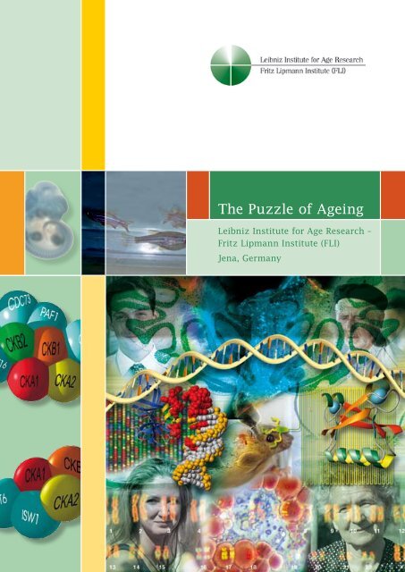
The Puzzle of Ageing - Leibniz Institute for Age Research
The Puzzle of Ageing - Leibniz Institute for Age Research
The Puzzle of Ageing - Leibniz Institute for Age Research

You also want an ePaper? Increase the reach of your titles
YUMPU automatically turns print PDFs into web optimized ePapers that Google loves.
<strong>The</strong> <strong>Puzzle</strong> <strong>of</strong> <strong><strong>Age</strong>ing</strong><br />
<strong>Leibniz</strong> <strong>Institute</strong> <strong>for</strong> <strong>Age</strong> <strong>Research</strong> –<br />
Fritz Lipmann <strong>Institute</strong> (FLI)<br />
Jena, Germany
2 Contents<br />
4 12 24 34<br />
7 <br />
<br />
<br />
<br />
<br />
<br />
<br />
<br />
<br />
<br />
<br />
<br />
<br />
<br />
<br />
<br />
<br />
<br />
18 29 40<br />
<br />
<br />
<br />
<br />
<br />
<br />
<br />
<br />
<br />
<br />
<br />
<br />
<br />
<br />
<br />
<br />
<br />
<br />
<br />
<br />
<br />
<br />
<br />
<br />
<br />
<br />
<br />
<br />
<br />
8 <br />
<br />
<br />
<br />
<br />
<br />
<br />
<br />
21 <br />
31 43<br />
<br />
<br />
<br />
<br />
<br />
<br />
<br />
<br />
<br />
<br />
<br />
<br />
<br />
<br />
<br />
<br />
<br />
<br />
<br />
<br />
<br />
6 15 26 37<br />
<br />
<br />
<br />
<br />
<br />
<br />
4 Preface by Peter Herrlich<br />
6 Fritz Lipmann – Biochemist,<br />
Noble Prize Laureate and<br />
Pioneer <strong>of</strong> <strong>Age</strong> <strong>Research</strong><br />
7 Solving the <strong>Puzzle</strong> <strong>of</strong> <strong><strong>Age</strong>ing</strong><br />
8 <strong>Research</strong> Concept: Many Roads –<br />
One Goal<br />
Selected Topics<br />
TUMOUR BIOLOGY<br />
12 How Fatal Growth Stimuli Are Blocked<br />
GENOME ANALYSIS<br />
15 Huddling Together <strong>for</strong> Safety:<br />
How Cells Cooperate<br />
BIOPHYSICS<br />
18 Virus-Induced Cancer:<br />
<strong>The</strong> Structural Basis <strong>of</strong><br />
Viral Cancerogenesis<br />
GENETICS OF AGEING<br />
21 What the Turquoise Killifish Can Tell Us<br />
about <strong><strong>Age</strong>ing</strong><br />
BIOINFORMATICS<br />
24 Analysis and Interpretation <strong>of</strong><br />
Complex Data<br />
Our Laboratories<br />
CALKHOVEN LAB<br />
26 Translational Control <strong>of</strong> Gene Expression<br />
CELLERINO LAB<br />
29 Using a Short-Lived Fish to Investigate<br />
the Biological Mechanisms Controlling<br />
Lifespan<br />
DIEKMANN LAB<br />
31 Early <strong><strong>Age</strong>ing</strong> and Premature Death:<br />
When Cellular Control Systems Fail<br />
ENGLERT LAB<br />
34 From Genes to Organs:<br />
How Genes Control Development<br />
FäNDRICH LAB<br />
37 Structure and Formation <strong>of</strong> Amyloid Fibrils<br />
GöRLACH LAB<br />
40 Biomolecular Matchmaking:<br />
How Molecules Contact Each Other<br />
GREULICH LAB<br />
43 Getting Sorted:<br />
Functional Molecule Blocks<br />
GROSSE LAB<br />
46 An Elegant Balancing Act:<br />
How Cells Maintain their Genetic<br />
Stability
Eberhard Fritz<br />
<br />
<br />
<br />
H. Lekscha; G.Bergner; E.Stöckl<br />
Diana Kirchh<strong>of</strong><br />
49 60 72 <br />
<br />
83<br />
Zhao-Qi Wang<br />
Swen Löhle<br />
<br />
<br />
<br />
Jürgen Sühnel<br />
<br />
Jürgen Sühnel<br />
52 64 74<br />
<br />
<br />
<br />
Peter Hemmerich<br />
<br />
Matthias 84 Platzer<br />
<br />
<br />
Jan Tuckermann<br />
<br />
<br />
Falk Weih<br />
<br />
<br />
55 66 <br />
<br />
<br />
<br />
Heike Heuer<br />
77 <br />
K.H. Gührs B. Schlott<br />
<br />
Matthias Görlach<br />
<br />
Eberhard Fritz<br />
<br />
<br />
Christian Hoischen<br />
<br />
<br />
C. Calkhoven, H. Heuer, C. Kaether<br />
<br />
<br />
M. Than<br />
<br />
74 Steroid Hormones:<br />
<br />
<br />
Chairman: Wolfram Eberbach<br />
<br />
<br />
<br />
Peter Herrlich<br />
46 58 68<br />
Head <strong>of</strong> Administration: Daniele Barthel<br />
80<br />
Muttermal Fehlgebildetes<br />
Muttermal<br />
Radiales<br />
Wachstum<br />
Vertikales<br />
Wachstum<br />
HERRLICH LAB<br />
49 Metastatic Migration:<br />
A Disastrous Property <strong>of</strong> Cancer Cells<br />
Tumor &<br />
Metastasenbildung<br />
HEUER LAB<br />
52 Influential Messengers:<br />
How Thyroid Hormones Affect the Brain<br />
KAETHER LAB<br />
55 Misguided Proteins:<br />
Looking <strong>for</strong> the Causes <strong>of</strong> Alzheimer’s<br />
Disease<br />
MORRISON LAB<br />
58 How Switch Proteins are Regulated<br />
to Control Proliferation and Neural<br />
Function<br />
PLATzER LAB<br />
60 Genomes, Diseases and <strong><strong>Age</strong>ing</strong><br />
PLOUBIDOU LAB<br />
64 Virus-Induced Signal Transduction<br />
and Oncogenesis<br />
SCHILLING LAB<br />
66 Molecular Mechanisms <strong>of</strong> Huntington’s<br />
Disease and <strong>The</strong>rapeutic Approaches<br />
SüHNEL LAB<br />
68 From In<strong>for</strong>mation to Knowledge:<br />
New Databases and Analysis Tools<br />
THAN LAB<br />
72 From Structure to Function:<br />
How Proteins Work in the Body<br />
TUCKERMANN LAB<br />
<br />
<br />
Chairman: Piet Borst<br />
<br />
<br />
<br />
<br />
<br />
Benita Rost<br />
<br />
<br />
Regulators <strong>of</strong> Tissue Integrity, Metabolism<br />
and Inflammation<br />
WANG LAB<br />
77 Out <strong>of</strong> Balance –<br />
How Genomic Instability Promotes<br />
Diseases and <strong><strong>Age</strong>ing</strong><br />
WEIH LAB<br />
80 Vital Communication: <strong>The</strong> NF-κB Signal<br />
Transduction Pathway in the Immune<br />
System<br />
83 Organisation Chart<br />
84 Imprint<br />
3
4 Preface<br />
As <strong>of</strong> 2003, the <strong>for</strong>mer <strong>Institute</strong> <strong>for</strong> Molecular Biotech-<br />
nology (IMB) has been systematically reorganised into an<br />
institute devoted to research on ageing. In 2005 our new<br />
name put the seal on this process. We are proud to<br />
present the first national institute in Germany with an ex-<br />
plicit research focus on the mechanisms <strong>of</strong> ageing and<br />
age-related diseases.<br />
Why research on ageing? And why in Jena? In view <strong>of</strong><br />
the increasing lifespan in industrialised nations, the<br />
theme is certainly timely. Equally important, however, is<br />
the current technological repertoire that enables us to<br />
study complex biological processes. What actually deter-<br />
mines ageing is still poorly understood. <strong>The</strong> field <strong>of</strong> age<br />
research today resembles that <strong>of</strong> cancer research some 30<br />
to 40 years ago, when environmental, genotoxic and ge-<br />
netic hypotheses existed side by side. Numerous ageing<br />
hypotheses (e.g. the “mitochondrial hypothesis on age-<br />
ing”, the “stem-cell hypothesis on ageing”, “genomic in-<br />
stability”, the “neuroendocrine hypothesis on ageing”)<br />
have found experimental support and co-exist without<br />
any proven, unifying theory. Likewise, age-related diseases<br />
are “multifactorial”, indicating that most <strong>of</strong> the mecha-<br />
nisms are yet unknown.<br />
Dear Readers,<br />
Several factors make Jena an ideal location <strong>for</strong> an in-<br />
stitute devoted to the study <strong>of</strong> ageing. <strong>The</strong> analysis <strong>of</strong><br />
complex genetic mechanisms requires high-quality ge-<br />
nome analysis. In the past, the institute has contributed<br />
significantly to the sequencing <strong>of</strong> the human genome<br />
(2% <strong>of</strong> the human genome) and the genomes <strong>of</strong> other or-<br />
ganisms. We have further improved and upgraded the se-<br />
quencing technology that now enables us to rapidly de-<br />
tect genetic and epigenetic traits. Another unique feature<br />
<strong>of</strong> the FLI has been created by a research focus on ge-<br />
nomic instability, particularly on DNA repair, an area<br />
which in the past has been by and large neglected in Ger-<br />
many. This area is important <strong>for</strong> age research because hu-<br />
man syndromes caused by DNA repair deficiencies are<br />
characterised by premature ageing. Interestingly, DNA re-<br />
pair diseases go hand in hand with neurodegeneration.<br />
Neurological ageing is another focus <strong>of</strong> research at FLI.<br />
Although the neurosciences are well established at vari-<br />
ous national centres, our institute in Jena chose to con-<br />
centrate on specific issues in age-associated neural dis-<br />
ease investigated by experts newly recruited to the FLI.<br />
Animal models have become a major tool in neurodegen-<br />
eration research. At the same time, the Medical Faculty <strong>of</strong><br />
the Friedrich Schiller University created a research focus<br />
on age-related diseases that complements our institute´s<br />
basic research. <strong>The</strong> increasing links with the Medical Fac-<br />
ulty will be pr<strong>of</strong>itable <strong>for</strong> both sides.
With the help <strong>of</strong> both the Federal government (BMBF)<br />
and Thuringian Ministry <strong>of</strong> Education and Cultural Affairs<br />
(TKM) major improvements <strong>for</strong> the infrastructure have<br />
been planned and are currently being implemented. <strong>The</strong><br />
FLI is to be housed in a new building designed by the ar-<br />
chitect M. Mackenrodt (Archiscape, Berlin). This design<br />
was chosen by a high-ranking jury consisting <strong>of</strong> the archi-<br />
tects Otto Steidle, Benedikt Tonon, Arno Lederer and Tho-<br />
mas Bahr. Completion is planned <strong>for</strong> 2009/2010. Although<br />
the scientists recruited to Jena are <strong>of</strong> prime importance,<br />
the laboratory conditions in the new building will cer-<br />
tainly add to our institute’s international visibility and at-<br />
tractiveness.<br />
With this brochure we want to present the overall sci-<br />
entific concept and the individual projects <strong>of</strong> our research<br />
activities. Additional in<strong>for</strong>mation on our institute is avail-<br />
able in our annual report “Facts and Figures 2006”.<br />
Peter Herrlich<br />
Scientific Director,<br />
Head <strong>of</strong> <strong>Institute</strong><br />
5
6 Fritz Lipmann<br />
Fritz Lipmann: Biochemist, Nobel Prize Laureate<br />
and Pioneer <strong>of</strong> <strong>Age</strong> <strong>Research</strong><br />
By choosing the name <strong>of</strong> Fritz Lipmann <strong>for</strong> our insti-<br />
tute, the FLI intends to honour an outstanding biochemist<br />
who contributed considerably to understanding the fun-<br />
damental factors involved in the ageing process. Fritz Lip-<br />
mann came from a Jewish family<br />
in Königsberg (now Kaliningrad).<br />
He received his chemical and<br />
medical education in Königs-<br />
berg, Munich and Berlin, and the<br />
Berlin-Dahlem research environ-<br />
ment influenced his first labora-<br />
tory work. After staying one year<br />
in New York, Fritz Lipmann<br />
moved to Copenhagen in 1932,<br />
and returned again to the United<br />
States in 1939, where he lived<br />
and worked in New York and<br />
Boston (1941 – 1957). In 1953 he<br />
was awarded the Nobel Prize <strong>for</strong><br />
medicine or physiology <strong>for</strong> the<br />
discovery <strong>of</strong> coenzyme A, one <strong>of</strong><br />
the most important factors in<br />
cellular metabolism. Fritz Lip-<br />
mann devoted much <strong>of</strong> his work<br />
to research on the energy me-<br />
tabolism <strong>of</strong> cells. In 1937 and<br />
1939 he presented a concept <strong>of</strong><br />
Fritz Lipmann investigated the energy metabolism<br />
<strong>of</strong> the cells and discovered coenzyme A<br />
ATP production by mitochondria, a process known today<br />
as “oxidative phosphorylation”. ATP stands <strong>for</strong> adenosine<br />
triphosphate, the most important “energy currency” <strong>of</strong><br />
the cell. In the 1940s Fritz Lipmann conducted research on<br />
the use <strong>of</strong> ATP <strong>for</strong> the regulation <strong>of</strong> pro-<br />
teins in the cell: A high-energy phosphate<br />
from ATP is transferred to a protein,<br />
thereby modulating its function. During<br />
this process, ATP is broken down into the<br />
lower energy <strong>for</strong>m ADP (adenosine di-<br />
phosphate).<br />
In 2000 the Biographical Memoirs <strong>of</strong><br />
the Royal Society had this to say about<br />
Lipmann’s achievements: “Fritz Lipmann<br />
was largely responsible <strong>for</strong> identifying<br />
and characterizing the connection be-<br />
tween metabolism and the energetics <strong>of</strong><br />
living systems that makes life possible.”<br />
<strong>The</strong> current, still rudimentary knowledge<br />
on the connection between metabolism,<br />
life expectancy and reduced energy pro-<br />
duction by the mitochondria in ageing<br />
organs is based on Fritz Lipmann’s find-<br />
ings and has laid the foundations <strong>for</strong> age<br />
research at the cellular level.<br />
Together with Hans<br />
Krebs, Fritz Lipmann<br />
was awarded the Nobel<br />
prize in 1953.
Solving the <strong>Puzzle</strong> <strong>of</strong> <strong><strong>Age</strong>ing</strong><br />
Why do we age? This question is not new, it has preoc-<br />
cupied humankind <strong>for</strong> a long time. What is new is the de-<br />
mographic development over the last 100 years, which in-<br />
dicates that average life expectancy in the industrialised<br />
nations increases by 3 months a year or 6 hours a day. <strong>The</strong><br />
resulting increase in the ageing population alarms politi-<br />
cians and prompts the biomedical community to increase<br />
its ef<strong>for</strong>ts to understand the ageing process and to ex-<br />
plore how we can <strong>of</strong>fset age-related diseases such as<br />
Alzheimer’s and cancer. <strong>The</strong> cost to society that is involved<br />
in this demographic development is by no means the only<br />
issue to be addressed, indeed it is not clear whether the<br />
health costs now rocketing around<br />
age 60 will increase considerably<br />
with the number <strong>of</strong> centenarians.<br />
Quite apart from the costs, the in-<br />
creased number <strong>of</strong> possibly un-<br />
healthy people is a considerable<br />
challenge <strong>for</strong> humanity. <strong>The</strong> desire<br />
to grow older in as healthy a condi-<br />
tion as possible is a pr<strong>of</strong>ound<br />
concern. One <strong>of</strong> the most pressing<br />
challenges involved in this research<br />
is that <strong>of</strong> contributing to healthy<br />
ageing.<br />
Healthy ageing<br />
<strong>Research</strong> on ageing is complex<br />
indeed. Numerous unanswered<br />
Life expectancy <strong>of</strong> people living in Europe<br />
has been rising steadily over the past 100 years.<br />
Exception: the war years<br />
<strong>The</strong> <strong>Puzzle</strong> <strong>of</strong> <strong><strong>Age</strong>ing</strong><br />
questions need to be addressed: How much do external<br />
and internal causes contribute to ageing? To which ex-<br />
tent is the individual ageing process predetermined by<br />
the genetic constitution? Do cellular functions simply de-<br />
teriorate in the older years <strong>of</strong> an organism, or is the<br />
lifespan predetermined during embryogenesis? Although<br />
currently disfavoured, not even the question <strong>of</strong> whether<br />
there is such a thing as a genetic clock dictating lifespan<br />
has been answered unequivocally.<br />
<strong>The</strong> prime and ultimate goal <strong>of</strong> biomedical age re-<br />
search is to break the link between ageing and disease. To<br />
achieve this goal, it is important to<br />
explore the mechanistic determi-<br />
nants <strong>of</strong> ageing and the links with<br />
disease, <strong>for</strong> instance to identify oper-<br />
ative differences between individu-<br />
als who live to an old age without<br />
ailments and others who suffer from<br />
diseases. <strong>The</strong> Fritz Lipmann <strong>Institute</strong><br />
hosts a number <strong>of</strong> research activities<br />
that address this overall goal. But to<br />
be successful, we need to focus on<br />
specific selected topics approached<br />
from a variety <strong>of</strong> different perspec-<br />
tives. <strong>The</strong> next section describes<br />
how the FLI research laboratories are<br />
thematically organised to address<br />
such topics.<br />
7
8 <strong>Research</strong> Concept<br />
Heads <strong>of</strong> laboratories at FLI<br />
Many Roads – One Goal<br />
Within the overall field <strong>of</strong> age research, the scientists<br />
at the Fritz Lipmann <strong>Institute</strong> are involved in two major<br />
programmes: “Mechanisms <strong>of</strong> <strong><strong>Age</strong>ing</strong> and Senescence”<br />
and “<strong>Age</strong>-Associated Diseases” (see diagram). <strong>The</strong> penta-<br />
gons <strong>for</strong> each programme indicate the subtopics covered<br />
by the research groups. <strong>The</strong> whole research area, with its<br />
<strong>for</strong>mally divided programmes, is characterised by an enor-<br />
mous degree <strong>of</strong> scientific and methodological overlap,<br />
both between and within the programmes and their sub-<br />
topics.<br />
<strong>The</strong> increasing scientific coherence <strong>of</strong> the research lab-<br />
oratories <strong>of</strong> the FLI over the last three years has brought<br />
with it a high degree <strong>of</strong> collaboration and synergy in their<br />
ef<strong>for</strong>ts to deal with related issues. As the illustration indi-<br />
cates, most FLI laboratories contribute to a variety <strong>of</strong> dif-<br />
ferent subtopics (pentagons), by investigating similar<br />
questions using individual experimental expertises and<br />
approaches. <strong>The</strong> laboratories will be presented separately<br />
in this brochure however the links between the labs will<br />
also be indicated.<br />
Scientific coherence in the institute is also reflected by<br />
a number <strong>of</strong> regular lab meetings, journal clubs and dis-<br />
cussion rounds shared between different laboratories.<br />
<strong>The</strong> „Metabolic Club“ (Bauer, Calkhoven, Heuer, Tucker-<br />
mann labs) and „Genomic Instability“ lab meeting (Greu-<br />
lich, Herrlich, Wang labs) may serve as prominent exam-<br />
ples <strong>for</strong> such joint regular meetings <strong>of</strong> several laboratories<br />
on collective research topics. <strong>The</strong> fact that many labs<br />
share a high degree <strong>of</strong> methodological overlap addition-<br />
ally promotes coherence <strong>of</strong> the research groups.
9
10 <strong>Research</strong> Concept<br />
Mechanisms <strong>of</strong> ageing and<br />
senescence<br />
Laboratories working on the “Mechanisms <strong>of</strong><br />
<strong><strong>Age</strong>ing</strong> and Senescence” encompass research on<br />
lifespan in a new model organism, on the identifi-<br />
cation <strong>of</strong> the determinants <strong>of</strong> healthy human age-<br />
ing and on selected aspects <strong>of</strong> cellular senescence<br />
under the heading <strong>of</strong> “Longevity”. A significant<br />
role in both organismic ageing and cellular senes-<br />
cence is probably played by “Destabilisation <strong>of</strong><br />
the genome”, which accordingly represents a<br />
major focus in the work done at the FLI: Several<br />
FLI laboratories concentrate on aspects <strong>of</strong> DNA<br />
replication, DNA repair, chromosome segrega-<br />
tion and telomere structure and maintenance.<br />
Errors in these processes cause premature ageing<br />
in cells and organisms and are key factors in the<br />
development <strong>of</strong> age-associated diseases. In addi-<br />
tion, the enormous expertise in genome analysis<br />
available at the FLI is being used to identify age-<br />
dependent DNA methylation <strong>of</strong> the human ge-<br />
nome, changes in gene copy number and in the<br />
pattern <strong>of</strong> splicing.<br />
<strong>Age</strong>-associated diseases<br />
<strong>The</strong> second major focus at the FLI is research on age-<br />
associated diseases, notably selected aspects <strong>of</strong> neurode-<br />
generation and cancer. In addition, specific issues are ad-<br />
dressed in the area <strong>of</strong> the metabolic syndrome, including<br />
associated conditions <strong>of</strong> hormonal dysregulation and<br />
atherosclerosis. Within the subtopic “Impaired tissue ho-<br />
moestasis”, osteoporosis and chronic inflammation are<br />
studied, among others. Since embryonic processes are<br />
partly recapitulated in tissue regeneration, developmen-<br />
tally active genes related to kidney and gonadal disorders<br />
are studied in a zebrafish model. In the subtopic “Geno-<br />
mic variability” the FLI is identifying disease-associated<br />
polymorphisms through genome analysis. Almost all FLI<br />
activities on age-associated diseases rely on the use <strong>of</strong> an-<br />
imal models, currently mice and fish.<br />
Support technologies<br />
As one might expect, a whole range <strong>of</strong> methods and<br />
technologies is needed to explore the processes involved<br />
in ageing and disease, from the study <strong>of</strong> single molecules<br />
and cells in culture to animal models and the study <strong>of</strong><br />
families whose members display longevity. Using such<br />
different mutually supportive methodologies and experi-<br />
mental approaches to answering basic questions about<br />
molecular functions is one <strong>of</strong> the strengths <strong>of</strong> the Fritz<br />
Lipmann <strong>Institute</strong>.<br />
<strong>The</strong> methodical synergies at FLI range from structural<br />
biology and protein biochemistry to cell biology and from<br />
the use <strong>of</strong> cell culture models to animal models <strong>for</strong> typical<br />
human age-associated diseases. We also analyse bio-<br />
probes from selected patient cohorts, while old-aged indi-<br />
viduals are used <strong>for</strong> genetic and molecular analysis <strong>of</strong> age-<br />
ing and pathogenic mechanisms in humans.
<strong>Research</strong> environment<br />
As in other areas, we expect that<br />
mechanistic knowledge will rapidly ac-<br />
cumulate in the area <strong>of</strong> age research. It<br />
takes, however, time be<strong>for</strong>e applications<br />
reach the patients or the ageing individ-<br />
ual. To catalyse the transfer <strong>of</strong> research<br />
results to clinical studies as quickly as<br />
possible is there<strong>for</strong>e within FLI’s atten-<br />
tion. In order to promote the interac-<br />
tions between basic scientists and clinicians, we are cur-<br />
rently setting up a <strong>Leibniz</strong> <strong>Research</strong> School <strong>for</strong> Clinician<br />
Scientists. <strong>The</strong> additional benefit <strong>of</strong> this school is to per-<br />
mit clinicians to develop their own research in a stimula-<br />
tory environment and with reduced clinical duties.<br />
<strong>The</strong> research interests <strong>of</strong> the scientists working at the<br />
Fritz Lipmann <strong>Institute</strong> are closely linked with those <strong>of</strong> the<br />
Friedrich Schiller University (FSU) in Jena, other research<br />
institutes on the local Beutenberg campus and many<br />
other cooperation partners at national and international<br />
institutions. Several research group leaders at the Fritz<br />
Lipmann <strong>Institute</strong> have pr<strong>of</strong>essorships at the Friedrich<br />
Schiller University and contribute substantially to teach-<br />
ing at the FSU. <strong>The</strong> recently established Graduate School<br />
<strong>of</strong> the FLI (the <strong>Leibniz</strong> Graduate School on <strong><strong>Age</strong>ing</strong> and<br />
<strong>Age</strong>-Related Diseases, LGSA) is designed to ensure future<br />
continuity in age research.<br />
More in<strong>for</strong>mation<br />
selected projects are highlighted.<br />
In this brochure the group leaders<br />
and scientists <strong>of</strong> the Fritz Lipmann Insti-<br />
tute introduce their research projects<br />
and scientific achievements to the gen-<br />
eral international public <strong>for</strong> the first<br />
time. Each research laboratory is pre-<br />
sented as an individual entity, pinpoint-<br />
ing its specific contributions to the over-<br />
all research design. In addition, a few<br />
For more in<strong>for</strong>mation, please visit our website<br />
www.fli-leibniz.de. An overview <strong>of</strong> the recent scientific<br />
output and the structure <strong>of</strong> the FLI is presented in our<br />
annual report “Facts and Figures 2006”, available on our<br />
website or in hard copy <strong>for</strong>m on request.<br />
11
12 Selected Topics: Tumour Biology<br />
How Fatal Growth Stimuli Are Blocked<br />
Normal cells in the developing organism know exactly when to stop proliferating. When an<br />
organ is <strong>for</strong>med and the space filled, normal cells no longer react to growth stimuli. Tumour<br />
cells, however, have <strong>for</strong>feited this property. <strong>The</strong>y go on growing, even pushing other tissues aside in<br />
the process. <strong>The</strong> laboratory headed by Helen Morrison explores the mechanisms involved in growth<br />
control. She describes a chain <strong>of</strong> reactions involving a protein cascade telling cells that they have<br />
made contact with their neighbours (cell-cell contact). This is a novel regulatory step in cellular<br />
signalling that goes <strong>of</strong>f the rails in tumour cells.<br />
Neur<strong>of</strong>ibromatosis type II (NF2) is a rare inherited dis-<br />
ease that affects 1 in 40,000 individuals. NF2 patients de-<br />
velop a variety <strong>of</strong> tumours in the nervous system, the<br />
most notable <strong>of</strong> which are vestibular schwannomas, tu-<br />
mours associated with hearing nerves. NF2 patients carry<br />
defects in a gene specifying the production <strong>of</strong> a protein<br />
called merlin. Defective merlin (or its absence) causes ab-<br />
normal and uncontrolled cell proliferation leading to the<br />
<strong>for</strong>mation <strong>of</strong> tumours. In most, if not all, normal cells <strong>of</strong><br />
the organism, merlin controls cell multiplication (and<br />
hence is called a tumour suppressor protein). Merlin ex-<br />
ists, however, in two states: an active state, in which it in-<br />
hibits cell proliferation, and an inactive state in which it<br />
permits cell proliferation. Accordingly, merlin itself is regu-<br />
lated and only becomes active under certain conditions.<br />
Switching between the active and inactive states appears<br />
to be controlled by the absence or presence <strong>of</strong> a phos-<br />
phate group on the merlin molecule at the location called<br />
ser518. <strong>The</strong>se are very basic mechanisms. <strong>The</strong> transfer <strong>of</strong> a<br />
high-energy phosphate from ATP to the proteins is<br />
known as phosphorylation (see the section on Fritz<br />
Lipmann), while removal <strong>of</strong> the phosphate is known as<br />
dephosphorylation.<br />
Tight control <strong>of</strong> merlin activity<br />
Merlin is inactive when the phosphate group is at-<br />
tached to ser518 and active when it is removed. Merlin is<br />
only dephosphorylated at the ser518 position following<br />
cell-cell contact and only such activated merlin stops cel-<br />
lular proliferation. <strong>The</strong> Morrison laboratory has explored<br />
how merlin manages to halt proliferation and how the<br />
dephosphorylation <strong>of</strong> merlin is achieved. Put briefly, the<br />
result was the discovery <strong>of</strong> a reaction cascade involving<br />
several stages. Merlin interferes with the activation <strong>of</strong> a<br />
well-known proliferation-promoting protein called Ras.<br />
Like merlin, Ras behaves as a switch which in the “on” po-<br />
sition predominantly induces cells to multiply. Lack <strong>of</strong>
Molecular signal cascades influence a cell’s proliferation:<br />
after an extracellular growth factor is bound to its receptor<br />
this message is further transferred via several intracellular<br />
proteins (Grb2, SOS, Ras, Raf, MEK, ERK). <strong>The</strong> transient nature<br />
<strong>of</strong> this signal is characterised by reversible changes <strong>of</strong> the<br />
proteins involved, i.e. modification <strong>of</strong> amino acids (pY) or<br />
exchange <strong>of</strong> co-factors (Ras-GDP & Ras-GTP). <strong>The</strong> cell will<br />
respond to this signal and adjust its behaviour accordingly,<br />
e.g. divide.<br />
P Y<br />
Growth<br />
factor<br />
receptor<br />
Y P<br />
Growth<br />
factor<br />
Grb2<br />
Sos<br />
Ras<br />
GDP<br />
Ras<br />
GTP<br />
Cell membrane<br />
Raf<br />
MEK<br />
ERK<br />
Growth<br />
merlin leads to a hyperactive <strong>for</strong>m <strong>of</strong> Ras resulting in un-<br />
controlled cell proliferation and thus ultimately contribut-<br />
ing to the <strong>for</strong>mation <strong>of</strong> cancer.<br />
Merlin is dephosphorylated in response to a stimulus<br />
from outside the cell that is provided by cell-cell contact.<br />
A significant step towards understanding this process was<br />
recently achieved by our discovery <strong>of</strong> the enzyme respon-<br />
sible <strong>for</strong> removing the phosphate from merlin. This en-<br />
zyme is a phosphatase known as the myosin phosphatase<br />
(MYPT-1–PP1δ) previously known to be involved in con-<br />
tractility, a process important <strong>for</strong> cell shape and motility.<br />
<strong>The</strong> enzyme specifically selects merlin from among many<br />
other proteins. <strong>The</strong> specificity is mediated by one <strong>of</strong> the<br />
sub-units <strong>of</strong> the phosphatase, the MYPT sub-unit. MYPT<br />
makes the contact and guides the catalytic sub-unit PP1δ<br />
to its target (merlin). Not surprisingly, a regulatory reac-<br />
tion <strong>of</strong> this importance is subject to further control. For<br />
example, the cells can produce an inhibitor called CPI-17,<br />
and both CPI-17 and the phosphatase itself are again regu-<br />
lated by phosphorylation. We now hope to identify other<br />
steps in the cascade mediating the activation <strong>of</strong> the tu-<br />
mour suppressor protein merlin in response to cell-cell<br />
contact.<br />
Our results extend the current model <strong>of</strong> cellular growth<br />
control by three essential findings: the growth factor is<br />
dependent on a co-receptor (mostly adhesion proteins). This<br />
co-receptor functions as an anchor on the intracellular face by<br />
binding to the protein ezrin, which connects to the cellular<br />
skeleton (actin filaments). Ezrin in turn associates with and<br />
activates the signalling component SOS thereby directly<br />
participating in the signal relay.<br />
P Y<br />
Growth<br />
factor<br />
Growth<br />
factor<br />
receptor<br />
Y P<br />
Grb2<br />
Adhesion<br />
receptor<br />
SOS<br />
Ezrin<br />
Ras<br />
GDP<br />
Actin filaments<br />
Extracellular<br />
matrix<br />
Cell membrane<br />
Ras<br />
GTP<br />
Dysregulation <strong>of</strong> merlin promotes<br />
tumourigenesis<br />
Raf<br />
MEK<br />
ERK<br />
Proliferation...<br />
Tumour cells arise when merlin is defective (as in neu-<br />
r<strong>of</strong>ibromatosis type 2). Can tumours <strong>for</strong>m when merlin is<br />
normal but cannot be activated by dephosphorylation?<br />
We have indeed found evidence <strong>for</strong> this possibility. Under<br />
certain conditions, elevated expression <strong>of</strong> the inhibitor<br />
CPI-17 causes cultured cells to become tumour-like. Re-<br />
cently we also discovered several human cancers that<br />
carry high levels <strong>of</strong> CPI-17. In these cells, merlin is inactive<br />
and the growth stimuli constantly push Ras into the “on”<br />
position.<br />
While merlin inactivation is a major factor keeping Ras<br />
in the “on” position, we also wanted to know whether this<br />
absence <strong>of</strong> an “<strong>of</strong>f” switch <strong>for</strong> merlin was sufficient in it-<br />
self. Our work on ezrin (see Morrison lab, page 58) indi-<br />
cates that merlin and ezrin (which is structurally related<br />
to merlin) compete <strong>for</strong> the same interaction sites on the<br />
plasma membrane. Ezrin promotes Ras-dependent signal-<br />
ling and subsequent cellular proliferation by binding Ras<br />
and localising it to a specific Ras-activating enzyme called<br />
SOS. In addition, ezrin can interact with SOS and speed up<br />
the catalytic activity <strong>of</strong> this enzyme. Merlin, however,<br />
does the opposite! While active dephosphorylated merlin<br />
can localise to the same sites in the plasma membrane as<br />
13
14 Selected Topics: Tumour Biology<br />
A single cell layer <strong>of</strong> contact inhibited cells is visible in the<br />
electron microscope. Experimental activation <strong>of</strong> the oncogene<br />
CPI-17 trans<strong>for</strong>ms cells characterised by loss <strong>of</strong> cell to cell<br />
contacts, change <strong>of</strong> shape, and enhanced proliferation.<br />
<strong>The</strong> graphic depicts the mode <strong>of</strong> action <strong>of</strong> CPI-17: it inhibits the<br />
myosin phosphatase MYPT-PP1δ. This phosphatase is a crucial<br />
activator <strong>of</strong> the tumour suppressing protein merlin.<br />
<br />
<br />
<br />
<br />
ezrin, merlin cannot interact with SOS and activate it.<br />
How do ezrin proteins know that they need to stop func-<br />
tioning? Both merlin and ezrin are regulated by the same<br />
mechanism. Ezrin is activated by the transfer <strong>of</strong> phos-<br />
phate, whereas phosphorylated merlin is inactive. Cell-cell<br />
contact triggers MYPT-PP1δ-dependent dephosphoryla-<br />
tion, which removes phosphates from both types <strong>of</strong> pro-<br />
tein, subsequently activating merlin and at the same time<br />
inactivating ezrin. This enables merlin to interfere with<br />
the action <strong>of</strong> the ezrin proteins and to block the essential<br />
signalling events promoting growth. When CPI-17 is ab-<br />
normally expressed, the phosphatase MYPT-PP1δ will be<br />
inhibited, thus causing the cells not only to lose merlin<br />
but also to acquire the tumour-promoting activity <strong>of</strong><br />
ezrin. Eventually we hope to discover tools that can inter-<br />
fere with the action <strong>of</strong> CPI-17.<br />
Authors: Helen Morrison, Tobias Sperka and Peter Herrlich<br />
Phone: 0049-3641-656139<br />
E-mail: helen@fli-leibniz.de<br />
Original publication:<br />
Tumorigenic trans<strong>for</strong>mation by CPI-17 through inhibition <strong>of</strong> a<br />
merlin phosphatase,<br />
Nature 442, 576-579.<br />
CPI-17 there<strong>for</strong>e arrests merlin in the inactive state allowing the<br />
cell to proliferate.<br />
δ<br />
<br />
<br />
<br />
<br />
<br />
<br />
<br />
<br />
<br />
<br />
<br />
<br />
<br />
Helen Morrison and the Thuringian Minister <strong>of</strong><br />
education and cultural affairs Jens Goebel during the<br />
awards ceremony <strong>for</strong> the Thuringian <strong>Research</strong> Award
Dictyostelium discoideum, a small model <strong>for</strong><br />
big questions: Why and how did<br />
multicellular organisms like humans evolve?<br />
Dictyostelium discoideum is an amoeba that<br />
remains unicellullar…<br />
Huddling Together <strong>for</strong> Safety:<br />
How Cells Cooperate<br />
What is it that enables single cells to aggregate into a<br />
multicellular organism? Though we still cannot give a<br />
complete answer to this question we have made consider-<br />
able progress in that direction. In 2005 a Dictyostelium<br />
Genome Analysis Consortium assembling scientists from<br />
the United States, the United Kingdom, Japan and Ger-<br />
many published a description and initial analysis <strong>of</strong> the<br />
genome <strong>of</strong> the amoeba Dictyostelium discoideum in the<br />
journal Nature.<br />
What makes an amoeba so interesting? Its specific life-<br />
<strong>for</strong>m makes Dictyostelium what it has been <strong>for</strong> decades al-<br />
ready: a model system <strong>for</strong> scientists attempting to find<br />
out how organisms develop, how cells transmit messages<br />
(signal transduction) and the role played by the cell skele-<br />
ton (cytoskeleton).<br />
Social when need be<br />
<strong>The</strong> most striking feature <strong>of</strong> these amoebas is their veg-<br />
etative life-cycle. It is characterised as follows: When ambi-<br />
Selected Topics: Genome Analysis<br />
Dictyostelium discoideum is an amoeba that remains unicellular as long as conditions are<br />
favourable. When conditions deteriorate, these individual cells congregate to <strong>for</strong>m a multicellular<br />
organism, which then develops a long, thin stalk and a fruiting body dispersing spores.<br />
How do these single cells communicate? What enables them to join <strong>for</strong>ces? How do they specialise?<br />
Gernot Glöckner has the answers to these intriguing questions and indicates their relevance <strong>for</strong><br />
human ageing.<br />
ent conditions become unfavourable, single autonomous<br />
cells aggregate into a multicellular fruiting body after un-<br />
dergoing several well-defined interim stages. Only a part<br />
<strong>of</strong> these cells will develop into viable <strong>for</strong>ms and survive<br />
the aggregation process. In no other branch <strong>of</strong> the tree <strong>of</strong><br />
life do individual and essentially autonomous cells cooper-<br />
ate in this way. <strong>The</strong>y assure the survival <strong>of</strong> the species by<br />
sacrificing themselves. This astonishing altruism can only<br />
function if the cells communicate and single cells special-<br />
ise into cell types per<strong>for</strong>ming specific tasks (cell differenti-<br />
ation). Incidentally, this conditional multicellularity <strong>of</strong> the<br />
amoeba is achieved largely with the same set <strong>of</strong> genes<br />
that permanently multicellular systems draw upon.<br />
Another remarkable feature <strong>of</strong> Dictyostelium is the<br />
amoeboid motility <strong>of</strong> the cells, which is assured by the<br />
components <strong>of</strong> the cytoskeleton. <strong>The</strong> components and dy-<br />
namics <strong>of</strong> the cytoskeleton are readily comparable to the<br />
cytoskeleton <strong>of</strong> cells moving freely in multicellular organ-<br />
isms, <strong>for</strong> example the macrophages, scavenger cells <strong>of</strong> the<br />
immune system.<br />
15
16 Selected Topics: Genome Analysis<br />
<strong>The</strong> amoeba D. discoideum has 6 chromosomes and around 12,000 genes. <strong>The</strong> adjacent<br />
picture shows the fruiting body, which is built as end-point <strong>of</strong> the vegetative multicellular<br />
life cycle <strong>of</strong> the amoeba. <strong>The</strong> lighter cells in this picture are destined to die, whereas the<br />
dark cells will survive and develop to spores.<br />
Happy Mapping<br />
Deciphering the genome <strong>of</strong> Dictyostelium discoideum<br />
was a stiff challenge. First, the percentage <strong>of</strong> complex<br />
repetitive elements (10%) is high <strong>for</strong> such a small genome<br />
(34 Mb). Another complicating factor was that the pro-<br />
portion <strong>of</strong> the nucleotides adenin and thymin is so high<br />
(78%) that bacterial subclones can only be kept stable up<br />
to a size <strong>of</strong> five kilobase (in genetics a kilobase, kb, is a<br />
unit used to measure lengths <strong>of</strong> DNA and corresponds to<br />
1,000 nucleotides). Accordingly, sequencing with the as-<br />
sistance <strong>of</strong> large bacterial clones was impossible. To en-<br />
sure that the entire genome was represented it was nec-<br />
essary to establish an accurate map <strong>of</strong> it. To this end, a<br />
second map charted with the “Happy Mapping” method<br />
was integrated into an existing rough genetic map to<br />
achieve an average landmark density <strong>of</strong> 1/60 kb in the ge-<br />
nome. A map <strong>of</strong> this accuracy was essential to identify the<br />
entire chromosomal structure. In the process a striking<br />
feature became apparent. At its end (terminus), every<br />
chromosome displays a region consisting largely <strong>of</strong> clus-<br />
ters <strong>of</strong> special retro-elements. Our conjecture is that these<br />
regions per<strong>for</strong>m the function <strong>of</strong> centromeres to which the<br />
spindle fibres attach during cell division in order to segre-<br />
gate the chromosomes after duplication. Another feature<br />
common to all the chromosomes is that their termini end<br />
in sequences deriving from the so-called rDNA palin-<br />
drome, which figures at various points in the cell nucleus<br />
and carries the in<strong>for</strong>mation <strong>for</strong> the ribosomal RNAs. <strong>The</strong><br />
transition from chromosomal to palindrome sequence<br />
does not appear to be defined, as the attachment point<br />
differs <strong>for</strong> each chromosome terminus. Thus one might<br />
regard the chromosomes as sequences embedded in an<br />
acentric palindrome. As the chromosomal termini do not<br />
display any differences from the rDNA palindrome, they<br />
may conceivably be repeatedly renewed from this reser-<br />
voir. In other words, Dictyostelium has found a way <strong>of</strong> us-<br />
ing the same components over and over again to <strong>for</strong>m<br />
protective caps <strong>for</strong> its chromosomes.<br />
Genes <strong>of</strong> an amoeba<br />
Dictyostelium’s small genome contains over 12,000<br />
genes, a similar number to the genomes <strong>of</strong> “genuine”<br />
metazoa (multicellular organisms) like the fruit-fly Dro-<br />
sophila. Apart from a small number <strong>of</strong> genes probably<br />
restricted to this evolutionary line, most <strong>of</strong> the Dictyostel-<br />
ium genes are similar to those <strong>of</strong> other organisms. Re-<br />
markable are a number <strong>of</strong> genes and gene families <strong>for</strong>-<br />
merly thought to be the “invention” <strong>of</strong> metazoa only.<br />
In relation to the number <strong>of</strong> genes, we found consider-<br />
ably fewer transcription factors than is customary <strong>for</strong><br />
other species. Transcription factor is the term used <strong>for</strong><br />
proteins that can switch genes on or <strong>of</strong>f. So far, just under<br />
100 transcription factors have been identified in Dictyos-<br />
telium, fewer than in yeasts. Transcription factors with the<br />
so-called “basic loop-helix-loop domain” are totally ab-<br />
sent. This motif has been identified in all other evolution-<br />
ary lines so far.
This relative absence <strong>of</strong> transcription factors in Dicty-<br />
ostelium may have to do with the use <strong>of</strong> more highly inte-<br />
grated regulation mechanisms. It is however more likely<br />
that most transcription factors are specifically adapted to<br />
the requirements <strong>of</strong> the genome and have yet to be dis-<br />
covered because they have little or no similarity to known<br />
counterparts in other organisms. Further investigation is<br />
necessary to clarify this issue and to use a systems biology<br />
approach to describe the regulatory network <strong>of</strong> these or-<br />
ganisms.<br />
What we have so far is the entire sequence, i.e. the<br />
complete order <strong>of</strong> all bases constituting the genome <strong>of</strong><br />
Dictyostelium. This makes it possible <strong>for</strong> us to analyse the<br />
functions <strong>of</strong> gene families. As the functions <strong>of</strong> gene prod-<br />
ucts (proteins) <strong>of</strong> a gene family frequently overlap, we use<br />
“knock-out” mutants in which individual groups <strong>of</strong> genes<br />
are systematically switched <strong>of</strong>f. In this way we can include<br />
the entire genetic background and describe the cause and<br />
effect relations.<br />
Another field <strong>of</strong> research opened up by our knowledge<br />
<strong>of</strong> the entire genome sequence <strong>of</strong> Dictyostelium is the<br />
comparison <strong>of</strong> the genomes <strong>of</strong> different organisms (com-<br />
parative genomics). Various social amoebas have been de-<br />
scribed morphologically and are present in strain collec-<br />
tions. Other members <strong>of</strong> the same evolutionary branch<br />
are organisms as various as Entamoeba histolytica or<br />
Physarum polycephalum. We now have the prospect <strong>of</strong><br />
embarking on in-depth study <strong>of</strong> the evolution <strong>of</strong> this fas-<br />
cinating group <strong>of</strong> organisms.<br />
Protective caps <strong>for</strong> chromosomes<br />
Telomeres <strong>for</strong>m the termini <strong>of</strong> linear chromosomes,<br />
closing them <strong>of</strong>f like protective caps. To ensure that no<br />
genetic material gets lost they have to remain intact<br />
throughout the entire life <strong>of</strong> the cell. If the machinery<br />
maintaining the telomeres is damaged, the cell will die.<br />
This factor may also be involved in the ageing <strong>of</strong> complex<br />
organisms like the roundworm Caenorhabditis elegans,<br />
the fruit fly Drosophila or humans. <strong>The</strong> structure <strong>of</strong> the<br />
telomeres <strong>of</strong> Dicytostelium discoideum differs fundamen-<br />
tally from the telomere structure <strong>of</strong> other model organ-<br />
isms used in age research and also from that <strong>of</strong> the hu-<br />
man organism. In a project entitled “From Comparative to<br />
Functional Genomics <strong>of</strong> Social Amoeba” funded by the<br />
German <strong>Research</strong> Foundation (DFG) we have set out to<br />
elucidate the mechanisms ensuring telomere preservation<br />
in Dictyostelium discoideum. We believe that this project<br />
will make a substantial contribution to our understanding<br />
<strong>of</strong> ageing.<br />
Author: Gernot Glöckner<br />
Phone: 0049-3641-656440<br />
E-mail: gernot@fli-leibiniz.de<br />
Original publication:<br />
<strong>The</strong> genome <strong>of</strong> the social amoeba Dictyostelium discoideum.<br />
Nature 435, 43-57<br />
Contrary to the situation in other<br />
organisms the telomeres <strong>of</strong><br />
Dictyostelium are not composed<br />
<strong>of</strong> simple repeated sequences.<br />
What mechanism do the<br />
amoebae use to maintain their<br />
chromosome ends? And can this<br />
mechanism be exploited to better<br />
understand ageing, even in<br />
humans? <strong>The</strong>se are two burning<br />
questions <strong>of</strong> the group <strong>of</strong> Gernot<br />
Glöckner. <strong>The</strong> figure shows the<br />
detailed telomere structure <strong>of</strong><br />
D. discoideum.<br />
17
18 Selected Topics: Biophysics<br />
Virus-Induced Cancer:<br />
<strong>The</strong> Structural Basis <strong>of</strong> Viral Cancerogenesis<br />
Certain viruses, the so-called papilloma viruses, may cause cancer in humans. <strong>The</strong> molecular<br />
details <strong>of</strong> how the virus manages to trans<strong>for</strong>m a healthy cell into a cancer cell are still enigmatic.<br />
Matthias Görlach explains how a virus removes the “brakes” in infected cells to drive them into<br />
uncontrolled proliferation. <strong>The</strong> laboratory has succeeded in determining a protein structure <strong>of</strong> the<br />
virus that appears to be an appropriate target <strong>for</strong> drugs intercepting the molecular pathways the<br />
virus draws upon to trigger cancer.<br />
Human papilloma viruses infect the basal layers <strong>of</strong> epi-<br />
thelia, such as skin or mucosa, thereby causing a number<br />
<strong>of</strong> medical conditions ranging from harmless warts to<br />
cancer. Accordingly, the papilloma viruses are categorised<br />
into different types posing low or high risk <strong>of</strong> causing can-<br />
cer (LR = low-risk; HR = high-risk).<br />
<strong>The</strong> virus types 16, 18 and 45 belong to the high-risk<br />
group. <strong>The</strong>y cause cervical carcinoma, the second most<br />
frequent <strong>for</strong>m <strong>of</strong> cancer affecting women worldwide.<br />
However, we still have only an imperfect understanding<br />
<strong>of</strong> how exactly an infection with these virus types ulti-<br />
mately leads to the development <strong>of</strong> cancer. It is known<br />
that, together with cellular factors, certain proteins <strong>of</strong> the<br />
virus, the so-called oncoproteins E6 and E7, contribute de-<br />
cisively to the trans<strong>for</strong>mation <strong>of</strong> cells in cervical mucosa.<br />
<strong>The</strong> oncoprotein E7 operates by interacting with cellular<br />
proteins, among them pRb and p21CIP. <strong>The</strong>se two proteins<br />
are involved in regulating the processes <strong>of</strong> the cell cycle.<br />
Our aim is to elucidate the structure <strong>of</strong> E7 proteins derived<br />
from low-risk and high-risk papilloma virus types. In addi-<br />
tion, we intend to investigate how this viral protein inter-<br />
acts with cellular proteins, thus shedding light on the<br />
structural basis <strong>of</strong> viral cancerogenesis. Once the modus<br />
operandi <strong>of</strong> the E7 oncoprotein is understood, i.e. the way<br />
it interacts with cellular proteins, it may be possible to<br />
find substances that can intercept such interactions. This<br />
in its turn could pave the way <strong>for</strong> the development <strong>of</strong><br />
medical drugs preventing progression <strong>of</strong> pre-cancerous<br />
conditions (so-called pre-malignant lesions) into full-<br />
blown tumours.
How the virus subverts cellular growth<br />
regulation<br />
<strong>The</strong> oncoprotein E7 comprises approx. 100 amino acids,<br />
the building blocks <strong>of</strong> proteins. E7 contains three regions<br />
CR1, CR2 and CR3, which are conserved among the differ-<br />
ent papillomavirus types (CR = conserved regions). Of<br />
those, CR1 is unique and found only in E7, while CR2 is sim-<br />
ilar to equivalent regions <strong>of</strong> the E1A protein from adenovi-<br />
ruses and the large T antigen <strong>of</strong> SV40 viruses. <strong>The</strong> CR3 re-<br />
gion <strong>of</strong> E7 contains two CxxC motifs, which are separated<br />
by 29 amino acids and which co-or-<br />
dinate a zinc ion, thereby stabilis-<br />
ing the structure <strong>of</strong> E7.<br />
<strong>The</strong> CR2 region <strong>of</strong> the on-<br />
coprotein E7 contains a<br />
strictly conserved sequence<br />
motif (LxCxE) that contrib-<br />
utes decisively to the interac-<br />
tion <strong>of</strong> E7 with a cellular protein,<br />
the tumour suppressor pRb. Binding<br />
<strong>of</strong> pRb by E7 and subsequent E7 CR1 de-<br />
pendent pRb degradation by the cellular proteasome lead<br />
to a release <strong>of</strong> pRb-bound transcription factors <strong>of</strong> the E2F<br />
family. Transcription factors are proteins that switch genes<br />
on or <strong>of</strong>f. Here, the transcription factors released initiate<br />
the read-out <strong>of</strong> genes that are necessary <strong>for</strong> the entry <strong>of</strong><br />
cells into the S-phase <strong>of</strong> the cell cycle. <strong>The</strong> S-phase is<br />
mainly characterised, among other things, by replication<br />
(duplication) <strong>of</strong> the cellular hereditary substance DNA.<br />
<strong>The</strong> human papilloma virus<br />
(here HPV 16) causes cervical carcinoma,<br />
the second most frequent tumour in women<br />
worldwide.<br />
<strong>The</strong> CR3 region <strong>of</strong> oncoprotein E7 mediates contact<br />
with a number <strong>of</strong> cellular regulatory proteins, including<br />
p21CIP, the NURD histone deacetylase complex, BRCA1 and<br />
the insulin-like growth factor binding protein IGFBP3.<br />
Binding to E7 by the cyclin-dependent kinase inhibitor<br />
(CDKI) p21CIP abrogates the inhibition <strong>of</strong> cyclin-dependent<br />
kinases. This interaction also diminishes the inhibition <strong>of</strong><br />
PCNA-dependent DNA replication as the binding sites <strong>for</strong><br />
E7 and PCNA in the C-terminal region <strong>of</strong> p21CIP overlap.<br />
In short, the binding <strong>of</strong> the viral oncoprotein E7<br />
to the cellular regulatory proteins, e.g.<br />
pRb and p21CIP, leads to a re-<br />
lease <strong>of</strong> “molecular brakes”.<br />
In a healthy, non-infected<br />
state these brakes pre-<br />
vent cell division and<br />
proliferation.<br />
Portrait <strong>of</strong> an<br />
oncogenic protein<br />
In order to elucidate the structural<br />
basis <strong>of</strong> this virus-induced dysregulation <strong>of</strong> the<br />
“molecular brakes”, we are investigating the structure and<br />
the interactions <strong>of</strong> the viral oncoprotein E7 from a number<br />
<strong>of</strong> papilloma virus types (both LR and HR types). As a first<br />
step, the full-length <strong>for</strong>m and the CR3 region <strong>of</strong> E7 from<br />
HR–HPV 45 were produced in bacteria in the presence <strong>of</strong><br />
the stable isotopes 13C and 15N, purified and prepared <strong>for</strong><br />
structural analysis by NMR spectroscopy. A comparison <strong>of</strong><br />
19
20 Selected Topics: Biophysics<br />
[1H,15N] HSQC – “fingerprint” – spectra <strong>of</strong> the two E7 con-<br />
structs revealed that the N–terminal part (CR1 and CR2) <strong>of</strong><br />
E7 is mainly unstructured, as has been observed in other<br />
regulatory proteins in the absence <strong>of</strong> their specific protein<br />
ligands. By contrast, the C-terminal CR3 region adopts a<br />
defined spatial structure, which in turn depends upon the<br />
presence <strong>of</strong> zinc ions. Complete structural analysis re-<br />
vealed that CR3 assembles into homodimers. Each mono-<br />
mer adopts a β1β2α1β3α2 topology and the stabilising zinc<br />
ion is co-ordinated by the four cysteine residues <strong>of</strong> the<br />
two CxxC motifs. This topology represents a novel pro-<br />
tein-folding motif, which is not found in other zinc-bind-<br />
ing cellular proteins. Relaxation measurements and mo-<br />
lecular dynamics simulations show that the β3 strand is<br />
<strong>for</strong>med as a consequence <strong>of</strong> dimerisation and stabilised<br />
by hydrogen bonding with the β2 strand <strong>of</strong> the other<br />
monomer. Accordingly, it is only stable in the dimer. <strong>The</strong><br />
hydrophobic core <strong>of</strong> each monomer is comparatively small<br />
and dimerisation occurs via exposed hydrophobic residues<br />
<strong>of</strong> the individual monomer cores. As a result, a larger com-<br />
bined and contiguous hydrophobic core <strong>for</strong>ms, which very<br />
likely contributes significantly to stabilising the CR3 struc-<br />
ture.<br />
NMR spectra <strong>of</strong> the viral protein E7<br />
constituting the basis <strong>for</strong> determination <strong>of</strong> its<br />
three-dimensional structure.<br />
<strong>The</strong> E7:p21CIP interaction was characterised by means<br />
<strong>of</strong> a series <strong>of</strong> titration experiments. Increasing amounts <strong>of</strong><br />
a peptide representing the C-terminus <strong>of</strong> p21CIP were<br />
added to the E7–CR3 and the perturbation <strong>of</strong> the chemical<br />
shift <strong>of</strong> CR3 amide resonances induced by the binding <strong>of</strong><br />
the p21CIP C-terminus was observed via NMR spectros-<br />
copy. This enabled us to “map” the binding site <strong>of</strong> p21CIP<br />
<strong>The</strong> NMR structure reveals surface properties<br />
important <strong>for</strong> contacts between the viral protein E7<br />
and cellular proteins.<br />
to a shallow groove between α1 and β2 <strong>of</strong> the CR3 <strong>of</strong> E7.<br />
Interestingly, this part <strong>of</strong> CR3 partially overlaps with one<br />
binding site <strong>of</strong> pRb on E7. Hence this region <strong>of</strong> the CR3 sur-<br />
face constitutes an initial target structure <strong>for</strong> the develop-<br />
ment <strong>of</strong> substances that may impede the binding <strong>of</strong><br />
p21CIP and pRb to E7 and thus interfere with the patho-<br />
genic “modus operandi” <strong>of</strong> oncoprotein E7.<br />
This project pr<strong>of</strong>its from internal collaborations with<br />
the labs <strong>of</strong> A. Ploubidou and H. Morrison, who study the<br />
role <strong>of</strong> the cytoskeleton in oncogenic progression and sig-<br />
nalling pathways in tumour cells, respectively.<br />
Author: Matthias Görlach<br />
Phone: 0049-3641-656220<br />
E-mail: mago@fli-leibniz.de<br />
Original publication:<br />
Solution structure <strong>of</strong> the partially folded high-risk human<br />
papilloma virus 45 oncoprotein E7<br />
Oncogene 25, 5953-5959.
What the Turquoise Killifish<br />
Can Tell Us about <strong><strong>Age</strong>ing</strong><br />
<strong>The</strong> turquoise killifish Nothobranchius furzeri is at<br />
home in East Zimbabwe and Mozambique, where it lives<br />
in seasonal waters such as those existing in the rain pe-<br />
riod only. To the best <strong>of</strong> our knowledge, fish <strong>of</strong> this spe-<br />
cies are among the vertebrates with the shortest lifespan.<br />
<strong>The</strong>y have to make optimal use <strong>of</strong> the “season” in their<br />
native habitats, reach sexual maturity after only a few<br />
weeks, mate, lay their eggs and die be<strong>for</strong>e the next dry<br />
period. During the dry period the eggs laid in the muddy<br />
soil remain in a state <strong>of</strong> arrested development until the<br />
next monsoon, when they hatch, thus assuring the sur-<br />
vival <strong>of</strong> the next generation.<br />
Even under optimal conditions fish <strong>of</strong> this kind kept in<br />
the laboratory live only a few months. In addition, differ-<br />
ent populations <strong>of</strong> Nothobranchius furzeri show differ-<br />
ences in lifespan depending on the characteristic <strong>of</strong> their<br />
local habitat. Since these differences are genetic, this<br />
makes it an ideal model <strong>for</strong> identifying genes which con-<br />
trol longevity and ageing processes in natural populations<br />
like humans are.<br />
Selected Topics: Genetics <strong>of</strong> <strong><strong>Age</strong>ing</strong><br />
<strong>The</strong> African turquoise killifish Nothobranchius furzeri has an extremely brief lifespan <strong>of</strong> only few<br />
months. This model system can be used to test interventions <strong>for</strong> healthy ageing which are<br />
eventually <strong>of</strong> relevance <strong>for</strong> humans and to identify the genes controlling ageing rates in natural<br />
populations. <strong>The</strong> laboratories headed by Alessandro Cellerino, Christoph Englert and Matthias<br />
Platzer are using complementary approaches to tackle these two issues.<br />
Interventions <strong>for</strong> healthy ageing<br />
<strong>The</strong> identification <strong>of</strong> molecules able to prevent age-<br />
related diseases is a <strong>for</strong>midable challenge <strong>for</strong> our society.<br />
From the study <strong>of</strong> yeasts, fruit flies and nematode worms<br />
we know that resveratrol, a substance found in grape<br />
skins and in red wine, prolongs lifespan in these simple<br />
models. Whether this natural substance can also have a<br />
life-prolonging effect on vertebrates and – more impor-<br />
tantly – prevent age-related diseases was unclear. One<br />
reason <strong>for</strong> this is that life-long pharmacological experi-<br />
ments in rodents – the most widely used lab vertebrate –<br />
take years to be completed. Now, however, observations<br />
<strong>of</strong> Alessandro Cellerino’s lab obtained in the short-lived<br />
vertebrate Nothobranchius furzeri have indicated that<br />
resveratrol can indeed prolong lifespan and counteract<br />
age-related illnesses.<br />
<strong>The</strong> natural substance, resveratrol, was added at<br />
different concentrations to the fish-food and caused<br />
significant life-extension. More importantly, resveratrol-<br />
treated fish were physically fit, fertile and did not show<br />
21
22 Selected Topics: Genetics <strong>of</strong> <strong><strong>Age</strong>ing</strong><br />
age-dependent brain degeneration and senile cognitive<br />
impairment. Similar effects can be achieved by lowering<br />
the water temperature in the aquarium from 25 to 22 de-<br />
grees Celsius.<br />
An embryo <strong>of</strong> Nothobranchius furzeri shortly be<strong>for</strong>e<br />
hatching: Eye and tail are clearly visible. Embryos <strong>of</strong><br />
the fish develop in the egg over several weeks, thereby<br />
passing through two to three dormant stages.<br />
This paradigm can now be extended to other classes<br />
<strong>of</strong> substances to identify drugs which can<br />
be suitable to prevent the occurrence <strong>of</strong><br />
age-related diseases in humans.<br />
First steps towards<br />
a genome project<br />
We have next to no molec-<br />
ular-genetic data <strong>for</strong> Notho-<br />
branchius furzeri. Accordingly,<br />
we initiated a “Notho-<br />
branchius furzeri genome<br />
project” to establish the turquoise<br />
killifish as a new model <strong>for</strong> age<br />
research. <strong>The</strong> lab headed by<br />
Matthias Platzer had already been involved in<br />
the deciphering <strong>of</strong> the human genome and<br />
that <strong>of</strong> other organisms. In the long-term perspective, the<br />
Nothobranchius furzeri genome project will represent the<br />
basis <strong>for</strong> all further molecular, cell-biological and whole-<br />
organism investigations.<br />
In the first stage, thousands <strong>of</strong> random sequences<br />
were determined in the Nothobranchius genome, indicat-<br />
ing how large the fish’s genome is and how many se-<br />
quences (chemical building blocks) recur regularly.<br />
Brain structure <strong>of</strong> the fish<br />
Nothobranchius furzeri grows to a maximum <strong>of</strong><br />
seven centimetres in length – and some <strong>of</strong> its species<br />
have a lifespan <strong>of</strong> only three months.<br />
Identification <strong>of</strong> genes controlling longevity<br />
in natural populations<br />
Our knowledge <strong>of</strong> the genetic control <strong>of</strong> ageing comes<br />
from studies where specific genes were artificially mu-<br />
tated in genetically homogeneous laboratory animals.<br />
Very little is known concerning the control <strong>of</strong> lon-<br />
gevity in genetically variable natural popula-<br />
tions.<br />
<strong>The</strong> lab headed by Alessandro<br />
Cellerino has discovered that different pop-<br />
ulations <strong>of</strong> Nothobranchius furzeri originating<br />
from regions with shorter or longer dura-<br />
tion <strong>of</strong> the monsoon show remarkable<br />
differences in longevity and timing <strong>of</strong><br />
expression <strong>of</strong> age-related pathologies.<br />
<strong>The</strong>se populations interbreed freely<br />
and produce <strong>of</strong>fspring <strong>of</strong> intermedi-<br />
ate lifespan. <strong>The</strong>se results set the ba-<br />
sis to identify the genes responsible <strong>for</strong><br />
these differences.<br />
<strong>The</strong> lab <strong>of</strong> Matthias Platzer has identified<br />
hundreds <strong>of</strong> so-called genomic landmarks which differ<br />
between populations. Combining functional and genomic<br />
approaches, the analysis <strong>of</strong> hybrids <strong>of</strong> short-lived and<br />
long-lived population can now allow the identification <strong>of</strong><br />
the chromosome regions which are responsible <strong>for</strong> differ-<br />
ences in longevity and age-related pathologies. In the sec-<br />
ond hybrid generation, a segregation takes places so that<br />
both shorter-lived and longer-lived individuals are gener-<br />
ated. <strong>The</strong> lifespan and occurrence <strong>of</strong> age-related disease<br />
in each hybrid fish is then recorded and correlated with<br />
inheritance <strong>of</strong> specific genomic landmarks.
Gills (green) <strong>of</strong> Nothobranchius in a microscopic image.<br />
Work on this issue will take place at the level <strong>of</strong> the<br />
entire genome but will also be complemented by compar-<br />
ative analysis <strong>of</strong> “candidate” genes which influence<br />
lifespan in other model systems.<br />
Can gene transfer prolong life?<br />
For some years now, the laboratory headed by Chris-<br />
toph Englert has been studying zebrafish to find out how<br />
genes control organ <strong>for</strong>mation. <strong>The</strong> functions <strong>of</strong> individ-<br />
ual genes can be detected by switching <strong>of</strong>f certain genes<br />
or introducing additional ones. In future we intend to ex-<br />
tend these experiments to Nothobranchius furzeri. <strong>The</strong><br />
crucial issue here is the molecular basis <strong>of</strong> the ageing<br />
process and the way it determines lifespan. At present we<br />
are isolating a number <strong>of</strong> candidate genes from the ge-<br />
nome <strong>of</strong> Nothobranchius furzeri, genes known from ex-<br />
periments on other species to be an operative factor in<br />
ageing. This in its turn is the prerequisite <strong>for</strong> systematic<br />
manipulation <strong>of</strong> age-associated genes. Above all, it will be<br />
interesting to introduce genes from short-lived Notho-<br />
branchius subspecies into those with a longer lifespan<br />
and vice versa. We may confidently expect these experi-<br />
ments to tell us something about the way humans age.<br />
<strong>Research</strong> expedition to Mozambique:<br />
Nothobranchius furzeri has been discovered in its<br />
natural habitat <strong>for</strong> the first time in 35 years.<br />
Authors: Alessandro Cellerino, Christoph Englert,<br />
Matthias Platzer<br />
Phone: 0049-3641-656336<br />
E-mail: acellerino@fli-leibniz.de,<br />
cenglert@fli-leibniz.de,<br />
mplatzer@fli-leibniz.de<br />
23
24 Selected Topics: Bioin<strong>for</strong>matics<br />
Analysis and Interpretation <strong>of</strong> Complex Data<br />
T he Jena Centre <strong>for</strong> Bioin<strong>for</strong>matics supports and links bioin<strong>for</strong>matics research in Jena. Bioin<strong>for</strong>matic<br />
analysis and interpretation is also <strong>of</strong> importance <strong>for</strong> the exceptionally challenging task <strong>of</strong><br />
research into ageing.<br />
Does bioin<strong>for</strong>matics have anything to do with ageing?<br />
<strong>The</strong> answer is a definite yes. After all, as in other fields <strong>of</strong><br />
the biosciences, the constantly increasing flood <strong>of</strong> experi-<br />
mental data related to ageing research requires both ana-<br />
lysis and interpretation with the methods supplied by bio-<br />
in<strong>for</strong>matics. This applies both to the biological process <strong>of</strong><br />
ageing and research on diseases associated with ageing,<br />
since both phenomena are dauntingly complex.<br />
In the last few decades, new experimental ap-<br />
proaches have developed that can be grouped under<br />
the heading <strong>of</strong> automation, miniaturisation and par-<br />
allelisation. Huge amounts <strong>of</strong> biological data have<br />
been generated at unimaginable speed. <strong>The</strong> data have to<br />
be recorded, validated, analysed and interpreted. For this<br />
purpose, new methods are required whose development<br />
and application has led to the establishment <strong>of</strong> a new bio-<br />
logical discipline – bioin<strong>for</strong>matics.<br />
In 2001 research groups in Jena succeeded in gaining<br />
assistance from a funding initiative <strong>of</strong> the Federal Minis-<br />
try <strong>of</strong> Education and <strong>Research</strong> (BMBF) amounting to over<br />
eight million euros <strong>for</strong> the development <strong>of</strong> bioin<strong>for</strong>matics.<br />
This resulted in the founding <strong>of</strong> the Jena Centre <strong>for</strong> Bioin-<br />
<strong>for</strong>matics, the “JCB”. <strong>The</strong> new centre is an association <strong>of</strong><br />
research groups from the Friedrich Schiller University, the<br />
Jena University <strong>of</strong> Applied Sciences, the Max Planck Insti-<br />
tute <strong>for</strong> Chemical Ecology, the <strong>Leibniz</strong> <strong>Institute</strong> <strong>for</strong> Natu-<br />
ral Product <strong>Research</strong> and Infection Biology (Hans Knöll In-<br />
stitute) and the <strong>Leibniz</strong> <strong>Institute</strong> <strong>for</strong> <strong>Age</strong> <strong>Research</strong> (Fritz<br />
Lipmann <strong>Institute</strong>). Also involved are the companies “Bio-<br />
Control”, “Clondiag Chip Technologies” and “Jenapharm”,<br />
all <strong>of</strong> which are located in Jena.<br />
<strong>The</strong> aim <strong>of</strong> the Jena Centre <strong>for</strong> Bioin<strong>for</strong>matics is to<br />
develop and link together the expertise available on<br />
the spot in the areas <strong>of</strong> bioin<strong>for</strong>matics and “computa-<br />
tional biology”. This has already improved and will<br />
continue to greatly improve both bioin<strong>for</strong>matics training<br />
and biomedical research and development. <strong>The</strong> focus <strong>of</strong><br />
JCB research is “molecular communications processes in<br />
normal and pathological cell states”. Both this general ori-<br />
entation and numerous individual projects and methodo-<br />
logical developments will provide an excellent supple-<br />
ment to the focus <strong>of</strong> the research activities at the Fritz<br />
Lipmann <strong>Institute</strong> <strong>for</strong> <strong>Age</strong> <strong>Research</strong>, which is represented<br />
by an above-average number <strong>of</strong> groups at the Jena Centre<br />
<strong>for</strong> Bioin<strong>for</strong>matics.
Impulses <strong>for</strong> research<br />
In this way, the genome analysis lab <strong>of</strong> the Fritz Lip-<br />
mann <strong>Institute</strong>, directed by Matthias Platzer, has partici-<br />
pated in a number <strong>of</strong> national and international genome<br />
projects aimed at unravelling the human genome. Funda-<br />
mental work done by this group on so-called alternative<br />
splicing has been published. <strong>The</strong> junior group “<strong>The</strong>oretical<br />
Systems Biology”, headed by Thomas Wilhelm at the Fritz<br />
Lipmann <strong>Institute</strong>, concentrated on the analysis <strong>of</strong> biologi-<br />
cal networks. Thomas has now moved to the <strong>Institute</strong> <strong>for</strong><br />
Food <strong>Research</strong> at Norwich (U.K.) as systems biology group<br />
leader. <strong>The</strong> structural biology lab headed by Matthias<br />
Görlach has most recently been concerned with the clari-<br />
fication <strong>of</strong> the structure <strong>of</strong> virus proteins. <strong>The</strong> junior group<br />
“<strong>The</strong>oretical Biophysics”, <strong>for</strong>merly led by Martin Zacharias,<br />
has contributed to the success <strong>of</strong> the Jena Centre <strong>for</strong> Bio-<br />
in<strong>for</strong>matics. In the meantime Martin has been appointed<br />
pr<strong>of</strong>essor at the International University <strong>of</strong> Bremen. Im-<br />
portant contributions have been made to improving the<br />
protein-docking procedure. In the biocomputing group<br />
under the leadership <strong>of</strong> Jürgen Sühnel, internationally rec-<br />
ognised databanks and analysis tools have been devel-<br />
oped. Amongst these are the Jena Library <strong>of</strong> Biological<br />
Macromolecules (www.fli-leibniz.de/IMAGE.html), the<br />
Jena Prokaryotic Genome Viewer (jpgv.fli-leibniz.de), the<br />
Spirochete Genome Browser (sgb.fli-leibniz.de) and the<br />
JCB Protein-Protein Interaction Website (www.fli-leibniz.<br />
de/jcb/).<br />
Active participation: Open house at the JCB<br />
Impulses <strong>for</strong> training<br />
In addition to carrying out research, the Jena Centre<br />
<strong>for</strong> Bioin<strong>for</strong>matics has also given a significant impulse to<br />
training. <strong>The</strong> core <strong>of</strong> bioin<strong>for</strong>matics training in Jena is the<br />
bioin<strong>for</strong>matics curriculum at the Friedrich Schiller Univer-<br />
sity. Additional training activities have included the intro-<br />
duction <strong>of</strong> bioin<strong>for</strong>matics components in the pharma-bio-<br />
technology and medical technology courses at the<br />
University <strong>of</strong> Applied Sciences and the JCB Centre <strong>for</strong> Post-<br />
graduate Training.<br />
Overall, the new centre has exerted a substantially<br />
positive effect on regional inter-linking and the interna-<br />
tional reputation <strong>of</strong> research in Jena. In 2005 and 2007, all<br />
national bioin<strong>for</strong>matics centres supported by the Federal<br />
Ministry BMBF were evaluated by a group <strong>of</strong> international<br />
experts in the field. <strong>The</strong> JCB made an excellent showing.<br />
In 2008 funding by the Federal Ministry will expire. <strong>The</strong>re-<br />
<strong>for</strong>e, the JCB currently undergoes reorganisation to adapt<br />
to this new situation without losing the Centre’s positive<br />
effects.<br />
Author: Jürgen Sühnel<br />
Phone: 0049-3641-656200,<br />
E-mail: jsuehnel@fli-leibniz.de<br />
www.fli-leibniz.de/jcb/<br />
25
26 Calkhoven Lab<br />
Translational Control <strong>of</strong> Gene Expression<br />
Regulated translation <strong>of</strong> specific mRNAs plays a pivotal role in the control <strong>of</strong> cell proliferation,<br />
differentiation and cellular functions. <strong>The</strong> laboratory <strong>of</strong> Cornelis Calkhoven is focussing on<br />
mechanisms <strong>of</strong> translation control, the signalling pathways involved and their physiological implica-<br />
tions by combining cell- and molecular-biological approaches and mouse models.<br />
Our work focuses on the regulation <strong>of</strong> expression <strong>of</strong><br />
key transcription factors in cellular differentiation, prolif-<br />
eration and senescence at the mRNA-translation level.<br />
Recently, the aetiologies <strong>of</strong> several human diseases, in-<br />
cluding cancer, have been linked to mutations in genes <strong>of</strong><br />
the translational control machinery or in cis-regulatory<br />
sequences <strong>of</strong> mRNAs. Accordingly, the development <strong>of</strong><br />
novel therapeutic strategies targeting translational con-<br />
trol <strong>of</strong>fers promising new prospects in the treatment <strong>of</strong><br />
human diseases.<br />
uORF-mediated translation<br />
Small upstream open reading frames (uORFs) in C/EBP<br />
and SCL/TAL1 mRNAs serve as cis-regulatory elements<br />
controlling the site <strong>of</strong> translation initiation. By monitoring<br />
the activity <strong>of</strong> translation initiation factors (eIFs), they de-<br />
termine the ratio <strong>of</strong> expression <strong>of</strong> distinct protein iso-<br />
<strong>for</strong>ms displaying different physiological functions. Hence,<br />
signal transduction pathways converging on the transla-<br />
tional machinery can affect cell fate through uORF-con-<br />
trolled translation. In order to examine the effect <strong>of</strong> aber-<br />
rant translation control <strong>of</strong> C/EBP mRNAs in vivo,<br />
C/EBPβ-uORF-deficient mice have been generated and<br />
C/EBPα-uORF-deficient mice are under construction.<br />
Deregulated translation <strong>of</strong> C/EBPα and<br />
-β protein is<strong>of</strong>orm expression in cancer<br />
Translational deregulation <strong>of</strong> C/EBP is<strong>of</strong>orm expres-<br />
sion through upregulation <strong>of</strong> translation initiation factor<br />
(eIF) activities results in disturbed adipocyte differentia-<br />
tion and cellular trans<strong>for</strong>mation in cell culture. In collabo-<br />
ration with Dr. Franziska Jundt and colleagues (Charité,<br />
Berlin) we have shown that inhibition <strong>of</strong> mTOR (mamma-<br />
lian target <strong>of</strong> rapamycin) mediated translational signalling<br />
by a pharmacological rapamycin derivate is strongly anti-<br />
proliferative in Hodgkin lymphoma (HL) and anaplastic<br />
large cell lymphoma (ALCL) cells and prevents lymph node<br />
metastasis <strong>of</strong> xenotransplanted lymphoma cells in mice.<br />
Rapamycin exerts its anti-tumour effect by the trans-<br />
lational down-regulation <strong>of</strong> pathologically high levels <strong>of</strong><br />
the proliferation-promoting truncated C/EBPβ is<strong>of</strong>orm.<br />
Accordingly, pharmacological inhibition <strong>of</strong> the mTOR<br />
pathway by rapamycin derivatives may represent a new<br />
treatment option in lymphoma therapy.
Translational protein is<strong>of</strong>orms <strong>of</strong> SCL/Tal1<br />
determine lineage outcome <strong>of</strong> primary<br />
bone-marrow cells<br />
<strong>The</strong> expression <strong>of</strong> different SCL/Tal1 protein is<strong>of</strong>orms is<br />
regulated by signal transduction pathways that modulate<br />
the function <strong>of</strong> the translation initiation factors (eIFs). A<br />
conserved small upstream open reading frame (uORF) in<br />
SCL transcripts acts as a cis–regulatory element control-<br />
ling is<strong>of</strong>orm expression. At the onset <strong>of</strong> erythroid differ-<br />
entiation, truncated SCL protein is<strong>of</strong>orms arise by alterna-<br />
tive translation initiation and favour the erythroid<br />
lineage. In comparison, full-length SCL proteins are<br />
more efficient in enhancing megakaryocytic dif-<br />
ferentiation. Our studies have revealed transla-<br />
tional controlled gene expression as a novel mech-<br />
anism regulating haematopoietic lineage outcome.<br />
Metabolic signalling pathways<br />
control C/EBP is<strong>of</strong>orm<br />
expression<br />
Nutrient and energy signalling through mTOR<br />
(mammalian target <strong>of</strong> rapamycin) and AMPK (AMP-acti-<br />
vated protein kinase) converge on the translational con-<br />
trol machinery to adapt global and specific mRNA transla-<br />
tion to the energy and nutritional status <strong>of</strong> the cell.<br />
Cancer cells have overcome the usual restriction by nutri-<br />
ents and energy checkpoints, enabling them to prolifer-<br />
ate and maintain high metabolic rates even when nutri-<br />
Insights into the inside <strong>of</strong> the cell,<br />
in the front: an adipocyte<br />
ents and energy are in short supply. In addition, the mTOR<br />
kinase pathway has been shown to regulate insulin sensi-<br />
tivity and is believed to be involved in the development <strong>of</strong><br />
type II diabetes. Our recent studies show that C/EBP trans-<br />
lation is strongly affected by the availability <strong>of</strong> nutrients.<br />
Interestingly, C/EBPα and -β are key regulators <strong>of</strong> genes<br />
involved in energy, glucose and lipid metabolism. <strong>The</strong>re-<br />
<strong>for</strong>e we are investigating how C/EBP translation is inte-<br />
grated into the growth factor and nutrient mTOR signal-<br />
ling network and whether the resulting adaptation <strong>of</strong><br />
C/EBP activity leads to a response in metabolic gene<br />
regulation and cell growth.<br />
C/EBP regulation in senescence and<br />
ageing<br />
Recently, several studies have linked C/EBPα and -β<br />
function to senescence and ageing. Interestingly, ad-<br />
equate expression <strong>of</strong> IGF-1 and insulin receptors,<br />
which regulate longevity in a conserved manner<br />
throughout species, depends on C/EBP transcrip-<br />
tion-factor activity. In addition, C/EBPα interaction<br />
with the chromatin-remodelling factor Brahma is associ-<br />
ated with a compromised regeneration capacity in the<br />
elderly liver. Our scientific aims are to reveal the role <strong>of</strong><br />
translationally controlled C/EBP expression in replicative<br />
senescence and ageing, to identify co-factors and co-regu-<br />
lators in C/EBP is<strong>of</strong>orm function and to study the signal<br />
transduction pathways involved. C/EBPα-uORF and<br />
C/EBPβ-uORF-deficient mice will be used to study ageing<br />
and senescence in vivo.<br />
27
28 Calkhoven Lab<br />
A translational control reporter system<br />
(TCRS)<br />
Several proteins <strong>of</strong> the translational control signalling<br />
network and machinery, as well as translationally control-<br />
led genes, are implicated in oncogenic, neurological, in-<br />
flammatory and metabolic disorders. It is anticipated that<br />
translational control in vertebrate development and dis-<br />
ease will prove to be <strong>of</strong> greater importance than previ-<br />
ously thought and that its exploration may provide novel<br />
targets <strong>for</strong> therapy. Accordingly, we have created a trans-<br />
lational control reporter system (TCRS) designed to iden-<br />
tify such agents and aid the development <strong>of</strong> novel thera-<br />
peutic strategies in treating cancer and other human<br />
diseases.<br />
Histological staining <strong>of</strong> liver with Sudan III: staining <strong>of</strong> fat droplets<br />
(fatty liver)<br />
<strong>The</strong> distribution and<br />
amount <strong>of</strong> proteins in the<br />
cell can be shown using<br />
immun<strong>of</strong>luorescence.<br />
Lab members: Cornelis Calkhoven, Christine Müller, Laura Maria<br />
Zidek, Sabrina Schubert, Sandra Schreiber, Thomas Niemietz.<br />
Not pictured: Anna Bremer, Götz Hartleben<br />
Author: Cornelis Calkhoven<br />
Phone: 0049-3641–656005<br />
E-mail: calkhoven@fli-leibniz.de
Cellerino Lab<br />
Using a Short-Lived Fish to Investigate the Biological<br />
Mechanisms Controlling Lifespan<br />
<strong><strong>Age</strong>ing</strong> research involves testing the effects <strong>of</strong> experimental or genetic manipulations on lifespan<br />
and age-related pathologies. However, the lifespan <strong>of</strong> the available model systems represent<br />
a hurdle that cannot be overcome. Alessandro Cellerino presents N. furzeri as a new model<br />
system <strong>for</strong> age research, its advantages over other models, and first data <strong>of</strong> his research projects.<br />
A suitable model <strong>for</strong> age research<br />
<strong>The</strong> annual fish Nothobranchius furzeri inhabits<br />
ephemeral pools in semi-desert areas with scarce and er-<br />
ratic precipitation. It adapts to the routine drying <strong>of</strong> the<br />
environment by evolving desiccation-resistant eggs that<br />
can survive <strong>for</strong> one or more years. Due to the very short<br />
duration <strong>of</strong> the rain season, the natural lifespan <strong>of</strong> these<br />
fish is only a few months and their captive lifespan is sim-<br />
ilarly brief. <strong>The</strong> inbred strain Gona Re Zhou develops from<br />
a larva to a sexually mature adult in 3-4 weeks, with me-<br />
dian survival <strong>of</strong> 8.5 weeks under laboratory conditions.<br />
<strong>The</strong>re are three reasons why this model is particularly<br />
suitable <strong>for</strong> studies on ageing:<br />
First, it displays highly accelerated rates in a series <strong>of</strong><br />
conserved age-related markers typical <strong>of</strong> vertebrates: i)<br />
Reduction <strong>of</strong> locomotion, as quantified by open-field ex-<br />
ploration, a standard behavioural test used <strong>for</strong> rodents<br />
that displays age-dependent decline. Such greatly acceler-<br />
ated decline is also present in N.furzeri and is already de-<br />
tectable at the age <strong>of</strong> 9 weeks. ii) Cognitive decline, as re-<br />
flected by an operant conditioning protocol (shuttlebox).<br />
Young fish (5 weeks old) show significantly higher per-<br />
<strong>for</strong>mance than old fish (9 weeks old). This simple test re-<br />
veals an age-dependent learning deficit in N. furzeri. iii)<br />
Neurodegeneration, as demonstrated in the brain using<br />
the specific dye Fluoro-JadeB. iv) Histological markers in<br />
peripheral organs: Lip<strong>of</strong>uscin (LF), the aut<strong>of</strong>luorescent<br />
„ageing pigment“, displays age-dependent accumulation<br />
in the liver, while senescence-associated beta-galactosi-<br />
dase (SA-β-Gal), an age-related marker in humans and<br />
zebrafish, is detectable at 9 weeks <strong>of</strong> age.<br />
Secondly, longevity and the expression <strong>of</strong> age-related<br />
markers are modulated by treatments known to increase<br />
lifespan in other model organisms. A reduction <strong>of</strong> water<br />
temperature by only 3°C induces an increase <strong>of</strong> median<br />
lifespan from 9 to 10 weeks and <strong>of</strong> maximum lifespan<br />
from 11 to 12.5 weeks. Simultaneously, it prevents the ex-<br />
pression <strong>of</strong> age-related locomotor and learning deficits at<br />
9 weeks and reduces the accumulation <strong>of</strong> the age-related<br />
markers. <strong>The</strong>se data strongly indicate that short lifespan<br />
in N. furzeri is not pathologically induced but linked to ac-<br />
celeration <strong>of</strong> ageing, possibly as result <strong>of</strong> adaptation to its<br />
particular environment.<br />
29
30 Cellerino Lab<br />
A view <strong>of</strong> the FLI Nothobranchius facility, where the fish<br />
are grown to per<strong>for</strong>m pharmacological studies.<br />
Thirdly, N. furzeri can be raised in relatively high num-<br />
bers and this has enabled us to per<strong>for</strong>m finely graded,<br />
age-dependent survival studies. It is possible to analyse<br />
more than a hundred experimental fish, deriving not only<br />
median and maximum survival but also the parameters <strong>of</strong><br />
mortality curves such as ageing rates and age-independ-<br />
ent mortality. This type <strong>of</strong> analysis is important in dis-<br />
cerning whether a treatment reduces mortality risk<br />
equally at all ages or retards the age-related acceleration<br />
<strong>of</strong> mortality so that the difference in mortality becomes<br />
progressively larger with increasing age.<br />
In summary, this model enables us to measure the ef-<br />
fects <strong>of</strong> experimental manipulations on mortality and<br />
age-related traits using a fraction <strong>of</strong> the time (and fund-<br />
ing) that would be required to per<strong>for</strong>m the same experi-<br />
ments on zebrafish or mice.<br />
Effects <strong>of</strong> pharmacological interventions<br />
on lifespan and age-related pathologies<br />
We have already used N.furzeri to test the effects <strong>of</strong><br />
life-long treatment with resveratrol on longevity and age-<br />
related markers. In N.furzeri, resveratrol induced a dose-<br />
dependent life extension <strong>of</strong> up to 60% increase in maxi-<br />
mum lifespan. This life-extension effect was linked to<br />
retardation in the onset <strong>of</strong> age-dependent cognitive and<br />
locomotive deficits and prevention <strong>of</strong> age-dependent<br />
neuronal degeneration. <strong>The</strong>se results identify resveratrol<br />
as the first small molecule able to increase lifespan in ani-<br />
mals as diverse as C. elegans, Drosophila and a fish. In fu-<br />
A fully developed embryo <strong>of</strong> Nothobranchius furzeri in the<br />
dry substrate waiting to hatch. If water is dropped onto the<br />
egg at this stage, the embryo produces proteases to free itself<br />
from the chorion and in a few hours a newborn fry will swim.<br />
ture, we will be studying both small molecules targeting<br />
specific biochemical pathways and candidate drugs,<br />
measuring their effects on lifespan, age-related patholo-<br />
gies and cognitive decline.<br />
N.furzeri also represents a model system <strong>for</strong> investi-<br />
gating the genetic basis <strong>of</strong> lifespan. This project, de-<br />
scribed on page 21, is conducted in close-collaboration<br />
with the two laboratories <strong>of</strong> Matthias Platzer and<br />
Christoph Englert at FLI.<br />
Author: Alessandro Cellerino<br />
Phone: 0049-3641-656439<br />
E-mail: acellerino@fli-leibniz.de<br />
Lab members: Eva Terzibasi, Alessandro Cellerino
All important functions<br />
<strong>of</strong> a cell are coordinated<br />
in its „command centre“,<br />
the nucleus.<br />
Early <strong><strong>Age</strong>ing</strong> and Premature Death:<br />
When Cellular Control Systems Fail<br />
During cell division (mitosis) the centromere/kineto-<br />
chore complex mediates the link between chromosomes<br />
and microtubuli <strong>of</strong> the spindle apparatus. Faithful guard-<br />
ing <strong>of</strong> this interaction is crucial <strong>for</strong> the precise distribution<br />
<strong>of</strong> the genetic material among the two daughter cells, a<br />
process called chromosome segregation. In higher organ-<br />
isms the correct distribution <strong>of</strong> the chromosomes is pre-<br />
cisely controlled by proteins <strong>of</strong> the mitotic „checkpoint“.<br />
Failure <strong>of</strong> this control unit causes aneuploidy and may re-<br />
sult in cancer. <strong>The</strong> aim <strong>of</strong> the lab is to describe the mecha-<br />
nisms essential <strong>for</strong> chromosome segregation and the<br />
functions <strong>of</strong> the proteins involved.<br />
Promyelocytic leukaemia nuclear bodies (PML NBs)<br />
locally accumulate a dynamic range <strong>of</strong> specific proteins,<br />
many <strong>of</strong> which are key regulators <strong>of</strong> cellular senescence,<br />
the DNA damage response, and transcriptional regulation.<br />
<strong>The</strong> precise function <strong>of</strong> PML NBs in these processes are<br />
not understood. <strong>The</strong> lab analyses the function <strong>of</strong> the PML<br />
protein and the dynamic composition <strong>of</strong> PML NBs in nor-<br />
mal and perturbed human cells.<br />
Diekmann Lab<br />
Important cell functions are organised and controlled in the cell nucleus. This organelle ensures<br />
correct chromosome distribution during cell division and proper repair <strong>of</strong> damaged sites in the<br />
DNA molecule. <strong>The</strong> major focus <strong>of</strong> research in Stephan Diekmann‘s laboratory are centromeres and<br />
promyelocytic leukaemia (PML) nuclear bodies<br />
<strong>The</strong> centromere/kinetochore complex:<br />
always in focus<br />
<strong>The</strong> number <strong>of</strong> proteins per<strong>for</strong>ming the complex task<br />
<strong>of</strong> chromosome segregation is surprisingly large. We are<br />
cloning all these proteins in order to make them visible in<br />
the cell interior. This enables us to determine their proper-<br />
ties in their natural environment and to draw conclusions<br />
about their molecular environment, reaction partners and<br />
motility. We establish where and when the proteins per-<br />
<strong>for</strong>m certain functions and whether or not they deal with<br />
different tasks in different places and at different times.<br />
Complex processes <strong>of</strong> this kind can now be studied in<br />
living cells, the most natural setting <strong>for</strong> a protein.<br />
To maintain genomic stability, check-point mecha-<br />
nisms supervise whether or not the cell is in good meta-<br />
bolic shape, whether all conditions are fulfilled to start<br />
DNA replication, whether replication was successful and<br />
complete, whether each <strong>of</strong> the chromosomes has been at-<br />
tached to the mitotic spindle, and whether distribution <strong>of</strong><br />
31
32 Diekmann Lab<br />
One <strong>of</strong> the most important events in the life cycle<br />
<strong>of</strong> a single cell is cell division, a complex<br />
mechanism following a precise choreography. <strong>The</strong><br />
correct distribution <strong>of</strong> DNA to the daughter cells is<br />
<strong>of</strong> particular importance <strong>for</strong> their future fate.<br />
the chromosomes to the newly arising daughter cells was<br />
successful. In case <strong>of</strong> an accident, cells halt the cell cycle<br />
and try to repair or remove the defect. One major regula-<br />
tor <strong>of</strong> cell division is the anaphase promoting complex/cy-<br />
closome (APC/C), dysfunction <strong>of</strong> which causes improper<br />
sister chromatid separation. <strong>The</strong> APC/C complex consists<br />
<strong>of</strong> at least 12 known core subunits and its activity is to de-<br />
lay anaphase onset until all chromosomes are correctly at-<br />
tached to the mitotic spindle. When completed, APC/C<br />
chemically modifies the cohesin inhibitor securin, leading<br />
to cohesin degradation followed by chromatid separation.<br />
Unattached kinetochores inhibit, through a complex in-<br />
cluding the protein BubR1, the E3 ubiquitin ligase activity<br />
<strong>of</strong> APC/C. We clone all proteins involved and study their<br />
interaction and interplay by life cell imaging.<br />
As an example, we study the checkpoint protein<br />
BubR1. If this protein is not available in sufficient quanti-<br />
ties, DNA will not be correctly distributed between the<br />
two daughter cells during mitosis. BubR1 depletion to<br />
5-10% <strong>of</strong> normal levels results in an ageing phenotype in<br />
mice, and, in cells, causes mitotic checkpoint failure as<br />
well as a compromised response to DNA damage, and<br />
early cellular senescence. Humans with BubR1 mutations<br />
show phenotypes <strong>of</strong> mosaic-variegated aneuploidy. To<br />
test the hypothesis that checkpoint protein function requires<br />
expression and availability above a threshold value<br />
in order to ensure the function <strong>of</strong> the mitotic checkpoint<br />
complex, BubR1 will be studied by live cell fluorescence<br />
microscopy. <strong>The</strong> hope is to elucidate function and interplay<br />
<strong>of</strong> mitotic checkpoint proteins, and maybe we can<br />
identify processes how they influence human ageing. Our<br />
objective is to understand the molecular processes underlying<br />
BubR1 deficiency-induced premature ageing.<br />
Mysterious nuclar bodies<br />
<strong>The</strong> cell nucleus accommodates not only chromosomes<br />
but also a number <strong>of</strong> so-called nuclear bodies (NBs). <strong>The</strong><br />
nucleolus represents the best known <strong>of</strong> the NBs and we<br />
have detailed textbook knowledge on how the nucleolus<br />
functions in ribosome biogenesis. On the other hand, the<br />
precise function(s) <strong>of</strong> PML bodies is still enigmatic.<br />
<strong>The</strong> abbreviation PML stands <strong>for</strong> “promyelocytic<br />
leukaemia”, a <strong>for</strong>m <strong>of</strong> blood cancer. PML is also the term<br />
used <strong>for</strong> the protein that figures most prominently in this<br />
macromolecular assembly, which contains 9 permanent<br />
and more than 60 transient protein members apart from<br />
PML. Strikingly, in cells <strong>of</strong> patients suffering from promyelocytic<br />
leukaemia the PML bodies are disrupted and a certain<br />
type <strong>of</strong> blood cells can no longer <strong>for</strong>m.<br />
<strong>The</strong> current model suggests that PML NBs serve as sites<br />
<strong>for</strong> specific nuclear protein modification, sequesteration<br />
and/or complex assembly. PML NBs specifically bind to<br />
DNA double-strand breaks, suggesting that they are capable<br />
<strong>of</strong> sensing these dangerous lesions. During the cellular<br />
senescence program PML bodies specifically accumulate<br />
proteins which mediate this process. In addition, these<br />
structures are spatially associated with specific genomic regions,<br />
which contain genes that might be under the transcriptional<br />
control <strong>of</strong> PML. Our aim is to establish the structure<br />
and function <strong>of</strong> PML nuclear bodies in these processes.
To this end we mark the protein components <strong>of</strong> these<br />
structures to make them visible under the microscope.<br />
This enables us to identify the structure and composition<br />
<strong>of</strong> these assemblies more accurately. In addition, we com-<br />
bine microscopy with biophysical methods to find out<br />
how quickly these proteins move in a living cell, how long<br />
the proteins stay at the nuclear body, what other proteins<br />
they react with and how they do their biochemical work<br />
on the DNA.<br />
Different fluorescence techniques allow to<br />
visualise different nuclear bodies, which are<br />
sub-nuclear compartments within the cell<br />
nucleus.<br />
In-house collaborations:<br />
<strong>The</strong> checkpoint mechanisms <strong>of</strong> the centromere/kineto-<br />
chor complex ascertain that DNA replication (Große lab)<br />
and DNA repair (Wang lab) have been properly completed.<br />
<strong>The</strong> protein which is defective in the human Nijmegen<br />
Breakage syndrome (NBS1; Wang lab) has been found to<br />
be associated with the PML bodies. <strong>The</strong> putative func-<br />
tional links are studied in collaboration.<br />
In a broader sense, the functional analyses <strong>of</strong> the cen-<br />
tromere and PML nuclear bodies shall help us to under-<br />
stand the basic principles underlying the functioning <strong>of</strong><br />
the cell nucleus in normal and defective cells.<br />
Lab members: Stephan Diekmann, Christian Hoischen, Marianne<br />
Koch, Sabine Ohndorf, Sandra Münch, Stefanie Weidtkamp-Peters,<br />
Sylke Pfeifer, Britta Reichenbächer, Adelheid Diete, Tobias Ulbricht,<br />
Christiane Hirsch, Peter Hemmerich. Not pictured: Daniela Hellwig,<br />
Christian Weber<br />
Authors: Stephan Diekmann, Peter Hemmerich, Christian Hoischen<br />
Phone: 0049-3641-656260<br />
E-mail: diekmann@fli-leibniz.de<br />
33
34 Englert Lab<br />
From Genes to Organs:<br />
How Genes Control Development<br />
Very little is known about the way in which organs <strong>for</strong>m, which genes control this complex process,<br />
how they do so and the way in which these genes are regulated. With reference to the<br />
development <strong>of</strong> kidneys and gonads, Christoph Englert is investigating how genes and their gene<br />
products contribute to “organogenesis” and the mal<strong>for</strong>mations and illnesses that ensue if the genes<br />
are unable to per<strong>for</strong>m their organogenetic functions.<br />
Apart from infectious diseases, most <strong>of</strong> the illnesses to<br />
which the human body is subject are caused by mutations,<br />
i.e. changes to hereditary material. How these mutations<br />
ultimately affect the structure or function <strong>of</strong> an organ is<br />
still largely unknown. To achieve a better understanding<br />
<strong>of</strong> these mechanisms we first need to discover the princi-<br />
ples underlying organ <strong>for</strong>mation. With reference to devel-<br />
opment in the urogenital area <strong>of</strong> the body (the <strong>for</strong>mation<br />
<strong>of</strong> the kidneys and the gonads), we are investigating the<br />
genes and gene products (proteins) that control the proc-<br />
ess <strong>of</strong> “organogenesis”. Mutations <strong>of</strong> these genes are re-<br />
sponsible <strong>for</strong> numerous human diseases.<br />
One <strong>of</strong> the genes involved in organogenesis is the<br />
“Wt1” gene and its gene product, the “Wilms tumour pro-<br />
tein Wt1”. This protein is indispensable <strong>for</strong> normal devel-<br />
opment <strong>of</strong> the kidneys and gonads. If the Wt1 protein is<br />
missing or not functioning properly, the result is the<br />
“Wilms tumour”, a <strong>for</strong>m <strong>of</strong> kidney cancer. One child in<br />
every 10,000 is affected by this tumour, other mal<strong>for</strong>ma-<br />
tions in the urogenital area have the same causes. We do<br />
not yet know how the Wt1 protein causes the organs to<br />
develop. It per<strong>for</strong>ms at least part <strong>of</strong> its functions as a<br />
“transcription factor”, which means that it switches genes<br />
on or <strong>of</strong>f.<br />
Of mice and fish<br />
At present we are using mice and zebrafish to investi-<br />
gate precisely which genes are switched on or <strong>of</strong>f by Wt1.<br />
For this purpose we compare normal tissue with tissues in<br />
which the Wt1 protein has been deactivated. In this way<br />
we have already identified a number <strong>of</strong> Wt1’s target<br />
genes. We know that one <strong>of</strong> these genes is responsible <strong>for</strong><br />
the production <strong>of</strong> an important receptor. This receptor is<br />
part <strong>of</strong> the signalling pathway phasing out “female” struc-<br />
tures during the development <strong>of</strong> male embryos. This ob-<br />
servation explains the specific kind <strong>of</strong> hermaphroditism<br />
sometimes leading to a complete sex reversal that is fre-<br />
quently encountered in patients with Wt1 mutations.
A zebrafish embryo at 17 hours post fertilization. <strong>The</strong> eyes<br />
(bottom left) as well as the somites (right) are already<br />
distinguishable. From the latter bones, muscle and skin<br />
develop.<br />
In future we intend to study the significance Wt1 has<br />
<strong>for</strong> the development <strong>of</strong> other organs. For this purpose we<br />
have produced so-called knockout mice in which the Wt1<br />
gene has been altered so that it can be switched <strong>of</strong>f in<br />
certain tissues and at certain times. In this way we can<br />
recognise the function <strong>of</strong> the gene or its protein in de-<br />
fined organs.<br />
We also use zebrafish to study the function and regu-<br />
lation <strong>of</strong> Wt1. As model organisms these fish have a<br />
number <strong>of</strong> advantages. <strong>The</strong>y are relatively easy to keep<br />
and to manipulate genetically, they produce a large<br />
number <strong>of</strong> <strong>of</strong>fspring and their embryos are transparent.<br />
In our initial experiments we established that, unlike<br />
many other creatures studied previously, zebrafish have<br />
two Wt1 genes. Though their activities overlap, these<br />
genes are clearly distinguished from one another.<br />
To find out how the two Wt1 genes are<br />
regulated, we bred fish in which the con-<br />
trol region <strong>of</strong> the relevant Wt1 gene<br />
was fused with a gene <strong>for</strong> green<br />
fluorescent protein (GFP). Accord-<br />
ingly, the fish embryos light up<br />
green where the corresponding<br />
Wt1 gene is active. With the help <strong>of</strong><br />
fish like these we intend in future to<br />
analyse the factors responsible <strong>for</strong><br />
the temporally and spatially specific<br />
activity <strong>of</strong> the two Wt1 genes.<br />
<strong>The</strong> activity <strong>of</strong> a gene that is important <strong>for</strong> kidney and gonad<br />
development (Anti Müllerian hormone receptor 2, Amhr2) is<br />
controlled by a protein called “Wt1”. In a normal 11-day old mouse<br />
embryo (left) Amhr2 is active in the developing gonads (dark blue<br />
color). When Wt1 is inactivated, Amhr2 is not turned on (right)<br />
and gonad development stagnates.<br />
wt1a α-tropomyosin<br />
wt1b α-tropomyosin<br />
Finally, we also intend to use these fish to study the<br />
function that Wt1 proteins per<strong>for</strong>m. Initial experiments<br />
have indicated that the two Wt1 proteins are essential <strong>for</strong><br />
the normal development <strong>of</strong> the kidneys. If one <strong>of</strong> the two<br />
genes is deactivated, the kidneys either cannot develop<br />
at all or mal<strong>for</strong>med cystic kidneys are the outcome.<br />
A number <strong>of</strong> the physically recognisable changes we have<br />
observed in these fish are very similar to disorders found<br />
in humans. This underlines the significance <strong>of</strong> zebrafish<br />
as a model <strong>for</strong> the study <strong>of</strong> human diseases.<br />
wt1a<br />
wt1b<br />
15 hpf 15 hpf 15 hpf<br />
A 12-day old mouse embryo: <strong>The</strong> dark blue<br />
colour indicates the activity <strong>of</strong> a particular<br />
gene that encodes a transcription factor. <strong>The</strong><br />
latter is a protein that can switch on and <strong>of</strong>f<br />
other genes. This particular gene is active in the<br />
limbs as well as in the head.<br />
35
36 Englert Lab<br />
A 35-hour old zebrafish is oriented such that the head is on the<br />
right and the tail on the left. At this age the body is still<br />
transparent and the inner organs can be seen; in this case the<br />
kidneys are visualised by fluorescence microscopy.<br />
“Eya” – an old gene family<br />
Other genes called Pax, Six and Eya are responsible <strong>for</strong><br />
the development <strong>of</strong> the kidneys and other tissues and<br />
organs like muscles, the eyes and hearing. Mutations <strong>of</strong><br />
the human Eya1 gene lead to pathogenic changes affect-<br />
ing hearing, the kidneys and other parts <strong>of</strong> the body. In<br />
evolutionary terms the genes <strong>of</strong> the Eya family have been<br />
well conserved. <strong>The</strong>y are very old and function in many<br />
different organisms, including plants, fruit flies and mam-<br />
mals. <strong>The</strong>y regulate cell differentiation (the maturation <strong>of</strong><br />
embryonic cells to <strong>for</strong>m a specific cell type with a specific<br />
function), proliferation and survival. In biochemical terms,<br />
the Eya proteins unite the activities <strong>of</strong> factors influencing<br />
transcription (the reading-<strong>of</strong>f <strong>of</strong> genes from the DNA)<br />
with the activities <strong>of</strong> signalling molecules and enzymes<br />
(phosphatases).<br />
To achieve a better understanding <strong>of</strong> the various func-<br />
tions <strong>of</strong> Eya1 we are attempting to identify interaction<br />
partners and target proteins <strong>of</strong> the phosphatases. <strong>The</strong>se<br />
experiments are being per<strong>for</strong>med initially on cell cultures.<br />
Subsequently we intend to investigate how the enzymatic<br />
activity <strong>of</strong> Eya1 is connected to its biological activity and<br />
thus identify which <strong>of</strong> the many biochemical functions<br />
are indispensable <strong>for</strong> organ <strong>for</strong>mation.<br />
“Eya1”, a member <strong>of</strong> the Eya family <strong>of</strong> proteins is translocated<br />
into the nucleus by Six proteins. In the absence <strong>of</strong> Six, Eya1 is<br />
distributed throughout the cell (left), while in the presence <strong>of</strong> Six,<br />
Eya1 is exclusively in the nucleus. In order to visualise Eya1, it has<br />
been tagged with a fluorescent label.<br />
Molecular basis <strong>of</strong> ageing<br />
In a common ef<strong>for</strong>t with the labs <strong>of</strong> Matthias Platzer<br />
and Alessandro Cellerino we have established fish colo-<br />
nies <strong>of</strong> the species Nothobranchius at FLI. <strong>The</strong> goal is to<br />
establish this short-lived fish as a novel model <strong>for</strong> ageing<br />
research. Our lab is particularly interested in identifying<br />
molecular pathways that control ageing. To that end we<br />
are analysing ageing-associated changes in gene expres-<br />
sion and are studying the role <strong>of</strong> reactive oxygen species<br />
(ROS) as well as <strong>of</strong> chromosomal end structures, the so-<br />
called telomeres in the ageing process.<br />
Author: Christoph Englert<br />
Phone: 0049-3641-656042<br />
E-mail: cenglert@fli-leibniz.de<br />
Lab members: Kathrin Landgraf, Michael Graf, Amna Musharraf,<br />
Birgit Besenbeck, Claudia Reichardt, Christina Ebert, Christoph Englert,<br />
Dagmar Kruspe, Eric Rivera-Milla, Nils Hartmann, Birgit Perner,<br />
Peter Reinhardt, Ronald Schmidt, Andreas Boland, Ralph Sierig,<br />
Martin Franke, Frank Bollig. Not pictured: Bianca Lanick
Structure and Formation <strong>of</strong> Amyloid Fibrils<br />
Biophysical principles <strong>of</strong> amyloid <strong>for</strong>mation<br />
and aggregation<br />
We have been able to show that the <strong>for</strong>mation <strong>of</strong> amy-<br />
loid fibrils is not a property specific to those polypeptide<br />
sequences that <strong>for</strong>m such structures inside the human<br />
body. In fact, these fibrils can also <strong>for</strong>m in vitro from<br />
polypeptide chain sequences not known to do so in vivo.<br />
Examples <strong>of</strong> such behaviour are myoglobin and polyamino<br />
acids. <strong>The</strong>se and other data suggest that amyloid fibrils<br />
represent a generic structural <strong>for</strong>m <strong>of</strong> the polypeptide<br />
chain that is primarily determined by the invariant inter-<br />
actions <strong>of</strong> the polypeptide main-chain. However, side<br />
Fändrich Lab<br />
Amyloid fibrils are fibrillar polypeptide aggregates that possess a cross-β structure. <strong>The</strong>se fibrils<br />
can occur inside the human body and are associated with ageing and disease. <strong>The</strong> best-known<br />
disorders involving amyloid <strong>for</strong>mation are Alzheimer‘s and Creutzfeldt-Jakob disease, where deposits<br />
<strong>of</strong> such fibrils occur inside the brain. In addition, amyloid fibrils can occur also outside the brain and<br />
in most, if not all tissues. Here they are <strong>for</strong>med from different polypeptide sequences, including the<br />
serum amyloid A protein or medin. <strong>The</strong> ability <strong>of</strong> natural polypeptide sequences to <strong>for</strong>m amyloid<br />
fibrils is particularly remarkable, given that the structure <strong>of</strong> the amyloid fibrils may be radically dif-<br />
ferent from the native structures <strong>of</strong> the same polypeptide chain. <strong>The</strong> main aim <strong>of</strong> the lab headed by<br />
Marcus Fändrich is to contribute to a better understanding <strong>of</strong> the structure <strong>of</strong> amyloid fibrils and<br />
the principles underlying their <strong>for</strong>mation.<br />
Deposits in the brain with serious consequences<br />
chains and the physico-chemical environment have pro-<br />
found effects on the kinetic partitioning between amyloid<br />
fibrils and other structural <strong>for</strong>ms <strong>of</strong> the polypeptide chain,<br />
as well as on the thermodynamics <strong>of</strong> aggregation. Using<br />
the Alzheimer’s Aβ(1-40) peptide as a model system, we<br />
have systematically explored the thermodynamic and ki-<br />
netic consequences <strong>of</strong> mutation and different side-chain<br />
properties on the aggregation process. We found that the<br />
speed at which different polypeptide chains nucleate and<br />
elongate depends very much on the extent to which the<br />
<strong>for</strong>mation <strong>of</strong> these aggregates is thermodynamically fa-<br />
vourable.<br />
37
38 Fändrich Lab<br />
Thin-layer chromatography <strong>of</strong> lipid extracts<br />
from amyloidotic tissue (AA, AL, ATTR) and<br />
normal tissue. Various lipids are analysed,<br />
CH: cholesterol, SM: sphingomyelin<br />
Structure <strong>of</strong> amyloid fibrils from<br />
Alzheimer‘s Aβ peptide<br />
All amyloid fibrils are defined by the presence <strong>of</strong> a com-<br />
mon structural motif, termed the cross-β con<strong>for</strong>mation.<br />
Accordingly, knowledge <strong>of</strong> fibril structure is a prerequisite<br />
<strong>for</strong> the understanding <strong>of</strong> the <strong>for</strong>ces and biophysical princi-<br />
ples stabilising these states and <strong>for</strong> structure-based meth-<br />
ods designed to interfere with their <strong>for</strong>mation. With<br />
Nikolaus Grigorieff from Brandeis University we are using<br />
circular dichroism and infrared spectroscopy, as well as<br />
high-resolution cryo-electron microscopy to study the<br />
structure <strong>of</strong> amyloid fibrils. Cryo-electron microscopy has<br />
been particularly useful in enabling us to reconstruct the<br />
Alzheimer’s Aβ(1-40) amyloid fibril and to analyse the way<br />
in which the peptide molecules are packed into the fibril<br />
quaternary structure. We found that the fibril is a left-<br />
handed helix <strong>for</strong>med from two prot<strong>of</strong>ilaments. Recently,<br />
we were able to refine the resolution <strong>of</strong> this structure to<br />
Amyloid fibrils <strong>of</strong> the brain, visualised by<br />
electron microscopy (left).<br />
Reconstruction <strong>of</strong> a single amyloid fibril,<br />
based on cryo-electron microscopy (right).<br />
less than 10 Å, one <strong>of</strong> the highest resolutions achieved so<br />
far <strong>for</strong> amyloid fibrils. It is now clear that the fibril mor-<br />
phology examined in this way is constructed from proto-<br />
filaments comprising a central structural spine <strong>of</strong> two<br />
closely packed cross β-sheets. <strong>The</strong> pairing <strong>of</strong> these β-<br />
sheets in the prot<strong>of</strong>ilament core resembles the one in re-<br />
cently proposed steric zipper structures.<br />
Con<strong>for</strong>mational antibody domains <strong>for</strong><br />
studying amyloid <strong>for</strong>mation<br />
From the recombinant library <strong>of</strong> camelid VHH-anti-<br />
body domains established by the group <strong>of</strong> Dr. Uwe Horn<br />
(Hans-Knöll-<strong>Institute</strong>) we were able to select, by phage<br />
display, an antibody domain termed B10 that acts in a<br />
con<strong>for</strong>mationally sensitive manner: it interacts strongly<br />
with the mature amyloid fibrils from Alzheimer’s Aβ pep-<br />
tide but not with disaggregated <strong>for</strong>ms <strong>of</strong> this peptide or<br />
specific Aβ oligomers. Hence, B10 differs from sequence-<br />
specific antibodies such as 22C4, which recognise both the<br />
monomeric Aβ peptide and Aβ fibrils. Using B10, we have<br />
examined twelve hippocampal sections from confirmed<br />
Alzheimer cases and ten age-matched controls. While<br />
Alzheimer cases show plaques when stained with B10, no<br />
such plaques were observed in the control samples. We<br />
conclude that the fibrils <strong>for</strong>med in vitro carry surface<br />
epitopes that are very similar to, if not identical with,<br />
amyloid fibrils from Alzheimer plaques. In vitro aggrega-<br />
tion assays <strong>of</strong> Aβ with or without B10 show that the anti-<br />
body domain potently interferes with the transition be-<br />
tween Aβ prot<strong>of</strong>ibrils and mature fibrils. B10 or B10<br />
derivatives may have future applications in amyloid de-<br />
tection and in therapeutic interference with the mecha-<br />
nism <strong>of</strong> amyloid <strong>for</strong>mation.
Lipid interactions in the cellular mechanism<br />
<strong>of</strong> amyloid fibril <strong>for</strong>mation<br />
Our analysis <strong>of</strong> amyloid fibrils derived from various<br />
human diseases (AA, ATTR, Aß2M, ALlambda and ALkappa<br />
amyloidosis) revealed that these are associated with a<br />
common lipid component that has a conserved chemical<br />
composition and is especially rich in cholesterol and<br />
sphingolipids, major components <strong>of</strong> cellular lipid rafts.<br />
This pattern is not notably affected by the purification<br />
procedure and no close lipid interactions can be detected<br />
when pre<strong>for</strong>med fibrils are mixed with lipids. <strong>The</strong>se data<br />
suggest the existence <strong>of</strong> common cellular mechanisms in<br />
the generation <strong>of</strong> different types <strong>of</strong> clinical amyloid de-<br />
posits. To study the possible relevance <strong>of</strong> amyloid-lipid in-<br />
teractions we have established several cellular amyloido-<br />
sis models, including one <strong>for</strong> AA amyloidosis. <strong>The</strong>se<br />
cellular models lead to the <strong>for</strong>mation <strong>of</strong> amyloid plaques<br />
that closely resemble the ones detected in the diseased<br />
Correlation between the thermodynamics<br />
(expressed with ΔG) and the kinetics <strong>of</strong><br />
aggregation (expressed in the lag time t1 or<br />
elongation rate k) <strong>for</strong> valine 18 mutants <strong>of</strong> Aβ<br />
(1-40) and disease-related variants.<br />
tissue. Significantly, they contain the same lipid compo-<br />
nents that are characteristic <strong>of</strong> the disease-associated de-<br />
posits. We are currently using cell culture and in vitro as-<br />
says to study the potential biological relevance <strong>of</strong> the lipid<br />
component <strong>for</strong> amyloid fibril <strong>for</strong>mation. <strong>The</strong>se investiga-<br />
tions are partly conducted in conjunction with Christoph<br />
Kaether’s lab.<br />
Author:<br />
Marcus Fändrich<br />
Phone: 0049-3641-656306,<br />
E-mail: fandrich@fli-leibniz.de<br />
New adress:<br />
Max Planck <strong>Research</strong> Unit <strong>for</strong> Enzymology <strong>of</strong> Protein Folding<br />
Halle; Germany<br />
Phone 0049-345-5524970,<br />
E-mail: fandrich@enzyme-halle.mpg.de<br />
Amyloid plaque <strong>for</strong>mation in the<br />
cell culture model <strong>of</strong> AA<br />
amyloidosis monitored by Congo<br />
red, left: bright field; right: dark<br />
field.<br />
Lab members: Jessica Meinhardt, Karin Wieligmann, Marcus Fändrich,<br />
Nicole Hartenstein, Michael Schuch, Christian Haupt, Carsten Sachse,<br />
Katharina Tepper, Ralf Friedrich, Karoline Klement<br />
39
40 Görlach Lab<br />
Biomolecular Matchmaking:<br />
How Molecules Contact Each Other<br />
Biological processes like growth, cell division or the repair <strong>of</strong> molecular damage require that<br />
the biomolecules involved establish specific contacts with each other. Severe diseases may be<br />
the outcome if contact <strong>of</strong> this kind is compromised or disrupted. Matthias Görlach explains how a<br />
modern biophysical technique called NMR spectroscopy can be used to investigate the three-dimen-<br />
sional structure <strong>of</strong> biomolecules and their mutual interaction.<br />
For biological processes to take their normal course<br />
there have to be mechanisms enabling biomolecules to<br />
specifically recognise each other. Disruptions <strong>of</strong> these in-<br />
teractions can lead to blockades or dysregulation operative<br />
in the onset <strong>of</strong> acute or chronic illnesses. <strong>The</strong> kind <strong>of</strong> inter-<br />
actions involved depends on the “fit” between molecular<br />
contact surfaces. In an attempt to understand the three-<br />
dimensional structure <strong>of</strong> the biomolecules involved we in-<br />
vestigate the molecules and their interaction with the help<br />
<strong>of</strong> nuclear magnetic resonance spectroscopy (NMR). In<br />
concrete terms, we concentrate on proteins and ribonu-<br />
cleic acids involved in anti-oxidative processes, degenera-<br />
tive neuromuscular disorders or playing a central role in<br />
cancer associated with virus infections.<br />
One <strong>of</strong> our projects centres around the repair <strong>of</strong> so-<br />
called oxidative damage caused by reactive oxygen species<br />
NMR structure <strong>of</strong> a viral RNA<br />
and modelled contact regions<br />
(red) between the RNA and a<br />
viral protein.<br />
(ROS). Such ROS molecules are <strong>for</strong>med either by normal<br />
metabolic processes or as a result <strong>of</strong> external factors. One<br />
way ROS cause damage is by chemically modifying amino<br />
acids, the building blocks <strong>of</strong> proteins. When damage <strong>of</strong><br />
this kind accumulates the outcome is referred to as “mo-<br />
lecular ageing”. For example, the amino acid methionine<br />
is oxidised by ROS into methionine sulfoxide (MetSO). <strong>The</strong><br />
repair <strong>of</strong> this damage is undertaken by special enzymes<br />
called MetSO reductases, MSR <strong>for</strong> short. <strong>The</strong>se enzymes<br />
are crucial <strong>for</strong> the anti-oxidative response in cells <strong>of</strong> all or-<br />
ganisms. We know that fruit flies producing a surplus <strong>of</strong><br />
such enzymes live considerably longer.<br />
MSR enzymes come in two classes (MSRA and MSRB).<br />
<strong>The</strong>ir three-dimensional structure differs significantly. In<br />
addition, MSRBs in the human organism display apprecia-<br />
ble differences in their amino acid sequence (the order <strong>of</strong>
amino acids) outside the catalytically active centre. This<br />
suggests that certain areas <strong>of</strong> the MSRBs are responsible<br />
<strong>for</strong> certain interactions with substrate proteins, which<br />
makes it conceivable that MSRBs play a regulatory role<br />
under oxidative stress conditions. Accordingly, the objec-<br />
tive <strong>of</strong> our project is to understand the structure <strong>of</strong> hu-<br />
man MSRBs and their interactions with substrate proteins.<br />
Virus-associated cancers<br />
In this project we study two viruses, the human papil-<br />
loma virus (HPV) family and the hepatitis B virus. Papillo-<br />
maviruses cause both harmless warts and malignant con-<br />
ditions such as cervical tumours. This work is carried out<br />
in collaboration with the laboratories <strong>of</strong> Aspasia Ploubi-<br />
dou and Helen Morrison, who study the role <strong>of</strong> the cy-<br />
toskeleton in oncogenic progression and signalling path-<br />
ways in tumour cells, respectively. For a detailed report<br />
see page 18.<br />
Solid state NMR spectra <strong>of</strong> the ribonucleic acid (CUG)n. Background: Semithin section <strong>of</strong> a muscle fiber.<br />
Chronic infection with hepatitis B viruses (HBV) leads<br />
to cirrhosis <strong>of</strong> the liver or liver cell cancer. <strong>The</strong> virus in-<br />
vades the nucleus <strong>of</strong> the human cell with its DNA. <strong>The</strong><br />
subsequent transcription <strong>of</strong> the virus DNA in the nucleus<br />
<strong>of</strong> the cell gives rise to intron-free messenger ribonucleic<br />
acids (mRNA) that have to be exported to the cytoplasm<br />
<strong>for</strong> the synthesis <strong>of</strong> viral proteins. All mRNAs <strong>of</strong> the virus<br />
contain a common component called the “HBV post-tran-<br />
scriptional regulatory element” (HPRE). Without it, mRNA<br />
transport from the cell nucleus to the cytoplasm is impos-<br />
sible. HPRE contains two characteristic signal structures<br />
(HSLα and HSLβ), which interact with cellular proteins<br />
during export. We have already succeeded in elucidating<br />
the structure <strong>of</strong> HSLα. At present we are investigating<br />
how it interacts with candidate cellular proteins.<br />
In its helical stem region, the stem-loop structure <strong>of</strong><br />
HSLα contains a single bulged nucleotide (guanine resi-<br />
due). This residue is located close to a loop (the apical<br />
pentanucleotide loop or “penta-loop”) that adopts a novel<br />
structure. Such a combination <strong>of</strong> structural elements<br />
(loop with single adjacent bulged nucleotide) also serves<br />
in other systems as a specific recognition site <strong>for</strong> proteins.<br />
Structural features operative in a severe<br />
muscular disorder<br />
Inaccuracy in the duplication <strong>of</strong> the hereditary sub-<br />
stance, the DNA, prior to cell division can lead to the ex-<br />
pansion <strong>of</strong> short repetitive sequences in the DNA. Such<br />
expanded repeats are frequently associated with illnesses.<br />
For example, an expansion <strong>of</strong> CTG triplets in the gene <strong>for</strong><br />
DM protein kinase (DMPK) leads to the emergence <strong>of</strong> long<br />
(CUG)n stretches in the 3’-noncoding region <strong>of</strong> DMPK<br />
mRNA. <strong>The</strong>y bind “muscleblind” proteins (MBNL) and<br />
withdraw them from the active splice-factor pool. This in<br />
its turn leads to defects in the processing <strong>of</strong> pre-mRNAs.<br />
<strong>The</strong> result is muscle cell proteins that are unable to fulfil<br />
their function, which ultimately leads to a complex syn-<br />
drome known as “myotonic dystrophy” (DM1). DM1 is as-<br />
sociated, amongst other things, with myotonia (muscle<br />
“stiffness”), myasthenia (muscle weakness) and cataract<br />
41
42 Görlach Lab<br />
UV-light<br />
smoke<br />
Reactive oxygen species (ROS) cause damage to<br />
cellular structures and functions. Specific enzymes,<br />
the methionine sulfoxide reductases, are capable to<br />
repair certain damages.<br />
DNA-damage<br />
<strong>for</strong>mation. Investigation <strong>of</strong> the structural basis <strong>of</strong> the in-<br />
teraction between MBNL and (CUG)n stretches via NMR<br />
in solution or X-ray structural analysis is impeded by the<br />
aggregation propensity <strong>of</strong> MBNL-(CUG)n complexes.<br />
Here, “magic angle spinning” solid-state NMR spectros-<br />
copy (MAS-NMR), which has recently developed into a<br />
powerful technique <strong>for</strong> elucidating biomolecular struc-<br />
tures, may provide a way out <strong>of</strong> the dilemma. At present<br />
MAS-NMR methods are available <strong>for</strong> proteins, but not <strong>for</strong><br />
RNA. Accordingly, we are working on MAS-NMR tech-<br />
niques <strong>for</strong> the determination <strong>of</strong> the structure <strong>of</strong> RNA. In<br />
this way we have already succeeded in demonstrating the<br />
double-stranded helical structure <strong>of</strong> (CUG)n sections.<br />
Signal structures in viruses<br />
So-called enteroviruses cause various acute and<br />
chronic diseases. For example, the coxsackie virus B3 is re-<br />
sponsible <strong>for</strong> acute and chronic <strong>for</strong>ms <strong>of</strong> myocarditis. <strong>The</strong><br />
hereditary material <strong>of</strong> the viruses consists <strong>of</strong> ribonucleic<br />
acid (RNA), serving as messenger RNA and <strong>for</strong>ming an ex-<br />
tensively structured untranslated 5’-region containing the<br />
internal ribosome entry site (IRES) and the 5’-terminal clo-<br />
verleaf (5’-CL). <strong>The</strong> latter functions as a signal <strong>for</strong> cellular<br />
and viral proteins in the assembly <strong>of</strong> the viral RNA replica-<br />
tion complex. An essential RNA-protein reaction in the 5’-<br />
CL takes place between the 3C domain <strong>of</strong> the viral<br />
polymerase precursor 3CDpro and a stem-loop structure<br />
(SLD) <strong>of</strong> the 5’-CL. Here it is mainly the apical loop <strong>of</strong> the<br />
SLD that is responsible <strong>for</strong> the specific interaction with<br />
3CDpro. This tetra-loop structure is remarkably conserved<br />
ROS<br />
repair<br />
protein- oxidation<br />
MSR<br />
protection<br />
in enteroviruses even though its sequence variability is<br />
surprisingly high. This analysis indicated that the specific<br />
recognition <strong>of</strong> the SLD signal structure by the 3C domain<br />
is governed by shape rather than by sequence specificity.<br />
This result has enabled us also to postulate a conceivable<br />
path <strong>for</strong> the transition between structurally unrelated<br />
tetra-loop families in the course <strong>of</strong> evolution. In toto,<br />
tetra-loops represent a fundamental structural element <strong>of</strong><br />
RNA.<br />
Author: Matthias Görlach<br />
Phone: 0049-3641-656220<br />
E-mail: mago@fli-leibniz.de<br />
Lab members: Yvonne Ihle, Sabine Häfner, Oliver Ohlenschläger,<br />
Michela Carella, Christiane Hirsch, Hansjörg Leppert, Angelika Heller,<br />
Thomas Seiboth, Ramadurai Ramachandran, Marina Baum, Christian<br />
Herbst, Christine Kamperdick, Matthias Görlach, Georg Peiter, Kerstin<br />
Riedel, Matthias Nestler. Not pictured: Anika Kirschstein
<strong>The</strong> best known process <strong>for</strong> the fusion <strong>of</strong> genetic properties<br />
<strong>of</strong> two cell types is sexual reproduction. Other cell types<br />
which do not fuse spontaneously, can be induced to fuse<br />
under microscopic control by a laser microbeam. Upper row:<br />
Fusion <strong>of</strong> protoplasts <strong>for</strong> plant breeding. Lower row: Fusion<br />
<strong>of</strong> immune cells with the potential to produce antibodies.<br />
Getting Sorted: Functional Molecule Blocks<br />
Greulich Lab<br />
At first glance, the countless molecules acting and interacting in the interior <strong>of</strong> the cell appear<br />
to be an impenetrable tangle. Karl Otto Greulich explains how scientists make sense <strong>of</strong> this<br />
apparent chaos and outlines the surprising insights into the life <strong>of</strong> the cell that can be gained by<br />
grouping molecular processes into larger, “functional blocks”. Techniques <strong>of</strong> this kind may one day<br />
enable us to understand the changes undergone by meaningful molecular units during the ageing<br />
process.<br />
One present objective <strong>of</strong> research in cell biology is to<br />
understand what goes on in the living cell at the molecu-<br />
lar level. We can see how ambitious this goal is if we bear<br />
the following figures in mind: the human genome con-<br />
tains the blueprint <strong>for</strong> over 20,000 different molecules;<br />
an average cell uses about 10,000 <strong>of</strong> these molecules, but<br />
a liver cell draws upon an entirely different set <strong>of</strong> genes<br />
from, <strong>for</strong> example, a cell in the pancreas.<br />
At present, scientists all over the world are investigat-<br />
ing the various ways in which different molecules interact<br />
in the interior <strong>of</strong> the cell. To do so, they attempt to group<br />
these interactions into “functional blocks”. This perspec-<br />
tive drastically reduces the number <strong>of</strong> possible interac-<br />
tions. To see why, we can draw an analogy with the way<br />
in which pairings are selected in a soccer tournament. If<br />
all the 16 soccer teams taking part in the European Cham-<br />
pionships were paired <strong>of</strong>f freely, the number <strong>of</strong> possible<br />
encounters <strong>for</strong> the first preliminary round alone would be<br />
43,680. But if each group is headed by a seeded team and<br />
the pairings are taken from three to six “pots” containing<br />
previously selected teams, then the number <strong>of</strong> potential<br />
pairings declines dramatically.<br />
A similar approach can be used to impose order on the<br />
molecules in the interior <strong>of</strong> the cell. Here too, the tangle<br />
<strong>of</strong> molecular interactions and potential molecular reac-<br />
tions become much easier to survey if we arrange them<br />
into sets that we call “functional blocks”. Such blocks<br />
may be metabolic pathways, such as glycolysis (the<br />
breakdown <strong>of</strong> sugar), or reaction pathways like DNA re-<br />
pair by enzymes. Another functional block is the one<br />
grouping interactions typical <strong>for</strong> the tumour suppressor<br />
molecule p53. This growth-regulating protein is able to<br />
curb the further proliferation <strong>of</strong> cancer cells and has al-<br />
most 100 reaction partners.<br />
To identify interesting connections with a compara-<br />
tively low degree <strong>of</strong> ef<strong>for</strong>t, it is best to desist from doing<br />
the measuring work oneself and to trust to the skill <strong>of</strong><br />
colleagues all over the world in per<strong>for</strong>ming (on average)<br />
correct measurements despite all the experimental pit-<br />
43
44 Greulich Lab<br />
Damaged DNA inside the cell nucleus can be visualized with the COMET assay. <strong>The</strong> cell is embedded in an electrophoresis gel<br />
and an electric field is applied. <strong>The</strong> negatively charged DNA migrates towards the positive pole <strong>of</strong> the field. Highly fractionated<br />
DNA migrates further than less fragmented DNA. Left: Nucleus with moderate DNA damage. Middle: DNA damage is visible.<br />
Right: After long time <strong>of</strong> electrophoresis and change <strong>of</strong> field direction, even undamaged chromosomes are stretched and can<br />
be observed directly. Such a direct view on stretched chromosomes is hardly available with other techniques.<br />
falls and potential errors that can interfere with their<br />
work. Access to very many measurement data published<br />
world-wide is almost always possible if we restrict our in-<br />
quiries to the amount <strong>of</strong> a given molecule present in a<br />
given tissue (or, in scientific parlance, how highly ex-<br />
pressed it is). An excellent source <strong>for</strong> in<strong>for</strong>mation <strong>of</strong> this<br />
kind is the “dbEST” database published by the National<br />
<strong>Institute</strong> <strong>of</strong> Health (NHI) in the United States. It contains<br />
data on 51 different healthy types <strong>of</strong> tissue and the cor-<br />
responding cancer tissues.<br />
<strong>The</strong> figure on page 45 illustrates a sur-<br />
prisingly simple outcome discovered in<br />
this way. <strong>The</strong> two columns in the figure<br />
indicate how high the sum <strong>of</strong> expression<br />
levels <strong>of</strong> a selection <strong>of</strong> 63 molecules inter-<br />
acting with the tumour suppressor mole-<br />
cule p53 is. <strong>The</strong> left-hand column shows<br />
the expression pattern in liver tissue,<br />
while the column on the right shows the<br />
expression pattern in pancreatic tissue. It<br />
is discernible at a glance that a gene<br />
called “STAT1” (violet) has a considerable<br />
share in the overall height <strong>of</strong> the column<br />
<strong>for</strong> liver tissue. By contrast, it is difficult<br />
to identify it in pancreatic tissue. Overall<br />
there appear to be far more genes in the<br />
liver than in the pancreas. <strong>The</strong> dominant<br />
gene in the pancreas is “Ddr1” (orange).<br />
Taken on their own, these observations<br />
are not particularly surprising. After all,<br />
the expression patterns <strong>for</strong> cells per<strong>for</strong>m-<br />
ing different tasks are themselves bound<br />
Direct view into DNA repair: Using a laser<br />
microbeam, DNA double strand breaks are<br />
induced, here in the <strong>for</strong>m F(ritz) L(ipmann)<br />
I(nstitute). <strong>The</strong> DNA repair, which starts<br />
after a few seconds, is visualized with a<br />
GFP labelled protein, which is recruited to<br />
the site <strong>of</strong> DNA damage and then activates<br />
a whole functional molecule block.<br />
to differ. <strong>The</strong> really surprising thing is the height <strong>of</strong> the<br />
columns. Though they are made up <strong>of</strong> different genes,<br />
they are almost equally high. This remains true if we com-<br />
pare not only the liver and the pancreas but 24 different<br />
types <strong>of</strong> tissue. Though expression <strong>for</strong> single genes may<br />
deviate by a factor <strong>of</strong> 100, the columns <strong>for</strong> all 24 types <strong>of</strong><br />
tissue are similar in height, with a standard deviation <strong>of</strong><br />
33 percent. If we then include the corresponding cancer<br />
tissues in our observations, we find columns heightened<br />
by a factor <strong>of</strong> 1.4 with a standard deviation <strong>of</strong> 16 percent.<br />
In other words, cancer both increases the cumulative ex-<br />
pression values (the height <strong>of</strong> the col-<br />
umns) and unifies them. Using these<br />
principles, a classifier has been devel-<br />
oped in cooperation with the Sühnel lab,<br />
which safely allows to distinguish colon<br />
cancer tissue from normal tissue on the<br />
basis <strong>of</strong> the 62 gene products which in-<br />
teract with the p53 molecule. Presently,<br />
it is shown, together with the Diekmann<br />
lab, that the genes <strong>for</strong> the different vari-<br />
ants <strong>of</strong> the centromere proteins CENP<br />
are expressed in a very tissue specific<br />
manner.<br />
We have established this fact not<br />
only in connection with the molecules<br />
interacting with p53 but also <strong>for</strong> glycoly-<br />
sis and DNA repair enzymes. In the case<br />
<strong>of</strong> the repair enzymes, we first identified<br />
similarities or differences in the expres-<br />
sion <strong>of</strong> single enzymes or whole repair<br />
chains in different kinds <strong>of</strong> tissue. At<br />
present we are testing this finding ex-
Interactions <strong>of</strong> the p53 tumour suppressor: Both columns show <strong>for</strong> each individual molecule<br />
(slice <strong>of</strong> the column) the expression level <strong>of</strong> a choice <strong>of</strong> 62 molecules which interact with p53.<br />
<strong>The</strong> left column gives the expression pattern in liver, the right one in pancreas. In liver, much more<br />
different genes are expressed than in pancreas, where the gene Ddr1 highly contributes to the height <strong>of</strong><br />
the column. In spite <strong>of</strong> the very different composition, both columns have almost the same height.<br />
perimentally with fluorescent, so-called GFP constructs <strong>of</strong><br />
the repair enzymes. <strong>The</strong>se experiments are per<strong>for</strong>med in<br />
tight cooperation with the Wang lab. To gain a dynamic<br />
view <strong>of</strong> DNA repair we use laser microscopy techniques.<br />
<strong>The</strong>se enable us to systematically cause DNA damage in<br />
the cell nucleus and subsequently to study repair mecha-<br />
nisms microscopically with temporal resolution in the<br />
vicinity <strong>of</strong> one second. In these areas we closely interact<br />
with the Diekmann and Wang laboratories.<br />
Our investigations are designed to show the extent to<br />
which the understanding <strong>of</strong> molecular connections in the<br />
cell can be simplified by observing gene blocks and their<br />
interactions. One day this will also enable us to under-<br />
stand the changes undergone by functional blocks in the<br />
ageing process.<br />
Author: Karl Otto Greulich<br />
Phone: 0049-3641-656400<br />
E-mail: kog@fli-leibniz.de<br />
Lab members: Paulius Grigaravicius, Sabine H<strong>of</strong>fmann, Kerstin<br />
Dreblow, Norman Gerstner, Anandhakumar Jayamani, Nikolina<br />
Kalchishkova, Gabriele Günther, Shamci Monajembashi, Roland<br />
Stracke, Kornelia Haus, Karl Otto Greulich, Marina Wollmann,<br />
Leo Wollweber, Silke Schulz, Konrad Böhm, Maria Yosifova Radeva.<br />
Not pictured: Teresa Keining, Christine Beck<br />
45
46 Große Lab<br />
An Elegant Balancing Act:<br />
How Cells Maintain their Genetic Stability<br />
Inner stability is as important <strong>for</strong> cells as <strong>for</strong> anything else. If cells lose their genetic balance,<br />
severe disorders like cancer may be the outcome. A loss <strong>of</strong> genetic stability can also be observed<br />
when cells age. Frank Große outlines the sophisticated repair mechanisms with which cells maintain<br />
their genetic stability even in the face <strong>of</strong> recurrent damage. He also describes what happens when<br />
cellular repair proteins fall down on the job.<br />
In the life <strong>of</strong> every organism there are recurrent cases<br />
<strong>of</strong> DNA damage that interferes with effective replication<br />
and transcription. Damage <strong>of</strong> this kind, which can occur<br />
during the DNA duplication process and/or the transcrip-<br />
tion <strong>of</strong> DNA in<strong>for</strong>mation into RNA, has to be recognised<br />
and reported to the cell’s repair apparatus. Repair failure<br />
due to the absence <strong>of</strong> an important enzyme leads to an<br />
accretion <strong>of</strong> mutations and in certain cases to an illegiti-<br />
mate reorganisation <strong>of</strong> genetic material resulting in<br />
heightened cancer risk.<br />
A long-term replication or transcription stop causes<br />
the cell to destroy itself by means <strong>of</strong> a genetic programme<br />
called apoptosis (programmed cell death). Its place is<br />
taken by a neighbouring cell. As the neighbouring cells<br />
only per<strong>for</strong>m a restricted number <strong>of</strong> divisions, the life ex-<br />
pectancy <strong>of</strong> the entire organism is reduced by the perma-<br />
nent stress caused by the absence <strong>of</strong> important control<br />
and repair systems. <strong>The</strong> Große lab investigates the con-<br />
nections between replication and transcription stalling,<br />
the triggering <strong>of</strong> signal pathways reporting these events<br />
to the cell cycle and repair machinery, and the responses<br />
with which cells protect themselves from mutations and<br />
chromosome rearrangements.<br />
Premature ageing<br />
When transcription is experimentally blocked, <strong>for</strong> ex-<br />
ample by administration <strong>of</strong> the inhibitor actinomycin D,<br />
the following processes are observable: Around the site<br />
<strong>of</strong> the damage triggered by actinomycin D a rapid change<br />
occurs affecting chromatin, the genetic material <strong>of</strong> the<br />
cell in the interim phase <strong>of</strong> cell division. Foci <strong>of</strong> a phos-<br />
phorylated <strong>for</strong>m <strong>of</strong> the rare histone H2AX are <strong>for</strong>med.<br />
<strong>The</strong>se foci mark the sites <strong>of</strong> the damage and direct fur-<br />
ther signal mediators and repair proteins to these places.<br />
Among the repair proteins there are two that are ca-<br />
pable <strong>of</strong> unwinding DNA: the helicases WRN and NDH II.<br />
We assume that NDH II removes incomplete transcripts,
whereas WRN prevents single-strand DNA from penetrat-<br />
ing transcription bubbles. This protects the cell from un-<br />
desirable recombination events.<br />
Humans without the repair protein WRN die prema-<br />
turely (no older than 54) and display all the features <strong>of</strong><br />
ageing. <strong>The</strong>ir hair goes grey, their skin is wrinkled, they<br />
develop cataracts and diabetes mellitus and are very likely<br />
to suffer from cancer. All these symptoms are known col-<br />
lectively as the “Werner syndrome”, named after the Kiel<br />
physician Otto Werner (1879-1936).<br />
How repair proteins cooperate<br />
As we have seen, the WRN helicase is important in<br />
preventing illegitimate recombination. But it also appears<br />
to per<strong>for</strong>m many other functions as well. One <strong>of</strong> them is<br />
to watch over the integrity <strong>of</strong> chromosome ends (telo-<br />
meres), another to dismantle complicated DNA structures<br />
<strong>of</strong> the kind occurring in replication arrests. To this end, the<br />
WRN helicase cooperates with another enzyme, the DNA<br />
topoisomerase I. Accordingly, patients without the WRN<br />
helicase respond very sensitively to the administration <strong>of</strong><br />
topoisomerase inhibitors <strong>of</strong> the kind used in cancer ther-<br />
apy. Normally, the topoisomerase removes the superheli-<br />
cal tensions from the DNA that invariably occur during<br />
replication and transcription. In addition, the topoisome-<br />
rase can “accidentally” induce recombination events that<br />
would otherwise be monitored and prevented by the<br />
WRN helicase. One <strong>of</strong> the aims <strong>of</strong> our research work is to<br />
achieve a better understanding <strong>of</strong> the way these two<br />
enzymes cooperate.<br />
A complex repair system<br />
assures that damage to the<br />
DNA is promptly removed<br />
and repaired. If this system<br />
fails, cells are prone to<br />
progress into cancer cells<br />
and/or to undergo premature<br />
ageing.<br />
Interestingly, topoisomerase I is strongly stimulated<br />
not only by the WRN helicase but also by the tumour sup-<br />
pressor protein p53. If p53 is not functioning properly, this<br />
results in the absence <strong>of</strong> an important control factor pre-<br />
serving the cell from uncontrolled divisions. Defective p53<br />
is to be found in about half <strong>of</strong> all human tumours. We<br />
have been able to demonstrate that via its amino acids<br />
47
48<br />
Große Lab<br />
302 to 320 p53 interacts with amino acids 156 to 170 <strong>of</strong><br />
topoisomerase I and per<strong>for</strong>ms its stimulatory activity by<br />
way <strong>of</strong> this interaction. It enhances the DNA-relaxing ac-<br />
tivity <strong>of</strong> the topoisomerase and draws upon the recombi-<br />
natory activity <strong>of</strong> that enzyme.<br />
<strong>The</strong>se findings allow the following tentative conclu-<br />
sion: Cancer cells contain high concentrations <strong>of</strong> defective<br />
p53. This protein can no longer prevent cells from dividing<br />
in an uncontrolled manner. However, it still interacts with<br />
topoisomerase and stimulates it to engender undesirable<br />
recombination events. This may explain why we frequently<br />
observe genomic instability in cancer cells.<br />
Undesirable arrangements<br />
Our investigations have shown that cells carrying out<br />
the cellular suicide programme (apoptosis) contain high<br />
concentrations <strong>of</strong> topoisomerase-DNA covalent com-<br />
plexes. Surprisingly, the <strong>for</strong>mation <strong>of</strong> these complexes<br />
also occurs when the DNA is not damaged. Is this an at-<br />
tempt on the part <strong>of</strong> the cell to avoid programmed cell<br />
death? Or does the topoisomerase perhaps join <strong>for</strong>ces<br />
with p53 and the WRN helicase to cause the DNA frag-<br />
mentation that precedes apoptosis? Another objective <strong>of</strong><br />
our ef<strong>for</strong>ts is to elucidate these connections.<br />
Undesirable chromosome rearrangements favour the<br />
<strong>for</strong>mation <strong>of</strong> cancer cells and probably also cell ageing.<br />
We hope that our investigations can contribute to a bet-<br />
ter understanding <strong>of</strong> the complex mechanisms involved,<br />
with a view to preventing the onset <strong>of</strong> cancer and other<br />
age-related diseases. We strongly interact, both within<br />
our scientific interests and on a methodological level,<br />
with the labs <strong>of</strong> Diekmann, Herrlich and Wang.<br />
Author: Frank Große<br />
Phone: 0049-3641-656291<br />
E-mail: fgrosse@fli-leibniz.de<br />
Left: TRF2, in red, lights up on<br />
the blue-coloured<br />
chromosomal DNA.<br />
Middle: <strong>The</strong> dividing cell is<br />
shown in phase contrast<br />
Right: Merge <strong>of</strong> both pictures<br />
Lab members: Frank Große, Anita Willitzer, Bernhard Schlott, Norma<br />
Baum, Marcel Kramer, Laura Steller, Irmgard Tiroke, Anja Rockstroh,<br />
Caroline Utermann-Kessler, Karl-Heinz Gührs, Annerose Schneider.<br />
Not pictured: Prasun Chakraborty, Sibyll Pollok, Suisheng Zhang
nuclear<br />
export signal<br />
dimerisation<br />
domain<br />
AUG1<br />
C H<br />
Zn<br />
C C<br />
AUG2<br />
1 2 3<br />
Trip6<br />
C H<br />
Zn<br />
C D/H/C<br />
Trip6 mRNA<br />
dimerisation<br />
domain<br />
C H<br />
Zn<br />
C C<br />
1 2 3<br />
nTrip6<br />
Focal adhesions Nucleus<br />
AAAAAA<br />
C H<br />
Zn<br />
C D/H/C<br />
An organiser protein with multiple interaction domains is<br />
made in two <strong>for</strong>ms, the longer one helping cell attachment to<br />
a surface, the short one directing genes in the nucleus.<br />
Metastatic Migration:<br />
A Disastrous Property <strong>of</strong> Cancer Cells<br />
In the past decade, the focus <strong>of</strong> our work has been on<br />
the process <strong>of</strong> metastasis <strong>for</strong>mation. We have been in-<br />
quiring how it is possible <strong>for</strong> tumour cells to leave their<br />
site <strong>of</strong> origin and settle at other locations in the body as<br />
metastases (secondary tumours). <strong>The</strong> outcome <strong>of</strong> our ini-<br />
tial investigations on this topic was the identification <strong>of</strong><br />
proteins (markers) on the surface <strong>of</strong> tumour cells. One <strong>of</strong><br />
these tumour cell markers, CD44v6, turned out to be a de-<br />
cisive helper (co-receptor) <strong>of</strong> the receptor tyrosine kinase<br />
“Met”. (V6 designates a protein segment which is only<br />
contained in a fraction <strong>of</strong> CD44 molecules. It is introduced<br />
into the protein by so-called alternative splicing.)<br />
Together with its helper, Met directs the movement <strong>of</strong><br />
cells in the organism, e.g. during the development <strong>of</strong> the<br />
embryo, when cells – in accordance with the blueprint <strong>of</strong><br />
the body – <strong>for</strong>m organs and tissues and have to migrate in<br />
order to do this. Met also controls the migration <strong>of</strong> cells in<br />
the adult, e.g. immune-response cells. Not surprisingly,<br />
inappropriate cellular migration also depends on Met and<br />
its helper protein. Tumour-cell migration is responsible <strong>for</strong><br />
incurable metastatic cancer.<br />
Herrlich Lab<br />
Tumour cells can detach from their site <strong>of</strong> origin, migrate to remote regions <strong>of</strong> the body and<br />
develop into secondary tumours or metastases. What it is that makes these cells capable <strong>of</strong><br />
embarking on this fateful journey is a focus <strong>of</strong> the scientific work carried out at the Herrlich labora-<br />
tory. Peter Herrlich has described molecules that make a decisive contribution to the metastatic<br />
cascade. Hopefully, this knowledge may be <strong>of</strong> use in the development <strong>of</strong> interference strategies.<br />
How does the helper protein function?<br />
Our aim is to decipher the mechanism behind it. Ac-<br />
cording to our current knowledge, CD44v6, which pro-<br />
trudes from the cell membrane and reaches into the cell<br />
with a cytoplasmic tail, contributes to migration control<br />
in three ways. It is required <strong>for</strong> the activation <strong>of</strong> Met in<br />
response to its ligand, the so-called hepatocyte growth<br />
factor (HGF). This activation can be disturbed by the addi-<br />
tion <strong>of</strong> an antibody directed against v6 or <strong>of</strong> small pep-<br />
tide sequences corresponding to part <strong>of</strong> the v6 sequence.<br />
This may be a promising way <strong>of</strong> interfering with metas-<br />
tasis <strong>for</strong>mation. Secondly, the cytoplasmic tail <strong>of</strong> CD44v6<br />
mediates the turn-on <strong>of</strong> the switch proteins Ras and Rac,<br />
a mechanism studied in conjunction with the Morrison<br />
laboratory. Since this mechanism involves the actin cy-<br />
toskeleton, which is also a target <strong>of</strong> viruses that cause hu-<br />
man cancer, we are attempting to identify intermediate<br />
components in this cancerous pathway (in collaboration<br />
with the Ploubidou laboratory). In a highly interesting<br />
third type <strong>of</strong> action, CD44 is cleaved to release the cyto-<br />
49
50 Herrlich Lab<br />
2.<br />
3.<br />
C<strong>of</strong>actor <strong>for</strong> growth factor receptors<br />
Linkage to actin cytoskeleton<br />
HGF<br />
Met CD44v6<br />
ezrin<br />
1. Cell-cell and cell-matrix contact<br />
F-actin<br />
plasmic tail, which enters the nucleus and turns on a pro-<br />
gramme <strong>of</strong> gene expression. This programme promotes<br />
cell proliferation and cell migration. <strong>The</strong> cleavage resem-<br />
bles that <strong>of</strong> the Alzheimer precursor protein, so the mech-<br />
anism <strong>of</strong> CD44 cleavage may also tell us something about<br />
control steps in the production <strong>of</strong> Alzheimer plaques (we<br />
are collaborating on this with the Kaether laboratory).<br />
In the meantime, the co-receptor concept has proved<br />
to be valid <strong>for</strong> other receptors responsible <strong>for</strong> the recep-<br />
tion and mediation <strong>of</strong> growth messages, including the<br />
receptors Ron, Sea, Trk and PDGF. Ron, Sea and Trk need<br />
CD44v6 as a co-receptor, while the PDGF receptor is de-<br />
pendent on an integrin, beta-1. CD44 and the integrins<br />
are, in addition, adhesion molecules with which the cells<br />
make contact with other cells or connective tissue struc-<br />
tures.<br />
CD44, an artist among proteins: it reaches from the outside <strong>of</strong><br />
the cell to the cell‘s interior, and it fulfils at least four<br />
functions: (i) mediating the contact <strong>of</strong> cells with its<br />
environment (the so-called extracellular matrix), (ii) helping<br />
several growth factor receptors, (iii) regulating through a link<br />
(by ezrin) to the actin cytoskeleton an important switch (see<br />
<strong>The</strong> Trip6 focal adhesion protein<br />
Several years ago the Herrlich lab and its collaborators<br />
discovered a chromatin-associated protein per<strong>for</strong>ming im-<br />
portant functions in gene regulation: Trip6. It does not<br />
bind to DNA directly but interacts with transcription fac-<br />
tors bound to its promoters. Through several specific do-<br />
mains Trip6 selects the transcription factor it binds to.<br />
Trip6 then appears to assemble various other regulatory<br />
factors required <strong>for</strong> the activation or repression <strong>of</strong> tran-<br />
scription. For instance, Trip6 enhances the transcription-<br />
activating function <strong>of</strong> AP-1 (Jun:Fos) and <strong>of</strong> NF-κB, whose<br />
actions promote cell survival, proliferation and migration.<br />
CD44<br />
full length<br />
Comet assay<br />
soluble CD44<br />
Ectodomain cleavage<br />
Intramembranous<br />
cleavage<br />
? ? ? ?<br />
CD44tail<br />
nucleus<br />
Morrison lab). Last but not least<br />
promoter<br />
CD44 is cleaved like the APP molecule,<br />
the precursor <strong>of</strong> the Alzheimer peptide.<br />
Cleavage releases the cytoplasmic tail which is transported to the<br />
nucleus where it promotes a gene program <strong>for</strong> migration and<br />
proliferation (right panel).<br />
Conversely, Trip6 mediates the repressive action <strong>of</strong> the<br />
gene<br />
expression<br />
glucocorticoid receptor on these transcription factors, a<br />
process studied in Jan Tuckermann’s laboratory. Interest-<br />
ingly, Trip6 is synthesized in two <strong>for</strong>ms: a nuclear <strong>for</strong>m,<br />
which we discovered, and a membrane-bound <strong>for</strong>m,<br />
where it is associated with so-called focal contacts. <strong>The</strong><br />
two <strong>for</strong>ms are generated by alternative translation, i.e.<br />
the transcript carries two start signals <strong>for</strong> protein synthe-<br />
sis. Our current ef<strong>for</strong>ts are directed towards understand-<br />
ing the transcriptional function <strong>of</strong> nuclear Trip6 and iden-<br />
tifying the roles the two <strong>for</strong>ms play in the mouse<br />
organism.<br />
Author: Peter Herrlich<br />
Phone: 0049-3641-656334<br />
E-mail: pherrlich@fli-leibniz.de
Homologous recombination, a rarely occuring DNA<br />
repair mechanism in human cells, can be visualised using<br />
appropriate reporter systems.<br />
Radiation sensitivity in tumour patients:<br />
the role <strong>of</strong> repair genes<br />
About five to ten percent <strong>of</strong> cancer patients undergo-<br />
ing radiotherapy respond so sensitively to radiation that<br />
the therapy causes adverse clinical symptoms. <strong>The</strong> aim <strong>of</strong><br />
Eberhard Fritz, a member <strong>of</strong> the Herrlich laboratory, is to<br />
identify cellular and molecular markers with which the<br />
sensitivity <strong>of</strong> individual patients to radiation can be pre-<br />
dicted. Suspected causes <strong>of</strong> increased sensitivity to irradi-<br />
ation are, <strong>for</strong> example, genes or proteins involved in the<br />
repair <strong>of</strong> the DNA molecules.<br />
Together with our clinical partners, we are currently in-<br />
vestigating sensitivity to radiation using blood cells from<br />
cancer patients. For this purpose, we make use <strong>of</strong> so-<br />
called alkaline comet assays (see page 50) that measure<br />
the cells’ ability to repair DNA molecules previously dam-<br />
aged by experimental irradiation. In addition, we are also<br />
investigating how many blood cells are subject to so-<br />
called programmed cell death (apoptosis) after irradia-<br />
tion. A third parameter, radiation-induced phosphoryla-<br />
tion <strong>of</strong> the histone H2A protein, is an indicator <strong>of</strong> active<br />
DNA repair. Following such cellular characterisations, the<br />
subsequent aim is to search <strong>for</strong> molecular causes <strong>for</strong> in-<br />
creased radiation sensitivity in cells identified as “abnor-<br />
mal”. <strong>The</strong> starting point in this search is analysis <strong>of</strong> the<br />
genes and proteins already known to be involved in DNA<br />
repair. For example, our intention is to identify gene vari-<br />
ants – polymorphisms – that may be responsible <strong>for</strong> in-<br />
creased sensitivity to radiation. A further aim is to locate<br />
DNA repair proteins (green and red) attached to the DNA<br />
(blue) in chromatin or <strong>of</strong> condensed chromosomes are<br />
visualised by immun<strong>of</strong>luorescence techniques.<br />
genes, unknown until now, that mediate increased sensi-<br />
tivity to radiation. In doing so, we also draw on the exper-<br />
tise and technical resources <strong>of</strong> the Genome Analysis labo-<br />
ratory headed by Matthias Platzer. All our cellular and<br />
molecular experimental data on radiation sensitivity will<br />
subsequently be correlated with clinical data describing<br />
the course <strong>of</strong> the illness <strong>for</strong> each patient.<br />
Author: Eberhard Fritz<br />
Phone: 0049-3641-656371<br />
E-mail: efritz@fli-leibniz.de<br />
Lab members: Pavel Urbanek, Beate Voigt, Birgit Pavelka, Eberhard<br />
Fritz, Harald Seeberger, Juliane Rübsam, Monika Stopinska, Kristin<br />
Dreffke, Peter Herrlich, Kristin Platzeck. Not pictured: Tobias Sperka<br />
51
52 Heuer Lab<br />
Influential Messengers:<br />
How Thyroid Hormones Affect the Brain<br />
Without thyroid hormones the brain neither develops properly nor functions as it should.<br />
So far, however, we do not know how thyroid hormones actually get into the brain, nor how<br />
they manage to influence nerve cells. Heike Heuer and her team attempt to trace the molecular<br />
pathways on which thyroid hormones transmit their messages.<br />
Thyroid hormones are important signalling substances<br />
that affect basically every organ, most prominently the<br />
brain. Children born without a functional thyroid gland<br />
and not treated in time with thyroid hormones will even-<br />
tually develop a syndrome known as cretinism. <strong>The</strong>se chil-<br />
dren will show severe mental impairments, suffer from<br />
hearing deficits and their fine motor functions will be dis-<br />
turbed. Neurological symptoms are also observed when<br />
thyroid hormone deficiency occurs later in life. But how<br />
do thyroid hormones make their way into the brain?<br />
Which processes inside the cells are regulated by thyroid<br />
hormones? And finally, how does ageing affect thyroid<br />
hormone functions in the brain? <strong>The</strong>se are crucial ques-<br />
tions we aim to address in our studies in greater detail.<br />
In order to act in the brain, the prohormone thyroxine<br />
(T4) has to be produced by the thyroid gland and to be se-<br />
creted into the circulation. T4 is then taken up by the brain<br />
via the so-called blood-brain barrier and transported into<br />
glia cells that express a special enzyme called deiodinase<br />
type 2 (D2). This enzyme is capable <strong>of</strong> trans<strong>for</strong>ming T4<br />
into the active thyroid hormone T3. Subsequently, the glia<br />
cells release T3 that is then taken up by neurons. Finally, T3<br />
binds to its receptors (thyroid hormone receptors, TR) that<br />
are located in the nucleus and act as transcription factors<br />
thereby regulating the expression <strong>of</strong> respective target<br />
genes.<br />
Enigmatic transport pathways<br />
Accordingly, a number <strong>of</strong> transport processes are re-<br />
quired <strong>for</strong> thyroid hormones to be effective in the brain.<br />
At the molecular level, however, we do not know much<br />
about the proteins that are involved in these transport<br />
processes. One exception is the so-called monocarboxy-<br />
late transporter (MCT8), which was first characterised in<br />
2003 as an efficient, thyroid hormone-specific transporter<br />
(Friesema et al., 2003).<br />
<strong>The</strong> importance <strong>of</strong> this transporter became evident<br />
when patients were identified who carry mutations or de-<br />
letions in the gene coding <strong>for</strong> MCT8 that is located on the<br />
X chromosome. All patients with an inactive MCT8 trans-<br />
porter suffer from a severe <strong>for</strong>m <strong>of</strong> psychomotor retarda-<br />
tion (also known as Allan-Herndon-Dudley syndrome), as-<br />
sociated with severe mental and physical disabilities
<strong>The</strong> effect <strong>of</strong> thyroid hormones on the brain can be<br />
demonstrated on Purkinje cells <strong>of</strong> the cerebellum. Right:<br />
Purkinje cells <strong>of</strong> normal mice. Left: Purkinje cells <strong>of</strong> mice born<br />
without a thyroid.<br />
(Friesema et al., 2004; Dumitrescu et al., 2004). In addi-<br />
tion, all patients exhibit very unusual serum thyroid hor-<br />
mone concentrations. This finding further supports the<br />
hypothesis that MCT8 also acts in vivo as an important<br />
thyroid hormone transporter.<br />
But why does the deactivation <strong>of</strong> the MCT8 trans-<br />
porter lead to such a severe disorder? In order to get<br />
insight into the pathogenic mechanisms we are<br />
currently studying genetically engineered mice<br />
mutants that are deficient in MCT8. Our pre-<br />
liminary analysis already revealed that<br />
MCT8 deficient mice show the same ab-<br />
normal thyroid hormone parameters as pa-<br />
tients with MCT8 mutations. As a conse-<br />
quence, all tissues analysed so far are affected<br />
with liver and kidney being in a hyperthyroid state<br />
while the brain was found to be in a hypothyroid condi-<br />
tion. Most importantly, by studying thyroid hormone<br />
transport processes we could demonstrate that in the ab-<br />
sence <strong>of</strong> MCT8 the uptake <strong>of</strong> the active thyroid hormone<br />
T3 into the brain is diminished. Based on these findings<br />
we speculate that an impaired uptake <strong>of</strong> thyroid hor-<br />
mones, especially during critical periods <strong>of</strong> brain develop-<br />
ment, might cause the severe neurological symptoms <strong>of</strong><br />
the patients. Further studies are ongoing whether ageing<br />
also affects thyroid hormone transport processes in the<br />
brain and might there<strong>for</strong>e result in a decrease <strong>of</strong> cognitive<br />
capacities.<br />
“MCT8“ is the name <strong>of</strong> a transporter protein that takes thyroid<br />
hormones to the nerve cells. <strong>The</strong> corresponding gene is<br />
particularly active in the hippocampus, a region <strong>of</strong> the brain that<br />
is, among other things, responsible <strong>for</strong> learning and memory.<br />
Communication within neurons<br />
We are not only interested in elucidating the role <strong>of</strong><br />
thyroid hormone transporters in the brain, but we also<br />
aim to define the signalling processes in neurons and glia<br />
cells that are controlled by thyroid hormones. Especially,<br />
we would like to understand by which<br />
pathways and proteins thyroid hor-<br />
mones regulate the development <strong>of</strong><br />
the cerebellum, a key brain area in-<br />
volved in motor control. For that pur-<br />
pose we are studying the impact <strong>of</strong><br />
thyroid hormones on Purkinje cells<br />
as the principle neuron <strong>of</strong> the cere-<br />
bellar cortex, and on Bergmann glia<br />
cells as an important neighbour support-<br />
ing Purkinje cell dendrite <strong>for</strong>mation. By analysing<br />
hypothyroid mice we already identified Purkinje cell and<br />
Bergmann glia specific genes that are regulated by thy-<br />
roid hormones. In primary neuronal cell cultures and orga-<br />
notypic slice cultures we will further investigate the func-<br />
tion <strong>of</strong> these genes in promoting dendritogenesis, a<br />
process highly dependent on proper thyroid hormone sup-<br />
ply. <strong>The</strong>se studies are linked to other FLI laboratories<br />
working on neurodegeneration, in particular to the labs<br />
Kaether and Wang.<br />
53
54<br />
Heuer Lab<br />
Hormone paths: <strong>The</strong> hormone T4 is produced by the<br />
thyroid; it must first overcome the blood-brain barrier and<br />
then enters the glia cells (supporting tissue) <strong>of</strong> the nervous<br />
system. Only the glia cells are able to convert the inactive<br />
hormone T4 to the active hormone T3. T3 then enters the<br />
neuron, presumably with the help <strong>of</strong> a transporter protein<br />
(MCT8). When it has reached its destination, T3 activates its<br />
receptor (TR), influences the transcription <strong>of</strong> genes and the<br />
production <strong>of</strong> special proteins by the cell.<br />
Thyroid hormones, neuropeptides and<br />
energy homeostasis<br />
Another focus <strong>of</strong> our research is to understand the<br />
mechanisms by which neuropeptides regulate body<br />
weight and food intake. By analysing mice mutants we<br />
are especially interested in studying the role <strong>of</strong> thyrotro-<br />
pin-releasing hormone (TRH) that not only functions as a<br />
hypothalamic-hypophysiotropic releasing factor thereby<br />
stimulating thyroid hormone production, but also acts as<br />
a neuropeptide in the CNS where it is involved in central<br />
circuits regulating energy homeostasis. This project pr<strong>of</strong>-<br />
its methodologically from collaborations within the FLI<br />
“metabolic club” comprised <strong>of</strong> members from the<br />
Calkhoven, Bauer, Heuer and Tuckermann labs.<br />
Author: Heike Heuer<br />
Phone: 0049-3641-656021<br />
E-mail: hheuer@fli-leibniz.de<br />
Thyroid hormones stimulate the growth <strong>of</strong> dendrites, which<br />
are the short, heavily branched projections <strong>of</strong> a neuron.<br />
Lab members: Sigrun Horn, Katja Seider, Jan Lukas, Heike Heuer,<br />
Andrea Hirsch, Sabine Landmann. Not pictured: Marija Trajkovic
Alzheimer’s plaque: protein<br />
deposits with serious<br />
consequences<br />
… the most frequent <strong>for</strong>m <strong>of</strong><br />
dementia in Germany and<br />
most developed Western<br />
countries<br />
Misguided Proteins:<br />
Looking <strong>for</strong> the Causes <strong>of</strong> Alzheimer’s Disease<br />
Kaether Lab<br />
As society is getting older all the time, more and more people will come down with Alzheimer’s<br />
disease. Today, this disorder and the complete loss <strong>of</strong> personality that goes with it is the most<br />
frequent <strong>for</strong>m <strong>of</strong> dementia in Germany. Christoph Kaether sums up what scientists know about the<br />
way the disease originates and explains the role played in its development by the misguided trans-<br />
port <strong>of</strong> proteins. <strong>The</strong> work being done by his research group in the general programme “age-related<br />
diseases” bears prospects <strong>for</strong> improved therapies.<br />
In 1906 the German neurologist Alois Alzheimer first<br />
described a syndrome involving a complete loss <strong>of</strong> per-<br />
sonality in persons suffering from it. At present, about 1.2<br />
million people in Germany are affected by Alzheimer’s dis-<br />
ease, which makes it the most frequent <strong>for</strong>m <strong>of</strong> dementia<br />
in the country. Alongside the individual suffering this ill-<br />
ness causes its victims and their families, Alzheimer is also<br />
a major challenge <strong>for</strong> a society in which the progressive<br />
ageing <strong>of</strong> the population will further increase the number<br />
<strong>of</strong> patients.<br />
Protein aggregates<br />
Alzheimer’s disease involves the <strong>for</strong>mation <strong>of</strong> abnor-<br />
mal protein deposits in the brain, so-called amyloid<br />
plaques and neur<strong>of</strong>ibrillary tangles. <strong>The</strong> tangles consist <strong>of</strong><br />
so-called tau proteins, while the amyloid plaques are<br />
largely made up <strong>of</strong> amyloid beta, small protein molecules<br />
consisting <strong>of</strong> 40 to 42 amino acids, the components <strong>of</strong> the<br />
proteins.<br />
Amyloid beta develops from a larger precursor mole-<br />
cule, the “amyloid precursor protein”, APP <strong>for</strong> short. This<br />
precursor molecule is anchored in the membrane surface<br />
<strong>of</strong> nerve cells. Enzymes called secretases progressively cut<br />
amyloid beta (Aß) out <strong>of</strong> this precursor molecule. Initially,<br />
the ß secretase cuts away the large N-terminal end <strong>of</strong> the<br />
molecule anchored in the cell. <strong>The</strong> result is a fragment<br />
that remains anchored in the membrane <strong>of</strong> the nerve cell<br />
(C99). C99 in its turn is cleft by the γ secretase. In this way<br />
Aß is released and can <strong>for</strong>m deposits outside the cell. <strong>The</strong><br />
second cleavage product (AICD) originating from this proc-<br />
ess makes its way into the interior <strong>of</strong> the cell. <strong>The</strong> alterna-<br />
tive process involves the α secretase. It cuts Aß in the mid-<br />
dle and thus prevents it from clumping to <strong>for</strong>m Alzheimer<br />
plaque. Normally, these two degradative pathways are in<br />
a state <strong>of</strong> equilibrium. In Alzheimer’s disease one <strong>of</strong> the<br />
pathways (ß) may be upregulated and the other (α) down-<br />
regulated.<br />
55
56 Kaether Lab<br />
Alzheimer “families”<br />
<strong>The</strong> decisive step in the <strong>for</strong>mation <strong>of</strong> amyloid beta is<br />
the processing (enzymatic trans<strong>for</strong>mation) <strong>of</strong> C99 by the γ<br />
secretase. This enzyme is a member <strong>of</strong> a recently charac-<br />
terised family <strong>of</strong> enzymes that share the same active cen-<br />
tre (a GXGD motif). <strong>The</strong> γ secretase is highly complex and<br />
is made up <strong>of</strong> four different proteins: presenilin (PS) 1 or 2,<br />
nicastrin (Nct), Aph1 and Pen2. Mutations <strong>of</strong> the PS genes<br />
can cause familial Alzheimer. So far, over 100 families with<br />
familial Alzheimer have been identified which carry a mu-<br />
tation <strong>of</strong> the PS 1 or 2 gene. <strong>The</strong> γ secretase processes not<br />
only APP but also a large number <strong>of</strong> other surface pro-<br />
teins. Of these the cell surface proteins “notch” is the one<br />
that has been most accurately characterised.<br />
At present our research work focuses on three major<br />
issues:<br />
1. axonal transport dysfunctions in Alzheimer’s disease<br />
(axons are the long extensions <strong>of</strong> nerve cells),<br />
2. the transport and processing <strong>of</strong> the “notch” cell sur-<br />
face protein in nerve cells,<br />
3. the assembly and transport <strong>of</strong> the γ secretase.<br />
γ secretase<br />
Presenilin Nicastrin Pen2 Aph1α/β<br />
Decisive step in the <strong>for</strong>mation <strong>of</strong> Alzheimer’s plaque: Conversion <strong>of</strong> Presenilin, a protein in the membrane <strong>of</strong> neurons, by the<br />
enzyme complex γ secretase.<br />
Transport problems<br />
APP is transported via fast axonal transport. We are<br />
not entirely sure where the processing that <strong>for</strong>ms amyloid<br />
beta actually takes place in nerve cells (neurons). Investi-<br />
gations undertaken so far suggest that in neurons <strong>of</strong><br />
Alzheimer patients axonal transport is disturbed. We are<br />
directly studying axonal transport in living neurons.<br />
<strong>The</strong> cell surface protein “notch” is important <strong>for</strong> the<br />
development and maturation <strong>of</strong> the nervous system. For<br />
example, notch is required to ensure that the nerve cells<br />
<strong>of</strong> the developing brain are correctly wired up to one an-<br />
other. In the adult brain notch is an important factor in<br />
memory processes.<br />
Like APP, notch is a substrate <strong>of</strong> the γ secretase. We in-<br />
vestigate the transport and processing <strong>of</strong> notch in the in-<br />
terior <strong>of</strong> nerve cells. For this purpose we use nerve cells<br />
from embryonic rat brains grown on coverslips. Subse-<br />
quently the nerve cells are examined using microscopes,<br />
video microscopes and biochemical procedures.<br />
In certain aspects the γ secretase resembles the chan-<br />
nels in cell membranes through which ions are trans-<br />
ported. Both the γ secretase and the ion channels consist<br />
<strong>of</strong> various protein sub-units. In both cases only fully as-<br />
sembled complexes leave the endoplasmatic reticulum<br />
(ER), one <strong>of</strong> the cell’s synthesis and transport systems. By<br />
contrast, unassembled sub-units are retained or trans-<br />
ported back into the ER. This process is mediated by sig-<br />
nals called “ER retention signals”. We have been able to<br />
identify such ER retention signals in two sub-units <strong>of</strong> the
γ secretase. One <strong>of</strong> these signals is recognised by a protein<br />
called Rer1. At present we are attempting to clarify the<br />
molecular details <strong>of</strong> this newly discovered mechanism<br />
with a view to achieving a better understanding <strong>of</strong> Alz-<br />
heimer’s disease and improving its treatment. We have<br />
close collaborations with the labs <strong>of</strong> Heuer, Than, Wang<br />
and Große to get support in histology, protein expression<br />
and analysis, transgenic mice and others. We extensively<br />
use the FACS and mouse facilities and the microscope<br />
user group.<br />
Author: Christoph Kaether<br />
Phone: 0049-3641-656230<br />
E-mail: ckaether@fli-leibniz.de<br />
Neurons have the enormous task <strong>of</strong><br />
transporting substances over large<br />
distances along their axons (the long<br />
projections arising from the cell body).<br />
Failures in this transport system can have<br />
dramatic consequences <strong>for</strong> the function and<br />
survival <strong>of</strong> neurons.<br />
<strong>The</strong> small images originate from a film<br />
showing the transport along the axon <strong>of</strong> a<br />
live neuron. <strong>The</strong> arrows point to transport<br />
units that move along the axon at a high<br />
speed.<br />
Lab members: Kerstin Hünniger, Slavomir Kacmar, Christina Valkova,<br />
Christoph Kaether, Daniela Reichenbach, Matthias Faßler<br />
57
58 Morrison Lab<br />
P Y<br />
Growth<br />
factor<br />
Growth<br />
factor<br />
receptor<br />
Y P<br />
Grb2<br />
Adhesion<br />
receptor<br />
SOS<br />
Ezrin<br />
Ras<br />
GDP<br />
Actin filaments<br />
Extracellular<br />
matrix<br />
How Switch Proteins Are Regulated to Control<br />
Proliferation and Neural Function<br />
G-proteins, their activation and function have been<br />
known <strong>for</strong> several decades. Recently, however, we discov-<br />
ered a novel step in the activation <strong>of</strong> small G-proteins like<br />
Ras and Rac. <strong>The</strong> activation requires ezrin, one <strong>of</strong> the<br />
ezrin-radixin-moesin-(ERM) family <strong>of</strong> proteins. For the ac-<br />
tivation <strong>of</strong> Ras and Rac, ezrin needs to link the cell’s mem-<br />
brane to the underlying actin cytoskeleton. <strong>The</strong> sites on<br />
the plasma membrane to which ERM proteins bind are co-<br />
receptors providing essential aid in the functioning <strong>of</strong> the<br />
receptor tyrosine kinases (RTK). Together with their co-re-<br />
ceptors, the RTKs’ job is to receive growth signals and<br />
transmit them to the cell´s interior. Co-receptors and re-<br />
ceptors are large transmembrane proteins that reach di-<br />
agonally through the cell membrane. <strong>The</strong> part <strong>of</strong> the co-<br />
receptor reaching into the inside <strong>of</strong> the cell encounters<br />
binding proteins such as the ERM proteins which, in turn,<br />
make contact with the actin filaments <strong>of</strong> the cell´s skele-<br />
ton (cytoskeleton). <strong>The</strong> most frequently encountered rep-<br />
resentative <strong>of</strong> these binding proteins is ezrin.<br />
When a growth stimulus reaches an RTK, a chain <strong>of</strong><br />
signalling events is triggered in the cell´s interior. One <strong>of</strong><br />
the important signalling steps involves the activation <strong>of</strong> a<br />
A cascade <strong>of</strong> signals started on the cells exterior and<br />
relayed into its interior is involved in proliferation control<br />
(see text <strong>for</strong> details).<br />
Cell membrane<br />
Ras<br />
GTP<br />
Raf<br />
MEK<br />
ERK<br />
Proliferation...<br />
In all cells a number <strong>of</strong> on-<strong>of</strong>f switches are accomplished by the loading <strong>of</strong> small so-called<br />
G-proteins with GTP. In the active GTP-bound state the G-proteins affect numerous cellular<br />
processes. Helen Morrison is interested in the tight control <strong>of</strong> the activation state <strong>of</strong> the G-proteins<br />
and particularly in how this control is exerted in proliferation and the establishment and function<br />
<strong>of</strong> neural synapses.<br />
switch protein called Ras. We have found that the pres-<br />
ence <strong>of</strong> ezrin at the co-receptor in conjunction with the<br />
link to the actin cytoskeleton is necessary <strong>for</strong> the activa-<br />
tion <strong>of</strong> Ras.<br />
Exactly how ezrin catalyses the activation <strong>of</strong> Ras is<br />
currently being investigated at our lab in collaboration<br />
with the Herrlich laboratory. Ras shifts between two<br />
functional states. In the “on” state it is associated with<br />
GTP, an energy-rich nucleotide, while in the “<strong>of</strong>f” state<br />
Ras is associated with the less energy-rich GDP. <strong>The</strong> on-<strong>of</strong>f<br />
switch is influenced by regulatory proteins. When an RTK<br />
receives a growth stimulus, the protein called SOS, a<br />
guanine nucleotide exchange factor (GEF) triggers the<br />
“on” state, inducing a rapid exchange <strong>of</strong> GDP <strong>for</strong> GTP.<br />
What does ezrin contribute to this reaction? We have<br />
discovered that ezrin interacts directly with Ras-GDP<br />
(inactive Ras). In association with the actin cytoskeleton,<br />
ezrin also interacts with SOS. In the absence <strong>of</strong> ezrin,<br />
these interactions cannot occur and the <strong>for</strong>mation <strong>of</strong><br />
Ras-GTP is impossible despite stimulation <strong>of</strong> the RTK.
<strong>The</strong> acquisition <strong>of</strong> genetic changes (mutations) confers new<br />
characteristics to a cell facilitating it to leave its place <strong>of</strong><br />
origin, move over to the blood or lymphatic system, and seed<br />
in a distant place <strong>for</strong>ming metastasis.<br />
<br />
<br />
<br />
<br />
<strong>The</strong> idea we are following up is that ezrin associated<br />
with the actin cytoskeleton not only brings SOS and Ras<br />
together but also unfolds and activates SOS. Though this<br />
cascade <strong>of</strong> interdependent activation processes may<br />
sound confusing, it is in fact characteristic <strong>of</strong> highly im-<br />
portant control steps. SOS carries two Ras binding sites,<br />
only one <strong>of</strong> which per<strong>for</strong>ms the GDP-to-GTP exchange.<br />
<strong>The</strong> other binding site greatly enhances catalytic action<br />
but is blocked by so-called autoinhibitory domains in the<br />
the structure <strong>of</strong> SOS. At present we do not know how this<br />
inhibition process is triggered but we believe that this is<br />
where ezrin comes into play, enabling SOS to expose the<br />
critical Ras binding site necessary <strong>for</strong> effective catalytic<br />
interaction with Ras. Is this simply the dissection <strong>of</strong> “mo-<br />
lecular mechanics”? This exciting new step in Ras activa-<br />
tion is regulated in its turn. Ezrin has a counterplayer -<br />
merlin! (see page 12)<br />
Signal transduction in neurons<br />
So far, our work on the regulation <strong>of</strong> Ras and Ras-like<br />
proteins in physiological and pathophysiological situa-<br />
tions has concentrated on the development <strong>of</strong> cancer. <strong>The</strong><br />
small G-protein Ras not only regulates essential cellular<br />
features such as proliferation but also controls other cellu-<br />
lar programmes such as cell death and differentiation. In<br />
addition, hyperactive Ras can induce senescence and cel-<br />
lular ageing, which, paradoxically, would help to avoid tu-<br />
mourigenesis.<br />
Our studies also focus on the conditions that influence<br />
the decision determining which Ras-dependent process is<br />
chosen. Here we plan to home in on the role <strong>of</strong> ERM and<br />
adhesion-dependent Ras regulation in the development<br />
<br />
<br />
<br />
<br />
<br />
<br />
<br />
Loss <strong>of</strong> contact inhibition <strong>of</strong> proliferation is one important<br />
feature in the malignant trans<strong>for</strong>mation process: the protein<br />
merlin indicates cellular contact and inhibits proliferation.<br />
Obstruction <strong>of</strong> merlin‘s function renders cells insensitive to<br />
contact signals leading to more proliferation and finally to<br />
metastasis.<br />
and function <strong>of</strong> neuronal synapses. It is well known that<br />
Ras and Ras-like proteins play a role in synaptic and struc-<br />
tural plasticity, the cellular and molecular basis <strong>for</strong> pro-<br />
cesses such as learning and memory. Synapses are dy-<br />
namic structures enabling nerve cells to communicate<br />
with one another through axons and dendrites, thus con-<br />
verting electrical impulses into chemical signals. <strong>The</strong><br />
knowledge that CPI-17 is expressed in the brain has en-<br />
couraged us to inquire whether neurons make use <strong>of</strong> CPI-<br />
17 to control Ras activity through the interplay <strong>of</strong> MYPT-1/<br />
PP1δ, merlin and ERM and, if so, which neurons are in-<br />
volved in this process.<br />
Author: Helen Morrison<br />
Phone: 0049-3641-656139<br />
E-mail: helen@fli-leibniz.de<br />
Lab members: Ingmar Scholl, Helen L. Morrison, Cui Yan, Sabine<br />
Reichert, Uta Petz, Katja Geißler, Ulrike Merkel<br />
59
60 Platzer Lab<br />
base pairs<br />
Genomes, Diseases and <strong><strong>Age</strong>ing</strong><br />
Cell<br />
histone<br />
nucleus<br />
chromatids<br />
chromosome<br />
DNA<br />
double strand<br />
For a better understanding <strong>of</strong> the way in which genes are involved in the origin <strong>of</strong> diseases and<br />
the process <strong>of</strong> ageing, scientists use intricate methods to investigate human chromosomes,<br />
compare the genomes <strong>of</strong> various organisms and draw upon new models, including a small fish<br />
During the international project devoted to the deci-<br />
phering <strong>of</strong> the human genome, our laboratory was in-<br />
volved in the analysis <strong>of</strong> the human chromosomes 8 and<br />
21 and the sex chromosome X. In the course <strong>of</strong> our work<br />
we were able to help in identifying and characterising<br />
genes responsible <strong>for</strong> human disorders, including short<br />
stature (SHOX), night blindness (CACNA1F), physical<br />
mal<strong>for</strong>mations (TRPS1), a skin disease (NEMO) and mental<br />
retardation (ATP6AP2), as well as a gene involved in DNA<br />
repair (NBS1).<br />
Remarkable chromosome 8<br />
Together with colleagues from the United States and<br />
Japan we discovered a remarkable characteristic <strong>of</strong> chro-<br />
mosome 8: a region <strong>of</strong> approximately 15 mb at the rear<br />
end <strong>of</strong> the so-called p-arm. Comparison <strong>of</strong> this chromo-<br />
some region <strong>of</strong> humans and chimpanzees reveals a major<br />
divergence. Also, the human population displays a much<br />
higher rate <strong>of</strong> polymorphisms (genetic differences within<br />
one population). This region contains a number <strong>of</strong> genes<br />
telomere<br />
centromere<br />
telomere<br />
among the shortest-lived vertebrates in existence. Matthias Platzer reports here on the research fin-<br />
dings and discusses genomic variations determining the individual risk <strong>for</strong> inflammation, infections,<br />
cancer, and adiposity, as well as ageing.<br />
significantly operative in the innate immunity system or<br />
the development <strong>of</strong> the nervous system. It is conceivable<br />
that these genes are subject to positive selection.<br />
Unique X chromosome<br />
Molecule <strong>of</strong> life: DNA<br />
and its organisation<br />
within the cell<br />
<strong>The</strong> biology <strong>of</strong> the X chromosome, the sex chromo-<br />
some shared by men and women, is unique, which is why<br />
geneticists have taken an interest in it from an early<br />
stage. Gene defects on the X chromosome mainly affect<br />
men because unlike women (= XX) men have only one X<br />
chromosome (= XY). <strong>The</strong> publication on the sequence <strong>of</strong><br />
the human X chromosome that we were involved in indi-<br />
cates the value <strong>of</strong> international and systematically coordi-<br />
nated studies <strong>for</strong> the elucidation <strong>of</strong> fundamental biologi-<br />
cal and medical issues. <strong>The</strong> detailed analysis <strong>of</strong> the X<br />
chromosome revealed that, comparatively speaking, it is<br />
a chromosome with a relatively low number <strong>of</strong> genes.<br />
However, a disproportionately large number <strong>of</strong> hereditary<br />
diseases are associated with this sex chromosome. <strong>The</strong><br />
remarkable thing is that about three-quarters <strong>of</strong> the
genes normally active in the male gonads and in tumours<br />
were found on the X chromosome. About 10 percent <strong>of</strong><br />
the genes on the X chromosome are members <strong>of</strong> this<br />
“cancer-testis-antigen gene family”. This finding supports<br />
the hypothesis that in the course <strong>of</strong> evolution the genes<br />
providing the male with a selective advantage have been<br />
evolved especially quick on the X chromosome. But where<br />
this is associated with disadvantages <strong>for</strong> the female or-<br />
ganism their activity became restricted to testis.<br />
Comparative genome analysis<br />
Once the international “Dictyostelium discoideum Ge-<br />
nome Project” deciphering the genome <strong>of</strong> the social<br />
amoeba D. discoideum was completed, we began work on<br />
a comparison <strong>of</strong> the genomes <strong>of</strong> typical representatives <strong>of</strong><br />
other groups <strong>of</strong> social amoebas. In this we have concen-<br />
trated on functional aspects. For example, we are at-<br />
tempting to identify “promoters” – those parts <strong>of</strong> a gene<br />
where transcription (the translation <strong>of</strong> DNA into RNA) be-<br />
gins – and are also characterising the telomeres, the “pro-<br />
tective caps” <strong>of</strong> the chromosomes. <strong>The</strong> telomere structure<br />
<strong>of</strong> D. discoideum was one <strong>of</strong> the big surprises produced by<br />
the genome project. It poses the question whether other<br />
social amoebas have developed mechanisms to maintain<br />
chromosome integrity. A second issue is whether this<br />
plays a role in the maturation and ageing <strong>of</strong> Dictyostelium<br />
cells.<br />
Detailed analysis <strong>of</strong> the human sex<br />
chromosomes (X,Y) shows that the Xchromosome<br />
belongs to the chromosomes<br />
that are rather poor in gene numbers.<br />
Quite revealing: Comparing the human chromosomes with<br />
the chromosomes <strong>of</strong> monkeys and apes.<br />
First disease gene <strong>for</strong> sarcoidosis<br />
In the field <strong>of</strong> functional genomics we collaborate<br />
closely with the Kiel campus <strong>of</strong> Schleswig-Holstein Uni-<br />
versity Hospital. A recent success this cooperation has<br />
come up with is the identification <strong>of</strong> a splicing-relevant<br />
polymorphism (rs2076530) in the BTNL2 gene. This gene<br />
variant is associated with increased risk <strong>of</strong> sarcoidosis, an<br />
ailment in which the immune system attacks tissues and<br />
organs in the body. Exchanging the base guanine <strong>for</strong> ade-<br />
nine in the last position <strong>of</strong> exon 5 activates a splicing do-<br />
nor four nucleotides further up the exon so that the alter-<br />
native splicing product displays a premature stop codon.<br />
As is usual in case <strong>for</strong> complex diseases, the individual<br />
sarcoidosis risk is only slightly increased. In the population<br />
as a whole, however, the probability <strong>of</strong> contracting this<br />
immune disease rose <strong>for</strong> the proband group by about<br />
23 percent.<br />
Fending <strong>of</strong>f bacteria and cancer<br />
“Defensins” are proteins produced by cells to fend <strong>of</strong>f<br />
bacteria and viruses. Defensins modulate cellular per-<br />
<strong>for</strong>mance and in recent years they have been given<br />
greater attention by cancer researchers. <strong>The</strong> largest<br />
number <strong>of</strong> the defensin genes (DEF genes) are to be found<br />
on chromosome 8, more precisely in a 2-Mb locus at p23.1.<br />
We have elucidated the complex and dynamic structure <strong>of</strong><br />
this locus. It is distinguished by extensive segmental du-<br />
plications and individual copy number variations. We have<br />
also been able to develop methods to accurately quantify<br />
61
62 Platzer Lab<br />
Human chromosomes; here: a female<br />
“karyotype” (XX)<br />
these structural polymorphisms and to show that the dip-<br />
loid copy number <strong>of</strong> a region as large as ~350 kb contain-<br />
ing eight DEF genes varies from 2-12.<br />
Experiments comparing the so-called haplotype pat-<br />
terns <strong>of</strong> patients with prostate cancer to those <strong>of</strong> healthy<br />
individuals indicated substantial differences between the<br />
two groups. Accordingly, we now intend to investigate the<br />
relation between such DEF haplotypes and DEF copies in<br />
cases <strong>of</strong> prostate cancer, notably with a view to providing<br />
a functional characterisation <strong>of</strong> predisposing haplotypes.<br />
Small causes, large protein diversity<br />
In the quest <strong>for</strong> genetic variations typically associated<br />
with complex diseases we have discovered a widespread,<br />
be<strong>for</strong>e largely neglected <strong>for</strong>m <strong>of</strong> alternative splicing.<br />
“Splicing” is a process in which DNA sequences not coding<br />
<strong>for</strong> a protein (introns) are removed from the messenger<br />
molecule (mRNA). <strong>The</strong> remaining sequences (exons) are<br />
subsequently connected, thus producing an mRNA as a<br />
blueprint <strong>for</strong> protein synthesis at the ribosomes. In the<br />
case <strong>of</strong> alternative splicing the exons are connected in a<br />
different way, so that different proteins can be produced<br />
from one gene.<br />
In the so far neglected <strong>for</strong>m <strong>of</strong> alternative splicing that<br />
we have described, two messenger molecules originate at<br />
each <strong>of</strong> the acceptor splicing sites with the sequence mo-<br />
tif NAGNAG. Some <strong>of</strong> these molecules contain the second<br />
NAG, others do not. NAGNAG or tandem acceptors have<br />
two acceptor AGs at distances <strong>of</strong> three nucleotides. This<br />
sequence motif frequently allows <strong>for</strong> the use <strong>of</strong> both AGs<br />
during the splicing process. Here the longer “E transcript”<br />
contains the second AG while the entire NAGNAG motif is<br />
intronic in the shorter “I transcript”. <strong>The</strong> introduction <strong>of</strong> a<br />
NAG into the coding sequence can either lead to the inclu-<br />
sion <strong>of</strong> a single amino acid or to the substitution <strong>of</strong> a com-<br />
pletely different dipeptide <strong>for</strong> one amino acid. A stop co-<br />
don can also be introduced. At the protein level this<br />
results in a wide range <strong>of</strong> variants, although this <strong>for</strong>m <strong>of</strong><br />
alternative splicing produces modified transcripts differ-<br />
ing only in three nucleotides.<br />
We have been able to show that NAGNAG acceptors<br />
are widespread both in the human genome and in others.<br />
About 30 percent <strong>of</strong> human RefSeq genes have at least<br />
one NAGNAG acceptor in the protein-coding sequence<br />
and experiments confirm that five percent have at least<br />
one functional NAGNAG. An interesting factor is that<br />
NAGNAG acceptors are not distributed randomly in the<br />
genome but display a number <strong>of</strong> significant features. Tan-<br />
dem acceptors demonstrate a marked inclination <strong>for</strong> in-<br />
trons in phase 1, lead much more frequently to the intro-<br />
duction/deletion <strong>of</strong> a single amino acid and result in polar<br />
amino acids enriching themselves on neighbouring exon<br />
boundaries. <strong>The</strong> proteins encoded by genes with NAGNAG<br />
acceptors interact very frequently with other proteins or<br />
nucleic acids. Depending on the use <strong>of</strong> the two acceptors,<br />
the <strong>for</strong>m <strong>of</strong> alternative splicing we have described causes<br />
tissue-specific differences. In addition, the tandem accep-<br />
tors are conserved in the mouse and account <strong>for</strong> about
Alternative splicing<br />
at NAGNAG acceptors<br />
half <strong>of</strong> all alternative acceptors conserved. <strong>The</strong>se facts<br />
substantiate the biological relevance <strong>of</strong> the subtle <strong>for</strong>m <strong>of</strong><br />
alternative splicing we have described.<br />
NAGNAG acceptors are frequently found in genes re-<br />
sponsible <strong>for</strong> the occurrence <strong>of</strong> diseases. For example,<br />
genes involved in adiposity or chronic inflammations <strong>of</strong><br />
the intestines have corresponding tandem acceptors.<br />
AIDS takes different courses<br />
Together with scientists from the German Primate<br />
Centre in Göttingen and the Universities <strong>of</strong> Cologne and<br />
Kiel, we are investigating the most important animal<br />
model <strong>for</strong> the immune deficiency syndrome AIDS <strong>for</strong> host<br />
factors and genetic variability. <strong>The</strong>se investigations are<br />
being conducted on related rhesus monkeys (Macaca mu-<br />
latta) infected with SIV (simian immune deficiency virus),<br />
in which the disease takes different courses. Re-sequenc-<br />
ing functional candidate genes has made it possible to<br />
identify a genetic variant that correlates with the variable<br />
course <strong>of</strong> the illness in rhesus monkeys. With the aid <strong>of</strong> a<br />
genome-wide mapping <strong>of</strong> the monkeys infected with SIV<br />
we are looking <strong>for</strong> other, hitherto unknown regions con-<br />
tributing to the inhibition <strong>of</strong> virus multiplication. Our fur-<br />
ther aim is to make a detailed study <strong>of</strong> candidate genes<br />
from this region and to add the knowledge thus acquired<br />
to the genomic characterisation <strong>of</strong> HIV patients displaying<br />
different illness courses.<br />
Massive-parallel sequencing<br />
(MPS) in picotiter plates<br />
(GS20, Roche)<br />
A fish model<br />
Lab members: Rica Zinsky, Andrew Heidel, Hella<br />
Ludewig, Stefan Taudien, Nadine Zeise, Patricia Möckel,<br />
Sabine Gallert, Niels Jahn, Cornelia Luge, Brigitte<br />
Küntzel, Bernd Senf, Silke Förste, Jeanette Kirschner,<br />
Tom H<strong>of</strong>mann, Ivonne Heinze, Susanne Fabisch,<br />
Ivonne Görlich, Marie-Luise Schmidt, Ulrike Gausmann,<br />
Klaus Huse, Kathrin Reichwald, Karol Szafranski,<br />
Markus Schilhabel, Roman Siddiqui, Matthias Platzer.<br />
Not pictured: Chris Lauber, Marcel Kramer, Ralf<br />
Dittmann, Stefanie Schindler, Oliver Müller,<br />
Rileen Sinha, Marita Liebisch, Christin Heinrich,<br />
Nicole Ulbricht, Ulrike Sauermann, Daniela Werler,<br />
Christoph Sponholz, Beate Szafranski, Gernot Glöckner,<br />
Marco Groth<br />
<strong>The</strong> African turquoise killifish Nothobranchius furzeri is<br />
one <strong>of</strong> the most short-lived vertebrates in existence. It is a<br />
suitable experimental model <strong>for</strong> age research as it can<br />
conceivably be used to identify genes determining<br />
lifespan. In conjunction with the laboratories headed by<br />
Christoph Englert and Alessandro Cellerino we have initi-<br />
ated the first steps towards a N. furzeri genome project.<br />
Once completed it will provide the foundation <strong>for</strong> all fur-<br />
ther molecular, cell-biological and whole-organism inves-<br />
tigations providing insights into molecuar mechanisms <strong>of</strong><br />
ageing and age-associated diseases.<br />
Author: Matthias Platzer<br />
Phone: 0049-3641-656241<br />
E-mail: mplatzer@fli-leibniz.de<br />
63
64 Ploubidou Lab<br />
Cytoskeletal signalling & trans<strong>for</strong>mation<br />
<strong>The</strong> cytoskeleton is made up <strong>of</strong> 3 types <strong>of</strong> filaments:<br />
actin micr<strong>of</strong>ilaments, microtubules and intermediate fila-<br />
ments. <strong>The</strong>se filaments <strong>for</strong>m highly organised structures<br />
(see figures) that have structural functions in the cell, are<br />
required <strong>for</strong> cell motility and are modulated through in-<br />
tra- and extra- cellular signals which are important <strong>for</strong><br />
overall cellular function and tissue homeostasis. In fulfill-<br />
ing these functions, the cytoskeleton acts as a signalling<br />
center, converting intra- and extra-cellular signals into<br />
structures and structure remodeling. This diversity <strong>of</strong> cy-<br />
toskeletal functions is reflected in the use and abuse <strong>of</strong><br />
the cytoskeleton in disease.<br />
Pathogens subvert cellular processes to their own ad-<br />
vantage and a common recurring target is the cytoskele-<br />
ton. Thus they provide efficient, manipulable systems to<br />
dissect cytoskeletal function, its regulation and the cross-<br />
talk among cytoskeletal components. We are using vac-<br />
<strong>The</strong> actin cytoskeleton. Actin<br />
filaments are shown in yellow<br />
and the nucleus in blue.<br />
Virus-Induced Signal Transduction and Oncogenesis<br />
Cancer is a major age-related pathology, its incidence exponentially increasing after the age <strong>of</strong><br />
50. <strong><strong>Age</strong>ing</strong> is thus the largest single risk factor in the development <strong>of</strong> cancer. This is consistent<br />
with the fact that accumulation <strong>of</strong> genetic and epigenetic changes contribute largely to tumouri-<br />
genesis. <strong>Research</strong> in Aspasia Ploubidou‘s laboratory investigates how viruses modify cytoskeletal<br />
signalling, leading to oncogenic trans<strong>for</strong>mation and cancer.<br />
cinia virus in such an approach, to dissect centrosome and<br />
microtubule remodeling pathways and their dysregula-<br />
tion. <strong>The</strong> emphasis <strong>of</strong> our work is on signalling molecules<br />
which act as switches <strong>for</strong> the organisation and function <strong>of</strong><br />
the cytoskeleton.<br />
Virus-induced oncogenesis<br />
<strong>The</strong> microtubule cytoskeleton.<br />
Microtubules are shown in blue<br />
and the nucleus in red.<br />
Approximately 20% <strong>of</strong> all cancers are virally induced.<br />
Our hypothesis is that virus-induced cytoskeletal trans<strong>for</strong>-<br />
mation plays a central role in infection-mediated onco-<br />
genesis. We are using human papillomavirus (HPV) as a<br />
model system <strong>for</strong> these studies. <strong>The</strong>re are over 100<br />
human papillomaviruses classified as “low-” or “high-risk”<br />
in accordance with their pathogenic potential, which cov-<br />
ers a range extending from low-grade epithelial lesions<br />
exhibiting cytological abnormalities to skin papillomas<br />
(benign tumours) to highly invasive carcinomas.
We are studying the molecular basis <strong>of</strong> cytoskeletal<br />
function and dysfunction in HPV-induced malignant<br />
trans<strong>for</strong>mation, by dissecting the signalling pathways tar-<br />
geted early in trans<strong>for</strong>mation and by defining the struc-<br />
tural basis <strong>of</strong> the identified protein-protein interactions<br />
(collaboration with Görlach lab). We are using a cell cul-<br />
ture system and relate the obtained results to molecular<br />
and cellular changes that we observe in different stages<br />
and types <strong>of</strong> human cancer.<br />
Four different approaches are employed: Expression<br />
<strong>of</strong> the oncoproteins E6 and E7 from high-risk and low-risk<br />
HPVs in a cell culture system allows analysis <strong>of</strong> cytoskele-<br />
tal reorganisation in their cancer-related cellular traits and<br />
facilitates comparison with other oncogenes, such as the<br />
E1A adenoviral oncoprotein (collaboration with Herrlich<br />
lab). In a second approach, cells expressing affinity<br />
tagged E6 <strong>of</strong> high-risk and low-risk HPVs are used to ana-<br />
lyse the differences <strong>of</strong> protein-protein interactions among<br />
the different E6 oncoproteins with cytoskeletal molecules,<br />
by quantitative MS (collaboration with Große lab). <strong>The</strong>se<br />
methods are complemented via the analysis <strong>of</strong> HPV-posi-<br />
tive tumours from patients at early and advanced cancer<br />
stages. As common cellular targets <strong>of</strong> oncogenic viruses<br />
are emerging, a systematic comparative study <strong>of</strong> viral on-<br />
cogenes is an efficient way to identify consensus host<br />
pathways manipulated by different oncoproteins. In a 4th<br />
approach, automated high-throughput technologies (HCS<br />
microscopy, chemical compound & RNAi libraries) that<br />
quantify cancer-related cellular traits are used to identify<br />
<strong>The</strong> microtubule cytoskeleton.<br />
Microtubules are shown in green<br />
and the nucleus in purple.<br />
new molecules, implicated in different stages <strong>of</strong> cancer<br />
progression (collaboration with Morrison, Kaether, Tucker-<br />
mann and Herrlich labs).<br />
Author: Aspasia Ploubidou<br />
Phone: 0049-3641-656468<br />
E-mail: ploubidou@fli-leibniz.de<br />
<strong>The</strong> cytoskeleton during mitosis.<br />
Microtubules are shown in<br />
green, actin in red and the<br />
nucleus in blue.<br />
Human papilloma virus 16:<br />
L1 capsid model,<br />
after Modis et al. (2002)<br />
Lab members: Dirk Schudde, Katja Bierhals, Jana Hamann,<br />
Juliane Simon, Yu-Chieh Lin, Aspasia Ploubidou, David Schmidt<br />
65
66 Schilling Lab<br />
Molecular Mechanisms <strong>of</strong><br />
Huntington’s disease (HD) is an autosomal dominant,<br />
progressive and fatal neurodegenerative disease that usu-<br />
ally starts in mid life. It occurs when the nucleotide repeat<br />
sequence CAG (encoding glutamine Q) near the N-termi-<br />
nus <strong>of</strong> the huntingtin (htt) protein expands to a length<br />
greater than 36 consecutive glutamines. <strong>The</strong> symptoms <strong>of</strong><br />
Huntington’s disease include motility disorder, cognitive<br />
impairment and psychiatric disturbances that lead to<br />
death after a period <strong>of</strong> 15-25 years. <strong>The</strong> neuropathological<br />
features <strong>of</strong> the disease include general brain atrophy and<br />
a dramatic loss <strong>of</strong> medium spiny neurons.<br />
In an earlier study we generated HD transgenic mice.<br />
<strong>The</strong>se mice express the first 171 amino acids <strong>of</strong> the hunt-<br />
ingtin (htt) protein, including glutamine stretches <strong>of</strong> dif-<br />
ferent lengths, either 18Q, 44Q or 82Q (HD-N171-82Q).<br />
Expression is controlled by the mouse prion protein pro-<br />
motor. Only the 82Q mice display loss <strong>of</strong> motoric function,<br />
abbreviated lifespans and widespread nuclear and cyto-<br />
plasmic aggregates <strong>of</strong> mutant htt protein in the brain. We<br />
are attempting to establish the way in which proteolytic<br />
processing <strong>of</strong> the htt protein may be important in HD and<br />
could hence serve as a target <strong>for</strong> therapy.<br />
Proteolytic processing <strong>of</strong> htt<br />
Various studies have demonstrated that proteolysis <strong>of</strong><br />
the htt protein is implicated in the pathogenesis <strong>of</strong> Hunt-<br />
ington’s disease. For example, neurons throughout the<br />
central nervous system harbour inclusion bodies in both<br />
the nucleus and cytoplasm that are immuno-reactive with<br />
antibodies directed against the N-terminal regions <strong>of</strong> htt<br />
but not against the C-terminal regions. N-terminal frag-<br />
ments <strong>of</strong> mutant htt proteins were identified in immuno-<br />
blots <strong>of</strong> homogenates from HD brains and transgenic<br />
mouse models.<br />
Map <strong>of</strong> the human huntingtin (htt)<br />
protein showing the localisation <strong>of</strong><br />
the polyclonal peptide rabbit<br />
antibodies generated against<br />
several epitopes within the N171<br />
transgenic protein.<br />
Huntington’s Disease and <strong>The</strong>rapeutic Approaches<br />
<strong>The</strong> genetic cause <strong>of</strong> Huntington’s disease (chorea Huntington) was discovered back in the<br />
1990s. But the molecular mechanisms leading to the accumulation <strong>of</strong> protein fragments and<br />
the reasons <strong>for</strong> their toxicity in brain cells are still unknown. Gabriele Schilling describes how small<br />
fragments <strong>of</strong> the huntingtin protein can be generated by specific cleavage and explains why these<br />
fragments are related to, or possibly even cause, the pathogenesis <strong>of</strong> Alzheimer’s disease.<br />
In cell-culture models, short N-terminal fragments <strong>of</strong><br />
mutant huntingtin protein are more toxic than full-length<br />
mutant htt proteins. Importantly, transgenic mice ex-
pressing only N-terminal portions <strong>of</strong> mutant htt protein<br />
develop pathological abnormalities that are nearly identi-<br />
cal to those found in humans. Accordingly, there is strong<br />
evidence suggesting that truncation <strong>of</strong> mutant htt pro-<br />
tein may play a role in creating a toxic, or toxic and aggre-<br />
gating, protein.<br />
Epitopes present in nuclear inclusions found in<br />
post-mortem HD brains.<br />
We had previously demonstrated that expression <strong>of</strong><br />
N-terminal fragments <strong>of</strong> huntingtin ending at residue 171<br />
with stretches <strong>of</strong> 18, 44 or 82 glutamines, results in accu-<br />
mulation <strong>of</strong> htt proteins in the brain. <strong>The</strong>se proteins com-<br />
prised both a fragment <strong>of</strong> the predicted size and a shorter<br />
C-terminally truncated fragment. We generated a panel <strong>of</strong><br />
antibodies targeting several sequences in the N-terminus<br />
<strong>of</strong> huntingtin protein <strong>for</strong> use in immuno-cytochemical<br />
studies (see figure on page 66) <strong>of</strong> HD-N171-82Q mice and<br />
post-mortem HD brains. Our goal was to further charac-<br />
terise the mutant human huntingtin fragments that accu-<br />
mulate in inclusions found in human HD and in our N171-<br />
82Q mouse model <strong>of</strong> HD. We have been able to show that<br />
the huntingtin fragments making up nuclear inclusions in<br />
the brains <strong>of</strong> HD patients (see figure, this page) and N171-<br />
82Q mice possess C-termini that end between antibody<br />
epitopes defined by two htt peptides (81-90 and 115-129).<br />
Using biochemical tools, we now hope to identify the<br />
exact cleavage site <strong>of</strong> htt by expressing a fragment com-<br />
prising roughly the first 100 amino acids (Cp-A). In addi-<br />
tion, we have identified interesting mutations in C-pA dis-<br />
playing reduced or increased accumulation <strong>of</strong> the<br />
fragments in cells. Our next ef<strong>for</strong>ts will focus on determin-<br />
ing the toxicity <strong>of</strong> these Cp-A mutants by investigating the<br />
aggregation and the nuclear localization <strong>of</strong> the mutants.<br />
We also intend to continue with the search <strong>for</strong> pro-<br />
tease inhibitors in our cell-culture system, which has pro-<br />
duced promising results so far. If we can reduce htt cleav-<br />
age, we may be able to prevent nuclear localization and<br />
aggregation <strong>of</strong> mutant htt, which we believe may cause<br />
toxicity in Huntington’s disease.<br />
Microtubule disruption in HD<br />
Htt has been shown to interact with a variety <strong>of</strong> pro-<br />
teins linked to microtubule transport, including endophilin<br />
A, kinesin light chain and dynactin p150Glued via HAP-1.<br />
It has been suggested that these proteins function to-<br />
gether with htt protein in the retrograde axonal transport<br />
<strong>of</strong> organelles along the microtubules. In cooperation with<br />
A. Ploubidou, we aim to determine the precise role <strong>of</strong> mi-<br />
crotubule transport and the extent <strong>of</strong> microtubule dys-<br />
function in the brains <strong>of</strong> our HD transgenic mice.<br />
Author: Gabriele Schilling<br />
Phone: 0049-3641-656042<br />
E-mail: schillling@fli-leibniz.de<br />
Lab members: Christina Weiße, Stefanie Sendelbach, Gabriele<br />
Schilling, Katrin Jünemann, Denise Reichmann<br />
67
68 Sühnel Lab<br />
From In<strong>for</strong>mation to Knowledge:<br />
New Databases and Analysis Tools<br />
In the last decade no other branch <strong>of</strong> science has equalled modern biomolecular research in producing<br />
colossal amounts <strong>of</strong> data requiring collection, storage, validation, analysis and interpretation.<br />
Bioin<strong>for</strong>matics is a new scientific discipline designed to assist in coping with this data deluge.<br />
Jürgen Sühnel describes new databases and tools developed in Jena to getting the most out <strong>of</strong> data<br />
relating to biological sequences and three-dimensional structures.<br />
In the last decade, biology has probably been the sci-<br />
ence that has produced the largest amount <strong>of</strong> new scien-<br />
tific data at the quickest rate. This data explosion has led<br />
to the emergence <strong>of</strong> a new discipline, bioin<strong>for</strong>matics. <strong>The</strong><br />
effects <strong>of</strong> this glut <strong>of</strong> new data are here to stay and they<br />
may well institute dramatic changes to the very approach<br />
to biological research. With the methods it has devised,<br />
bioin<strong>for</strong>matics is an operative factor in gradually taking<br />
this branch <strong>of</strong> science back outside the frontiers <strong>of</strong> reduc-<br />
tionism and towards a holistic, systemic and integrative<br />
approach to biological research.<br />
Bioin<strong>for</strong>matics has paved the way <strong>for</strong> this new per-<br />
spective by developing and supplying databases with<br />
which the plethora <strong>of</strong> new data can be captured, stored,<br />
tested and made available to users <strong>for</strong> analysis and inter-<br />
pretation. Largely speaking, modern biological research<br />
would be inconceivable without databases <strong>of</strong> this nature.<br />
But there are problems involved with these new sources<br />
<strong>of</strong> in<strong>for</strong>mation. Users find it increasingly difficult to locate<br />
the data that are <strong>of</strong> interest to them. Here, database pro-<br />
ducers can improve matters by devising methods <strong>for</strong><br />
greater data integration. A second problem has to do with<br />
the fact that many data resources have been developed<br />
<strong>for</strong> bioin<strong>for</strong>matics specialists, so that little attention has<br />
been paid to ensuring that they can be used intuitively<br />
without expert knowledge <strong>of</strong> in<strong>for</strong>matics or computers.<br />
One <strong>of</strong> the key concerns in our work over the past few<br />
years has been to develop data resources and analytic<br />
tools with which the problems outlined above can be<br />
avoided. Two <strong>of</strong> these resources are described in the fol-<br />
lowing.<br />
<strong>The</strong> Jena Library <strong>of</strong> Biological Macromolecules, JenLib<br />
<strong>for</strong> short (www.fli-leibniz.de/IMAGE.html), serves to en-<br />
hance access to in<strong>for</strong>mation on the three-dimensional<br />
structures <strong>of</strong> biological macromolecules. <strong>The</strong> s<strong>of</strong>tware and<br />
database system GenColors (www.gencolors.fli-leibniz.de)<br />
is the basis <strong>for</strong> a series <strong>of</strong> browsers <strong>for</strong> prokaryotic ge-<br />
nomes.<br />
Three-dimensional<br />
DNA tetraplex<br />
structure: It is part<br />
<strong>of</strong> the telomeres,<br />
the „protective<br />
caps“ <strong>of</strong> the<br />
chromosomes.
Structure <strong>of</strong> a subunit <strong>of</strong> the ribosome (the place where proteins<br />
are produced) <strong>of</strong> the bacterium Haloarcula marismortui.<br />
<strong>The</strong> JenaLib database has been in existence since 1993.<br />
Starting in 2005 it has been substantially expanded in the<br />
framework <strong>of</strong> a project run by the National Genome Re-<br />
search Network. <strong>The</strong> first genome browsers based on<br />
GenColors were released in 2005. <strong>The</strong>y developed in the<br />
course <strong>of</strong> a joint project conducted by the Jena Bioin<strong>for</strong>-<br />
matics Centre in conjunction with the “Genome Analysis”<br />
lab headed by Matthias Platzer.<br />
“JenaLib”: Three-dimensional structures <strong>of</strong><br />
proteins and nucleic acids<br />
<strong>The</strong> Jena Library <strong>of</strong> Biological Macromolecules uses<br />
structural in<strong>for</strong>mation from primary data sources on<br />
three-dimensional structural data <strong>for</strong> biological macro-<br />
molecules, the Protein Data Base (PDB) and the Nucleic<br />
Acids Database (NDB). In addition, it makes numerous an-<br />
alytic methods <strong>of</strong> its own available to users and places<br />
special emphasis on data integration. <strong>The</strong> value added to<br />
the original data in this way is considerable. <strong>The</strong> JenaLib<br />
database consists <strong>of</strong> two parts. On the one hand it pro-<br />
vides general in<strong>for</strong>mation on the architecture <strong>of</strong> biological<br />
macromolecules, on the other it contains a structural at-<br />
las with currently about 50,000 entries. With the aid <strong>of</strong> a<br />
specially designed system <strong>of</strong> cross-references UniProt pro-<br />
tein sequences and sequences extracted from structural<br />
files can be related to one another. In this way in<strong>for</strong>ma-<br />
tion hitherto only available on the sequence plane can be<br />
mapped onto three-dimensional structures. We have ap-<br />
plied this mapping technique to SAPs (single amino acid<br />
polymorphisms) and PROSITE motifs (PROSITE is a data-<br />
Structure <strong>of</strong> the protein Leptin.<br />
It plays an important role in<br />
regulating the lipid<br />
metabolism.<br />
base <strong>for</strong> functionally significant sequential motifs) as well<br />
as to the domain organisation <strong>of</strong> proteins provided by the<br />
Pfam database and to exon-exon boundaries. Recently,<br />
we have integrated in<strong>for</strong>mation from the Gen<strong>Age</strong> data-<br />
base that <strong>of</strong>fers data on genes related to ageing.<br />
Visualisation is especially important <strong>for</strong> three-dimen-<br />
sional structures. Recently we developed the JenaLib<br />
Viewer that is based on the open-source Jmol Viewer<br />
(jmol.source<strong>for</strong>ge.net) and <strong>of</strong>fers a user-friendly and flex-<br />
ible Javascript interface. Unlike other visualisation tools<br />
the JenaLib-Jmol Viewer is plat<strong>for</strong>m-independent and en-<br />
ables the user to analyse biological macromolecules in a<br />
wide variety <strong>of</strong> ways with one and the same tool. One op-<br />
tion is the simple, alternative display <strong>of</strong> biological and<br />
asymmetrical units and standard visualisations <strong>of</strong><br />
PROSITE, SCOP, CATH and SAP data.<br />
Flexible search options enable the user to home in on<br />
selected structures. JenaLib contains links to over 30 other<br />
databases and is quoted as a cross-reference by databases<br />
like SwissProt, OCA and PDBsum. <strong>The</strong> JenaLib database<br />
has already been reviewed twice by the renowned scien-<br />
tific journal Science. Images from the database have been<br />
widely made use <strong>of</strong> in books and exhibitions. In addition,<br />
the journal RNA (www.rnajournal.org) has used molecule<br />
images from JenaLib <strong>for</strong> its cover pages since 2003. <strong>The</strong><br />
combination <strong>of</strong> available analysis tools and effective data<br />
integration makes the JenaLib database a unique resource<br />
<strong>for</strong> three-dimensional structures <strong>of</strong> biological macromole-<br />
cules.<br />
69
70 Sühnel Lab<br />
Images <strong>of</strong> the „Jena Library“ can be found on the<br />
covers <strong>of</strong> international scientific journals. Right:<br />
Circular representation <strong>of</strong> the hereditary material <strong>of</strong><br />
the bacterium Escherichia coli, generated with the<br />
„Jena Prokaryotic Genome Viewer“.<br />
“GenColors”: Analysis <strong>of</strong> prokaryotic<br />
genomes made easy<br />
GenColors is a new s<strong>of</strong>tware and database system<br />
that can be accessed via the internet or installed locally.<br />
<strong>The</strong> system makes the analysis <strong>of</strong> the genomes <strong>of</strong><br />
prokaryotes (unicellular organisms, e.g. bacteria, whose<br />
cell nucleus is not membrane-bound) both better and<br />
faster. Genome comparisons are heavily drawn upon in<br />
the process. <strong>The</strong> system provides <strong>for</strong> the seamless incor-<br />
poration <strong>of</strong> data from ongoing genome projects into ge-<br />
nomes that are already complete. Export and import fil-<br />
ters facilitate simple data exchange, both with assembly<br />
programmes like GAP4 and with genomic data in Gen-<br />
Bank <strong>for</strong>mat. Most comparative genomics methods rest<br />
on the identification <strong>of</strong> so-called best bi-directional hits<br />
<strong>for</strong> protein sequences. <strong>The</strong>se are used <strong>for</strong> the analysis <strong>of</strong><br />
gene sequences, syntenies and gene core units in two or<br />
more genomes. Precalculated UniProt hits <strong>for</strong> all protein-<br />
coding genes make <strong>for</strong> effective annotation. To the extent<br />
that they are available, base-specific quality data (confi-<br />
dence, coverage) can also be processed. <strong>The</strong> GenColors<br />
system can be used both <strong>for</strong> annotation in ongoing ge-<br />
nome projects and as a tool <strong>for</strong> the analysis and presenta-<br />
tion <strong>of</strong> genomic data pertaining to complete genomes.<br />
<strong>The</strong> genome browsers based on GenColors come in<br />
two varieties, either as so-called dedicated browsers or in<br />
the <strong>for</strong>m <strong>of</strong> the Jena Prokaryotic Genome Viewer (JPGV).<br />
Dedicated genome browsers contain in<strong>for</strong>mation on a set<br />
<strong>of</strong> related genomes and provide a complete range <strong>of</strong> op-<br />
tions <strong>for</strong> genome comparison. <strong>The</strong> system was employed<br />
in the sequencing <strong>of</strong> Borrelia garinii, a European species<br />
causing borreliosis, and is at present being used in ongoing<br />
genome projects related to strains <strong>of</strong> Borrelia, Legionella,<br />
Escherichia and Pseudomomas. One <strong>of</strong> these dedicated<br />
browsers, the Spirochetes Genome Browser SGB (sgb.fli-<br />
leibniz.de) featuring Borrelia, Leptospira and Treponema<br />
genomes, is already freely accessible.
Unlike the dedicated browsers, the Jena Prokaryotic<br />
Genome Viewer (jpgv.fli-leibniz.de) contains in<strong>for</strong>mation<br />
on all currently known complete bacterial genomes. At<br />
present, the JPGV contains some 1,140 genomic elements<br />
(chromosomes, plasmids) <strong>for</strong> 293 species.<br />
Alongside the functions outlined so far, both the dedi-<br />
cated browsers and the JPGV boast flexible and effective<br />
quick-search and advanced-search options and provide<br />
various possibilities <strong>for</strong> the generation <strong>of</strong> linear and circu-<br />
lar genome topologies. Predictions on horizontal gene<br />
transfer are the latest development to be incorporated.<br />
Future work will focus on improving the analysis <strong>of</strong> inter-<br />
genic regions and the search <strong>for</strong> small functional RNA<br />
molecules.<br />
Lab members: Eberhard Schmitt, Rolf Hühne, Marius Felder,<br />
Jürgen Sühnel, Friedrich Haubensak, Gerhard Müller, David Krahmer.<br />
Not pictured: Kristina Mehliß<br />
Author: Jürgen Sühnel<br />
Phone: 0049-3641-656200<br />
E-mail: jsuehnel@fli-leibniz.de<br />
71
72 Than Lab<br />
From Structure to Function:<br />
How Proteins Work in the Body<br />
In order to truly understand the function <strong>of</strong> proteins and biomolecules, as well as their interaction<br />
with other molecules at the atomic level, it is essential to have a detailed knowledge <strong>of</strong> their threedimensional<br />
structures. We employ the complex method <strong>of</strong> protein crystallography to determine the<br />
structures <strong>of</strong> proteins crucial <strong>for</strong> the ageing process and <strong>for</strong> age-associated diseases, making use <strong>of</strong><br />
the diffraction <strong>of</strong> X-rays on the lattice structure <strong>of</strong> protein crystals. Manuel Than, head <strong>of</strong> the pro-<br />
tein crystallography laboratory, gives examples <strong>of</strong> applications and explains the importance <strong>of</strong> the<br />
method, which is also an essential basis <strong>for</strong> the target-oriented development <strong>of</strong> drugs.<br />
We explore the detailed three-dimensional structure<br />
<strong>of</strong> proteins in order to understand their function and their<br />
interactions during life, development and ageing at the<br />
atomic level. Such studies are also essential <strong>for</strong> the ra-<br />
tional development <strong>of</strong> organic compounds that influence<br />
protein activities and thus represent a basis <strong>for</strong> drugs tar-<br />
geting diseases that are difficult to treat or have even<br />
been incurable to date. Our laboratory uses a combi-<br />
nation <strong>of</strong> protein crystallography and biochemical<br />
and biophysical methods to analyse the structure<br />
and its impact on the function <strong>of</strong> proteins involved<br />
in the development <strong>of</strong> Alzheimer’s dis-<br />
ease and <strong>of</strong> proteolytic enzymes trans-<br />
<strong>for</strong>ming inactive pro-<strong>for</strong>ms <strong>of</strong> proteins into<br />
their active, mature <strong>for</strong>m. This activation<br />
process is indispensable <strong>for</strong> the cellular gen-<br />
eration <strong>of</strong> a large number <strong>of</strong> proteins and<br />
factors required <strong>for</strong> cancer progression and<br />
metastasis but it also activates bacterial and<br />
viral pathogens.<br />
Structure and function <strong>of</strong> proteins related<br />
to Alzheimer’s disease<br />
Alzheimer’s disease is the most common <strong>for</strong>m <strong>of</strong> de-<br />
mentia worldwide. In the brains <strong>of</strong> affected patients, pro-<br />
teins aggregate to <strong>for</strong>m plaques, mainly consisting <strong>of</strong> so-<br />
called amyloid β-peptides (Aβ).<strong>The</strong>se peptides are<br />
generated when a larger precursor protein, the β-amyloid<br />
precursor protein (APP), is cleaved by a specific set <strong>of</strong> pro-<br />
teolytic enzymes. Of special importance is the cleavage<br />
by γ-secretase, a membrane-integral enzyme complex<br />
<strong>of</strong> high molecular weight, which results in the liberation<br />
<strong>of</strong> Aβ by a chemical cleavage mechanism that can not be<br />
understood by known protease structures. In addition, a<br />
second cleavage product (AICD) is released, which is be-<br />
lieved to play a central role in cellular signal transduction<br />
(see also Kaether lab, page 55). In recent years, cell biolog-<br />
ical and biochemical research has provided a tremendous<br />
number <strong>of</strong> new insights into the pathological processes<br />
that eventually lead to the <strong>for</strong>mation <strong>of</strong> Aβ. However, little
An enzyme gets in contact with its inhibitor.<br />
is known so far about the detailed atomic structures <strong>of</strong> the<br />
molecules involved, their interactions and the functions<br />
that these proteins normally fulfil in the healthy organism.<br />
<strong>The</strong> desire to learn more about these proteins, their struc-<br />
ture and function is the motivation behind many <strong>of</strong> our re-<br />
search projects, which are embedded in our institute’s re-<br />
search activities on Alzheimer’s disease.<br />
Activation <strong>of</strong> protein precursors<br />
Cells in an organism produce numerous proteins re-<br />
leased into their surroundings by the secretion machinery<br />
<strong>of</strong> the cell. Many <strong>of</strong> these secreted proteins are initially<br />
produced by the cell as inactive pro-proteins. <strong>The</strong> neces-<br />
sary activation takes place in the secretory pathway, just<br />
be<strong>for</strong>e their release from the cell, and is carried out by<br />
certain proteolytic enzymes constituting an enzyme fam-<br />
ily <strong>of</strong> their own, the so-called proprotein/prohormone<br />
convertases (PCs). Proteolytic cleavage by the PCs occurs<br />
in a calcium-dependent manner and only at very specific<br />
sites within the substrates. Proteins activated in this way<br />
include the blood-sugar regulating hormone insulin, vari-<br />
ous growth and differentiation factors and extra-cellular<br />
proteolytic enzymes. <strong>The</strong> latter are believed to play a cru-<br />
cial role in the development <strong>of</strong> neurodegenerative dis-<br />
eases, in oncogenesis and in metastasis. Also certain bac-<br />
terial toxins and viral coat proteins have to be activated in<br />
this way to become pathogenic. As they activate a large<br />
number <strong>of</strong> pathogenic proteins, the PCs constitute a very<br />
interesting pharmacological target.<br />
During the last few years we have been able to deter-<br />
mine the three-dimensional structure <strong>of</strong> the biochemically<br />
best characterised member <strong>of</strong> the entire PC-family, furin,<br />
Protein crystals are the basis<br />
<strong>for</strong> x-ray crystallography.<br />
by protein crystallography. Moreover, modelling studies<br />
have led us to propose very accurate structures <strong>of</strong> other<br />
family members as well. <strong>The</strong>se studies enabled us <strong>for</strong> the<br />
first time to analyse the exact architecture <strong>of</strong> the PCs, to<br />
understand the substrate specificity <strong>of</strong> furin and <strong>of</strong> its<br />
close homologues and to develop a clear defined struc-<br />
tural concept <strong>of</strong> the activation <strong>of</strong> the PCs. Our next aim is<br />
to explore the structure <strong>of</strong> other family members, some<br />
<strong>of</strong> which differ considerably from furin in that they recog-<br />
nise different substrate sequences <strong>for</strong> cleavage. In addi-<br />
tion, we would like to advance the rational, structure-<br />
based development <strong>of</strong> inhibitors.<br />
Methodological improvements<br />
Our main goal is to use our protein-crystallographic<br />
expertise to investigate and understand the structure <strong>of</strong><br />
our target proteins. However, the specific issues posed by<br />
a given project <strong>of</strong>ten necessitate the adaptation <strong>of</strong> exist-<br />
ing methods or the development <strong>of</strong> new ones. One <strong>of</strong> our<br />
latest developments is a special electron density map<br />
that made it possible to precisely identify the number<br />
and spatial location <strong>of</strong> calcium ions within the furin mol-<br />
ecule. Moreover, several projects have benefited greatly<br />
from a special treatment <strong>of</strong> protein crystals. Using spe-<br />
cific and tightly controlled changes <strong>of</strong> the humidity in the<br />
crystal environment we were able to improve the internal<br />
order <strong>of</strong> the protein crystals and thus facilitate the deter-<br />
mination <strong>of</strong> their structures.<br />
Author: Manuel Than<br />
Phone: 0049-3641-656170<br />
E-mail: than@fli-leibniz.de<br />
Lab members: Dietmar Schwertner,<br />
Miriam Küster, Sven Dahms, Janine Roy,<br />
Dirk Röser, Yvonne Schaub, Manuel Than<br />
73
74 Tuckermann Lab<br />
Steroid Hormones:<br />
Regulators <strong>of</strong> Tissue Integrity, Metabolism and<br />
Inflammation<br />
Steroids, in particular glucocorticoids (GCs) are widely<br />
used to treat allergic and autoimmune diseases due to<br />
their unsurpassed anti-inflammatory efficacy. However,<br />
their application is accompanied by severe side effects<br />
such as insulin resistance, fat redistribution, muscle and<br />
skin atrophy and osteoporosis.<br />
GC actions are exerted through the GC receptor (GR)<br />
(Figure 1). This nuclear receptor resides in the cytoplasm<br />
under resting conditions. After hormone binding the GR<br />
translocates into the nucleus where it alters gene expres-<br />
sion by acting as a transcription factor via several modes<br />
<strong>of</strong> actions. Those include the binding <strong>of</strong> a dimerised GR<br />
Fig. 1: <strong>The</strong> glucocorticoid receptor (GR) controls regulation <strong>of</strong> genes in two major<br />
ways: Two receptor molecules team up to a pair, a dimer, and bind to the DNA.<br />
<strong>The</strong> second mechanism is the interaction <strong>of</strong> a single GR molecule with other DNA<br />
bound transcription factors controlling inflammation (AP-1, NF-kB and STAT5).<br />
Steroids e.g. glucocorticoids are frequently used compounds to treat chronic inflammatory<br />
disorders and are involved in the treatment <strong>of</strong> certain cancers. <strong>The</strong> lab <strong>of</strong> Jan Tuckermann aims<br />
to get insight into physiological processes that are targets <strong>of</strong> the therapeutic effects and side<br />
effects <strong>of</strong> steroid hormones. We use <strong>for</strong> this purpose cell type and function selective gene targeted<br />
mice <strong>for</strong> the estrogen receptor, the glucocorticoid receptor and interacting factors such as STAT5.<br />
From these studies we could make significant contributions to the understanding <strong>of</strong> cellular and<br />
molecular action <strong>of</strong> steroids in contact allergy, in the septic shock response, glucocorticoid induced<br />
osteoporosis and in the contribution <strong>of</strong> the niche <strong>of</strong> hematopoeietic stem cells.<br />
molecule to palindromic elements in the promoter <strong>of</strong> GC-<br />
regulated genes and the interaction <strong>of</strong> the monomeric re-<br />
ceptor with DNA-bound transcription factors such as NF-<br />
kB, AP-1, IRF-3, and STAT5. Currently, suppression <strong>of</strong> those<br />
transcription factors is believed to underlie at least in part<br />
the anti-inflammatory effects <strong>of</strong> GCs, whilst dimerisation<br />
<strong>of</strong> the GR was hypothesised to contribute to the majority<br />
<strong>of</strong> side effects.<br />
In order to test this hypothesis we are employing func-<br />
tion selective GR mutant mice which carry a GR that is not<br />
able to dimerise and bind to palindromic elements, but re-<br />
tains its capacity to interact with pro-inflammatory tran-
wt<br />
GR dim<br />
CD68<br />
A<br />
Ox Ox + Dex<br />
CD3<br />
DAPI<br />
B<br />
C D<br />
scription factors (Fig. 2). Using these mice we have a pow-<br />
erful tool in hand to measure the contribution <strong>of</strong> the<br />
dimerisation in therapeutical and side effects <strong>of</strong> GC ther-<br />
apy. We employed these mice to determine the impact <strong>of</strong><br />
dimerisation <strong>of</strong> the GR versus monomeric functions in<br />
contact allergy, sepsis and osteoporosis. <strong>The</strong>se experi-<br />
ments were complemented by cell type specific deletions<br />
<strong>of</strong> the GR gene in mice to identify the relevant cell type<br />
mediating the GC action in vivo. This allowed us to investi-<br />
gate primary actions <strong>of</strong> the GR ex vivo on activation or<br />
differentiation and on the identification <strong>of</strong> dimer-depend-<br />
ent or dimer-independent down stream factors.<br />
<strong>The</strong> role <strong>of</strong> the GR in<br />
inflammation – Contact hypersensitivity<br />
Contact dermatitis is a common allergic<br />
reaction <strong>of</strong> the skin in which usually<br />
glucocorticoids (GCs) are prescribed<br />
as standard therapy. We could show,<br />
that in contact allergy anti-inflam-<br />
matory action <strong>of</strong> GCs is dispensable<br />
in keratinocytes, T-cells and antigen<br />
presenting cells, but absolutely required<br />
in macrophages and neutrophils to prevent<br />
CHS. Suppression <strong>of</strong> contact allergy by GCs requires the<br />
dimerisation <strong>of</strong> the glucocorticoid receptor (GR), since<br />
mice with a dimerisation defective GR (GR dim ) were largely<br />
resistant to GC treatment.<br />
Septic shock<br />
Sepsis is viewed as a complex dysregulation <strong>of</strong> inflam-<br />
mation arising when the host is unable to successfully de-<br />
Fig 2: During contact allergy, elicited by agents<br />
such as oxazolone (ox) leukocytes, in particular<br />
CD68 positive macrophages (green fluorescence)<br />
immigrate into the inflamed tissue in wild type<br />
(wt) and mice with a GR deficient in dimerisation<br />
(C, GRdim[superscript]). Glucocorticoid treatment<br />
(Ox + Dex) suppress potently the immigration <strong>of</strong><br />
macrophages in wild type animals (B), but not in<br />
animals with a glucocorticoid receptor defective<br />
in dimerisation (D).<br />
feat an infection. We could show that <strong>for</strong> resolution <strong>of</strong><br />
lethal inflammation in septic shock the dimerisation<br />
function <strong>of</strong> the GR in macrophages is required, since mice<br />
with the GR dim mutation in the hematopoeitic system and<br />
lacking the GR in macrophages (GR LysMCre ) exhibit a higher<br />
lethality to sepsis. <strong>The</strong> enhanced lethal symptoms in LPS<br />
treated GR dim and GR LysMCre mice were associated with a<br />
prolonged drop <strong>of</strong> glucose levels, whereas in wild type<br />
mice the glucose levels after an initial drop could be sta-<br />
bilised. This was a consequence <strong>of</strong> an impaired suppres-<br />
sion <strong>of</strong> IL-1b in the serum by endogenous GCs. Using re-<br />
combinant IL-1 receptor antagonist we could rescue the<br />
drop <strong>of</strong> glucose levels in GR dim and GR LysMCre mice.<br />
Glucocorticoid induced osteoporosis<br />
Osteoporosis is a severe adverse effect <strong>of</strong><br />
glucocorticoid (GC) therapy. To explore<br />
the cell autonomous role <strong>of</strong> the GR in<br />
osteoblasts, we generated mice with a<br />
deletion <strong>of</strong> the GR in osteoblasts<br />
(GR Runx2Cre ). Treatment <strong>of</strong> GR Runx2Cre with<br />
GCs renders them completely resistant<br />
against GC induced bone loss and impair-<br />
ment <strong>of</strong> bone <strong>for</strong>mation. In contrast, mice car-<br />
rying a DNA binding defective GR (GR dim ) responded to<br />
prednisolone treatment with an effective inhibition <strong>of</strong><br />
bone <strong>for</strong>mation leading to bone loss. <strong>The</strong>se results indi-<br />
cate that selective GR agonists designed <strong>for</strong> non-dimeri-<br />
zation <strong>of</strong> the GR might not be suitable to avoid GC in-<br />
duced osteoporosis as a side effect <strong>of</strong> steroid therapy.<br />
75
76 Tuckermann Lab<br />
A B<br />
C D<br />
Fig 3: Macrophages isolated from wild type mice (A) react<br />
strongly to glucocorticoid treatment and reduce their<br />
inflammatory activity (B). Macrophages from mice with a<br />
dimerisation defective glucocorticoid receptor (C) are resistant to<br />
glucocorticoid treatment (D) and remain active.<br />
Regulation <strong>of</strong> the hematopoeitic niche by<br />
osteogenic hormones<br />
Stem cells are required <strong>for</strong> tissue homeostasis and re-<br />
generation. During ageing an exhaustion <strong>of</strong> stem cell self-<br />
renewal and decrease <strong>of</strong> the capacity to maintain tissue<br />
homeostasis occurs and accelerate the ageing process and<br />
the onset <strong>of</strong> age related diseases. We investigate the inter-<br />
action <strong>of</strong> hematopoeitic stem cells (HSCs) with their he-<br />
matopoeitic stem cell niche, comprised <strong>of</strong> either osteo-<br />
blasts or stromal/endothelial cells. We could show that<br />
estrogen treatment also enhances the numbers <strong>of</strong> hemat-<br />
opoeitic stem cells. Surprisingly, estrogen treatment only<br />
increases hematopoeitic stem cell fraction that is not as-<br />
sociated to bone, indicating that estrogens might act on<br />
the vascular niche. Currently we are testing the require-<br />
ment <strong>of</strong> the estrogen receptor alpha and beta <strong>for</strong> this ef-<br />
fect and if these receptors are acting cell autonomously in<br />
the hematopoeitic stem cells or in the cellular compart-<br />
ments <strong>of</strong> the niche.<br />
Metabolism and adipocyte differentiation<br />
Hormonal control is a major aspect <strong>of</strong> both tissue<br />
homeostasis and metabolism. Adipositas and insulin re-<br />
sistance are controlled by peripheral hormones. <strong>The</strong> cata-<br />
bolic actions <strong>of</strong> GCs contribute to these metabolic side ef-<br />
fects. Fat tissue in the obese individuals <strong>of</strong>ten secretes<br />
elevated amounts <strong>of</strong> GCs. To unravel these complex phe-<br />
nomena we investigate the effects <strong>of</strong> the GR on obesity<br />
and insulin resistance in mice carrying mutations <strong>of</strong> the<br />
A B<br />
C D<br />
Fig. 4: Bone <strong>for</strong>mation determined by fluorescent calcein<br />
incorporation (A) is suppressed by glucocorticoids leading to<br />
osteoporosis (B). In mice lacking the glucocorticoid receptor<br />
in bone <strong>for</strong>ming osteoblasts (C) the glucocorticoid<br />
suppression <strong>of</strong> bone <strong>for</strong>mation is abrogated (D).<br />
GR in various cell types. <strong>The</strong> mechanisms involved in the<br />
metabolic syndrome are studied in in-house collabora-<br />
tions with the laboratories <strong>of</strong> H. Heuer, C. Calkhoven and<br />
F. Weih (“Metabolic Club”). <strong>The</strong> immunological aspects <strong>of</strong><br />
our work which employ differentiation <strong>of</strong> blood cells in-<br />
flammatory models and the interactions <strong>of</strong> NF-kB with<br />
steroid receptors in several cell types are per<strong>for</strong>med in<br />
close collaboration with the Weih lab. Inflammation-<br />
associated cancer and molecular aspects <strong>of</strong> GR-AP-1 inter-<br />
actions are analysed together with the Herrlich labora-<br />
tory. RNAi-screens to identify novel effectors <strong>of</strong> nuclear<br />
receptors are per<strong>for</strong>med together with the Ploubidou,<br />
Morrison, Herrlich and Kaether laboratories.<br />
Author: Jan Tuckermann<br />
Phone: 0049-3641-656134<br />
E-mail: jan@fli-leibniz.de<br />
Lab members: Anita Neumann, Susanne Ostermay, Kerstin Jungert,<br />
Anna Kleyman, Anett Illing, Katrin Buder, Jan Tuckermann.<br />
Not pictured: Ulrike Baschant, Alexander Rauch, Sabine Hübner
Tissue section <strong>of</strong> a<br />
mouse cerebellum<br />
Out <strong>of</strong> Balance –<br />
Many specific molecules involved in DNA damage re-<br />
sponse play a critical role in DNA repair, cell-cycle control<br />
and the operation <strong>of</strong> apoptosis. Study <strong>of</strong> the mechanism<br />
<strong>of</strong> DNA damage response is important <strong>for</strong> improving our<br />
understanding <strong>of</strong> fundamental cellular processes and age-<br />
ing-related pathologies. Due to the restricted material <strong>for</strong><br />
these processes in human studies, the experimental mod-<br />
els to mimicking human pathological symptoms is consid-<br />
ered highly desirable by the scientific community. Geneti-<br />
cally engineered animals, e.g. transgenic and knockout<br />
mice produced by gene-targeting technology in which one<br />
or more genes are modified, provide a powerful tool <strong>for</strong><br />
scientists and clinicians in elucidating pathological<br />
processes in humans. <strong>The</strong>se animal models also enhance<br />
our ability to evaluate a range <strong>of</strong> biomarkers prior to their<br />
clinical use and to validate environmental factors and<br />
therapeutic strategies in the treatment <strong>of</strong> pathological<br />
changes.<br />
<strong>Research</strong> activities<br />
Wang Lab<br />
How Genomic Instability Promotes Diseases and <strong><strong>Age</strong>ing</strong><br />
<strong>The</strong> hereditary material, DNA, is constantly being remodelled in our cells, e.g. in order to allow<br />
<strong>for</strong> production <strong>of</strong> active proteins or to repair damages <strong>of</strong> the DNA structure. Such processes are<br />
tightly controlled since they may lead to alterations <strong>of</strong> the DNA sequence containing the genetic<br />
in<strong>for</strong>mation, which may in turn favour the cells transition to ageing, diseases and cancer. <strong>The</strong> labora-<br />
tory <strong>of</strong> Zhao-Qi Wang investigates the delicate balance <strong>of</strong> DNA damage responses that enable DNA<br />
repair processes while maintaining genomic stability and cellular homeostasis.<br />
Our laboratory generates genetically engineered mice<br />
and cells that serve as model systems <strong>for</strong> corresponding<br />
human conditions. In particular, we focus our studies on<br />
analysing: (I) the role <strong>of</strong> DNA damage response molecules<br />
[ATM, ATR, Fanconi anaemia proteins, MRE11/RAD50/<br />
NBS1] in DNA repair pathways, genomic instability, tu-<br />
mourigenesis and tissue degeneration; (II) the biological<br />
function <strong>of</strong> poly(ADP-ribosyl)ation homeostasis modu-<br />
lated by PARP-1 and PARG in genomic stability, tissue in-<br />
jury and age-related pathologies. <strong>The</strong>se two major topics,<br />
which are embedded in the overall interest in DNA repair<br />
shared by several labs at FLI, are outlined in the following.<br />
77
78 Wang Lab<br />
Blastocysts <strong>for</strong>med three days after fertilization <strong>of</strong> mouse<br />
oocytes<br />
DNA damage response and pathogenesis<br />
<strong>The</strong> Nijmegen breakage syndrome (NBS) is a rare in-<br />
herited syndrome characterised by DNA repair deficien-<br />
cies, chromosomal instability, microcephaly, mental retar-<br />
dation, immunodeficiency and cancer susceptibility. <strong>The</strong><br />
product <strong>of</strong> the NBS gene (NBS1) <strong>for</strong>ms a complex with<br />
RAD50 and MRE11 (M/R/N). This complex plays a multi-<br />
functional role in DNA damage signalling, ge-<br />
nomic stability and cell-cycle regulation. To in-<br />
vestigate the function <strong>of</strong> NBS1 in vivo and<br />
study the causal role <strong>of</strong> NBS1 mutations<br />
in the human NBS phenotype, we dis-<br />
rupted the Nbs1 gene in mice. Nbs1 ho-<br />
mozygous mutant mice die embryonically and<br />
Nbs1 heterozygous mutant mice developed various<br />
tumours in later life, suggesting that Nbs1 is an essential<br />
gene and plays a role in suppressing tumourigenesis.<br />
To further investigate the biological function <strong>of</strong> Nbs1<br />
and overcome embryonic fatality in Nbs1-null mice, we<br />
have generated “conditional” Nbs1 mutant mice using<br />
the Cre-loxP technique, which makes it possible to intro-<br />
duce a null mutation in specific tissues and cell types.<br />
When Nbs1 was deleted in neural tissues, the mice dis-<br />
played a combination <strong>of</strong> the neurological anomalies <strong>of</strong><br />
NBS, ataxia telangiectasia (AT) and AT-like disorder (AT-<br />
LD), including microcephaly, growth retardation, cerebel-<br />
lar defects and ataxia due to proliferation and apoptosis<br />
defects. Nbs1-deficient neuroprogenitor cells contained<br />
more chromosomal breaks. Depletion <strong>of</strong> p53 significantly<br />
<strong>of</strong>fset the neurological defects <strong>of</strong> these mice. <strong>The</strong>se re-<br />
sults identify an essential role <strong>for</strong> NBS1 and the DNA dam-<br />
age response in neurological anomalies <strong>of</strong> NBS. Moreover,<br />
inactivation <strong>of</strong> the Nbs1 gene in the mouse lens causes<br />
progressive cataract <strong>for</strong>mation due to disruption <strong>of</strong> epi-<br />
thelial cells differentiation in the lens. <strong>The</strong>se studies dem-<br />
onstrate that defects in DNA damage response<br />
pathways play a causative role in the pathogene-<br />
sis <strong>of</strong> ageing phenotypes, including neurode-<br />
generation and progressive cataractogenesis.<br />
Finally, by constructing mouse Nbs1-null embry-<br />
onic stem cells and embryonic fibroblast cells,<br />
we found that Nbs1 determines the branching<br />
pathways <strong>of</strong> DNA repair by promoting homologous<br />
repair while repressing non-homologous end-joining.<br />
Our research is expected to yield key in<strong>for</strong>mation on<br />
the mechanisms responsible <strong>for</strong> genomic instability, tel-<br />
omere stability, neuronal degeneration and immunodefi-<br />
ciency in genomic instability syndromes, namely A-T, A-<br />
TLD and NBS. Such knowledge has not been available so<br />
far and may facilitate the development <strong>of</strong> novel strategies<br />
<strong>for</strong> dealing with neurodegenerative components and ma-<br />
lignancy.<br />
Using a microinjection pipette (right), embryonic stem<br />
cells are injected into a blastocyst, which is held by a<br />
holding pipette (left).<br />
Right: Tissue section <strong>of</strong> a mouse cerebellum: We<br />
characterised defined genetic changes in the neurons<br />
that cause disturbed neurological function.
<strong>The</strong> homeostasis <strong>of</strong> poly(ADP-ribosyl)ation<br />
in genomic stability, tumourigenesis and<br />
tissue injury<br />
Poly(ADP-ribosyl)ation is an immediate cellular re-<br />
sponse to DNA damage. This post-translational modifica-<br />
tion is an extensive but transient modification modulated<br />
by poly(ADP-ribose) polymerase (PARP-1) and metabolized<br />
by poly(ADP-ribose) glycohydrolase (PARG). Genetic and<br />
molecular studies have demonstrated the involvement <strong>of</strong><br />
PARP-1 in DNA repair, apoptosis, genomic stability and<br />
proliferation, as well as in transcription regulation. In col-<br />
laboration with others, we have found that in animal<br />
models, PARP-1 deficiency promotes tumourigenesis <strong>of</strong><br />
the liver, breast and brain and accelerates ageing, but pro-<br />
tects mice from diabetes, strokes and inflammations. A<br />
single nucleotide polymorphism in humans is associated<br />
with breast cancer. Although PARP-1 is dispensable in the<br />
repair <strong>of</strong> DNA double-strand breaks, it is involved in the<br />
repair and reactivation <strong>of</strong> stalled replication <strong>for</strong>ks.<br />
To further study the homeostasis <strong>of</strong> poly(ADP)ribo-<br />
syl)ation, we have generated mice lacking the principal<br />
is<strong>of</strong>orm (110 kD) <strong>of</strong> PARG (PARG110) and found that these<br />
mutant mice are hypersensitive to alkylating agents and<br />
ionizing radiation. <strong>The</strong>se mice however were also suscep-<br />
tible to diabetes, stroke and endotoxic shock. PARG110<br />
knockout mice are protected from renal injury, intestinal<br />
ischemia-reperfusion damage, experimental spinal-cord<br />
trauma and DNBS-induced colon injury, apparently due to<br />
Lab members: Tangliang Li, Christopher Bruhn, Ralph Gruber,<br />
Zhao-Qi Wang, Tjard Joerß, Mareen Welzel, Bénazir Siddek,<br />
Kristin Kiesow, Laura Perucho Aznar, Wookee Min, Amal Saidi,<br />
Sandra Orthaus, Anja Krüger. Not pictured: Mikhail Sukchev<br />
a function <strong>of</strong> PARG in inflammation response. In addition,<br />
cellular studies showed delayed DNA repair, concomitant<br />
with increased sister chromatid exchange, micronuclei<br />
and chromosomal aberrations, as well as hyper-amplifica-<br />
tion <strong>of</strong> centrosomes, all <strong>of</strong> which are hallmarks <strong>of</strong> ge-<br />
nomic instability. <strong>The</strong>se results suggest that modulation<br />
<strong>of</strong> poly(ADP-ribosyl)ation is important in genomic stabil-<br />
ity, inflammatory response, tissue homeostasis, and the<br />
prevention <strong>of</strong> carcinogenesis. <strong>The</strong>se studies also suggest<br />
potential use <strong>of</strong> PARG inhibitors in therapy.<br />
Author: Zhao-Qi Wang<br />
Phone: 0049-3641-656415<br />
E-mail: zqwang@fli-leibniz.de<br />
79
80 Weih Lab<br />
Vital Communication: <strong>The</strong> NF-κB Signal Transduction<br />
Pathway in the Immune System<br />
An intact and functional immune system is essential <strong>for</strong> maintaining a good health status in<br />
particular in the elderly. In the immune system‘s response to pathogenic agents, the activation<br />
<strong>of</strong> specific genes by transcription factors plays a crucial role that is significant <strong>for</strong> development and<br />
functioning <strong>of</strong> the immune system itself. This is also true <strong>for</strong> inflammatory processes and cancer<br />
genesis. <strong>The</strong> lab <strong>of</strong> Falk Weih studies how one very important family <strong>of</strong> transcription factors<br />
influences the immune system and indicates how knowledge <strong>of</strong> this kind can help us to better<br />
understand age-related immune deficiencies and disease.<br />
Transcription factors are proteins with a positive or<br />
negative effect on the expression <strong>of</strong> genes. Certain tran-<br />
scription factors – those <strong>of</strong> the Rel/NF-κB family – play an<br />
important role in immune responses, inflammatory pro-<br />
cesses, the regulation <strong>of</strong> apoptosis and cancer. Using<br />
genetically altered mice, we are analysing NF-κB signalling<br />
in both normal development and pathological alterations<br />
<strong>of</strong> the immune system. Our work centres on the activation<br />
<strong>of</strong> the NF-κB family member “RelB” via the recently<br />
described “alternative” pathway.<br />
<strong>The</strong> development <strong>of</strong> important organs in<br />
the immune system<br />
Spleen, lymph nodes and Peyer’s patches in the small<br />
intestine are indispensable <strong>for</strong> an effective immune re-<br />
sponse to pathogenic agents invading the body. <strong>The</strong>y rep-<br />
resent strategic locations where native T and B lym-<br />
phocytes are activated by so-called antigen presenting<br />
Thymus<br />
Appendix<br />
cells. <strong>The</strong> analysis <strong>of</strong> mice that lack the RelB protein<br />
shows that RelB controls the development <strong>of</strong> the micro-<br />
structure <strong>of</strong> the spleen. In the absence <strong>of</strong> RelB, lymph<br />
nodes remain in a rudimentary state, while Peyer’s<br />
patches in the small intestine do not develop at all. Bio-<br />
chemical and genetic studies have demonstrated that<br />
RelB has to be activated by the lymphotoxin-ß receptor<br />
(LTßR) <strong>for</strong> secondary lymphoid organs like the spleen,<br />
lymph nodes and Peyer’s patches to develop normally. In<br />
addition to “alternative” RelB activation there is also acti-<br />
vation <strong>of</strong> RelA via the “classical” signalling pathway (see<br />
diagram on next page). This pathway can also be acti-<br />
vated by the LTßR and loss <strong>of</strong> RelA also impairs normal de-<br />
velopment <strong>of</strong> secondary lymphoid organs. At present we<br />
are trying to understand the specific roles <strong>of</strong> RelA and<br />
RelB in the development <strong>of</strong> lymphoid organs and in au-<br />
toimmunity. In addition, we are looking <strong>for</strong> target genes<br />
that are transcriptionally regulated by RelA or RelB down-<br />
stream <strong>of</strong> the LTßR.<br />
Palatine Tonsils<br />
Tonsils<br />
Lymph Nodes<br />
Lymphatics<br />
Spleen<br />
Peyer“s<br />
Patches<br />
Bone Marrow
p100<br />
α α<br />
p52<br />
p52<br />
P P<br />
RelB<br />
RelB<br />
RelB<br />
LTβR<br />
Alternative NF-κB<br />
Activation<br />
NIK<br />
Pathway<br />
IKKα activation<br />
p100<br />
phosphorylation<br />
p100<br />
ubiquitination<br />
and processing<br />
p100<br />
C-term.<br />
Lymphoid organ development<br />
Adaptive immunity<br />
Regulation <strong>of</strong> natural killer T cell and B<br />
cell development by NF-κB<br />
Ub Ub<br />
P P<br />
Natural killer T cells (NKT cells) are a small but impor-<br />
tant subpopulation <strong>of</strong> T cells in the immune system.<br />
Along with an invariant T cell receptor, NKT cells also carry<br />
the NK1.1 marker. Once activated, they produce certain<br />
messenger substances <strong>of</strong> the immune system, the cy-<br />
tokines IL-4 and IFN-γ, and it has been proposed that they<br />
repress autoimmune disorders and mount anti-tumour re-<br />
sponses. In experiments with mice we have been able to<br />
show that the activation <strong>of</strong> the RelB protein in stromal<br />
cells <strong>of</strong> the thymus – the natural training ground <strong>for</strong> all T<br />
cells – is indispensable <strong>for</strong> natural killer T cells to develop<br />
normally. <strong>The</strong> classical NF-κB signalling pathway, <strong>for</strong> its<br />
part, controls the development <strong>of</strong> precursor cells into ma-<br />
LTα 1 β 2<br />
Cytoplasm<br />
UbUb Ub<br />
P P<br />
Ub<br />
Bα<br />
Iκ<br />
Nucleus<br />
MEKK3<br />
γ<br />
α β<br />
P P<br />
Classical<br />
IκBα<br />
p50 RelA<br />
p50 RelA<br />
p50 RelA<br />
IKKβ activation<br />
IκBα<br />
phosphorylation<br />
IκBα<br />
ubiquitination<br />
and proteasomal<br />
degradation<br />
Cell survival, inflammation<br />
Innate immunity<br />
ture NKT cells in a cell-autonomous way. At present we<br />
are investigating the molecular pathways through which<br />
RelA regulates the development <strong>of</strong> NKT cells in the thy-<br />
mus.<br />
Based on complex signalling pathways, organs and<br />
specialised cells <strong>of</strong> the immune system are generated<br />
during embryogenesis.<br />
We are also studying how development <strong>of</strong> B cells in<br />
the bone marrow and the differentiation <strong>of</strong> various B cell<br />
subpopulations in the spleen take place. Our results indi-<br />
cate that the constitutive activation <strong>of</strong> the alternative NF-<br />
κB pathway and RelB activity interferes with I) early B cell<br />
development in the bone marrow; II) normal differentia-<br />
tion <strong>of</strong> B cell subpopulations in the spleen; and III) a well<br />
structured and functional splenic marginal zone. Thus,<br />
81
82 Weih Lab<br />
alternative NF-κB activation has to be tightly controlled to<br />
prevent aberrant B cell development and a disorganised<br />
spleen microarchitecture.<br />
Atherosclerosis and neurodegenerative<br />
diseases<br />
Atherosclerosis constitutes the single most important<br />
contributor to cardiovascular disease, the leading cause <strong>of</strong><br />
death and illness in developed countries. In collaboration<br />
with the <strong>Institute</strong> <strong>of</strong> Vascular Medicine at the Friedrich-<br />
Schiller-University Jena we have begun to investigate the<br />
role <strong>of</strong> lymphotoxin signalling <strong>for</strong> the development <strong>of</strong> so-<br />
called tertiary lymphoid organs in the aorta adventitia<br />
and <strong>for</strong> atherosclerosis in aged hyperlipidemic mice.<br />
We also use mouse models to investigate cerebral<br />
ischemia (stroke), another leading cause <strong>of</strong> death and dis-<br />
ability worldwide. In collaboration with the <strong>Institute</strong> <strong>of</strong><br />
Pharmacology in Heidelberg we started to investigate the<br />
role <strong>of</strong> the NF-κB family members RelA and RelB, repre-<br />
senting the classical and alternative NF-κB activation<br />
pathway, respectively, in induced focal cerebral ischemia.<br />
We also collaborate with a neurology lab in Jena on the<br />
role <strong>of</strong> NF-κB in neuroprotection and regeneration <strong>of</strong> the<br />
injured optic nerve.<br />
<strong>The</strong> Peyer’s patches <strong>of</strong> the small intestine are structures <strong>of</strong> the immune system located in a strategically<br />
important place: Invading pathogens here encounter defending immune cells. Peyer’s Patches cannot develop<br />
without the protein RelB: <strong>The</strong> images show Peyer’s Patches <strong>of</strong> healthy mice (wild-type) and <strong>of</strong> mice whose gene<br />
<strong>for</strong> this important protein has been switched <strong>of</strong>f (relB-/-).<br />
In-house collaborations include work on the role <strong>of</strong><br />
the Nijmegen Breakage Syndrome (Nbs-1) gene in the im-<br />
mune system (Wang laboratory) as well as NF-κB func-<br />
tion in osteoblasts and in septic shock models (Tucker-<br />
mann laboratory).<br />
Author: Falk Weih<br />
Phone: 0049-3641-656048<br />
E-mail: fweih@fli-leibniz.de<br />
Lab members: Falk Weih, Debra Weih, Iwona Powolny, Verena Wolf,<br />
Elke Meier, Feng Guo, Simone Tänzer, Marc Riemann, Agnes Lovas,<br />
Alexander Werner
Chairman: Wolfram Eberbach<br />
<br />
<br />
Peter Herrlich<br />
Head <strong>of</strong> Administration: Daniele Barthel<br />
<br />
<br />
Eberhard Fritz<br />
<br />
H. Lekscha; G.Bergner; E.Stöckl<br />
<br />
Zhao-Qi Wang<br />
<br />
Jürgen Sühnel<br />
<br />
Jürgen Sühnel<br />
<br />
Peter Hemmerich<br />
<br />
Matthias Platzer<br />
<br />
Jan Tuckermann<br />
<br />
Falk Weih<br />
<br />
Heike Heuer<br />
<br />
<br />
K.H. Gührs B. Schlott<br />
<br />
Matthias Görlach<br />
<br />
Eberhard Fritz<br />
<br />
Christian Hoischen<br />
<br />
C. Calkhoven, H. Heuer, C. Kaether<br />
<br />
M. Than<br />
<br />
Benita Rost<br />
as <strong>of</strong> January 2008<br />
<br />
Chairman: Piet Borst<br />
<br />
<br />
<br />
<br />
Diana Kirchh<strong>of</strong><br />
<br />
Swen Löhle<br />
<br />
<br />
<br />
Picture credits<br />
Organisation Chart<br />
Pictures and graphics were supplied by the authors<br />
and members <strong>of</strong> FLI, unless stated otherwise.<br />
Portraits were taken by Gerhard Müller, FLI, and<br />
Rolf Hühne, FLI.<br />
Peter Scheere (FSU) contributed the portraits on<br />
pages: 31, 33, 36, 39, 40, 42, 43, 45, 46, 48, 54, 55, 57,<br />
60, 63, 68, 71, 74, 76, 77, 79, 80, 82.<br />
<strong>The</strong> editor FLI intended to obtain permission <strong>for</strong><br />
using copyrighted material wherever possible.<br />
We thank the following contributors:<br />
Front cover: Herbert Thum<br />
Page 4: Birgitta Kowsky<br />
Page 5, portrait gallery: Herbert Thum<br />
Page 6: We thank Mr. S. Lipmann, son <strong>of</strong><br />
Fritz Lipmann, <strong>for</strong> releasing the material<br />
Paage 7, demography chart: J. Vaupel, K.G. von<br />
Kistowski (MPI Rostock)<br />
Page 12: Tino Zippel, Osttühringische Zeitung<br />
Page 14, portrait: Eckhardt Hoenig<br />
Pages 15 and 16, Dictyostelium: M. J. Grimson &<br />
R. L. Blanton<br />
Page 19, graphics: Herbert Thum<br />
Page 62: Andreas Bolzer, Gregor Kreth, Daniela<br />
Koehler, Kaan Saracoglu, Christine Fauth, Stefan<br />
Müller, Roland Eils, Christoph Cremer, Michael<br />
Speicher, Thomas Cremer<br />
Page 70: We thank the journal „RNA“<br />
(www.rnajournal.org) <strong>for</strong> release <strong>of</strong> cover pictures<br />
Page 80, graphics: Herbert Thum<br />
Back cover: Herbert Thum<br />
83
Imprint<br />
Publisher<br />
<strong>Leibniz</strong> <strong>Institute</strong> <strong>for</strong> <strong>Age</strong> <strong>Research</strong> –<br />
Fritz Lipmann <strong>Institute</strong> (FLI)<br />
Beutenbergstraße 11<br />
07745 Jena, Germany<br />
www.fli-leibniz.de<br />
Concept and Realisation<br />
Eberhard Fritz<br />
Scientific Coordinator<br />
Text Editing and Translations<br />
Andrew Jenkins<br />
Debra Weih<br />
Claudia Eberhard-Metzger<br />
www.eberhard-metzger.de<br />
Layout and Design<br />
Herbert Thum<br />
Viskon Kommunikation & Design<br />
www.viskon.de<br />
Print<br />
Nino Druck GmbH<br />
Neustadt/Weinstraße, Germany


![Programm [pdf]](https://img.yumpu.com/20944039/1/184x260/programm-pdf.jpg?quality=85)