VELOCITY Unity - Fujifilm
VELOCITY Unity - Fujifilm VELOCITY Unity - Fujifilm
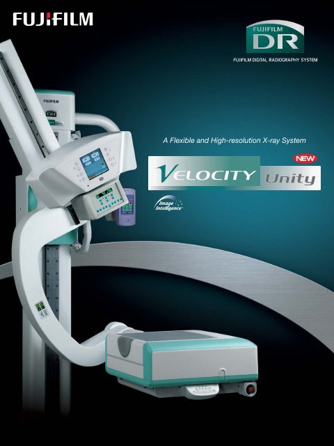
A Flexible and High-resolution X-ray System
A Flexible and High-resolution X-ray System
A Speedy and Easy-to-operate X-ray System<br />
Performing a Wide Range of Exams.<br />
The <strong>Fujifilm</strong> DR <strong>VELOCITY</strong> <strong>Unity</strong> enables radiographic exams in various positions including<br />
supine and upright. The X-ray tube and the detector both on the U-arm can be perfectly aligned,<br />
and the detector can be easily tilted for exams of angled parts such as the knee and the skull.<br />
The unit has an exceptionally high image processing capability, and is fully motorized ensuring<br />
rapid set-up at exam.<br />
HD LineScan Technology<br />
For image acquisition, the <strong>Fujifilm</strong> DR <strong>VELOCITY</strong> <strong>Unity</strong> uses a<br />
revolutionary HD LineScan technology which employs a<br />
wide-view CCD and a built-in Deviced IP. The detector unit is<br />
significantly slimmer than previous models yet has an<br />
increased throughput, reading images at a high resolution<br />
of 100µm.<br />
HIGH IMAGE QUALITY<br />
Deviced IP<br />
Laser Diode<br />
Laser Focusing<br />
Lens<br />
Laser Beam<br />
Light Collecting<br />
Lens<br />
Wide View CCD<br />
UNPRECEDENTED SPEED<br />
Processing up to 240 IP/hour at 10-pixel/mm resolution, the<br />
<strong>Fujifilm</strong> DR <strong>VELOCITY</strong> <strong>Unity</strong> ensures immediate results for the<br />
operator and less waiting for the patient. The productivity gains<br />
seen with this unit are more than just impressive throughput.<br />
Since images are processed using FUJIFILM’s refined image<br />
processing tools, they are in the optimum state for diagnosis<br />
when they are displayed. Radiologists do not waste valuable<br />
time trying to make the image look acceptable before<br />
transmitting them to PACS.<br />
Image Intelligence <br />
FNC<br />
<strong>VELOCITY</strong> <strong>Unity</strong><br />
“Image Intelligence” is a set of<br />
sophisticated digital image processing<br />
software technologies incorporated in the<br />
<strong>Fujifilm</strong> DR <strong>VELOCITY</strong> <strong>Unity</strong>. An optimum<br />
image for examination is generated with<br />
this Image Intelligence technology.<br />
Software Technology Examples<br />
Flexible Noise Control<br />
FNC selectively suppresses noise components while<br />
maintaining signal contrast, improving granularity in<br />
“noisy” anatomical regions.<br />
MFP Multiple Frequency Processing<br />
MFP is optional software that provides greater<br />
diagnostic information from a single exposure image<br />
through frequency enhancement. MFP improves<br />
visibility of both dense and peripheral tissues,<br />
simultaneously applying edge-enhancement processing<br />
to all structures in an image.<br />
Image is on display in<br />
approximately 9 9 seconds<br />
Image Data<br />
CR Console<br />
• A 100µm image resolution • Large image detection area (up to 17” ✕ 17” or 43cm ✕ 43cm)<br />
• Takes approximately 9 seconds for image displaying • Flexible positioning of patient at X-ray exposure<br />
• Small footprint • User-friendly graphic menus, easy operability • Safety design
The <strong>Fujifilm</strong> DR <strong>VELOCITY</strong> <strong>Unity</strong> can perform versatile movements<br />
enabling a wide range of radiographic exams with just one unit.<br />
-45°<br />
Motorized angulations<br />
of the detector (+45°/-45°)<br />
0°<br />
45°<br />
-30°<br />
Source to Image Distance<br />
(SID) adjustable between<br />
1000 mm (39”) and<br />
1800 mm (71”) with<br />
continuous motorized<br />
movement.<br />
Examination of various body parts<br />
FLEXIBILITY<br />
Motorized angulations<br />
of the tube detection<br />
arm (+120°/-30°)<br />
Vertical movement of<br />
the U-arm between<br />
500mm (20”) and<br />
1530mm (60”)<br />
Skull Exams Chest Views Extremity Elbow<br />
0°<br />
120°<br />
90°<br />
Simple and automatic operation<br />
Easy operation using the following:<br />
• Touch buttons located in the X-ray tube cover,<br />
around the touch screen display control<br />
• Control panel at the detector side<br />
• Generator console located outside the room<br />
• Infrared remote controller<br />
Easy positioning, better productivity<br />
PATIENT SAFETY<br />
The system’s dual-speed<br />
motorized movement when a<br />
button or control is pressed<br />
and the intelligent anti-collision<br />
mechanism which uses two<br />
sensor laser beams ensure<br />
patient safety, and quick and<br />
easy patient positioning.<br />
EASY TO CONTROL<br />
Generator console<br />
IR Remote controller<br />
(option)<br />
The following can be set automatically from the touch screen panel by<br />
selecting a specific position number from the pre-set position list displayed<br />
on the screen: SID, arm angle, detector angle, and height.<br />
Also the exposure condition (kV, mA, ms) can be set automatically by<br />
selecting a body part from the body part list displayed on the CR Console.*<br />
These auto-setting functions greatly alleviate the workload of radiologists.<br />
USER-FRIENDLY INTERFACE<br />
A convenient verification screen<br />
clearly indicates the patient<br />
name for quick and easy<br />
confirmation, minimizing patientdata<br />
errors. A suitable grid size<br />
can be selected from this screen<br />
to match the type of exam.<br />
*The CR Console is the central controlling device of the CR system.<br />
CR Console
FUJIFILM DR <strong>VELOCITY</strong> <strong>Unity</strong> SPECIFICATIONS<br />
Image Detector (Model: CR-IR 371 RU)<br />
• Image size 43 ✕ 43 cm (17 ✕ 17 inch)<br />
• Pixels 4280 ✕ 4280<br />
• Pixel size 100 microns<br />
• Spatial frequency (Nyquist) 5.0 cy/mm<br />
• Bit Depth 12 bit<br />
• Preview image Approx. 9 seconds<br />
Universal Arm Stand (Model: VERSO F)<br />
• Vertical height max. 1530 mm or more<br />
min. 500 mm<br />
• Rotation of arm +120/-30 degree<br />
• Tilting of detector +/-45 degree*<br />
• SID 1000 – 1800 mm<br />
*The absolute angle of detector is limited from 0 to +110 degree by software.<br />
Generator (Model: SHF515 / SHF535 / SHF635 / SHF835)<br />
• Output 50kW (SHF515, SHF535)/64kW (SHF635)/80kW (SHF835)<br />
(SHF835 available in some countries)<br />
• Range of output 40 to 150kVp in 1kVp steps.<br />
• Maximum current 640mA (SHF515, SHF535,SHF635)/800mA (SHF835)<br />
• Console (Model: TPC 12”) 12 inch Monitor and Touch screen display<br />
X-Ray Tube (Model: E7252X / E7869X)<br />
• Range of voltage 40 – 150kV<br />
• Focal spot 1.2 mm (Large focus)/0.6 mm (Small focus)<br />
• Anode heat storage capacity 300 kHU (E7252X)/600 kHU (E7869X)<br />
Collimator (Model: 150PBL Collimator)<br />
• Automatic collimator<br />
Image & Information Processor (Model: CR-IR 348CL): Option<br />
• Spatial frequency processing<br />
• Gradation processing<br />
• Dynamic Range compression processing<br />
• Multi-frequency processing (option)<br />
• Flexible noise control processing (option)<br />
• Grid pattern removal processing (option)<br />
Specifications are subject to change without notice.<br />
All brand names or trademarks are the property of their respective owners.<br />
All products require the regulatory approval of the importing country.<br />
For details on their availability, contact our local representative.<br />
FUJIFILM DR <strong>VELOCITY</strong> <strong>Unity</strong> (Model: CR-IR371)<br />
Dimensions<br />
Unit mm (in.):<br />
310±25 (12±1)<br />
Dimensions* (W ✕ D ✕ H)<br />
• Universal arm stand** 2320 ✕ 1465 ✕ 2650 mm (91” ✕ 58” ✕ 104”)<br />
• Arm controller 592 ✕ 422 ✕ 600 mm (23” ✕ 17” ✕ 24”)<br />
• Generator 592 ✕ 422 ✕ 690 mm (23” ✕ 17” ✕ 27”)<br />
*This is approx. value. ** Detector is included<br />
Weight*<br />
• Universal arm stand** 500 kg (1102 lbs.)<br />
• Arm controller 65 kg (143 lbs.)<br />
• Generator 110 kg (243 lbs.)<br />
*This is approx. value. ** Detector is included<br />
Power Supply Requirement<br />
• Detector 200 – 240V±10%, 2.1 2.5A, 50/60Hz (Single phase)<br />
• Universal arm stand 230/240V±10%, 50/60Hz (Single phase)<br />
• Generator 230/240V, 50/60Hz (Single phase) for output 50kW<br />
400V, 50/60Hz (3-phase) for output 50kW<br />
400V, 50/60Hz (3-phase) for output 64kW<br />
480V, 50/60Hz (3-phase) for output 80kW<br />
Environment Condition<br />
• Temperature 15 – 30 degree<br />
• Humidity 40 – 75%Rh<br />
• Atmospheric pressure 750 – 1060 hPa<br />
Optional Accessories<br />
• Mobile table Carbon fiber table (Patient Capacity: 200 kg)<br />
• Grid 10:1 or 8:1, 36 lines/cm, FID 100/140/180 cm<br />
• Ion chamber (Model: ICX-127) 3 field<br />
• Infrared remote controller<br />
• Ceiling support<br />
• Cassette holder<br />
• Long view cassette holder<br />
• QC phantom holder<br />
700(28)<br />
2650 (104)<br />
1655 (65)<br />
310 (12)<br />
50 (2)<br />
45°<br />
45°<br />
1000~1800 (39~71)<br />
1520~2320 (60~91)<br />
Ref. No. XB-861ER (SK·08·07·F1120·F9711) Printed in Japan ©2008 FUJIFILM Corporation<br />
30°<br />
90°<br />
1290 (51)<br />
500 (20)<br />
30°<br />
1530 (60)


