Bardet-Biedl Syndrome Associated With Brachial Amyotrophy
Bardet-Biedl Syndrome Associated With Brachial Amyotrophy
Bardet-Biedl Syndrome Associated With Brachial Amyotrophy

You also want an ePaper? Increase the reach of your titles
YUMPU automatically turns print PDFs into web optimized ePapers that Google loves.
346<br />
Yıldırım et al.<br />
<strong>Bardet</strong>-<strong>Biedl</strong> <strong>Syndrome</strong> <strong>With</strong> Cerebral-Cerebellar Atrophy<br />
accompanied by shoulder, elbow, and wrist pain. The patient’s<br />
history revealed an extra digit on his right foot, poor vision, and<br />
mental retardation since birth. He was the ninth child of seconddegree<br />
consanguineous parents. The family history showed that his<br />
mother had two stillbirths and that a brother died from leukemia at<br />
the age of 44 years. His living 45-year-old brother had obesity,<br />
retinitis pigmentosa, polydactyly, and infertility problems.<br />
Additionally, the patient had four healthy sisters and two healthy<br />
brothers. Because of socioeconomic problems, we could not<br />
examine his brother with the history of obesity, retinitis pigmentosa,<br />
polydactyly, and infertility problems.<br />
On physical examination of the patient, obesity, strabismus, poor<br />
vision, micrognathia, facial dysmorphism, dental anomalies (high<br />
arched palate, hypodontia, small teeth), and polydactyly were<br />
observed. He was not able to fixate on or visually follow objects, and<br />
there was only a blink response to direct light during his ocular<br />
examination. Moderate mental retardation was noted.<br />
Neuromuscular examination revealed motor deficits. Shoulder<br />
and elbow 4/5, wrist 3/5, flexion of fingers 3/5, extension of fingers<br />
2/5, and abduction and adduction of fingers were 1/5 in his left arm.<br />
No motor deficit was detected in the other three extremities. Tendon<br />
reflexes of the upper extremities were brisker on the left side than on<br />
the right. In the lower extremities, bilateral patella reflexes were<br />
absent and Achilles reflexes were normal. Plantar response was<br />
extensor in the left lower limb. Severe sensory impairment (touch and<br />
pinprick) of the left upper extremity, such as hypoesthesia in C6 and<br />
T1, anesthesia in C7, and C8 dermatomes, was observed. In the<br />
other three extremities, sensory examination was normal. In all<br />
extremities proprioceptive sensory joint movement and position was<br />
unaffected. Atrophy of the thenar and hypothenar eminence muscles<br />
was noted. At the cerebellar system examination, horizontal<br />
nystagmus was positive. There was no asymmetry in pupillary size,<br />
but both pupils were unresponsive to light. Moreover, bilateral loss of<br />
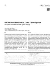
Figure 1. Sagittal T1-weighted image demonstrating atrophy in the<br />
cerebellar vermis.<br />
Turk J Phys Med Re hab 2011;57 Suppl 2; 345-7<br />
Türk Fiz T›p Re hab Derg 2011;57 Özel Sayı 2; 345-7<br />
vision and absence of palatal reflexes were observed on cranial nerve<br />
examination.<br />
There was no pathological finding of the genitourinary system,<br />
except for hypospadias.<br />
Laboratory analysis included a complete blood cell count, and<br />
measurement of electrolytes, serum glucose, and lipid levels, renal<br />
and liver function tests, and gonadal hormone tests, all of which<br />
were within normal limits, except for high blood triglyceride level.<br />
Upper and lower abdominal ultrasonography (US) showed normal<br />
kidneys and normal findings, except grade II hepatosteatosis.<br />
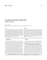
Figure 2. Transverse T2-weighted image showing peripheral sulcal<br />
dilation (according to cortical atrophy in the temporo-frontoparietal<br />
lobes of the right cerebral hemisphere).<br />
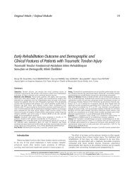
Figure 3. T1-weighted frontal image revealed atrophy in the right<br />
temporal lobe.