Pearls for Cataract Removal - Drs. Fine, Hoffman and Packer
Pearls for Cataract Removal - Drs. Fine, Hoffman and Packer
Pearls for Cataract Removal - Drs. Fine, Hoffman and Packer

You also want an ePaper? Increase the reach of your titles
YUMPU automatically turns print PDFs into web optimized ePapers that Google loves.
INTRODUCTION<br />
No two cataract surgeries are alike. Although some cases<br />
are uncomplicated <strong>and</strong> progress smoothly from start to finish,<br />
others are plagued with complications that require the<br />
surgeon to make difficult decisions.<br />
The lesson all cataract surgeons have learned at some<br />
point is that there is no st<strong>and</strong>ard technique that can be<br />
used <strong>for</strong> all procedures. It is best to adjust your technique<br />
depending on the case <strong>and</strong> the cataract type. In<br />
this article, key opinion leaders in cataract surgery offer<br />
their pearls <strong>for</strong> cataract removal in soft, medium, <strong>and</strong><br />
hard cataract cases. As most imply, you must be able to<br />
change your strategy at any time to achieve the optimal<br />
outcome in any given case.<br />
DAVID ALLEN, FRCS, FRCOPHTH<br />
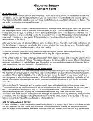
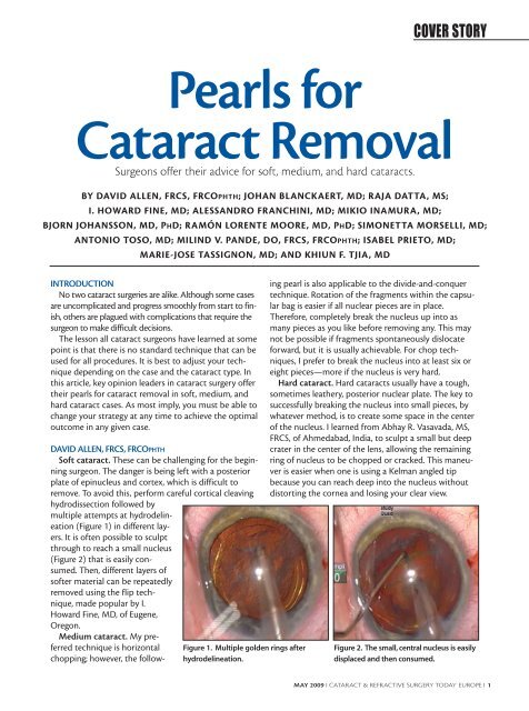
Soft cataract. These can be challenging <strong>for</strong> the beginning<br />
surgeon. The danger is being left with a posterior<br />
plate of epinucleus <strong>and</strong> cortex, which is difficult to<br />
remove. To avoid this, per<strong>for</strong>m careful cortical cleaving<br />
hydrodissection followed by<br />
multiple attempts at hydrodelineation<br />
(Figure 1) in different layers.<br />
It is often possible to sculpt<br />
through to reach a small nucleus<br />
(Figure 2) that is easily consumed.<br />
Then, different layers of<br />
softer material can be repeatedly<br />
removed using the flip technique,<br />
made popular by I.<br />
Howard <strong>Fine</strong>, MD, of Eugene,<br />
Oregon.<br />
Medium cataract. My preferred<br />
technique is horizontal<br />
chopping; however, the follow-<br />
COVER STORY<br />
<strong>Pearls</strong> <strong>for</strong><br />
<strong>Cataract</strong> <strong>Removal</strong><br />
Surgeons offer their advice <strong>for</strong> soft, medium, <strong>and</strong> hard cataracts.<br />
BY DAVID ALLEN, FRCS, FRCOPHTH; JOHAN BLANCKAERT, MD; RAJA DATTA, MS;<br />
I. HOWARD FINE, MD; ALESSANDRO FRANCHINI, MD; MIKIO INAMURA, MD;<br />
BJORN JOHANSSON, MD, PHD; RAMÓN LORENTE MOORE, MD, PHD; SIMONETTA MORSELLI, MD;<br />
ANTONIO TOSO, MD; MILIND V. PANDE, DO, FRCS, FRCOPHTH; ISABEL PRIETO, MD;<br />
MARIE-JOSE TASSIGNON, MD; AND KHIUN F. TJIA, MD<br />
Figure 1. Multiple golden rings after<br />
hydrodelineation.<br />
ing pearl is also applicable to the divide-<strong>and</strong>-conquer<br />
technique. Rotation of the fragments within the capsular<br />
bag is easier if all nuclear pieces are in place.<br />
There<strong>for</strong>e, completely break the nucleus up into as<br />
many pieces as you like be<strong>for</strong>e removing any. This may<br />
not be possible if fragments spontaneously dislocate<br />
<strong>for</strong>ward, but it is usually achievable. For chop techniques,<br />
I prefer to break the nucleus into at least six or<br />
eight pieces—more if the nucleus is very hard.<br />
Hard cataract. Hard cataracts usually have a tough,<br />
sometimes leathery, posterior nuclear plate. The key to<br />
successfully breaking the nucleus into small pieces, by<br />
whatever method, is to create some space in the center<br />
of the nucleus. I learned from Abhay R. Vasavada, MS,<br />
FRCS, of Ahmedabad, India, to sculpt a small but deep<br />
crater in the center of the lens, allowing the remaining<br />
ring of nucleus to be chopped or cracked. This maneuver<br />
is easier when one is using a Kelman angled tip<br />
because you can reach deep into the nucleus without<br />
distorting the cornea <strong>and</strong> losing your clear view.<br />
Figure 2. The small, central nucleus is easily<br />
displaced <strong>and</strong> then consumed.<br />
MAY 2009 I CATARACT & REFRACTIVE SURGERY TODAY EUROPE I 1
COVER STORY<br />
JOHAN BLANCKAERT, MD<br />
Soft cataract. In most soft cataract cases, the patient is<br />
young <strong>and</strong> would like to continue leading an active lifestyle. A<br />
premium IOL solution or a multifocal IOL is the best option<br />
<strong>for</strong> these active patients. Its obvious that in any cataract case<br />
you always need to do whatever you can to avoid posterior<br />
capsule rupture <strong>and</strong> zonulolysis. These two complications can<br />
compromise IOL centration, an essential component when<br />
implanting premium IOLs. For this reason, in those softer<br />
cataracts I frequently opt to do either a phaco roll or nucleus<br />
rotation technique, which has been so well described by Jose<br />
L. Guell, MD, of Spain. This technique reduces zonular stress<br />
<strong>and</strong> is safe <strong>for</strong> the posterior capsule. The most important<br />
pearl is achieving a good hydrodissection.<br />
Medium cataract. Patients with medium cataracts are<br />
typically st<strong>and</strong>ard cataract patients. My first pearl would be:<br />
Do not fall into a routine. Secondly, a good anterior capsulorrhexis<br />
must be centered on the first Purkinje reflex of the<br />
cornea. A 5.5-mm capsulorrhexis is the best choice in these<br />
patients because it is large enough to avoid anterior capsule<br />
contraction <strong>and</strong> small enough to provide an overlap of the<br />
IOL optic. If posterior capsule rupture <strong>for</strong>ces you to use a<br />
sulcus-fixated IOL, then optic capture can still be done with<br />
excellent IOL centration.<br />
Hard cataract. Avoiding corneal burn is the most important<br />
point in patients with hard cataracts. I would choose<br />
either torsional phaco with the Infiniti plat<strong>for</strong>m (Alcon<br />
Laboratories, Inc., Fort Worth, Texas) or the Whitestar<br />
Signature Ice plat<strong>for</strong>m (Abbott Medical Optics, Inc., Santa<br />
Ana, Cali<strong>for</strong>nia). Both systems are notorious <strong>for</strong> the cool<br />
phaco needle temperature needed in those hard, long<br />
cataract emulsifications.<br />
RAJA DATTA, MS<br />
Soft cataract. Cracking the nucleus <strong>and</strong> completely<br />
separating the quadrants is difficult in very soft<br />
cataracts. The procedure is not made easier by soft lens<br />
material <strong>and</strong> cortical matter that continuously enter the<br />
anterior chamber <strong>and</strong> decrease visibility. To combat<br />
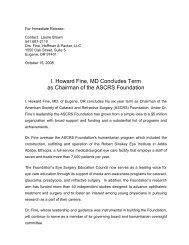
these difficulties, the central two-thirds of the soft<br />
nucleus should be hydrodelineated after initial<br />
hydrodissection. Following separation of the central<br />
nucleus from the thick surrounding epinucleus (Figure<br />
3A), it is slowly brought out into the anterior chamber<br />
(Figures 3B <strong>and</strong> C). Depending on its hardness, either<br />
this soft central nucleus can be aspirated with the<br />
phaco tip (footswitch in position 2), keeping the vacuum<br />
at no more than 150 mm Hg <strong>and</strong> aspiration flow<br />
rate (AFR) no more than 30 cc/minute, or a little phaco<br />
energy may be used (Figure 3D).<br />
The thick epinucleus can be aspirated with the phaco tip<br />
using a maximum vacuum of 100 mm Hg <strong>and</strong> AFR of 25<br />
2 I CATARACT & REFRACTIVE SURGERY TODAY EUROPE I MAY 2009<br />
A B<br />
C D<br />
Figure 3. (A) Hydrodelineation of the central nucleus. (B) The<br />
nucleus core is brought into the anterior chamber. (C) The<br />
nucleus core in the anterior chamber. (D) The nucleus is<br />
phacoemulsified in the anterior chamber.<br />
A B<br />
C D<br />
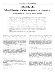
Figure 4. (A) Deep Sculpting of a hard cataract. (B) The two<br />
instruments used <strong>for</strong> cracking are kept together in the deepest<br />
part of the groove. (C) Achieving complete division of the<br />
nucleur fragments. (D) Separating the intranuclear fibers.<br />
cc/minute. Alternatively, you may use a coaxial I/A tip. This<br />
method is easier <strong>and</strong> takes little time, especially in young<br />
patients (up to 50 years of age) or those with posterior subcapsular,<br />
central, or very early nuclear cataracts.<br />
Medium cataract. These cataracts are the easiest to tackle.<br />
During sculpting of these nuclei, it is not necessary to go<br />
as deep as you do with very hard ones. Sculpting up to twothirds<br />
of the total depth of the nucleus will crack it completely.<br />
Going deeper may often lead to passage into the vitreous<br />
cavity because the resistance of the central nucleus is<br />
much less than in harder cataracts.<br />
While one is removing the last piece of nucleus, the vacu-
um should be between 100 <strong>and</strong> 150 mm Hg <strong>and</strong> AFR<br />
between 25 <strong>and</strong> 30 cc/minute. The reason <strong>for</strong> these settings<br />
is not because of the jumping fragments that may hit <strong>and</strong><br />
rupture the posterior capsule, but in case of accidental occlusion<br />
of the phaco tip with posterior cortical matter <strong>and</strong> the<br />
posterior capsule, which can cause a rent. With these parameters<br />
set low in the event of accidental occlusion, you can<br />
immediately switch to footswitch position 1, which should<br />
release the posterior capsule without causing damage.<br />
Hard cataract. Cracking the nucleus in the two extremes<br />
of nuclear hardness (ie, very soft <strong>and</strong> very hard) is a difficult<br />
task. Hard to very hard cataracts, especially those that are<br />
amber or black in color, are the most difficult to crack. In<br />
these cases, it is advisable to adopt the four-quadrant or<br />
stop-<strong>and</strong>-chop method.<br />
Sculpting should be done deeply, until the posterior<br />
nuclear plate is reached (Figure 4A). The two instruments<br />
used to crack the nucleus are placed so that the tips touch<br />
each other as well as the floor of the groove (Figure 4B). The<br />
tips are then slowly separated to create a crack in the nucleus<br />
that should start from the periphery, under full visibility,<br />
<strong>and</strong> gradually move toward the center. Care must be taken<br />
to ensure complete separation of the nuclear fragments,<br />
including the posterior nuclear plate (Figure 4C).<br />
Difficulty arises in these cases due to the resilient intranuclear<br />
fibers, which are tenacious <strong>and</strong> difficult to separate. It is<br />
always advisable to break these fibers by separating the fragments<br />
at their site of origin (Figure 4D).<br />
Utmost care must be taken while separating the<br />
nuclear fragments to achieve a complete crack, which<br />
should extend throughout the depth of the nucleus.<br />
Since these nuclei are not only hard <strong>and</strong> leathery but<br />
also extra large in size, they occupy almost the whole of<br />
the capsular bag, which is distended, with little or no<br />
cortical matter separating them.<br />
Care must also be taken while creating the crack. Pulling<br />
the pieces away from each other to crack the nucleus transmits<br />
pressure directly to the capsular bag. The more distance<br />
between pieces, especially at the periphery, the more<br />
the stretch <strong>for</strong>ce on the already distended capsular bag,<br />
which may lead to a capsular tear. There<strong>for</strong>e, it is imperative<br />
to start cracking at the periphery <strong>and</strong> go slowly down to the<br />
center of the nucleus.<br />
I. HOWARD FINE, MD<br />
Soft cataract. For soft nuclei, I hydroexpress the lens<br />
into the capsulorrhexis plane <strong>and</strong> carousel it with the<br />
bevel of the phaco tip pointed toward the lens equator.<br />
I place the second instrument above the nucleus, protecting<br />
the cornea.<br />
Medium cataract. For these cataracts, I prefer horizontal<br />
chopping. Prior to embedding the tip, I go over<br />
COVER STORY<br />
the equator with the horizontal chopper, pulling up <strong>and</strong><br />
toward the incision to stabilize the nucleus as I bury the<br />
phaco tip. This ensures that I am not transmitting any<br />
<strong>for</strong>ce to the capsule or zonules.<br />
Hard cataract. For hard cataracts, I use vertical chopping<br />
<strong>and</strong> per<strong>for</strong>m all chopping in the traditional, longitudinal<br />
phaco mode prior to mobilizing the segments. There<strong>for</strong>e,<br />
the tip is not widened as it is buried. Then I change parameters<br />
<strong>for</strong> segment mobilization to include torsional or elliptical<br />
phaco.<br />
ALESSANDRO FRANCHINI, MD<br />
Soft cataract. Removing very soft nuclei <strong>and</strong> cracking the<br />
lens into small fragments can be harder than it would seem<br />
during either divide-<strong>and</strong>-conquer or chop techniques.<br />
Especially <strong>for</strong> surgeons in training, it can be difficult to separate<br />
the two epinuclei or the four nuclear quadrants after<br />
sculpting. The nuclear fragment substance can be gummy,<br />
<strong>and</strong> manipulators or choppers can easily penetrate the substance<br />
without applying the necessary strength to separate<br />
it. In these cases, it may be useful to deepen the groove as<br />
much as possible; however, this maneuver risks a posterior<br />
capsule break, especially in the h<strong>and</strong>s of a young surgeon. If<br />
the division does not appear in the middle of the nucleus,<br />
<strong>and</strong> two different-sized fragments result, one should refracture<br />
the larger piece.<br />
I prefer to use cracking <strong>for</strong>ceps in patients with soft nuclei.<br />
These <strong>for</strong>ceps have two flat paddles that apply lateral separation<br />
onto a larger area of tissue, allowing the perfect division<br />
of two equal fragments—even if the groove has not<br />
been well per<strong>for</strong>med or if the nucleus is particularly soft.<br />
The most recent cracking <strong>for</strong>ceps models can be easily<br />
inserted into the anterior chamber through a 2.2-mm incision.<br />
Furthermore, the lower part of the two paddles has a<br />
fine edge so that if the groove is not sufficiently deep, they<br />
can be used to per<strong>for</strong>m a type of karate chop.<br />
Medium cataract. Separating the lens material from the<br />
capsule (ie, hydrodissection) <strong>and</strong> the nucleus from the epinucleus<br />
(ie, hydrodelineation) represent the most crucial steps<br />
in patients with a medium-hard cataract. These cataracts are<br />
considered the easiest to remove; however, the surgeon<br />
should avoid beginning phacoemulsification be<strong>for</strong>e the<br />
nucleus is mobilized with these two maneuvers <strong>and</strong> then<br />
rotated. When a good separation between the lens layers is<br />
obtained, difficulties aspirating the cataract are avoided.<br />
Avoid initiating hydrodissection if the anterior chamber is<br />
over-inflated with an ophthalmic viscosurgical device<br />
(OVD). When per<strong>for</strong>ming microincision cataract surgery<br />
(MICS), be aware of the risk of increased pressure in the<br />
anterior chamber.<br />
To per<strong>for</strong>m hydrodissection, a cannula is inserted into the<br />
anterior chamber, under the capsulorrhexis border <strong>and</strong><br />
MAY 2009 I CATARACT & REFRACTIVE SURGERY TODAY EUROPE I 3
COVER STORY<br />
between the capsule <strong>and</strong> lens cortex. This is done in an<br />
attempt to slightly raise the capsule <strong>and</strong> direct the fluid<br />
stream along the inner surface of the capsular bag. Balanced<br />
saline solution is injected to separate the lens from the capsule.<br />
It is necessary to continue injecting fluid until the wave<br />
completely crosses behind the nucleus. The center of the<br />
nucleus is pushed gently backward to allow the fluid to pass<br />
through the capsular equator <strong>and</strong> obtain complete separation<br />
from the capsule. This maneuver is repeated as many<br />
times as necessary to facilitate good lens rotation.<br />
Using a straight cannula, the surgeon then injects balanced<br />
saline solution into only the contraincisional portion<br />
of the bag. It is preferable to use a 90º angled cannula to<br />
inject fluid in the right <strong>and</strong> left portions of the bag, guaranteeing<br />
complete hydrodissection.<br />
To facilitate aspiration of a medium-hard cataract, the<br />
nucleus is separated from the epinucleus with hydrodelineation.<br />
The cannula is inserted deeply inside the periphery<br />
of the nucleus, <strong>and</strong> balanced saline solution is injected until<br />
a golden ring appears between the nucleus <strong>and</strong> epinucleus.<br />
After hydrodissection <strong>and</strong> hydrodelineation have been<br />
well per<strong>for</strong>med, the surgeon can easily aspirate the nucleus,<br />
epinucleus, <strong>and</strong> cortex without moving the phaco tip from<br />
the pupillary field. This technique decreases surgical time<br />
<strong>and</strong> increases efficiency <strong>and</strong>, above all, safety.<br />
Hard cataract. Often with the brunescent cataract, we<br />
are dealing with a nucleus of enlarged dimension <strong>and</strong><br />
increased density. There is no epinucleus <strong>and</strong> often no cortical<br />
layer. Hard cataracts are somewhat like a pillow in that<br />
they absorb the transmission of <strong>for</strong>ce applied to the posterior<br />
capsule <strong>and</strong> zonula. There<strong>for</strong>e, mechanical <strong>for</strong>ces applied<br />
during sculpting, cracking, <strong>and</strong> rotation are transmitted<br />
straight to the posterior capsule <strong>and</strong> the zonula, which may<br />
already be weak in these patients. The nuclear fragments are<br />
often hard <strong>and</strong> sharp, increasing the risk <strong>for</strong> a break in the<br />
posterior capsule.<br />
The enlarged depth <strong>and</strong> width of the cataract can create a<br />
number of problems, whether we per<strong>for</strong>m a divide-<strong>and</strong>-conquer<br />
or horizontal chop technique. Breaking the most posterior<br />
layers of the nuclear plate requires digging deep <strong>and</strong> close<br />
to the posterior capsule. If one per<strong>for</strong>ms horizontal chop,<br />
inserting the chopper as deeply as possible can be dangerous<br />
because of the absence of an epinucleus. However, it is often<br />
difficult to obtain a clean fracture with horizontal chop<br />
because tissue bridges connecting the two fragments can<br />
remain intact. A vertical chop technique is generally more<br />
successful in breaking these bridges, so this may be the best<br />
technique <strong>for</strong> patients with brunescent cataract.<br />
With both the divide-<strong>and</strong>-conquer <strong>and</strong> horizontal chopping<br />
techniques, we cut only part of the nucleus to avoid<br />
breaking the posterior capsule. To complete the fracture, we<br />
separate the two heminuclei; however, the <strong>for</strong>ce of either<br />
4 I CATARACT & REFRACTIVE SURGERY TODAY EUROPE I MAY 2009<br />
technique only partially spreads the nuclei posteriorly. In the<br />
case of very hard nuclei, the propagating fracture may continue<br />
only horizontally without advancing further posteriorly.<br />
This is the reason the tissues bridges remain together.<br />
Alternatively, the backward-<strong>and</strong>-<strong>for</strong>ward vertical movement<br />
applied during vertical chop favors propagation of the<br />
fracture to the most posterior layers. In patients with a<br />
white cataract with a very hard nucleus, vertical chop is the<br />
best choice in terms of efficiency <strong>and</strong> safety, making it possible<br />
to work only in the pupillary field without concern <strong>for</strong><br />
where the posterior capsule is located.<br />
MIKIO INAMURA, MD<br />
Soft cataract. The Infiniti OZil Torsional phaco on the<br />
Infiniti Vision System (Alcon Laboratories, Inc., Fort Worth,<br />
Texas) is the instrument of choice <strong>for</strong> all my cataract procedures.<br />
Soft cataract cases can be completed without any<br />
problems, regardless of how I approach the nucleus. In cases<br />
in which the nucleus is extremely soft, I use the Akahoshi<br />
Prechopper (ASICO, Westmont, Illinois) or the Inamura<br />
Eagle Prechopper (ASICO) to divide the nucleus in half<br />
(Figures 5A <strong>and</strong> B), or into quadrants if that is not too difficult,<br />
be<strong>for</strong>e emulsifying the nucleus with low power torsional<br />
phaco. To treat soft cataracts, a high power setting with<br />
torsional phaco is unnecessary because too much power<br />
would result in dislodgment of the nucleus fragments<br />
(Figure 5C).<br />
The settings I use are pulse mode, 30 pulses/second, torsional<br />
amplitude 80% (linear), on time 80%, aspiration flow<br />
rate 25 mL/minute (linear), <strong>and</strong> maximum vacuum 400 mm<br />
Hg (linear). I recommend using the Kelman Mini flared tip<br />
(Alcon Laboratories, Inc.) because it allows superior followability<br />
together with the ability to maintain anterior chamber<br />
stability with ease.<br />
Medium cataract. Torsional phaco also demonstrates<br />
efficiency when dealing with medium density cataracts. In<br />
medium cataract procedures, I like to use the Inamura Eagle<br />
Prechopper to divide the nucleus. This prechopper can easily<br />
divide most nuclei (from very soft to hard); rotating the<br />
nucleus inside the capsule is easy with this device.<br />
It is important that hydrodissection be per<strong>for</strong>med completely,<br />
making the nucleus easy to rotate. The double nozzle<br />
Inamura Hydro Cannula (Duckworth & Kent<br />
[Hert<strong>for</strong>dshire, Engl<strong>and</strong>] <strong>and</strong> ASICO) makes accomplishing<br />
hydrodissection easy.<br />
After hydrodissection, I reinject the OVD, preferably<br />
Healon5 (Abbott Medical Optics, Inc., Santa Ana,<br />
Cali<strong>for</strong>nia), into the anterior chamber. Then, using the<br />
Inamura Eagle Prechopper, I divide the nucleus in half. The<br />
Eagle Prechopper has a sharp tip, somewhat like an eagle’s<br />
beak, which I insert into the middle of the nucleus to chop<br />
it in half. Its sharp tip will penetrate <strong>and</strong> chop even highly
A B C<br />
dense nuclei. I then use the tip to snag the divided halves of<br />
the nucleus, rotate them 90° inside the capsule, <strong>and</strong> divide<br />
them further into quadrants (Figures 6A <strong>and</strong> B). The procedures<br />
after this are more efficient if the nuclear fragments<br />
are divided even more.<br />
For torsional phaco in these cases, I use the 45° beveled<br />
Kelman Mini Flared tip, <strong>and</strong> more recently the 12° Partial<br />
Kelman Mini Flared tip (OZil 12). Alternating traditional<br />
COVER STORY<br />
Figure 5. (A) Dividing the nucleus into two pieces with the Inamura Eagle Prechopper. (B) Two nucleus halves after division. (C)<br />
Aspirating soft nucleus without longitudinal phaco.<br />
A B C<br />
Figure 6. (A) Dividing the nucleus into four pieces with the Inamura Eagle Prechopper. (B) Four nucleus quadrants after division.<br />
(C) Four quadrants are emulsified in torsional pulse mode.<br />
A B<br />
C D<br />
Figure 7. (A) Semicircular crater is dug with phaco <strong>and</strong> torsional<br />
ultrasound. (B) Chopping with phaco tip <strong>and</strong> a second<br />
instrument. (C) Dividing by chopping into four pieces. (D)<br />
Emulsifying four quadrants.<br />
longitudinal phaco <strong>and</strong> torsional phaco pulses makes<br />
removal of the first quadrant of the nucleus efficient. After<br />
the first quadrant is removed, I switch to pulse mode torsional<br />
phaco. Use of the foot pedal makes the transition<br />
smooth. Note that the phaco tip bevel is positioned parallel<br />
to the nucleus fragment, so that the fragments rotate as if<br />
they occlude the phaco tip <strong>and</strong> there is no chattering.<br />
The settings I use <strong>for</strong> removal of the first nucleus quadrant<br />
are 30 pulses/second, longitudinal oscillation 70% (linear),<br />
on time 20%, torsional amplitude 100% (linear), on<br />
time 80%, aspiration flow rate 30 mL/minute (linear), <strong>and</strong><br />
maximum vacuum 300 mm Hg (linear). For removing the<br />
rest of the nucleus quadrants, settings are 40 pulses/second,<br />
torsional amplitude 100%, on time 80%, aspiration flow rate<br />
28 mL/min (linear), <strong>and</strong> maximum vacuum 350 mm Hg<br />
(linear; Figure 6C).<br />
Hard cataract. If the nucleus is so hard that the Inamura<br />
Eagle Prechopper does not penetrate, I immediately switch<br />
to the phaco-chop method. With this method, I can<br />
approach the hardest cataract cases safely, even when the<br />
zonules are in a weakened state. After hydrodissection, I dig<br />
a semicircular crater on the proximal surface of the nucleus<br />
(Figure 7A). I then insert the phaco tip into the opposite<br />
side of the nucleus <strong>and</strong>, using a second instrument to hold<br />
the nucleus from the other side, I divide the nucleus (Figure<br />
7B) very surely. Further, I rotate the nucleus 90°, insert the<br />
phaco tip into the opposite wall <strong>and</strong> similarly divide the<br />
nucleus (Figure 7C).<br />
MAY 2009 I CATARACT & REFRACTIVE SURGERY TODAY EUROPE I 5
COVER STORY<br />
After dividing the nucleus in this way, I set about to<br />
remove the fragments with torsional phaco (Figure 7D).<br />
Aspirating dense nuclear material with torsional phaco<br />
alone can cause clogging, but when torsional is combined<br />
with longitudinal oscillations, clogging can be minimized.<br />
Another way to prevent clogging is by slowly emulsifying<br />
the nuclear fragments piece by piece. It is most important<br />
to avoid completely occluding the phaco tip with nucleus<br />
fragments. This is strikingly different from traditional longitudinal<br />
phaco <strong>and</strong> must be observed.<br />
The settings <strong>for</strong> removal of the first nucleus quadrant are<br />
30 pulses/second, longitudinal oscillation 70% (linear), on<br />
time 20%, torsional amplitude 100%, on time 80%, aspiration<br />
flow rate 30 mL/minute (linear), <strong>and</strong> maximum vacuum 300<br />
mm Hg (linear). For removal of the rest of the nuclear quadrants,<br />
settings are 40 pulses/second, torsional amplitude<br />
100%, on time 80%, aspiration flow rate 28 mL/minute (linear),<br />
<strong>and</strong> maximum vacuum 350 mm Hg (linear).<br />
If clogging occurs, I get rid of it by switching the setting to<br />
longitudinal oscillation, declogging mode at 20 pulses/second,<br />
longitudinal oscillation 100% (linear), on time 20%,<br />
aspiration flow rate 25 mL/min fixed, <strong>and</strong> maximum vacuum<br />
300 mm Hg (linear).<br />
Recently I have been using the Akahoshi Bent wobble<br />
square tip (ASICO), <strong>and</strong> I found it to be efficient <strong>for</strong> removing<br />
the nuclear fragments. It causes less injury to the cornea<br />
<strong>and</strong> iris compared to the Kelman Mini Flared tip, <strong>and</strong> it is<br />
easier to use because of its straight tip. It can be used on<br />
cataracts of all densities. It is also not prone to clogging. This<br />
tip is not yet on the market, but I feel there is great potential<br />
<strong>for</strong> its use.<br />
BJORN JOHANSSON, MD, PHD<br />
Soft cataract. If the soft cataract goes unnoticed preoperatively,<br />
you will recognize it as you initiate hydrodelineation<br />
after cortical cleaving hydrodissection—two crucial steps in<br />
every safe cataract procedure. Your blunt cannula will not<br />
meet resistance, or you will find a very small nucleus when<br />
balanced saline solution is injected into the deeper layers of<br />
the lens.<br />
Attempts to crack or chop soft nuclei are, of course, seldom<br />
fruitful. In these cases, aspiration without ultrasound is<br />
usually enough to evacuate the lens, either by stopping at<br />
position 2 on a monolinear pedal or activating only vacuum/aspiration<br />
on a dual-linear foot control. I consume the<br />
central nucleus first <strong>and</strong> then catch the anterior rim of the<br />
remaining bowl opposite the phaco tunnel, flipping it<br />
upside-down with help from the second instrument. This<br />
makes it easy to aspirate efficiently at a safe distance from<br />
the posterior capsule in the center of the pupil.<br />
Medium cataract. Several years ago, I transitioned from a<br />
stop-<strong>and</strong>-chop technique to a direct horizontal chop with<br />
6 I CATARACT & REFRACTIVE SURGERY TODAY EUROPE I MAY 2009<br />
the Nagahara chopper (Katena Products, Inc., Denville, New<br />
Jersey). Earlier, I was a traditional four-groove divide-<strong>and</strong>conquer<br />
surgeon. Creating four grooves is advantageous <strong>for</strong><br />
the beginning surgeon because it allows you to get a good<br />
grasp of the anatomy of the lens <strong>and</strong> capsule in various scenarios,<br />
including exfoliation <strong>and</strong> shallow or deep chambers. I<br />
found it a helpful precursor to chop techniques.<br />
When I first tried direct horizontal chop, it was a bit difficult.<br />
If you wish to progress to chopping, first master stop<strong>and</strong>-chop:<br />
Create one initial groove; crack the nucleus into<br />
halves, which can usually be mobilized; capture one half of<br />
the nucleus with the phaco tip, with vacuum increased<br />
from the groove setting; <strong>and</strong> chop it with the side instrument.<br />
This technique allows you to try various choppers on<br />
lens pieces that are freed from the capsule, which I think<br />
makes learning easier.<br />
Hard cataract. With hard nuclei, I often have a problem<br />
getting a chop completely down through to the back side of<br />
the nucleus, which can be fibrotic <strong>and</strong> leathery. In these<br />
cases, my tactic is one of the following: Either I use the side<br />
instrument <strong>and</strong> viscoelastic cannula, inserted through the<br />
phaco tunnel, to separate the nuclear pieces (after divide<strong>and</strong>-conquer,<br />
stop-<strong>and</strong>-chop, or direct chop); or, if the<br />
instrument is available, I use an Akahoshi prechopper. Both<br />
methods allow one to easily apply <strong>for</strong>ces at the deepest possible<br />
point in the nucleus <strong>and</strong> to direct the splitting <strong>for</strong>ce in<br />
the most efficacious directions.<br />
RAMÓN LORENTE MOORE, MD, PHD<br />
Soft cataract. Although often considered an easy surgery,<br />
the soft cataract may pose refractive <strong>and</strong> technical challenges.<br />
As a refractive cataract procedure, there is the<br />
dem<strong>and</strong> to achieve accurate biometry <strong>and</strong> uneventful surgery,<br />
leading to extra pressure <strong>for</strong> the surgeon. Technical<br />
problems when h<strong>and</strong>ling the soft nuclei during phacoemulsification<br />
are twofold: First, there is difficulty rotating the<br />
nucleus-cortex complex because cortical fibers tend to stick<br />
to the capsule. Second, there is an inability to divide the<br />
nucleus due to its softness. The following points summarize<br />
my strategy to overcome these problems.<br />
An adequate hydroprocedure is m<strong>and</strong>atory to achieve<br />
complete nuclear rotation. First, cortical cleaving hydrodissection<br />
must be per<strong>for</strong>med to detach the cortex from the<br />
capsule so that the cortex remains attached to the epinucleus.<br />
It may be helpful to repeat the same maneuver in the<br />
opposite distal quadrant. Given that an excessive amount of<br />
OVD can increase the resistance to fluid egress from the<br />
capsular bag via the capsulorrhexis, an important step<br />
be<strong>for</strong>e starting hydrodissection is to evacuate some of OVD<br />
from the anterior chamber. After hydrodissection, the<br />
nucleous-cortex complex is rotated bimanually (using the<br />
two-Sinskey hook method) to confirm that there is no
adherence between the cortex <strong>and</strong> the capsule. The next<br />
step is hydrodelineation, which by cleaving the central<br />
nucleus from the epinucleus, facilitates phacoemulsification.<br />
A golden ring of the delineated nucleus is its hallmark sign.<br />
My preferred method <strong>for</strong> nuclear emulsification of soft<br />
nuclei is the chip-<strong>and</strong>-flip technique using AquaLase technology<br />
(Alcon Laboratories, Inc.). It is not only the safest<br />
technology but it could also be associated with a lower rate<br />
of posterior capsule opacification.<br />
The rest of the procedure is the same that will be<br />
described <strong>for</strong> medium cataracts.<br />
Medium cataract. My surgical technique in medium<br />
cataracts corresponds to the procedure I per<strong>for</strong>m in st<strong>and</strong>ard<br />
cases. Thus, I will describe my personal technique in<br />
this setting. I use topical anesthesia with intracameral preservative-free<br />
lidocaine 2%. The anterior chamber is filled<br />
with a viscoadaptative OVD (DisCoVisc; Alcon Laboratories,<br />
Inc.) through the paracentesis, <strong>and</strong> a temporal clear corneal<br />
incision (2.2 x 1.75 mm) is made. Should a multifocal IOL be<br />
implanted, the incision is placed at the steepest meridian. A<br />
circular, well-centered, 5.25-mm capsulorrhexis is perfomed<br />
with Utrata <strong>for</strong>ceps. The hydroprocedures are per<strong>for</strong>med as<br />
previously described <strong>for</strong> soft cataracts.<br />
During phacoemulsification, I usually work with torsional<br />
ultrasound technology <strong>for</strong> two main reasons: it is easier <strong>and</strong><br />
safer. These advantages are related to its shearing action,<br />
which minimizes repulsion <strong>and</strong> allows use of lower vacuum<br />
parameters to decrease surge. To optimize the torsional<br />
effect, achieve better holdability, <strong>and</strong> reduce clogging, a<br />
Kelman Mini flared 45º bevel tip is strongly recommended.<br />
My preferred technique is vertical chop using a Rosen chopper<br />
to divide the nucleous into four or five fragments with<br />
the following parameters: 90% torsional amplitude with<br />
burst mode (50 milliseconds [mSec] on, 150 mSec off); vacuum,<br />
420 mm Hg; flow, 35 cc/minute; <strong>and</strong> bottle height, 95<br />
cm.<br />
Once the nucleus is divided, parameters are adapted to<br />
emulsify the fragments: continuous torsional ultrasound<br />
(maximum amplitude, 90% <strong>and</strong> starting amplitude, 20%);<br />
vacuum, 320 mm Hg; flow, 25 cc/minute; bottle height, 95<br />
cm. The footpedal set-up is adjusted to 18% to shorten the<br />
range of footpedal position 3. There is no need <strong>for</strong> longitudinal<br />
ultrasound.<br />
During irrigation <strong>and</strong> aspiration, the cortex is aspirated<br />
with a curved silicone I/A tip (Alcon Laboratories, Inc.) due<br />
to its greater levels of safety—the incidence of posterior<br />
capsule rupture is lower than using metal tip. Whenever<br />
subincisional cortex removal becomes difficult, it may be a<br />
good option to use two separate cannulas, one <strong>for</strong> irrigating<br />
the anterior chamber <strong>and</strong> another <strong>for</strong> aspirating cortical<br />
material. This bimanual I/A technique requires two paracentesis<br />
placed approximately 50º apart from the main incision.<br />
COVER STORY<br />
I routinely polish the posterior capsule with the silicone tip<br />
or with an irrigating polisher.<br />
I typically implant the AcrySof IQ (Alcon Laboratories,<br />
Inc.) using the Monarch III injector <strong>and</strong> Monarch D cartridge<br />
(Alcon Laboratories, Inc.). I do not need to enlarge<br />
the incision. Residual OVD trapped under the IOL is then<br />
removed. Cefuroxime is used both <strong>for</strong> intracameral administration<br />
<strong>and</strong> stromal hydration at both edges of the clear<br />
corneal incision.<br />
Hard cataract. H<strong>and</strong>ling the hard nucleus is a major challenge,<br />
even <strong>for</strong> the experienced surgeon. It requires higher<br />
power ultrasound <strong>and</strong> prolonged phaco time. Preoperative<br />
evaluation is of paramount importance. We must consider<br />
the following points: endothelial cell count, anterior chamber<br />
depth, zonular instability, pupil dilation, <strong>and</strong> a B-scan if<br />
there is no view of posterior fundus.<br />
Phacoemulsification in hard cataracts requires the use of<br />
specific strategies that differ from routine phacoemulsification.<br />
The main modifications of each surgical step to successfully<br />
operate such cataracts are as follows. Local anesthesia<br />
is recommended in cases of weak zonulas, poor collaboration,<br />
or risk of intraoperative complications. When<br />
the red reflex is poor, we must stain the capsule with trypan<br />
blue 0.06% to enhance capsular visualization enough to per<strong>for</strong>m<br />
capsulorrhexis <strong>and</strong> also visualization of the edge of the<br />
rhexis during phacoemulsification.<br />
OVD plays an important role in protecting the endothelium<br />
in hard cataracts. For this reason, I use Steven<br />
Arshinoff´s soft shell technique, using Viscoat to coat the<br />
endothelium <strong>and</strong> a highly cohesive OVD (Provisc; Alcon<br />
Laboratories, Inc.) to maintain the anterior chamber.<br />
Phacoemulsification <strong>for</strong> hard lenses poses two main risks:<br />
higher endothelial cell loss <strong>and</strong> increased risk of posterior<br />
capsule rupture. Furthermore, dividing the hard nucleus<br />
becomes difficult. Posterior layer fibers can be cohesive <strong>and</strong><br />
tenacious <strong>and</strong> resist to all conventional methods of division.<br />
Extra precautions must be taken to protect the endothelium<br />
<strong>and</strong> the posterior capsule.<br />
Torsional ultrasound is the best technology to protect<br />
the endothelium due to minimal repulsion <strong>and</strong> less turbulence<br />
in the anterior chamber. My preferred technique is<br />
vertical chop because it causes less endothelial cell loss <strong>and</strong><br />
less stress on the zonules versus divide <strong>and</strong> conquer. I do not<br />
change many parameters with respect to medium cataracts,<br />
apart from elevating the starting torsional amplitude setting<br />
to 30%. However, there are a few variations to my technique<br />
in hard cataracts: (1) I use a karate chopper, which is longer<br />
<strong>and</strong> sharper, to facilitate embedding the dense nucleus without<br />
displacing it. (2) The irrigation sleeve must be retracted<br />
more than usual. This will expose a longer segment of the<br />
metal needle <strong>and</strong> maximize penetration of the tip, which is<br />
crucial to divide the nucleus. It is easier to begin by sculpting<br />
MAY 2009 I CATARACT & REFRACTIVE SURGERY TODAY EUROPE I 7
COVER STORY<br />
a small, deep, pit centrally. This pit allows the nucleus to be<br />
impaled more deeply. (3) It is more efficient to alter the<br />
angle of the vertical chop slightly <strong>and</strong> approach the embedded<br />
phaco tip more diagonally. This provides more of a horizontal<br />
vector that pushes the nucleus against the tip while<br />
the vertical vector initiates the downward fracture, combining<br />
the mechanical advantages of both strategies. (4) If there<br />
are leathery fibers at the posterior layer, it is best to transect<br />
them with the chopper while the nucleus is engaged <strong>and</strong><br />
stabilized by the vacuum of the phaco tip. (5) The nucleus<br />
should be divided in smaller fragments to emulsify them<br />
securely. (6) To maximize endothelial protection, we should<br />
refill the anterior chamber with Viscoat during fragment<br />
emulsification. (7) A dispersive OVD injected behind the last<br />
remaining fragments creates <strong>and</strong> artificial epinucleus that<br />
will restrain the lax <strong>and</strong> fragile posterior capsule from trampolining<br />
toward the phaco tip, minimizing the risk of rupture.<br />
SIMONETTA MORSELLI, MD;<br />
AND ANTONIO TOSO, MD<br />
Soft cataract. We use very high vacuum <strong>and</strong> very low<br />
ultrasound power to remove soft cataracts. We suggest<br />
using a manipulator rather than a chopper, avoiding damage<br />
to the posterior capsule during nuclear fragment<br />
removal with high vacuum.<br />
Medium cataract. We use the Stellaris (Bausch & Lomb,<br />
Rochester, New York), a phacoemulsification plat<strong>for</strong>m created<br />
specifically <strong>for</strong> MICS. This system is exceptionally fast <strong>and</strong><br />
safe. Its capability <strong>for</strong> high vacuum allows quick removal of<br />
nuclear fragments while the chamber remains perfectly stable.<br />
This machine gives one the ability to set the ultrasound<br />
in two different modes, burst <strong>and</strong> micropulse. By changing<br />
the duration <strong>and</strong> the duty cycle of the ultrasound, we can<br />
adapt the power to any type of cataract.<br />
We set sub mode 1 <strong>for</strong> cataracts of 2+ to 3+ hardness;<br />
sub mode 2 <strong>for</strong> 3+ to 4+ cataracts; <strong>and</strong> sub mode 3 <strong>for</strong> 5+<br />
to 6+ cataracts.<br />
Our parameters <strong>for</strong> sub mode 1 <strong>for</strong> normal or medium<br />
2+ to 3+ cataracts are: dual foot pedal system; 10% linear<br />
ultrasound, pulse mode 80 pps; 35% duty cycle.<br />
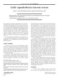
Hard cataract. Even with very hard cataracts, we are able<br />
to remove pieces with not more than 10% ultrasound<br />
power.<br />
Our parameters <strong>for</strong> sub mode 2, used <strong>for</strong> 3+ to 4+<br />
cataracts, are: dual foot pedal system; 10% linear ultrasound;<br />
fixed burst 160 millisecond duration; 320 millisecond interval<br />
of pulse duration.<br />
Our parameters <strong>for</strong> sub mode 3, used <strong>for</strong> 5+ to 6+<br />
cataracts, are: dual foot pedal system; 10% fixed ultrasound;<br />
multiple burst 40 millisecond burst duration; 60% duty<br />
cycle.<br />
8 I CATARACT & REFRACTIVE SURGERY TODAY EUROPE I MAY 2009<br />
Figure 8. MICS with very hard cataract.<br />
With these multiple modes set, we are able to change to<br />
any of these parameters during surgery. For example, to<br />
chop the nucleus at the beginning of surgery, we might use<br />
sub mode 3. To remove the final little pieces floating into<br />
the anterior chamber toward the end of the case, we might<br />
use sub mode 1.<br />
Our machine is set so that we can change sub modes<br />
during surgery using the left foot pedal on the dual foot<br />
pedal system. With the right foot pedal, we control ultrasound<br />
power with a lateral motion <strong>and</strong> vacuum by depressing<br />
the pedal. The surgeon can change the sub mode at any<br />
moment during surgery.<br />
With this system we can remove very hard cataract using<br />
MICS (Figure 8) with low trauma to the intraocular structures.<br />
The most important thing is to know the capabilities<br />
of the machine to obtain high efficacy with minimal trauma<br />
<strong>for</strong> the patient.<br />
Another pearl to manage hard cataracts is to use a<br />
chop technique with a new OVD, Ovidio (Sifi, SpA, Aci<br />
S. Antonio, Italy). This new viscoelastic substance contains<br />
sodium hyaluronate 2% with a molecular weight<br />
of 2.3 million daltons, giving it dispersive properties<br />
with a medium viscosity. It provides good anterior<br />
chamber stability <strong>and</strong> reliable adhesion to the corneal<br />
endothelium, protecting the intraocular structures. This<br />
OVD gives the surgeon the option to per<strong>for</strong>m a large<br />
<strong>and</strong> well-controlled capsulorrhexis. Unlike other dispersive<br />
OVDs, this one does not allow adhesion of cataract<br />
fragments to the posterior surface of the cornea. For<br />
these reasons, this OVD provides better visualization <strong>for</strong><br />
the surgeon during phacoemulsification.<br />
To remove a hard cataract we per<strong>for</strong>m a large capsulorrhexis<br />
(6–6.5 mm), allowing us to move the nucleus out of<br />
the capsular bag <strong>and</strong> into the anterior chamber <strong>for</strong><br />
hydrodissection. This allows us to safely fracture the nucleus<br />
with a chop technique. The chopper is positioned at the<br />
equator of the nucleus without the risk of damaging the<br />
capsular bag, even in case of pseudoexfoliation or zonular
weakness. A small amount of OVD is injected under the<br />
nucleus <strong>and</strong> over the capsular bag. The phaco tip, with<br />
bevel down, impales the nucleus as deeply into the center as<br />
possible. The sleeve must be retracted to obtain as much as<br />
1.8 to 2 mm of tip length, thus penetrating the center of the<br />
nucleus. A deep nuclear fracture is then obtained with the<br />
chopper. After the first crack, the nucleus is divided into<br />
small slices <strong>and</strong> emulsified; using the manipulator avoids<br />
damage to the capsular bag. During these maneuvers, it is<br />
necessary to refill the anterior chamber with OVD to protect<br />
the endothelium <strong>and</strong> maintain stabilization of the<br />
nucleus.<br />
MILIND V. PANDE, DO, FRCS, FRCOPHTH<br />
Soft cataract. Adjust your flow <strong>and</strong> vacuum settings<br />
down when you want move the nucleus in one piece. If the<br />
vacuum or flow is too high, it will nibble away at the nucleus<br />
rather than enable you to move the whole nucleus. It is a<br />
bit like moving a blob of jelly without breaking it up; it<br />
requires just the right amount of <strong>for</strong>ce.<br />
Medium cataract. When you are chopping a medium<br />
cataract, your angle of attack with the phaco probe must be<br />
acute. A shallow angle will drive the phaco tip superficial<br />
<strong>and</strong> into the peripheral (<strong>and</strong> thinner) aspect of the nucleus,<br />
increasing the risk of capsular rupture. An acute, almost vertical<br />
angle of approach will embed the phaco probe into the<br />
central part of the nucleus, engaging it securely on the<br />
phaco tip <strong>for</strong> a chop.<br />
Hard cataract. The key in these cases is to be patient <strong>and</strong><br />
dismantle the nucleus bit by bit. A combination of horizontal<br />
<strong>and</strong> vertical chop will allow segmental dismantling,<br />
much like removing the petals of a flower one by one, leaving<br />
the central posterior leathery core to be emulsified at<br />
the end. Chipping safely in these nuclei means taking smaller<br />
bites; using lots of OVD protection; <strong>and</strong> adopting a<br />
patient, methodical approach.<br />
ISABEL PRIETO, MD<br />
Soft cataract. Whether one faces a soft, medium, or hard<br />
cataract, it is important to have a plan preoperatively. Under<br />
the microscope light, many dense cataracts appear like soft<br />
cataracts, so it is important to know ahead of time what<br />
you are facing <strong>for</strong> each case <strong>and</strong> have the right h<strong>and</strong>piece,<br />
the right phaco tip, the right technique. The goal in all cases<br />
is to customize cataract surgery depending on the needs of<br />
the patient.<br />
My specialty is complicated cases, so often the cases I see<br />
are not soft cataracts. However, it is possible <strong>for</strong> cataracts to<br />
be very soft <strong>and</strong> yet still complicated. An example of a soft<br />
but difficult cataract could be, <strong>for</strong> instance, a posterior polar<br />
cataract, which may be more challenging than a harder<br />
nuclear cataract.<br />
COVER STORY<br />
My preferred technique is microcoaxial phaco with a 2.2mm<br />
incision. For soft cataracts, I like to use the AquaLase<br />
mode on the Infiniti phacoemulsification system (Alcon<br />
Laboratories, Inc.). This is a safe mode because it has a lower<br />
risk of capsule rupture. Normally in soft lenses I use a modified<br />
chip-<strong>and</strong>-flip technique with a Barraquer spatula<br />
(Moria, Antony, France) as my second instrument. To<br />
remove cortical material, I use bimanual irrigation <strong>and</strong> aspiration<br />
because of improved cleaning, especially of the subincisional<br />
cortex. It is important in these patients to clean the<br />
capsular bag thoroughly of cortical remnants to reduce the<br />
postoperative inflammatory response.<br />
Medium cataract. In both normal <strong>and</strong> hard<br />
cataracts, I favor a chopping technique. There are many<br />
names <strong>for</strong> the various chopping techniques, but as<br />
David F. Chang, MD, of Cali<strong>for</strong>nia, has pointed out,<br />
there are basically two types of chop: horizontal <strong>and</strong><br />
vertical. I favor quick chop, which is a vertical chop<br />
technique. This approach reduces stress on the zonulas,<br />
which can be important in complicated cases.<br />
Currently, I use torsional phaco with the OZil h<strong>and</strong>piece<br />
on the Infiniti, with a 12° reverse phaco tip. This works<br />
well with the quick chop technique because the end of<br />
the tip is pointed down. To per<strong>for</strong>m the quick chop, I<br />
impale the nucleus deeply using a little phaco energy. I<br />
do not need much energy because I use high vacuum to<br />
secure the nucleus. I chop the nucleus into several<br />
pieces with a quick chopper, <strong>and</strong> then I emulsify <strong>and</strong><br />
aspirate the nuclear pieces. This technique allows me to<br />
work most of the time in the center of the bag, which is<br />
useful in nondilating pupils or difficult cases like subluxated<br />
cataract. I like to work fast because the less time<br />
spent inside the eye, the better. But with complicated<br />
cases sometimes a slower approach is necessary. I am<br />
careful to create a good incision, per<strong>for</strong>m a good capsulorrhexis,<br />
<strong>and</strong> maintain the structures of the eye with as<br />
much balance of fluid dynamics as possible. It is important<br />
to avoid surge. Although I prefer a vertical chop,<br />
sometimes it is necessary to use a horizontal chop<br />
approach. You must be flexible <strong>and</strong> respond to what<br />
you encounter in the eye.<br />
Hard cataract. In hard cataracts, I use the same basic<br />
techniques as in medium cataracts, but in these cases it is<br />
more difficult to break the nucleus. Sometimes there may<br />
be problems with capsular bag. In more elastic cataracts, I<br />
use high-density OVDs to help crack the nucleus (ie, viscocrack).<br />
If there is no capsular bag instability, I still use high<br />
vacuum <strong>and</strong> high flow, <strong>and</strong> I combine longitudinal <strong>and</strong> torsional<br />
phaco energy to break the nucleus in pieces. Then, I<br />
remove the pieces with torsional energy only.<br />
For me, there is not a great difference between soft, medium,<br />
<strong>and</strong> hard cataracts, as long as I am prepared. Of course<br />
MAY 2009 I CATARACT & REFRACTIVE SURGERY TODAY EUROPE I 9
COVER STORY<br />
in soft cataracts usually the surgical time is shorter, <strong>and</strong> in<br />
very hard cataracts it is longer. Also, in softer cataracts we<br />
do not need a great amount of phaco energy, using mostly<br />
mechanical <strong>for</strong>ce <strong>and</strong> aspiration. In hard cataracts we need<br />
more energy to impale <strong>and</strong> secure the nucleus <strong>and</strong> to emulsify<br />
the pieces. The surgical approach is decided preoperatively,<br />
so that we know what we are facing.<br />
One concern <strong>for</strong> me is the patient’s expectations. Some<br />
young patients with soft cataracts have great expectations<br />
about quick visual recovery <strong>and</strong> excellent postoperative<br />
vision, while older patients with very hard cataracts may not<br />
have such high visual dem<strong>and</strong>s. They want to see well, of<br />
course, but perhaps not 20/20 uncorrected, especially if they<br />
are used to wearing glasses.<br />
Every cataract is different, so the best approach is to be<br />
prepared. Know be<strong>for</strong>eh<strong>and</strong> what you are doing <strong>and</strong> how<br />
you are doing it. Know the plat<strong>for</strong>m well, know the patient<br />
well, <strong>and</strong> know how to approach complications that you<br />
may encounter.<br />
MARIE-JOSE TASSIGNON, MD<br />
Soft cataract. Only in children or young adults one can<br />
be sure that the cataract will be soft. <strong>Cataract</strong> surgery in<br />
most patients in this age group is per<strong>for</strong>med under general<br />
anesthesia. In these cases, it is my preference to use two incisions<br />
of 1 mm each to remove the lens. Prior to per<strong>for</strong>ming<br />
anterior capsulorrhexis, a ring caliper is positioned on top of<br />
the lens capsule. Because of the elasticity of the lens capsule<br />
in this group of patients <strong>and</strong> the important regenerative<br />
power of the lens epithelial cells (LECs), a 4.5 mm in diameter<br />
capsulorrhexis is per<strong>for</strong>med to tightly seal the bag-inthe-lens<br />
(BIL; Morcher GmbH, Stuttgart, Germany), the<br />
st<strong>and</strong>ard lens that I use. This lens can incorporate a toric<br />
correction when needed.<br />
Hydrodissection is per<strong>for</strong>med except in eyes with posterior<br />
polar cataract. In these cases, hydrodelineation is preferred.<br />
The principle of the BIL implantation is based on a<br />
twin capsulorrhexis. After protecting the anterior vitreous<br />
hyaloid by injecting a low molecular weight OVD<br />
into Berger’s space, a posterior capsulorrhexis is per<strong>for</strong>med<br />
of the same size as the anterior capsulorrhexis.<br />
Both capsules are glided into the groove on the edge of<br />
the IOL optic, defined by the elliptical anterior <strong>and</strong> posterior<br />
flanges (haptics). Anterior vitrectomy is not per<strong>for</strong>med<br />
unless the anatomy of the anterior vitreous<br />
presents abnormalities such as persistent hyperplastic<br />
primary vitreous or in case of vitreous loss, as may happen<br />
in posterior polar cataract.<br />
Lens centration is per<strong>for</strong>med based on the first <strong>and</strong><br />
fourth Purkinje reflexes from the microscope light.<br />
The BIL implantation technique requires perfect con-<br />
10 I CATARACT & REFRACTIVE SURGERY TODAY EUROPE I MAY 2009<br />
trol of the pressure of both intraocular compartments:<br />
anterior chamber <strong>and</strong> vitreous body. The use of adequate<br />
OVD is there<strong>for</strong>e crucial. The posterior capsulorrhexis<br />
can be per<strong>for</strong>med only after the anterior chamber<br />
has been filled with high molecular weight cohesive<br />
OVD <strong>and</strong> the capsular bag is in a horizontal position.<br />
The capsular bag is not inflated. Both the anterior <strong>and</strong><br />
posterior capsules must stick to each other to allow<br />
them to glide simultaneously into the lens groove.<br />
Once the BIL is injected into the anterior chamber, it<br />
is positioned on top of the anterior capsule, <strong>and</strong> in<br />
front of both rhexis openings. The lens is kept in this<br />
position with the use of low molecular weight cohesive<br />
viscoelastic material. It is then moved slightly laterally in<br />
order to allow the posterior haptic to be positioned<br />
behind the posterior capsule <strong>and</strong> to insert the peripheral<br />
capsular bag in the lens groove by exerting small pressure<br />
on the four cardinal points of the lens optic at the<br />
level of the groove (the transition between haptic <strong>and</strong><br />
optic).<br />
Medium cataract. In adult eyes with low density<br />
cataracts of approximately 25% as measured with the<br />
Scheimpflug lens densitometer, an incision of 2.5 to 2.8 mm<br />
is per<strong>for</strong>med. Surgery is per<strong>for</strong>med under topical anesthesia<br />
unless patients have neurological problems, are mentally<br />
challenged, or at the patient’s specific request. The incision<br />
size will depend of the power <strong>and</strong> thickness of the IOL to be<br />
inserted.<br />
Anterior capsulorrhexis is calibrated at 5 mm diameter<br />
using a ring caliper. Centration of the ring on the anterior<br />
capsular surface is based on the first <strong>and</strong> fourth Purkinje<br />
reflexes. The fourth Purkinje reflex corresponds to the<br />
reflection of the microscope light at the level of the posterior<br />
face of the lens. The first Purkinje reflex corresponds to<br />
the reflection of the microscope light on the anterior surface<br />
of the cornea.<br />
Hydrodissection is per<strong>for</strong>med in all cases. Rotation of the<br />
lens within the capsular bag is m<strong>and</strong>atory be<strong>for</strong>e phacoemulsification<br />
begins. Exception is made <strong>for</strong> posterior<br />
polar cataracts, in which case only hydrodelineation is per<strong>for</strong>med.<br />
A one-piece phaco <strong>and</strong> irrigation-aspiration technique is<br />
used. The lens pieces are aspirated out of the capsular bag,<br />
<strong>and</strong> ultrasound is applied at the iris plane. The fragments<br />
are aspirated with very little phacoemulsification.<br />
A posterior capsulorrhexis of the same diameter as the<br />
anterior capsulorrhexis allows insertion of a monofocal BIL<br />
with or without toric component, depending on the residual<br />
corneal astigmatism.<br />
A high molecular weight cohesive OVD is used to fill<br />
the anterior chamber <strong>and</strong> ensure proper counterpressure<br />
<strong>for</strong> the posterior segment. A light molecular weight
cohesive OVD is used to fill Berger’s space behind the<br />
posterior capsule.<br />
Hard cataract. In very hard nuclei, the surgical steps are<br />
similar to those <strong>for</strong> medium cataracts. However, there are<br />
slight differences. The anterior capsulorrhexis is 5 mm in<br />
diameter, no smaller, <strong>and</strong> again measured with a ring caliper.<br />
Centration of the ring caliper on the Purkinje reflexes is not<br />
possible because the lens is too dense. The microscope light<br />
is absorbed by the lens, prohibiting the light to be reflected<br />
from the posterior lens surface. As a result, it cannot be<br />
observed by the surgeon. Centration is based on the pupil<br />
instead.<br />
Hydrodissection is per<strong>for</strong>med, <strong>and</strong> rotation of the lens<br />
material is again m<strong>and</strong>atory.<br />
All cracking maneuvers <strong>and</strong> emulsification of lens fragments<br />
are per<strong>for</strong>med at the lenticular plane, within the capsular<br />
bag. The reason is basically to avoid endothelial damage<br />
due to the prolonged <strong>and</strong> high power ultrasound that<br />
must be used.<br />
Again a BIL with or without toric component depending<br />
on the corneal astigmatism is used following the techniques<br />
previously described. Because in hypermature cataracts the<br />
capsule is often very thin <strong>and</strong> large, a capsular tension ring is<br />
inserted to avoid capsular donesis.<br />
The golden rule in cases with hard nuclei is, Take your<br />
time <strong>and</strong> be patient.<br />
KHIUN F. TJIA, MD<br />
Soft cataract. Often inexperienced surgeons complain<br />
about removal of very soft cataracts. All sideport<br />
instruments to manipulate or crack the lens tend to<br />
slice through the very soft lens instead of moving it.<br />
Actually, these very soft lenses are quite easy to deal<br />
with, but one should not try to manipulate the lens<br />
within the capsular bag.<br />
Instead, after initial complete hydrodissection, the<br />
surgeon should create several hydrodelineation planes<br />
from the periphery inward, like the layers of an onion.<br />
In a very soft lens, the volume of the injected balanced<br />
saline solution will push the overlying material out of<br />
the bag. With subsequent hydrodelineation maneuvers,<br />
a significant part of the lens will be prolapsed into the<br />
anterior chamber, from which it can be aspirated easily<br />
with any moderate fluidics settings.<br />
Medium cataract. A yellow nuclear cataract is easiest<br />
to crack or chop. Careful manipulation will normally<br />
result in a successful outcome. A not-so-soft, but also<br />
not really nuclear cataract, however, can sometimes be<br />
bothersome. After hydrodissection with or without<br />
hydrodelineation, <strong>and</strong> then nucleus removal, an epinuclear<br />
bowl can remain in the capsular bag. Multiple<br />
attempts to engage the anterior edge of the epinucleus,<br />
COVER STORY<br />
with potential risk <strong>for</strong> anterior capsule ruptures, may<br />
only result in nibbling away at the edge of the bowl circumferentially,<br />
leaving a nuclear soup dish on top of the<br />
posterior capsule. It is important to have a specific<br />
epinucleus setting with moderate vacuum <strong>and</strong> aspiration<br />
flow in linear mode. In this way, you can engage<br />
the edge of the epinucleus gently <strong>and</strong> gradually by<br />
pressing the footswitch down slowly (with there<strong>for</strong>e low<br />
fluidics parameters). By slowly increasing the vacuum,<br />
you can pull at the epinucleus, subluxate it from the<br />
capsular bag, <strong>and</strong> emulsify it with low power ultrasound.<br />
High fixed vacuum <strong>and</strong> flow will only take direct<br />
bites from the epinucleus edge <strong>and</strong> result in the frustrating<br />
nibbling away of the edge.<br />
If this strategy ultimately fails, do not try to attack the<br />
nuclear plate with the phaco tip within the capsule. This<br />
carries a high risk <strong>for</strong> capsule rupture. It is almost always<br />
possible to subluxate the epinucleus by hydroexpression—<br />
similar to hydrodissection—<strong>and</strong> then attempt emulsification<br />
with the epinucleus setting.<br />
If this strategy also fails, viscoexpression is the ultimate<br />
gateway to successfully finishing the case. I prefer a dispersive<br />
OVD <strong>for</strong> this purpose because it tends to move more<br />
easily posterior to the epinucleus.<br />
Hard cataract. Very dense nuclei require good surgical<br />
skills. You will learn from all experienced surgeons that the<br />
leathery fibers in the posterior plate of the nucleus are<br />
extremely difficult to separate. Additionally, all mature<br />
cataracts, whether nuclear or cortical, also include the risks<br />
of weak zonules <strong>and</strong> fragile capsules. Referring the patient to<br />
a more experienced colleague is the best option <strong>for</strong> a beginning<br />
surgeon.<br />
In this article, other surgeons have provided several tips<br />
<strong>and</strong> tricks on how to completely crack or chop a hard <strong>and</strong><br />
rubbery nucleus. It all comes down to carefully <strong>and</strong> meticulously<br />
moving <strong>for</strong>ward step by step. Patience is the key to<br />
success.<br />
I would like to add one pearl: Repeated injection of a dispersive<br />
OVD should be per<strong>for</strong>med to protect the corneal<br />
endothelium. Emulsification of a very dense cataract always<br />
involves elevated levels of dissipated ultrasound energy <strong>and</strong><br />
prolonged high turbulence in the anterior chamber. Both<br />
can affect the endothelium significantly, <strong>and</strong> corneal<br />
decompensation is not rare after phacoemulsification of a<br />
very hard cataract.<br />
I also recommend a low aspiration flow rate setting<br />
(eg, 15 mL/minute) which will not aspirate the dispersive<br />
OVD quickly, leaving the protective OVD layer<br />
intact. With torsional ultrasound, such a low aspiration<br />
flow does not affect the efficiency of emulsification<br />
efficiency.<br />
My preferred phaco tip <strong>for</strong> a very dense nucleus <strong>and</strong> tor-<br />
MAY 2009 I CATARACT & REFRACTIVE SURGERY TODAY EUROPE I 11
COVER STORY<br />
sional phaco is the 45° Kelman Microtip, which has no<br />
tapered lumen, eliminating potential tip obstruction.<br />
Lastly, I also recommend injecting a dispersive OVD<br />
behind the nucleus when you have succeeded in obtaining<br />
an initial crack in the periphery. This will provide a certain<br />
safety zone <strong>for</strong> subsequent cracking or chopping maneuvers.<br />
■<br />
David Allen, FRCS, FRCOphth, is a Consultant<br />
Ophthalmologist specializing in cataract surgery at Sunderl<strong>and</strong><br />
Eye Infirmary, Engl<strong>and</strong>. Dr. Allen states that he no financial<br />
interest in the products or companies mentioned. He may be<br />
reached at tel: +44 191 5699067; e-mail:<br />
allen401@btinternet.com.<br />
Johan Blanckaert, MD, is the Director of the Eye<br />
& Refractive Center, Belgium. Dr. Blanckaert states<br />
that he is a paid consultant to Abbott Medical<br />
Optics, Inc., <strong>and</strong> receives travel <strong>and</strong> research support<br />
from Abbott Medical Optics, Inc., Novartis,<br />
Pfizer, <strong>and</strong> Alcon Laboratories, Inc. Dr. Blanckaert may be<br />
reached at tel: +32 57 202300; fax: +32 57 215616; e-mail: oogartsen@p<strong>and</strong>ora.be<br />
or<br />
Johan.blanckaert@uz.kuleuven.ac.be.<br />
Raja Datta, MS, practices at the Eye Care <strong>and</strong><br />
Laser Centre, Jamshedpur, India. Dr. Datta states<br />
that he has no financial interest in the products or<br />
companies mentioned. He may be reached at tel:<br />
+916572224845; e-mail:<br />
drrajadatta@gmail.com.<br />
I. Howard <strong>Fine</strong>, MD, is a Clinical Professor of Ophthalmology<br />
at the Casey Eye Institute, Oregon Health & Science University,<br />
Portl<strong>and</strong>, Oregon, <strong>and</strong> the cofounder of the Oregon Eye Surgery<br />
Center <strong>and</strong> the Oregon Eye Institute. Dr. <strong>Fine</strong> is also in private<br />
practice at <strong>Drs</strong>. <strong>Fine</strong>, <strong>Hoffman</strong>, & <strong>Packer</strong> LLC, Eugene, Oregon.<br />
Dr. <strong>Fine</strong> is a member of the CRST Europe Global Advisory<br />
Board. He states that he is a paid consultant to<br />
Abbott Medical Optics, Inc., Bausch & Lomb,<br />
iScience, Carl Zeiss Meditec, <strong>and</strong> Omeros Corp. Dr.<br />
<strong>Fine</strong> also states that he receives research <strong>and</strong> travel<br />
support from Alcon Laboratories, Inc., STAAR<br />
Surgican, <strong>and</strong> Rayner, Ltd. Dr. <strong>Fine</strong> may be reached at tel: +1<br />
541 687 2110; e-mail: hfine@finemd.com.<br />
Aless<strong>and</strong>ro Franchini, MD, is Professor at the School of<br />
Ophthalmological Specialization, University of Florence, in Italy.<br />
Dr. Franchini states that he has no financial interest in the<br />
companies or products mentioned. He may be reached at email:<br />
aless<strong>and</strong>rofranchini@yahoo.it.<br />
Mikio Inamura, MD, practices at Inamura<br />
Ganka Clinic in Yokohama, Japan. Dr. Inamura<br />
states that he has no financial relationships<br />
regarding products or companies mentioned. He<br />
may be reached at tel: +81-45-263-1771; fax: +81<br />
12 I CATARACT & REFRACTIVE SURGERY TODAY EUROPE I MAY 2009<br />
45 263 1772; e-mail: m-ina@sa2.so-net.ne.jp.<br />
Björn Johansson, MD, PhD, practices in the Department of<br />
Ophthalmology, Linköping University Hospital, Sweden, <strong>and</strong> is<br />
the Secretary of Swedish Ophthalmological Society. Dr.<br />
Johansson states that he has no financial interest in the products<br />
or companies mentioned. He may be reached at tel: +46<br />
13 223068; fax: +46 13 223065; e-mail: bjorn.johansson@lio.se.<br />
Ramón Lorente Moore, MD, is the Chairman of the<br />
Department of Ophthalmology, Complejo<br />
Hospitalario Orense, Spain. Dr. Lorente Moore states<br />
that he has no financial interest in the companies<br />
or products mentioned. He may be reached at email:<br />
rlorenteoftal@yahoo.es.<br />
Simonetta Morselli, MD, is head of ophthalmology at<br />
Bassano del Grappa City Hospital. Dr. Morselli is a member of<br />
the CRST Europe Editorial Board. She states that she has no<br />
financial interest in the products or companies mentioned. She<br />
can be reached at e-mail: simonetta.morselli@gmail.com.<br />
Milind V. P<strong>and</strong>e, DO, FRCS, FRCOphth, is Head<br />
of the Vision Surgery & Research Centre, in East<br />
Yorkshire, United Kingdom. Dr. P<strong>and</strong>e states that<br />
he has no financial interest in the products or companies<br />
mentioned. He may be reached at tel: +44<br />
01482 339515.<br />
Isabel Prieto, MD, is Medical Chief of Ophthalmology,<br />
Department of the Professor Fern<strong>and</strong>o Fonseca Hospital,<br />
Lisbon, Portugal. Dr. Prieto states that she receives travel grants<br />
from Alcon Laboratories, Inc., but has no financial interest in<br />
the products or companies mentioned. She may be reached at<br />
tel: +35 1 217781991 or +35 214348290; e-mail:<br />
isabelprieto@netcabo.pt.<br />
Marie-José Tassignon, MD, PhD, FEBO, is Head of the<br />
Department of Ophthalmology at the Antwerp<br />
University Hospital, Belgium. She is immediate past<br />
president of the European Board of Ophthalmology<br />
<strong>and</strong> initiator of a network of educational programs.<br />
Dr. Tassignon states that she has a patent<br />
ownership with Morcher GmbH. She may be reached at tel:<br />
+32 3 821 33 77; fax +32 3 825 19 26; e-mail: Marie-<br />
Jose.Tassignon@uza.be.<br />
Khiun F. Tjia, MD, is an Anterior Segment Specialist at the<br />
Isala Clinics, in Zwolle, Netherl<strong>and</strong>s. Dr. Tjia is the<br />
Co-Chief Medical Editor of CRST Europe. He states<br />
that he is a research consultant to Alcon<br />
Laboratories, Inc. Dr. Tjia may be reached at email:<br />
kftjia@planet.nl.<br />
Antonio Toso, MD, is a consultant in ophthalmology at<br />
Bassano del Grappa City Hospital. Dr. Toso states<br />
that he has no financial interest in the products or<br />
companies mentioned. He can be reached via email<br />
at: antonio.toso@gmail.com.
COVER STORY<br />
MAY 2009 I CATARACT & REFRACTIVE SURGERY TODAY EUROPE I 13