

Colcemid and the mitotic cycle
Colcemid and the mitotic cycle
Colcemid and the mitotic cycle

You also want an ePaper? Increase the reach of your titles
YUMPU automatically turns print PDFs into web optimized ePapers that Google loves.
Journal of Cell Science 102, 387-392 (1992)<br />
Printed in Great Britain © The Company of Biologists Limited 1992<br />
COMMENTARY<br />
<strong>Colcemid</strong> <strong>and</strong> <strong>the</strong> <strong>mitotic</strong> <strong>cycle</strong><br />
CONLY L. RIEDER*<br />
Wadsworth Center for Labs <strong>and</strong> Research, P.O. Box 509, Albany, NY 12201-0509, USA <strong>and</strong> Department of Biomedical Sciences, State<br />
University of New York, Albany, NY 12222, USA<br />
<strong>and</strong> ROBERT E. PALAZZO<br />
Marine Biological Laboratory, Woods Hole, MA 02543, USA<br />
*Author for correspondence at Wadsworth Center for Labs <strong>and</strong> Research<br />
Introduction<br />
The precise segregation of replicated chromosomes to<br />
daughter cells during mitosis depends on <strong>the</strong> formation<br />
of a bipolar spindle composed primarily of microtubules<br />
(MTs). Since MTs are highly dynamic structures whose<br />
spatial organization is critical for proper spindle<br />
function, physical <strong>and</strong> chemical agents that interfere<br />
with MT behavior invariably disrupt mitosis. Perhaps<br />
<strong>the</strong> most notable of <strong>the</strong>se agents is colchicine, derived<br />
from plants of <strong>the</strong> genus Colchicum, which has long<br />
been known to be a potent inhibitor of cell division<br />
through its effects on spindle MT assembly (reviewed<br />
by Eigsti <strong>and</strong> Dustin, 1955; Dustin, 1978; Sluder, 1991).<br />
Over <strong>the</strong> years <strong>the</strong> action of colchicine, <strong>and</strong> <strong>the</strong> closely<br />
related but less-toxic compound demecolcine (<strong>Colcemid</strong>),<br />
has been mostly elucidated <strong>and</strong> o<strong>the</strong>r drugs (e.g.<br />
podophyllotoxin, steganacin, vinblastine, Nocodazole)<br />
have been discovered that interfere similarly with<br />
mitosis through <strong>the</strong>ir action on MTs (e.g. see Eigsti <strong>and</strong><br />
Dustin, 1955; Deysson, 1968; Mareel <strong>and</strong> DeMets,<br />
1984).<br />
The functional basis of how colchicine <strong>and</strong> <strong>Colcemid</strong><br />
disrupt <strong>the</strong> spindle is now well understood (e.g. see<br />
Taylor, 1965; Wilson et al., 1976; Dustin, 1978; Mareel<br />
<strong>and</strong> DeMets, 1984). However, much of our knowledge<br />
of how mitosis proceeds in <strong>the</strong> presence of <strong>the</strong>se drugs<br />
(C-mitosis; Levan, 1938) is based on cytological<br />
examinations of fixed cells conducted prior to 1955<br />
(summarized by Eigsti <strong>and</strong> Dustin, 1955; Dustin, 1978).<br />
Although <strong>the</strong>se pioneering studies provided fundamental<br />
data regarding <strong>the</strong> effects of colchicine/<strong>Colcemid</strong> on<br />
spindle formation in plants <strong>and</strong> animals, <strong>and</strong> established<br />
much of <strong>the</strong> terminology still used to characterize<br />
<strong>the</strong> process of C-mitosis, few addressed <strong>the</strong> ultimate<br />
fate of C-<strong>mitotic</strong>s in animal tissues. Moreover, those<br />
that did failed to reach a consensus concerning <strong>the</strong> extent<br />
that colchicine/<strong>Colcemid</strong> permanently blocks cells<br />
in mitosis, or whe<strong>the</strong>r <strong>the</strong>se drugs inhibit <strong>the</strong> disjunction<br />
(i.e. anaphasic separation) of replicated chromosomes.<br />
Both of <strong>the</strong>se issues are germane to, <strong>and</strong> have<br />
387<br />
been impacted by, recent <strong>and</strong> important findings on <strong>the</strong><br />
control mechanisms by which <strong>the</strong> cell monitors progress<br />
through, <strong>and</strong> ultimately exits, mitosis (e.g. see Hartwell<br />
<strong>and</strong> Weinert, 1989; Murray <strong>and</strong> Kirschner, 1989).<br />
The aim of this commentary is to oultine <strong>the</strong> process<br />
of C-mitosis in plant <strong>and</strong> animal cells with an emphasis<br />
on new data that provide possible explanations for why<br />
various cell types behave differently during mitosis in<br />
<strong>the</strong> presence of drugs that disrupt MT function.<br />
Although our focus is on colchicine/<strong>Colcemid</strong>, many of<br />
<strong>the</strong> conclusions may be applicable to similar drugs that<br />
disrupt mitosis through <strong>the</strong>ir action on MTs.<br />
The '<strong>mitotic</strong> block'<br />
Over a wide range of concentrations colchicine <strong>and</strong><br />
<strong>Colcemid</strong> do not affect <strong>the</strong> rate at which cells enter<br />
mitosis (reviewed by Eigsti <strong>and</strong> Dustin, 1955; Sluder,<br />
1979). When applied well before nuclear envelope<br />
breakdown (NEB) <strong>and</strong> in a sufficient concentration<br />
<strong>the</strong>se drugs completely inhibit <strong>the</strong> formation of spindle<br />
MTs. As a result, during NEB <strong>the</strong> chromosomes are<br />
released into <strong>the</strong> cytoplasm where <strong>the</strong>y remain r<strong>and</strong>omly<br />
dispersed throughout <strong>the</strong> prolonged period of Cmitosis<br />
(Fig. 1). It is noteworthy that <strong>the</strong> chromosome<br />
condensation <strong>cycle</strong> (see Mazia, 1987) continues during<br />
C-mitosis (Fig. 2), so that over time <strong>the</strong> chromosomes<br />
may become quite condensed, reducing <strong>the</strong>ir regular<br />
length by 1-1.5x (Ludford, 1936; Bajer, 1959; reviewed<br />
by Eigsti <strong>and</strong> Dustin, 1955; Mazia, 1961). During <strong>the</strong><br />
later stages of condensation <strong>the</strong> sister chromatids<br />
usually separate along <strong>the</strong>ir length, except in <strong>the</strong><br />
centromeric region, to form X-shaped chromosomes or<br />
"C-pairs" (for plants, see Levan, 1938; Ostergren, 1943;<br />
Mole-Bajer, 1958; for animals, see Ludford, 1936;<br />
Stubblefield, 1964; Cooke et al., 1987; Figs 1,2).<br />
In his classic 1938 paper on <strong>the</strong> effects of colchicine<br />
Key words: colcemid, <strong>mitotic</strong> <strong>cycle</strong>, microtubules, cell <strong>cycle</strong>.
388 C. L. Rieder <strong>and</strong> R. E. Palazzo<br />
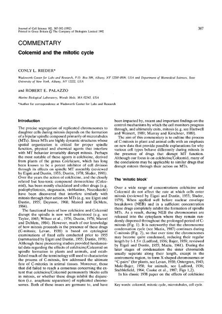
Fig. 1. Sequential phase-contrast photomicrographs, taken from a time-lapse video light-microscopic recording, of a newt<br />
lung cell proceeding through C-mitosis in <strong>the</strong> presence of 20 fjM Nocodazole. The chromatids comprising each chromosome<br />
are well separated along <strong>the</strong>ir length, except in <strong>the</strong> centromere region, in C. C-anaphase is initiated between D <strong>and</strong> E,<br />
during which time <strong>the</strong> chromatids of each chromosome disjoin in <strong>the</strong> centromeric region (e.g., cf. centromere regions noted<br />
by arrows in C-E). Approximately 30 min later (G) <strong>the</strong> chromatids undergo telophase changes that lead to <strong>the</strong> formation<br />
of a restitution nucleus (H). Bar in H, 50 /jm.<br />
Fig. 2. Schematic drawing of<br />
<strong>the</strong> chromosome <strong>cycle</strong> during<br />
C-mitosis. After nuclear<br />
envelope breakdown (A-B) <strong>the</strong><br />
chromosomes continue to<br />
thicken <strong>and</strong> shorten. Over<br />
time <strong>the</strong> two chromatids<br />
comprising each chromosome<br />
become separated along <strong>the</strong>ir<br />
length (C-D), but remain connected in <strong>the</strong> centomere region (E). During C-anaphase <strong>the</strong> chromatids completely disjoin (F)<br />
to form "pairs of skis". After a short time, relative to <strong>the</strong> duration of C-mitosis, <strong>the</strong> chromatids undergo telophase<br />
decondensation (G) to form ultimately a micronucleated restitution nucleus (H).
Levan states "<strong>the</strong> prophases arrive at metaphase <strong>and</strong><br />
are kept at that state for a long period...". This<br />
statement was based on Strasburger's (1884; see page<br />
120 of Wilson, 1925) terminology of <strong>the</strong> time, which<br />
separated <strong>the</strong> <strong>mitotic</strong> <strong>cycle</strong> into prophase, metaphase,<br />
anaphase <strong>and</strong> telophase without an intervening stage of<br />
prometaphase. The impetus for establishing "prometaphase"<br />
as a distinct stage of mitosis occurred between<br />
<strong>the</strong> publication of Schrader's first (1944) <strong>and</strong> second<br />
(1953) books on mitosis, well after Levan's initial<br />
studies. As first emphasized by Nebel <strong>and</strong> Ruttle in 1938<br />
(see also Ostergren, 1943), <strong>and</strong> more recently by Sluder<br />
(1979, 1988), C-<strong>mitotic</strong>s are blocked in prometaphase<br />
not metaphase. Indeed, after prolonged periods in Cmitosis,<br />
recovering sea urchin cells still require <strong>the</strong><br />
same 10 minute prometaphase interval to construct a<br />
spindle <strong>and</strong> congress chromosomes that is normally<br />
required in untreated controls (Sluder, 1979; see also<br />
Brinkley et al., 1967). Regardless, <strong>the</strong> erroneous notion<br />
that colchicine/<strong>Colcemid</strong> blocks <strong>the</strong> <strong>mitotic</strong> <strong>cycle</strong> at<br />
metaphase is still perpetuated as evidenced by <strong>the</strong><br />
continued widespread use of <strong>the</strong> terms "metaphase<br />
arrest", "C-<strong>mitotic</strong> metaphase", "maintained in metaphase",<br />
"held in metaphase", "colchicine (or C)metaphase",<br />
"metaphase-blocked", etc.<br />
A clear distinction between a <strong>mitotic</strong> block at<br />
prometaphase <strong>and</strong> metaphase should not be viewed as a<br />
trivial matter. It becomes increasingly important as<br />
molecular-genetic <strong>and</strong> cell-free systems are used to<br />
dissect more closely, <strong>and</strong> to define, <strong>the</strong> sequence of<br />
biochemical events comprising mitosis. Indeed, <strong>the</strong><br />
term "metaphase arrest" is commonly used to characterize<br />
various somatic cell mutants blocked in mitosis,<br />
<strong>and</strong> to describe <strong>the</strong> outcome of experimental treatments<br />
on <strong>mitotic</strong> cells, even under conditions in which spindle<br />
formation is largely or completely inhibited. These<br />
"metaphase arrested" cells contrast sharply with those<br />
oocytes that are naturally arrested at true metaphase I<br />
or II of meiosis with fully fomed spindles (reviewed by<br />
Longo, 1973), <strong>and</strong> those (few) somatic cells that can be<br />
induced by various treatments to arrest permanently in<br />
mitosis with fully formed (e.g. see Shoji-Kasai et al.,<br />
1987; Jordan et al., 1991) or nearly fully formed (Hirano<br />
et al., 1988) spindles.<br />
Escaping <strong>the</strong> <strong>mitotic</strong> block<br />
Most, if not all plant cells undergo repeated cell <strong>cycle</strong>s<br />
in <strong>the</strong> presence of colchicine (e.g. see Levan, 1938;<br />
Nebel <strong>and</strong> Ruttle, 1938; Eigsti <strong>and</strong> Dustin, 1955), a fact<br />
that has been widely utilized for generating polyploid<br />
strains of commercially valuable crops. Similarly, many<br />
types of animal cells, including some from Chinese<br />
hamsters (Stubblefield, 1964), newts (Fig. 1), rat<br />
kangaroos (Jensen et al., 1987), mice (Kung et al.,<br />
1990), humans (Chamla et al., 1980) <strong>and</strong> sea urchins<br />
(Sluder, 1979), are capable of completing one or more<br />
rounds of C-mitosis in <strong>the</strong> presence of <strong>the</strong> drug. Thus,<br />
contrary to <strong>the</strong> implications of such common terms as<br />
"<strong>mitotic</strong> arrest", "stathmokinesis", "metaphase ar-<br />
C-mitosis 389<br />
rest", "blocked or arrested in mitosis", "C-<strong>mitotic</strong><br />
arrest", "halted at metaphase", etc., colchicine, <strong>Colcemid</strong><br />
<strong>and</strong> drugs with similar actions do not permanently<br />
block plant <strong>and</strong> many animal cells in mitosis. Ra<strong>the</strong>r,<br />
when compared with controls, most drug-treated cells<br />
invariably spend a significantly greater period of time<br />
(up to 10-fold; Eigsti <strong>and</strong> Dustin, 1955) in (prometaphase<br />
of) mitosis prior to entering interphase of <strong>the</strong><br />
next cell <strong>cycle</strong>.<br />
The prolongation of <strong>the</strong> <strong>mitotic</strong> period during Cmitosis<br />
is not a unique response to <strong>the</strong> destruction of <strong>the</strong><br />
spindle by colchicine <strong>and</strong> similar drugs. On <strong>the</strong><br />
contrary, concentrations of <strong>Colcemid</strong> or vinblastine<br />
that have little or no discernable effect on spindle<br />
formation in sea urchins (Sluder, 1988) or HeLa-S3 cells<br />
(Jordon et al., 1991) significantly prolong mitosis (sea<br />
urchins) or may even permanently arrest <strong>the</strong> cells at<br />
true metaphase (HeLa). Similarly, prolongation of <strong>the</strong><br />
<strong>mitotic</strong> period is not a unique response to <strong>Colcemid</strong> or<br />
o<strong>the</strong>r drugs that disrupt MTs; <strong>the</strong> duration of prometaphase<br />
in untreated cells is greatly extended by <strong>the</strong><br />
presence of mal-oriented chromosomes (Mazia, 1961;<br />
Zirkle, 1970; Rieder <strong>and</strong> Alex<strong>and</strong>er, 1989), <strong>and</strong>/or by<br />
<strong>the</strong> absence of normal spindle bipolarity (Sluder <strong>and</strong><br />
Begg, 1983; Hunt et al., 1992).<br />
It has been proposed by Hartwell <strong>and</strong> Weinert (1989)<br />
that cells possess control mechanisms, termed "checkpoints",<br />
which function to ensure that <strong>the</strong> events of <strong>the</strong><br />
cell <strong>cycle</strong> are properly coordinated. The fact that <strong>the</strong><br />
onset of anaphase is considerably delayed by partial or<br />
total disruption of <strong>the</strong> spindle (as in C-mitosis),<br />
treatments that minimally compromise MT function, by<br />
<strong>the</strong> lack of spindle bipolarity, <strong>and</strong>/or by mal-oriented<br />
chromosomes on a bipolar spindle, reveals that <strong>the</strong><br />
process of spindle formation is "monitored" by such a<br />
surveillance checkpoint. As emphasized by Mazia<br />
(1961,1987), <strong>and</strong> more recently by o<strong>the</strong>rs (Hartwell <strong>and</strong><br />
Weinert, 1989; Murray <strong>and</strong> Kirschner, 1989), this<br />
checkpoint appears to control cell entry into anaphase,<br />
<strong>and</strong> passage through this point triggers a cascading<br />
series of events that allow a rapid escape from mitosis,<br />
advancing <strong>the</strong> cell to interphase of <strong>the</strong> next cell <strong>cycle</strong>.<br />
It has recently become clear that <strong>the</strong> nuclear <strong>and</strong><br />
cytoplasmic events that lead to mitosis are regulated, in<br />
part, by <strong>the</strong> sequential syn<strong>the</strong>sis <strong>and</strong> accumulation of<br />
"cyclin" proteins A <strong>and</strong> B. These proteins are cofactors<br />
required for <strong>the</strong> catalytic activity of <strong>the</strong> protein kinase,<br />
p34cdc2 (Solomon et al., 1990). Cyclin syn<strong>the</strong>sis drives<br />
cells into mitosis (Murray <strong>and</strong> Kirschner, 1989), while<br />
<strong>the</strong> initiation of anaphase <strong>and</strong> <strong>the</strong> cells' subsequent exit<br />
from mitosis is coincident with <strong>the</strong> rapid proteolytic<br />
destruction of <strong>the</strong>se proteins (Evans et al., 1983;<br />
reviewed by Murray <strong>and</strong> Kirschner, 1989; Whitfield et<br />
al., 1990). More specifically, in somatic cells cyclin A<br />
appears to reach peak levels just before NEB, <strong>and</strong> is<br />
<strong>the</strong>n degraded during prometaphase as <strong>the</strong> spindle<br />
forms. By contrast, <strong>the</strong> cyclin B level remains high until<br />
<strong>the</strong> metaphase-anaphase transition, at which time it<br />
drops precipitously. Importantly, cyclin A is degraded<br />
but cyclin B levels remain high throughout <strong>the</strong><br />
prolonged prometaphase exhibited by C-<strong>mitotic</strong>s (Kung
390 C. L. Rieder <strong>and</strong> R. E. Palazzo<br />
et al., 1990; Whitfield et al., 1990) <strong>and</strong> cells containing<br />
monopolar spindles (Hunt et al., 1992). Toge<strong>the</strong>r <strong>the</strong>se<br />
data strongly support <strong>the</strong> argument that passage<br />
through <strong>the</strong> spindle-formation surveillance checkpoint<br />
is triggered by declining levels of cyclin B. If true, it will<br />
become important to elucidate how <strong>the</strong> life expectancy<br />
of cyclin B is determined by <strong>the</strong> "state of microtubules<br />
<strong>and</strong> form of <strong>the</strong> spindle" (Hunt et al., 1992). The recent<br />
isolation of <strong>mitotic</strong> arrest-deficient (mad) mutants in<br />
yeast (Hoyt et al., 1991; Li <strong>and</strong> Murray, 1991), in which<br />
<strong>the</strong> cells fail to arrest at mitosis in response to loss of<br />
MT function, offers a promising approach for underst<strong>and</strong>ing<br />
how <strong>the</strong> cell monitors spindle formation.<br />
Not all animal cells ultimately pass through C-mitosis<br />
<strong>and</strong> enter <strong>the</strong> next cell <strong>cycle</strong> in <strong>the</strong> presence of drugs<br />
that disrupt MT function. For example, cells of certain<br />
mammalian lines (including HeLa S3, Vero, Tera2) die<br />
after 72 h in C-mitosis (see references quoted by Eigsti<br />
<strong>and</strong> Dustin, 1955; Kung et al., 1990), possibly from <strong>the</strong>ir<br />
inability to syn<strong>the</strong>size mRNA (Dustin, 1959). In some<br />
cases, a significant proportion of <strong>the</strong> cells within a<br />
<strong>mitotic</strong>ally arrested population escape <strong>the</strong> block while<br />
o<strong>the</strong>rs die in mitosis (i.e. <strong>the</strong> block is leaky; e.g. see<br />
Shoji-Kasai et al., 1987; Jordan et al., 1991). Kung et al.<br />
(1990) have recently shown that <strong>the</strong> ability of a cell type<br />
to survive C-mitosis is somewhat species-specific, <strong>and</strong> is<br />
positively correlated with its ability to degrade cyclin B<br />
during <strong>the</strong> prolonged <strong>mitotic</strong> period. Although <strong>the</strong>se<br />
experiments do not distinguish whe<strong>the</strong>r cyclin B<br />
degradation causes, or simply results from, <strong>the</strong> biochemical<br />
changes leading to escape from mitosis, <strong>the</strong><br />
former does provide a possible molecular basis for why<br />
some cells are truly "arrested" in mitosis by colchicine<br />
or <strong>Colcemid</strong> while o<strong>the</strong>rs can ultimately advance to<br />
interphase. Clearly, "<strong>the</strong> stringency of <strong>the</strong> [spindle<br />
formation surveillance checkpoint]...varies among different<br />
cell lines" (Kung et al., 1990).<br />
Chromatid disjunction in <strong>the</strong> absence of a<br />
spindle<br />
In actively cycling cells <strong>the</strong> initiation of anaphase, <strong>and</strong><br />
thus exit from mitosis, is signaled by <strong>the</strong> disjunction of<br />
replicated chromatids. In some types of cells <strong>the</strong><br />
chromatids of each replicated chromosome separate at<br />
<strong>the</strong> centromeric region near <strong>the</strong> end of <strong>the</strong> C-<strong>mitotic</strong><br />
period. This "C-anaphase" (Levan, 1938) phenomenon<br />
appears to occur in all plant cells (reviewed by Levan,<br />
1954; Eigsti <strong>and</strong> Dustin, 1955), where it has been<br />
especially well characterized owing to <strong>the</strong> absence of<br />
rounding during <strong>the</strong> division process (Mole-Bajer, 1958;<br />
Lambert, 1980). In Haemanthus each pair of replicated<br />
chromosomes requires a 1-2 min period to separate (see<br />
Fig. 6 of Lambert, 1980), <strong>and</strong> all chromatids of <strong>the</strong><br />
genome separate in near but not perfect synchrony (see<br />
Eigsti <strong>and</strong> Dustin, 1955; Mole-Bajer, 1958; Lambert,<br />
1980) in <strong>the</strong> complete absence of MTs (Lambert, 1980).<br />
Shortly after separation <strong>the</strong> chromatids begin to swell<br />
<strong>and</strong> undergo telophase events to form a 4N or polyploid<br />
restitution nucleus. The total duration of C-anaphase is<br />
similar to <strong>the</strong> time of anaphase in untreated cells (Mole-<br />
Bajer, 1958).<br />
There is currently no consensus concerning <strong>the</strong> extent<br />
to which C-anaphase occurs in animal cells (e.g. see<br />
Levan, 1954; Mazia, 1961; Rao <strong>and</strong> Engelberg, 1966;<br />
Mclntosh, 1979), <strong>and</strong> <strong>the</strong>re are several obvious reasons<br />
for this confusion. Unlike plants, <strong>the</strong> ultimate fate of<br />
individual chromosomes during C-mitosis in animals is<br />
difficult to follow clearly because most cells progressively<br />
round throughout this process. Moreover, few<br />
investigators have studied <strong>the</strong> course of C-mitosis in<br />
animal cells with <strong>the</strong> explicit goal of determining<br />
whe<strong>the</strong>r <strong>the</strong> chromatids disjoin.<br />
C-anaphase figures are seen in many types of animal<br />
cells when assayed by using squashed or dropped<br />
chromosome preparations. These include, but are not<br />
limited to, grasshopper spermatogonium (Sokolow,<br />
1939), human lymphocytes (Gabarron et al., 1986),<br />
mouse carcinoma (Ludford, 1936), ascites tumor<br />
(Levan, 1954), Chinese hamster ovary (Stubblefield,<br />
1964), rat kangaroo kidney epi<strong>the</strong>lia (Vig, 1981) <strong>and</strong><br />
Drosophila neuroblasts (Gonzalez et al., 1991). (See<br />
Eigsti <strong>and</strong> Dustin (1955), for additional references on<br />
C-anaphase in chromosome spreads of animal cells.)<br />
Studies on premature centromere separation (e.g. see<br />
Fitzgerald et al., 1975), <strong>and</strong> <strong>the</strong> sequence of centromere<br />
separation (e.g. see Vig, 1981), reveal that <strong>the</strong> harsh<br />
preparative treatments used for <strong>the</strong>se analyses (hypotonic<br />
swelling, fixation in acetic acid/ethanol, squashing<br />
or dropping onto slides) are not likely to induce<br />
chromatid separation artificially.<br />
C-anaphase has also been clearly demonstrated in sea<br />
urchin embryos fixed <strong>and</strong> lightly flattened between two<br />
coverslips (Sluder, 1979). Moreover, C-anaphase figures<br />
represent approx. 1-2% of all <strong>mitotic</strong>s in PtK<br />
cultures fixed after 18 h in a concentration (20 fxM) of<br />
nocodazole sufficient to deplete <strong>the</strong> cells of MTs (C.L.<br />
Rieder <strong>and</strong> R.W. Cole, unpublished). We have also<br />
observed <strong>the</strong> process of C-anaphase directly by timelapse<br />
video light microscopy of similarly treated newt<br />
lung cells (Fig. 1). With respect to <strong>the</strong>se findings it is<br />
noteworthy that individual chromosomes within <strong>the</strong><br />
cytoplasm of PtK (Brenner et al., 1980) <strong>and</strong> newt<br />
(Rieder <strong>and</strong> Alex<strong>and</strong>er, 1989) cells, which fail to attach<br />
to <strong>the</strong> normally forming spindle, still separate <strong>the</strong>ir<br />
chromatids at <strong>the</strong> onset of anaphase. Chromatid<br />
disjunction also occurs during monopolar mitosis in<br />
newts (Rieder et al., 1986) <strong>and</strong> sea urchins (Mazia et<br />
al., 1981).<br />
The mechanism responsible for chromatid separation<br />
remains mysterious. It is clear from studies on C<strong>mitotic</strong>s<br />
that it is not dependent on antagonistic pulling<br />
forces, generated by <strong>the</strong> spindle, that act on sister<br />
kinetochores within <strong>the</strong> centromeric region. This<br />
conclusion contrasts sharply with those models for<br />
chromatid separation in yeast, generated to explain <strong>the</strong><br />
apparent need for spindle MT-dependent forces during<br />
DNA decatenation by topoisomerase II (Holm et al.,<br />
1985, 1989; Uemura <strong>and</strong> Yanagida, 1986). In some<br />
animal cells chromatid disjunction exhibits a close temporal<br />
coupling to <strong>the</strong> Ca 2+ -mediated inactivation of <strong>the</strong>
p34 cdc2 /cyclin B complex <strong>and</strong> <strong>the</strong> destruction of cyclin B<br />
(Hunt et al., 1992; Shamu <strong>and</strong> Murray, 1992). It also<br />
probably requires DNA topoisomerase II activity<br />
(Downes et al., 1991; Shamu <strong>and</strong> Murray, 1992) <strong>and</strong><br />
perhaps <strong>the</strong> modification of INCENP (Cooke et al.,<br />
1987) <strong>and</strong> CLiP (Rattner et al., 1988), proteins unique<br />
to that region of <strong>the</strong> centromere spanning <strong>the</strong> sister<br />
kinetochores. In this context it is noteworthy that<br />
chromatids maintain firm centromeric connections<br />
prior to C-anaphase, after becoming separated along<br />
<strong>the</strong> remainder of <strong>the</strong>ir length (see above). Thus <strong>the</strong><br />
processing of chromatin that leads to chromatid<br />
separation is multi-phasic (i.e. <strong>the</strong> decatenation <strong>and</strong><br />
subseqeunt separation of chromosome arms <strong>and</strong> telomeres<br />
occurs well before that of <strong>the</strong> centromeres).<br />
It remains to be determined whe<strong>the</strong>r C-anaphase is a<br />
characteristic feature of C-mitosis in all animal cells.<br />
Statements that it does not occur must be re-evaluated<br />
in <strong>the</strong> context of those considerations that tend to mask<br />
its appearance. However, as discussed above some cell<br />
types ultimately die in C-mitosis, apparently because<br />
<strong>the</strong>y cannot degrade cyclin B to initiate those anaphase<br />
events that allow <strong>the</strong>m to exit <strong>the</strong> <strong>mitotic</strong> <strong>cycle</strong> (Kung et<br />
al., 1990; Whitfield et al., 1990; Hunt etal., 1992). Since<br />
<strong>the</strong> initiation of anaphase is normally heralded by<br />
chromatid separation, cells that are unable to exit Cmitosis<br />
may never disjoin <strong>the</strong>ir chromatids. In such cells<br />
spindle formation would be necessary for chromatid<br />
separation (i.e. anaphase) only because it is a prerequisite<br />
for initiating cyclin B degradation to allow passage<br />
through <strong>the</strong> spindle-formation surveillance checkpoint,<br />
not because chromatid separation is based on forces<br />
generated by <strong>the</strong> spindle (e.g. see Gonzalez et al.,<br />
1991).<br />
Although chromatid disjunction is normally temporaly<br />
coincident with cyclin B destruction, it may not be<br />
mediated, even indirectly, by declining cyclin B levels<br />
but by some o<strong>the</strong>r independent signal. Under <strong>the</strong>se<br />
circumstances cells would be able to separate <strong>the</strong>ir<br />
chromatids without necessarily initiating those o<strong>the</strong>r<br />
events of anaphase that allow <strong>the</strong>m to exit mitosis.<br />
Reports that certain mutant human cells appear to<br />
remain arrested for considerable periods of time in<br />
mitosis, with some or all of <strong>the</strong>ir chromatids disjoined<br />
(Fitzgerald et al., 1975; Rudd et al., 1983; Gabarron et<br />
al., 1986), argue in favor of this hypo<strong>the</strong>sis. By contrast,<br />
it is also possible that <strong>the</strong> events of anaphase that allow<br />
<strong>the</strong> cell to exit mitosis can occur in <strong>the</strong> absence of<br />
chromatid separation. Such a "relief of dependence"<br />
(Hartwell <strong>and</strong> Weinert, 1989) is suggested by <strong>the</strong><br />
observation that treatments that inhibit chromatid<br />
separation in mammalian cells do not necessarily<br />
prevent exit from mitosis (Downes et al., 1991).<br />
Conclusions<br />
We have reviewed <strong>the</strong> evidence that, for many cells,<br />
disruption of <strong>the</strong> <strong>mitotic</strong> spindle with <strong>Colcemid</strong>,<br />
colchicine <strong>and</strong> similar drugs delays but does not inhibit<br />
progression through <strong>the</strong> <strong>mitotic</strong> <strong>cycle</strong>. Whe<strong>the</strong>r a<br />
C-mitosis 391<br />
particular cell type can exit C-mitosis depends on its<br />
ability to overcome <strong>the</strong> spindle-formation surveillance<br />
checkpoint in <strong>the</strong> absence of a spindle, an ability that<br />
may depend on whe<strong>the</strong>r <strong>the</strong> cell can ultimately degrade<br />
cyclin B while in C-mitosis. C-<strong>mitotic</strong>s capable of<br />
passing through this checkpoint normally advance to<br />
interphase by way of a C-anaphase. C-anaphase is<br />
indicated by <strong>the</strong> separation of sister chromatids <strong>and</strong> this<br />
event does not depend on forces generated by <strong>the</strong><br />
spindle.<br />
We thank Drs. G. Sluder, S.P. Alex<strong>and</strong>er, S.S. Bowser <strong>and</strong><br />
J.G. Ault for <strong>the</strong>ir scientific comments, <strong>and</strong> Ms. S. Nowogrodzki<br />
for editorial assistance. This work was supported, in<br />
part, by grants from <strong>the</strong> National Institutes of Health,<br />
General Medical Sciences R01-40198 (to C.L.R.) <strong>and</strong> R01-<br />
43264 (to R.E.P.), by grant no. 2725 from <strong>the</strong> Council for<br />
Tobacco Research (to R.E.P.), <strong>and</strong> by American Cancer<br />
Society grant JFRA 62121 (to R.E.P.).<br />
References<br />
Bajer, A. S. (1959). Change of length <strong>and</strong> volume of <strong>mitotic</strong><br />
chromosomes in living cells. Hereditas 45, 579-596.<br />
Brenner, S. L., Liaw, L.-H. <strong>and</strong> Berns, M. W. (1980). Laser<br />
microirradiation of kinetochores in <strong>mitotic</strong> PtK2 cells. Cell.<br />
Biophys. 2, 139-151.<br />
Brinkley, B. R., Stubblefield, E. <strong>and</strong> Hus, T. C. (1967). The effects of<br />
colcemid inhibition <strong>and</strong> reversal on <strong>the</strong> fine structure of <strong>the</strong> <strong>mitotic</strong><br />
apparatus of Chinese hamster cells in vivo. J. Ultrastruct. Res. 19,1-<br />
18.<br />
Chamla, Y., Roumy, M., Lassegues, M. <strong>and</strong> Battin, J. (1980). Altered<br />
sensitivity to colchicine <strong>and</strong> PHA in human cultured cells. Hum.<br />
Genet. 53, 249-253.<br />
Cooke, C. A., Heck, M. S. <strong>and</strong> Earnshaw, W. C. (1987). The inner<br />
centromere protein (INCENP) antigens: Movement from inner<br />
centromere to midbody during mitosis. J. Cell Biol. 105, 2053-2067.<br />
Deysson, G. (1968). Anti<strong>mitotic</strong> substances. Int. Rev. Cytol. 24, 99-<br />
148.<br />
Downes, C. S., Mullinger, A. M. <strong>and</strong> Johnson, R. T. (1991).<br />
Inhibitors of DNA topoisomerase II prevent chromatid separation<br />
in mammaliancells but do not prevent exit from mitosis. Proc. Nat.<br />
Acad. Sci. USA 88, 8895-8899.<br />
Dustin. P. (1959). The quantitative estimation of <strong>mitotic</strong> growth in <strong>the</strong><br />
bone marrow of <strong>the</strong> rat by <strong>the</strong> stathmokinetic (colchicinic) method.<br />
In The Kinetics of Cellular Proliferation (ed. F. Stohlman, Jr), pp.<br />
50-56. New York, London: Grune <strong>and</strong> Stratton.<br />
Dustin, P. (1978). Microtubules, pp. 452. New York: Springer Verlag.<br />
Eigsti, O. J. <strong>and</strong> Dustin, P. (1955). Colchicine in Agriculture,<br />
Medicine, Biology <strong>and</strong> Chemistry, pp. 470. Aimes, Iowa: Iowa<br />
State Coll. Press.<br />
Evans, T., Rosenthal, E. T., Youngblom, J., Distel, D. <strong>and</strong> Hunt, T.<br />
(1983). Cyclin: A protein specified by maternal mRNA in sea<br />
urchin eggs that is destroyed at each cleavage division. Cell 33, 389-<br />
396.<br />
Fitzgerald, P. H., Pickering, A. F., Mercer, J. M. <strong>and</strong> Miethke, P. M.<br />
(1975). Premature centromere division: A mechanism of nondisjunction<br />
causing X chromosome aneuploidy in somatic cells of<br />
man. Ann. Hum. Genet. 38, 417-428.<br />
Gabarron, J., Jimenez, J. <strong>and</strong> Glover, G. (1986). Premature<br />
centromere division dominantly inherited in a subfertile family.<br />
Cytogenet. Cell Genet. 43, 69-71.<br />
Gonzalez, C, Jimenez, J. C, Ripoll, P. <strong>and</strong> Sunkel, C. E. (1991). The<br />
spindle is required for <strong>the</strong> process of sister chromatid separation in<br />
Drosophila neuroblasts. Exp. Cell Res. 192, 10-15.<br />
Hartwell, L. H. <strong>and</strong> Weinert, T. A. (1989). Checkpoints: Controls<br />
that ensure <strong>the</strong> order of cell <strong>cycle</strong> events. Science 246, 629-634.<br />
Hirano, T., Hiraoka, Y. <strong>and</strong> Yanagida, M. (1988). A temperaturesensitive<br />
mutation of <strong>the</strong> Schizosaccharomyces pombe gene nuc2+<br />
that encodes a nuclear-scaffold-like protein blocks spindle<br />
elongation in anaphase. J. Cell Biol. 106, 1171-1183.
392 C. L. Rieder <strong>and</strong> R. E. Palazzo<br />
Holm, C, Goto, T., Wang, J. C. <strong>and</strong> Botstein, D. (1985). DNA<br />
topoisomerase II is required at <strong>the</strong> time of mitosis in yeast. Cell 41,<br />
553-563.<br />
Holm, C, Stearns, T. <strong>and</strong> Botstein, D. (1989). DNA topoisomerase II<br />
must act at mitosis to prevent nondisjunction <strong>and</strong> chromosome<br />
breakage. Mol. Cell Biol. 9, 159-168.<br />
Hoyt, M. A., Totis, L. <strong>and</strong> Roberts, B. T. (1991). S. cerevisiae genes<br />
required for cell <strong>cycle</strong> arrest in response to loss of microtubule<br />
function. Cell 66, 507-517.<br />
Hunt, T., Luca, F. C. <strong>and</strong> Ruderman, J. V. (1992). The requirements<br />
for protein syn<strong>the</strong>sis <strong>and</strong> degredtion, <strong>and</strong> <strong>the</strong> control of destruction<br />
of cyclins A <strong>and</strong> B in <strong>the</strong> meiotic <strong>and</strong> <strong>mitotic</strong> cell <strong>cycle</strong>s of teh clam<br />
embryo. /. Cell Biol. 116, 707-724.<br />
Jensen, C. G., Davison, E. A., Bowser, S. S. <strong>and</strong> Rieder, C. L. (1987).<br />
Primary cilia <strong>cycle</strong> in PtKl cells: Effects of colcemid <strong>and</strong> taxol on<br />
cilia formation <strong>and</strong> resorption. Cell Modi. Cyloskel. 7, 187-197.<br />
Jordan, M. A., Thrower, P. <strong>and</strong> Wilson, L. (1991). Mechanism of<br />
inhibition of cell proliferation by vinca alkaloids. Cancer Res. 51,<br />
2212-2222.<br />
Rung, A. L., Sherwood, S. W. <strong>and</strong> Schimke, R. T. (1990). Cell linespecific<br />
differences in <strong>the</strong> control of cell <strong>cycle</strong> progression in <strong>the</strong><br />
absence of mitosis. Proc. Nat. Acad. Sci. USA 87, 9553-9557.<br />
Lambert, A-M. (1980). The role of chromosomes in anaphase trigger<br />
<strong>and</strong> nuclear envelope activity in spindle formation. Chromosoma<br />
76, 295-308.<br />
Levan, A. (1938). The effect of colchicine on root mitoses in Allium.<br />
Hereditas 24, 471-486.<br />
Levan, A. (1954). Colchicine-induced C-mitosis in two mouse ascites<br />
tumors. Hereditas 40, 1-64.<br />
Li, R. <strong>and</strong> Murray, A. W. (1991). Feedback control of mitosis in<br />
budding yeast. Cell 66, 519-531.<br />
Longo, F. J. (1973). Fertilization: A comparative ultrastructural<br />
review. Biol. Reprod. 9, 149-215.<br />
Ludford, R. J. (1936). The action of toxic substances upon <strong>the</strong><br />
division of normal <strong>and</strong> malignant cells in vitro <strong>and</strong> in vivo. Arch.<br />
Exp. Zellforsch. 18, 411-441.<br />
Mareel, M. M. <strong>and</strong> DeMets, M. (1984). Effect of microtubule<br />
inhibitors on invasion <strong>and</strong> on related activities of tumor cells. Int.<br />
Rev. Cytol. 90, 125-168.<br />
Mazia, D. (1961). Mitosis: <strong>the</strong> physiology of cell division. In The Cell.<br />
vol. Ill (ed. J. Brachet <strong>and</strong> A.E. Mirksy), pp. 78-412. New York:<br />
Academic Press.<br />
Mazia, D. (1987). The chromosome <strong>cycle</strong> <strong>and</strong> <strong>the</strong> centrosome <strong>cycle</strong> in<br />
<strong>the</strong> <strong>mitotic</strong> <strong>cycle</strong>. Int. Rev. Cytol. 100, 49-92.<br />
Mazia, D., Paweletz, N., Sluder, G. <strong>and</strong> Finze, E.-M. (1981).<br />
Cooperation of kinetochores <strong>and</strong> pole in <strong>the</strong> establishment of<br />
monopolar <strong>mitotic</strong> apparatus. Proc. Nat. Acad. Sci. USA 78, 377-<br />
381.<br />
Mclntosh, J. R. (1979). Cell division. In Microtubules (ed. K. Roberts<br />
<strong>and</strong> J.S. Hyams), pp. 382-441. New York: Academic Press.<br />
Mole-Bajer, J. (1958). Cine-micrographic analysis of C-mtosis in<br />
endosperm. Chromosoma 9, 332-358.<br />
Murray, A. W. <strong>and</strong> Kirschner, M. W. (1989). Dominoes <strong>and</strong> clocks:<br />
The union of two views of <strong>the</strong> cell <strong>cycle</strong>. Science 246, 614-621.<br />
Nebel, B. R. <strong>and</strong> Ruttle, M. L. (1938). The cytological <strong>and</strong> genetical<br />
significance of colchicine. J. Hered. 29, 3-9.<br />
Ostergren, G. (1943). Elastic chromosome repulsions. Hereditas 29,<br />
444-450.<br />
Rao, P. N. <strong>and</strong> Engelberg, J. (1966). Mitotic non-disjunction of sister<br />
chromatids <strong>and</strong> anomalous mitosis induced by low temperatures in<br />
HeLa cells. Exp. Cell Res. 43, 332-342.<br />
Rattner, J. B., Kingwell, B. G. <strong>and</strong> Fritzler, M. J. (1988). Detection<br />
of distinct structural domains within <strong>the</strong> primary constriction using<br />
autoantibodies. Chromosoma 96, 360-367.<br />
Rieder, C. L. <strong>and</strong> Alex<strong>and</strong>er, S. P. (1989). The attachment of<br />
chromosomes to <strong>the</strong> <strong>mitotic</strong> spindle <strong>and</strong> <strong>the</strong> production of<br />
aneuploidy in newt lung cells. In Mechanisms of Chromosome<br />
Distribution <strong>and</strong> Aneuploidy (ed. M.A. Resnick <strong>and</strong> B.K. Vig), pp.<br />
185-194. New York: Alan R. Liss.<br />
Rieder, C. L., Davison, E. A., Jensen, L. C. W., Cassimeris, L. <strong>and</strong><br />
Salmon, E. D. (1986). Oscillatory movements of monooriented<br />
chromosomes <strong>and</strong> <strong>the</strong>ir position relative to <strong>the</strong> spindle pole result<br />
from <strong>the</strong> ejection properties of <strong>the</strong> aster <strong>and</strong> half-spindle. J. Cell<br />
Biol. 103, 581-591.<br />
Rudd, N. L., Teshima, I. E., Martin, R. H., Sisken, J. E. <strong>and</strong><br />
Weksberg, R. (1983). A dominantly inherited cytogenetic anomaly:<br />
A possible cell division mutant. Hum. Genet. 65, 117-121.<br />
Schrader, F. (1944). Mitosis, 1st cdn, pp. 110. Columbia Univ. Press,<br />
New York.<br />
Schrader, F. (1953). Mitosis, 2nd edn, pp. 170. Columbia Univ. Press,<br />
New York.<br />
Shamu, C. E. <strong>and</strong> Murray, A. W. (1992). Sister chromatid separation<br />
in frog egg extracts requires DNA topoisomerase II activity during<br />
anaphase. J. Cell Biol. (in press).<br />
Shoji-Kasai, Y., Senshu, M., Iwashita, S. <strong>and</strong> Imahori, K. (1987).<br />
Thiol proteasc-spccific inhibitor E-64 arrests human cpidermoid<br />
carcinoma A431 cells at <strong>mitotic</strong> metaphasc. Proc. Nat. Acad. Sci.<br />
USA 85, 146-150.<br />
Sluder, G. (1979). Role of spindle microtubules in <strong>the</strong> control of cell<br />
<strong>cycle</strong> timing. J. Cell Biol. 80, 674-691.<br />
Sluder, G. (1988). Control mechanisms of Mitosis: The role of spindle<br />
microtubules in <strong>the</strong> timing of <strong>mitotic</strong> events. Zool. Sci. 5, 653-665.<br />
Sluder, G. (1991). The practical use of colchicine <strong>and</strong> colcemid to<br />
reversibly block microtubule assembly in living cells. In Advanced<br />
Techniques in Chromosome Research (ed. K.W. Adolph), pp. 427-<br />
447. New York: Marcel Dckker.<br />
Sluder, G. <strong>and</strong> Begg, D. A. (1983). Control mechanisms of <strong>the</strong> cell<br />
<strong>cycle</strong>: Role of <strong>the</strong> spatial arrangement of spindle components in <strong>the</strong><br />
timing of <strong>mitotic</strong> events. J. Cell Biol. 97, 877-886.<br />
Sokolow, I. (1939). Einfluss des Colchicins auf die<br />
Spermatogenialmitosen bei den Orthopetcren. CR (Doklady)<br />
Acad. Sci. URSS 24, 298-300.<br />
Solomon, M. J., Glotzer, M., Lee, T., Philippe, M. <strong>and</strong> Kirschner, M.<br />
(1990). Cyclin activation of p34cdc2. Cell 63, 1013-1024.<br />
Stubblefield, E. (1964). DNA syn<strong>the</strong>sis <strong>and</strong> chromosomal<br />
morphology of Chinese hamster cells cultured in media containing<br />
N-deacetyl-N-methylcolchicine (<strong>Colcemid</strong>). In Cytogenetics of<br />
Cells in Culture (ed. R.J. Harris), pp. 223-248. New York:<br />
Academic Press.<br />
Taylor, E. W. (1965). The mechanism of colchicine inhibition of<br />
mitosis. J. Cell Biol. 25, 145-160.<br />
Uemura, T. <strong>and</strong> Yanagida, M. (1986). Mitotic spindle pulls but fails to<br />
separate chromosomes in type II DNA topoisomerase mutants:<br />
uncoordinated mitosis. EMBO J. 5. 1003-1010.<br />
Vig, B. K. (1981). Sequence of centromere separation: An analysis of<br />
<strong>mitotic</strong> chromosomes from long-term cultures of Potorus cells.<br />
Cytogenet. Cell Genet. 31, 129-136.<br />
Whitfield, W. G. F., Gonzalez, C, Maldonado-Codina, G. <strong>and</strong><br />
Glover, D. M. (1990). The A- <strong>and</strong> B-type cyclins of Drosophila are<br />
accumulated <strong>and</strong> destroyed in temporally distinct events that define<br />
separable phases of <strong>the</strong> G2-M transition. EMBO J. 9, 2563-2572.<br />
Wilson, E. B. (1925). The Cell in Development <strong>and</strong> Heredity, pp. 1232.<br />
New York: MacMillan Co.<br />
Wilson, L., Anderson, K. <strong>and</strong> Chin, D. (1976). Nonstoichiometric<br />
poisoning of microtubule polymerization: A model for <strong>the</strong><br />
mechanism of action of <strong>the</strong> vinca alkaloids, popodhylcotoxin <strong>and</strong><br />
colchicine. In Cell Motility (ed. Goldman, R.. Pollard, T. <strong>and</strong><br />
Rosenbaum, J.), vol. 3, pp. 1051-1064. Cold Spring Harbor<br />
Laboratory Press, NY.<br />
Zirkle, R. E. (1970). Ultraviolet-microbeam irradiation of newt-cell<br />
cytoplasm: Spindle destruction, false anaphase, <strong>and</strong> delay of true<br />
anaphase. Radial. Res. 41, 516-537.<br />
Note added in proof<br />
While this manuscript was at <strong>the</strong> printers Gosh <strong>and</strong><br />
Paweletz (Exp. Cell Res. 200, 215-217, 1992) reported<br />
that okadaic acid inhibits sister chromatid separation in<br />
HeLa cells without inhibiting exit from mitosis. As a<br />
result, at <strong>the</strong> next mitosis diplochromosomes are<br />
formed that contain 4 unseparated chromatids. These<br />
data support <strong>the</strong> hypo<strong>the</strong>ses that phosphatase 1 activity<br />
is required for sister chromatid separation, <strong>and</strong> that<br />
chromatid separation is not an obligatory event for<br />
escape from mitosis.


