

SPHENOPHRYNE - American Museum of Natural History
SPHENOPHRYNE - American Museum of Natural History
SPHENOPHRYNE - American Museum of Natural History

You also want an ePaper? Increase the reach of your titles
YUMPU automatically turns print PDFs into web optimized ePapers that Google loves.
2000 ZWEIFEL: PARTITION OF <strong>SPHENOPHRYNE</strong><br />
107<br />
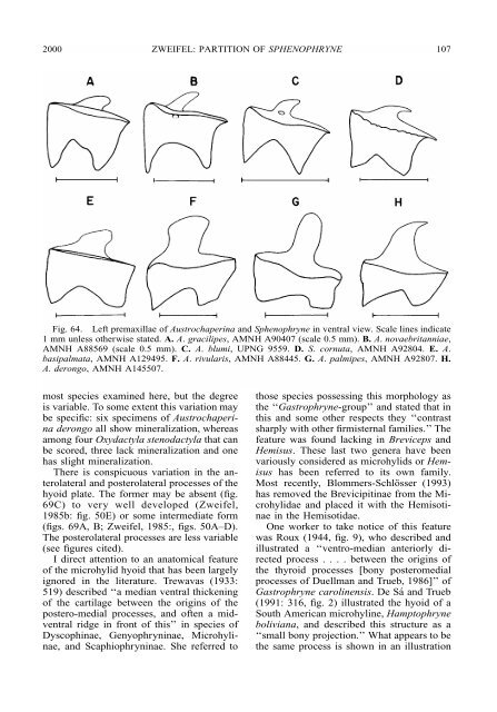
Fig. 64. Left premaxillae <strong>of</strong> Austrochaperina and Sphenophryne in ventral view. Scale lines indicate<br />
1 mm unless otherwise stated. A. A. gracilipes, AMNH A90407 (scale 0.5 mm). B. A. novaebritanniae,<br />
AMNH A88569 (scale 0.5 mm). C. A. blumi, UPNG 9559. D. S. cornuta, AMNH A92804. E. A.<br />
basipalmata, AMNH A129495. F. A. rivularis, AMNH A88445. G. A. palmipes, AMNH A92807. H.<br />
A. derongo, AMNH A145507.<br />
most species examined here, but the degree<br />
is variable. To some extent this variation may<br />
be specific: six specimens <strong>of</strong> Austrochaperina<br />
derongo all show mineralization, whereas<br />
among four Oxydactyla stenodactyla that can<br />
be scored, three lack mineralization and one<br />
has slight mineralization.<br />
There is conspicuous variation in the anterolateral<br />
and posterolateral processes <strong>of</strong> the<br />
hyoid plate. The former may be absent (fig.<br />
69C) to very well developed (Zweifel,<br />
1985b: fig. 50E) or some intermediate form<br />
(figs. 69A, B; Zweifel, 1985:, figs. 50A–D).<br />
The posterolateral processes are less variable<br />
(see figures cited).<br />
I direct attention to an anatomical feature<br />
<strong>of</strong> the microhylid hyoid that has been largely<br />
ignored in the literature. Trewavas (1933:<br />
519) described ‘‘a median ventral thickening<br />
<strong>of</strong> the cartilage between the origins <strong>of</strong> the<br />
postero-medial processes, and <strong>of</strong>ten a midventral<br />
ridge in front <strong>of</strong> this’’ in species <strong>of</strong><br />
Dyscophinae, Genyophryninae, Microhylinae,<br />
and Scaphiophryninae. She referred to<br />
those species possessing this morphology as<br />
the ‘‘Gastrophryne-group’’ and stated that in<br />
this and some other respects they ‘‘contrast<br />
sharply with other firmisternal families.’’ The<br />
feature was found lacking in Breviceps and<br />
Hemisus. These last two genera have been<br />
variously considered as microhylids or Hemisus<br />
has been referred to its own family.<br />
Most recently, Blommers-Schlösser (1993)<br />
has removed the Brevicipitinae from the Microhylidae<br />
and placed it with the Hemisotinae<br />
in the Hemisotidae.<br />
One worker to take notice <strong>of</strong> this feature<br />
was Roux (1944, fig. 9), who described and<br />
illustrated a ‘‘ventro-median anteriorly directed<br />
process . . . . between the origins <strong>of</strong><br />
the thyroid processes [bony posteromedial<br />
processes <strong>of</strong> Duellman and Trueb, 1986]’’ <strong>of</strong><br />
Gastrophryne carolinensis. DeSáand Trueb<br />
(1991: 316, fig. 2) illustrated the hyoid <strong>of</strong> a<br />
South <strong>American</strong> microhyline, Hamptophryne<br />
boliviana, and described this structure as a<br />
‘‘small bony projection.’’ What appears to be<br />
the same process is shown in an illustration


