
Create successful ePaper yourself
Turn your PDF publications into a flip-book with our unique Google optimized e-Paper software.
When the contents are special...<br />
The container should be too!<br />
XENETIX<br />
®<br />
Iobitridol<br />
In<br />
Xenetix ® 350, solution for injection (350 mgI/ml); Xenetix ® 300, solution for injection (300 mgI/ml); Xenetix ® 250, solution for injection (250 mgI/ml) – COMPOSITION per 100 ml: Xenetix ® 350: 76.78 g<br />
of iobitridol (corresponding to 35 g of iodine), Xenetix ® 300: 65.81 g of iobitridol (corresponding to 30 g of iodine), Xenetix ® 250: 54.84 g of iobitridol (corresponding to 25 g of iodine) –Indications and<br />
approvals may vary in different countries. Please refer to the local Summary of Product Characteristics (SPC) before prescribing. Further information available on request. CLINICALS PARTICULARS: therapeutic<br />
indications: this product is for diagnostic use only. Contrast agent for use in: Xenetix ® 350 intravenous urography, computed tomography, intravenous digital substraction angiography, arteriography, angiocardiography<br />
– Xenetix ® 300: intravenous urography, computed tomography, intravenous digital substraction angiography, arteriography, angiocardiography, endoscopic retrograde cholangiopancreatography, arthrography,<br />
hysterosalpingography – Xenetix ® 250: phlebography, computed tomography, intra-arterial digital substraction angiography, arteriography, endoscopic retrograde cholangiopancreatography – Posology and<br />
method of administration: the doses should be adapted to the type of examination and the region to be opacified as well as the weight and renal function of the patient, especially in children – Contraindication:<br />
hypersensitivity to iobitridol or any of the excipients, history of major immediate or delayed cutaneous reaction (see undesirable effects) to Xenetix ® , manifest thyrotoxicosis, hysterosalpingography during pregnancy.<br />
General particulars corresponding to all iodinated contrast agents: in the absence of any specific studies, myelography is not an indication for Xenetix ® . All iodinated contrast media can cause minor or<br />
major reactions that can be life-threatening. These can occur immediately (within 60 minutes) or be delayed (within 7 days) and are often unpredictable. Because of the risk of major reactions, emergency<br />
resuscitation equipment should be available for immediate use – Precautions for use: intolerance to iodinated contrast media, renal insufficiency, hepatic insufficiency, asthma, dysthyroidism, severe cardiovascular<br />
diseases, central nervous system disorders, phechromocytoma, myasthenia, intensification of side effects: Interaction with other medicinal products and other forms of interaction – Association requiring<br />
precaution of use: beta-blocking agents, diuretics, metformin, radiopharmaceuticals – Association to take into account: interleukin II – Pregnancy and lactation – Undesirable effects: anaphylactoid reactions<br />
and hypersensibility: skin-mucosa (very rare), respiratory (very rare), cardiovascular (very rare), neurosensorial (very rare), digestive (very rare), renal (infrequent), thyroid (very rare), local effects (frequent): 1- benign and<br />
transient local pain and oedema can occur at the injection site in the absence of extravasation of the injected contrast agent. Following intra-arterial administration, the sensation of pain at the injection site depends on<br />
the osmolality of the product injected. In case of extravasation (< 0.01%), a local inflammatory reaction or even tissue necrosis may be observed. 2- thrombophlebitis (not reported with Xenetix ® ). 3- articular pain with<br />
arthrography. 4- pelvic pain with hysterosalpingography – French presentation and marketing authorisation number: Xenetix ® 350: 337 710.7: 20 ml vial (glass) – 337 711.3: 50 ml vial (glass) – 560 154.3:<br />
50 ml vial x25 (glass) – 337 910.6: 60 ml vial (glass) – 337 713.6: 100 vial (glass) – 560 156.6: 100 ml vial x10 (glass) – 337 714.2: 150 ml vial (glass) – 337 715.9: 200 ml vial (glass) - 337 718.8:<br />
60 ml set – 369 154.2: 100 ml bag (polypropylene:pp) – 570 816 9: 100 ml bag x10 (pp) – 369 156.5: 150 ml bag (pp) – 570 817.5: 150 ml bag x10 (pp) – 369 158.8: 200 ml bag pp) – 570 818.1:<br />
200 ml bag x10 (pp) – 369 160.2: 500 ml bag (pp) – 570 819.8: 500 ml bag x10 (pp) - Xenetix ® 300: 337 767.9: 20 ml vial (glass) – 337 768.5: 50 ml vial (glass) – 560 157.2: 50 ml vial x25 (glass) –<br />
337 769.1: 60 ml vial (glass) – 337 771.6: 100 ml vial (glass) – 560 158.9: 100 ml vial x10 (glass) – 337 772.2: 150 ml vial (glass) – 337 705.3: 200 ml vial (glass) – 337 709.2: 60 ml set –<br />
369 144.7: 100 ml bag (pp) – 570 820.6: 100 ml bag x10 (pp) – 369 147.6: 150 ml bag (pp) – 570 821.2: 150 ml bag x10 (pp) – 369 149.9: 200 ml bag (pp) – 570 822.9: 200 ml bag x10 (pp) –<br />
369 151.3: 500 ml bag (pp) – 570 823.5: 500 ml bag x10 (pp) - Xenetix ® 250: 560 159.5: 50 ml vial x25 (glass) – 337 762.7: 100 ml vial (glass) – 560 160.3: 100 ml vial x10 (glass) – 337 763.3: 200 ml vial<br />
(glass) – 337 766.2: 50 ml set – Guerbet – BP 57400 F-95943 Roissy CdG cedex – tel: +33.(0)1.45.91.50.00 - www.guerbet.com - For detailed information, see Dictionnaire Vidal - Revised October 2007. Terre<br />
Neuve - P 08 024 XEN - 03/2008.<br />
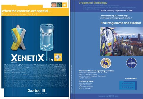
Selbstverlag, München ISBN 978-3-00-025479-6 ESUR 2008<br />
Urogenital Radiology<br />
Special Focus: Prostate, Pelvic Floor and Urinary Tract<br />
Final Programme and Syllabus of the 15th Symposium of the European Society<br />
of Urogenital Radiology<br />
Munich, Germany - September 11-14, 2008<br />
Chairman of the local organizing committee:<br />
Assoc. Prof. Ullrich Mueller-Lisse, MD, MBA<br />
Dept. of Clinical Radiology<br />
Munich University Hospitals – Innenstadt<br />
Munich, Germany<br />
Conference Venue:<br />
University of Munich<br />
HighTech Campus<br />
Munich-Grosshadern<br />
Munich, Germany<br />
Jahresfortbildung AG Uroradiologie<br />
der Deutschen Röntgengesellschaft e.V.<br />
Final Programme and Syllabus<br />
www.esur2008.org<br />
supported by:
Programme<br />
Edited by:<br />
Ulrike L. Müller-Lisse, MD<br />
Published by:<br />
European Society of Urogenital Radiology Deutsche Röntgengesellschaft
www.esur2008.org<br />
2<br />
Content<br />
Preface 3<br />
Acknowledgments 4<br />
ESUR 2008 Organisation 5<br />
ESUR 2008 Faculty 6<br />
Accreditation 7<br />
Social Programme 8<br />
General Information 9<br />
Maps of the Conference Area 13<br />
Programme Overview 14<br />
Detailed Programme 15<br />
Thursday, 11.09.2008 15<br />
Friday, 12.09.2008 16<br />
Saturday, 13.09.2008 23<br />
Sunday, 14.09.2008 24<br />
Poster Session 25<br />
Abstracts 29<br />
Members’ Day Sessions 30<br />
Scientific Session 37<br />
Workshops 55<br />
Course Lectures 68<br />
Posters 82
www.esur2008.org<br />
Dear colleagues and friends,<br />
Welcome to the 15th Symposium of the European Society of Urogenital Radiology, ESUR.<br />
Supported by the European Society of Radiology, ESR, the Society of Urogenital Radiology, SUR, the<br />
German Roentgen Society (DRG) AG, Uroradiologie of DRG, and the European Association of<br />
Urologists, EAU, ESUR 2008 meets in Munich, Germany.<br />
Scientific sessions, workshops, lectures, lunch symposia sponsored by industrial partners, the opening<br />
ceremony and the welcome reception, the industrial exhibition and the scientific poster exhibition<br />
take place from September 11th through 14 th, 2008, at the High Tech Campus of the University<br />
of Munich. The social programme and the accompanying persons’ programme will take you to<br />
interesting sites throughout the world-renowned city of Munich.<br />
Urogenital radiology is rapidly advancing, with new imaging and contrast technologies on the one<br />
hand and increasing demand for imaging and intervention services by urologists, gynaecologists, and<br />
radiation oncologists on the other.<br />
The main topics of ESUR 2008, prostate cancer, lower urinary tract, and pelvic floor, and other<br />
important topics, including gynaecologic imaging, upper urinary tract, urogenital trauma, and new<br />
developments in contrast media, are covered by internationally acclaimed specialists in interdisciplinary<br />
lecture sessions. The lunch symposia focus on cross-sectional imaging in urogenital radiology<br />
and important safety issues in the application of contrast media, including contrast-induced<br />
nephropathy and nephrogenic systemic fibrosis.<br />
We welcome physicians with a specialty or a keen interest in radiology, nuclear medicine, radiation<br />
oncology, gynaecology, and urology to share their knowledge and their visions with one another, to<br />
study together, to make new friends, and to renew old friendships across Europe, the Mediterranean<br />
and many other countries.<br />
We warmly welcome you at ESUR 2008 in Munich!<br />
On behalf of the local organizing committee of ESUR 2008,<br />
Ullrich G. Mueller-Lisse, M.D., M.B.A.,<br />
Attending Radiologist, Associate Professor of Radiology<br />
University of Munich Hospitals,<br />
Ulrike L. Mueller-Lisse, M.D.,<br />
Clinical Fellow in Urology<br />
University of Munich Hospitals<br />
www.esur2008.org<br />
3
www.esur2008.org<br />
4<br />
Acknowledgments<br />
ESUR 2008 gratefully acknowledges the support of the following sponsors<br />
Honorary Sponsor<br />
Prof. Maximilian Reiser, MD<br />
Dean-elect of the Faculty of Medicine, LMU Munich<br />
GE imagination at work<br />
Main Sponsors<br />
Sponsors<br />
Celon AG, Teltow, Germany<br />
Medrad Medizinische Systeme GMBH, Volkach, Germany<br />
Optimed Medizinische Instrumente GmbH, Ettlingen, Germany<br />
Terumo GmbH, Eschborn, Germany
www.esur2008.org<br />
ESUR 2008 Organisation<br />
ESUR Board<br />
S. Morcos (UK), President<br />
B. Hamm (D), President-Elect<br />
G. Heinz-Peer (A), Secretary/ Treasurer<br />
L. Derchi (I), Past President<br />
J. Jakobsen (N), Member-at-Large<br />
Programme Committee<br />
J. Barentsz (NL)<br />
R. Cohan (USA)<br />
K. Darge (USA)<br />
L. Derchi (I)<br />
T. El-Diasty (ET)<br />
F. Frauscher (A)<br />
B. Hamm (D)<br />
G. Heinz-Peer (A)<br />
K. Kinkel (CH)<br />
G. Malachias (GR)<br />
S. Morcos (UK)<br />
U. Mueller-Lisse (D)<br />
M. Riccabona (A)<br />
Scientific Secretariat<br />
Ullrich Mueller-Lisse<br />
Dept. of Clinical Radiology<br />
University of Munich Hospitals- Innenstadt<br />
Ziemssenstrasse 1<br />
80336 Munich, Germany<br />
Tel: +49 - (0)89 - 5160-9101<br />
Fax: +49 - (0)89 - 5160-9102<br />
info@esur2008.org<br />
Organizer<br />
Scientific Programme:<br />
European Society of Urogenital Radiology<br />
Industrial Exhibition and Sponsorship:<br />
EUROKONGRESS GmbH<br />
Local Committee<br />
E.M. Coppenrath<br />
C. Degenhart<br />
T. Meindl<br />
U.G. Mueller-Lisse<br />
U.L. Mueller-Lisse<br />
M.K. Scherr<br />
Organizing Secretariat<br />
EUROKONGRESS GmbH<br />
Congress + Event + Exhibition Management<br />
Schleissheimer Str. 2<br />
80333 München, Germany<br />
Tel. +49 (0)89/210 98 60<br />
Fax +49 (0)89/210 98 698<br />
info@eurokongress.de<br />
esur2008@eurokongress.de<br />
www.eurokongress.de<br />
5
www.esur2008.org<br />
Faculty 2008<br />
J. Barentsz (NL)<br />
D. Babnik-Peskar (SI)<br />
M.-F. Bellin (F)<br />
D. Beyersdorff (D)<br />
V. Berg-Løgager (DK)<br />
B. Brkljacic (HR)<br />
C. Chapple (UK)<br />
R. Cohan (USA)<br />
E. Coppenrath (D)<br />
F. Cornud (F)<br />
N. Cowan (UK)<br />
T.M. Cunha (P)<br />
N. Curry (USA)<br />
L. Dalla Palma (I)<br />
K. Darge (USA)<br />
C. Degenhart (D)<br />
L. Derchi (I)<br />
A. Dimopoulou (S)<br />
V. Dogra (USA)<br />
T. El-Diasty (ET)<br />
R. Farouk El Sayed (ET)<br />
R. Figueiras (E)<br />
F. Frauscher (A)<br />
K. Friese (D)<br />
R. Forstner (A)<br />
J. Fuetterer (NL)<br />
S. Goldman (USA)<br />
N. Grenier (F)<br />
P. Hallscheidt (D)<br />
B. Hamm (D)<br />
S. Hanna (ET)<br />
G. Heinz-Peer (A)<br />
J. Jakobsen (N)<br />
A. Lienemann (D)<br />
L. Lobo (P)<br />
6<br />
J. Kemper (D)<br />
P. Kenney (USA)<br />
K. Kinkel (CH)<br />
A. Magnusson (S)<br />
G. Malachias (GR)<br />
T. Meindl (D)<br />
S. Morcos (UK)<br />
S. Moussa (UK)<br />
U.G. Mueller-Lisse (D)<br />
U.L. Mueller-Lisse (D)<br />
C. Nolte-Ernsting (D)<br />
R. Oyen (B)<br />
P. Pavlica (I)<br />
R. Pozzi-Mucelli (I)<br />
P. Ramchandani (USA)<br />
M. Reiser (D)<br />
M. Riccabona (A)<br />
J. Richenberg (UK)<br />
C. Roy (F)<br />
E. Rummeny (D)<br />
C. Sandler (USA)<br />
J. Scheidler (D)<br />
M. Scherr (D)<br />
H. Schlemmer (D)<br />
K. Schneider (D)<br />
J. Spencer (UK)<br />
C. Stief (D)<br />
H. Thoeny (CH)<br />
H. Thomson (DK)<br />
A. Tuncay Turgut (TR)<br />
A. Van der Molen (NL)<br />
N. Wasserman (USA)<br />
R. Zagoria (USA)<br />
N. Zantl (D)
www.esur2008.org<br />
Accreditation<br />
The 15th European Symposium on Urogenital Radiology is accredited by the European Accreditation<br />
Council for Continuing Medical Education (EACCME) to provide the following CME activity for medical<br />
specialists. The EACCME is an institution of the European Union of Medical Specialists (UEMS).<br />
www.uems.be<br />
The 15th European Symposium on Urogenital Radiology is designed for a maximum of 18 hours of European<br />
external CME credits. Each medical specialist should only claim those hours of credit that he/ she<br />
actually spent in the educational activity.<br />
EACCME credits are recognized by the American Medical Association towards the Physician´s Recognition<br />
Award (PRA). To convert EACCME credits to AMA PRA category I credit, please contact AMA.<br />
The 15th European Symposium on Urogenital Radiology has been awarded 26 CME points by the Bavarian<br />
Chamber of Physicians (Bayerische Landesärztekammer).<br />
Distribution of CME Credits for ESUR 2008 is as follows:<br />
September 11, 2008: 6 Credits<br />
September 12, 2008: 6 Credits<br />
September 13, 2008: 6 Credits<br />
September 14, 2008: 4 Credits<br />
You may earn a total of 26 category "A" credits if you attend ESUR 2008 on all 4 days.<br />
The CME credit certificate will be handed out with your congress documents.<br />
7
www.esur2008.org<br />
Accompanying Persons’ Programme<br />
The fee for accompanying persons includes a guided walking tour in the Munich city centre,<br />
the Welcome Reception and a 3-day-ticket for the public transport (inner circle Munich).<br />
The guided walking tour will take place on Friday, September 12, 2008, from 16:00h to 18:00h.<br />
The language of the tour is English.<br />
Please meet the guide directly in the city centre at the “Fish Fountain” located on the famous<br />
“Marienplatz”. The walking tour includes the most famous sights of Munich: the city center Marienplatz,<br />
Munich´s oldest church St. Peter and the cathedral, the Opera House and the home of the Bavarian<br />
Rulers – the Residenz. You will have a look at the most elegant avenue Maximilianstreet, at the<br />
Hofbräuhaus and the most popular foodmarket “Viktualienmarkt”.<br />
Evening Programme<br />
Members´ Dinner<br />
On Thursday, September 11, 2008 the traditional ESUR-Members´ Dinner will be held at “Der Pschorr”.<br />
The restaurant is located in the historical city center at the famous “Viktualienmarkt”.<br />
The entrance tickets will be handed out on site at the conference venue (if registered).<br />
On site registrations are subject to availability.<br />
Welcome Reception<br />
All participants are warmly invited to join the Welcome Reception at the conference<br />
venue on Friday, September 12, 2008 at 20:00h. The entrance is free of charge for all registered<br />
participants.<br />
Course Dinner<br />
On Saturday, September 13, 2008 the Course Dinner, a “Bavarian Evening”, will be held at the<br />
“Augustinerkeller”. The “Augustinerkeller” is one of the most popular beer gardens in Munich and offers<br />
typical Bavarian specialties. The entrance tickets will be handed out on site at the conference venue<br />
(if registered). On site registrations are subject to availability.<br />
Please contact the conference office by Friday, September 13, 2008, 13:00 at the latest.<br />
8<br />
Social programme
www.esur2008.org<br />
General Information<br />
Abbreviations<br />
MS Members´ Session<br />
LS Lecture Session<br />
WS Workshop<br />
ScS Scientific Session<br />
P Poster Session<br />
YR Young Radiologists´ Forum<br />
Badges<br />
It is mandatory for all participants to wear their<br />
badges visibly throughout the meeting as it is the<br />
entrance ticket to all sessions. In the event of<br />
badge loss, please contact the registration desk.<br />
Banks<br />
There are several banks near the conference<br />
venue at both sides of the subway station<br />
“Großhadern”.<br />
Cancellation/ No-show<br />
A refund on the registration fees less an administrative<br />
fee of 40,00 € has been made when a<br />
written cancellation was received by<br />
August 11, 2008 .<br />
Thereafter no refunds can be made.<br />
Cash Bar<br />
Outside the official coffee breaks beverages,<br />
snacks and food can be purchased at a Snack<br />
Bar on the ground floor.<br />
Certificate of Attendance<br />
A certificate of attendance including a CME<br />
credit certificate as well as an UEMS certificate<br />
will be handed out with your congress<br />
documents.<br />
Coffee Breaks<br />
Coffee, beverages and food will be available<br />
for registered participants during the designed<br />
coffee break times on the ground floor.<br />
Conference Language<br />
Conference language is English.<br />
Conference Venue<br />
University of Munich<br />
High Tech Campus Munich-Grosshadern<br />
Institutes of Chemistry and Pharmacy<br />
Butenandtstr. 5-13<br />
81377 Munich, Germany<br />
Conference hours<br />
Thursday, September 11<br />
(Members’ Day)<br />
14:00 - 19:00<br />
Friday, September 12 08:30 - 20:00<br />
Saturday, September 13 09:00 - 18:00<br />
Sunday, September 14 09:00 - 13:30<br />
Conference Office/ Registration<br />
Registration is possible at the Registration Desk in<br />
the foyer.<br />
On site registrations and day tickets are subject<br />
to availability.<br />
Opening hours:<br />
Thursday, September 11 10:00 - 19:00<br />
Friday, September 12 07:30 - 20:00<br />
Saturday, September 13 08:30 - 19:00<br />
Sunday, September 14 08:30 - 13:30<br />
9
www.esur2008.org<br />
Registration fee<br />
ESUR / SUR Member 390,00 €<br />
Non-Member 440,00 €<br />
Residents and Technologists 290,00 €<br />
Accompanying Persons 135,00 €<br />
Members’ Dinner 60,00 €<br />
Course Dinner 70,00 €<br />
The conference fees include the participation in<br />
the scientific sessions, coffee breaks, lunches, the<br />
Welcome Reception as well as a 3-day-ticket for<br />
public transportation (inner circle Munich).<br />
Hotel Reservation<br />
Eurokongress has reserved special room<br />
allotments close to the conference and in the<br />
city center. For detailed information, booking<br />
and/or changes, please contact Eurokongress at<br />
the conference office on site. A city map of the<br />
respective hotels is included in this programme<br />
(see page 12 ).<br />
Industrial Exhibition<br />
The industrial exhibition is located in the foyer in<br />
front of lecture hall “Buchner”. Please refer to the<br />
floor plan. The exhibition is open during conference<br />
hours.<br />
Lecture Halls<br />
The lecture halls are named as follows:<br />
Lecture hall Buchner Ground Floor<br />
Lecture hall Baeyer Lower Level<br />
Lecture hall Willstätter Lower Level<br />
Lecture hall Wieland Lower Level<br />
Lecture hall Butenandt Lower Level<br />
10<br />
Liability<br />
The organizer cannot be held responsible for any<br />
personal injury, accident, damage to private<br />
property or additional expenses incurred to a<br />
result of changes of dates, venue, programme<br />
or else.<br />
Lunches<br />
Lunches on Friday, September 12 and Saturday,<br />
September 13 are included in the registration<br />
fee. Lunch packages will be handed out at the<br />
Lunch Symposia. Please follow the announcement<br />
and signs.<br />
Photographing/ Recording<br />
It is strongly prohibited to take pictures, record<br />
or tape any presentations or sessions without<br />
official permission of the organizers.<br />
Poster Exhibition<br />
Scientific posters are exhibited in the poster area<br />
on the lower level of the conference venue in<br />
front of the lecture halls.<br />
The Poster exhibition is open from Thursday,<br />
September 11, 2008, 12:00 until Sunday, September<br />
14, 2008, 12:30. All poster presenters are<br />
kindly asked to be available at their respective<br />
poster during “Best of ESUR” to allow poster<br />
discussion.<br />
Poster Prizes<br />
The three best scientific exhibits will be awarded<br />
a diploma during the opening ceremony.<br />
Evaluation of the posters will be based on novelty,<br />
accuracy, educational value and design.
www.esur2008.org<br />
Preview Centre<br />
The preview center is located on the lower level in<br />
front of lecture hall “Butenandt”.<br />
Opening hours:<br />
Thursday, September 11 11:00 - 17:00<br />
Friday, September 12 07:30 - 17:00<br />
Saturday, September 13 08:00 - 17:00<br />
Sunday, September 14 08:00 - 12:30<br />
All lectures, workshops, and scientific papers have<br />
to be submitted to the presentation center at<br />
least 2 hours prior to the beginning of the respective<br />
session. It will not be possible to use your own<br />
laptop computer. Please adhere strictly to restrictions<br />
on Powerpoint software (no younger than<br />
Powerpoint 2003 for Windows) and Windows operating<br />
systems (no younger than Windows 2000<br />
or XP). For example, it will not be possible to make<br />
a presentation based on Windows Vista or on<br />
Powerpoint 2007 for Windows. Please note that<br />
if you have prepared your presentation on a<br />
Macintosh computer, you should convert it such<br />
as to match with Windows programmes as mentioned<br />
above.<br />
There will be no chance of presenting conventional<br />
slides or overhead sheets or other lecture<br />
modalities.<br />
Please refrain from bringing your own personal<br />
computer for your presentation, since it may not<br />
be compatible with the equipment on site.<br />
The preferred data storage device to carry your<br />
scientific presentation, lecture or workshop would<br />
be a memory stick or similar device which is USB-2compatible;<br />
however, if you prefer, you may bring<br />
a CD ROM.<br />
Public Transportation<br />
All registered participants receive a 3-day-ticket<br />
for public transportation at the conference<br />
office on site.<br />
To get to the conference venue we recommend<br />
using underground line U6, direction to ‘Klinikum<br />
Grosshadern’ and get off at the stop ‘Grosshadern’.<br />
The underground line U6 takes you also to<br />
the city centre and Marienplatz. For detailed<br />
information about public transportation please<br />
have a look at the brochure handed out at the<br />
conference office on site. Participants arriving by<br />
car have the possibility to park close to the congress<br />
venue.<br />
Wardrobe<br />
The wardrobe for jackets and bags can be found<br />
on the lower level of the conference venue<br />
(please follow the signs). Please note that the<br />
organizers cannot provide security staff or other<br />
means to attend property left at the wardrobe<br />
and cannot be held responsible for damage or<br />
loss of property left at the wardrobe.<br />
Young Radiologists´ Forum<br />
Young radiologists are defined as radiologists in<br />
training, or recently accredited radiologists not<br />
older than 35 years. To encourage and promote<br />
research, the three best <strong>abstracts</strong> submitted by<br />
young radiologists as first authors will be<br />
honoured and awarded.<br />
11
City Map Munich<br />
List of Hotels<br />
1 Eden Hotel Wolff, Arnulfstr. 4, 80335 Munich<br />
2 Anna Hotel, Schützenstr. 1, 80335 Munich<br />
3 Hotel Exquisit, Pettenkoferstr. 3, 80336 Munich<br />
4 Hotel Carmen, Hansastr. 146-148, 81373 Munich<br />
5 Hotel Neumayr, Heiglhofstr. 18, 81377 Munich<br />
6 Hotel Thalmair, Heiglhofstr. 3, 81377 Munich<br />
7 Hotel am Klinikum, Würmtalstr. 99, 81375 Munich<br />
12<br />
Conference Venue<br />
A University of Munich<br />
HighTech Campus Munich-Grosshadern<br />
Butenandtstr. 5-13, 81377 Munich<br />
Evening Programme<br />
B Augustinerkeller, Arnulfstr. 52, 80335 Munich<br />
C Der Pschorr, Viktualienmarkt 15, 80331 Munich
Lecture Halls on the lower level<br />
Poster Exhibition Industrial Exhibition<br />
Lecture Halls on the ground level<br />
13
www.esur2008.org<br />
14<br />
ESUR 2008 Programme Overview<br />
8:00<br />
8:30<br />
9:00<br />
9:30<br />
10:00<br />
10:30<br />
11:00<br />
11:30<br />
12:00<br />
12:30<br />
13:00<br />
13:30<br />
14:00<br />
14:30<br />
15:00<br />
15:30<br />
16:00<br />
16:30<br />
17:00<br />
17:30<br />
18:00<br />
18:30<br />
19:00<br />
19:30<br />
20:00<br />
20:30<br />
21:00<br />
Registration & Poster Exhibition<br />
Thursday, September 11 th Friday, September 12 th<br />
Members' Day<br />
Session I<br />
Coffee Break<br />
Members' Day<br />
Session II<br />
General Assembly<br />
Registration & Poster Exhibition<br />
Scientific Sessions I-IV<br />
Coffee Break<br />
Workshops I-IV<br />
Best of ESUR 2008*<br />
Lunch /<br />
Lunch Symposia<br />
Bayer Schering Pharma<br />
Guerbert<br />
Workshops I-V<br />
repetitions<br />
Coffee Break<br />
Opening Ceremony<br />
Lecture Session I:<br />
Prostate Cancer<br />
SUR Honorary Lecture<br />
Members' Dinner Welcome Reception<br />
Registration & Poster Exhibition<br />
Saturday, September 13 th Sunday, September 14 th<br />
Lecture Session II:<br />
Upper Urinary Tract<br />
Course Dinner<br />
Poster Exhibition<br />
Coffee Break Coffee Break<br />
Lecture Session III:<br />
Lower Urinary Tract<br />
Best of ESUR 2008*<br />
Lunch /<br />
Lunch Symposium<br />
GE HealthCare<br />
Lecture Session IV:<br />
Pelvic Trauma<br />
Coffee Break<br />
Lecture Session V:<br />
Contrast Media and<br />
Beyond<br />
ESUR Subcommittee<br />
meetings<br />
Lecture Session VI:<br />
Female Pelvic Cancers<br />
+ Guidelines For<br />
Imaging<br />
Lecture Session VII:<br />
Pelvic Floor<br />
Best of ESUR 2008*<br />
Closing Ceremony<br />
Programme subject to change *Best of ESUR 2008 includes on site Discussion of Posters<br />
8:00<br />
8:30<br />
9:00<br />
9:30<br />
10:00<br />
10:30<br />
11:00<br />
11:30<br />
12:00<br />
12:30<br />
13:00<br />
13:30<br />
14:00<br />
14:30<br />
15:00<br />
15:30<br />
16:00<br />
16:30<br />
17:00<br />
17:30<br />
18:00<br />
18:30<br />
19:00<br />
19:30<br />
20:00<br />
20:30<br />
21:00
Scientific Programme<br />
Thursday, September 11, 2008<br />
10:00 – 18:00 Registration<br />
12:00 – 18:00 Poster Exhibition<br />
Lecture Hall: Buchner<br />
ESUR Members Day<br />
14:00 – 15:30 Members’ Day Session I<br />
Moderators: L. Derchi (I), J. Jakobsen (N)<br />
Abstracts M1-M9<br />
14:00 – 14:10 M1 Trends in urogenital radiology research: a survey of publications from ESUR Members<br />
L. Derchi (Genova, Italy)<br />
14:10 – 14:20 M2 Pregnancy after uterine artery embolization for symptomatic fibroids: a series of 15<br />
pregnancies among 119 patients<br />
K. Firouznia, H. Ghanaati, R. Bohloul, A. Jalali, M. Shakiba (Teheran, Iran)<br />
14:20 – 14:30 M3 Usefulness of Contrast-Enhanced Sonography (CEUS) to characterize solid renal masses<br />
(angiomyolipoma excluded)<br />
C. Roy, B. Sauer, L. Gengler, H. Lang (Strasbourg, France)<br />
14:30 – 14:40 M4 Furosemide CT urography: is a routine CT KUB necessary?<br />
J.Y. Keanie, D. Alcorn, S.A. Moussa (Edinburgh, United Kingdom)<br />
14:40 – 14:50 M5 Compex renal stones - the benefit of an extended preoperative planning with 3D CT<br />
A. Magnusson, M. Brehmer, M. Beckman (Uppsala, Sweden)<br />
14:50 – 15:00 M6 US, IVU and CT findings in patients with renal small calyx stones and papillary calcification<br />
V.M. Builov, Y.N. Patrunov, Y.B. Smolyakov (Yaroslavl, Russia) withdrawn<br />
15:00 – 15:10 M7 Contrast-induced nephropathy following the intravenous injection of iso-osmolar and lowosmolar<br />
contrast media: a pooled analysis<br />
H.S. Thomsen, S.K. Morcos, C.M. Erley, L. Romano, D. Sahani (Herlev, Denmark)<br />
15:10 – 15:20 M8 Functional semi-automated segmentation of renal DCE-MRI sequences: preliminary results<br />
M. Claudon, D. Mandry, B. Chevaillier, C. Pasquier, J.L. Collette, O. Pietquin (Nancy, France)<br />
15:20 – 15:30 M9 Renal lesions associated with autoimmune pancreatitis<br />
C. Triantopoulou, G. Malachias, P. Maniatis, J. Anastopoulos, I. Siafas, J. Papailiou<br />
(Athens, Greece)<br />
15:30 – 16:00 Coffee break<br />
16:00 – 17:30 Members’ Day Session II<br />
Moderators: A. Magnusson (S), G. Malachias (GR)<br />
Abstracts: M10-M18<br />
www.esur2008.org<br />
16:00 – 16:10 M10 The endocrine and hematologic meaning of adrenal myelipoma: a new evidence requiring<br />
a different attention<br />
F.M. Danza, E. Sacco, G. Regine, G.M. Latagliata, L. Bonomo (Rome, Italy)<br />
16:10 – 16:20 M11 Dedicated testicular MRI (dtMRI) in adults with congenital adreno-genital syndrome<br />
(AGS) and testicular adrenal rest tumors (TARTs)<br />
M.K. Scherr, N. Reisch, M. Bartenhauser, M. Reincke, M.F. Reiser, U.G. Müller-Lisse<br />
(Munich, Germany)<br />
15
www.esur2008.org<br />
16:20 – 16:30 M12 Localisation of non palpable prostate cancer by quantitative dynamic contrast-enhanced<br />
MRI at 1.5T: correlation with results of whole-mount radical prostatectomy specimens<br />
examination<br />
F. Cornud, F. Beuvon, F.Thévenin, A.Vieillefond, T.Flam (Paris, France)<br />
16:30 – 16:40 M13 Prostate cancer patients younger than 55 years<br />
P. Pavlica, M. De Matteis, L. Barozzi, M. Valentino (Bologna, Italy)<br />
16:40 – 16:50 M14 Comparison of real-time gray-scale TRUS and T2-weighted endorectal MR imaging at 3T in<br />
local staging of prostate cancer<br />
S.W.T.P. Hejmnk, T. Hambrock, C.A. Hulsbergen-v.d. Kaa, J.A. Witjes, J.O. Barentzs<br />
(Nijmegen, The Netherlands)<br />
16:50 – 17:00 M15 Value of Diffusion-weighted MR In staging of urinary bladder carcinoma<br />
M.E. Abou El-ghar, A.M. El-Assmy, H.F. Refaie, T.A. El-Diasty (Mansoura, Egypt)<br />
17:00 – 17:10 M16 Noninvasive Detection of Pelvic Lymph node Metastases in Patients with Bladder and<br />
Prostate Cancer by DW-MRI: Correlation with Histology as Gold Standard<br />
H.C. Thoeny, M. Triantafyllou, F. Birkhaeuser, T. Binser, A. Fleischmann, U.E. Studer,<br />
P. Vermathen (Bern, Switzerland)<br />
17:10 – 17:20 M17 Role of MDCT in evaluation of the urethral lesions<br />
S.A.Z. Hanna, S.F. Abd El-Rahman (Cairo, Egypt)<br />
17:20 – 17:30 M18 Intraluminal optical coherence tomography of the human upper urinary tract in vivo: initial<br />
experience<br />
U.L. Mueller-Lisse, M. Bader, Y. Hocaoglu, M. Pühls, C.G. Stief, M.F. Reiser, U.G. Mueller-<br />
Lisse (Munich, Germany)<br />
17:30 – 19:00 General Assembly<br />
20:00 Members’ Dinner at “Der Pschorr” at the Viktualienmarkt<br />
08:00 – 18:00 Registration/ Poster Exhibition<br />
Lecture Hall: Baeyer<br />
08:30 – 10:30 Scientific Session I: Young Radiologists’ Forum<br />
Moderators: B. Hamm (D), A. Dimopoulous (S)<br />
Abstracts YR1-YR12<br />
08:30 – 08:40 YR1 3 Tesla MR Guided Biopsy to Detect Prostate Cancer Recurrence following Radiotherapy<br />
T. Hambrock, D. Yakar, J. Fütterer, H. Huisman, E. van Lin, J.O. Barentsz (Nijmegen, The<br />
Netherlands)<br />
08:40 – 08:50 YR2 Transgluteal CT-guided prostate needle biopsies in men without a rectum: a retrospective<br />
review of 70 cases<br />
W.G. Alleman, T.J. Welch, B.F. King, J.R. Karnes (Rochester, MN, U.S.A.)<br />
08:50 – 09:00 YR3 MR Imaging of prostate cancer: Diffusion Weighted Imaging and MR Spectroscopy in<br />
Comparison with Histology<br />
J. Yamamura, G. Salomon, J. Graessner, G. Adam, U. Wedegaertner (Hamburg, Germany)<br />
09:00 – 09:10 YR4 Dynamic Contrast-Enhanced Magnetic Resonance Imaging in the Detection of Local<br />
Recurrence after Radical Prostatectomy and Radiation Therapy for Prostate Cancer<br />
D. Yakar, T. Hambrock, E. Van Lin, J.A. Witjes, J.O. Barentsz, J.J. Futterer (Nijmegen, The<br />
Netherlands)<br />
09:10 – 09:20 YR5 Dynamic contrast-enhanced MR imaging in evaluating Placental Perfusion<br />
G. Masselli, E. Casciani, E. Polettini, G. Gualdi (Rome, Italy)<br />
16<br />
Friday, September 12, 2008
09:20 – 09:30 YR6 Accuracy of Preoperative MRI in Staging Primary Cervical Cancer - 3 year experience<br />
S. Rajaram, A. Chopra, S. Abdi (Sheffield, United Kingdom)<br />
09:30 – 09:40 YR7 Diffusion-Weighted MR imaging in endometrial cancer<br />
M. Vrang, H. H. Johannesen, V. Løgager, J.M. Møller, Berit J Mosgaard (Herlev, Denmark)<br />
09:40 – 09:50 YR8 1H-Magnetic Resonance Spectral Analisys in Different Adnexal Lesions<br />
A. Iotti, Federica Fiocchi, G. Ligabue, I. Di Monte, V.M. Iasonni, Pietro Torricelli (Modena, Italy)<br />
09:50 – 10:00 YR9 Dynamic Magnetic Resonance Imaging for Grading Pelvic Organ Prolapse using Three<br />
Reference Lines: Intra- and Interobserver Variability<br />
J.J. Fütterer, Suzan Broekhuis, Mark Vierhout, Jelle O. Barentsz (Nijmegen, The Netherlands)<br />
10:00 – 10:10 YR10 Small renal masses: assessment of dynamic contrast enhanced MRI<br />
M.M.H. Abd Ellah, L. Pallwein, C. Kremser, R. Peschl, G. Bartsch, M. Gregor, W. Jaschke,<br />
F. Frauscher (Innsbruck, Austria)<br />
10:10 – 10:20 YR11 Evaluation of renal transplant vascularization with contrast-enhanced ultrasonography (CE-US)<br />
I. Mancarella, A. Grossi, N.Caproni, P. D’Alimonte, G. Cappelli, P. Torricelli (Modena, Italy)<br />
10:20 – 10:30 YR12 Excretory MR-urography at 1.5 and 3 Tesla: Comparison with MDCT-urography<br />
M. Regier, C. Nolte-Ernsting, G. Adam, J. Kemper (Hamburg, Germany)<br />
Lecture Hall: Willstätter<br />
08:30 – 10:30 Scientific Session II: Members and Young Radiologists<br />
Moderators: P. Pavlica (I), L. Dalla Palma (I)<br />
Abstracts YR13-YR16 und M19-M26<br />
08:30 – 08:40 YR13 MR imaging in the evaluation of pregnant patients with acute pelvic pain<br />
G. Masselli, E. Casciani, E. Polettini, G. Gualdi (Rome, Italy)<br />
08:40 – 08:50 YR14 Intraindividual Comparison of Image Quality in MR-Urography at 1.5 and 3 Tesla<br />
using an Animal Model<br />
M. Regier, C. Nolte-Ernsting, G. Adam, J. Kemper (Hamburg, Germany)<br />
08:50 – 09:00 M19 Assessment of acute unilateral ureteral obstruction by functional MRI<br />
H.C. Thoeny, T. Binser, T.M. Kessler, U.E. Studer, P. Vermathen<br />
(Bern, Switzerland)<br />
09:00 – 09:10 YR15 Experimental study – bladder phantom and mixture of Iohexol<br />
P. Dahlman, J. Tilly (Uppsala, Sweden)<br />
09:10 – 09:20 M20 Diffusion-weighted MR imaging in patients with gross hematuria<br />
M.E.Abou El-Ghar, A.M.El-Assmy, H.F.Refaie, T.A.El-Diasty (Mansoura, Egypt)<br />
09:20 – 09:30 M21 Primary vesicoureteral reflux associated with mild antenatal hydronephrosis-Usefulness<br />
of voiding urosonography in the diagnosis<br />
F. Papadopoulou, E. Siomou, A. Charisiadi, V. Giapros, G. Makridimas, S. Andronikou,<br />
C. Tsamboulas (Ionnanina, Greece)<br />
09:30 – 09:40 M22 Contrast enhanced Ultrasonography in the evaluation of renal trauma<br />
P. Pavlica, M. Valentino, L. Barozzi (Bologna, Italy)<br />
09:40 – 09:50 M23 A cohort study demonstrating improved survival in patients with relapsed FIGO stage<br />
IIIC ovarian cancer with CT pattern of ‘lymph node only’ disease compared to those<br />
with additional peritoneal disease<br />
N. Tahir, C. Haigh, G. Hall, J.A. Spencer (Leeds, United Kingdom)<br />
09:50 – 10:00 M24 Size, location & number of fibroids are not associated with success rate &<br />
complications in uterine artery embolization<br />
K. Firouznia, H. Ghanaati, R. Bohloul, A.H. Jalali, M. Shakiba (Teheran, Iran)<br />
www.esur2008.org<br />
10:00 – 10:10 M25 Radiologic and Ultrasonographic aspects of Cowper’glands and ducts pathology<br />
P. Pavlica, M. De Matteis , L. Barozzi, M. Valentino (Bologna, Italy)<br />
10:10 – 10:20 M26 Prediction of subsequent biopsy results after negative prostate biopsy: comparison of<br />
DRE, PSA, and MRI and MR spectroscopy of the prostate<br />
U.G. Mueller-Lisse, M.K. Scherr, M. Seitz, C.G. Stief, M.F. Reiser (Munich, Germany)<br />
17
www.esur2008.org<br />
10:20 – 10:30 YR16 Value of 3 Tesla Multi-modality directed MR Guided Biopsy to Detect Prostate Cancer in<br />
high-risk patients after at least two previous negative biopsies<br />
T. Hambrock, R. Somford, J.J. Fütterer, H.J. Huisman, J.P. van Basten, I. van Oort,<br />
F.J.A. Witjes, J.O. Barentsz (Nijmegen, The Netherlands)<br />
Lecture Hall: Wieland<br />
08:30 – 10:30 Scientific Session III: Genital tract<br />
Moderators: R. Figueiras (E), V. Dogra (USA)<br />
Abstracts ScS1-ScS12<br />
08:30 – 08:40 ScS1 Preliminary experience of cadence contrast-pulse sequence ultrasound technique in<br />
prostate cancer diagnosis<br />
J. Gradl, E. Pallwein, L. Pallwein, V. Spiss, F. Aigner, W. Jaschke, G. Bartsch,<br />
F. Frauscher (Innsbruck, Austria)<br />
08:40 – 08:50 ScS2 Quantitative Perfusion Analysis Using DCE-DSC-MRI: Differentiation of Normal Prostate<br />
from Low-grade and High-grade Prostate Cancer<br />
T. Franiel, L. Lüdemann, B. Rudolph, H. Rehbein, A. Staack, M. Taupitz, B. Hamm,<br />
D. Beyersdorff (Berlin, Germany)<br />
08:50 – 09:00 ScS3 US guided transrectal prostate biopsy: how many samples?<br />
M. Valentino, M. De Matteis, L. Barozzi, P. Pavlica (Bologna, Italy)<br />
09:00 – 09:10 ScS4 Is real-time elastography targeted biopsy able to enhance prostate cancer detection?<br />
An analysis of detection rate using an elasticity-scoring system<br />
E. Pallwein, F. Aigner, L. Pallwein, V. Spiss, W. Horninger, W. Jaschke, G. Bartsch,<br />
F. Frauscher (Innsbruck, Austria)<br />
09:10 – 09:20 ScS5 Prostate cancer (PCa) detection in patients with a total PSA (tPSA) < 10 ng/ml:<br />
targeted biopsy with a sonographic triple approach and a reduced number of cores<br />
versus systematic 10 core biopsy<br />
F. Aigner, E. Pallwein, L. Pallwein, V. Spiss, W. Horninger, W. Jaschke, G. Bartsch,<br />
F. Frauscher (Innsbruck, Austria)<br />
09:20 – 09:30 ScS6 Can T2w endorectal MRI play a role in MRI guided prostate interventions? – A<br />
histopathologic correlation with whole-mount sections in 70 patients with prostate cancer<br />
M.P. Lichy, L. Jurgschat, U. Vogel, D. Schilling, A. Anastasiadis, C.D. Claussen, H.-P.<br />
Schlemmer (Tuebingen, Germany)<br />
09:30 – 09:40 ScS7 Detection of prostate cancer recurrence with 11C-Acetate PET-CT in patients with early<br />
biochemical failure after prostatectomy<br />
A. Bergman, I. Verbiene, D. Kudrén, I. Turesson, J. Sörenssen (Uppsala, Sweden)<br />
09:40 – 09:50 ScS8 Percutaneous Intensity-modulated Irradiation of Prostate Cancer: Monitoring Radiation-induced<br />
Tissue Changes Using DCE-DSC-MRI<br />
T. Franiel, L. Lüdemann, B. Rudolph, C. Stephan, M. Taupitz, D. Böhmer, B. Hamm,<br />
D. Beyersdorff (Berlin, Germany)<br />
09:50 – 10:00 ScS9 Prostate MRI: Tissue Characterization Using Volume and Perfusion Parameters and<br />
Correlation with the Histologic Prognostic Factor Mean Vessel Density<br />
T. Franiel, L. Lüdemann, M. Taupitz, C. Stephan, B. Hamm, D. Beyersdorff<br />
(Berlin, Germany)<br />
10:00 – 10:10 ScS10 Testicular Masses: Value of Sono Elastography for Differentation of Neoplastic and<br />
non-Neoplastic Disease – a Preliminary Study<br />
R. Faschingbauer, L. Pallwein, F. Aigner, G. Bartsch, H. Steiner, F. Frauscher<br />
(Innsbruck, Austria)<br />
10:10 – 10:20 ScS11 Testicular ultrasound: Are we helping?<br />
S.B. Nair, N. Venkatanarasimha, J. Isaacs (Plymouth, United Kingdom)<br />
10:20 – 10:30 ScS12 Pre-treatment tumor necrosis evaluated by magnetic resonance dynamic contrastenhanced<br />
subtraction imaging as a predictor of chemoradiotherapy response in<br />
advanced cervical cancer<br />
L. Mannelli, E. Sala, A. Priest, D. Lomas (Cambridge, United Kingdom)<br />
18
Lecture Hall: Buchner<br />
08:30 – 10:30 Scientific Session IV: Urinary tract<br />
Moderators: D. Babnik-Peskar (SI), R. Pozzi-Mucelli (I)<br />
Abstracts SCS13-SCS24<br />
08:30 – 08:40 ScS13 Imaging and Doppler finding in dual kidney transplantation (DKT) from “marginal”<br />
donors ( MD)<br />
M.B. Damasio, D. Rolla, G. Cittadini, M. Gherzi, G. Cannella, L.E. Derchi (Genova, Italy)<br />
08:40 – 08:50 ScS14 Preoperative assessment of potential live kidney donors from IV-DSA to 64<br />
multidetector CT: Single center experience<br />
H.F.Refaie, M.E. Abou El-Ghar, T.A. El-Diasty, A.F.Refaie, A.A. Shokeir, M.A.Ghoneim<br />
(Mansoura, Egypt)<br />
08:50 – 09:00 ScS15 Evaluation of Randall’s plaque theory with CT attenuation value an observational study<br />
N. Bhuskute, W.W. Yap, T.M. Wah (Leeds, United Kingdom)<br />
09:00 – 09:10 ScS16 Quantative Enhancement Washout Analysis of Solid Malignant and Benign Cortical<br />
Renal Masses Using MDCT<br />
H.M. Shebl, T.A. El-Diasty, K.Z. Sheir, H.M. Abou El Atta, A. Mosbah, A.A. Shaaban<br />
(Mansoura, Egypt)<br />
09:10 – 09:20 ScS17 Differentiation of renal carcinoma subtypes by multislice computed tomography<br />
M. El-Saied El-Azab, T. Abd El-Moniem El-Diasty, K.Z. Sheir,<br />
A.A. Shabaan (Mansoura, Egypt)<br />
09:20 – 09:30 ScS18 Efficacy of post-CT KUB in patients with traumatic and non-traumatic genitourinary Disease<br />
S. Namkung, J.Y. Lee, J.Y. Jang, I.K. Hwang, M.S. Hong, H.C. Kim (Chuncheon, South Korea)<br />
09:30 – 09:40 ScS19 Excretory Urography: Trends in Clinical Usage Since CT Urography<br />
W.M. Pabon-Ramos, E.M. Caoili, R.H. Cohan, T. Stephens, I.R. Francis, J.H. Ellis,<br />
M. Korobkin, M. Schipper (Ann Arbor, MI, USA)<br />
09:40 – 09:50 ScS20 Multi-Detector row CT urography using a 16-row CT scanner, in the evaluation of<br />
patients presenting with hematuria<br />
K.G. Chlapoutakis, S.D. Yarmenitis, G. Hatzakis, F. Sofras, N.C. Gourtsoyiannis<br />
(Heraklion, Greece)<br />
09:50 – 10:00 ScS21 Incidence of contrast induced nefropathy (CIN) in a general CT population: A<br />
retrospective cohort study.<br />
H.J. Kingma, R.W.F. Geenen, P. Algra, T. van der Ploeg, I.M.M. van Haelst (Alkmaar,<br />
The Netherlands)<br />
10:00 – 10:10 ScS22 Delineation of intra-renal arteries with ultra high resolution flat panel based volume CT<br />
– outer limits of spatial resolution<br />
M. Neukamm, M. Palmowski, S. Schawo, S. Bartling, U. Rietdorf, J. Kuntz, H.U.Kauczor,<br />
P. Hallscheidt (Heidelberg, Germany)<br />
10:10 – 10:20 ScS23 The Broad Spectrum Images of Bladder Rupture<br />
H.J. Jeon, S.Il Jung, Y.J. Kim, S.W Park, H.J. Shin, Y.C. Choi, K. Kim (Seoul, South Korea)<br />
10:20 – 10:30 ScS24 CT Urography after Bladder Reconstruction<br />
O. Portnoy, S. Apter, J. Ramon (Tel-Aviv, Israel)<br />
10:30 – 11:00 Coffee break<br />
Lecture Hall: Buchner<br />
11:00 – 12:30 Workshop I: Prostate cancer: a case-based approach<br />
Moderator: J. Richenberg (UK)<br />
11:00 – 11:30 Ultrasonography and ultrasound-guided biopsy of the prostate<br />
N. Wasserman (USA)<br />
11:30 – 12:00 MRI and MRS of the prostate<br />
U.G. Mueller-Lisse (Germany)<br />
www.esur2008.org<br />
19
www.esur2008.org<br />
12:00 – 12:30 Local Recurrence and metastases in prostate cancer<br />
H. Schlemmer (Germany)<br />
Lecture Hall: Baeyer<br />
11:00 – 12:30 Workshop II: Pelvic Floor and gynaecologic tumors: a case-based approach<br />
Moderator: V. Berg-Løgager (DK)<br />
11:00 – 11:30 Pelvic floor disorders<br />
R. Farouk El-Sayed (Egypt)<br />
11:30 – 12:00 Uterine corpus and cervical tumors<br />
T.M. Cunha (Portugal)<br />
12:00 – 12:30 Ovarian lesion characterization<br />
K. Kinkel (Switzerland)<br />
Lecture Hall: Willstätter<br />
11:00 – 12:30 Workshop III: Imaging the urinary tract: limits to radiation exposure<br />
Moderator: J. Kemper (D)<br />
11:00 – 11:30 CTU and IVU Radiation Dose<br />
A v.d. Molen (The Netherlands)<br />
11:30 – 12:00 CT urography: clinical indications and protocols<br />
E. Coppenrath (Germany)<br />
12:00 – 12:30 Examinations of the urinary tract in children: highlights from the EU study<br />
K. Schneider (Germany)<br />
Lecture Hall: Wieland<br />
11:00 – 12:30 Workshop IV: Renal and adrenal imaging: a case-based approach<br />
Moderator: H. Thoeny (CH)<br />
11:00 – 11:30 Adrenal imaging<br />
P. Kenney (USA)<br />
11:30 – 12:00 Renal imaging<br />
P. Hallscheidt (Germany)<br />
12:00 – 12:30 Minimal-invasive treatment<br />
R. Zagoria (USA)<br />
13:00 – 14:30 Lunch break<br />
Lunch Symposia organized by Bayer Schering Pharma and by Guerbet<br />
Lecture Hall: Baeyer<br />
13:00 – 14:30 BAYER SCHERING PHARMA<br />
Moderator: P. Hallscheidt (D)<br />
„Modern Urological Imaging and Patient Safety”<br />
Safety Issues of Contrast Media: CIN and NSF<br />
P. Persson (Germany)<br />
MRI and CT in Urogenital imaging<br />
P. Hallscheidt (Germany)<br />
20
Lecture Hall: Willstätter<br />
13:00 – 14:30 GUERBET<br />
Moderator: C. Loewe (A)<br />
“How to optimize the use of iodinated contrast Agents”<br />
Introduction and objectives<br />
C. Loewe (Austria)<br />
Abdominal vascular imaging in MSCT - Xenetix 350 concentration: the good choice<br />
C. Loewe (Austria)<br />
Dynamic CT imaging in the anti-angiogenic answer in kidney tumors<br />
C. A. Cuenod (France)<br />
C.I.N. (Contrast-Induced Nephropathy): what’s new<br />
L. Juillard (France)<br />
Questions and conclusion<br />
C. Loewe (Austria)<br />
Lecture Hall: Buchner<br />
14:30 – 16:00 Workshop I repetition: Prostate cancer: a case-based approach<br />
Moderator: N.N.<br />
14:30 – 15:00 Ultrasonography and ultrasound-guided biopsy of the prostate<br />
A. Tuncay Turgut (Turkey)<br />
15:00 – 15:30 MRI and MRS of the prostate<br />
J. Scheidler (Germany)<br />
15:30 – 16:00 Local Recurrence and metastases in prostate cancer<br />
M. Scherr (Germany)<br />
Lecture Hall: Baeyer<br />
14:30 – 16:00 Workshop II repetition: Pelvic Floor and gynaecologic tumors: a case-based approach<br />
Moderator: A. Lienemann (D)<br />
14:30 – 15:00 Pelvic floor disorders: perineal ultrasound<br />
C. Roy (France)<br />
15:00 – 15:30 Staging of cervical cancer<br />
B. Hamm (Germany)<br />
15:30 – 16:00 Ovarian tumors<br />
R. Forstner (Austria)<br />
Lecture Hall: Willstätter<br />
14:30 – 16:00 Workshop III repetition: Imaging the urinary tract<br />
Moderator: S. Moussa (UK)<br />
14:30 – 15:00 Images of the urinary tract: a case-based approach to endoscopy and CT urography<br />
N. Cowan (United Kingdom)<br />
15:00 – 15:30 Images of the urinary tract: a case-based approach to conventional urography<br />
T. Meindl (Germany)<br />
15:30 – 16:00 Images of the urinary tract: a case-based approach to CT urography<br />
C. Degenhart (Germany)<br />
www.esur2008.org<br />
21
www.esur2008.org<br />
Lecture Hall: Wieland<br />
14:30 – 16:00 Workshop IV repetition: Renal and adrenal imaging: a case-based approach<br />
Moderator: F.M. Danza (T)<br />
14:30 – 15:00 Adrenal imaging<br />
G. Heinz-Peer (Austria)<br />
15:00 – 15:30 Renal mass characterization by CT<br />
N. Curry (USA)<br />
15:30 – 16:00 Renal interventions<br />
B. Brkljacic (Croatia)<br />
Lecture Hall: Butenandt<br />
14:30 – 16:00 Workshop V: Science to practice: new methods in urogenital imaging<br />
Moderator: E. Rummeny (D)<br />
14:30 – 15:00 Genitourinary MRI at 3T: science to practice<br />
J. Fuetterer (The Netherlands)<br />
15:00 – 15:30 USPIOs for lymphadenopathy: science to practice<br />
S. Takahashi (Japan)<br />
15:30 – 16:00 Optical coherence tomography of the urinary tract: science to practice<br />
U.L. Mueller-Lisse (Germany)<br />
16:00 – 16:30 Coffee break<br />
Lecture Hall: Buchner<br />
16:30 – 17:30 Opening Ceremony<br />
Moderator: U.G. Mueller-Lisse (D)<br />
Introduction<br />
U.G. Mueller-Lisse (Germany)<br />
Greetings from the president of ESUR<br />
S. Morcos (United Kingdom)<br />
Greetings from the Dean-elect of the Faculty of Medicine, LMU Munich<br />
M.F. Reiser (Germany)<br />
“Lending wings to a radiologist”<br />
T. El-Diasty (Egypt)<br />
Award-winning Presentations<br />
tba<br />
Lecture Hall: Buchner<br />
17:30 – 19:00 Lecture Session I: Prostate cancer<br />
Moderators: R. Oyen (B), F. Cornud (F)<br />
17:30 – 18:00 Imaging of prostate cancer: the urologist’s perspective<br />
C. Stief (Germany)<br />
18:00 – 18:30 Prostate cancer: ultrasonography<br />
F. Frauscher (Austria)<br />
18:30 – 19:00 MRI in Prostate cancer<br />
J. Barentsz (The Netherlands)<br />
Lecture Hall: Buchner<br />
19:00 – 19:45 SUR Honorary Lecture<br />
Moderators: S. Morcos (UK), U.G. Mueller-Lisse (D)<br />
19:00 – 20:00 Bladder cancer and Bladder cancer Imaging<br />
R. Cohan (USA)<br />
20:00 – 22:00 Welcome Reception at the Conference Venue kindly supported by<br />
22
Saturday, September 13, 2008<br />
08:00 – 18:00 Registration/ Poster Exhibition<br />
Lecture Hall: Buchner<br />
09:00 – 10:30 Lecture Session II: Upper Urinary Tract<br />
Moderators: N. Cowan (UK), A. v.d. Molen (NL)<br />
09:00 – 09:30 Imaging of the upper urinary tract: the urologist’s perspective<br />
N. Zantl (Germany)<br />
09:30 – 10:00 Imaging of the upper urinary tract: current status in adults<br />
C. Nolte-Ernsting (Germany)<br />
10:00 – 10:30 Imaging of the upper urinary tract: current status in children<br />
K. Darge (USA)<br />
10:30 – 11:00 Coffee break<br />
11:00 – 12:30 Lecture Session III: Lower Urinary Tract<br />
Moderators: P. Prassopoulos (GR), E. Coppenrath (D)<br />
11:00 – 11:30 Imaging of the lower urinary tract: the urologist’s perspective<br />
C. Chapple (United Kingdom)<br />
11:30 – 12:00 Imaging of the lower urinary tract: current status in adults<br />
D. Beyersdorff (Germany)<br />
12:00 – 12:30 Imaging of the lower urinary tract: current status in children<br />
M. Riccabona (Austria)<br />
13:00 – 14:30 Lunch break<br />
Lunch Symposium organized by GE Healthcare<br />
Lecture Hall: Buchner<br />
13:00 – 14:30 GE Healthcare<br />
Moderator: U.G. Mueller-Lisse (D)<br />
“Managing the risk of CIN and NSF in radiology practice: Interpreting the evidence”<br />
Chairman’s Welcome & Introduction<br />
U.G. Mueller-Lisse (Germany)<br />
How to interpret CIN trials and their impact on patient care<br />
M. Laville (France)<br />
NSF: Facts, theories, speculation, and implications for clinical practice<br />
D. Reddan (Ireland)<br />
14:30 – 16:00 Lecture Session IV: Pelvic and Urinary Trauma<br />
Moderators: S. Goldman (USA), S. Hanna (ET)<br />
14:30 – 15:00 Radiological managment in trauma of the upper urinary tract<br />
P. Ramchandani (USA)<br />
15:00 – 15:30 Imaging in trauma of the adult genital and lower urinary tract<br />
C. Sandler (USA)<br />
15:30 – 16:00 Imaging in trauma of the pediatric genital and lower urinary tract<br />
L. Lobo (Portugal)<br />
www.esur2008.org<br />
23
www.esur2008.org<br />
16:00 – 16:30 Coffee break<br />
16:30 – 18:00 Lecture Session V: Contrast media and beyond<br />
Moderators: M. Reiser (D), S. Morcos (UK)<br />
16:30 – 17:00 ESUR 2008 update on contrast media safety<br />
H. Thomsen (Denmark)<br />
17:00 – 17:30 ESUR 2008 update on molecular imaging and new contrast media<br />
N. Grenier (France)<br />
17:30 – 18:00 Use of Ultrasmall Superparamagnetic Iron Oxide in Lymph Node MR Imaging<br />
J. Barentsz (The Netherlands)<br />
18:00 – 19:00 ESUR subcommittee meetings<br />
20:00 Course Dinner “Bavarian Evening” at the “Augustinerkeller”<br />
08:00 – 13:00 Poster Exhibition<br />
Lecture Hall: Buchner<br />
09:00 – 10:30 Lecture Session VI: Female pelvic cancers and guidelines for imaging<br />
Moderators: K. Kinkel (CH), K. Friese (D)<br />
09:00 – 09:10 Introduction<br />
K. Kinkel (Switzerland)<br />
09:10 – 09:30 ESUR 2008 update on Endometrial Cancer<br />
K. Kinkel (Switzerland)<br />
09:30 – 09:50 ESUR 2008 update on Cervical Cancer<br />
T.M. Cunha (Portugal)<br />
09:50 – 10:10 MR imaging of the US indeterminate adnexal mass: an algorithmic approach<br />
J. Spencer (United Kingdom)<br />
10:10 – 10:30 ESUR 2008 update on Ovarian Cancer Staging<br />
R. Forstner (Austria)<br />
10:30 – 11:00 Coffee break<br />
11:00 – 12:30 Lecture Session VII: Pelvic Floor and Functional Imaging<br />
Moderators: T. El-Diasty (ET), M.F. Bellin (F)<br />
11:00 – 11:30 Functional Imaging of the Urinary Tract in Children: morphology of the urinary bladder,<br />
bladder neck and the urethra<br />
K. Schneider (Germany)<br />
11:30 – 12:00 Functional Anatomy of the Pelvic Floor<br />
R. Farouk El-Sayed (Egypt)<br />
12:00 – 12:30 Functional Imaging of the Pelvic Floor: MRI and its alternatives<br />
A. Lienemann (Germany)<br />
12:30 – 13:00 Best of ESUR 2008<br />
13:00 – 13:30 Closing Ceremony<br />
24<br />
Sunday, September 14, 2008
Poster Session<br />
P1-P17: Female Imaging<br />
P1 Magnetic Resonance Imaging versus Double Contrast Barium Enema for the assessment of rectal or colonic<br />
localization of endometriosis<br />
G. Restaino, M. Occhionero, M. Missere, E. Cucci, M. Ciuffreda, G. Sallustio (Campobasso, Italy)<br />
P2 MR Images of endometriosis: 8 years of experience<br />
L. Buñesch, M. C. Sebastià, S. Rafael, F. Carmona, C. Nicolau (Barcelona, Spain)<br />
P3 MR imaging of non-squamous cervical cancer: discriminant features<br />
R.E. Hyland, S.E. Swift, N. Wilkinson, J.A. Spencer (Leeds, United Kingdom)<br />
P4 Multidetector CT features of benign adnexal masses<br />
A.C. Tsili, A. Charisiadi, O. Papanikolaou, I. Ntova, Ev. Paraskevaidis, K. Tsampoulas (Ioannina. Greece)<br />
P5 Adnexal Masses In Women with Acute Abdomen: Differential Diagnosis and Diagnostic Pitfalls On CT<br />
Imaging<br />
Y. Lee, H.P. Hong, H.W. Park (Seoul, South Korea)<br />
P6 Problem solving MR imaging pathway: an algorithmic approach to the sonographically ‘indeterminate’<br />
adnexal mass<br />
S. Ghattamaneni, N. Bhuskute, F. Lang, J.A. Spencer (Leeds, United Kingdom)<br />
P7 Fallopian tubal diseases: discriminant morphologic characteristics and manifestations on MR imaging<br />
S. Ghattamaneni, N. Bhuskute, J.A. Spencer (Leeds, United Kingdom)<br />
P8 CT Findings of Tuboovarian Abscess<br />
J. Kim (Daejeon, South Korea)<br />
P9 Management impact of CT in women undergoing neoadjuvant chemotherapy and interval debulking<br />
surgery for ovarian cancer<br />
N. Bhuskute, S. Ghattamaneni, J.A. Spencer (Leeds, United Kingdom)<br />
P10 Role of a Computed Tomography derived score (PIrad) for the prediction of optimal cytoreduction at<br />
primary surgery in patients with ovarian cancer<br />
E. Cucci, L. Aquilani, M. Missere, G. Restaino, M. Ciuffreda, G. Sallustio (Campobasso, Italy)<br />
P11 Ovarian metastasis in endometrial carcinoma: incidence and imaging findings with Computed Tomography<br />
and Magnetic Resonance in a referral center for Gynaecological Oncology.<br />
G. Restaino, M. Occhionero, S. Giambersio, E. Cucci, M. Guerriero, G. Sallustio (Campobasso, Italy)<br />
P12 Comparison of 18F-FDG PET/CT and CT or MRI for the preoperative staging of ovarian cancer<br />
J.Y. Byun, Y.J. Lee, G.W. Jung, S.N. Oh, S.E. Rha, I.R. Yoo, S.H. Kim, S.K. Chung (Seoul, South Korea)<br />
P13 Review of imaging in cases of pelvic actinomycosis masquerading as gynenocological malignancy<br />
N. Bhuskute, S. Ghattamaneni, S. Munot, S. Osborn, J.A. Spencer (Leeds, United Kingdom)<br />
P14 Radiological Findings of Gynecologic Conditions that Cause Chronic Pelvic Pain<br />
H. C.Henriques, M. Duarte, T.M. Cunha (Lisboa, Portugal)<br />
P15 Imagiological features of gynecologic pathology in women with acute pelvic pain<br />
M. Duarte, H. C.Henriques, T.M. Cunha (Lisboa, Portugal)<br />
P16 A Case Report of Angiomyofibroblastoma Arising from the Posterior Perivesical Space: MR Findings<br />
K.J. Lim, J.H. Moon, D.Y. Yoon, J.H. Cha (Seoul, South Korea)<br />
P17 Magnetic Resonance Imaging in the diagnosis of suspected placental invasion: correlation with color<br />
Doppler ultrasound<br />
G. Masselli, E. Casciani, E. Polettini, G. Gualdi (Rome, Italy)<br />
P18-P24: Paediatric Imaging<br />
P18 What’s that retrovesical cystic structure? A rare sonographic finding in childhood<br />
M. Vakaki, E. Dagiakidi, C. Karanikas, N. Evlogias, C. Koumanidou (Athens, Greece)<br />
www.esur2008.org<br />
P19 Acute idiopathic scrotal edema in children: why should we be aware of its sonographic appearance?<br />
M. Vakaki, G. Pitsoulakis, R. Sfakiotaki, D. Berati, C. Koumanidou (Athens, Greece)<br />
25
www.esur2008.org<br />
P20 Testicular epidermoid cyst or teratoma? A state of the art review of sonographic findings<br />
M. Vakaki, G. Pitsoulakis, R. Sfakiotaki, D. Berati, C. Koumanidou (Athens, Greece)<br />
P21 Bilateral nephroblastoma - case report<br />
E. Łuczynska, J. Aniol, A. Stelmach (Cracow, Poland)<br />
P22 All types of congenital hydrocele, from testicular to abdomino-scrotal: embryology, state of the art US<br />
imaging and differential diagnosis<br />
M. Vakaki, G. Pitsoulakis, C. Koumanidou (Athens, Greece)<br />
P23 High-resolution US and multicystic dysplastic kidney: what is the pediatric radiologist looking for?<br />
M. Vakaki, G. Pitsoulakis, E. Dagiakidi, C. Karanikas, N. Evlogias, C. Koumanidou (Athens, Greece)<br />
P24 Unsual pelvic mass in a adolescent girl<br />
S.M P. Palma, J. Leitão, L. Lobo, M. Abecasis, I. Távora, A. Melo, D. Nogueira (Lisboa, Portugal)<br />
P25-P40: Imaging of the Urinary Tract<br />
P25 An approximation algorithm is useful for evaluating split renal function from CT<br />
H. Björkman, P. Dahlman, A. Magnusson (Uppsala, Sweden)<br />
P26 Renal malignancies with sarcomatous elements: MDCT features<br />
A.C. Tsili, K. Christakis, D. Giannakis, A. Zioga, N. Sofikitis, K. Tsampoulas (Ioannina, Greece)<br />
P27 Synchronous renal tumors of different histology in the same kidney: report of seven cases<br />
E. Capaccio, V. Varca, A. Simonato, C. Toncini, G. Carmignani, L.E. Derchi (Genova, Italy)<br />
P28 Diffuse Renal Involvement: Differential diagnosis based on Imaging findings<br />
R.G. Figueiras, C.V. Martin, S.B. Gonzalez, A.G. Figueiras, M.A. Rego, A.N. Parga, I.R. Isidro<br />
(Santiago de Compostela, Spain)<br />
P29 Primary small cell carcinomas arising from the genitourinary tract: imaging features<br />
A.C. Tsili, V. Malamou-Mitsi, D. Giannakis, A. Charisiadi, N. Sofikitis, Ev. Paraskevaidis, K. Tsampoulas<br />
(Ioannina, Greece)<br />
P30 Synchronous primary malignancies of the genitourinary tract: imaging features<br />
A.C. Tsili, A. Charisiadi, G. Koliopoulos , M. Doukas, Ev. Paraskevaidis, K. Tsampoulas (Ioannina, Greece)<br />
P31 Papillary Renal Cell Carcinoma: Radiologic and Clinical Spectrum<br />
R. Vikram, C.S. Ng, P. Tamboli, N.M. Tannir, S.F. Matin, E. Jonasch, C.G. Wood, C.M. Sandler (Houston, Tx, USA)<br />
P32 The Treated Kidney: A Pictorial Review of Imaging Findings Following Kidney Directed Interventions, Short<br />
and Long Term Follow-up.<br />
C.M. Sandler, C.A. Farinas, M. Patnana, S. Carter, J. Szklaruk (Houston, Tx, USA)<br />
P33 MR urography with a new negative oral contrast agent<br />
T. Gokan, M. Kawahara, Y. Ohgiya, M. Hirose (Tokyo, Japan)<br />
P34 Virtual Pyelo-ureteroscopy: Role of 16-row MDCT<br />
S.D. Yarmenitis, K.G. Chlapoutakis, A. Papadakis, F. Sofras, N.C. Gourtsoyiannis (Heraklion, Greece)<br />
P35 Benefit vs Risk in patients with acute flank pain examined with CT<br />
M. Lönnemark, S.A. Hamdeh, L. Jangland (Uppsala, Sweden)<br />
P36 Usefulness of MDCT urography after excretory urography in obstructed kidneys<br />
C. Sebastià, L. Buñesch, C. Nicolau, S. Quiroga, R. Salvador, R. Boyé (Barcelona, Spain)<br />
P37 Usefulness of curved multiplanar reconstructions (MPR) and curved thin maximum intensity projection<br />
(thin-MIP) reconstructions in urinary tract studies by means of MDCT<br />
C. Sebastià, L. Buñesch, C. Nicolau, S. Quiroga, R. Salvador, R. Boyé (Barcelona, Spain)<br />
P38 Assessment of renal vascular changes after ESWL: measured by Ultrasound Resistive Index (RI), ASL<br />
(FLASH-STAR) and contrast perfusion MRI<br />
M.M.H. Abd Ellah, L. Pallwein, F. Aigner, C. Kremser, M. Schocke, C. Wolf, G. Bartsch, F. Frauscher<br />
(Innsbruck, Austria)<br />
P39 Percutaneous nephrostomy under CT guidance in mild hydronephrosis and complicated cases<br />
P. Maniatis C. Triantopoulou, I. Siafas, D. Papadimitriou, I. Fagadaki, J. Papailiou (Athens, Greece)<br />
P40 The diagnostic performance of Urethrocisto-MR in the evaluation of male urethral stenotic lesion<br />
G. Regine, C. Pace, C. Parola, M. Atzori, M. Gaffi, F.M. Danza (Rome, Italy)<br />
26
P42-P50: Male Imaging<br />
P42 The value of perfusion CT in evaluation the locoregional staging in post-radical prostatectomy patients with<br />
elevated PSA level in blood serum<br />
E. Łuczynska, J. Aniol, A. Stelmach (Cracow, Poland)<br />
P43 Correlation between 3T MRI Apparent Diffusion Coefficient and Prostate Cancer Gleason Score in radical<br />
Prostatectomy Specimens<br />
T. Hambrock, R. Somford, H.J. Huisman, C. Hulsbergen-van de Kaa, I. van Oort, F.J.A. Witjes, J.O. Barentsz<br />
(Nijmegen, The Netherlands)<br />
P44 Effect of Computer Assisted Diagnosis on Characterization of Prostate Lesions on Dynamic Contrast<br />
Enhanced MR Imaging<br />
T. Hambrock, P. Vos, H.J. Huisman, F.J.A. Witjes, C. Hulsbergen-van de Kaa, J.O. Barentsz (Nijmegen,<br />
The Netherlands)<br />
P45 Contrast enhanced colour Doppler targeted prostate biopsy for prostate cancer detection:<br />
Results of 2008 men<br />
F. Aigner, E. Pallwein, L. Pallwein, V. Spiss, M. Mitterberger, W. Jaschke, G. Bartsch, F. Frauscher<br />
(Innsbruck, Austria)<br />
P46 Case Report: GIST masquerading as a prostatic mass<br />
J.Y. Keanie, C. Reddy, S.A. Moussa (Edinburgh, United Kingdom)<br />
P47 MR imaging of scrotal masses<br />
A.C. Tsili, K. Christakis, D. Giannakis, I. Ntasiou, N. Sofikitis, K. Tsampoulas (Ioannina, Greece)<br />
P48 Effect of vasectomy on testicular blood flow evaluated by color Doppler Ultra-sound resistive index<br />
measurement<br />
R. Faschingbauer, E. Pallwein, L. Pallwein, F. Aigner, M. Mitterberger, F. Frauscher (Innsbruck, Austria)<br />
P49 Erectile Dysfunction: Spectrum of Penile Doppler US Findings<br />
N. Bhuskute, M.J. Weston (Leeds, United Kingdom)<br />
P50 Neurinoma of the penis: a case report with radiologic-pathologic correlation and review of the literature<br />
G.M. Argiolas, G. Catani, S. Mallocci, D. Sirigu, G.T. Bitti (Cagliari, Italy)<br />
P51-P52: New Developments in Imaging<br />
P51 Signal intensities in whole-body MRA at 3T: a randomized trial of gadofosveset and gadoterate<br />
Y.W. Nielsen, V.B. Løgager, H.S. Thomsen (Herlev, Denmark)<br />
P52 The Internet portal dedicated to urologic ultrasound as a multidisciplinary educational space<br />
W. Bialek P. Michalak, K. Bar, J. Michalak (Lublin, Poland)<br />
www.esur2008.org<br />
27
Scientific Abstracts<br />
Selbstverlag, München<br />
ISBN 978-3-00-025479-6<br />
29
www.esur2008.org<br />
14:00 – 15:30 Members’ Day Session I<br />
Lecture Hall: Buchner<br />
Abstracts M1-M9<br />
M1 Trends in urogenital radiology research: a survey of publications from ESUR Members<br />
L. Derchi (Genova, Italy)<br />
Purpose:<br />
To analyze trends in urogenital radiology research basing on the scientific publications of ESUR Members.<br />
Materials and Methods:<br />
We interrogated the PubMed database for publications from ESUR Members (Membership list Sept 06). E.mail asking for a<br />
publications list was sent to all those in whom difficulties were encountered due to coincidence of names. The % of papers<br />
on GU was calculated for each Member. Papers were then classified according to their topics. Paper from multiple members<br />
were counted only once.<br />
Results:<br />
Data were available from 170/230 Members (73.9%). There were 9242 hits in PubMed; 4054 (43.8%) were on GU. A total of<br />
3454 papers were classified according to topics. Kidney (830), Contrast Media (446), Female imaging (322) Prostate (225), Urinary<br />
Tract (211), Pediatrics (154) and Kidney Transplantation (147) were the most common topics of research. Among clinical<br />
ones, tumor imaging was the most frequent argument. In Contrast Media research, iodinated contrast media were the most<br />
studied, and bench and bedside research shared 50% of these titles. Most papers were published in radiological journals<br />
(2385 – 69%). Of the 1069 (31%) papers in clinical journals, there were 11 printed in the NEJM and 13 published in The Lancet.<br />
Only 3 papers were dealing with patient information in contrast media and only 7 were on cost/effectiveness evaluation.<br />
Conclusions:<br />
ESUR Members are highly productive in research. They cover both basic and clinical research, deal with a large variety of<br />
topics and publish in highly respected journals. Oncology is the most frequent clinical argument.<br />
M2 Pregnancy after uterine artery embolization for symptomatic fibroids: a series of 15<br />
pregnancies among 119 patients<br />
K. Firouznia, H. Ghanaati, R. Bohloul, A. Jalali, M. Shakiba (Teheran, Iran)<br />
Purpose:<br />
To report on pregnancies and their outcome after uterine artery embolization (UAE) for uterine fibroids.<br />
Materials and Methods:<br />
119 patients managed with bilateral UAE by 500-710 µm polyvinyl alcohol particles for their symptomatic<br />
uterine fibroids from 2001 to 2005. The mean age of patients was 36±6.4 years(20-49). The mean uterus<br />
volume was 576±632 cm 3 (41-4656) and dominant fibroid size was 234±349 cm 3 (14-2618) before the<br />
procedure.<br />
We followed the patients for two years; whether they had been trying to achieve pregnancy and whether<br />
they had been successful. In women who reported pregnancies, we reviewed their pregnancy and<br />
obstetric records.<br />
Results:<br />
25 women (21%) had been seeking to become pregnant and 14 (56%) of them became pregnant (nine were<br />
nulliparous). One case had two times pregnancies. Fourteen pregnancies were spontaneous and one was by<br />
using zygote intrafallopian transfer (zift). We had two miscarriages in 12th and 16th weeks in gestational age.<br />
Thirteen pregnancies were full-term and non-complicated which delivered by elective cesarean. All newborns<br />
were healthy with apgar scores higher than eight. The mean weight of neonates was 3274±514.4g. One<br />
newborn was small for gestational age (2100g).<br />
Conclusions:<br />
Despite the small sample size, UAE does not seem to affect fertility in women treated for uterine fibroids;<br />
thus, this procedure may provide new hopes to the patients as a substitution for previous invasive operations<br />
like hysterectomy or myomectomy.<br />
Additional studies including randomized trials comparing UAE with myomectomy should be performed to<br />
prove UAE as a safe procedure for women who desire future fertility.<br />
30<br />
Scientific Sessions<br />
Thursday, September 11, 2008
M3 Usefulness of Contrast-Enhanced Sonography (CEUS) to characterize solid renal masses<br />
(angiomyolipoma excluded)<br />
C. Roy, B. Sauer, L. Gengler, H. Lang (Strasbourg, France)<br />
www.esur2008.org<br />
Purpose:<br />
Assess the performance of CEUS to characterize solid renal masses after indeterminate CT or MR examination.<br />
Materials and Methods:<br />
30 patients (33 renal lesions, 1.5 – 7.5 cm) studied with CEUS after indeterminate CT (29), MRI (4) or both (14). Subjective<br />
analysis was focused on dynamic aspect of tumoral enhancement in comparison with adjacent renal parenchyma.<br />
Two readers independently reviewed digital dynamic data with knowledge of CT or MR examinations.<br />
Results were correlated with final pathological diagnosis obtained by surgery (28) or biopsy (3). It was 20 RCC (7 CC,<br />
13 Papi), 4 metastasis, 7 oncocytomas. 2 pseudo-masses were confirmed directly by CEUS.<br />
Results:<br />
CCRCC had early diffuse heterogeneous (8) or homogeneous (1) enhancement. PapiRCC presented delayed enhancement<br />
with more homogeneous pattern except in one case. Metastasis presented heterogeneous intermediate enhancement.<br />
On delayed analysis (3 min) all malignant masses were hypoechoic to normal parenchyma. Oncocytomas presented<br />
a very early and radiated enhancement and then homogeneous pattern with central or eccentric hypointense stellate scar.<br />
On delayed analysis (3 min), an isoechoic appearance of the lesion to the adjacent renal parenchyma was present for all<br />
oncocytoma . One of those criteria was lacking for one CCRCC diagnosed as oncocytoma (case of renal insufficiency) and<br />
for one PapiRCC diagnosed as CCRCC. Image quality was excellent. Sensitivity to discriminate benign from malignant masses<br />
was high (23/24).<br />
Conclusions:<br />
CEUS is highly accurate to differentiate malignant from benign solid tumor. The main criteria for oncocytoma seems to be a<br />
low wash-out of contrast in the contrary to rapid wash-out for malignancy.<br />
M4 Furosemide CT urography: is a routine CT KUB necessary?<br />
JY Keanie, D. Alcorn, S.A. Moussa (Edinburgh, United Kingdom)<br />
Purpose:<br />
CT urography (CTU) is rapidly replacing the conventional IVU for investigation of haematuria due to its superior imaging<br />
qualities, but this has resulted in significantly increased radiation dose to the patient population. Recommended protocols<br />
involve an unenhanced scan (CT KUB), chiefly to identify calculi, followed by one or more post-contrast scans. The purpose<br />
of this study was to determine whether the CT KUB could be routinely omitted when furosemide is used as an adjunct.<br />
Materials and Methods:<br />
The scans of 100 consecutive patients who underwent furosemide-assisted CTU at a single institution were retrospectively<br />
analysed. All examinations were carried out on the same 16-slice MDCT. A two-phase protocol was used: one plain and one<br />
split-bolus post-contast scan through the urinary tract. Three reviewers with a special interest in uroradiology (two consultants<br />
and one specialist registrar) independently evaluated the post-contrast examination before making comparison with the<br />
CT KUB. Hounsfield values of opacified urine and calculi were also measured.<br />
Results:<br />
All calculi demonstrated on the CT KUB had already been seen on the post-contrast scan when viewed on “wide windows”.<br />
This is accounted for by the higher density of calculi compared to the relatively dilute opacified urine obtained by using<br />
furosemide. (Results subject to final analysis)<br />
Conclusions:<br />
The CT KUB can be routinely omitted in furosemide CTU, allowing a reduction in dose to the patient population. The small<br />
minority of patients found to have possibly complex cysts can be recalled for a plain scan of the renal areas only.<br />
M5 Compex renal stones - the benefit of an extended preoperative planning with 3D CT<br />
A. Magnusson, M. Brehmer, M. Beckman (Uppsala, Sweden)<br />
Purpose:<br />
To evaluate the impact of an extended radiological planning with 3D CT in patients with complex renal stones.<br />
Materials and Methods:<br />
29 consecutive patients with complex renal stones were included in a prospective study. Initially the patients were examined<br />
with IVP or abdominal CT. In consensus an endourologist and a radiologist decided an optimal track from these images. All<br />
patients were then examined with CT with thin slices and 3D reconstructions of the collecting system and the stone/stones. In<br />
order to simulate positioning during surgery the patients were scanned in a prone position and with a wedge cushion under<br />
the lower part of the abdomen. In consensus and blinded for the result from the initial decision a track was estimated from<br />
the 3D CT. All patients underwent percutaneous nephrolithotripsy (PNL).<br />
Results:<br />
From the initial examination a track was planned in 22 patients. The decision was changed in 13 patients after the 3D CT. With<br />
3D CT it was possible to estimate a track for all patients. In 8/9 patients a track for PNL was established in agreement with the<br />
decision from the initial examination and in 19/20 patients in agreement with the 3D CT-exmination. 23 patients were successfully<br />
treated with one PNL session and in 20 of these patients only one track was established. 6 patients required further treatment<br />
with ESWL (3), PNL (1) ureteroscopy (2).<br />
Conclusions:<br />
An extended preoperative planning with 3D CT results in successful treatment of most complex renal stones through one<br />
track.<br />
31
www.esur2008.org<br />
M6 US, IVU and CT findings in patients with renal small calyx stones and papillary calcification<br />
V.M. Builov, Y.N. Patrunov, Y.B. Smolyakov (Yaroslavl, Russia) withdrawn<br />
Purpose:<br />
To estimate criteria for differential diagnosis of calyx stones (CS) and papillary calcifications (PC).<br />
Materials and Methods:<br />
US, IVU, un-enhanced polypositional (in the back and on the stomach) CT with 1-2 mm scans were performed in 43 patients<br />
with 2-4 mm CS and 23 patients with PC.<br />
Results:<br />
US data in patients with CS and PC was the same: small hyperechoic structures with acoustic shadow and/or twinkling<br />
artifact in color doppler. The same was their density – 250-750HU depending on the size. On plane X-ray PC were always of<br />
triangle shape with the apex directed inside that differed them from round-shaped CS. Un-enhanced polypositional CT<br />
reveal CS movement. On the contrary PC always kept their place. IVU showed that both small CS and PC “disappear”<br />
mimicking contrast agent, but fornix and calyx shape in case of PC never change. In CS there appeared calicoectasia<br />
and fornix smoothing. Orthostatic reaction in patients with PC didn’t change differing from always incomplete contrast<br />
evacuation out of ca-lyx in case of CS.<br />
Conclusions:<br />
Small calyx stones and papillary calcifications may be differentiated on the data of plane X-ray, IVU and unenhanced<br />
polypositional CT that is necessary for treatment strategy.<br />
M7 Contrast-induced nephropathy following the intravenous injection of iso-osmolar and<br />
low-osmolar contrast media: a pooled analys<br />
H.S. Thomsen, S.K. Morcos, C.M. Erley, L. Romano, D. Sahani (Herlev, Denmark)<br />
Purpose:<br />
To compare the incidence of contrast-induced nephropathy (CIN) after administration of low-osmolar contrast media<br />
(iomeprol-400 or iopamidol-370) or iso-osmolar iodixanol-320 in patients with chronic kidney disease undergoing multidetector<br />
computed tomography (MDCT).<br />
Materials, Methods and Procedures:<br />
301 patients with creatinine clearance (CrCl) < 60 mL/min received iopamidol or iomeprol (LOCM Group, N=153), or iodixanol<br />
(IOCM Group, N=148) in two double-blind studies. All patients received a similar CM dose (40 gI) IV at 4 mL/s. Blood<br />
samples were collected predose and at 2-3 days postdose for serum creatinine (SCr) measurements. All laboratory investigations<br />
were performed by the same central laboratory for all patients. CIN endpoint was an increase in SCr >/= 0.5 mg/dL from<br />
baseline for the entire population and for the subset of patients (N=67 in LOCM Group, N=52 in IOCM Group) with CrCl < 40<br />
mL/min and SCr >/= 2.0 mg/dL. Baseline patient characteristics were compared using a chi-square test or the unpaired<br />
t-test, as appropriate. CIN rates were compared using a chi-square test.<br />
Results:<br />
The two groups were comparable at baseline for all the variables tested (age, sex, body weight, presence of diabetes, CM<br />
dose, hydration, premedication, concomitant use of nephrotoxins, renal function). Baseline SCr was 1.62±0.51 mg/dL in<br />
Group A and 1.62±0.62 in Group B (p=0.9). A significantly greater change in SCr from baseline was seen in patients receiving<br />
iodixanol compared with patients receiving low-osmolar CM (0.05±0.25 vs. -0.02±0.18 mg/dL, p=0.008). The rate of post dose<br />
SCr increases >/= 0.5 mg/dL was 4.7% (7/148) after iodixanol and 0/153 in the LOCM Group (p=0.007). In patients with CrCl <<br />
40 mL/min and SCr >/= 2.0 mg/dL, the rate of CIN was 11.5% (6/52) in the IOCM group, 0/67 in the LOCM Group (p=0.006).<br />
Significance of the Conclusions:<br />
In high-risk patients receiving IV doses of contrast, the rate of CIN may be higher following the IOCM iodixanol than the<br />
LOCM iopamidol and iomeprol.<br />
M8 Functional semi-automated segmentation of renal DCE-MRI sequences: preliminary results<br />
M. Claudon, D. Mandry, B. Chevaillier, C. Pasquier, J.L. Collette, O. Pietquin (Nancy, France)<br />
Purpose:<br />
In contrast-enhanced MRI (CE-MRI), manual segmentation of cortex, medulla and pelvo-caliceal cavities is necessary for<br />
functional assessment, but appears time-consuming and is subject to interoperator variability. The objective of the present<br />
study was to evaluate the interest of a new, semi-automated functional method, based on time-intensity curves of renal<br />
pixels, for renal compartments.<br />
Materials and Methods:<br />
Eight CE-MRI sequences of normal kidney perfusion, obtained with a 2562 matrix and for 10’ after gadolinium chelates injection,<br />
were analyzed after rigid registration. Manual segmentation, performed independently by two radiologists, was considered<br />
as the reference method. The tested functional method associated a classification of kidney pixels in several clusters<br />
according to their contrast evolution using a vector quantization algorithm, followed by a merging process to get the three<br />
renal compartments. To compare manual and functional segmentation methods, four discrepancy indices were calculated<br />
for each compartment (percentage overlap, percentage extra, similarity index and mean distance between contours).<br />
Results:<br />
For manual segmentation, mean variation coefficients between radiologists were 17.7%, 24% and 6.2% for respectively the<br />
cortex, medulla and cavities, while the values range depending on the number of clusters classes were 8.3-11.1%, 4.1-11.9%,<br />
3.3-19.4% for the functional method. Segmentation time ranged from 12 to 15 minutes for the manual registration versus 30<br />
seconds for the semi-automatic method.<br />
32
www.esur2008.org<br />
Conclusions:<br />
These preliminary results indicate that the present functional semi-automatic method shows lower variability and much higher<br />
efficiency than the manual method for the segmentation of renal cortex, medulla and cavities in CE-MRI.<br />
M9 Renal lesions associated with autoimmune pancreatitis<br />
C. Triantopoulou, G. Malachias, P. Maniatis, J. Anastopoulos, I. Siafas, J. Papailiou (Athens, Greece)<br />
Purpose:<br />
Autoimmune pancreatitis (AIP) is a chronic inflammatory condition characterized by IgG4-positive plasma cells. Recent evidence<br />
suggest that it is a systemic disease affecting various organs. Tubulointerstitial nephritis has been reported in association<br />
with AIP. The purpose of our study was to investigate renal involvement in patients with AIP.<br />
Materials and Methods:<br />
13 patients with no history of renal disease and diagnosis of AIP (on the basis of histopathologic findings or a combination of<br />
characteristic imaging features, increases serum IgG4 levels and response to steroid treatment) were included. All patients<br />
underwent CT imaging and the follow-up duration ranged from 6 months to 2 years. CT images were reviewed for the presence<br />
of renal lesions.<br />
Results:<br />
5 patients had renal involvement (38.5%). None of the lesions were visible on non-contrast enhanced CT scan. Parenchymal<br />
lesions appeared as multiple peripheral nodules showing decreased enhancement (3 cases). In the differential diagnosis<br />
pyelonephritis, lymphoma or metastases were considered. An ill defined low-attenuation mass-like lesion was found in one<br />
patient, while diffuse thickening of renal pelvis wall was also evident in the last case. Renal lesions regressed in all patients<br />
after steroid treatment, the larger one leaving a fibrous cortical scar.<br />
Conclusions:<br />
Renal lesions in patients with AIP are relatively common. These findings support the proposed concept of IgG4-related<br />
systemic disease. Autoimmune disease should be suspected in cases of renal involvement in association with pancreatic<br />
focal or diffuse enlargement.<br />
16:00 – 17:30 Members’ Day Session II<br />
Lecture Hall: Buchner<br />
Abstracts: M10-M18<br />
M10 The endocrine and hematologic meaning of adrenal myelipoma: a new evidence requiring a<br />
different attention<br />
F.M. Danza, E. Sacco, G. Regine, G.M. Latagliata, L. Bonomo (Rome, Italy)<br />
Purpose:<br />
To analize the clinical and radiological aspects of 7 giant myelolipomas (ML), with particular attention to their differential diagnosis<br />
criteria, searcing for their endocrinologic and haematologic correlations.<br />
Materials and Methods:<br />
the authors review the clinical and radiological data of a group of 7 patients with myelolipomas (ML) bigger than 5 cm, in<br />
one case associated with an endocrinological syndrome, to analize in primis the radiological differential diagnosis of these<br />
rare forms, that can create problems with liposarcomas (cysts, haemorrhage, solid areas mixed with a lipid stroma). The<br />
radiological appearence of such neoplasms has some similarities with the estrusive form of extra medullary ematopoiesys .<br />
The authors also reviewed the known physio-pathological mechanisms that can play a role in the development of such particular<br />
masses, not to be considered as simple amartomas:<br />
Results:<br />
The results of a re-evaluation of the clinical and radiological reports of a compreensive group of 7 cases with MLs over 5 cm<br />
in dimensions are reported. In four of our cases a possibile association with problems of erythropoiesis was investigated. Two<br />
molecules are probably involved, ACTH and erythropoietin: the chronic stimulation by ACTH iper-incretion or broadly speaking<br />
the co-existence of an iper-corticism, are probably at the base of the development of MLs. In our case with a-drenogenital<br />
sindrome caused by 21-alfa-hydroxylase deficiency, this stimulation is probably expanded by the chronic ipoxia due to<br />
the associated thalassemic trait of the patient. The ACTH is also involved in the erythropoiesis regulation (anemias associated<br />
to the ipopitituarism), and stimulates the iuxtaglomerular cells to produce erythropoietin. Some others teories are proposed to<br />
explain the association with the syndromes with ipercorticism.<br />
Conclusions:<br />
The old vision of ML as a benign amartoma lacking clinical interest and only present in the differential diagnosis list, is to be<br />
reviewed, due to interesting correlations with the adrenal function and with the erythrogenesis. So this rare lesion, must be<br />
investigated in a different way: a banal morphologic finding becomes indicator of a complex clinical condition with hematologic<br />
and endocrine implications.<br />
33
www.esur2008.org<br />
M11 Dedicated testicular MRI (dtMRI) in adults with congenital adreno-genital syndrome<br />
(AGS) and testicular adrenal rest tumors (TARTs)<br />
M.K. Scherr, N. Reisch, M. Bartenhauser, M. Reincke, M.F. Reiser, U.G. Müller-Lisse<br />
(Munich, Germany)<br />
Purpose:<br />
Testicular adrenal rest tumors (TARTs) and their influence on fertility are rare and occur in the small population<br />
of young males suffering from congenital adreno-genital syndrome (AGS). The aim of our study was to evaluate<br />
presence, extent, morphology, and influence on fertility of TARTs using dedicated testicular MRI (dtMRI).<br />
Materials and Methods:<br />
18 Patients with congenital AGS were examined at 1.5T. Patient age at time of MRI was 19 to 48 years (mean<br />
30.8 years). Thin slice TSE series (3mm T1w cor/ax, T2w cor/ax and two consecutive T1w cor/ax after<br />
administration of Gadolinium) were acquired using a 14 cm loop flex coil placed on the elevated testicles. MRI<br />
data were evaluated for TART presence, extent and relative signal intensity in pre and post contrast sequences<br />
compared to surrounding parenchyma. Segmentation of testes and presenting TARTs revealed volumes of<br />
remaining functional testicular tissue. Sperm count was correlated with volume of functional testicular tissue.<br />
Results:<br />
MRI revealed TARTs in 9/18 patients, with a total volume of 0.37 to 21.56 ccm of TART per patient, resulting in a<br />
functional testicular volume of 11.86 to 35.25 ccm. Spermatozoal concentration (1 azoospermia, 8 patients with<br />
0.8 to 187.2 million/ccm) correlated with remaining functional testicular volume (r=0.84, p0,2cc of the prostatectomy specimen<br />
map. The dynamic parameters included Ktrans, Kep and the concentration of gadolinium ([Gd]) in the first 60 seconds after<br />
injection. Tumor volume was estimated on a per patient basis and correlated with the pathological volume.<br />
Results:<br />
Mean values of Ktrans, Kep and [Gd] were significantly higher in malignant foci of the OG (p
www.esur2008.org<br />
Conclusions:<br />
These results underline that prostate cancer can be observed in young patients when clinical or US signs are suggestive of a<br />
neoplastic lesion, independently of serum PSA values. Prostate biopsy is mandatory when PSA is higher to 4 ng/ml because<br />
of the high incidence of carcinoma. There was no correlation between serum PSA values and mean Gleason score.<br />
M14 Comparison of real-time gray-scale TRUS and T2-weighted endorectal MR imaging at 3T in<br />
local staging of prostate cancer<br />
S.W.T.P. Hejmink, T. Hambrock, C.A. Hulsbergen-v.d. Kaa, J.A. Witjes, J.O. Barentzs (Nijmegen,<br />
The Netherlands)<br />
Purpose:<br />
To compare the local staging accuracy of real-time gray-scale transrectal ultrasound (TRUS) and T2-weighted 3T endorectal<br />
(ERC) MRI with whole-mount section histopathology.<br />
Materials and Methods:<br />
After written informed consent, 25 consecutive patients with biopsy-proven and clinically localized prostate cancer underwent<br />
3T MRI and TRUS before radical prostatectomy. Three-directional T2-weighted ERC imaging was obtained (TR/TE<br />
5000/153ms; voxel size: 0.26x0.26x2.50mm 3 ). Real-time axial gray-scale TRUS using tissue harmonic imaging (6 MHz probe frequency,<br />
Aplio system, Toshiba) movie clips were recorded. Radiologist A and B, with different levels of experience, independently<br />
read all imaging sets and scored the local disease stage on a 5-point probability scale. Whole-mount section histopathology<br />
was the standard of reference. Areas under the ROC curve (AUC) were determined and diagnostic performance<br />
parameters were calculated. P
www.esur2008.org<br />
Materials and Methods:<br />
Nineteen patients with bladder cancer (n=8), prostate cancer (n=7) or both (n= 4) underwent MRI of the pelvis on a 3T MRsystem.<br />
Axial DW-MRI was performed covering the entire pelvis (slice-thickness 4mm, gap 1mm, 3 b-values (0, 500,<br />
1000sec/mm 2 ), isotropic, 6 acquisitions, respiratory-triggering when indicated).<br />
All patients underwent template lymphadenectomy (n=16) or biopsy (n=3). The analysis of DW-images and ADC-maps was<br />
performed blinded to histology results. Lymph nodes with high intensities on DW-images and low ADC values (
Friday, September 12, 2008<br />
08:30 – 10:30 Scientific Session I: Young Radiologists’ Forum<br />
Lecture Hall: Baeyer<br />
Abstracts YR1-YR12<br />
www.esur2008.org<br />
YR1 3 Tesla MR Guided Biopsy to Detect Prostate Cancer Recurrence following Radiotherapy<br />
T. Hambrock, D. Yakar, J. Fütterer, H. Huisman, E. van Lin, J.O. Barentsz (Nijmegen, The Netherlands)<br />
Purpose:<br />
To assess the potential value of 3T MR guided prostate biopsy of tumor suspicious regions on Dynamic Contrast Enhanced<br />
(DCE) MRI to detect prostate cancer recurrence following radiotherapy.<br />
Materials and Methods:<br />
In this pilot study, 14 patients with prostate cancer previously treated with radiotherapy (> 1 year before) underwent an<br />
endorectal 3T MRI (Siemens, Germany) following 3 consecutive rises in PSA. Two radiologists in consensus determined tumor<br />
suspicious regions (TSR) from DCE-MR images. A prototype MR biopsy device (Invivo, Germany) was used in conjunction with<br />
a 32 channel phased array coil, to perform prostate biopsies under 3T MR guidance. Anatomical landmarks and features on<br />
T2-weighted Turbo Spin Echo and TRUE-FISP images were used to relocate prior determined TSRs. In total 13/14 patients had<br />
TSR’s on DCE-MR images. 8 Patients received MR guided biopsies while 4 due to evidence metastatic disease, subsequently<br />
did not . One patient refused the MR guided biopsy.<br />
Results:<br />
The average duration of MR guided biopsies was 30 min. In total 26/26 TSR representative biopsies were obtained. Prostate<br />
cancer was found in 7/8 patients. 21/26 (81%) of biopsies from TSR regions were positive for tumor. One biopsy showed<br />
normal tissue while 4/26 (1 patient) remaining biopsies showed radiotherapy induced reactive atypia. No complications<br />
occurred.<br />
Conclusions:<br />
This pilot study shows, that 3T MR guided biopsy of DCE-MRI tumor suspicious regions has a potential value to improve detection<br />
of local prostate cancer recurrence following radiotherapy.<br />
YR2 Transgluteal CT-guided prostate needle biopsies in men without a rectum: a retrospective review of 70 cases<br />
W.G. Alleman, T.J. Welch, B.F. King, J.R. Karnes (Rochester, MN, U.S.A.)<br />
Purpose:<br />
Diagnosis and staging of prostate cancer in men without a rectum is particularly challenging, for standard procedures such<br />
as digital rectal examination and transrectal ultrasound-guided biopsy are unavailable. CT-guided transgluteal biopsies of<br />
the prostate have become the preferred method of prostate cancer diagnosis in these patients at our institution. The purpose<br />
of this study is to evaluate the procedure and results of CT-guided transperineal prostate biopsies in men without a viable<br />
rectum.<br />
Materials and Methods:<br />
We retrospectively reviewed all patients at Mayo Clinic, Rochester, MN, U.S.A. who underwent transgluteal CT-guided<br />
biopsies between 1988 and 2007. Multiple variables were assessed, including PSA at time of biopsy, core biopsy needle size,<br />
total number of biopsies, pathologic results, and documented complications. Treatment in patients with positive results and<br />
clinical follow-up if negative were documented. False negative studies and non-diagnostic studies were examined to determine<br />
an underlying cause of failure.<br />
Results:<br />
70 biopsies were performed in 56 patients. Prostate adenocarcinoma was diagnosed in 27 samples (39%). Eight patients<br />
subsequently underwent radical retropubic prostatectomy, of which 4 were graded up, 1 graded down, and 3 concurred.<br />
Nine of the biopsies (13%) were unsuccessful (5 false negative and 4 non-diagnostic samples). The number of core samples<br />
taken in unsuccessful procedures was significantly less than successful procedures (mean of 4.4 samples compared to 7.1,<br />
P=0.003). No significant complications were recorded.<br />
Conclusions:<br />
CT-guided transperineal prostate biopsy is a valid method for diagnosing pro-state cancer in men without an accessible<br />
rectum. Diagnostic accuracy is im-proved with increased number of core specimens.<br />
YR3 MR Imaging of prostate cancer: Diffusion Weighted Imaging and MR Spectroscopy in<br />
Comparison with Histology<br />
J. Yamamura, G. Salomon, J. Graessner, G. Adam, U. Wedegaertner (Hamburg, Germany)<br />
Purpose:<br />
The purpose of this study was to evaluate the impact of diffusion weighted imaging (DWI) and MR spectroscopy in the<br />
detection of prostatic cancer in comparison to histology.<br />
37
www.esur2008.org<br />
Materials and Methods:<br />
40 patients with clinical suspicion of prostate cancer underwent a combined endorectal-body-phased-array MRI at a 1.5T<br />
MRI scanner (Siemens, Erlangen, Germany). DWI was performed using b-values of 50, 400, 800 s/mm 2 . For MR spectroscopy a<br />
3D CSI sequence was used (8 slices; FOV 120 mm). The prostate was divided in sixtants including the apex, middle aspect<br />
and base of the left and right side separately to evaluate the ADC-values for DWI and the choline+creatine/citrate ratios for<br />
spectroscopy. The results of DWI and spectroscopy were compared with the findings of the histological examination of radical<br />
prostatectomy specimens or endorectal sonographically guided sextant biopsy, erspectively. For statistical analysis a<br />
t-test was used.<br />
Results:<br />
Histology revealed no cancer in 25 patients and cancer in 15 patients. For DWI ADC mean values were 1.67x10⁻³ mm 2 /s<br />
[standard deviation (SD): ±0.08] for healthy tissue and 1.08x10⁻³ mm 2 /s [SD: ±0.1] for cancer. The ADC value was significantly<br />
higher in benign tissues than in cancer (p
YR6 Accuracy of Preoperative MRI in Staging Primary Cervical Cancer - 3 year experience<br />
S. Rajaram, A. Chopra, S. Abdi (Sheffield, United Kingdom)<br />
www.esur2008.org<br />
Purpose:<br />
To evaluate the diagnostic accuracy of preoperative MRI in predicting parametrial invasion, differentiating localized from<br />
advanced disease and lymph node involvement in patients with primary cervical cancer.<br />
Materials and methods:<br />
A retrospective study of 92 patients with primary cervical cancer referred to our Hospital between January 2003 to December<br />
2006. All patients in the study had histologically confirmed cervical malignancy. The preoperative MRI staging was<br />
compared with post operative histolopathology. The outcome was measured as specificity, sensitivity, positive predictive<br />
value, negative predictive value and accuracy of MRI in staging cervical cancer.<br />
Results:<br />
Of the 92 cases analysed there were 67 patients with localized disease and sensitivity and specificity of MRI in predicting this<br />
stage were 94% and 92% respectively. The sensitivity for identifying advanced disease was 96% and specificity was 95%. For<br />
staging parametrial involvement the sensitivity of MRI was 93% with specificity of 69%. 57 patients had lymph node histologically<br />
analysed and accuracy of MRI based on size criteria was 79%. The overall accuracy of MRI in identifying local and<br />
advanced cervical cancer were 93% and 95% respectively.<br />
Conclusion:<br />
Our study shows that MRI is highly accurate in differentiating local from advanced cervical cancer. However preoperative<br />
MRI showed a low PPV for parametrial invasion in cervical cancer. Detecting metastatic Lymph Node involvement by using<br />
the accepted size criteria for nodal involvement has a poor PPV and sensitivity.<br />
YR7 Diffusion-Weighted MR imaging in endometrial cancer<br />
M. Vrang, H. H. Johannesen, V. Løgager, J.M. Møller, Berit J Mosgaard (Herlev, Denmark)<br />
Purpose:<br />
MR imaging is increasingly being used in the planning of surgery and treatment of endometrial cancer. Recent studies have<br />
shown that Diffusion-Weighted MRI (DW-MRI) has a potential in distinguishing between normal tissue and cancer tissue in the<br />
endometrium. The purpose of this retrospective study is to investigate whether the ADC (Apparent Diffusion Coefficient)<br />
value of endometrial cancer differ from the value of the normal endometrium.<br />
Materials and Methods:<br />
DW MR images of 46 females, mean age 69 years, with surgically proven macroscopic endometrial cancer were examined.<br />
The control group included 14 females, mean age 61 years, with no known endometrial disease, but MR-scanned because<br />
of vulva or rectum cancer. The ADC values of the endometrial cancer and the normal endometrium were calculated and<br />
compared. The distributions were checked using the Anderson-Darling test and found to confirm their normality. For statistical<br />
analysis an unpaired t-test was used. A p-value of less than 0.05 was considered statistically significant.<br />
Results:<br />
The mean ADC value of the endometrial cancer was 0.80 (* 10-3 mm 2 /second), 95% CI 0.73 to 0.86, which was significantly<br />
lower than the mean ADC value of the normal endometrium, 1.3 (* 10-3 mm 2 /second), 95% CI 1.2 to 1.4. There was an<br />
overlap between the cancer group and the control group distributions. The range was from 0.49 to 1.3 in the cancer group<br />
and 1.0 to 1.8 in the control group.<br />
Conclusions:<br />
Since there is a significant difference between the ADC values of the two groups, the DW MRI can be used to facilitate the<br />
detection of macroscopic tumor in the endometrium in pelvic MRI and as a supplement to gadolinium contrast enhanced<br />
and non-enhanced MR images to outline the tumor from normal endometrial tissue in known endometrial cancer cases.<br />
YR8 1H-Magnetic Resonance Spectral Analisys in Different Adnexal Lesions<br />
A. Iotti, F. Fiocchi, G. Ligabue, I. Di Monte, V.M. Iasonni, P. Torricelli (Modena, Italy)<br />
Purpose:<br />
To explore the in vivo Magnetic Resonance (MR) spectral features of adnexal lesions and to characterize the spectral patterns<br />
of various pathologic lesions<br />
Materials and Methods:<br />
Thirty patients with undefined lesions underwent contrast enhanced MR ( ce-MR) and MR spectroscopy (MRs) prior to surgery.<br />
Histopathology was considered the reference standard<br />
Results:<br />
26 among 30 patients underwent surgery. In 4 patients ce-MR and MRs identified benign lesions : 3 lymphoceles and 1 normal<br />
ovary, in which MRs revealed only water peak at 4.5 ppm. MRs revealed the presence of choline at 3.2 ppm and lipid in 6<br />
serous carcinomas, 1 immature teratoma, 2 granulose-theca cell tumors, 6 metastases (4 form bowel and 2 from cervix<br />
carcinomas). MRs revealed lactate at 1.3 ppm and lipid in 3 endometrioid carcinomas.In 2 cases of serous cystoadenoma<br />
and in 3 cases of non-malignant lesion MRs detected water peak only<br />
Conclusions:<br />
1H-MRs demonstrates significant differences in metabolite concentration between benign and malignant ovarian tumors.<br />
MRs may therefore be helpful in the differential diagnosis of adnexal lesions, but further investigation is required.<br />
39
www.esur2008.org<br />
YR9 Dynamic Magnetic Resonance Imaging for Grading Pelvic Organ Prolapse using Three<br />
Reference Lines: Intra- and Interobserver Variability<br />
J.J. Fütterer, S. Broekhuis, M. Vierhout, J.O. Barentsz (Nijmegen, The Netherlands)<br />
Purpose:<br />
To assess the intra- and interobserver variability of dynamic MR imaging in grading pelvic organ prolapse using three different<br />
reference lines.<br />
Materials and Methods:<br />
Fifty patients with genital prolapse were enrolled in this study. They underwent physical examination and dynamic MR imaging<br />
using a 3T MR scanner. The rectum was filled with ultrasound gel (100-150ml). MR images were obtained in the midsagittal<br />
plane using a Half-Fourier acquisition single-shot turbo spin-echo sequence (temporal resolution of 1 second).<br />
Standard manoeuvres consisted of several repeated cycles of maximal and sustained straining. Two observers performed the<br />
MR imaging measurements of pelvic organ prolapse using three different reference lines: the mid-pubic line, pubococcygeal<br />
line and the horizontal line. Measurements were repeated twice according to each line. The intra-class coefficient was used<br />
to estimate intra-observer and inter-observer variability.<br />
Results:<br />
The intra-observer and inter-observer agreement of dynamic MR imaging measurements for pelvic organ prolapse were in<br />
general excellent for all lines. The intra-class correlation coefficient for the mid-pubic line, pubococcygeal line and the<br />
horizontal line ranged from 0.84-0.99, 0,75-0.98 and 0.67-0.99, respectively. Intra-class coefficients were better for the pubococcygeal<br />
line compared to the mid-pubic and horizontal line, however not significant.<br />
Conclusion:<br />
Dynamic MR imaging demonstrated excellent inter- and intra-observer agreement using reference lines in pelvic organ<br />
prolapse grading.<br />
YR10 Small renal masses: assessment of dynamic contrast enhanced MRI<br />
M.M.H. Abd Ellah, L. Pallwein, C. Kremser, R. Peschl, G. Bartsch, M. Gregor, W. Jaschke,<br />
F. Frauscher (Innsbruck, Austria)<br />
Purpose:<br />
We evaluated dynamic contrast MRI for differential diagnosis of small renal masses.<br />
Materials and Methods:<br />
We examined 15 patients with small renal masses (size < 3cm) (based on previous imaging studies). A T1-map dynamic<br />
contrast perfusion MRI was done for all patients using 1.5T Magnetom VISION (Siemens, Erlangen, Germany). All the obtained<br />
data were analyzed using the software ImageJ, with curves obtained for all examinations showing the pattern of enhancement.<br />
All patients underwent partial nephrectomy, and the histopathological findings were compared with the MRI findings.<br />
Results:<br />
Out of 16 patients, 11 showed RCC and 5 showed oncocytoma on pathohistology. Contrast dynamics (curves) of oncocytoma<br />
appeared different from RCC. Oncocytoma curves showed rapid enhancement with delayed wash-out. In comparison<br />
RCC showed also a rapid enhancement but a rapid washout.<br />
These differences in contrast dynamics might reflect the differences in microvasculature of these tumours.<br />
Conclusions:<br />
Dynamic contrast enhanced MRI has shown differences between RCC and oncocytoma. Even the data are obtained in a<br />
small series, this technique migght have the potential in differentiating small renal masses.<br />
YR11 Evaluation of renal transplant vascularization with contrast-enhanced ultrasonography (CE-US)<br />
I. Mancarella, A. Grossi, N.Caproni, P. D’Alimonte, G. Cappelli, P. Torricelli (Modena, Italy)<br />
Purpose:<br />
Compare CE-US with Power-DopplerUS(PD-US), S-creatinine clearance level and GFR in the assessment renal graft function.<br />
Materials and Methods:<br />
From December 2006 to April 2008, 56 patients submitted to renal transplantation (total of 62 kidneys) were enrolled. Each<br />
patient underwent S-creatinine (mg/dL) and GFR (ml/min)evaluation. CE-US and PD-US at day 15 post-transplant. At CE-US it<br />
was quantitatively analyzed the CE peak intensity (PI %=media of at least 2 ROIs located in the renal cortex) and the regional<br />
blood flow (RBF:ml/s). At PD-US it was evaluated the median Resistance Index (RI=media between IR of the main renal artery,<br />
the arcuate artery and the interlobar artery). Biopsy was indicated to confirm acute renal rejection. Correlation was considered<br />
significant if p
YR12 Excretory MR-Urography at 1.5 and 3 Tesla: Comparison with MDCT-Urography<br />
M. Regier, C. Nolte-Ernsting, G. Adam, J. Kemper (Hamburg, Germany)<br />
www.esur2008.org<br />
Purpose:<br />
To intraindividually compare the morphologic accuracy of contrast-enhanced MR-urography (MRU) and MDCT-urography<br />
(MDCTU) in a porcine model.<br />
Materials and Methods:<br />
After intravenous injection of low-dose furosemide, urographic imaging was performed in four pigs using a 1.5 and 3T MR-unit<br />
and a 16-row MDCT-scanner. A T1w 3D-GRE (TR/TE:5.1ms/2.3ms; matrix:256x179) and a high-resolution (HR) T1w 3D-GRE<br />
(TR/TE:5.4ms/2.4ms; matrix:464x354) sequence were used at both field-strengths. SENSE-imaging was additionally utilized at 3T.<br />
For MDCTU (collimation:16x1.5mm; tube-voltage:120kV), current-time product was set at 4 gradiations (200, 125, 70 and 30<br />
eff.mAs). A 4-point-grading-scale was applied for comparing both modalities regarding the depiction of the urinary tract.<br />
Statistics included kappa-analysis and Wilcoxon`s-test.<br />
Results:<br />
Anatomical depiction of the upper urinary tract was superior at 1.5 and 3T MRU compared to MDCTU at 30 eff.mAs. At 70<br />
eff.mAs, MDCTU was superior to MRU at 1.5T using a T1w 3D-GRE sequence (mean 2.56; p=0.012), but slightly inferior to HR T1w<br />
3D-GRE MRU at 3T (mean 3.17; p>0.05). With additional use of SENSE at 3T, the HR T1w 3D-GRE sequence was rated significantly<br />
higher than MDCTU at 70 eff.mAs (mean 3.44; p=0.014). With higher current-time products of 125 and 200 eff.mAs,<br />
MDCTU showed better delineation of pelvicalices and ureters, irrespective of sequence parameters or field strength.<br />
Conclusions:<br />
MRU with parallel image acquisition at 3T enables superior delineation of the upper urinary tract compared to low-dose<br />
MDCTU. Only at higher tube current-time products, MDCTU allows for a detailed depiction of the urinary tract and is superior<br />
to MRU at 1.5 and 3T.<br />
08:30 – 10:30 Scientific Session II: Members and Young Radiologists<br />
Lecture Hall: Willstätter<br />
Abstracts YR13-YR16 und M19-M26<br />
YR13 MR imaging in the evaluation of pregnant patients with acute pelvic pain<br />
G. Masselli, E. Casciani, E. Polettini, G. Gualdi (Rome, Italy)<br />
Purpose:<br />
The diagnosis of pelvic conditions during pregnancy can be challenging. Aim was to assess the performance of MR imaging<br />
in the evaluation and triage of pregnant patients presenting with acute pelvic pain.<br />
Materials and Methods:<br />
89 pregnant patients who underwent to MR examination because of acute pelvic pain between June 2006 and May 2008<br />
were included in this study.<br />
A US examination was performed in all patients before MR. Multiplanar HASTE, TRUE FISP and GRE MR images were obtained .<br />
In 71/89 (79%) of studies, gadolinium was not administered. The prospective clinical MR interpretations were compared with<br />
follow-up and surgical records to de-termine the correctness of the interpretation.<br />
Results:<br />
MR examinations demonstrated abnormalities in 34 patients: adnexal lesions (n = 3), ovarian torsion (n=1), urinary pathology<br />
(n = 5), degenerating fibroid (n = 3), placenta abruption (n=7), DVT (n = 2), hernia (n = 1), colitis (n = 1), thick terminal ileum<br />
(n = 1), rectus hematoma (n = 1), appendiceal abscess (n=1), appendicitis (n=2), intraabdominal and rectus muscle abscess<br />
(n=1), intussusception (n=1), ulcerative colitis (n=1), Crohn’s disease with diffuse peritoneal inflammation (n=3).<br />
The sensitivity, specificity, accuracy, positive predictive values (ppv), and negative predictive values (npv) of MR imaging<br />
were 95% , 96% , 96% , 95%, 94%, respectively.<br />
Conclusions:<br />
The intrinsic safety of MRI and its ability to accurately show pelvic disease in pregnant patients make it highly useful in the<br />
evaluation of these patients.<br />
YR14 Intraindividual Comparison of Image Quality in MR-Urography at 1.5 and 3 Tesla using an Animal Model<br />
M. Regier, C. Nolte-Ernsting, G. Adam, J. Kemper (Hamburg, Germany)<br />
Purpose:<br />
To compare image quality and anatomical depiction of the upper urinary tract using MR-urography at 1.5 and 3 T<br />
in a porcine model.<br />
Materials and Methods:<br />
Using a T1w 3D-GRE sequence (TR:5.1ms; TE:2.3ms; FOV:34.9x29.2cm; matrix:256x179) and a high-resolution<br />
(HR) T1w 3D-GRE sequence (TR:5.4ms; TE:2.4ms; FOV:32.9x29.8cm; matrix:464x354) four healthy domestic<br />
pigs were examined at 1.5 and 3 T. The MR-urographic scans were performed after intravenous injection of<br />
Gadolinium-DTPA (0.1 mmol/kg) and low-dose furosemide (0.1 mg/kg). Image evaluation was performed by<br />
two independent radiologists blinded to applied sequence parameters and field strength. Image analysis included<br />
grading of image quality of the segmented collecting system based on a 5-point-grading scale regarding the<br />
41
www.esur2008.org<br />
anatomical depiction and degree of artefacts observed. The signal-to-noise ratio (SNR) was determined.<br />
Statistical evaluation included kappa-analysis and Wilcoxon`s-test.<br />
Results:<br />
The mean scores for MR-urographies at 1.5 T were 2.83 for the T1w 3D-GRE and 3.48 for the HR T1w 3D GRE<br />
sequence. Significantly higher values were determined using the corresponding sequences at 3 Tesla, averaging<br />
3.19 for the T1w 3D-GRE (p=0.047) and 3.92 for the HR T1w 3D-GRE (p=0.023) sequence. Delineation of the<br />
pelvicaliceal system was rated significantly higher at 3 Tesla compared to 1.5 Tesla (T1w 3D-GRE: p=0.015; HR<br />
T1w 3D-GRE: p=0.006). At 3 Tesla the mean SNR was 45.6 % (p=0.039) higher. A Kappa of 0.67 indicated a<br />
good correlation among the two observers.<br />
Conclusions:<br />
In an experimental setup MR-urography at 3 T allowed for a significantly higher image quality and SNR compared to 1.5 T.<br />
M19 Assessment of acute unilateral ureteral obstruction by functional MRI<br />
H.C. Thoeny, T. Binser, T.M. Kessler, Urs E. Studer, Peter Vermathen (Bern, Switzerland)<br />
Purpose:<br />
To prospectively assess the potential of diffusion-weighted (DW-MRI) to detect changes in diffusion and micro-perfusion<br />
during acute unilateral ureteral obstruction.<br />
Materials and Methods:<br />
Sixteen patients (age 43.3+10.7years) with acute unilateral ureteral obstruction due to a calculus underwent MRI of the kidneys<br />
on a 1.5T-system. Next to conventional MRI (cor-onal HASTE, axial and coronal T1-w sequences), coronal DW-MRI was<br />
performed with the following parameters: slice thickness 5mm, gap 1mm, 6 acquisitions with isotropic diffusion-weighting,<br />
TE=71ms, respiratory-triggering. Ten b-values (0-900sec/mm2) were applied. Standard processing yielded ADCtot, whereas<br />
micro-perfusion and diffusion contributions were separated by biexponential fitting, yielding the perfusion-fraction Fp and the<br />
“pure” diffusion coefficient ADCD. The Wilcoxon signed rank test was used to statistically compare obstructed and nonobstructed<br />
kidney. P0.03), while this difference was not significant in cortex.<br />
The perfusion fraction Fp of the obstructed kidney was significantly lower compared to the nonobstructed kidney in medulla<br />
(18.0+5.9 and 21.4+6.8, respectively, P
M20 Diffusion-weighted MR imaging in patients with gross hematuria<br />
M.E.Abou El-Ghar, A.M.El-Assmy, H.F.Refaie, T.A.El-Diasty (Mansoura, Egypt)<br />
www.esur2008.org<br />
Purpose:<br />
To evaluate the usefulness of diffusion-weighted (DW) MRI in evaluating patients with gross hematuria of lower urinary tract<br />
origin.<br />
Materials and Methods:<br />
Between April 2007 to March 2008, 130 patients who had gross hematuria and whose upper urinary tract had a normal<br />
appearance on routine ultra-sound examination were prospectively included in our study. All patients were evaluated with<br />
T2 weighted high resolution MR of the urinary bladder, then DW MRI. Two radiologists independently interpreted the MR<br />
images, and discrepancies were resolved by consensus. All patients also underwent conventional cystoscopy. Using cystoscopy<br />
as the gold standard; the accuracy of DW MRI in identifying bladder tumors and detecting the source of hematuria<br />
was evaluated.<br />
Results:<br />
The causes of hematuria were bladder tumors in 106 patients, cystitis in 14, prostatic adenocarcinoma in 7, ureteric tumor in 1<br />
and colonic carcinoma invading the bladder in 2. In determining bladder tumors, the reviewers’ interpretations of DW MRI<br />
data agreed with their interpretations of cystoscopic data in 104 patients. Two malignant bladders were misdiagnosed as<br />
normal. The agreement between DW MRI and cystoscopic findings was excellent for identification of bladder neoplasm<br />
(kappa=0.94). For the identification of bladder tumors on DW MRI we found the following parameters: sensitivity, 98.1%; specificity,<br />
92.3%; positive predictive value, 98.1%; negative predictive value, 92.3%; and accuracy, 96.9%. For identifying the<br />
cause of hematuria the sensitivity and positive predictive value were 96.1% and 100% respectively.<br />
Conclusions:<br />
DW MRI is a promising imaging modality for evaluation of patients with gross hematuria.<br />
M21 Primary vesicoureteral reflux associated with mild antenatal hydronephrosis - Usefulness of<br />
voiding urosonography in the diagnosis<br />
F. Papadopoulou, E. Siomou, A. Charisiadi, V. Giapros, G. Makridimas, S. Andronikou, C. Tsamboulas<br />
(Ionnanina, Greece)<br />
Purpose:<br />
To evaluate the occurrence and severity of vesicoureteral reflux (VUR) in young infants with mild prenatal hydronephrosis<br />
and the usefulness of contrast-enhanced-harmonic voiding urosonography (VUS) in the diagnosis of VUR.<br />
Materials and Methods:<br />
Sixty infants (123 kidney-ureter-units (KUU]), with an anterior-posterior pelvic diameter of 5-9 mm and no other abnormalities<br />
shown on US during 21th-30th gestational week, were enrolled in the study. Postnatal ultrasound was performed within the<br />
first month of life. Voiding cystourethrography (VCUG) and VUS at same session, were performed at 1.5-2.5 months of age.<br />
Results:<br />
Postnatal ultrasound verified mild hydronephrosis in all infants. VUR was found on either method in 26/123KUU [21%] (grade<br />
I:1, II:11, III:12, IV:2) or in 19/60 (31.6%) infants. VUR was detected by both VCUG and VUS in 11/26KUU (42.4%), only by VCUG<br />
in 1 KUU (3.8%) and only by VUS in 14/26KUU (53.8%). VUR that was missed by VCUG was more severe (9 KUU grade II and 5<br />
grade III), compared with that one missed by VUS (1 grade I)[p=0.002]. Agreement between VUS and VCUG was found in<br />
108/123 KUU (87.8%, k=0.53).<br />
Conclusions:<br />
Mild prenatal hydronephrosis was associated with an important occurrence of VUR although of mild or moderate severity.<br />
Comparison between the two imaging methods showed that VUR missed by VUS was of no clinical significance (grade I),<br />
whereas VUR missed by VCUG was more severe. So, VUS as a more sensitive method in depicting VUR could be an alternative<br />
way of imaging infants with a mild prenatal hydronephrosis, thus avoiding the radiation exposure.<br />
M22 Contrast enhanced Ultrasonography in the evaluation of renal trauma<br />
P. Pavlica, M. Valentino, L. Barozzi (Bologna, Italy)<br />
Purpose:<br />
To prospectively compare the diagnostic value of ultrasound (US) and contrast-enhanced ultrasound (CE-US) with computed<br />
tomography (CT) for detection of renal injuries in blunt abdominal trauma patients.<br />
Materials and Methods:<br />
Materials and methods. US, CE-US and CT were performed to assess possible abdominal organ injuries in 152 non consecutive<br />
hemodynamically stable patients with blunt abdominal trauma and a strong clinical suspicion of abdominal lesions. US<br />
and CE-US findings were compared with CT findings, the reference standard technique. 24 out of 152 patients (15,7%) had<br />
renal lesions, the severity of which was graded according to the organ injury severity scale of the American Association for<br />
the Surgery of Trauma (AAST).<br />
Results:<br />
Eleven out of 24 traumatic parenchymal lesions had perirenal fluid collection at baseline US. 27 renal parenchymal lesions,<br />
with or without perirenal or retroperitoneal haematoma, were identified at contrast-enhanced US. The sensitivity and specificity<br />
of US were 45.8 % and 91.4 % respectively. CE-US had a sensitivity of 96.4 %, a specificity of 100 % and a positive and<br />
negative predictive value of 100% and 92.5 % respectively.<br />
43
www.esur2008.org<br />
Conclusions:<br />
CE-US was found to be more sensitive than US and almost as sensitive as CT in the detection of traumatic renal traumatic injuries.<br />
It can therefore be proposed as a useful tool in the assessment of blunt abdominal trauma. CE-US reduced radiation<br />
exposure and can be used also in patients with reduced renal function and/or with previous adverse reaction to iodinated<br />
contrast media.<br />
M23 A cohort study demonstrating improved survival in patients with relapsed FIGO stage IIIC ovarian cancer<br />
with CT pattern of ‘lymph node only’ disease compared to those with additional peritoneal disease<br />
N Tahir, C Haigh, G Hall, JA Spencer (Leeds, United Kingdom)<br />
Purpose:<br />
Recent work suggests that women whose only stage IIIC disease from ovarian cancer (OC) at presentation is lymph node<br />
metastasis have a better prognosis than those with peritoneal carcinomatosis (PC). We determined if women at our centre<br />
with relapsed FIGO stage IIIC ovarian cancer had a better prognosis with CT ‘lymph node only’ disease than with PC.<br />
Materials and methods:<br />
A cohort of 180 women with newly diagnosed OC were identified from 2000-2001 to allow a minimum 5-year outcome<br />
assessment. Women who subsequently relapsed with stage IIIC pattern were identified as having ‘lymph node only’ disease<br />
or PC on CT. Outcome after this first relapse was assessed with longitudinal documentation of CT patterns and sites of disease<br />
after retreatment.<br />
Results:<br />
Six women of the 180 had primary relapse with a CT ‘lymph node only’ disease. Average time from diagnosis to relapse was<br />
21 months. 5-year survival of these women was 86% compared with only 26% in the remainder of the cohort. Four of the 6<br />
women are still alive but with persistent disease.<br />
Conclusions including clinical relevance:<br />
Ovarian cancer which relapses with the ‘lymph node only’ CT pattern of metastasis represents a small subgroup identifiable<br />
by CT but one with a far more favourable prognosis than stage IIIC peritoneal relapse. These data correlate with a recent<br />
study at initial diagnosis of OC. The FIGO staging classification of ovarian cancer might be modified to reflect this more<br />
favourable outcome of ‘lymph node only’ metastatic disease.<br />
M24 Size, location & number of fibroids are not associated with success rate & complications in<br />
uterine artery embolization<br />
K. Firouznia, H. Ghanaati, R. Bohloul, A.H. Jalali, M. Shakiba (Teheran, Iran)<br />
Purpose:<br />
To evaluate whether the size, location, or number of the fibroids affects therapeutic efficacy or complications of<br />
uterine artery embolization (UAE).<br />
Materials and Methods:<br />
Patients with symptomatic uterine fibroids (n = 165) were treated by selective bilateral UAE using 500 to 710 µm<br />
polyvinyl alcohol (PVA) particles. Baseline measures of clinical symptoms, sonography, and MRI; taken before the<br />
procedure were compared to those taken 1, 3, 6, and 12 months later.<br />
Complications and outcomes were analyzed for associations with fibroid size, location, and number.<br />
Results:<br />
Reductions in mean volume of the dominant fibroid were similar in patients with single (61.5%±38.3) and multiple (65.9%±25.3)<br />
fibroids after 12 months (P= 0.60). Menstrual improvement was not statistically different in patients with single (93.9%) and multiple<br />
(81.4%) fibroids (P= 0.11). In multivariate linear regression analysis, between primary volumes of dominant fibroids, their<br />
location, and number, as independent variables and percentage reduction of fibroid volume after 1 year yielded a weak R2<br />
equal to 0.10. This analysis was not significant for uterine volume reduction too (R2=0.09, P=0.12).<br />
Multivariate regression models revealed no statistically or clinically significant coefficients or odds ratios for three independent<br />
variables (primary size of the dominant fibroid, total number, and fibroid location) and all outcome variables (percent<br />
reduction of dominant fibroid volumes in 1 year, improvement of clinical symptoms [menstrual, bulk related, and urinary] in 1<br />
year, and complications after UAE).<br />
Conclusions:<br />
UAE outcome was not statistically or clinically affected by the primary size, location, or total number of fibroids.<br />
M25 Radiologic and Ultrasonographic aspects of Cowper’glands and ducts pathology<br />
P. Pavlica, M. De Matteis, L. Barozzi, M. Valentino (Bologna, Italy)<br />
Purpose:<br />
Cowper’s glands are accessory sexual glands and are made of a main gland situated on either side of the bulbar urethra at<br />
the level of the urogenital diaphragm. Their long ducts are drained into the bulbar urethra by small paramedian orifices. The<br />
glands can be the site of cystic dilatation or inflammation and their ducts can show a dilatation that can produce voiding<br />
obstruction. The indicative signs of this particular pathology are not specific and voiding cystography, retrograde urethrography<br />
or sono-urethrography are the non invasive methods commonly employed to diagnose them.<br />
44
www.esur2008.org<br />
Materials and Methods:<br />
23 adult patients with Cowper’s gland or duct pathology were observed. Sonography was used in 16 cases, radiography in<br />
23 and cystoscopy in 7.<br />
Results:<br />
The most common symptoms (15 pts) were stranguria and post-urinary dribbling, two perineal pain during straining and six<br />
were asymptomatic were investigated for different pathology. In two cases small calcifications were detected at the level of<br />
the urogenital diaphragm on the plain film and transrectal sonography. Abscess of the gland was observed in 1 patient and<br />
cystic dilatation in 2 cases. The dilated Cowper’s duct or syringocele was the most frequent pathology.<br />
Conclusions:<br />
Cowper’s glands and duct pathology is considered rare but the diagnosis should be entertained in any male presenting with<br />
long standing irritative or obstructive symptoms when no other explanation is found. The diagnosis is obtained only with voiding<br />
or retrograde urethrography. Transperineal sonourethrography is very helpful in all cases prior any therapeutic procedure.<br />
M26 Prediction of subsequent biopsy results after negative prostate biopsy: comparison of DRE,<br />
PSA, and MRI and MR spectroscopy of the prostate<br />
UG Mueller-Lisse, MK Scherr, M Seitz, CG Stief, MF Reiser (Munich, Germany)<br />
Purpose:<br />
To assess predictive values for results of subsequent prostate biopsy of digital rectal examination (DRE), prostate-specific<br />
antigen (PSA), and combined MRI and MR spectroscopy (MRI+MRS) of the prostate in patients with suspicion of prostate<br />
cancer (PCA) and previously negative prostate biopsy (pnpbx).<br />
Material and Methods:<br />
Patients with pnpbx, continuing suspicion of PCA at DRE or PSA (>4 ng/ml), MRI+MRS of the prostate with an endorectal-coilbody-phased-array-coil<br />
system at 1.5T between 05/2003 and 04/2007, and subsequent prostate biopsy in our institution were<br />
identified. Findings at DRE, PSA, and MRI+MRS and biopsy results were retrospectively compared.<br />
Results:<br />
Among 42 patients (age+/-s.d., 64+/-6 years, PSA 11.0+/-6.7 ng/ml, 2+/-1 [1-6] pnpbx, no previous prostate treatment), subsequent<br />
prostate biopsy revealed PCA in 12 (29%, Gleason sums of 6+/-1). PSA did not differ significantly between patients with<br />
(14.7+/-9.6 ng/ml) and without PCA (9.5+/-4.2 ng/ml, t-test, p=0.1094). Respective sensitivity, specificity, positive and negative<br />
predictive values for PCA at subsequent biopsy were 6/12 (50%), 22/30 (73%), 6/14 (43%), and 22/78 (79%) for DRE, 12/12<br />
(100%), 10/30 (33%), 12/32 (38%), and 10/10 (100%) for MRI+MRS, and 11/12 (92%), 27/30 (90%), 11/14 (79%), and 27/28 (96%)<br />
for MRI+MRS with signs of PCA in more than one half prostate sextant of the peripheral zone.<br />
Conclusions:<br />
It appears that, after negative prostate biopsy, MRI+MRS of the prostate detect both patients who would not benefit from<br />
subsequent prostate biopsy and patients whose subsequent biopsy is highly likely to show prostate cancer.<br />
YR16 Value of 3 Tesla Multi-modality directed MR Guided Biopsy to Detect Prostate Cancer in highrisk<br />
patients after at least two previous negative biopsies<br />
T. Hambrock, R. Somford, J.J. Fütterer, H.J. Huisman, J.P. van Basten, I. van Oort, F.J.A.<br />
Witjes, J.O. Barentsz (Nijmegen, The Netherlands)<br />
Purpose:<br />
Determine the tumor detection rate, Gleason score distribution and location of tumors detected with MR guided prostate<br />
biopsies of tumor suspicious regions (TSR) identified on T2-weighted, Diffusion Weighted and Dynamic Contrast Enhanced MR<br />
imaging in patients with high PSA (> 4 ng/ml) and multiple previous negative biopsies.<br />
Materials and Methods:<br />
63 Patients with high PSA and previous negative prostate biopsies received a multi-modality 3T MRI for prostate cancer<br />
detection. TSRs were determined using the multi-modality images. An MR biopsy device was used to perform prostate biopsies<br />
of these TSRs.<br />
Results:<br />
Patients had a median PSA value of 13 ng/ml (range 4-123) and median of 2 (range 2–7) previous biopsies. 106 TSRs in 63<br />
patients were biopsied and 240 cores obtained. The tumor detection rate was 57% (36/63 patients). 42/106 (40%) TSRs contained<br />
tumor. Prevalence of Gleason 5 tumors was 5%; Gleason 6, 50%; Gleason 7, 29%; Gleason 8, 14% and Gleason 9<br />
tumors, 2%. Location of tumors were: transition zone 57%, peripheral zone 33% and central zone 10%. Median number of biopsies<br />
obtained was 4 (range 1-7) and mean duration of MR biopsies was 31 min (range 14-75 min). One transient transurethral<br />
hemorrhage and one urinary tract infection were the only procedure related complications.<br />
Conclusions:<br />
MR guided biopsies targeted towards multi-modality 3T MRI determined TSRs is an effective method for detecting prostate<br />
cancer in high risk patients with multiple previous negative biopsies. A large number (45%) of aggressive tumors (Gleason ≥ 7)<br />
were found and the predominant location of tumor was the transition zone.<br />
45
www.esur2008.org<br />
08:30 – 10:30 Scientific Session III: Genital tract and MCU<br />
Lecture Hall: Wieland<br />
Abstracts ScS1-ScS12<br />
ScS1 Preliminary experience of cadence contrast-pulse sequence ultrasound technique in prostate<br />
cancer diagnosis<br />
J. Gradl, E. Pallwein, L. Pallwein, V. Spiss, F. Aigner, W. Jaschke, G. Bartsch, F. Frauscher (Innsbruck, Austria)<br />
Purpose:<br />
Cadence contrast-pulse sequence (CPS) ultrasound technique has shown to be useful for detection of liver tumours and<br />
follow-up after radiofrequency ablation therapy of hepatocellular carcinomas. In a small series of 20 patients CPS was evaluated<br />
for detection of prostate cancer.<br />
Materials and Methods:<br />
CPS technique was used to assess the intraprostatic vasculature during microbubble administration. Transrectal US was performed<br />
using a 8C4 probe with a transmitting frequency varying between 4 and 5.0 MHz. To reduce micobubble destruction<br />
a low mechanical index (0.14) was used. The US contrast agent SonoVue, was administered by bolus injection, to a maximum<br />
dose of 4.8 ml. The blood flow of the peripheral zone was evaluated, and areas of earlier and stronger contrast enhancement<br />
were defined as suspicious for malignancy. Up to five targeted biopsies were performed from suspicious areas, and<br />
subsequently another investigator performed ten systematic biopsies in a standard spatial distribution. The cancer detection<br />
rates were compared.<br />
Results:<br />
CPS imaging found suspicious areas of contrast enhancement in 11 of 20 cases (55%). CPS targeted biopsy revealed cancer<br />
in eight of the 11 cases (73%). In comparison were in the systematic biopsy in five of 20 subjects (25%) cancer found. Overall<br />
CPS detected cancer in 8/20 (40%) and systematic biopsy in 5/20 (25%) subjects. Therefore cancer detection was significantly<br />
improved (p
www.esur2008.org<br />
Materials and Methods:<br />
The biopsy results were analysed in 2350 patients in which the procedure was performed between January 2004<br />
and June 2007. The patients were divided in 3 groups on the base of the sample number performed: Group A up<br />
to 6 cores (874 pts), Group B performed 7-8 cores (1175 pts) and Group C who performed more than 8 samples<br />
(310 pts). There was not a statistical difference between age and serum PSA values between the three groups.<br />
Results:<br />
The incidence of prostate carcinoma was of 43.9% in Group A, of 47,9% in Group B and of 46.4% in Group C.<br />
Precancerous lesions (HG PIN and ASAP) displayed the same incidence in the groups considered.<br />
Conclusions:<br />
Our data show that the detection rate of prostate cancer does not change statistically with the increase number<br />
of the samples performed, as is emerging from some prospective non randomized studies. Controversy continues<br />
to surround the optimal number and placement of biopsies for detection of cancer. The increase number of<br />
samples is associated to higher costs, discomfort for the patient and increased incidence of complications.<br />
ScS4 Is real-time elastography targeted biopsy able to enhance prostate cancer detection?<br />
An analysis of detection rate using an elasticity-scoring system<br />
E. Pallwein, F. Aigner, L. Pallwein, V. Spiss, W. Horninger, W. Jaschke, G. Bartsch, F. Frauscher<br />
(Innsbruck, Austria)<br />
Purpose:<br />
Real-time elastography (RTE) has already shown its ability to detect PCa. This prospective study was performed to evaluate<br />
RTE for targeted prostate biopsy in a PSA first line screening population in comparison to cancer detection rate of systematic<br />
biopsy.<br />
Materials and Methods:<br />
Included were 383 patients with elevated PSA (mean: 7.0± 13.8) and scheduled for systematic biopsy. Before systematic<br />
approach a targeted biopsy with a limited number of cores (maximum 5) was performed. Targeted biopsy was based on<br />
findings in RTE. Stiff lesions were considered malignant. Appearance of elasticity of outer gland areas was divided into: score<br />
1- normal (regular stiffness), score 2- indeterminate (inhomogeneously increased stiffness), and score 3-suspicious (homogeneously<br />
increased stiffness). PCa detection rate of each stiffness grades were compared with findings of systematic biopsy.<br />
Results:<br />
Sensitivity for PCa detection (134 of 383 patients; 35%) was 91.0% (122/134) for RTE targeted biopsy and 76.9% (103/134) for<br />
systematic biopsy. Score 1 elasticity pattern was found in 129 patients, 3 of them (2.3%) showed cancer, Score 2 elasticity<br />
pattern in 146 patients, 42 of them (28.8%) showed cancer, and Score 3 elasticity pattern in 108 patients, 89 of them (82.4%)<br />
showed cancer. The correlation between stiffness grade and Gleason Score was significant. The prostate volume and the<br />
PSA also were correlated with the stiffness grades.<br />
Conclusions:<br />
RTE has already shown its value for PCa detection. The use of a stiffness grading systems seems to be able to further enhance<br />
the PCa detection rate and can increase the diagnostic accuracy of RTE.<br />
ScS5 Prostate cancer (PCa) detection in patients with a total PSA (tPSA) < 10 ng/ml: targeted biopsy with<br />
a sonographic triple approach and a reduced number of cores versus systematic 10 core biopsy<br />
F. Aigner, E. Pallwein, L. Pallwein, V. Spiss, W. Horninger, W. Jaschke, G. Bartsch, F. Frauscher<br />
(Innsbruck, Austria)<br />
Purpose:<br />
Results of a targeted biopsy approach based on findings in grey scale (GS), color Doppler (CD) and real-time elastographical<br />
(RTE) imaging were compared with systematic biopsy findings for detection of prostate cancer.<br />
Materials and Methods:<br />
94 patients (mean age: 61, 8; range: 42-86) with a tPSA serum value lower than 10 ng/ml (mean: 5,1; range:1,3-10) underwent<br />
a 5 core targeted biopsy by an uroradiologist. Targeted cores only were taken from areas with pathologic patterns in<br />
GS ,CD, or RTE imaging (Hitachi EUB-8500). In the same session a 10 core systematic biopsy was performed by a blinded urologist.<br />
PCa detection rates were compared.<br />
Results:<br />
28/94 patients showed PCa (mean gleason score: 6,3; range: 5-9). Systematic approach detected 18/28 (64.2%), targeted<br />
biopsy approach detected 23/28 (82.1%). In the group of patients with a tPSA of < 4 ng/ml, 12 patients showed cancer and<br />
targeted biopsy was able to detect 10 and systematic biopsy 7 of these12. The detection rate in the group with tPSA of 4-10<br />
ng/ml was 15/16, for targeted biopsy and 11/16 for systematic biopsy.<br />
Conclusions:<br />
The targeted biopsy based on a sonographical triple approach with a reduced number of cores seems to increase PCa<br />
detection rate even in patients with a tPSA of < 10 ng/ml. Therefore the targeted technique may be able to reduce costs<br />
and complications of pros-tate biopsy.<br />
47
www.esur2008.org<br />
ScS6 Can T2w endorectal MRI play a role in MRI guided prostate interventions? – A histopathologic<br />
correlation with whole-mount sections in 70 patients with prostate cancer<br />
M.P. Lichy, L. Jurgschat, U. Vogel, D. Schilling, A. Anastasiadis, C.D. Claussen, H.-P. Schlemmer<br />
(Tuebingen, Germany)<br />
Purpose:<br />
To evaluate the diagnostic performance of T2w endorectal MRI in localization of tumors foci within the prostatic gland.<br />
Materials and Methods:<br />
70 patients with prostate cancer (PCa) were examined with endorectal MRI (endoMRI) for therapy planning before radical<br />
prostatectomy. MRI was performed on a standard 1.5T whole-body with T2w turbo spin echo sequences. For evaluation of<br />
MRI, two radiologists evaluated each tumor focus and documented localization, size and configuration. After radical<br />
prostatectomy, the prostates were prepared as whole-mount sections, according to the slice orientation and slice thickness<br />
of the transversal T2w MRI. Histology and endoMRI were compared for each slice. Based on whole-mount section, 315 slices<br />
were evaluated. Totally, 533 tumor lesions were documented (8+/-5 lesions per patient).<br />
Results:<br />
Based on the T2w endoMRI, 210 tumor lesions were described. In 130/211, histology could prove these lesions. For the other<br />
423 histological confirmed also on a separate restrospective evaluation no clear demarcation of these lesions on T2w MRI<br />
was observed. Additionally, the lesions were categorized according their maximal diameter on the whole-mount sections.<br />
MRI was able to visualize 0 / 56 lesions with a maximum size of < 0.3 cm (detection rate 0%), between 0.3–0.5cm 4/116 (3%),<br />
between 1–0.5cm 22/169 (13%), between 2–1 cm 61/135 (45%) and for >2cm 43/57 (75%). False positive endoMRI findings<br />
were: 2cm 15.<br />
Conclusions:<br />
Conventional T2w MRI is able to visualize tumor lesions down to 1cm within the prostate with acceptable diagnostic accuracy.<br />
ScS7 Detection of prostate cancer recurrence with 11C-Acetate PET-CT in patients with early<br />
biochemical failure after prostatectomy<br />
A. Bergman, I. Verbiene, D. Kudrén, I. Turesson, J. Sörenssen (Uppsala, Sweden)<br />
Purpose:<br />
Localisation of prostate cancer recurrence in patients with early biochemical failure after prostatectomy with conventional<br />
image techniques is often not possible. Treatment decisions are different for relapse in the prostate bed, pelvic lymph nodes<br />
and distant metastases. The main purpose of our study was to evaluate the role of 11C-Acetate PETCT in prostate cancer<br />
patients with early biochemical failure after prostatectomy.<br />
Materials and Methods:<br />
52 patients with PSA relapse underwent 11C-Acetate PETCT and uptake was interpreted as pathological, non<br />
pathological or equivocal. PSA ranged between 0.12 ng/ml and 4.3 ng/ml (median 0.9 ng/ml).<br />
Results:<br />
PETCT showed pathological uptake in 36 patients: 13 in prostate bed only, 24 in pelvic lymph nodes with or without uptake<br />
in prostate and 6 in distant metastases. Pathological uptake in pelvic lymph nodes among the 24 patients was distributed as:<br />
iliaca externa, iliaca interna and iliaca communis region in 86%, 17%, and 31% of cases respectively. Totally 12 patients<br />
underwent a new PETCT after treatment with bicalutamid, and in 10 regression of pathological 11C-Acetate uptake was<br />
observed.<br />
Conclusions:<br />
Our findings indicate that 11C-Acetate PETCT has the diagnostic potential in prostate cancer patients with early<br />
biochemical failure after prostatectomy.<br />
ScS8 Percutaneous Intensity-modulated Irradiation of Prostate Cancer: Monitoring Radiation<br />
induced Tissue Changes Using DCE-DSC-MRI<br />
T. Franiel, L. Lüdemann, M. Taupitz, C. Stephan, B. Hamm, D. Beyersdorff (Berlin, Germany)<br />
Purpose:<br />
To quantify pharmacokinetic parameters and their posttherapeutic course after percutaneous intensity-modulated radiotherapy<br />
of prostate cancer.<br />
Materials and Methods:<br />
Six patients with biopsy-proven prostate cancer (initial PSA, 6.0-81.4 ng/ml) and a continuous posttherapeutic PSA decrease<br />
(PSA 2 years after radiotherapy, 0.2-1.1 ng/ml) underwent MRI at 1.5 Tesla using a combined endorectal/body phased-array<br />
coil and a dynamic contrast-enhanced inversion-prepared dual-contrast gradient echo sequence (T1w and T2*w; temporal<br />
resolution, 1.65s). MRI was performed before and immediately after radiotherapy, at 3 months and at 1 year. A sequential<br />
3-compartment model was used to calculate the following pharmacokinetic parameters in prostate cancer and normal prostate<br />
for all four time points: perfusion, blood volume, mean transit time, delay, dispersion, interstitial volume, and extraction<br />
coefficient. A trend test with baseline adjustment was applied to evaluate parameter changes over time.<br />
Results:<br />
Prostate cancer and normal prostate tissue showed a statistically significant decrease in perfusion (p=0.006, p=0.001) and<br />
increase in extraction coefficient (p=0.004, p
www.esur2008.org<br />
Statistically significant differences between prostate cancer and normal prostate tissue were only observed before radiotherapy,<br />
when prostate cancer showed significantly higher perfusion (1.84 vs. 0.12 ml/cm 3 *min, p=0.028), shorter mean transit<br />
time (4.48 vs. 10.72 s, p=0.046), and a smaller extraction coefficient (0.42 vs. 0.64, p=0.028).<br />
Conclusions:<br />
Two pharmacokinetic parameters, perfusion and extraction coefficient, appear to allow monitoring changes induced by<br />
percutaneous intensity-modulated radiotherapy of prostate cancer.<br />
ScS9 Prostate MRI: Tissue Characterization Using Volume and Perfusion Parameters and<br />
Correlation with the Histologic Prognostic Factor Mean Vessel Density<br />
T. Franiel, L. Lüdemann, M. Taupitz, C. Stephan, D. Böhmer, B. Hamm, D. Beyersdorff (Berlin, Germany)<br />
Purpose:<br />
To determine and quantify MRI parameters that allow calculation of histological mean vessel density (MVD), mean vessel<br />
area (MVA), and mean interstitial area (MIA).<br />
Materials and Methods:<br />
Thirty-eight consecutive patients with biopsy-proven prostate cancer (PSA: 1.4-31.4ng/ml) were examined with a dynamic<br />
contrast-enhanced inversion-prepared dual-contrast gradient echo sequence (temporal resolution, 1.65s) at 1.5 Tesla using<br />
the combined endorectal/body phased-array coil to calculate blood volume, interstitial volume, and perfusion. In 35<br />
patients who underwent prostatectomy, blood and interstitial volumes and perfusion were correlated with MVD, MVA, and<br />
MIA in 95 areas (36x prostate cancer, 27x chronic prostatitis, 32x normal prostate tissue) in immunohistochemically prepared<br />
histologic sections corresponding to the MR images. For each MRI area, 5 1mm2 squares (100x magnification) of the matching<br />
histologic area were analyzed.<br />
Results:<br />
Blood volume correlated adequately with MVD (RS=0.252, p=0.014) but not with MVA (p=0.759). Interstitial volume did not<br />
correlate with MIA (p=0.507). Blood volume differed significantly between prostate cancer and normal prostate (1.49 vs.<br />
0.84%, p
www.esur2008.org<br />
Results:<br />
Only 48% of the referrals were for clinically valid indications. These were considered to include testicular masses/ swellings,<br />
acute scrotal pain, epididymo-orchitis, undescended and atrophic testis, postoperative complications and one case each<br />
of haematospermia and infertility. The urologists referred patients for valid indications in 63% of cases compared to 39% for<br />
GPs. Invalid indications included longstanding pain/ discomfort, epididymal cysts/lumped outside testes and hydrocele<br />
amongst others<br />
Ultrasound findings confirmed clinically suspected abnormalities in only 16% of referrals with a valid indication. Scans were<br />
done within a median time of 25 days. The 2 week target for suspected testicular cancer scans was met.<br />
Conclusions:<br />
Ultrasound is a valuable non-ionising tool in the assessment of testes, but referrals for sonographic examination must ideally<br />
be following Urologist review or follow referral guidelines in order that the department’s limited resources be directed to help<br />
those patients in whom there is a potential health benefit.<br />
ScS12 Pre-treatment tumor necrosis evaluated by magnetic resonance dynamic contrast-enhanced<br />
subtraction imaging as a predictor of chemoradiotherapy response in advanced cervical cancer<br />
L. Mannelli, E. Sala, A. Priest, D. Lomas (Cambridge, United Kingdom)<br />
Purpose:<br />
To retrospectively evaluate dynamic contrast enhanced magnetic resonance (DCE-MRI) subtracted imaging as predictor of<br />
chemoradiotherapy response in patients with advanced cervical cancer.<br />
Materials and Methods:<br />
13 patients with advanced cervical cancer treated with chemo-radiotherapy underwent DCE-MRI at 3 time-points: before<br />
treatment, after 2 weeks of chemoradiotherapy and at the completion of chemoradiotherapy (5 weeks) but before the start<br />
of brachytherapy. The MRI protocol included T1W axial and T2W sagittal, axial and axial oblique images followed by a T1W<br />
perfusion sequence (PWI). This consisted of a 3D T1W fast spoiled gradient echo (TR/TE = 4.8/1.5 ms, flip angle = 18o) of 4<br />
contiguous sagittal sections repeated every 3 seconds for a total of 180 seconds after contrast administration. Subtraction<br />
imaging was performed at 18, 78 and 138 seconds after contrast medium injection using GE-AW 4.2_03 Image Combination<br />
(Version 3.0.63) subtraction tool based on a voxel-by-voxel method. The percentage of tumour necrosis was assessed on<br />
subtracted images using a visual analogous scale and was correlated with radiological tumour response.<br />
Results:<br />
13 patients had a total of 38 MRI examinations. The pre-treatment percentage tumour necrosis assessed using arterial subtraction<br />
imaging (at 18 seconds) showed an excellent inverse correlation with percentage of tumour regression (r = -0.934,<br />
p < 0.001).<br />
Conclusions:<br />
This study shows that pre-treatment tumour necrosis assessed with arterial image subtraction predicts the radiation response<br />
in cervix cancer. This measurement may allow a tailored therapy for patients with cervix cancer.<br />
08:30 – 10:30 Scientific Session IV: Urinary tract<br />
Lecture Hall: Buchner<br />
Abstracts ScS13-ScS24<br />
ScS13 Imaging and Doppler finding in dual kidney transplantation (DKT) from “marginal” donors (MD)<br />
M.B. Damasio, D. Rolla, G. Cittadini, M. Gherzi, G. Cannella, L.E. Derchi (Genova, Italy)<br />
Purpose:<br />
To analyze imaging and Doppler findings in patients (pts) with dual kidney transplantation (DKT) from “marginal” donors (MD)<br />
and to compare renal volume and RI values with kidneys of pts with single kidney transplantation (SKT) from “ideal “donors.<br />
Methods and materials:<br />
We reviewed clinical and imaging (30 Doppler-US examinations, 6 MR and 1 CT) findings in 31 pts with DKT. Only three had clinical<br />
graft malfunction. Renal volumes and RIs of DKT were compared with those of 14 pts with SKT and comparable levels of<br />
renal function.<br />
Results:<br />
The 3 pts with graft malfunction had chronic rejection, encrusted pyeloureteritis of left graft and occluded main artery of left<br />
graft. Unilateral pathologies were seen in additional 7 cases (1 stenosis of left iliac artery, 1 terminal ureteritis, 3 pelvic dilatations<br />
with urothelial thickening, 2 lymphoceles).<br />
In asymptomatic DKT patients, no differences between right and left kidneys were observed in renal length (10,47 vs 10,37<br />
cm), volumes (115,23 vs 114,89 cc), cortical echogenicity and RIs (0,76 vs 0,75). Two of the pts with graft malfunction had the<br />
highest Ris (mean 0.89 and 0.86). At comparable levels of renal function, renal length, volumes and RIs resulted statistically<br />
different between DKTs and SKTs (respectively: lower length and volume, higher Ris).<br />
Conclusion:<br />
Imaging and Doppler provide useful information in patients with DKT, allowing detection of unilateral diseases not suspected<br />
on clinical and laboratory grounds.<br />
At comparable levels of renal function, DKT patients have higher RIs and lower volumes than SKTs, probably related to the<br />
older age of donors.<br />
50
www.esur2008.org<br />
ScS14 Preoperative assessment of potential live kidney donors from IV-DSA to 64 multidetector CT:<br />
Single center experience<br />
H.F. Refaie, M.E. Abou El-Ghar, T.A. El-Diasty, A.F. Refaie, A.A. Shokeir, M.A. Ghoneim (Mansoura, Egypt)<br />
Purpose:<br />
In this study we represent our experience at Mansoura Urology & Nephrology center –Egypt in preoperative evaluation of the<br />
potential live kidney donors .<br />
Materials and Methods:<br />
We retrospectively studied 1916 potential live kidney donors who underwent open surgical nephrectomy from October 1984<br />
till April 2008.We revised our published data about intravenous digital subtraction angiography (IV-DSA) and intra arterial-<br />
DSA(IA-DSA), the MR angiography(MRA) and 4 row CT angiography(CTA) and we included the results of the newly used 64<br />
multidetector CTA. IVU was a complimentary study in all cases of IV-DSA also CTU in cases studied with CT and MRU in cases<br />
studied with MRI. Functional evaluation was also tried with CT and MR and compared with Isotope renography that was<br />
done for all cases.<br />
Results:<br />
Among our donors there were 1521 cases underwent IV-DSA with the sensitivity, specificity and overall accuracy was<br />
96%,57% and 93% respectively while for 213 cases who underwent IA –DSA was 95%,75% and 90% respectively, and it was<br />
100% ,94% and 96 % respectively in 811 case underwent MRA.80 cases were examined using 4 row CTA with sensitivity,<br />
specificity and overall accuracy were 100% ,86% and 97% respectively and it was 100% ,100% and 100% for 25 cases<br />
examined 64 multidetector CT.<br />
Conclusions:<br />
At this study we represent our experience in preoperative assessment of potential live kidney donors from the conventional<br />
IV-DSA till the 64 multidetector CT.<br />
ScS15 Evaluation of Randall’s plaque theory with CT attenuation values- an observational study<br />
N. Bhuskute, W.W. Yap, T.M. Wah (Leeds, United Kingdom)<br />
Purpose:<br />
We examined the computed tomography attenuation values (HU) of renal papillae in stone formers (SF) to determine whether<br />
nephrolithiasis is associated with radiographic changes in renal papillae to investigate the Randall’s plaque theory.<br />
Materials and Methods:<br />
Two observers independently and retrospectively recorded the HU of the renal medullae in 90 patients with a unilateral single<br />
calculus within kidney or ureter, and in 104 cases in control group (CG) matched for age and renal functions.<br />
Results:<br />
The patient ages were similar in the stone former and control groups. However, the male-female ratio was significantly<br />
greater in the SF group (68:22) than in the CG (42:62, P < .0001). Left- right ratio in SF group was 50:40. The inter-rater agreement<br />
was = 0.53 (95% CI :0.42, 0.64).<br />
Mean HU of all papillae of affected side in stone-formers (ASSF) was significantly greater than that in CG (39.6 versus 29.6,<br />
p
www.esur2008.org<br />
ScS17 Differentiation of renal carcinoma subtypes by multislice computed tomography<br />
M. El-Saied El-Azab, T. Abd El-Moniem El-Diasty, K. Zaky Sheir, A.A. Shabaan (Mansoura, Egypt)<br />
Purpose:<br />
We differentiated renal cell carcinoma subtypes using multislice computerized tomography (CT).<br />
Materials and Methods:<br />
We reviewed the CT images of 87 patients with renal cell carcinoma. Three subtypes of renal cell carcinoma were noted,<br />
including clear cell in 37 cases, papillary in 26 and chromophobe in 24. Biphasic CT (unenhanced, corticomedullary and<br />
excretory phases ) was done in all patients . We compared patient age and sex, tumor size, enhancement degree and<br />
pattern ( homogenous , heterogenous and predominantly peripheral ), the presence or absence of calcification or cystic<br />
degeneration ( necrotic or hemorrhagic areas within the tumors ) and tumor spreading patterns, including perinephric<br />
change, venous invasion and lymphadenopathy, in the 3 subtypes .<br />
Results:<br />
The degree of enhancement was significantly different among the 3 subtypes in the corticomedullary and excretory phases<br />
(P < 0.001). Cystic degeneration was more evident in the clear cell subtype than in the other subtypes regardless of tumor<br />
size ( P < 0.001 ). A hypervascular pattern (higher tumor enhancement after contrast material injection due to higher<br />
vascularity ) was noted in 48.6% of clear cell subtype in comparison to 15.4% of papillary and 4.2% of chromophobe subtypes<br />
( P < 0.001 ) . The chromophobe subtype showed homogenous enhancement in 75% of cases in comparison to 29.7% and<br />
30.8% of clear cell and papillary subtypes (P > 0.05). Calcification was evident in 21.6%, 23.1% and 25% of clear cell, papillary<br />
and chromophobe subtypes, respectively (P > 0.05).<br />
Conclusions:<br />
To differentiate the subtypes of renal cell carcinoma the degree of enhancement is the most valuable parameter. The<br />
presence or absence of cystic degeneration, vascularity and enhancement patterns can serve supplemental role in differentiating<br />
renal cell carcinoma subtypes.<br />
ScS18 Efficacy of post-CT KUB in patients with traumatic and non-traumatic genitourinary Disease<br />
S. Namkung, J.Y. Lee, J.Y. Jang, I.K. Hwang, M.S. Hong, H.C. Kim (Chuncheon, South Korea)<br />
Purpose:<br />
To evaluate an efficacy of post-CT KUB (PC-KUB) for diagnosis and treatment planning in patients with traumatic and nontraumatic<br />
genitourinary (G-U) disease.<br />
Materials and Methods:<br />
We retrospectively evaluated 706 PC-KUBs obtained immediate after CT between 15 and 120 min in 678 patients<br />
(M:F=432:246, mean age; 47.2 years) with history of abdominal trauma (n=362) and clinical suggestion of G-U disease<br />
(n=344). In 228 PC-KUBs among them showed abnormal findings, which included urine leakage (n=16), G-U tumor (n=24),<br />
morphological change of G-U tract (n=82), urinary stone with/without obstruction (n=89), congenital anomaly (n=8) and<br />
post-operative/post-traumatic follow-up (n=9). In 32 patients, intravenous (IVP) or retrograde pyelourograms (RGP) were performed.<br />
We evaluated imaging quality of excretory urogram on PC- KUB and possible additional findings or critical finding<br />
over CT findings, and compared with IVP/RGP.<br />
Results:<br />
All cases showed good excretory urograms except for a little limitation of urinary bladder evaluation with indwelling Forley<br />
catheter. In patients with urine leakage, PC-KUB showed injured site more exactly than CT with unopacified renal pelvis and<br />
ureter due to early scanning time. In patients with G-U tumor, PC-KUB showed properly the size, shape and location except<br />
for cases (n=4) with surface spreading bladder tumor and with severe urinary obstruction. In patients with urinary obstruction,<br />
PC-KUB informed a possible functional impairment. PC-KUB showed comparable excretory urograms as compared with<br />
IVP/RGP in all cases.<br />
Conclusions:<br />
PC-KUB is very useful for evaluation of traumatic and non-traumatic G-U disease with comparable excretory urogram and<br />
simple and easy accessibility.<br />
ScS19 Excretory Urography: Trends in Clinical Usage Since CT Urography<br />
W.M. Pabon-Ramos, E.M. Caoili, R.H. Cohan, T. Stephens, I.R. Francis, J.H. Ellis, M. Korobkin,<br />
M. Schipper (Ann Arbor, MI, U.S.A.)<br />
Purpose:<br />
To assess for changes in clinical usage and diagnostic yield of excretory urography (EU) following the introduction of CT urography<br />
(CTU).<br />
Materials and Methods:<br />
We retrospectively reviewed reports from 6,313 EUs performed between 7/95 and 2/06. The clinical specialty of the ordering<br />
physician and clinical indication for the study were recorded, as were any collecting system, ureter, or bladder abnormalities<br />
identified at EU. Findings were compared prior to and after May 2000, when CTU was introduced.<br />
Results:<br />
Demand for EU by all physicians has decreased threefold. Proportional demand by urologists has not changed (p=.105).<br />
Since 2000, there has been a decrease in the proportion of EUs performed for the following indications: obstruction (p
www.esur2008.org<br />
nary tract infection (p60ml/min/1.73m2 and did develop CIN.<br />
ScS22 Delineation of intra-renal arteries with ultra high resolution flat panel based volume CT– outer<br />
limits of spatial resolution<br />
M. Neukamm, M. Palmowski, S. Schawo, S. Bartling, U. Rietdorf, J. Kuntz, H.U.Kauczor,<br />
P. Hallscheidt (Heidelberg, Germany)<br />
Purpose:<br />
In this study we aimed to determine the maximum spatial resolution and delineation of intrarenal vessels with a flat panel<br />
based volume CT system (VCT) in an experimental setting.<br />
53
www.esur2008.org<br />
Materials and Methods:<br />
11 porcine renal specimens were prospectively evaluated for delineation of intrarenal vessels using a prototype gantrybased<br />
flat-panel cone-beam CT system. The gantry incorporates an array of 40 x 30 cm2 CsI-amorphous silicon flat-panel<br />
detector consisting of a 2,048 x 1,536 matrix. After catheterizing the renal artery with a 5F end hole catheter a contrastenhanced<br />
scan was performed using barium sulfate (BaS) as a contrast medium at a dilution of 200mg/ml. Manual injection<br />
of 15 ml contrast media was initiated directly before the start of the respective scan, with 8 s injection time. The diameter of<br />
all definable arterial branches was determined using a software tool based on MITK allowing a semi-automatic segmentation<br />
of the vessel tree. In a fist step the vessel tree is segmented by a 3D region growing algorithm. Following its medial axis the<br />
vessel tree is extracted and converted to a representation including the diameters of the vessels.<br />
Results:<br />
In each kidney an average of 47454 (SD±23245) arterial branches could be delineated. The diameter of the branches ranged<br />
from 0,029 mm (mean 0,03245, SD±0,0026) to 3,44 mm (mean 1,8126, SD±0,6633) with a median of 0,206 mm. 2,7% of the<br />
intra renal arteries visible had a vessel diameter of 0,029 mm.<br />
Conclusions:<br />
The very high spatial resolution of VCT may improve the assessment of renal vessel architecture in healthy and pathologic<br />
conditions by visualization of intrarenal arterioles and capillaries down to a diameter of 0,03 mm.<br />
ScS23 The Broad Spectrum Images of Bladder Rupture<br />
H.J. Jeon, S.Il Jung, Y.J. Kim, S.W Park, H.J. Shin, Y.C. Choi, K. Kim (South Korea)<br />
Purpose:<br />
To illustrate various appearance of bladder rupture which resulted by many causes.<br />
Materials and Methods:<br />
CT images including US, Cystography from 14 patients with surgically, clinically proven bladder rupture during 30 months were<br />
reviewed. 14 cases of bladder rupture were found. We studied the images of 14 cases of bladder rupture respectively. The<br />
age range was between 34 years and 74 years<br />
Results:<br />
Out of 14 cases, 12 cases were intraperitoneal rupture, one case was extraperitoneal rupture and another one case was<br />
both intraperitoneal and extraperitoneal rupture.<br />
We missed one case of bladder rupture, which was perforated at dome site. Free fluid was seen in the pelvic cavity in all<br />
cases. The causes of bladder rupture were various. 9 cases of 14case were caused by trauma. Post cystoscopic perforation<br />
happened in one case. After radiation therapy for uterine cervical ca, bladder rupture occurred in one case. Non traumatic<br />
bladder rupture in one case. Bladder cancer with perforation in one case, Bladder hematoma with rupture in one case.<br />
Conclusions:<br />
Knowledge of the varied features and causes of the bladder rupture should help clinician in suggesting a diagnosis of<br />
bladder rupture.<br />
ScS24 CT Urography after Bladder Reconstruction<br />
O. Portnoy, S. Apter, J. Ramon (Tel-Aviv, Israel)<br />
Purpose:<br />
Imaging of patients after cystectomy and bladder reconstruction from various bowel parts is challenging. Neobladder evaluation<br />
using US, IVP, standard abdominal CT and cystography offers partial information.<br />
The purpose of this study was to assess the value of CT Urography (CTU) in demonstrating the revised anatomy and related<br />
complications.<br />
Materials and Methods:<br />
During 15 months, consecutive patients were scanned using 16 and 64 slice MDCT (GE lightspeed). CTU indications included:<br />
hematuria (n=4), pain (n=2), urinary obstruction (n=2), renal function deterioration (n=1), tumor recurrence (n=4), fistula or<br />
leak (n=2) and follow-up (n=32). CTU protocol included 3 phases: non contrast, nephrographic and excretory (10-60 min).<br />
Retrograde neobladder filling with diluted (4%) contrast was added in 8 patients with cutaneous diversion. Multiplanar reformats<br />
(MPR), maximal intensity projections (MIP) and a 360° three dimensional neobladder view were created using a workstation.<br />
Results:<br />
42 patients (37-79 years, 35 men, 7 women) underwent CTU, 10 days-7.5 years after surgery. Eight different surgical urinary<br />
diversions including bladder augmentation (2 patients) were observed. Visualization of the renal parenchyma, the collecting<br />
systems and the neobladder was optimal in 39/42 patients (92.8%). More than 80% of ureters’ length was demonstrated with<br />
contrast in 34/42 patients (81%). Complications involving the urinary system were seen in 13/42 patients (31%) including: stones<br />
(n=5), indolent abscess (n=4), fistula (n=2), mucus masses (n=5), significant hydronephrosis (n=11), stricture (n=3) ureteral<br />
thickening (n=4), renal mass (n=1).<br />
Conclusions:<br />
CTU offers comprehensive evaluation of patients with a neobladder. It has the potential to be a single rapid mean of<br />
diagnosing their pathologies.<br />
54
Workshops<br />
Friday, September 12, 2008<br />
11:00 – 12:30 Workshop I: Prostate cancer: a case-based approach<br />
Lecture Hall: Buchner<br />
Ultrasonography and ultrasound-guided biopsy of the prostate<br />
N. F. Wasserman (USA)<br />
www.esur2008.org<br />
After attending this workshop, the attendee will understand biopsy strategies for the following clinical situations: 1. Biopsy of<br />
the patient without a rectum. When to do it and how to do it with TRUS guidance. 2. Biopsy of the post radical prostatectomy<br />
patient with rising PSA. When to do it, Post-operative anatomy, and how to do it with TRUS guidance. 3. Biopsy of the<br />
patient with multiple negative biopsies and continuing rising PSA. TRUS biopsy strategies and role of MRI.<br />
MRI and MRS of the prostate<br />
U.G. Mueller-Lisse, M.K. Scherr (Germany)<br />
Purpose:<br />
To demonstrate basic technical and biochemical aspects and current clinical applications of magnetic resonance imaging<br />
(MRI) and magnetic resonance spectroscopy (MRS) of the prostate in a case-based approach.<br />
Material and Methods:<br />
Clinical cases involving combined MRI and MRS of the prostate are demonstrated and reviewed in view of pertinent radiological<br />
and biochemical literature as retrieved via electronic media (medline®, pubmed®). Basic concepts of MRS of the<br />
prostate and its clinical applications are included in the discussion of the cases.<br />
Background for MRS of the prostate:<br />
The prostate lends itself to MRS due to its unique production, storage, and secretion of citrate. While healthy prostate tissue<br />
demonstrates high levels of citrate and low levels of choline that marks cell wall turnover, prostate cancer (PCA) utilizes citrate<br />
for energy metabolism and shows high levels of choline. The ratio of (choline+creatine)/citrate distinguishes between<br />
healthy prostate tissue and PCA. The combination of magnetic resonance imaging (MRI) and 3-dimensional MRS (3D-MRSI<br />
or 3D-CSI) of the prostate localizes PCA to a sextant of the peripheral zone of the prostate with sensitivity/specificity of up to<br />
80%/80%. When MRS and MRI agree on PCA presence, the positive predictive value is up to 90%.<br />
Conclusions:<br />
Combined MRI and 3D-MRSI lends itself to the planning of prostate biopsy and therapy. For broad clinical application, it will<br />
be necessary to facilitate MRS examinations and their evaluation and make MRS available to a wider range of institutions.<br />
Learning Objectives:<br />
1. to know the technical principles of combined MRI and MRS of the prostate<br />
2. to understand the basics of prostate physiology as demonstrated by 3D-MRS<br />
3. to know potential clinical applications of combined MRI and MRS of the prostate<br />
Local Recurrence and metastases in prostate cancer: A Case-Based Approach<br />
H. Schlemmer (Germany)<br />
Prostate cancer encompasses a biological continuum from indolent to highly aggressive tumours. As surgery and radiotherapy<br />
alone will cure only a fraction of high-risk patients, testing for prostate-specific antigen (PSA) is important for monitoring<br />
of treatment response and detection of disease recurrence. Biochemical recurrence is defined as rising PSA after initial and<br />
in most cases curative intended therapy. In that case, the site(s) of recurrence have to be localized defining the therapeutic<br />
options including radiotherapy, hormone therapy, and chemotherapy. Imaging has an important role in diagnostic work-up<br />
for detection and localization of local recurrence, lymph node and distant metastasis. Dynamic contrast-enhanced MRI,<br />
diffusion-weighted MRI and proton MR spectroscopic imaging are helpful for detecting local recurrence. Conventional bone<br />
scintigraphy is currently the gold standard for detection of bone metastases. Recently it has been shown, however, that<br />
whole-body MRI and [18F]fluoride PET/CT are more sensitive for early detection. Whole-body MR has the additional capability<br />
of evaluating organ metastases with high accuracy. A dilemma is still the detection of lymph node metastases. Conventional<br />
CT and MRI have to rely on the weak approach of considering lymph node size. Lymph node MR imaging with ultrasmall<br />
superparamagnetic iron oxide particles (USPIO) has achieved superb results, but the contrast medium is not available for<br />
clinical use. [11C]cholin/acetate PET/CT is promising particularly for detection of local recurrence, its role for detecting lymph<br />
node and distant metastases has still to be evaluated in comparative clinical studies.<br />
Learning objectives:<br />
1. Clinical dilemma of managing biochemical recurrence of prostate cancer.<br />
2. Current role of imaging against the background of evidence-based treatment options and risk-stratified approach.<br />
3. State-of-the-art imaging options in treatment recommendations.<br />
4. Prospects of methodological advances and multimodal imaging approaches.<br />
55
www.esur2008.org<br />
11:00 – 12:30 Workshop II: Pelvic Floor and gynaecologic tumors: a case-based approach<br />
Lecture Hall: Baeyer<br />
Pelvic floor Disorder: a case based approach<br />
R. Farouk El-Sayed (Egypt)<br />
Pelvic floor dysfunction (PFD) corresponds to a wide variety of clinical conditions including stress urinary incontinence (SUI),<br />
pelvic organ prolapse (POP), fecal incontinence, defecatory dysfunction, sexual dysfunction and several chronic pain<br />
syndromes. The first three conditions are the most common clinical conditions.<br />
Magnetic resonance imaging is a novel noninvasive imaging modality that can be used for the assessment of pelvic floor<br />
dysfunction. It relies on static sequences with a high spatial resolution to study the urethral supporting structures, the vaginal<br />
supporting structures and the anal sphincter complex, and on fast imaging dynamic sequences during contraction, rest, and<br />
straining for functional correlation. Prolapse of the various pelvic compartments is detected with respect to organ position<br />
relative to the pubococcygeal line during dynamic phases.<br />
The learning objectives of this workshop are<br />
1. To give a quick review of the normal MR imaging features of the pelvic floor supporting system (urethral, vaginal and anal<br />
sphincter complex)<br />
2. Based on a case based approach we will discuss how to identify the types of defects in each supporting structure and<br />
how to classify.<br />
3. To practice how to report the MR findings at static and dynamic images in an easy, systematic and comprehensive way.<br />
Uterine corpus and cervical tumors<br />
T.M. Cunha (Portugal)<br />
Endometrial carcinoma is the most common malignancy of the female reproductive tract. Therapeutic orientation should be<br />
modified accordingly to prognostic factors such as histological subtype, grade of differentiation and stage of the disease.<br />
Endometrial sampling can predetermine the morphology of the tumor but myometrial and cervical invasion data are only<br />
available during surgery or by imaging. Deep myometrial and cervical invasion are associated with an increase risk of pelvic<br />
and paraaortic lymph nodes metastases. Preoperative knowledge of these two prognostic factors allows referring patients to<br />
oncologic surgery centers. Significant accuracy of MRI evaluating tumoral invasion of the myometrium (superficial/deep) as<br />
well as cervical extension has been accepted.<br />
Cervical carcinoma is the third most common gynecologic malignancy. FIGO staging system provides worldwide epidemiological<br />
and treatment response data. However, on treatment planning there are significant inaccuracies in this staging<br />
system. MRI (not included in FIGO) is now widely accepted as optimal for evaluation of important prognostic factors such as<br />
lesion volume and metastatic lymph node involvement that will help to determine treatment strategy. In general, T2WI more<br />
clearly delineate cervical carcinoma and are preferred for evaluation of the lymph nodes. Dynamic T1WI may help to identify<br />
smaller tumors and fistulous tracts.<br />
Learning Objectives:<br />
1. To learn the staging classification of primary cervical and endometrial tumors<br />
2. To understand the implications of the staging classification on patient management<br />
3. To get familiar with the patterns of tumor spread and preferred imaging technique<br />
4. To know typical imaging findings at different stages of disease<br />
5. To recognize imaging pitfalls<br />
Ovarian lesion characterization<br />
K. Kinkel (Switzerland)<br />
Ovarian masses are divided into functional and neoplastic lesions, the latter persist three months later. Endovaginal ultrasound<br />
with color Doppler is the preferred imaging modality to differentiate benign from malignant masses. The negative<br />
predictive value of ultrasound is high enough to stop further investigation, particularly if smooth borders and a water content<br />
are identified. Benign sonographic features of a typical dermoid cyst or an ovarian fibroid don’t require further investigation.<br />
If sonography reports an indeterminate mass, MRI is the optimal method of lesion characterization including T2 and<br />
T1-weighted sequences with and without fat suppression. Differentiation between benign and malignant tumors requires<br />
injection of contrast material and may benefit from dynamic image acquisition with contrast uptake comparison between<br />
the normal myometrium and the solid portion of the ovarian mass. A patient with a highly suspicious sonographic mass for<br />
ovarian cancer containing cystic and solid portions with ascitis, peritoneal implants or other indirect signs of malignancy<br />
should under CT for staging to identify disease that cannot be treated by primary debulking surgery.<br />
MRI can differentiate the ovarian, tubal, uterine or extra-genital origin of a large pelvic mass. Incomplete septa and the<br />
“diaphragm sign” are excellent diagnostic arguments to confirm the tubal nature of a mass whereas subserosal leiomyoma<br />
exhibit surrounding myometrium extending from the uterus to the periphery of the mass (“claw sign”).<br />
Learning objectives<br />
1. Technical recommendation and diagnostic criteria to increase accuracy in ovarian lesion characterization and organ<br />
determination at ultrasound and MRI<br />
56
Workshop III: Imaging the urinary tract: limits to radiation exposure<br />
Lecture Hall: Willstätter<br />
CTU and IVU Radiation Dose<br />
A.J. van der Molen (The Netherlands)<br />
www.esur2008.org<br />
For a long time the imaging of the urinary tract has relied on intravenous urography (IVU) as the primary imaging modality. In<br />
more recent times, CT urography (CTU) and (to a lesser extent) MR urography have become the primary means of urographic<br />
imaging. This process has been catalyzed by fast technological advancements such as the ease of use of multidetector-row<br />
CT. Throughout Europe, many departments have completely abolished IVU, but in others it still remains a frequently<br />
used examination. Since evidence-based data are lacking, this abolition is often premature and not only based on thorough<br />
scientific evidence. Factors such as local preferences, technical improvements, and the general trend of substituting projection<br />
by cross-sectional imaging techniques may play an important role.<br />
One of the drawbacks of multiphase CTU is its relatively high radiation dose compared to IVU. While the average effective<br />
dose of IVU on digital equipment is in the order of 2-4 mSv, effective doses for CTU in the range of 20-30 mSv are no exception,<br />
especially when image acquisition is not fully optimized. Therefore, if used as a primary examination, multiphase CTU<br />
should probably be limited to patients at increased risk for urologic cancer. However, the examination should always be<br />
tailored to the clinical question and CTU examinations with a reduced number of phases and/or CTU examinations at<br />
reduced dose could well play an important role in the problem-solving of benign diseases, such as chronic symptomatic<br />
urolithiasis. Because of its cost, low radiation dose, high in-plane spatial resolution, and the dynamic character of the examination,<br />
IVU could still play a role in young patients with benign diseases and for assisting (ultrasound- or fluoroscopy-guided)<br />
interventional procedures. For children and pregnant-patients, modern high-resolution MR Urography is probably a more<br />
complete and attractive alternative.<br />
CT urography – clinical indications and protocols<br />
E. Coppenrath, T. Meindl, C. Degenhart, M. Reiser, U. Müller-Lisse (Germany)<br />
CT urography is defined as CT imaging of kidneys and upper urinary tract including the application of intravenous contrast<br />
media. Imaging of excretory phase and reconstruction with thin slices are mandatory. Compared to conventional urography<br />
CT urography offers an uncompromised 3-dimensional imaging of kidney parenchym, renal pelvis, ureter lumen, and<br />
ureter wall as well as of the neighbour organs.<br />
The major indications are hematurie, renal congestion, tumors and compression of the ureter by adjacent abdominal tumors.<br />
CT urography can also be helpful in kidney trauma and in understanding of malformations. The clinical question is vital for<br />
chosing the proper protocol.<br />
For investigation of tumors in kidney and ureter three phases are necessary: native phase (without intravenous contrast<br />
media), nephrographic phase (90 seconds after administration of i.v. contrast media) and excretory phase (10 to 15 minutes<br />
after contrast media injection). The examination of an ureter compression by adjacent tumors requires either a split bolus<br />
technique (two boluses of contrast media 10 minutes and 2 minutes prior to data acquisition) or a portal-venous phase<br />
followed by the excretory phase in low dose technique.<br />
Suspecting kidney stones a native scan in low dose technique often suffices (but this protocol is not suggested to belong to<br />
CT urography).<br />
Dose exposition in CT urography is high especially in a three phase technique.(about 12 to 22 mSv). Dose limitation and<br />
reduction can be achieved by automatic mAs-modulation of modern multi-slice CT scanners or in a weight adapted reduction<br />
of mAs-product. A relevant dose reduction can possibly be achieved by reduction of voltage but no evident-based<br />
recommendations exist so far. Novel processing options like maximum intersity projection (MIP), multiplanar recontruction<br />
(MPR) and volume rendering technique (VRT) provide a better imaging but have no influence on total dose. Guideline<br />
recommendations of ESUR have recently been published (European Radiology 2008).<br />
Examination of the urinary tract in children: Highlights from an EU study on micturition cysturethrography (MCU)<br />
K. Schneider (Germany)<br />
Introduction<br />
MCU is the most frequent fluoroscopy in paediatric patients. It is necessary to get all information on the renal tract, especially<br />
to detect vesico-ureteric reflux (VUR). The unique capability of the MCU is to visualize the entire renal tract during all phases<br />
of MCU if necessary simultaneously from the bladder, the urethra and the in the case of reflux including both kidneys. The<br />
severity of the reflux can be graded and the occurrence and extent of intra-renal reflux detected. Basically, three factors<br />
determine the radiation exposure of the patient. The measured dose values, preferably the dose-area product (DAP),<br />
depend on the equipment, beam quality and the investigation technique used by the radiologist.<br />
Results<br />
In a European wide study, a literature survey and a 30 year dosimetric survey in our department great variation of the dose<br />
could be found. The mean values of the DAP in infants varied in Europe wide survey by a factor of 230, minimum values 0,8<br />
mean 48 and max 189,8 cGy • cm 2 . Factors, that increased the dose above the 3rd quartile of this study were: no additional<br />
filtration, more than 4 radiographs, screening time over 60 seconds and ins some cases the use of electronic magnification.<br />
The image quality varied greatly. Positioning criteria (five criteria) were frequently fulfilled by all participants, but the so-called<br />
visibility criteria (three criteria) were often missed and only 2 of 12 departments fulfilled them in more than 70% of exams, both<br />
in the low dose group (1st quartile). In our own department dosimetric follow- up showed a reduction of entrance dose levels<br />
in MCU by a factor of 100.<br />
57
www.esur2008.org<br />
Conclusion<br />
There is a great need in optimization procedures to improve the image quality and lowering the dose. Pulsed fluoroscopy is<br />
the most important technical innovation to reach this goal. The other point must be reached by better education and training<br />
of young radiologists. The most effective way is standardization of the MCU.<br />
Learning objectives<br />
1. Standardization of the MCU investigation technique<br />
2. Discussion of image quality criteria<br />
3. Dose reduction strategies<br />
11:00 – 12:30 Workshop IV: Renal and adrenal imaging: a case-based approach<br />
Lecture Hall: Wieland<br />
Adrenal imaging<br />
P. Kenney (USA)<br />
Learning objectives:<br />
1. Describe current protocols for MRI and MDCT for adrenal diagnosis<br />
2. Compare contrast MRI and MDCT for detection, localization, and characterization of functional and nonfunctional<br />
adrenal lesions<br />
3. Define potential strengths and limitations of PET/CT for adrenal diagnosis.<br />
4. Recognize key feature of active adrenal bleeding.<br />
5. Correlate imaging characteristics of unusual adrenal lesions with appearance of those entities in other organs, such as<br />
pseudocyst, hemangioma and oncocytoma<br />
The combination of advances in cross sectional and other imaging techniques and multiple investigations has improved<br />
noninvasive diagnosis of adrenal disorders. This workshop will utilize demonstrative cases to illustrate useful diagnostic<br />
approach to typical lesions but emphasize some limitations remaining. Current MRI and Multidetector CT (MDCT) techniques<br />
allow for very high sensitivity in detection of functional and nonfunctional adrenal lesions, and for differentiation in the majority<br />
of cases between common malignant lesions such as metastatic disease and primary adrenal carcinoma vs incidental<br />
nonhyperfunctioning adenoma. The ability to detect lipid in lesions using chemical shift MRI and CT attenuation including<br />
histogram analysis will be illustrated as well as potential challenge of suprarenal liposarcoma. Case based illustration of<br />
unusual lesions such as adrenal cysts, acute adrenal hemorrhage (with active bleeding) adrenal oncocytoma and hemangioma<br />
as well as unusual congenital varants will be presented. Recognition of the existence of these entities and typical characteristics<br />
will improve the participants ability to make diagnosis of such entities.<br />
Renal imaging<br />
P. Hallscheidt (Germany)<br />
With the advances of modern imaging modalities different renal tumors have to be assessed by different imaging modailites<br />
with its capabilities.<br />
Pathogenesis and Incidence<br />
Renal cell carcinoma is third most common genitourinary tumor and accounts for 3% of all malignancies in adults. The tumor<br />
usually is detected in older patients. Some known risk factors for renal cell carcinoma are von Hippel-Lindau disease and<br />
smoking. With the increased use of ultrasonography, MRI and CT, the detection rate of carcinomas in a asymtomatic state is<br />
increasing. These incidentally detected tumors usually have smaller size, a lower tumor stage and a better survival of the<br />
patients.<br />
MRI allows data acquisition with a very good soft tissue contrast and multiplanar image acquisition. Especially in patients with<br />
extensive tumor with invasion of the caval vein MRI determined the cranial extension of the thrombus much more effective<br />
than single slice computed tomography. Before the introduction of MS CT scanners, MRI used to be the image modalitiy of<br />
choice for delineation of tumor thrombus. Multislice helical CT scanners are able to generate multiplanar reconstructions with<br />
a high resolution, with similar staging results for MS-CT and MRI for staging renal cell carcinomas with extension of the thrombus<br />
into the IVC. MRI allows, in contrast to CT, to detect the pseudocapsule of the tumor in patients with small renal cell<br />
carcinomas. For nephron sparing sugery the infiltration into the perinephric fat has to be excluded and therefore the presence<br />
of a pseudocapsule opens the possibilities to perform a nephron sparing surgery in these small tumors. MR imaging<br />
showed superior results than CT in detecting cystic renal cell carcinomas, as the better soft tissue contrast allows to detect<br />
enhancement even in thin cystic septa or walls .<br />
In differential diagnosis of solid renal lesions, MRI and CT have similar advantages. The diagnosis angiomyolipoma can be<br />
made with CT and MRI, whereas both modalities have limitations in oncocytomas.<br />
MS-CT allows similar staging results as MRI, especially in extensive tumors. In earlier studies, single slice CT and MRI has similar<br />
staging capabilities for limited renal cell carcinomas. The introduction of MS-CT allowed to improve the staging accuracy of<br />
extensive tumors with caval thrombus. Althought MS CT provides images with a high spatial resolution and multiplane reconstructions,<br />
its role in planning nephron sparing surgery is, as MRI, still limited. Despite the high spatial resolution, it still remains<br />
an unsolved problem to detect infiltration of the tumor into intrarenal vessels and the renal pelvis.<br />
58
Learning objectives:<br />
1. To use MRI and CT according the capabilities of each modility<br />
2. To learn the differentiate and to stage the different renal tumors.<br />
Renal and adrenal imaging: Minimal-invasive treatment<br />
R. Zagoria (USA)<br />
www.esur2008.org<br />
The short- and medium-term efficacy of RFA for small (
www.esur2008.org<br />
of prostate cancer. MRI has a significantly higher sensitivity for tumor detection than TRUS, but specificity is suboptimal. The<br />
addition of metabolic information from magnetic resonance spectroscopy (MRS) to morphologic data from MRI may allow<br />
more specific diagnosis and localization of prostate cancer compared to MR imaging alone.<br />
The aim of this workshop is to review current techniques and applications of MR imaging and MR spectroscopy in prostate<br />
cancer.<br />
After this course, participants<br />
1. will be able to implement a MR protocol for endorectal MR of the prostate<br />
2. are familiar with the MR appearance of the normal prostate, benign prostate hyperplasia, acute and chronic prostatitis<br />
and prostate cancer<br />
3. may appropriately select patients based on clinical findings that may benefit from endorectal MR<br />
4. are aware of the clinical questions to radiologists with respect to initial cancer detection, localization and staging<br />
5. learned how to reduce false positive diagnosis of cancer and extracapsular tumor spread<br />
6. participated in the discussion of the value of contrast-enhancement and fat saturation<br />
7. gained insight into advanced techniques including 3D MR spectroscopy, dynamic perfusion imaging, diffusion imaging<br />
and MR lymphography<br />
Local Recurrence and metastases in prostate cancer<br />
M. Scherr (Germany)<br />
Detection of metastasis in proven prostate carcinoma prior to therapy decision is already a challenge. It is even more<br />
sophisticated to localize the origin of a post therapeutic PSA relapse after prostatectomy or irradiation.<br />
Radiology and nuclear medicine provide a lot of imaging procedures and make it difficult to choose the right one.<br />
Besides conventional X-ray of chest and skeleton, ultrasound of abdomen and pelvis, bone scan and CT, there are more<br />
complex imaging strategies like SPECT and PET and dedicated MR exams. Whole body imaging using modern tracers in<br />
PET(-CT) and the capability of covering the whole body in one MR exam are useful methods detecting distant metastases.<br />
There is still a lack in certainty imaging lymph nodes. Availability of iron nano particles provided, MR-Lymphography will be a<br />
reliable method to visualize lymph node metastasis influencing especially prior/secondary surgery or irradiation.<br />
Another diagnostic dilemma is the patient with a marginal PSA relapse, profiting from a reliable diagnosis of a local recurrence<br />
resulting in a more focused therapy.<br />
Dedicated pelvic MRI (with/without endorectal coil, contrast enhancement or complemented by MR-Spectroscopy) could<br />
contribute especially in local recurrence and are currently in competition with PET-CT using the most recent tracers.<br />
Learning objectives:<br />
1. Are conventional X-Ray, ultrasound, bone scan and CT still adequate?<br />
2. Which findings are expected based on patient history, lab tests and clinical presentation?<br />
3. Choosing the appropriate imaging modality per patient<br />
4. Comparison of latest MRI- and PET(-CT)-based imaging procedures<br />
5. Influence on further patient treatment<br />
14:30 – 16:00 Workshop II repetition Pelvic Floor and gynaecologic tumors:<br />
a case-based approach<br />
Lecture Hall: Baeyer<br />
Pelvic floor disorders: perineal ultrasound<br />
C. Roy (France)<br />
Ultrasound offers major advantages over x-rays or MRI for imaging the pelvic floor diseases. It provides good soft-tissue<br />
morphologic analysis of the urethrovesical junction and anal canal with in addition a dynamic bladder neck imaging with<br />
quantified movements. Examinations are easy, relatively quick (10 to 15 mins), and inexpensive. Evaluation is limited, however,<br />
to the lowest pelvic compartments, and requires operators to have experience and knowledge of urodynamics. Three<br />
different approaches to ultrasound are available: external ultrasound (transabdominal, perineal, and introital); endosonography<br />
(transvaginal and transrectal); and endoluminal (intraurethral). Perineal and introital ultrasound seem to be the easiest<br />
techniques using a common endoluminal probe. Evaluation is first performed at rest in lateral decubitus with knee flexion and<br />
in the standing position. Dynamic studies are then carried out during pelvic floor contraction and maximum straining in each<br />
position. The differential diagnosis between two main entities that is internal sphincter deficiency and hypermobility of the<br />
bladder neck is rather easy to perform. Associated or additional abnormalities are seen. After unsuccessful treatment, US will<br />
find the cause of this bad result.Ultrasound assessment of bladder neck descent has good reproducibility using the perineal<br />
approach.<br />
Learning objectives:<br />
1. Understanding perineal ultrasound technique<br />
2. Knowledge of main pathological processes<br />
3. Evaluate post treatment devices<br />
60
Staging of cervical cancer<br />
U. Lemke, B. Hamm (Germany)<br />
www.esur2008.org<br />
Epidemiology<br />
Eighth most common malignancy in women. Incidence has decreased in countries with screening programs. Second most<br />
common cause of cancer death in developing countries. Mean age at presentation is 52 years. Two peaks in incidence: at<br />
35 and 70 years.<br />
Etiology<br />
Squamous cell carcinoma is the most common type (> 80%), arises in the transformation zone due to persistent infection with<br />
high-risk human papilloma virus and cofactors. Stepwise development: epithelial proliferation, dysplasia, precancerous<br />
states, carcinoma in situ, invasive carcinoma. 15% of the cases are adenocarcinomas with unknown etiology.<br />
Role of MR-Imaging<br />
Indication for MR-imaging is pre-treatment staging before surgery or radiation. For local tumor staging MR has highest accuracy<br />
in comparison to computed tomography, ultrasound or physical examination. Important findings for the clinician: tumor<br />
size (>4 cm), tumor extent, esp. suspected parametrial invasion or involvement of other pelvic organs, pelvic lymph node<br />
status, paraaortic nodal status in advanced disease (those all are prognostic factors). Original staging according to the FIGO<br />
criteria not involving MR-findings.<br />
MR-Protocoll<br />
Preparation includes moderately urinary bladder, antiperistaltic agents and vaginal filling.<br />
Torso-pelvic phased-array coils are recommended.<br />
T2-TSE sagittal and axial oblique for outlining the tumor.<br />
T1-TSE/ PD axial for lymph nodes.<br />
Optional:<br />
Contrast enhanced dynamic T1-FSE axial (fat saturated) for urinary bladder or rectal involvement.<br />
T2-TSE axial (fat saturated) with respiratory triggering for abdominal evaluation in advanced cases.<br />
T2-GRE (HASTE) coronal for suspected urinary retention.<br />
Contrast enhanced T1-TSE (fat saturated) sagittal and T2-TSE (inversion recovery) axial for suspected fistulas.<br />
MR-Findings<br />
Hyperintense cervical mass on T2-weighted images. Growth pattern: polypoid exophytic, diffusely infiltrating, necrotic<br />
exulcerative.<br />
FIGO I<br />
A: invasive cancer identified only microscopically, not seen on MRI.<br />
B: cancer not extending beyond the cervix uteri (for FIGO staging purposes infiltration of corpus uteri is not important).<br />
FIGO II<br />
A: tumor involves up to two third of the vagina (sparing of the lower third).<br />
B: infiltration of the hypointense cervical stroma with tumor in adjacent parametria.<br />
FIGO III<br />
A: tumor involves the lower third of the vagina, segmental high signal intensity in the otherwise hypointense vaginal wall.<br />
B: tumor extends to the pelvic wall or causes hydronephrosis.<br />
FIGO IV<br />
A: tumor invades mucosa of the bladder or rectum, segmental high signal intensity in the normally hypointense bladder or<br />
rectal wall.<br />
B: tumor extends beyond the true pelvis or metastatic spread to distant organs.<br />
Metastatic spread<br />
Involvement of pelvic lymph nodes is the most important prognostic factor. Metastases of paraaortic nodes are classified as<br />
distant metastases. Late hematogenous spread to bones, lungs, peritoneum and liver.<br />
Fistulas<br />
Rectovaginal and vesicovaginal fistulas are typical findings in locally extended cervical cancers and after radiation. A fistula<br />
is seen as a hypointense defect within the tumor in contrast enhanced T1-TSE and as hyperintense tubular structure with<br />
T2-TSE (inversion recovery).<br />
Prognosis<br />
5-year survival rate of 88% in FIGO stage I, 73% in FIGO stage IIB, and 30% in case of pelvic organ involvement. Paraaortic<br />
lymph node metastases reduce survival by 50%.<br />
Learning objectives<br />
1. to review the normal MRI appearance of the cervix uteri<br />
2. to understand the role of MRI imaging in pretherapeutic staging<br />
3. to learn rational planning of an MRI study including preparation, pulse sequences, imaging planes and indication for<br />
contrast application<br />
4. to become familiar with the staging criteria of cervical cancer<br />
Ovarian tumors<br />
R. Forstner (Austria)<br />
Sonography is the first line modality in the assessment of the adnexae. In sonografically indeterminate lesions further imaging<br />
is warranted, as it will influence patient management including choice of treatment and referral practice.<br />
Purpose of further characterization of an indeterminate or suspected adnexal mass is a.) defining its origin, b.) differentiation<br />
of benign from malignant adnexal masses, c.) differentiation of benign lesions that require intervention from benign that do<br />
not and d.) staging of ovarian cancer.<br />
61
www.esur2008.org<br />
MDCT is useful in further characterization of cystic adnexal lesions and in staging ovarian cancer.<br />
However, when it comes to complex indeterminate lesions MRI has been established as the most powerful imaging modality.<br />
Criteria for characterization of adnexal masses depend on clinical information, morphologic features, SI, and perfusion. Lesions<br />
that may unequivocally be identified as benign include dermoids, cysts, endometriomas, cystadenomas, and hydrosalpinx.<br />
MRI allows also the differentiation of solid lesions, e.g. fibrothecomas from uterine fibroids.<br />
In acute pelvic pain CT may be performed alternatively to MRI to provide further information in suspected tubo- ovarian<br />
abscesses, adnexal torsion or non-gynecological masses.<br />
Furthermore, image guided biopsy is emerging as an additional diagnostic tool in adnexal masses and can obviate more<br />
invasive procedures.<br />
Learning objectives<br />
1. To learn of the role of CT and MRI in characterization of adnexal masses<br />
2. To become familiar with imaging findings that assist in characterization of adnexal masses with emphasis on origin and<br />
tissue characteristics<br />
3. To learn of the integrative role of Radiology in patient management.<br />
14:30 – 16:00 Workshop III repetition: Imaging the urinary tract<br />
Lecture Hall: Willstätter<br />
Images of the urinary tract: a case-based approach to endoscopy and CT urography<br />
N. Cowan (United Kingdom)<br />
Learning objectives:<br />
1. To maximize upper tract collecting system and ureter opacification on CTU.<br />
2. To learn how to manage patients with incompletely opacified ureters on CTU.<br />
3. To be able to confirm positive CTU findings of upper urinary tract<br />
urothelial cancer by using interventional uroradiological techniques.<br />
Images of the urinary tract: a case-based approach to conventional urography<br />
T. Meindl (Germany)<br />
First introduced in medicine in the early 1930s, conventional urography (intravenous urography, excretory urography) is the<br />
radiologic examination used most widely for detection and differential diagnosis of disease of the urinary tract. Undergoing<br />
steady improvement over years, the urogram demonstrates anatomic and pathologic features of the kidneys, the upper<br />
urinary tract and the bladder and provides additionally information about the function of the urinary system. Despite the<br />
advent of computed tomography in imaging the urinary tract, conventional urography can still stand any competition due<br />
to its availability, low radiation dose and cost. Urologists, radiologists and any referring physician should be familiar with the<br />
technique and clinical application and be aware of the limitations and pitfalls of this technique.<br />
Starting with the plain film, a complete urogram is accomplished after intravenous contrast media application and acquisition<br />
of several films. A properly performed conventional urogram is suited as sole imaging method for demonstration of<br />
urinary tract anomalies and diversions, for clarification of calculous disease and stone burden as well as giving hints on<br />
malignancy. Due to its superior anatomical resolution, small urothelial lesions are detected with high sensitivity.<br />
Although epitaphs to conventional urography have been manifold written in the last years, the usage of this favoured<br />
diagnostic procedure is going on routinely and successfully.<br />
Organization and Learning objectives of this workshop include:<br />
- history, definitions and technique of conventional urography in a nutshell<br />
- the normal urogram and anomalies of the upper urinary tract<br />
- Infectious diseases<br />
- calculous disease<br />
- urinary stasis and obstructive disease<br />
- malignancy<br />
- non-infectious, non-malignant disease<br />
- traumaImages of the urinary tract: a case-based approach to CT urography<br />
Images of the urinary tract: a case-based approach to CT- Urography<br />
C. Degenhart (Germany)<br />
For about one decade, CT- Urography has been used as a diagnostic tool for a multiplicity of pathologies including renal<br />
masses, upper urinary tract urothelial tumors, urolithiasis, infection, anatomical variations and trauma. Generally, this technique<br />
is defined as a dedicated multiphasic CT scanning technique, which is optimized for the imaging of the urinary tract.<br />
Best possible opacification and filling of the hollow organs with contrast agent is the crucial point in CTU. To obtain this,<br />
diverse strategies are used at different institutions. The applied techniques vary in detail, e.g. regarding positioning,<br />
prehydration, timing and scanning parameters.<br />
62
www.esur2008.org<br />
Learning objectives:<br />
A literature survey to different imaging strategies will be given and our institution´s own experience regarding indications and<br />
technical considerations will be demonstrated on the basis of several cases.<br />
14:30 – 16:00 Workshop IV repetition: Renal and adrenal imaging: a case-based approach<br />
Lecture Hall: Wieland<br />
Adrenal imaging<br />
G. Heinz-Peer (Austria)<br />
The increased use of imaging modalities such as ultrasound, computed tomography, and magnetic resonance imaging has<br />
increased the number of incidentally detected adrenal masses. Up to 5% of abdominal CT- examinations performed for<br />
reasons unrelated to adrenal dysfunction or suspected dysfunction will demonstrate an adrenal mass. This percentage could<br />
be even increased, since we know from autopsy studies that the prevalence of adrenal masses is about 7% in those 70 years<br />
of age or older (1-3). In the absence of extraadrenal malignancies the majority of these lesions will turn out to be nonhypersecretory<br />
and benign. However, this is not true for a patient with a known malignancy. In this special case it is difficult to assess<br />
the relative likelihood of an adenoma and a malignant lesion. Until few years ago, adrenal biopsy, resection, or clinical follow-up<br />
were the only methods of distinguishing benign adenomas from malignancies. Improvements of both, CT and MRI<br />
techniques have increased the reliability of these imaging methods in differentiating benign and malignant adrenal masses<br />
(4-15). Additionally, nuclear medicine studies using specific radiopharmaceuticals have the advantage of providing functional<br />
metabolic information for adrenal lesion characterization. PET-CT using 18-FDG allows malignant adrenal lesions to be<br />
characterized and gives detailed morphological information.<br />
In this workshop strengths and limitations of the various imaging techniques in adrenal mass imaging and characterization of<br />
adrenal masses will be addressed. The clinical management of adrenal incidentalomas will be considered. Cases will be presented<br />
in an interactive mode. Pitfalls in adrenal imaging will be included.<br />
Learning objectives:<br />
1. To learn about imaging modalities and protocols in adrenal imaging<br />
2. To learn how to differentiate benign and malignant adrenal masses<br />
3. To understand how to manage adrenal incidentalomas<br />
4. To show how to avoid pitfalls in adrenal imaging<br />
1. Lau J, Balk E, Rothberg M, Ioannidid JPA, DeVine D, Chew P, Kupelnick B, Miller K. Management of clinically inapparent<br />
adrenal mass. AHRQ Publication no. 02-E014. Rockville, MD, Agency for Healthcare Research and Quality. Evidence<br />
Report/Technology Assessment, 2002; no. 56.<br />
2. Mansmann G, Lau J, Balk E, Rothberg M, Miyachi Y, Bornstein SR. The clinically inapparent adrenal mass: update in diagnosis<br />
and management. Endocrine Reviews 2004; 25:309-340<br />
3. Angeli A, Osella G, Ali A, Terzolo M. Adrenal incidentaloma: an overview of clinical and epidemiological data from the<br />
National Italian Study Group. Hormone Research 1997; 47:279-283<br />
4. Szolar DH, Kammerhuber F. Quantitative CT evaluation of adrenal gland masses: a step forward in the differentiation between<br />
adenomas and nonadenomas? Radiology 1997; 202:517-521<br />
5. Szolar DH, Kammerhuber FH. Adrenal adenomas and nonadenomas: assessment of washout at delayed contrast enhanced<br />
CT. Radiology 1998; 207:369-375<br />
6. Korobkin M, Brodeur FJ, Yutzy GG, Francis IR, Quint LE, Dunnick NR, Kazerooni EA. Differentiation of adrenal adenomas from<br />
nonadenomas using CT attenuation values. Am J Roentgenol 1996; 166: 531-536.<br />
7. Kievit J, Haak HR. Diagnosis and treatment of adrenal incidentaloma. A cost-effectiveness analysis. Endocrinology and<br />
Metabolism Clinics of North America 2000; 29:69-90<br />
8. Hahn PF, Blake MA, Boland GWL. Adrenal lesions: attenuation measurement differences between CT scanners. Radiology<br />
2006;240:458-463<br />
9. Korobkin M, Brodeur FJ, Francis IR, Quint LE, Dunnick NR, Londy F. CT-time attenuation washout curves of adrenal adenomas<br />
and nonadenomas. Am J Roentgenol 1998; 170:747-752<br />
10. Korobkin M, Brodeur FJ, Yutzy GG, Francis IR, Quint LE, Dunnick NR, Kazerooni EA. Differentiation of adrenal adenomas<br />
from nonadenomas using CT attenuation values. Am J Roentgenol 1996; 166:531-536<br />
11. Caoili EM, Korobkin M, Francis IR, Cohan RH, Platt JF, Dunnick NR, Raghupathi KI. Adrenal masses: characterization with<br />
combined unenhanced and delayed enhanced CT. Radiology 2002; 222:629-633<br />
12. Mitchell DG, Crovello M, Matteucci T, Petersen RO, Miettinen MM. Benign adrenocortical masses: diagnosis with chemical<br />
MR imaging. Radiology 1992; 185:345-351<br />
13. Krestin GP, Steinbrich W, Friedmann G. Adrenal masses: evaluation with fast gradient-echo MR imaging and Gd-DTPAenhanced<br />
dynamic studies. Radiology 1989; 171:675-680<br />
63
www.esur2008.org<br />
14. Korobkin M, Lombardi TJ, Aisen AM, et al. Characterization of adrenal masses with chemical-shift and gadolinium-enhanced<br />
MR imaging. Radiology 1995; 197:411-418<br />
15. Heinz-Peer G, Hönigschnabl S, Schneider B, Niederle B, Kaserer K, Lechner G. Characterization of adrenal masses using<br />
MR imaging with histopathologic correlation. Am J Roentgenol 1999; 173:15-22<br />
Renal mass characterization by CT<br />
N. Curry (USA)<br />
Cystic and solid lesions of the kidney are common and may be benign or malignant. Those over 3 cm can usually be accurately<br />
characterized by dedicated renal CT. By analyzing lesion characteristics such as the presence or absence of calcification,<br />
mural thickening, internal septations, loculations or nodules, cystic masses can be grouped into those lesions which can<br />
be ignored, followed or require surgery. When a solid lesion is encountered, its attenuation, presence of calcification, growth<br />
pattern, degree of homogeneity and vascularity, and enhancement features help suggest a specific diagnosis. Occasionally,<br />
sonography and renal mass biopsy are necessary, however, in distinguishing between benign and malignant entities.<br />
Learning objectives:<br />
1. Distinguish among surgical and non-surgical cystic renal masses by the Bosniak classification system.<br />
2. Recognize clinical features and CT imaging characteristics of differing histiologic subtypes of renal cell carcinoma<br />
3. Understand and recognize common and uncommon infiltrative solid renal masses (transitional cell carcinoma, lymphoma,<br />
metastases, medullary and collecting duct carcinoma)<br />
4. Learn when renal mass biopsy is useful and when it is best avoided.<br />
5. Become familiar with variant presentations of angiomyolipoma and lymphoma<br />
Renal interventions<br />
B. Brkljacic (Croatia)<br />
Learning objectives:<br />
To present the indications, technique, outcome and complications of common renal interventions – nephrostomy, renal and<br />
retroperitoneal drainage, thermal ablation of renal tumors, renal vascular interventions.<br />
The indications and technique will be presented for the drainage of renal collecting system (percutaneous nephrostomy),<br />
as well as for the drainage of kidney and retroperitoneal abscesses; few cases will be presented. Indications and technique<br />
for image-guided thermal ablations of renal tumors will be discussed, with examples of ultrasound guided and CT guided<br />
procedures. The role of interventional image-guided procedures in the treatment of renovascular hypertension (RVHT) will be<br />
reviewed extensively. The relationship of renal artery stenosis to renovascular hypertension will be discussed. Indications and<br />
technique of percutaneous transluminal renal angioplasty and PTRA with stenting for treatment of RVHT will be reviewed.<br />
Examples of PTRA in patient with fibromuscular dysplasia will be presented, as well as example of PTRA and stenting in the<br />
patient with ostial atherosclerotic stenosis. The role of imaging, and particularly ultrasound, in the selection of patients for<br />
renal vascular intervention will be discussed, as well as in the follow-up of patients after endovascular interventions. Complications<br />
of endovascular treatment will be reviewed, as well as the results. The overview of published studies that compare<br />
endovascular therapy to the best medical treatment will be presented, and utility of endovascular intervention in RVHT will<br />
be discussed.<br />
Workshop V: Science to practice: new methods in urogenital imaging<br />
Lecture Hall: Butenandt<br />
Genitourinary MRI at 3T: science to practice<br />
J. Fuetterer (The Netherlands)<br />
High field strength MR scanners (i.e. 3T) are becoming more widely available in the scientific and clinical setting. The increased<br />
signal-to-noise ratio inherent at 3T as compared to 1.5T offers potential for clinical MR imaging like shortening of imaging<br />
time and increased spatial resolution or a combination of these two. Beside the increase in signal-to-noise ratio, the spectral<br />
resolution at 3T is intrinsically increased with a factor of two, which offers an increased potential for spectroscopic imaging.<br />
This workshop will address the advantages and drawbacks of 3T MR in genitourinary imaging.<br />
Learning Objectives:<br />
1. To describe the impact of 3T functional MR in genitourinary imaging<br />
2. To apply new body MR techniques in their practice<br />
3. Use multiparametric MRI for improved diagnosis of genitourinary diseases<br />
4. To provide new female pelvic and prostate protocols, introducing these methods and compare them to 1.5T<br />
64
USPIOs for lymphadenopathy: science to practice<br />
Satoru Takahashi (Japan)<br />
www.esur2008.org<br />
Learning objectives:<br />
1. Review the basis of Ferumoxtran-10 enhanced MR imaging for the Prostate cancer patients<br />
2. Show how to evaluate MRL in combination with dedicated software for visualizing 3D localization of lymph nodes.<br />
Ferumoxtran-10 is an ultrasmall superparamagnetic iron-oxide nanoparticle (USPIO) MR contrast agent, which targets a<br />
reticuloendothelial system. USPIO is transported to the lymph nodes via the lymphatic vessels after intravenous administration,<br />
and then, internalized by macrophage in the normal lymph nodes. As a lymph node with metastasis lacks uptake of USPIO,<br />
a metastatic lymph node can be clearly visualized as a high-signal nodule on T2*-weighted MR image regardless of size.<br />
Recent reports show that MR lymphography (MRL) using USPIO is extremely accurate for assessing the lymph nodes status in<br />
the prostate cancer patients.<br />
In this workshop, we will discuss potential clinical usefulness of USPIO in the diagnosis, pre-treatment assessment, and followup<br />
in prostate caner patients.<br />
First, we will review current status of USPIO based on our experiences in Nijmegen, including comparison between MRL and<br />
11C-Choline PET, which is also highly accurate novel molecular imaging techniques for assessing the prostate cancer. Then,<br />
we will discuss how to utilize the findings of MRL for treatment planning; such as 3D visualization for surgeons, radiation<br />
oncologists. We also briefly demonstrate the feasibility of image registration between MRL and CT as a potential tool for<br />
highly targeted radiation planning.<br />
Optical coherence tomography of the urinary tract: science to practice<br />
U.L. Mueller-Lisse (Germany)<br />
Learning Objectives:<br />
1. to understand the physcial and technical principles of optical coherence tomography (OCT)<br />
2. to understand the principles of catheter-based optical coherence tomography (OCT)<br />
3. to know about current experimental and initial clinical experience in optical coherence tomography (OCT)<br />
Optical coherence tomography (OCT) applies physical properties of near infrared light, such as transmission, reflection,<br />
optical interference, and optical extinction of light waves, to demonstrate changes in optical density between different<br />
objects, such as, e.g., different tissue layers within a biological organ. In contrast to clinical ultrasonography, which correlates<br />
time between emission and reception of a sound wave with depth of reflection of that sound wave within biological tissue,<br />
light waves travel at too fast a speed to allow for depth resolution in this manner. Rather, to obtain information on tissue<br />
depth, the physical and technical properties of a Michelson Interferometer are applied. In principle, a beam of optically<br />
coherent light is split in two arms by means of a semi-permeable mirror, namely, a reference arm whose length is altered by<br />
means of a rotating reflective mirror, and a measurement arm, whose length is determined by the sum of ist technical length<br />
and the depth of reflection of the emitted light within the object (e.g., biological tissue) under scrutiny. Only when the length<br />
of both arms is equal within the limits of coherence and wave lengthof the emitted light beam is there a signal provided by<br />
the interference of the light waves that are reflected back into the respective arms. While initially limited to optical laboratory<br />
settings and ex vivo studies, OCT has since become available even as a catheter-mounted device, with light transmission<br />
through long glass fibers. This has allowed access to tissues arranged in the form of long, narrow tubes, such as blood vessels,<br />
bronchi, or the lumina of the upper urinary tract. It is now possible to distinguish between different wall layers of those biological<br />
structures, such that arteriosclerotic plaque imaging and the demonstration of tumorous lesions within the urinary tract<br />
have become accessible by means of OCT.<br />
65
www.esur2008.org<br />
19:00 – 19:45 SUR Honorary Lecture<br />
Bladder Cancer and Bladder Cancer Imaging<br />
R. Cohan (USA)<br />
Learning Objectives<br />
1. To review the currently understood epidemiology and histology of bladder cancer.<br />
2. To review the CTU and MRU appearance of bladder cancer.<br />
3. To review pitfalls in the CTU and MRU detection of bladder cancer (providing examples of patients in whom false positive<br />
and false negative diagnoses were made)<br />
4. To review the role of imaging in assessing patients with suspected or known bladder cancer.<br />
5. To review the role of imaging in following patients after treatment of bladder cancer.<br />
Background<br />
Bladder cancer is the 2nd most common cancer of the urinary tract, after prostate cancer, affecting men 3-4 times as often<br />
as women (1). Risk factors include cigarette smoking, exposure to aromatic amines, medication exposure (cyclophosphamide,<br />
phenactin), and having a first degree relative with bladder cancer (2). Alcohol, coffee, and artificial sweeteners have<br />
not been confirmed as risk factors (2).<br />
Cell types<br />
While it was previously thought that nearly all bladder cancers belonged to one of three cell types, with most representing<br />
transitional cell carcinomas, it is now known that up to 25% of bladder neoplasms have mixed histology, containing clear cell,<br />
glandular cell, lymphoepithelial, micropapillary plasmacytoid, sarcomatoid, small cell/neuroendocrine, and/or squamous<br />
cell, as well a transitional cell components (3). Identification of variant cell populations is important, as most mixed tumors<br />
are more aggressive than pure transitional cell neoplasms (3). Identification of a “nested” pattern of cell growth also<br />
indicates a worse prognosis (4).<br />
Superficial versus invasive tumors<br />
Bladder cancers are classified as superficial (confined to the mucosa) or invasive. >70% of initially diagnosed bladder cancers<br />
are superficial (5, 6). Most superficial tumors have a papillary growth pattern and are low-grade (Ta); however, some<br />
superficial tumors have a flat growth pattern. Flat superficial tumors are more often high-grade and invasive (5). Most consider<br />
tumors that invade through the mucosa into the lamina propria, but not into the muscularis mucosa (T1) as superficial,<br />
although there is some controversy about this (7).<br />
Superficial bladder tumors are resected endoscopically, with treatment frequently supplemented by intravesical immunotherapy<br />
(BCG) or chemotherapy (mitomycin) (5). While these treatments are considered curative, 70% of patients with<br />
superficial tumors develop new bladder tumors within 3 years of treatment, 10-20% of which are invasive (5). Thus, patients<br />
with superficial cancers must be followed closely after treatment (6), and even with close follow up, new bladder tumors<br />
may not be detected before they become invasive (7).<br />
Organ-confined invasive bladder tumors are treated with radical cystectomy and lymph node dissection. Despite this aggressive<br />
treatment, many patients develop local and/or distant recurrences, with most recurrences appearing within 2-3 years.<br />
Imaging of patients with suspected bladder cancer<br />
While most patients with bladder cancer have hematuria, most patients with hematuria do not have bladder cancer (8).<br />
Nonetheless, because of this association, patients with hematuria undergo cystoscopy and upper tract imaging (intravenous<br />
urography [IVU], CT urography [CTU], or MR urography [MRU]).<br />
CTU has good to excellent sensitivity in detecting bladder cancer (9-11) and can be used for this purpose and to guide subsequent<br />
cystoscopy. Its only disadvantage is its high radiation dose (12). Most investigators recommend that CTU be performed<br />
using thin section excretory phase images (i.e.- 9, 12), although several studies have shown that thin section enhanced<br />
CT images obtained prior to renal excretion also detects most bladder cancers, due primarily to their early hyperenhancement<br />
(13, 14).<br />
Although MRU cannot detect some tiny tumors seen with CTU (due to lesser resolution), bladder tumor enhancement with<br />
gadolinium-based MR contrast media is pronounced, permitting effective tumor detection even in the absence of bladder<br />
wall thickening. Preliminary research on use of diffusion MR imaging to detect bladder cancers has also shown promise (15).<br />
On CTU and MRU, bladder cancers may produce asymmetric wall thickening, focal masses, or small filling defects. Rarely,<br />
bladder cancers can produce symmetric bladder wall thickening. Some bladder tumors are not detected with CTU or MRU,<br />
particularly small tumors, tumors located at the bladder base (which can be ascribed wrongly as representing part of an<br />
enlarged prostate) (10, 11), and tumors in inflamed or scarred bladders. However, CTU and MRU occasionally detect tumors<br />
missed at cystoscopy.<br />
Imaging for staging of bladder cancer<br />
Both CT and MRI have limited efficacy in staging bladder cancer, unless bulky disease is present (5, 8). The biggest problem<br />
relates to limitations in determining depth of mural invasion and in detecting microscopic extravesical spread. MRI enhanced<br />
with iron-oxide particle contrast agents can detect metastases in normal sized lymph nodes (i.e.-16).<br />
66<br />
SUR Honorary Lecture<br />
Friday, September 12, 2008
www.esur2008.org<br />
Imaging follow up of patients after treatment<br />
The most important use of imaging after treatment of superficial and invasive bladder tumors relates to the evaluation of the<br />
upper urinary tracts for metachronous tumors (occurring in 2-7% of patients), and to assess patients for metastatic disease (9).<br />
On occasion, unsuspected recurrent bladder tumors also will be identified on follow up studies, although the treated bladder<br />
can be difficult to evaluate (on imaging studies and at cystoscopy), because areas of fibrosis at tumor resection sites or<br />
occurring as a result of intravesical therapy can obscure adjacent tumors or even mimic tumors, leading to false negative<br />
and false positive diagnoses.<br />
Conclusion<br />
CTU and MRU have assumed a large role in evaluating patients with suspected or known bladder cancer. Nonetheless, these<br />
studies have limitations that will prevent them from replacing cystoscopy completely.<br />
Selected references<br />
1. Zhang J, Gerst S, Lefkowitz RA, et al. Imaging of bladder cancer. Radiol Clin N Am 2007; 45:183-205.<br />
2. Murta-Nascimento C, Schmitz-Drager BJ, Zeegers MP, et al. Epidemiology of urinary bladder cancer: from tumor development<br />
to patient’s death. World J Urol 2007; 25:285-295.<br />
3. Wasco MJ, Daignault S, Zhang Y, et al. Urothelial carcinoma with divergent histologic differentiation (mixed histologic<br />
features) predicts the presence of locally advanced bladder cancer when detected at transurethral resection. Urology<br />
2007; 70:69-74.<br />
4. Dhall D, Al-Ahymadie H, Olgac S. Nested variant of urothelial carcinoma. 2007; 131:1725-1727.<br />
5. Ng CS. Radiologic diagnosis and staging of renal and bladder cancer. Semin Roentgenol 2006; 41:121-137.<br />
6. Bradford TJ, Montie JE, Hafez KS. The role of imaging in the surveillance of urologic malignancies. Urol Clin N Am 2006;<br />
33:377-396.<br />
7. Soloway MS, Lee CT, Steinberg GD, et al. Difficult decisions in urologic oncology: management of high-grade T1 transitional<br />
cell carcinoma of the bladder. Urol Oncol Semin Orig Invest 2007; 25:338-340.<br />
8. Beyersdorff D, Zhang J, Schoder H, et al. Bladder cancer: can imaging change patient management? Curr Opin Urol<br />
2008; 18:98-104.<br />
9. Turney BW, Willatt JM, Nixon D, et al. Computed tomography urography for diagnosing bladder cancer. BJU Int 2006;<br />
98:345-348.<br />
10. Chow LC, Kwan SW, Olcott EW, et al. Split-bolus MDCT urography with synchronous nephrographic and excretory phase<br />
enhancement. AJR 2007; 189:314-322.<br />
11. Mueller-Lisse UG, Mueller-Lisse UL, Hinterberger J, et al. Multidetector-row computed tomography (MDCT) in patients with<br />
a history of previous urothelial cancer or painless macroscopic hematuria. Eur Radiol 2007; 17:2794-2803.<br />
12. van der Molen AJ, Cowan NC, Mueller-Lisse UG, et al. CT urography: definition, indications, and techniques, A guideline<br />
for clinical practice. Eur Radiol 2008; 18:4-17.<br />
13. Jinzaki M, Tanimoto A, Shinmoto H, et al. Detection of bladder tumors with dynamic contrast-enhanced MDCT.<br />
AJR 2007; 188:913-918.<br />
14. Park SB, Kim JK, Lee HJ, et al. Hematuria: portal venous phase multi-detector row CT of the bladder – a prospective<br />
study. Radiology 2007; 245:798-805.<br />
15. Matsuki M, Inadi Y, Tatsugami F, et al. Diffusion-weighted MR imaging for uinrary bladder carcinoma: initial results.<br />
Eur Radiol 2007; 17:201-204.<br />
16. Saksena MA, Dahl FM, Harasinghani MG. New modalities in bladder cancer. World J Urol 2006;24:473-480.<br />
67
www.esur2008.org<br />
17:30 – 19:00 Lecture Session I: Prostate cancer<br />
Lecture Hall: Buchner<br />
Imaging of prostate cancer: the urologist´s perspective<br />
C. Stief (Germany)<br />
In the context of a suspected or established cancer of the prostate, the urologist’s perspective on imaging must be focused<br />
on the treatment options that can be offered to an individual patient.<br />
Of utmost importance is staging of the carcinoma of the prostate: Is there any sign that the tumor has spread beyond the<br />
prostatic capsule? Any sign of infiltration of the seminal vesicles, the urethra or the bladder wall? Besides local extension,<br />
information of the pelvic lymph node status as well as of distant metastases, especially of the bones, is crucial.<br />
Given a localized T2 tumor of the prostate and the planning of a nerve-sparing surgical procedure, detailed knowledge of<br />
the anatomy of the gland itself as well as of the adjacent structures as neuro-vascular bundles of intravesical prostatic lobes<br />
are most helpful for the surgical strategy. If a intrafascial nerve sparing technique (maximum preservation of autonomic nerves)<br />
is planned, a distribution of the cancer foci within the prostate and a precise description of their individual localization<br />
and Gleason grading does greatly enhance the surgical strategy.<br />
Thus, advanced imaging of the prostate gland does improve both the functional outcome of prostate cancer surgery as well<br />
as the oncological result since it enables a stratification for more aggressive and extensive surgery for lcally advanced cases<br />
versus a maximal nerve sparing approach in organ defined patients.<br />
Prostate cancer: ultrasonography<br />
F. Frauscher (Austria)<br />
Prostate cancer (PCa) is the most common cancer in men. Improvement of PCa diagnosis is a main topic of diagnostic<br />
imaging. Systematic prostate biopsy is the „gold standard” of PCa diagnosis but may miss cancer in up to 35%. Contrast<br />
enhanced ultrasonography and elastography are evolving methods that may significantly change the role of ultrasonography<br />
for PCa diagnosis.<br />
Contrast enhanced ultrasonography: Prostate cancer tissue is associated with an increased microvessel density (MVD) due to<br />
neovessels. Ultrasonography contrast agents enable improved detection of low-volume blood flow by increasing the signalto-noise<br />
ratio and therefore allow a more complete delineation of the neovasculature. Contrast-enhanced ultrasonography<br />
can assess the MVD in PCa. Contrast enhanced Doppler ultrasonography can improve PCa detection, and targeted biopsy<br />
are up to 4.1-fold more likely to detect cancer than systematic biopsy. Furthermore contrast-enhanced biopsy detected<br />
cancers with significantly higher Gleason scores compared with systematic biopsy, and this techniques may allow identification<br />
of more aggressive cancers, which is important for defining prognosis and deciding treatment.<br />
New contrast-specific imaging techniques, such as grey-scale harmonic ultrasonography, offer a greater temporal and<br />
spatial resolution, and allow for excellent microbubble detection. Halpern et al. evaluated gray-scale harmonic ultrasonography<br />
for directed biopsy for PCa detection. When a suspicious site was evaluated with an additional biopsy core, the site<br />
was five times more likely to have a biopsy with positive findings than a standard systematic site. Though contrast-enhanced<br />
gray-scale harmonic US improves the ultrasonographic detection of malignant foci in the prostate, and allows for targeted<br />
biopsy. Recently, more sensitive contrast-enhanced ultrasonography techniques came available, such as cadence contrastpulse<br />
sequence (CPS) technique. We have used CPS imaging for detection of PCa in 20 patients. CPS technique was used to<br />
assess the intraprostatic vasculature during microbubble administration. CPS imaging found suspicious areas on contrast<br />
enhancement in 11 of 20 cases and targeted biopsy revealed cancer in 8 of the 11 cases. Systematic biopsy found cancer<br />
in 5 of 20 subjects. In the 9 subjects without any abnormal findings on CPS, systematic biopsy was negative for cancer.<br />
Elastography: It is known that cancer tissue shows both, an increase in vessel and cell density. Krouskop et al. described that<br />
there is a significant difference in stiffness between normal and neoplastic prostate tissue. For detection of changes in tissue<br />
elasticity Ophir et al. developed 1991 an imaging technique based on static deformation and called it „strain imaging“ (= elastography).<br />
This technique can visualize displacements between ultrasonographic image pairs of tissue under „compression.<br />
Studies have demonstrated, that elastography allows for PCa detection with a high degree of sensitivity. Furthermore the<br />
detection rate for elastography targeted biopsy cores was significantly better than for systematic biopsy cores. Therefore<br />
elastography allows for assessment of the tissue elasticity and therefore for differentiation between benignity and malignity.<br />
In summary the new ultrasonography techniques are promising for PCa detection, grading and staging and seem to offer a<br />
novel and great potential in PCa diagnosis.<br />
Learning objectives:<br />
1. To understand the importance of new ultrasonographic imaging techniques for prostate cancer diagnosis<br />
2. To learn the value of contrast enhanced ultrasonography for prostate cancer detection and grading.<br />
3. To understand the value of elastography for prostate cancer detection and staging.<br />
68<br />
Course Lectures<br />
Friday, September 12, 2008
MRI in Prostate cancer<br />
J. Barentsz (The Netherlands)<br />
www.esur2008.org<br />
With the increasing number and earlier detection of men with PCa the call for adequate imaging becomes urgent, not only<br />
for decision making in more advanced cancers, but in particular for earlier stage cancers (specific diagnosis of significant<br />
tumors). Functional and molecular MR methods are excellent potential tools for this purpose. Since the introduction of MR to<br />
address diagnostic and biomedical questions in PCa, there is a continuing quest for better spatial resolution, sensitivity and<br />
specificity. Recent MR developments, such as higher field magnets in combination with approaches to assess anatomy at<br />
high resolution, vascularity, metabolism, cellular density and visualization of contrast targeted or loaded cells, promise to<br />
provide the required specifications.<br />
The clinical capabilities of new techniques (and contrast agents) will be shown using modern clinical MR scanners<br />
(3T systems), with the help of advanced post-processing tools.<br />
There are 4 clinical issues of importance concerning MRI of prostate cancer:<br />
Detection: Reliable detection of PCa without an endorectal coil (ERC) is possible at 3T using dynamic contrast enhanced<br />
(DCE) and diffusion weighted (DWI) MR images. The use of these functional MR exam, will be discussed. In addition, the value<br />
of a recently developed, clinically approved, 3T MR-biopsy device to improve PCa detection in patients with persisting<br />
increased PSA and negative TRUS biopsy will be shown.<br />
Localization: High accuracy can be obtained in localizing prostate cancer with DCE, high resolution T2-weighted images and<br />
MRSI at 1.5T. At 3T the use of DWI, DCE MRI, and MRSI will give even better tumor localization. This improved localization will<br />
yield more representative biopsy results and thus a better pre-operative prediction of tumor aggression. Also, the fusion of<br />
functional MR data with CT, showing the dominant lesion can be used to guide IMRT.<br />
Local Staging: Improved detection of even minimal (0.5 mm) capsular penetration using 3T ERC MRI (sensitivity 87%,<br />
specificity 96%) is currently possible at 3T. The information where minimal capsular penetration is located, will most likely alter<br />
surgical management and results (e.g. % of free resection margins, possibility for nerve sparing surgery).<br />
Nodal Staging: Using iron-nanoparticle MR-contrast to detect lymph node metastasis (MRL), results in high detection accuracy<br />
for small nodes. The findings of a recently completed multi-center study (sensitivity and specificity both 93%) will be shown. In<br />
patients with a negative MRL lymph node dissection could be safely omitted. Even better results are obtained at 3T; currently<br />
3 mm nodal metastases can be detected. The accuracy of this technique with high-resolution 3T MRI will be discussed. In<br />
addition, the role in patients with PSA relapse after therapy will be presented. By prospectively investigating the impact of<br />
local treatment of MRL detected metastatic lymph nodes, on patient outcome, the true additive value of MRL will be shown.<br />
Saturday, September 13, 2008<br />
09:00 – 10:30 Lecture Session II: Upper Urinary Tract<br />
Lecture Hall: Buchner<br />
Imaging of the upper urinary tract: the urologist´s perspective<br />
N. Zantl (Germany)<br />
The urologist’s perspective of imaging of the upper urinary tract is basically affected by clinical and practical<br />
issues. The most striking questions arise out of everyday’s routine patient management and can be classified into<br />
five categories: emergency diagnostics (stone disease, trauma), primary diagnostics (hydronephrosis, hematuria,<br />
small renal masses, urothelial carcinoma of the upper urinary tract), staging of urologic malignant disease and<br />
management of postoperative complications. Besides achievement of the best imaging result, quantified by<br />
sensitivity and specificity, other criterions such as side effects of contrast media (in renal insufficiency) and<br />
radiation exposure (most notably in children and pregnant women) or the possibility of immediate intervention<br />
(e.g. implantation of an ureteral catheter, insertion of a percutaneous abscess drainage or selective embolization<br />
of bleeding vessels) have to be considered.<br />
This lecture will mainly focus on the role of the cross sectional imaging techniques computed tomography (CT:<br />
multidetector helical CT, CT-urography) and magnetic resonance imaging (MRI, MRI-urography, MRI for imaging<br />
of renal function) including the significance of the MRI-contrast media in renal impairment.<br />
Imaging of the upper urinary tract: current status in adults<br />
C. Nolte-Ernsting (Germany)<br />
No doubt, a groundbreaking change in urographic imaging during the past decade was substantiated by the<br />
clinical introduction of multidetector computed tomography (CT) and magnetic resonance imaging (MRI). Both CT<br />
urography and MR urography are going to replace conventional urography and invasive endourologic diagnostics<br />
in a growing number of indications. To date, unenhanced CT is already regarded the diagnostic gold standard in<br />
imaging of urolithiasis providing a sensitivity and specificity of near 100%. For contrast-enhanced CT of the upper<br />
urinary tract, diverse examination protocols have been promoted in the literature, including intravenous singlebolus<br />
and split-bolus techniques, single-phase and multiphase data acquisition protocols, and supplementation of<br />
69
www.esur2008.org<br />
saline or low-dose furosemide. Whatever variant of CT urographic technique we prefer, it is clear that we always<br />
have to strive for a compromise between the best possible urinary tract opacification and the lowest acceptable<br />
radiation exposure. Initial studies underscore that because of its high spatial resolution, multidetector CT<br />
urography must be part of the diagnostic algorithm for the noninvasive detection of urothelial cancer in patients,<br />
older than 40 years, presenting with painless hematuria.<br />
As a matter of fact, MR urography provides typical urographic views without resorting to radiation exposure.<br />
Moreover, MR urography is the only technology featuring two inherently different, but complementary strategies<br />
for imaging the upper urinary tract: The first MR urographic technique uses heavily T2-weighted turbo spin-echo<br />
sequences for obtaining unenhanced static-water images of the urinary tract. The second MR urographic<br />
technique imitates conventional intravenous urography and is, therefore, referred to as excretory MR urography.<br />
For this purpose, a gadolinium contrast agent is intravenously injected and after its renal excretion, the<br />
gadolinium-enhanced urine is visualized using fast T1-weighted gradient-echo sequences.<br />
Static-fluid MR urograms have proved to be excellent for depicting the markedly dilated urinary tract, even if the<br />
renal excretory function is quiescent. Static-fluid MR urography is less suitable for imaging of disorders that occur<br />
in the nondilated collecting system. On the other hand, gadolinium excretory MR urography provides high-quality<br />
images of both nondilated and obstructed urinary tracts in patients with normal or moderately impaired renal<br />
function.T2- and T1-weighted MR urographic imaging strategies can be applied either seperatly or in combination<br />
for obtaining the best comprehensive assessment.<br />
MR urography has proved to be of diagnostic value in numerous urinary tract disorders in adults and children.<br />
However, because of cost restraints in our health care systems, it is obvious that MR urography will mainly be<br />
regarded as a diagnostic tool of secondary preference following ultrasonography, conventional urography and CT<br />
urography. Nevertheless, MR urography offers a number of first-choice applications and should not be limited to<br />
patients who do not tolerate iodinated contrast agents. In a growing number of radiology departments, MR<br />
urography has already replaced conventional urography for diagnosing urinary tract disorders in children.<br />
Especially MR imaging, rather than CT, suggests a great potential for combining morphologic plus functional<br />
imaging performed as a single-session “all-in-one” examination. Such an integrative diagnostic approach will<br />
probably favor the use of MR imaging in the long term in uroradiologic patient care.<br />
Formerly, a good uroradiologist was a radiologist who could produce a good conventional urogram. To date, a<br />
good uroradiologist is a radiologist who can produce good CT and MR urograms!<br />
Learning objectives:<br />
1. To get reviewed the modern way of urographic imaging with use of CT and MRI.<br />
2. To understand the primary CT urographic imaging strategies.<br />
3. To learn about the differences in T2- and T1-weighted MR urography techniques.<br />
4. To become informed about potential clinical applications for CT urography and MR urography.<br />
Imaging of the upper urinary tract: current status in children<br />
K. Darge (USA)<br />
Learning objectives:<br />
1. To be familiar with recent advances in pediatric uroradiologic imaging of the urinary tract.<br />
2. To comprehend the usefulness of ultrasound harmonic, color Doppler artifact and 3D imaging modalities in pediatric<br />
urosonography.<br />
3. To learn the indications and procedural details of static and dynamic MR urographies in children.<br />
4. To be aware of recent recommendations on diagnostic algorithms for the upper urinary tract in children.<br />
The higher risk of radiation exposure in children compared to adults is a decisive factor in the selection of<br />
diagnostic imaging modalities of the urinary tract. Ultrasound [US] is the primary and most important one. It is<br />
free of radiation and widespread. Recent advances in US have reinforced and expanded its applicability. High<br />
resolution transducers enable the visualization of focal or diffuse parenchymal changes much easier. Harmonic<br />
imaging provides images of the kidneys artifact-free and with higher contrast and spatial resolution. The<br />
delineation of cysts is improved and concrements become more conspicuously visible. The use of color Doppler<br />
generating the twinkling artifact for stone detection is gaining more application in children. Three dimensional<br />
[3D] US provides better morphometry of kidneys with dilated pelvicalyectasis. It is possible to measure separately<br />
the pelvicalyceal and parenchymal volumes. MR imaging and particularly MR urography [MRU] is increasingly<br />
being used for the evaluation of the urinary tract in children. The pre-contrast images using 3D T2-weighted<br />
sequences provide excellent morphological detail. Particularly, in cases of renal duplication and complex<br />
pathologies such as combination of renal fusion and obstruction MRU has become invaluable. The added<br />
advantage of the contrast-enhanced images is improving the morphological depiction and separation of non- or<br />
poorly functioning part from that normally functioning. The post-contrast 3D dynamic images yield impressive<br />
morphological and functional information. These incorporate an MR angiography phase and thus information of<br />
vascular anomalies. Further post-processing of the post-contrast images can yield the split renal function based<br />
on volume and on the glomerular filtration rate [GFR] [Patlak split function]. The mapping of the GFR gives an<br />
overview of the renal functional distribution. In the end recent recommendations on imaging in urinary tract<br />
infection and postnatal imaging in mild-to-moderate neonatal hydronephrosis will be discussed.<br />
70
11:00 – 12:30 Lecture Session III: Lower Urinary Tract<br />
Lecture Hall: Buchner<br />
Functional Imaging of the lower urinary tract: the urologist´s perspective<br />
C. Chapple (United Kingdom)<br />
www.esur2008.org<br />
The lower urinary tract acts as a single (vesico-urethral) unit during normal lower urinary tract function. The role of this unit is to:<br />
• adequately store urine (Storage)<br />
• efficiently empty urine (Voiding)<br />
If the function of this vesicourethral unit is disturbed then urinary dysfunction along with associated lower urinary tract<br />
symptoms (LUTS) may occur. Management of lower urinary tract dysfunction is based on the findings of:<br />
• A focussed history and physical examination<br />
• Appropriate laboratory studies<br />
• Endoscopy and radiography – to provide structural information when clinically indicated<br />
• Urodynamic studies – to provide functional information when clinically indicated<br />
LUTS are broadly categorised into 3 groups related to their timing within the bladder (voiding) cycle. The 3 stages of the<br />
bladder cycle are:<br />
Storage Voiding Post Micturition<br />
● Urgency<br />
● Increased Daytime<br />
Frequency<br />
● Nocturia<br />
● Urinary Incontinence<br />
● Altered Bladder<br />
Sensations<br />
● Hesitancy<br />
● Intermittency<br />
● Slow Stream<br />
● Splitting or Spraying<br />
● Straining<br />
● Terminal Dribble<br />
Urodynamic evaluation<br />
Appropriate urodynamic tests can only be interpreted after taking an adequate history, with the formation of specific clinical<br />
questions, Since the principal aims of the urodynamic assessment are to answer the clinical question by evaluating the<br />
function of the vesicourethral unit. Urodynamics is particularly of benefit in objectively identifying functional abnormalities of<br />
the lower urinary tract such as urinary incontinence and bladder outflow obstruction (BOO). However (particularly with video<br />
cystometry) it may also assist in identifying structural abnormalities such as a prolapse associated with stress urinary incontinence<br />
(SUI), vesico-vaginal fistulae, urethral diverticula or associated upper tract vesico-ureteric reflux; or in the context of<br />
the male patient, it can be useful in demonstrating attenuation of the prostatic urethra in association with prostatic outlet<br />
obstruction or confirming the diagnosis of bladder neck obstruction by demonstrating associated trapping of contrast in the<br />
posterior urethra during the “stop test”.<br />
The term “urodynamics” encompasses any investigation of lower urinary tract dysfunction from the simple to the sophisticated,<br />
these include:<br />
• Frequency/Volume Chart (FVC)<br />
• Bladder Diary<br />
• Pad testing<br />
• Uroflowmetry ± ultrasound residual estimation<br />
• Pressure/Flow Studies<br />
● - Cystometry<br />
● - Video Cystometry<br />
● - Ambulatory Urodynamics<br />
• Urethral Pressure Studies<br />
• Other Studies<br />
● - Intravenous Urodynamogram<br />
● - Ultrasound Cystodynamogram<br />
Generally the term “urodynamics” has become synonymous with Pressure/Flow studies, with most clinicians referring to either<br />
cystometry or video cystometry when they use the term “urodynamics”<br />
An experienced clinician with an understanding of urodynamic techniques should carry out the urodynamic study and<br />
should interpret the study in the context of the patient’s symptoms. As with all practical skills there is a learning curve, with<br />
the interpretation becoming easier with increasing experience. The true clinical value or “art” of urodynamics is in applying<br />
the objective findings of a well executed study to the individual patient, taking into consideration subtleties in the history and<br />
physical examination that may be clinically important.<br />
Imaging of the lower urinary tract: current status in adults<br />
D. Beyersdorff (Germany)<br />
● Feeling of<br />
incomplete emptying<br />
● Post micturition<br />
dribble<br />
In adults the reason for imaging of the lower urinary tract is the differentiation of hematuria, staging of tumors of<br />
the urinary bladder or other pelvic organs and to identify bladder outlet obstruction. Other reasons are pelvic<br />
71
www.esur2008.org<br />
trauma and recurrent urinary tract infections. Congenital malformations are a rare cause of imaging of the lower<br />
urinary tract in adults.<br />
Intravenous pyelography (IVP) has been largely replaced with multiphasic computed tomography urography<br />
(CTU) performed on multislice CT scanners. The new CT technique allows direct and indirect detection of<br />
urothelial cancer throughout the urothelium and also identifies other causes of hemorrhage in patients presenting<br />
with hematuria. Thin-slice and multiplanar reconstructions have improved CT staging of bladder cancer. In<br />
addition, CT allows evaluation for lymphatic and distant metastases in a single examination. Magnetic resonance<br />
imaging (MRI) continues to have a role in evaluating the depth of tumor invasion into the bladder wall but the<br />
assessment is difficult after transurethral resection of the bladder (TURB). Retrograde urethrography is still<br />
necessary to identify bladder outlet obstruction and traumatic injury. Voiding cystourethrography is used to<br />
evaluate for vesicoureteral reflux in adults. This diagnostic tool is also used postoperativly to exclude<br />
complications after prostatectomy and cystectomy.<br />
Imaging of the lower urinary tract: current status in children<br />
M. Riccabona (Austria)<br />
“Lower urinary tract” is a somewhat undefined area – the urethra? The bladder? The ureters or at least the distal<br />
ureter? And in neonates – does this not often include imaging of the (inner) genitalia? In This context I decided<br />
to include all these aspects in my lecture.<br />
This lecture will try to discuss basic imaging techniques that are used for imaging of all these structures<br />
mentioned above such as ultrasound, voiding cystourethrograhy, ante- / retrograde urethrography, MRI / MRU,<br />
rarely (contrast-enhanced) CT including some remarks on modern applications such as virtual cystoscopy. Some<br />
basic technical details will be presented as well as a few more general remarks concerning the various imaging<br />
procedures. Additionally this lecture will try to give some image examples of typical pathologies, to present<br />
indications and restrictions for various imaging procedures, and to discuss the changing attitude towards invasive<br />
or radiating imaging, as well as to recommend some imaging strategies for common and important paediatric<br />
lower urinary tract conditions in the light of (presently) changing management concepts.<br />
In conclusion, imaging plays an essential part in the assessment of the lower (genito-)urinary tract in children;<br />
however, procedures have to be adapted to the paediatric situations to reduce radiation burden and procedural<br />
hazards. Furthermore, existing imaging algorithms for common paediatric queries such as assessment of VUR or<br />
imaging in UTI must be adapted to new knowledge and changing therapy concepts.<br />
Learning objectives:<br />
1. To understand the various imaging modalities used for assessment of the paediatric “lower urinary tract”, the<br />
requisites and their proper application.<br />
2. To appreciate the potential, the risks and the restrictions of common imaging procedures in this body area.<br />
3. To learn about recommendations for imaging algorithms applied to typical lower urinary tract conditions in infants<br />
and children.<br />
14:30 – 16:00 Lecture Session IV: Pelvic and Urinary Trauma<br />
Lecture Hall: Buchner<br />
Radiological managment in trauma of the upper urinary tract:<br />
P. Ramchandani (USA)<br />
Learning Objectives:<br />
1. Demonstrate key imaging findings in adrenal gland trauma and discuss the influence of imaging findings on the<br />
management of patients with adrenal hematomas.<br />
2. Demonstrate key imaging findings in renal trauma and discuss the role of imaging guided interventions in the<br />
management of patients with renal trauma.<br />
3. Discuss key imaging findings in the diagnosis of ureteral trauma and discuss the role of imaging guided<br />
interventions in the management of ureteral trauma.<br />
Trauma to the GU organs is identified in 0.5 -3.5 % of severe blunt trauma admissions with renal trauma in<br />
43-67% of cases, testicular trauma in 24% of cases, and bladder trauma in 16% of cases of motor vehicle collisions.<br />
Adrenal Trauma<br />
Adrenal injuries occur in patients with severe trauma and thus, association with other visceral and skeletal injuries is common.<br />
The majority of adrenal injuries occur on the right side. Bilateral hematomas are the least common manifestation of adrenal<br />
trauma. If an adrenal hematoma is identified in a patient history of minimal or no trauma, or if there are bilateral hematomas,<br />
spontaneous hemorrhage should be considered, which can be due to coagulopathy or bleeding in an underlying pathology<br />
such as metastases and primary tumors. It is important to keep in mind that a mass in the adrenal gland of a trauma<br />
patient may be a preexisting incidental lesion and not necessarily represent an adrenal injury.<br />
Renal Trauma<br />
Solid organ injuries (Liver, Spleen, Kidneys, Pancreas) are classified into grades of severity by the American Association for the<br />
Surgery of Trauma (AAST); CT grading systems are adapted from AAST classification with 95% of blunt renal trauma represent-<br />
72
www.esur2008.org<br />
ing minor injuries (AAST grades I-III). Optimal evaluation of renal trauma requires a high quality contrast enhanced CT examination.<br />
Imaging evaluation in renal trauma is directed towards answering the following key questions: determining the extent<br />
of parenchymal injury, presence of active arterial extravasation and other vascular injuries, and assessing for collecting<br />
system injury. When the trauma history is insignificant, it is important to look for underlying renal pathology such as hydronephrosis,<br />
renal cyst, tumor, or congenital anomalies which may have predisposed the kidney to injuries.<br />
Most renal injuries are minor and are managed non-operatively. These consist of renal contusion, hematoma, and lacerations.<br />
Deep lacerations may extend into the collecting system, and require excretory phase imaging for depiction. Most small<br />
urine leaks resolve spontaneously but large untreated urinary leaks may require urinary diversion with a ureteral stent.<br />
Arterial Extravasation may be present in 21 % of renal injuries; 18% of clinically stable trauma patients may have extravasation<br />
and the majority of these patients may require further therapy such as surgery or embolization. Injury to main or segmental<br />
renal vessels may be due to intimal injury to the renal artery in deceleration injuries, or to extension of parencymal lacerations<br />
into the vascular branches and result in pseudoaneurysma, arterio-venous fistulae, or segmental or total renal infarction. Isolated<br />
venous Injury without arterial injury is very uncommon in blunt abdominal trauma.<br />
Ureteral Injury<br />
Ureteral injuries from external blunt trauma are rare. Deceleration injuries may cause ureteropelvic junction (UPJ) injuries, and<br />
the same mechanism of injury can also damage the renal vascular pedicle. Typically, these injuries do not present with<br />
hematuria, and there is often a delay in their diagnosis. UPJ injury should be suspected when there is good excretion of contrast<br />
from the collecting system with massive urinary extravasation in the absence of parenchymal injury.<br />
Penetrating trauma and iatrogenic injuries are more often associated with ureteral injuries than blunt trauma. Gynecologic<br />
surgery accounts for >1/2 of iatrogenic ureteral injuries which occur predominantly in the distal 1/3 of ureter. Preoperative<br />
prophylactic imaging to delineate ureteral anatomy, and even the presence of ureteral stents do not prevent intraoperative<br />
damage. Common types of injury are ureteral ligation, kinking by suture, or transection, which can cause devascularization<br />
and lead to delayed necrosis and stricture. Over 65% of iatrogenic ureteral injuries are recognized only after a delay of several<br />
days to weeks after the surgery.<br />
References:<br />
Paparel P, N’Diaye A , Laumon B, Caillot J-L, Perrin P, Ruffion A. The epidemiology of trauma of the genitourinary system<br />
after traffic accidents : analysis of a register of over 43 000 victims. BJU Int ; 2006: 97: 338-341<br />
Rana AI, Kenney PJ, Lockhart ME, et al. Adrenal gland hematomas in trauma patients. Radiology 2004; 230:669-675<br />
Yao DC, Jeffrey RB, Mirvis SE, et al. Using contrast-enhanced helical CT to visualize arterial extravasation after blunt<br />
abdominal trauma. AJR 2002; 178:17-20<br />
Kawashima A, Sandler CM, MD, Corl FM, et al. Imaging of renal trauma: A comprehensive review. Radiographics.<br />
2001;21:557-574.)<br />
Acute bladder and urethral injuries<br />
C. Sandler (USA)<br />
BLADDER INJURIES<br />
Injury of the bladder may occur as a result of blunt, penetrating or iatrogenic trauma. The susceptibility of the bladder to<br />
injury varies with its degree of filling; a collapsed or nearly empty bladder is much less vulnerable to injury than is a distended<br />
organ.<br />
Radiologic Examination<br />
The static or CT cystogram is the examination of choice for the diagnosis of a bladder injury. At least 300 ml of contrast<br />
should be used and with a conventional cystogram, a postdrainage radiograph following as complete emptying of the<br />
bladder as possible must be included. Studies have shown that the diagnosis of bladder injury may be established on this<br />
radiograph only in approximately 10% of the cases.<br />
Bladder Injury in Blunt Pelvic Trauma:<br />
Major bladder injury occurs in approximately 10% of patients suffering pelvic fracture. Such injuries are classified radiologically<br />
as:<br />
– Bladder Contusion<br />
– Intraperitoneal Rupture<br />
– Extraperitoneal Rupture<br />
– a) Simple<br />
– b) Complex<br />
– Combined Bladder Injury<br />
Intraperitoneal rupture occurs when there is a sudden rise in intravesicle pressure as a result of a blow to the lower abdomen<br />
in a patient with a distended bladder. The increased intravesicle pressure results in rupture of the weakest portion of the<br />
bladder, the dome, where the bladder is in contact with the peritoneal surface. Intraperitoneal rupture accounts for approximately<br />
one-third of major bladder injuries. Approximately 25% of such injuries occur in patients without pelvic fracture. On<br />
cystography, contrast material extravasation into the paracolic gutters and outlining loops of small bowel will be present.<br />
The classically described mechanism for extraperitoneal bladder rupture is laceration of the bladder by a bone spicule in<br />
association with an anterior pelvic arch fracture. Extraperitoneal rupture represents approximately 60% of major bladder injuries.<br />
With simple extraperitoneal rupture, contrast extravasation is limited to the pelvic extraperitoneal space. With complex<br />
extraperitoneal rupture, contrast material extravasation may extend into the anterior abdominal wall, the penis, the scrotum<br />
73
www.esur2008.org<br />
and the perineum. Combined bladder injury results when both intraperitoneal and extraperitoneal bladder injury is present.<br />
This injury represents approximately 5% of major bladder injuries.<br />
External Penetrating Bladder Injury:<br />
Penetrating injury of the bladder occurs as a result of bullet or knife wounds of the pelvis or perineum or as the result of impalement<br />
of the bladder by a variety of objects. Penetrating injuries are classified as intraperitoneal rupture, extraperitoneal<br />
rupture or combined bladder injury.<br />
URETHRAL INJURIES<br />
In 1997, Goldman proposed a modification to the well accepted Colapinto and McCallum classification to make it anatomically<br />
consistent and more comprehensive so that it now includes all urethral injuries which occur following blunt trauma. This<br />
classification is as follows:<br />
Type I Posterior urethra is stretched but intact. No contrast extravasation is present on urethrography. A hematoma collects<br />
in the prostatic fossa, resulting in dislocation of the bladder base from the pelvis.<br />
Type II Partial or complete pure posterior injury with tear of the membranous urethra above the urogenital diaphragm.<br />
Urethrography demonstrates contrast extravasation into the pelvic extraperitoneal space above an intact urogenital<br />
diaphragm.<br />
Type III Partial or complete combined anterior/posterior urethral injury with disruption of the urogenital diaphragm. With this<br />
injury, contrast extravasation on urethrography is present below the urogenital diaphragm into the perineum.<br />
Type IV Bladder neck injury with extension into the urethra.<br />
Type IV (A) Injury of the base of the bladder with periurethral extravasation simulating a true type IV urethral injury.<br />
Type V Partial or complete pure anterior urethral injury.<br />
Types I, II and III injuries collectively occur in approximately 5% of male patients with pelvic fracture. The prostate is sheared<br />
from its connection to the urogenital diaphragm as the puboprostatic ligaments are ruptured. The urethral injury is attributable<br />
to a violent force which disrupts the soft tissues rather than to a laceration of the urethra by a bony spicule. A hematoma<br />
collects in the retropubic and perivesicle spaces. Urine, however does not escape from the bladder unless it too is injured or<br />
the bladder neck sphincter mechanism has been disrupted by prior surgery. Type III injuries are the most common; both Type<br />
I and II are relatively uncommon since the integrity of the urogenital diaphragm is frequently compromised by the force of<br />
the injury.<br />
Type IV injuries are also relatively uncommon injuries that extend from the base of the bladder into the bladder neck. Such<br />
injuries have been estimated to occur in approximately 5% of women with pelvic fracture but may also occur in males. Since<br />
the bladder neck is the site of the internal urethral sphincter, such injuries must be carefully evaluated to be certain that the<br />
sphincter is not compromised. Type IV (a) injuries have a similar radiographic pattern of extravasation but do not involve the<br />
urethra; the injury is limited to the base of the bladder only, and as such, actually constitute an extraperitoneal bladder rupture.<br />
Straddle injuries (Type V) occur as a result of a blow to the perineum. This occurs commonly when the male patient falls astride<br />
a hard object such as the crossbar of a bicycle. Complete anterior urethral disruption is uncommon, but is suggested on<br />
urethrography when there is extensive extravasation without filling of the posterior urethra. Partial urethral injury demonstrates<br />
some filling of the posterior urethra. If Buck’s fascia remains intact the extravasation is limited to the space between Buck’s<br />
fascia and the tunica albuginea of the corpus spongiosum.<br />
Imaging in trauma of the pediatric genital and lower urinary tract<br />
L. Lobo (Portugal)<br />
Learning Objectives:<br />
1. To present the current concepts of imaging evaluation in pediatric lower urinary tract and genital trauma.<br />
2. To point out pediatric specificities regarding physiopathology, clinical issues, patterns of injury and risks, and<br />
their implications on imaging strategies.<br />
3. To describe imaging findings of lower urinary tract and genital trauma in children.<br />
4. To emphasize the role of various imaging modalities, their rational use, pitfalls and limitations.<br />
Trauma is a major cause of morbidity and mortality in children, especially after one year of age. Among all pediatric trauma,<br />
the urogenital tract is affected in about 10% of cases, with renal trauma accounting for the majority of those. It is usually a<br />
result of blunt abdominal injury. Penetrating and iatrogenic injuries are less common. Non-accidental injury, including sexual<br />
abuse, should also be considered.<br />
Imaging strategies in pediatric lower urinary tract and genital trauma are not so much different from those commonly<br />
applied for adults. However, there are some anatomic and physiologic particularities in children that are important to recognize,<br />
as they can have an impact on imaging evaluation and management.<br />
Children can stay clinically stable for a long time after blunt trauma, even in the presence of severe injuries. In children, the<br />
urinary bladder has a higher position than in adults, with a larger peritoneal surface increasing the risk of intraperitoneal<br />
bladder rupture. In boys, the posterior urethra is less protected by the prostate gland, and it can be injured elsewhere.<br />
Likewise, bladder neck disruption is more common in children than in adults. In girls, urethral trauma is very rare, usually<br />
associated with vaginal and perineal injuries. Testicular trauma is uncommon in children, being more frequent in adolescents<br />
and often related to sport injuries.<br />
Hematuria, seat-belt ecchymosis and pelvic fractures can be associated with lower urinary tract trauma in children. Seat-belt<br />
ecchymosis on a child abdominal wall is a strong predictor of bladder injury.<br />
74
www.esur2008.org<br />
Imaging modalities for the evaluation of genital and lower urinary tract trauma in children include urinary tract<br />
and genital ultrasound, retrograde urethrography and cystography, CT cystography and/or contrast-enhanced CT with<br />
delayed acquisition. Fluoroscopic and CT parameters should be adapted to the pediatric age group, as well as contrast<br />
medium dose and time of image acquisition. In the emergency setting, MRI has no definite role. In cases of perineal injuries<br />
with extensive muscle disruption, MRI can be helpful prior to delayed surgical reconstruction. Imaging evaluation has also<br />
an important role in the follow-up of children after lower urogenital trauma, for detection and characterization of<br />
complications such as urethral stenosis.<br />
16:30 – 18:00 Lecture Session V: Contrast media and beyond<br />
Lecture Hall: Buchner<br />
ESUR 2008 update on contrast media safety<br />
H. Thomsen (Denmark)<br />
Since its establishment in 1996 the Contrast Media Safety Committee has released several guidelines on various<br />
safety aspects of the use of contrast media. Version 6.0 of the booklet with guidelines was released at ECR´06.<br />
Thanks to the evolution the Contrast Media Safety Committee plans to introduce some minor changes in version 7<br />
compared to version 6. The topics are following:<br />
Metformin and nomal renal function<br />
The risk of contrast induced nephropathy is very low in patients with diabetes but without nephropathy. Until now<br />
it has been recommended that metformin intake was stopped before and 48 hours after contrast medium<br />
administration. Serum creatinine should be measured and be within normal range before intake was resumed. It<br />
is proposed that intake of metformin can be continued in patients with diabetes but without nephropathy defined<br />
as eGFR above 60 ml/min. There is no reason for control of renal function or a pause in metformin intake. For<br />
patients with diabetic nephropathy guideline is unchanged.<br />
Gadolinium based contrast agents and pregnancy/lactation<br />
As there is a risk of small amount of gadolinium may be left in the body after exposure to gadolinium based<br />
contrast agents and they enter foetal circulation these agents should not be given to pregnant women except<br />
when it is clinically justified. In such cases agents, which leaves the smallest amount of gadolinium should be<br />
used. The same applies to lactating women. If administered lactation should be stopped for 24 hours. Milk<br />
produced during those 24 hours should be thrown away.<br />
NSF and Gadolinium based contrast agents<br />
The ESUR guideline on NSF should be introduced in version 7 of the booklet. After EMEA contraindicated the use<br />
of the least stable agents in patients with CKD 4 & 5 and cautioned their used in patients with CKD 3, no new<br />
cases of NSF has been reported. The ESUR guideline is in accordance with the European regulations. The new<br />
European classification of the various agents (high, intermediate or low risk of NSF) will be used. Optimark,<br />
Omniscan and Magnavist belong to the high risk group. Multihance, Primovist and Vasovist are in the<br />
intermediate group. Dotarem, Prohance and Gadovist (all macrocyclic agents) belong to the low risk group.<br />
Release<br />
It is expected that version 7.0 will be available at ECR’09 and in the 2nd edition of the book Contrast Media:<br />
Safety issues and ESUR guidelines.<br />
ESUR 2008 update on molecular imaging and new contrast media<br />
N. Grenier (France)<br />
Specific diagnosis of most renal diseases still requires pathological examination after percutaneous biopsy.<br />
Molecular imaging techniques aim to identify and to characterize cellular and molecular processes in vivo. New<br />
developments of MR systems, providing higher signal-to-noise ratio and higher spatial and/or temporal resolution,<br />
and specific MR contrast agents, offer the opportunity to drive new challenges for obtaining functional and<br />
biological information on tissue characteristics relevant for diagnosis, prognosis and treatment follow-up.<br />
MR imaging of intrarenal macrophage activity<br />
Intravenous injection of ultrasmall superparamagnetic particles of iron oxide (USPIO) has been proposed for<br />
targeting phagocytic cells in inflammatory renal diseases. Such targeting was performed in several models in rats.<br />
These studies showed that the degree of signal intensity (SI) decrease, 24h after IV injection of USPIO, was<br />
correlated with the number of macrophages within each renal compartment. Location of signal decrease<br />
depended on the type of disease : global when infiltration was interstitial (hydronephrosis (1, 2), intoxication with<br />
puromycin amino-nucleoside (1), acute and chronic rejection (3)), cortical when intraglomerular (autoimmune<br />
glomerulonephritis (4)) and medullary only in acute tubular necrosis (5).<br />
In humans, MR imaging was performed 3 days after USPIO injection (6). According to biopsy, significant SI<br />
decrease was noted diffusely in patients with an inflammatory component, and within the medulla only in those<br />
with ATN. Patients with chronic fibrotic disease did not show any change.<br />
MR imaging and intrarenal stem cell therapy<br />
Mesenchymal stem cells (MSC) are able to repair damaged tissues, whether genetically modified or not (7, 8).<br />
After intravascular administration, renal distribution of SPIO-labeled MSC has been investigated. When<br />
75
www.esur2008.org<br />
administered into the renal artery of normal kidneys, labeled MSC could be detected in vivo within cortex as long<br />
as 7 days after injection and iron-loaded cells were identified in renal glomeruli using histology (9). When<br />
administered intravenously, a homing effect could be detected ex vivo within diseased kidneys (10).<br />
MR imaging and monitoring of gene therapy<br />
Up to now, MR imaging of transgene expression has been limited to demonstration of proof-of-principle<br />
experiments, using changes of SI under control of expression of a reporter gene (11),(12, 13). MR imaging could<br />
also help in controlling gene expression in space and time, using heat-sensitive promoters (HSP70) (14). Heating<br />
can be achieved with high intensity focused ultrasound. This approach has been experimented in vivo within rat<br />
kidney using modified MSCs expressing luciferase reporter gene under control of hsp70B promoter, administered<br />
through left renal artery (15). Expression was controlled ex vivo (15) and in vivo (16).<br />
MR-targeting of PSMA<br />
PSMA, type II transmembrane glycoprotein, is a potential cell surface antigen overexpressed in prostatic<br />
carcinomas (17). PSMA content has been shown to be correlated with tumor grade and progression. This antigen<br />
has been targeted with radioactive antibodies (Prostascint®) (18) but images are lacking spatial resolution for<br />
intraprostatic localization. Recently, anti-PSMA antibodies conjugated with iron oxide nanoparticles were used ex<br />
vivo to specifically image prostate cancer cells (19).<br />
MR imaging of apoptosis<br />
Targeting apoptosis within kidney would play a major role for a pathophysiological point-of-view and for follow-up<br />
of acute ischemic disease under treatment (20). This approach requires labeling of apoptotic cells using USPIOs<br />
as marker and phosphatidylserine (present on outer leaflet of plasma membrane of apoptotic cells) as target (21).<br />
This approach has never been applied to ischemic kidney model.<br />
MR imaging of enzyme synthesis or activity<br />
* Metalloproteases (MMP) are zinc-containing endopeptidases, involved in degradation and remodeling<br />
extracellular matrix, crucial for tissue development and homeostasis (22). Within kidney, aberrant MMP<br />
expression is related to number of acute and chronic renal pathologies. A Gd-chelate coupled with a MMPinhibitor<br />
was recently developed and applied on atherosclerotic plaques (23).<br />
* Myeloperoxidase (MPO) is one of most abundant enzymes secreted by inflammatory mononuclear cells<br />
(neutrophils and macrophages), able to generate reactive oxygen species. Within kidney, this reaction leads to a<br />
variety of compounds that in turn may cause dysfunction of cells in different compartments of renal parenchyma<br />
(24). A MPO-sensitive Gd-based MR contrast agent has been developed recently targeting in vivo reperfused<br />
ischemic myocardium (26) and inflammatory demyelinating plaques in brain (25).<br />
Applications of these techniques within the kidney should be numerous in the future.<br />
References<br />
1. Hauger O, Delalande C, Trillaud H, et al. MR imaging of intrarenal macrophage infiltration in an experimental model of<br />
nephrotic syndrome. Magn Reson Med 1999; 41:156-162.<br />
2. Schreiner GF, Harris KP, Purkerson ML, Klahr S. Immunological aspects of acute ureteral obstruction: immune cell infiltrate<br />
in the kidney. Kidney Int 1988; 34:487-493.<br />
3. Ye Q, Yang D, Williams M, et al. In vivo detection of acute rat renal allograft rejection by MRI with USPIO particles. Kidney<br />
Int 2002; 61:1124-1135.<br />
4. Cattell V. Macrophages in acute glomerular inflammation. Kidney Int 1994; 45:945-952.<br />
5. Jo SK, Hu X, Kobayashi H, et al. Detection of inflammation following renal ischemia by magnetic resonance imaging.<br />
Kidney Int 2003; 64:43-51.<br />
6. Hauger O, Grenier N, Deminere C, et al. USPIO-enhanced MR imaging of macrophage infiltration in native and transplanted<br />
kidneys: initial results in humans. Eur Radiol 2007; 17:2898-2907.<br />
7. Baksh D, Song L, Tuan RS. Adult mesenchymal stem cells: characterization, differentiation, and application in cell and<br />
gene therapy. J Cell Mol Med 2004; 8:301-316.<br />
8. Pittenger MF, Martin BJ. Mesenchymal stem cells and their potential as cardiac therapeutics. Circ Res 2004; 95:9-20.<br />
9. Bos C, Delmas Y, Desmouliere A, et al. In vivo MR imaging of intravascularly injected magnetically labeled mesenchymal<br />
stem cells in rat kidney and liver. Radiology 2004; 233:781-789.<br />
10. Hauger O, Frost EE, Deminière C, et al. MR evaluation of the glomerular homing of magnetically labeled mesenchymal<br />
stem cells in a rat model of nephropathy. Radiology 2006; 238:200-210.<br />
11. Louie AY, Huber MM, Ahrens ET, et al. In vivo visualization of gene expression using magnetic resonance imaging. Nat<br />
Biotechnol 2000; 18:321-325.<br />
12. Moore A, Josephson L, Bhorade RM, Basilion JP, Weissleder R. Human transferrin receptor gene as a marker gene for MR<br />
imaging. Radiology 2001; 221:244-250.<br />
13. Weissleder R, Mahmood U. Molecular imaging. Radiology 2001; 219:316-333.<br />
14. Rome C, Couillaud F, Moonen CT. Spatial and temporal control of expression of therapeutic genes using heat shock<br />
protein promoters. Methods 2005; 35:188-198.<br />
15. Letavernier B, Salomir R, Delmas Y, et al. Ultrasound induced expression of a heat-shock promoter-driven transgene<br />
delivered in the kidney by genetically modified mesenchymal stem cells. A feasibility study. In: Hynynen K, Jolesz F, eds.<br />
MR-guided focused ultrasound surgery. New-York: Taylor & Francis, 2007.<br />
76
www.esur2008.org<br />
16. Eker O, Quesson B, Frulio N, et al. In vivo visualization of an optical reporter gene expression transported by a cellular<br />
vector and locally activated by MRI-guided HIFU in rat kidney. In: Medicine ISoMRi, ed. International Society of Magnetic<br />
Resonance in Medicine. Toronto, 2008.<br />
17. Chang SS. Overview of prostate-specific membrane antigen. Rev Urol 2004; 6 Suppl 10:S13-18.<br />
18. Hinkle GH, Burgers JK, Neal CE, et al. Multicenter radioimmunoscintigraphic evaluation of patients with prostate carcinoma<br />
using indium-111 capromab pendetide. Cancer 1998; 83:739-747.<br />
19. Serda RE, Adolphi NL, Bisoffi M, Sillerud LO. Targeting and cellular trafficking of magnetic nanoparticles for prostate cancer<br />
imaging. Mol Imaging 2007; 6:277-288.<br />
20. Ortiz A, Justo P, Sanz A, Lorz C, Egido J. Targeting apoptosis in acute tubular injury. Biochem Pharmacol 2003; 66:1589-1594.<br />
21. Hakumaki JM, Brindle KM. Techniques: Visualizing apoptosis using nuclear magnetic resonance. Trends Pharmacol Sci<br />
2003; 24:146-149.<br />
22. Catania JM, Chen G, Parrish AR. Role of matrix metalloproteinases in renal pathophysiologies. Am J Physiol Renal Physiol<br />
2007; 292:F905-911.<br />
23. Lancelot E, Amirbekian V, Brigger I, et al. Evaluation of matrix metalloproteinases in atherosclerosis using a novel noninvasive<br />
imaging approach. Arterioscler Thromb Vasc Biol 2008; 28:425-432.<br />
24. Malle E, Buch T, Grone HJ. Myeloperoxidase in kidney disease. Kidney Int 2003; 64:1956-1967.<br />
25. Chen JW, Querol Sans M, Bogdanov A, Jr., Weissleder R. Imaging of myeloperoxidase in mice by using novel amplifiable<br />
paramagnetic substrates. Radiology 2006; 240:473-481.<br />
26. Nahrendorf M, Sosnovik D, Chen JW, et al. Activatable magnetic resonance imaging agent reports myeloperoxidase<br />
activity in healing infarcts and noninvasively detects the antiinflammatory effects of atorvastatin on ischemia-reperfusion<br />
injury. Circulation 2008; 117:1153-1160.<br />
Use of Ultrasmall Superparamagnetic Iron Oxide in Lymph Node MR Imaging<br />
J. Barentsz, Jurgen J. Fütterer (The Netherlands)<br />
Learning objectives:<br />
To show the technique, lymph node anatomy, appearance of normal and abnormal nodes, and pitfalls of MR using a<br />
macrophage-specific magnetic resonance (MR) contrast agent.<br />
A macrophage-specific magnetic resonance (MR) contrast agent allows the detection of small and otherwise undetectable<br />
lymph node metastases in patients with prostate cancer. This has an important clinical impact, as the diagnosis will be more<br />
precise and less invasive to obtain. Subsequently, this will reduce morbidity and health care costs. However, thorough<br />
knowledge of sequence parameters and planes, lymph node anatomy, appearance of normal and abnormal nodes, and<br />
pitfalls is essential when using this technique. This will be elaborated in this presentation.<br />
Sunday, September 14, 2008<br />
09:00 – 10:30 Lecture Session VI: Female pelvic cancers and guidelines forimaging<br />
Lecture Hall: Buchner<br />
ESUR 2008 update on Endometrial Cancer<br />
K. Kinkel (Switzerland)<br />
Endometrial cancer is the fourth most frequent cancer in women. Surgical treatment options depend on the local extent of<br />
disease. In low risk patients only hysterectomy with bilateral oophorectomy is performed whereas in selected high risk<br />
patients lymphadenectomy, omental and peritoneal biopsies are included in the surgical treatment. Patient selection for<br />
primary lymphadenectomy at the time of hysterectomy is therefore a current debate in oncologic gynecology.<br />
In patients with endometrial cancer, histological tumor grade and depth of myometrial invasion strongly correlate with lymph<br />
node metastases and patient survival. Therefore patients are considered at high risk when high tumor grade is diagnosed at<br />
endometrial biopsy or when deep myometrial invasion is identified before surgery. Preoperative identification of deep myometrial<br />
invasion in patients with endometrial cancer is best performed with contrast-enhanced MRI of the pelvis with an<br />
accuracy of about 91%. However pitfalls in diagnosing the extent of endometrial cancer have been reported with MRI when<br />
associated adenomyosis or large leiomyomas impair exact assessment of depth of myometrial invasion. To reduce potential<br />
pitfalls or misinterpretation of false negative or false positive findings of myometrial or cervical invasion, MRI images should be<br />
acquired with enough technical quality to allow adequate image interpretation.<br />
Oblique coronal and axial slice orientation according to the long and short axis of the endometrial cavity should be applied<br />
under medical supervision. For optimal image quality T2W images covering the pelvis should be acquired with a small FOV<br />
(20-25cm) and a 512x512 matrix. Slice section of 4mm in T2W imaging should avoid difficulties in analyzing small postmenopausal<br />
uteri or underestimation of myometrial depths invasion due to thin overstretched myometrial walls by large tumors.<br />
Disruption on early subendometrial contrast enhancement on dynamic imaging is a landmark for the diagnosis of myometrial<br />
invasion. In postmenopausal woman such an enhancement may be impaired by the low spatial resolution of dynamic contrast<br />
enhanced images. Slice section of 2mm with enough spatial resolution is only possible in 3D gradient echo sequences.<br />
77
www.esur2008.org<br />
The time point after intravenous injection of contrast media in 3D gradient echo images to obtain highest contrast to noise<br />
ratio between the endometrial tumor and the myometrium has been demonstrated at 2min30. A single high spatial resolution<br />
3D gradient echo sequence is suggested as the optimal post-contrast sequence for the diagnosis of myometrial invasion by<br />
endometrial cancer. The increased length of those sequences usually allows fat suppression during image acquisition and a<br />
matrix of 512x512 that should increase detection of deep myometrial invasion and allow the diagnosis of transmyometrial<br />
invasion with higher confidence.<br />
Indications for MRI with proven endometrial cancer include: high grade, serous or papillary subtypes at biopsy; differentiation<br />
stage IB from IC as a basis for performing lymphadenectomy, suspicion of advanced disease (IIA) and confirmation of stage<br />
III and IV disease; screening for lymph node enlargement as a roadmap for lymph node sampling; medical contraindication<br />
for surgical staging, and suspected endometrial cancer inability of curettage (e.g. cervical stenosis).<br />
Learning objectives:<br />
1. Understand what staging information changes treatment options in endometrial cancer<br />
2. How to obtain high quality images of the uterus at MRI of the pelvis and particularly for the diagnosis of deep myometrial<br />
invasion<br />
3. Learn more about optimal reading conditions and interpretation criteria for the endometrial cancer TNM classification<br />
in MRI<br />
4. How to improve your MRI report for endometrial cancer staging<br />
ESUR 2008 update on Cervical Cancer<br />
C. Balleyguier (France), T.M. Cunha (Portugal)<br />
The learning objectives are the following:<br />
1. To understand the role of MRI in cervical cancer staging<br />
2. To know which sequences are required in cervical cancer staging<br />
3. To know how writing the MRI report in cervical cancer staging<br />
4. To know the new European guidelines for cervical cancer staging<br />
Indications<br />
MRI is in 2008 the best imaging method for uterine cervical cancer staging, especially for large tumors > 2 cm. Final staging is<br />
still based on physical examination, on FIGO classification, but more and more additional informations such as the 3D size of<br />
the tumor, the presence of pelvic or abdominal lymph node are mandatory to decide which treatment should be done. MRI<br />
evaluation should be performed in any cases of cervical cancer, superior to FIGO IB.<br />
MRI is useful to evaluate the response after radiochemotherapy. There is a debate on the delay to realize the examination,<br />
between 4 to 12 weeks after the end of the treatment. A late delay (after 6 weeks) may reduce the uptake of inflammatory<br />
changes and facilitate the reading of the images.<br />
The risk of recurrence after uterine cervical carcinoma is about to 3-4 % /year in case of conservative treatment. There is still<br />
no consensus for a systematic MR follow-up after cervical carcinoma.<br />
MRI should be performed in case of suspicion of local recurrence: recent ureteral dilatation, clinical suspicion on vaginal<br />
examination, abnormal papsmear evaluation etc…MRI can detect a local recurrence and locate if it’s central or lateral.<br />
A central recurrence can be surgically treated (pelvectomy) as a lateral recurrence is not treatable surgically.<br />
Technique<br />
At least two orthogonal planes on T2w sequences are necessary to visualize the tumor, which appears as a hyperintense cervical<br />
mass in comparison with the myometrium. T2w sequences must be acquired without a fat suppression, because a fatty<br />
contrast is needed to clearly establish the tumor limits. T2w oblique sequences, with thin slices (4mm/0.4 mm) in a perpendicular<br />
plan to cervical axis, are useful to detect a parametrium infiltration. Axial T2w sequences must cover pelvis and abdomen<br />
to the left renal veine. Thus, T1w sequences are nearly useless, and can be avoided in cervical cancer staging. Contrast<br />
ratio in T1w sequences is poor on the pelvis. These sequences are mostly useful to evaluate the contrast uptake after IV injection,<br />
and might be not performed if the injection is not required. So, we don’t recommend systematic acquisition of T1w<br />
images, when no injection is performed.<br />
Bowel preparation or vaginal opacification are optional. Gadolinium chelate injection can be useful in small lesions or after<br />
chemoradiotherapy, but are not so helpful in large tumors. In case of dynamic acquisition, contrast enhancement of the<br />
tumor is lower than the myometrium in the early phase, as on the late phases, tumor signal is higher than the myometrium.<br />
Thus, the reading of the images is easier on the earlier phases. But as the analysis of the images is largely based on T2w<br />
sequences, contrast injection might be not so accurate.<br />
After treatment, most of the authors in the literature are performing an IV injection, to differentiate the residual tumor or a<br />
recurrence from fibrosis. Dynamic injection must be preferred to detect subtle lesions, no more visible on the late phases.<br />
Diffusion weighted imaging (DWI)<br />
DWI seems to be very promising in the evaluation of uterine cervical cancer in MRI; even there is only few published data.<br />
Apparent Diffusion Coefficient (ADC) decreases in case of cervical carcinoma compared to the normal cervix and increases<br />
after radiochemotherapy. DWI can help to detect residual tumor or suspicious lymph node, and might be competitive<br />
with PET-imaging. We recommend to perform optional DWI sequences on the pelvis and the abdomen, in controlled studies,<br />
to allow a better standardization of the technique.<br />
Report<br />
The following check list is helpful for a comprehensive and easy to read report.<br />
- Description of the lesions<br />
tumor size must be evaluated in at least two orthogonal views; it is necessary to precise if the lesion size is superior to 4 cm<br />
or not, because treatment might be different for larger tumors.<br />
78
www.esur2008.org<br />
- Local staging<br />
Vaginal extension: precise if the extension is anterior, posterior. If the lesion reaches the 2/3 superior of the vagina, it’s a<br />
FIGO IIA; if the lesion invades the 1/3 inferior of the vagina, it is a FIGO IIIA.<br />
- Parametrial extension<br />
The best sign to assess a parametrial extension is the interruption of the hypointense line circumscribing the cervix on<br />
coronal T2w sequence with a mass effect of the lesion within the parametrium. A parametrial extension is certain if there<br />
is a ureteral dilatation. The side of the dilatation must be noted.<br />
- Isthmic extension: as it is not possible to evaluate the extent to isthmus clinically, it should be precised in the report.<br />
- Lymph node staging<br />
The FIGO classification does not include the nodal status, as it is a clinical classification. Yet, lymph node spreading is one<br />
of the most important prognostic factor in cervical carcinoma. Accuracy of MRI is quite low for lymph node staging, with<br />
a sensitivity varying from 38 to 89 %, and a specificity from 78 to 99 % according to the studies. That’s why PET-CT must be<br />
done in case of locally advanced cervical carcinoma with no abnormal lymph node depicted in the pelvis or the abdomen.<br />
Size criterion for a supicious lymph node is a short axis superior to 1 cm, in the pelvis or the abdomen. As we know<br />
that smaller lymph nodes may be malignant, especially in the pelvis, it’s important to describe other round, hyperintense<br />
lymph nodes even they are smaller than 1 cm.<br />
MR imaging of the US indeterminate adnexal mass: an algorithmic approach<br />
J. Spencer (United Kingdom)<br />
For the majority of women with suspected adnexal masses US is able to provide all the information required to plan further<br />
investigation and treatment but a small proportion cannot be designated as benign or malignant by US. MR imaging has<br />
been shown to correctly diagnose the great majority these lesions, most of which are infact complex benign masses [1].<br />
These US indeterminate masses are not rare and exotic lesions but common adnexal pathologies such as mature teratomas<br />
(dermoids), haemorrhagic lesions and solid fibrous masses (ovarian fibroma and uterine fibroids).<br />
The ESUR Female Imaging Sub-Committee has discussed the use of an algorithmic approach to the MR imaging assessment<br />
of the US indeterminate mass based on a decision tree which divides masses into three groups on the basis of their dominant<br />
characteristic on basic T1W and T2W sequences:<br />
T1 ‘bright’ masses containing T1 high signal intensity. These masses require fat suppressed T1W imaging to distinguish fat from<br />
blood.<br />
T2 ‘dark’ solid masses containing solid tissue which has T2 signal intensity lower than skeletal muscle. Solid adnexal masses<br />
which are very dark usually contain fibrous tissue and require oblique T2W imaging to see if they are an ovarian fibroma or a<br />
uterine fibroid. T2 intermediate solid masses are usually malignancies. After IV gadolinium injection these show significant<br />
enhancement. Rarer endocrine tumours also enhance. Fibromas show little or no enhancement.<br />
Complex cystic or cystic-solid masses. US diagnoses the majority of these but problems may arise in distinction of blood clot<br />
from solid material. The former produces a T1 bright mass. The latter can be diagnosed by demonstrating enhancing components<br />
after IV gadolinium administration.<br />
This tailored approach should allow distinction of benign masses which can be either followed up or resected depending on<br />
symptoms from potentially malignant masses which require referral for staging and surgical assessment by a specialist gynaecological<br />
oncologist.<br />
For the examination there is minimal patient preparation. Opinions regarding the need for patient fasting are divided but<br />
there are data showing the value of IV smooth muscle relaxants [2]. The IV cannula should be left in place in case IV gadolinium<br />
is indicated.<br />
Imaging should comprise as a minimum a T2W sagittal sequence and a pair of T1W and T2W sequences covering the mass in<br />
the same plane with similar slice thickness. These allow precise comparison of the tissue characteristics. There are ‘signature’<br />
appearances of fat and blood on these sequences. Blood and iron products of haemorrhage produce T2* effects with<br />
dependent shading in cysts and T2 dark rings in the walls of haemorrhagic cysts. Small fat deposits in dermoids result in the<br />
chemical shift artefact along the frequency encoding axis.<br />
Adnexal masses are supplied from the gonadal pedicle but pedunculated uterine fibroids are connected by a stalk which<br />
contains bridging vessels.<br />
For most cases simple pre- and post-gadolinium fast spin echo T1W imaging suffices to assess enhancement. Another option<br />
is the use of dynamic contrast enhanced MR imaging which can be bracketed between these two sequences. Early data<br />
suggest that enhancement profiles can help in identifying lesions which require cancer surgery [3].<br />
Key references<br />
1. Sohaib SA et al. Clin Radiol 2005; 60: 340-348.<br />
Description of use of MR imaging for ‘problem solving’ US indeterminate masses<br />
2. Johnson W et al. Clin Radiol 2007; 62: 1087-1093. Use of hyoscine butyl bromide improves image quality and diagnosis in<br />
pelvic MR imaging.<br />
3. Thomassin-Naggara I et al. Radiology 2008; 248: 148-159. Use of DCEMR, correlation with tumour angiogenesis.<br />
79
www.esur2008.org<br />
ESUR 2008 update on Ovarian Cancer Staging<br />
R. Forstner (Austria)<br />
Learning Objectives:<br />
1. To make familiar with imaging features of ovarian cancer<br />
2. To understand the role of Radiology in treatment decision in ovarian cancer<br />
3. To introduce the guidelines for staging ovarian cancer suggested by the ovarian cancer staging workgroup<br />
Ovarian cancer continues to be a major challenge for oncologists and radiologists, and is still one of the most lethal cancers<br />
in females. However, recent advents have been noted. In ovarian cancer comprehensive pretherapeutic imaging is gaining<br />
acceptance as an integral part of initial patient management in patients with gynecologic neoplasm. Thus, the findings<br />
assessed by Radiology are becoming pivotal in a more individualized patient care. Central to this is a multidisciplinary team<br />
approach in the management of ovarian cancer (1).<br />
However, for radiologists this requires profound knowledge of staging ovarian cancer including imaging findings, of pathways<br />
of tumor spread and knowledge of treatment options.<br />
The goals of preoperative staging of ovarian cancer are:<br />
a) Confirmation of a sonographically malignant adnexal mass<br />
b) Assessment of tumor burden, mapping of the distribution of metastatic disease and diagnosis of possible complications<br />
e.g. bowel obstruction, hydronephrosis, venous thrombosis<br />
c) Exclusion of a primary site in the breast, GI tract or pancreas whose metastatic spread might mimic primary ovarian cancer.<br />
Thus radiology can substantially contribute to the management in a patient with a newly diagnosed ovarian cancer in<br />
surgery planning, in selection of candidates for neoadjuvant chemotherapy, and in pretherapeutic tissue sampling (2).<br />
CA-125 provides only limited information for staging, but is the mainstay in tumor surveillance.<br />
Guidelines for staging ovarian cancer have been developed by the Ovary working group of the ESUR and were completed<br />
by the input of 11 European experts of the female genitourinary subgroup. Based upon the literature and personal preferences<br />
there was uniform consensus that CE-multidetector CT is the modality of choice for staging ovarian cancer, as it<br />
provides all relevant information in a short examination time (3). MRI continues to be an alternative modality, particularily in<br />
contraindications for CT and in young females. Sonography does not provide sufficient information for staging, but is useful in<br />
image guided biopsy. The value of PET/CT has yet to be assessed, however it may be useful in suspected distant spread.<br />
These guidelines including the indications for staging ovarian cancer, technical details for CT and MRI, key imaging features,<br />
and criteria for non- optimally respectable disease will be summarized. Furthermore, a structured report including all relevant<br />
findings in a patient preoperatively assessed for ovarian cancer will presented in detail in this presentation.<br />
References:<br />
1. Spencer JA (2005) A multidisciplinary approach to ovarian cancer at diagnosis. BJR 78:S94-S10).<br />
2. Forstner R (2007) Radiological staging of ovarian cancer: imaging findings and contribution of CT and MR. Eur Radiol<br />
17:3223-3246.I<br />
3. Tempany CM, Zou KH, Silverman, et al. (2000) Staging of advanced ovarian cancer: comparison of imaging modalitiesreport<br />
from the Radiology Oncology Group. Radiology 215:761-767<br />
11:00 – 12:30 Lecture Session VII: Pelvic Floor and Functional Imaging<br />
Lecture Hall: Buchner<br />
Functional imaging of the urinary tract in children: morphology of the urinary bladder, bladder neck and the urethra<br />
K. Schneider (Germany)<br />
Introduction<br />
Normal children learn bladder control and uninhibited voiding at relatively low pressures in the first five years of life. Patients<br />
with neurogenic bladder, developmental anomalies of the lower urinary tract, behavioural voiding disturbances and<br />
recurrent lower urinary infection are predisposed to show abnormal voiding patterns.<br />
Discussion<br />
The evaluation of the voiding phase during micturition cysturethrography (MCU) allows the demonstration of normal opening<br />
of the bladder neck, visualisation of the shape of the urethra and the effectiveness of bladder control. The normal landmarks<br />
of the urethra during peak of voiding are explained and some pathological cases discussed.<br />
Correlation of morphological criteria of the bladder base and bladder neck diseases diagnosed with a MCU with bladder<br />
manometric studies are presented and the most frequent abnormal voiding patterns in children shown.<br />
Learning objectives<br />
1. Demonstration of the normal functional anatomy of the bladder base and the urethra<br />
2. Demonstration of bladder base and some urethral abnormalities<br />
Functional Anatomy of the Pelvic Floor<br />
R. Farouk El-Sayed (Egypt)<br />
The term “Pelvic Floor” refers to the pelvic diaphragm, the sphincter mechanism of the lower urinary tract, the<br />
upper and lower vaginal support, and the internal and external anal sphincter. Pelvic Floor dysfunction (PFD) is a<br />
80
www.esur2008.org<br />
general term that has come to describe genital prolapse and conditions that adversely affect the female continence<br />
mechanisms (urinary and fecal). Although, multiple factors predispose for PFD, the precise pathological mechanism is poorly<br />
understood, and treatment is often started regardless of the specific anatomic lesion involved possibly due to a lack of<br />
understanding of normal anatomy and physiology of the pelvic floor. A report from Olsen et al [1] indicated that 29% of<br />
the procedures performed for incontinence and prolapse are repeat surgeries, suggesting the need for advances in both<br />
diagnosis and management of these disorders. DeLancey [2] emphasized the need for specific tests to pinpoint the specific<br />
anatomic defect responsible for PFD in each patient. Static and dynamic Magnetic Resonance (MR) imaging has revolutionized<br />
the ability to demonstrate muscular anatomy and movement of visceral organs of pelvic floor. Rather than the classic<br />
passive and active supporting elements and based on MR imaging findings the pelvic organ support system was divided into<br />
the urethral supporting structures, the vaginal supporting structures and the anal sphincter complex [3].<br />
The learning objectives of this lecture are:<br />
1. To give a comprehensive illustration how to asses the anatomy of the pelvic floor from a specific functional point of view.<br />
2. To recognise and classify the types of defects in each supporting structures.<br />
3. To understand how combined analysis of both static and dynamic MR images of patients reporting stress urinary incontinence,<br />
pelvic organ prolpase and anal incontinence revealed a noteworthy relationship between precise anatomic<br />
defects in the pelvic organ support system and specific pelvic floor dysfunction [3].<br />
4. To explain how this relationship was able to pinpoint the underlying structural defects and can allows for a defect-specific<br />
approach to pelvic floor dysfunction for each patient [3].<br />
5. How to report the MRI findings in a systematic way in respect to the urologist, gynecologist and proctologist clinical point<br />
of view and requirements [3].<br />
References:<br />
1. Olsen AL, Smith VJ, Bergstrom JO, Colling JC, Clark AL. Epidemiology of surgically managed pelvic organ prolapse and<br />
urinary incontinence. Obstet Gynecol 1997; 89: 501–506<br />
2. DeLancey JO. The hidden epidemic of pelvic floor dysfunction: achievable goals for improved prevention and treatment.<br />
Am J Obstet Gynecol 2005; 192:1488–1495.<br />
3. El Sayed RF, El Mashed S, Farag A, Morsy MM, Abel Azim MS. Pelvic floor dysfunction: assessment with combined<br />
analysis of static and dynamic MR imaging findings. Radiology, August 2008 [in press]<br />
Functional Imaging of the Pelvic Floor: MRI and its alternatives:<br />
A. Lienemann (Germany)<br />
Pelvic floor dysfunction and associated pelvic organ prolapse represent a major problem in our present-day society, mostly<br />
afflicting parous women. Cine Magnetic resonance imaging (cine MRI) is assuming an increasingly important role in the more<br />
accurate delineation of the extent of the problem. It is superior to other radiological modalities such as viscerography or<br />
defecography when assessing the movement of the pelvic floor. As a more general definition cine MRI of the pelvic floor<br />
is a synthesis of an imaging modality (MRI with its distinct features) and an adequate method of examination (captation of<br />
movements).<br />
At present imaging sequences do not allow a sufficient depiction of movement using true 3D- techniques.<br />
Therefore two different approaches are used: multiple single slices at the same slice position or one stack of<br />
slices covering a whole anatomical region. Out of the literature the smallest common denominator for functional cine MRI<br />
can be defined as follows: High field system; patient either in supine or sitting position; fast gradient echo sequence; midsagittal<br />
slice orientation; patient at rest or straining.<br />
The examination can be divided into 6 parts: [1] No preparation. [2] The patient should be familiarized with the intention and<br />
course of the examination. [3] Adequate opacification of pelvic organs is mandatory. We recommend opacification of the<br />
vagina and the rectum. [4] MRI is performed on a 1.5 T system using a body-array-surface coil with the patient supine. [5] We<br />
combine static (T2-weighted turbo spin-echo sequences axial and sagittal) and dynamic (single-slice True-FISP sequence (TR<br />
5.8 ms, TE 2.5 ms, flip angle 70 degrees, matrix 220 x 256, FOV field of view 270 mm, a total of 30 measurements, in-plane resolution<br />
1.02 mm)) pulse sequences. The patient is asked to increase the intraabdominal pressure by straining and to defecate.<br />
[6] Image analysis using two different reference lines and looking at the bony-, muscular pelvis as well as ligaments.<br />
A summary of important findings within the different compartments are visualized. Clinical applications, strengths and limitations<br />
of this imaging technique are mentioned.<br />
All except two publications stress the usefulness of functional cine MRI in the evaluation of patients with organ descent<br />
and prolapse. This well accepted method allows for the visualization of all relevant structures. It is especially useful in the<br />
detection of enteroceles, and provides a reliable postoperative follow-up tool. It is the modality of choise in depicting<br />
pelvic floor disorders.<br />
Learning objectives:<br />
1. What is Functional cine MRI of the pelvic floor?<br />
2. Image analysis: what is necessary?<br />
3. Case studies: What are the most important findings?<br />
81
www.esur2008.org<br />
P1-P17: Female Imaging<br />
P1 Magnetic Resonance Imaging versus Double Contrast Barium Enema for the assessment of rectal or colonic<br />
localization of endometriosis<br />
G. Restaino, M. Occhionero, M. Missere, E. Cucci, M. Ciuffreda, G. Sallustio (Campobasso, Italy)<br />
Purpose:<br />
To assess the accuracy of magnetic resonance imaging (MRI) for the identification of rectal or colonic localization<br />
of pelvic endometriosis in comparison with double contrast barium enema (DCBE) and laparoscopic or surgical findings.<br />
Materials and Methods:<br />
15 patients with clinical and US suspicion of rectal or colonic localization of pelvic endometriosis were prospectively evaluated<br />
with MRI and DCBE.<br />
MRI was performed with 1.5 T magnet and phased-array surface coil; high resolution T2-weighted and T1-weighted<br />
fat-suppressed images were acquired after administration of a spasmolytic agent. DCBE was performed according to the<br />
classical technique with air and barium.<br />
MRI and DCBE were independently evaluated by two radiologists: quality of examination, visibility of colonic wall, presence,<br />
number, location and extent of rectal/colonic endometriosis localization were recorded. Results of the two procedures were<br />
compared with laparoscopic or surgical findings.<br />
Results:<br />
MRI and DCBE correctly identified colonic and/or rectal localizations of endometriosis in 6/15 patients with 100% sensitivity.<br />
In 2 patients with false positive results at DCBE, MRI correctly excluded endometriosis infiltration, showing better specificity<br />
than DCBE (100% vs 81%). Moreover, MRI identified multiple localization of deep pelvic endometriosis that were missed<br />
by DCBE.<br />
Conclusions:<br />
MRI is more accurate than DCBE for the diagnosis of rectal/colonic localization of endometriosis, can identify foci of deep<br />
endometriosis and has no radiation burden, which is crucial in childbearing age endometriosis patients.<br />
P2 MR images of endometriosis: 8 years experience<br />
L. Buñesch, M. C. Sebastià, S. Rafael, F. Carmona, C. Nicolau (Barcelona, Spain)<br />
Purpose:<br />
To describe the common and uncommon MR features of endometriomas and deep endometriosis.<br />
Materials and Methods:<br />
We reviewed the MR studies (1.5 – 3 T; T2, T1 and fat sat sequences) and surgical results of 37 patients with clinical suspicion<br />
of deep endometriosis.<br />
For the analysis of our findings, we classified endometriosis into ENDOMETRIOMAS (ovarian cysts) and DEEP ENDOMETRIOSIS<br />
(fibrotic lesions). We subclassified deep lesions according to their location (torus uterinus, anterior cul-de-sac, posterior<br />
cul-de-sac (rectovaginal septum)).<br />
Results:<br />
ENDOMETRIOMAS were present in 17 patients (46%). DEEP ENDOMETRIOSIS was the most common finding:<br />
84% (torus uterinus: 17p (46%), anterior: 3p (1%), posterior: 18p (48%)).<br />
We observed a significative association between endometriomas and torus uterinus fibrosis: 88% of the patients with torus<br />
uterinus fibrosis also presented endometriomas while only 38% of the women with posterior and anterior lesions did.<br />
Torus uterinus lesions had the appearance of fibrotic plaques while posterior and anterior fibrosis were nodular.<br />
Fibrotic plaques caused distortion of adjacent structures (sigma (65%), ovaries (53%) and uterus (35%)) while<br />
nodules were infiltrative: rectum (28%) and bladder (67%).<br />
Fibrosis T2 hypointensity provided the diagnosis in 36 patients. In the remaining patient, the characteristic T1-hyperintense<br />
dots led to the diagnosis. T1-hyperintense dots were absent in 44% of posterior lesions and in 47% of torus uterinus lesions.<br />
Surprisingly, the anterior nodules showed T2 hyperintense dots with a frequency significantly superior to that of the posterior<br />
nodules (100% vs 28%).<br />
Conclusions:<br />
Knowledge of the various MR features of endometriosis is helpful in reaching a correct diagnosis and staging of<br />
the process.<br />
P3 MR imaging of non-squamous cervical cancer: discriminant features<br />
R.E. Hyland, S.E. Swift, N. Wilkinson, J.A. Spencer (Leeds, United Kingdom)<br />
Purpose:<br />
Squamous carcinoma is the most common type of cervical cancer and is routinely staged using MR imaging.<br />
We describe the MR imaging features of non-squamous cancer of the cervix accrued from cases managed in the largest<br />
gynaecological cancer centre in the UK. Our aim is to emphasise problems, pitfalls and solutions to assessment of the<br />
undiagnosed cervical mass.<br />
82<br />
Posters
www.esur2008.org<br />
Materials and Methods:<br />
This comprises women referred from a population of 4 million for treatment within their regional Cancer Centre with histologically<br />
diagnosed non-squamous cancer of the cervix.<br />
Results:<br />
The commonest non-squamous epithelial cancers involving the cervix were adenocarcinoma, mucinous carcinoma and<br />
downward spread of endometrial cancer. Rarer intrinsic pathologies included small cell carcinoma, carcinoid tumour,<br />
sarcoma, lymphoma. Metastatic disease, both haematogenous spread from sites such as the breast and direct invasion<br />
from adjacent structures such as the rectum, may occasionally present as a cervical mass or with cervical stenosis. These<br />
tumours may predominantly affect the endocervix or the cervical stroma and thus present with different clinical symptoms<br />
and signs. There may be difficulty in achieving a histological diagnosis from cervical smear and colposcopic biopsy and MR<br />
imaging may be valuable in guiding clinical approaches to biopsy and in suggesting alternative diagnoses.<br />
Conclusions:<br />
Whilst the majority of cancer of the cervix is squamous and women present for MR imaging with a clear histological diagnosis,<br />
a minority have different histological types and presentations and MR imaging can be most valuable in guiding diagnosis<br />
and management.<br />
P4 Multidetector CT features of benign adnexal masses<br />
A.C. Tsili, A. Charisiadi, O. Papanikolaou, I. Ntova, Ev. Paraskevaidis, K. Tsampoulas (Ioannina, Greece)<br />
Purpose:<br />
Multidetector CT greatly improved the accuracy of CT in detecting and characterizing adnexal masses.<br />
The purpose of this study was to present the MDCT features of benign adnexal masses.<br />
Materials and Methods:<br />
We report the MDCT imaging features of 67 histologically proven benign adnexal masses. Pathologic diagnoses<br />
were nonneoplastic adnexal cyst (n=16), endometrioma (n=12), teratoma (n=7), serous cystadenoma (n=13), mucinous<br />
cystadenoma (n=3), fibroma or fibrothecoma (n=8), and a variety of benign adnexal lesions, including mixed benign<br />
ovarian tumor, pelvic inflammatory disease and ovarian torsion (n=8). MDCT protocol included scanning of the abdomen<br />
after the iv administration of iodinated contrast material (portal phase) using a detector collimation of 16 X 0.75 mm and<br />
a pitch of 1.2.<br />
Results:<br />
Ovarian cysts had a characteristic CT appearance of a circumscribed cystic lesion, with homogeneous water density,<br />
smooth, thin wall and occasionally a few septa. Serous and mucinous cystadenomas were detected as unilocular or multilocular<br />
cystic tumors, with thin wall or septa, containing serous fluid or liquids of higher than water CT density, respectively.<br />
Dilated fallopian tube was seen as an oblong, tubular, fluid-filled structure. MDCT was accurate to characterize mature cystic<br />
teratomas. Endometriomas had a variable CT appearance, including a unilocular or multilocular cystic mass and a homogeneous<br />
hyperdense mass lesion. Fibrous tissue had a 50 HU CT density in patients with fibromas or fibrothecomas.<br />
Conclusions:<br />
Multidetector CT enables a reliable preoperative characterization of benign adnexal masses.<br />
P5 Adnexal Masses In Women with Acute Abdomen: Differential Diagnosis and Diagnostic Pitfalls On CT Imaging<br />
Y. Lee, H.P. Hong, H.W. Park (Seoul, South Korea)<br />
Clinical diagnosis of adnexal masses with acute abdomen in women is often difficult to assess and can occur due to a<br />
variety of causes. We discuss and illustrate the spectrum of MDCT findings emphasis on differential diagnosis and diagnostic<br />
pitfalls in variable gynecological and nongynecological etiologies ; pelvic inflammatory disease including tubo-ovarian<br />
abscess, ectopic pregnancy, ruptured or hemorrhagic ovarian cyst, ovarian hyperstimulation syndrome, endometriosis,<br />
ovarian torsion, perforated appendicitis, and diverticulitis et al.<br />
CT plays a very important role in the diagnosis and management of adnexal masses in patients with acute abdomen.<br />
Tuboovarian abscess is recognized as a cystic mass having a thick wall that is strongly enhanced. Hemoperitoneum<br />
associated with hemorrhagic cyst strongly indicates rupture of the cyst. Torsion of the adnexa can be diagnosed by<br />
the pedicle between the ovary and uterus. Rupture of endometrial cysts and cystic teratomas can cause acute chemical<br />
peritonitis. In ectopic pregnancy, findings of adnexal cyst associated with strong enhancement of the tubal wall frequently<br />
contribute to the diagnosis.<br />
P6 Problem solving MR imaging pathway: an algorithmic approach to the sonographically ‘indeterminate’<br />
adnexal mass<br />
S. Ghattamaneni , N. Bhuskute, F. Lang, J.A. Spencer (Leeds, United Kingdom)<br />
Purpose:<br />
1. To illustrate a problem solving pathway of MR imaging for dealing with complex and ‘indeterminate’ adnexal masses<br />
detected on ultrasound scan.<br />
2. To recognise the role of particular MR sequences to achieve a tissue specific diagnosis<br />
83
www.esur2008.org<br />
Materials and Methods:<br />
1. Ultrasound (US) is the first line investigation for suspected adnexal pathology. However, a troublesome proportion of these<br />
remain ‘indeterminate’ and MR imaging is the preferred next modality for further evaluation.<br />
2. We describe a simple algorithmic approach to MR imaging of an US indeterminate adnexal mass. This has been developed<br />
in the largest gynaecological network in the UK based on an experience of over 500 examinations in the last 5 years.<br />
3. Initial T1 and T2-weighted images allow identification of the organ of origin and le<br />
sions can be categorised into one of the following based on signal characteristics:<br />
a) T1 ’bright’<br />
b) T2 ‘dark’ or<br />
c) Cystic / cystic-solid<br />
4. Subsequent application of fat-suppression and /or contrast enhanced sequences are pivotal for evaluation of contents<br />
and nature of the mass and permit a tissue specific diagnosis.<br />
5. This exhibit will illustrate the pathway described with pictorial examples to aid better understanding of this common<br />
problem. Key MR features of common US ‘indeter-minate’ adnexal lesions are emphasised.<br />
Conclusions:<br />
1. MR imaging is highly accurate for characterization of US ‘indeterminate’ adnexal mass and allows tissue specific diagnosis.<br />
2. Confident distinction of benign from malignant adnexal disease may define management strategy towards conservative,<br />
limited or radical surgical options.<br />
P7 Fallopian tubal diseases: discriminant morphologic characteristics and manifestations on MR imaging<br />
S. Ghattamaneni, N. Bhuskute, J.A. Spencer (Leeds, United Kingdom)<br />
Purpose:<br />
1. To review and emphasise the cardinal signs of Fallopian tube masses on MR imaging with illustrative examples<br />
2. To correlate these signs on the basis of anatomy and histopathology<br />
3. To describe two new MR signs of tubal diseases; ‘synechiae’ and ‘amorphous shading’<br />
Materials and Methods:<br />
Review:<br />
1. Characteristic features of hydrosalpinx on ultrasound<br />
2. MR imaging sequences for discriminating an adnexal mass as tubal reflecting specific inflammatory processes<br />
Describe:<br />
• Key morphological MR imaging features of dilated Fallopian tube: tubular/ convoluted shape; incomplete mucosal<br />
folds (plicae); mural nodules; ‘waist’ and ‘beak’ signs; and ‘synechiae’<br />
• Patterns of shading with illustrative examples: ‘amorphous’ pattern represents pus and ‘gravitational’ shading is<br />
characteristic of endometriosis<br />
• Differential diagnoses and pitfalls<br />
Conclusions:<br />
1. By recognizing key MR imaging signs, masses of tubal origin can be reliably discriminated from other complex adnexal<br />
diseases.<br />
• A tubular or cystic mass with any of the findings of plicae, synechiae, T2- dark mural nodules, waist sign and beak signs is<br />
highly suggestive of tubal origin.<br />
• The etiology of a lesion is suggested by the pattern of shading within the mass; ‘amorphous’ in pyosalpinx and ‘gravitational’<br />
in endometriosis.<br />
2. A confident diagnosis of Fallopian tubal disease impacts on management as the great majority are benign and may be<br />
treated conservatively or with limited surgery.<br />
P8 CT Findings of Tuboovarian Abscess<br />
J. Kim (Daejeon, South Korea)<br />
Purpose:<br />
To present various CT findings of tuboovarian abscess (TOA).<br />
Materials and Methods:<br />
Among the patients of pathologically proven TOA at our hospital, a review of available CT scans was performed. A retrospective<br />
analysis of the CT findings was done without knowledge of the pathologic diagnosis, in terms of site, size, nature<br />
and shape of the fluid density mass, the thickness and enhancement pattern of the cyst wall, reactive lymphadenopathy,<br />
adjacent organ involvement, etc.<br />
Results:<br />
TOA revealed various CT findings such as fluid density mass, especially multilocular ; thick, uniform, enhancing wall, especially<br />
of multi-layered appearance ; complex cystic mass with enhancing thickened septa ; pyosalpinx ; internal gas bubbles ;<br />
thickening of uterosacral and/or broad ligaments ; loss of definition of the uterine border ; reactive paraaortic lymphadenopathy<br />
; adjacent involvement such as rectum, sigmoid, ureter, etc.<br />
Conclusions:<br />
CT is a helpful imaging modality in the diagnosis and differential diagnosis of TOA.<br />
84
www.esur2008.org<br />
P9 Management impact of CT in women undergoing neoadjuvant chemotherapy and interval debulking<br />
surgery for ovarian cancer<br />
N. Bhuskute, S. Ghattamaneni , J.A. Spencer (Leeds, United Kingdom)<br />
Purpose:<br />
To illustrate:<br />
• The role of CT in patient selection for management of advanced ovarian cancer at initial diagnosis.<br />
• The imaging features of ovarian cancer unlikely to be successfully debulked by surgery and therefore better treated by<br />
neoadjuvant chemotherapy (NCTx) and interval debulking surgery (IDS).<br />
• The role of CT imaging in providing a histological diagnosis, monitoring disease response and planning for IDS.<br />
Abstract:<br />
• Patients with newly diagnosed ovarian cancer should be treated by radical debulking (cytoreductive) surgery, aiming for<br />
complete tumor resection, followed by adjuvant chemotherapy.<br />
• Some 70% patients present with advanced disease and for some optimal debulking cannot be obtained or they have a<br />
performance status which precludes radical surgery. These patients have been shown to benefit from NCTx followed by<br />
IDS.<br />
• CT at initial diagnosis not only provides alerts to ‘unresectable disease’ but also provides a method of core biopsy to<br />
achieve a firm histological diagnosis when surgery will not occur, a technique described, validated and audited in extensive<br />
experience in our institution.<br />
• CT thus avoids non-therapeutic surgery with potential morbidity and mortality and monitors response to neoadjuvant<br />
chemotherapy.<br />
• In this exhibit we illustrate the areas of disease spread likely to prevent adequate cytoreductive surgery, describe CT<br />
guided biopsy, monitoring of disease response and planning of IDS.<br />
Conclusions:<br />
1. CT is useful to identify patients who might benefit from NCTx.<br />
2. CT has an important role in providing a tissue diagnosis, monitoring disease<br />
response and identifying cases for IDS.<br />
P10 Role of a Computed Tomography derived score (PIrad) for the prediction of optimal cytoreduction at<br />
primary surgery in patients with ovarian cancer<br />
E. Cucci, L. Aquilani, M. Missere, G. Restaino, M. Ciuffreda, G. Sallustio (Campobasso, Italy)<br />
Purpose:<br />
Residual disease after primary surgery is one of the crucial prognostic factors in advanced ovarian cancer. We designed<br />
this prospective study to assess the role of a CT derived score (PIrad) in the preoperative prediction of ovarian cancer<br />
optimal cytoreduction (residual tumor ≤1 cm).<br />
Materials and Methods:<br />
109 pts underwent abdominal helical contrast enhanced CT scan before exploratory laparotomy and were evaluated for<br />
potential cytoreduction. CT images evaluation was performed without knowledge of the operative findings, and included<br />
9 parameters: peritoneal and diaphragmatic carcinosis, bowel mesentery involvement (≥2 cm), supra- or infrarenal<br />
paraaortic lymphnodes (≥2 cm), liver involvement, spleen, stomach, or lesser sac localization, frozen pelvis, and ascites. The<br />
presence/absence of each sign was assigned a score which produced an overall radiologic resectability predictive index<br />
(PIrad) for each patient.<br />
Results:<br />
Median value of PIrad was 6 (0-15). After laparotomy, 56 patients out of 109 (49.6%) were optimally cytoreduced,<br />
while 54 patients (47.8%) were considered to have a non-resectable disease. At a PIrad cut-off value ≥ 12 corresponded<br />
a PPV = 87.5% and a NPV = 51.5%.<br />
Conclusions:<br />
A PIrad ≥ 12 showed high PPV, acceptable NPV, and high accurateness in identifying pts with advanced ovarian carcinoma<br />
unlikely to undergo optimal primary cytoreduction. Moreover, since it’s based on progressive cut-off values, PIrad allows to<br />
adjust for acceptable rates of unnecessary exploration and inappropriate lack of exploration according to patients’ age,<br />
performance status, and co-morbidities.<br />
P11 Ovarian metastasis in endometrial carcinoma: incidence and imaging findings with Computed Tomography<br />
and Magnetic Resonance in a referral center for Gynaecological Oncology<br />
G. Restaino, M. Occhionero, S. Giambersio, E. Cucci, M. Guerriero, G. Sallustio (Campobasso, Italy)<br />
Purpose:<br />
Ovarian metastases (OM) are an independent prognostic factor in stage I endometrial carcinoma (EC). Purpose of our study<br />
was to retrospectively evaluate incidence of OM from primitive EC in a referral center for Gynaecological Oncology and to<br />
illustrate their CT and/or MRI appearance<br />
Materials and Methods:<br />
We conducted a retrospective study on 212 patients with EC treated at Catholic University of Campobasso from 2004<br />
to 2007 by total abdominal hysterectomy. In the electronic database of Pathology Department we sought for patients<br />
with postoperative histological diagnoses of OM, thereafter we restored and evaluated the preoperative CT and/or<br />
MRI studies.<br />
85
www.esur2008.org<br />
Results:<br />
Examination of Pathology database revealed OM in 10 patients (4.7%). Imaging studies were found in 7/10 patients,<br />
specifically 4 MR and 3 CT examinations. Involved adnexa mostly appeared adhered to uterus, with normal or mildly-moderately<br />
enlarged volume, solid appearance and slightly inhomogeneous density or signal intensity and inhomogeneous<br />
enhancement after contrast agent i.v. administration. In one MR case the OM presented as a solid round capsulated mass,<br />
3.7 cm wide, isointense to primary tumor.<br />
Conclusions:<br />
Incidence of OM in EC in our referral center for Gynaecological Oncology resulted 4.7%, consistently with previous reported<br />
papers. Imaging findings in OM are aspecific and may be subtle. Knowledge of CT and MR appearance of OM in EC may<br />
help the radiologists to correctly achieve the diagnosis.<br />
P12 Comparison of 18F-FDG PET/CT and CT or MRI for the preoperative staging of ovarian cancer<br />
J.Y. Byun, Y.J. Lee, G.W. Jung, S.N. Oh, S.E. Rha, I.R. Yoo, S.H. Kim, S.K. Chung (Seoul, South Korea)<br />
Purpose:<br />
To compare the diagnostic performance of 18F-FDG PET/CT with CT or MRI for the preoperative staging of ovarian cancer.<br />
Materials and Methods:<br />
28 patients (mean age, 45 years; range, 15-67years) with surgically confirmed to malignant ovarian carcinoma (mean size,<br />
11.2cm; range 4-30cm) underwent both preoperative 18F-FDG PET/CT and CT or MRI (CT on 20 patient, and MR on 10<br />
patient). 18F-PET/CT and CT or MR images were evaluated separately and imaging results were compared with pathologic<br />
and operative findings. We analyzed the image findings focused on the following characteristics: 18F-FDG uptake or<br />
enhancing solid portion, pelvic lymph node metastasis, paraaortic lymph node metastasis, distant metastasis, peritoneal<br />
carcinomatosis and staging which was based on FIGO criteria.<br />
Results:<br />
6 tumors (21%) show no abnormal uptake on 18F-FDG PET/CT and one tumor (4%) shows no evidence of enhancing solid<br />
portion on CT or MRI. Staging reveled stage I disease in eleven patients (IA, n=8; IC, n=3), stage III in twelve (IIIA, n=2, IIIC,<br />
n=11), stage IV in five, according to the FIGO criteria. 18-FDG PET/CT staging correlated with post-operative staging in 19<br />
of 28 patients (68%), while CT or MRI in 17 of 28 patients (61%).<br />
Conclusions:<br />
In patients of malignant ovarian cancer, 18F-FDG PET/CT has no additional benefit to accurate diagnosis of preoperative<br />
staging, compared to the CT or MRI.<br />
P13 Review of imaging in cases of pelvic actinomycosis masquerading as gynenocological malignancy<br />
N. Bhuskute, S. Ghattamaneni, S. Munot, S. Osborn, J.A. Spencer (Leeds, United Kingdom)<br />
Introduction:<br />
Actinomycosis in the pelvic region is an uncommon diagnosis often related to the use of intra uterine device (IUD).<br />
The clinical presentation is, however, non-specific and often these cases present as complex pelvic masses which may mimic<br />
malignant disease. We illustrate the imaging appearances of such cases presenting to our regional multidisciplinary team<br />
which provides treatment for over 600 new cases of gynecological cancer each year.<br />
Abstract:<br />
Between Jan2004 and Nov 2007, 6 cases of pelvic actinomycosis which presented to the gynecological oncology multidisciplinary<br />
team were identified. We present a pictorial review of these cases with emphasis of the different appearances<br />
mimicking malignancy. Only two cases were associated with recent use of an IUD. Diagnosis was established by histological<br />
examination of removed tissue in all cases and Actinomycoses was demonstrated.<br />
Various presentations masquerading as gynenocological malignancy included cystic, solid and cavitating enhancing<br />
masses similar to primary ovarian cancer. Spread across the tissue planes, enhanced by the proteolytic enzymes produced<br />
by the organism, may reach the pelvic sidewall as may be seen with cervical cancer or spread to peritoneal cavity.<br />
One case involved a solid mass over the surface of the liver simulating a metastatic deposit from an ovarian primary tumour.<br />
Conclusion:<br />
Actinomycosis remains an uncommon but important differential diagnosis of the complex pelvic mass and may be seen<br />
without IUD use. This exhibit illustrates the varied imaging appearances of this condition illustrating pitfalls encountered by an<br />
experienced multidisciplinary team.<br />
P14 Radiological Findings of Gynecologic Conditions that Cause Chronic Pelvic Pain<br />
H.C. Henriques, M. Duarte, T.M. Cunha (Lisboa, Portugal)<br />
Purpose:<br />
To describe transvaginal ultrasound (US), computed tomography (CT) and magnetic resonance (MR) imaging<br />
appearances of gynecologic conditions that can cause chronic pelvic pain.<br />
Materials and Methods:<br />
Chronic pelvic pain is a common and disabling problem among female patients caused by numerous organic pathologies.<br />
It is defined as nonmenstrual pain of at least 6 months duration. The gynecologic causes are frequently overlooked and<br />
under diagnosed, resulting in inappropriate referral and inadequate treatment.<br />
US is generally accepted as the first imaging modality for the evaluation of chronic pelvic pain in the female patient, but<br />
sometimes US is not completely reliable for diagnosis.MR imaging have proved to be valuable, is objective, highly accurate,<br />
and not operator dependent. CT also has a role in the evaluation of chronic pelvic pain.<br />
86
www.esur2008.org<br />
Results:<br />
The authors make a comprehensive review on the imaging appearances of gynecologic conditions that can cause chronic<br />
pelvic pain. US, CT and MR images illustrate these pathologies, including endometriosis, adenomyosis, pelvic congestion<br />
syndrome, chronic pelvic inflammatory disease, ovarian remnant, pelvic relaxation, retained ovary syndrome, uterine<br />
leiomyomas and adhesions.<br />
Conclusions:<br />
Familiarity with the spectrum of radiologic imaging findings allows the radiologist to diagnose accurately the conditions<br />
producing chronic pelvic pain in most cases and to guide appropriate treatment.<br />
P15 Imagiological features of gynecologic pathology in women with acute pelvic pain<br />
M. Duarte, H.C. Henriques, T.M. Cunha (Lisboa, Portugal)<br />
Purpose:<br />
To describe and illustrate the ultrasonographic (US), computed tomography (CT) and magnetic resonance (MR)<br />
features of gynecologic conditions in women presenting with acute pelvic pain.<br />
Materials and Methods:<br />
Acute pelvic pain in women is a frequent cause of emergency hospital admission.<br />
Imaging techniques have enabled diagnosis of acute gynecologic conditions, which are characterized by sudden<br />
onset of lower abdominal pain, fever, genital bleeding, intraperitoneal bleeding or symptoms of shock.<br />
US is the first imaging tool in the assessment of acute pelvic pain in female patient, however usually is not conclusive.<br />
CT and MR are performed if US findings are equivocal, if the abnormality extends beyond the field of view<br />
achievable with the transvaginal probe or if further characterization of pelvic disease is required. MRI is also a<br />
complement to US when iodinated contrast media or radiation is not desirable.<br />
Results:<br />
The authors make a review of the characteristic imaging appearances of many gynecologic conditions that cause acute<br />
pelvic pain. US, CT and MR images illustrate these pathologies, including uterine disorders, ovarian disorders, endometriosis,<br />
pelvic inflammatory disease, pregnancy related conditions, postoperative or postpartum complications.<br />
Conclusions:<br />
Rapid and accurate diagnosis is essential for the appropriate management of acute pelvic conditions. It is important to<br />
differentiate diseases that require an immediate surgical or interventional approach, from conditions that can be treated<br />
with conservative therapy.<br />
Radiologists familiar with the clinical and radiologic characteristics of the underlying causes of acute pelvic pain will be able<br />
to diagnose accurately, guide appropriate treatment and may eliminate the need for further evaluation.<br />
P16 A Case Report of Angiomyofibroblastoma Arising from the Posterior Perivesical Space: MR Findings<br />
K.J. Lim, J.H. Moon, D.Y. Yoon, J.H. Cha (Seoul, South Korea)<br />
Purpose:<br />
We present a case of AMFB arising from the posterior perivesical space and emphasize the magnetic resonance (MR)<br />
imaging features.<br />
Materials and Methods:<br />
A 48 year-old woman presented with a vaginal mass, which was incidentally detected lesion for gynecological cancer<br />
screening.<br />
Results:<br />
The tumor was characterized by numerous, small to medium sized, and thin walled intratumoral blood vessels. A variable<br />
number of inflammatory cells and myxoid fibrous stroma were also detected through the lesion. At immunohistochemical<br />
analysis, the tumor cells were positive for actin (Figure 3). These distinctive histological features were compatible with the<br />
diagnosis of AMFB.<br />
Conclusions:<br />
A well-marginated mass involving the external genitalia in women shows hetero-geneous hypointensity on T2-weighted<br />
MR images and intense and delayed contrast enhancement, AMFB should be included in the differential diagnosis.<br />
P17 Magnetic Resonance Imaging in the diagnosis of suspected placental invasion: correlation with<br />
color Doppler ultrasound<br />
G. Masselli, E. Casciani, E. Polettini, G. Gualdi (Rome, Italy)<br />
Purpose:<br />
The purpose of this study was to compare the value of pelvic ultrasound with color Doppler and MRI in: 1) the diagnosis of<br />
placental adhesive disorders (PAD) 2 ) the definition of the degree of placenta invasiveness 3 ) determining the topographic<br />
correlation between the diagnostic images and the surgical results.<br />
Materials and Methods:<br />
56 pts in the third trimester of pregnancy (mean age 31 years; range 22-38 years) with a diagnosis of placenta previa and<br />
at least one previous caesarean section underwent color Doppler US and MR. The sonographic and MRI diagnoses were<br />
compared with the final pathologic or operative findings.<br />
Outcomes at delivery were as follows: normal placenta (n = 43) and PAD (n = 13).<br />
87
www.esur2008.org<br />
Results:<br />
MR and US-doppler showed no statistically difference in identiyfing patients with PAD (p=0.74), while MR was statistically better<br />
of US-doppler in evaluating the depth of placenta infiltration (P
www.esur2008.org<br />
Purpose:<br />
To present the sonographic findings of the above two entities and to highlight the role of sonography in the correct preoperative<br />
diagnosis, essential for the management decisions.<br />
Materials and Methods:<br />
The sonograms of 4 boys with testicular epidermoid cysts and 9 boys with testicular teratoma were reviewed. Patient age<br />
ranged from 47 days to 9 years, with a painless scrotal mass as the most common presentation (n=11). Two boys presented<br />
with acute scrotum due to testicular ap-pendiceal torsion and scrotal trauma, respectively and testicular mass was an<br />
incidental finding.<br />
Results:<br />
Epidermoid cysts were demonstrated as sharply demarcated by an echolucent or echogenic rim, solid-appearing masses<br />
with a target or onion ring configuration with alternating layers of hyper- and hypoechogenicity, avascular at Doppler<br />
examination. Intraoperative US-guided localization of the epidermoid cyst and excisional biopsy permitted a testis-sparing<br />
surgery in the most recent two cases.<br />
Teratomas appeared as wellcircumscribed predominantly cystic lesions with peripheral solid components, or complex<br />
masses with calcifications, echogenic fat and cystic areas, with internal blood flow at Doppler examination.<br />
All cases were histopathologically confirmed.<br />
Conclusions:<br />
Despite sonographic similarities, knowledge and recognition of the characteristic features of teratomas and epidermoid<br />
cysts is essential, and facilitates accurate diagnosis and patient treatment. The latter are candidates for a testis-sparing<br />
approach instead of an orchiectomy.<br />
P21 Bilateral nephroblastoma - case report<br />
E. Łuczynska, J. Aniol, A. Stelmach (Cracow, Poland)<br />
Introduction:<br />
Wilms Tumor is the most common renal tumor in children. Synchronous bilateral Wilms tumor ( BWT) accounts for 5% of all<br />
patients registered to the National Wilms Tumor Study Group ( NWSTG).<br />
Case study:<br />
A 28-year-old female patient was presented to Oncology Institute with right kidney tumor. Her left kidney was resected due<br />
to Wilms tumor in the second month of her life. Abdominal ultrasound was performed and demonstrated a massive<br />
right kidney tumor. Than abdominal CT was performed with the use of 16-slice CT scanner.<br />
The examination revealed:<br />
Right kidney showing signs of compensative overgrowth was rotated and moved to the top. In the lower and peripheral<br />
part of the kidney a nodular mass 7x10x9 cm in size was visible. The tumor was well-demarkated, showing mixed attenuation,<br />
extends from the inferio-lateral kidney pole. The tumor was adjacent to psoas muscle and abdominal cavity walls; no infiltration<br />
of those structures was noted. Described lesion surrounds inferiomedial part of the kidney and approaches kidney pelvis<br />
without infiltrating them. Supero-anterior and medial part of the kidney shows normal structure. Lymph nodes enlargement<br />
within periaortal area was not detected.<br />
Surgical procedure was performed resulting in excision of the tumor and saving the kidney. Histopathology examination<br />
revealed nephroblastoma. The tumor was surrounded by a thick, fibrous capsule. Surrounding parenchyma and fat tissue<br />
was not involved.<br />
CT examination performed 4 months after nephron sparing surgery revealed: right kidney of 137x51 mm in size,<br />
normal location, normal structure and correct function.<br />
P22 All types of congenital hydrocele, from testicular to abdomino-scrotal: embryology, state of the art US<br />
imaging and differential diagnosis<br />
M. Vakaki, G. Pitsoulakis, C. Koumanidou (Athens, Greece)<br />
Purpose:<br />
Failure of complete closure of the processus vaginalis in males during fetal life results in a spectrum of abnormalities.<br />
The purpose of this study was to evaluate the role of state of the art sonography in the diagnosis and differentiation of the<br />
various types of congenital hydrocele, to highlight the clinical significance of the prompt and proper sonographic diagnosis<br />
and to correlate the sonographic findings with embryology.<br />
Materials and Methods:<br />
The sonograms of 220 boys, 10-d to 10-y old, with ipsilateral ( =105) or bilateral ( =25) hydrocele, were retrospectively studied.<br />
Sonographic examination of the inguinal and scrotal areas was performed with high-frequency 5-12 MHz broad band transducer,<br />
supplemented by dynamic examination, color Doppler and abdominal sonogram, when necessary.<br />
Results:<br />
The sonographic examination revealed 70 cases of communicating hydrocele, 29 cases of non-carcerated inguinal/scrotal<br />
hernia, 14 cases of incarcerated inguinal/scrotal hernia, 22 cases of funicular spermatic cord hydrocele, 41 cases of encysted<br />
hydrocele of the spermatic cord, 67 cases of testicular hydrocele and one case of bilateral abdominoscrotal hydrocele.<br />
The various sonographic findings with anatomic and embryologic correlation and the sonographic differential diagnosis are<br />
presented.<br />
Conclusions:<br />
The spectrum of congenital hydrocele in males is wide, with very common, as well as very rare types. Sonography with the<br />
use of state of the art equipments is the imaging method of choice for the diagnosis of all of them. Pediatric radiologist’s<br />
awareness is the first and most essential step for an accurate sonographic diagnosis, which is fundamental for the determination<br />
of the appropriate management on time.<br />
89
www.esur2008.org<br />
P23 High-resolution US and multicystic dysplastic kidney: what is the pediatric radiologist looking for?<br />
M. Vakaki, G. Pitsoulakis, E. Dagiakidi, C. Karanikas, N. Evlogias, C. Koumanidou (Athens, Greece)<br />
Purpose:<br />
To present the various sonographic appearances of multicystic dysplastic kidney (MCDK) and their changes over time, and<br />
to highlight the associated congenital urogenital abnormalities.<br />
Materials and Methods:<br />
This retrospective 8-year study included 30 children, 3-day to 11-year old, with MCDK. The abdominal sonograms were performed<br />
on high definition sonographic equipments with high frequency transducers, depending on the patient’s age.<br />
Results:<br />
The sonographic examination demonstrated: multiple non-communicating cysts of varying size and shape, no identifiable<br />
renal pelvis and absent/ dysplastic renal tissue in the renal fossa (n=22), a solitary cystic mass (n=3), small cysts surrounding a<br />
large central cyst (hydronephrotic form) (n=1), or one of the above appearances in the upper pole of a duplicated kidney<br />
(segmental MCDK) (n=4). Of the contralateral kidneys, 16 kidneys showed compensatory hypertrophy. Associated ipsilateral<br />
genital malformations included: uterus didelphys (one girl), uterus didelphys with hydrocolpos ipsilateral to MCDK (one girl)<br />
and Müllerian duct remnant (three boys). Associated contralateral renal abnormalities that were sonographically detected<br />
were: ureteropelvic junction obstruction (n=2), duplicated kidney (n=3), and findings suggestive of vesicoureteral reflux, established<br />
by voiding cysteourethrography (n=8). At 1-year sonographic follow-up, 9 infantile MCDKs disappeared and 11 were<br />
involuting. Within 3 years 27 MCDKs disappeared. One growing MCDK was surgically removed and two children were lost to<br />
follow-up.<br />
Conclusions:<br />
High-resolution sonography can provide an accurate diagnosis of MCDK, can reliably suggest the possible associated congenital<br />
urogenital malformations, and can be used for the long-term follow-up. For obtaining detailed information, the Pediatric<br />
Radiologist’s awareness is an essential prerequisite.<br />
P24 Unsual pelvic mass in a adolescent girl<br />
S.M P. Palma, J. Leitão, L. Lobo, M. Abecasis, I. Távora, A. Melo, D. Nogueira (Lisboa, Portugal)<br />
The authors present a case of uterus didelphys in a previously healthy 13 years-old girl associated with a longitudinal<br />
vaginal septum and left renal agenesis.<br />
The initial clinical presentation was abdominal pain and a volumous pelvic mass . Abdominal and pelvic Ultrasound and pelvic<br />
Magnetic Resonance (MRI) were performed, allowing for the diagnosis. MRI revealed a pelvic mass with regular borders,<br />
filled with blood and related to three tubular structures, two directed to the left side and one to the right. Only one kidney<br />
was visible on the right side. Findings were consistent with hematometracolpos in a uterus didelphys associated with a longitudinal<br />
vaginal septum, and unilateral renal agenesis.<br />
Uterus didelphys represents 5% of the Müllerian duct anomalies (MDA’s) and is defined as two widely separated endometrial<br />
cavities. A longitudinal vaginal septum occurs in 75% of cases. Due to close embryologic relationship of the Müllerian and<br />
Wolffian systems, MDA’s may be associated with renal anomalies.<br />
MRI is the most accurate non-invasive modality available for detection and characterization of a MDA, providing information<br />
about the external fundal contour, shape of the uterine cavity and septa constitution.<br />
This case represents a rare congenital anomaly of the female genital tract associated with left kidney agenesis illustrated<br />
by interesting and relevant MR images. MRI allowed the characterization of this uterine anomaly which was matched with<br />
physical examination findings.<br />
P25-P40: Imaging of the Urinary Tract<br />
P25 An approximation algorithm is useful for evaluating split renal function from CT<br />
H. Björkman, P. Dahlman, A. Magnusson (Uppsala, Sweden)<br />
Purpose:<br />
The purpose of this study was to evaluate the accuracy of a simplified model for approximation of split renal function (the<br />
ratio of the kidneys’ function) from single phase contrast enhanced CT. A secondary aim was to evaluate the agreement<br />
between different contrast phases for the measurement.<br />
Materials and Methods:<br />
Sixty-four patients undergoing CT of the kidneys were studied retrospectively. As reference method for assessment of split<br />
renal function, the volume and mean contrast attenuation of each kidney was obtained with a volumetric tool. An approximation<br />
based on renal length, maximum cross sectional area and contrast attenuation in a single slice was applied and<br />
compared with the reference method. Intra- and inter-observer repeatability and comparison between corticomedullary<br />
and nephrographic contrast phase was made. For assessing agreements, 95 % limits of agreement (95% LA) were calculated.<br />
Results:<br />
Repeated measurements by the same observer with the approximation method differed acceptably, with 95% LA of -1.7 and<br />
2.3 percentage points. The inter-observer agreement was similarly close, with 95% LA of -2.4 and 3.2 percentage points. The<br />
approximation method differed from the reference method with 95% LA of -3.9 and 2.4 percentage points. The agreement<br />
between contrast phases had 95% LA of -1.4 and 2.0 percentage points.<br />
90
www.esur2008.org<br />
Conclusions:<br />
For routine purposes, split renal function can be estimated from contrast enhanced CT by means of an approximation formula.<br />
When high accuracy is needed, an exact volumetric method is recommended. The method gives comparable results<br />
when applied in either corticomedullary or nephrographic contrast phase.<br />
P26 Renal malignancies with sarcomatous elements: MDCT features<br />
A.C. Tsili, K. Christakis, D. Giannakis, A. Zioga, N. Sofikitis, K. Tsampoulas (Ioannina, Greece)<br />
Purpose:<br />
Sarcomatoid renal cell carcinoma (SRCC) represents a high-grade transformation of an epithelial neoplasm of the kidney.<br />
Transitional cell carcinoma of the renal pelvis may also coexist with a sarcomatous component. These are rare, highly-aggressive<br />
neoplasms that are associated with a poor prognosis. We describe one case of a sarcomatoid renal cell carcinoma<br />
and a case of sarcomatoid carcinoma of the renal pelvis. The role of multidetector CT (MDCT) and its contribution to the<br />
diagnosis are discussed.<br />
Materials and Methods:<br />
A 64-year old woman, with a pathologically proven early-stage SRRC, was referred for multidetector CT examination of the<br />
abdomen, due to an incidental right renal mass. We present also a case of 68-year old man with gross painless hematuria<br />
and a sarcomatoid carcinoma of transitional cell origin of the renal pelvis, evaluated also by MDCT.<br />
Results:<br />
MDCT examination in the first case revealed a lower pole renal mass, sharply demarcated and heterogeneously enhancing,<br />
the last finding suggestive of malignancy. A heterogeneously enhancing left pelvic mass, infiltrating the renal parenchyma<br />
was detected in the second patient.<br />
Conclusions:<br />
Based on the MDCT findings, the presence of heterogeneous enhancement may suggest an aggressive nature of renal<br />
malignancies.<br />
P27 Synchronous renal tumors of different histology in the same kidney: report of seven cases<br />
E. Capaccio, V. Varca, A. Simonato, C. Toncini, G. Carmignani, L.E. Derchi (Genova, Italy)<br />
Purpose:<br />
To report a series of 7 patients with two parenchymal tumors of different histology involving synchronously the same kidney.<br />
Materials and Methods:<br />
We reviewed the pathology reports in a series of 381 consecutive patients who underwent surgery for primary renal tumors<br />
from January 2000 to September 2007 for presence of synchronous renal tumors of different histology within the same kidney.<br />
The files of these patients were reviewed for clinical history, preoperative diagnosis, postoperative course and follow-up.<br />
Special attention was given to the results of imaging studies. All patients had CT (a variety of single and multilayer spiral<br />
equipment); five had also ultrasonography.<br />
Results:<br />
Seven out of 381 patients (1.84%) had coexistence of two primary tumors of different histology within the same kidney. Both<br />
lesions were detected preoperatively by CT in 6/7 cases; in the remaining one, a 1 cm lesion (examined with single layer<br />
equipment) was not visible, even in retrospect.<br />
5/7 CT studies were available for review. The two lesions had different enhancement patterns in 3/5 cases only.<br />
Conclusions:<br />
Coexistence of multiple, synchronous tumors of different histology within the same kidney has been only rarely described in<br />
the literature. This condition, however, is not exceedingy rare. Detection of two tumors with different enhancement patterns<br />
in the same kidney by imaging studies may help proper management of the patient.<br />
P28 Diffuse Renal Involvement: Differential diagnosis based on Imaging findings<br />
R.G. Figueiras, C.V. Martin, S.B. Gonzalez, A.G. Figueiras, M.A. Rego, A.N. Parga, I.R. Isidro<br />
(Santiago de Compostela, Spain)<br />
Purpose:<br />
1. To list the diseases that may involve the kidney in a diffuse fashion<br />
2. To describe its imaging features on US, CT, and MRI<br />
3. To establish the differential diagnosis of various diffuse renal processes<br />
Materials and Methods:<br />
Imaging features of different entities are shown, including congenital diseases, cystic lesions, metabolic diseases,<br />
inflammatory processes, ischemic conditions, haematologic disorders, and neoplastic diseases.<br />
Characteristic Imaging findings that could help to make their differential diagnosis are reviewed.<br />
Results:<br />
Many pathologic entities could involve the kidneys in a diffuse fashion, being either infiltrative or multifocal.<br />
US, CT and MRI could provide some clues to make the right diagnosis.<br />
Conclusions:<br />
Diffuse renal Involvement is a usual finding in many different entities. It is important to make the right diagnosis because the<br />
management of the patient may be very different. Although the radiologic features are often non-specific and correlation<br />
with clinical findings and symptoms is necessary, familiarity with the imaging features and differential diagnoses of various<br />
diffuse renal processes will facilitate prompt, accurate diagnosis and treatment.<br />
91
www.esur2008.org<br />
P29 Primary small cell carcinomas arising from the genitourinary tract: imaging features<br />
A.C. Tsili, V. Malamou-Mitsi, D. Giannakis, A. Charisiadi, N. Sofikitis, Ev. Paraskevaidis, K. Tsampoulas<br />
(Ioannina, Greece)<br />
Purpose:<br />
Primary small cell carcinomas (SCCs) arising from the genitourinary tract are uncommon. The purpose of this study was to<br />
analyze the multidetector CT and MR imaging features of two cases of SCCs, one of the urinary bladder and another of the<br />
endometrium.<br />
Materials and Methods:<br />
A 74-year old man with pathologically proven SCC of the urinary bladder underwent multidetector CT examination of the<br />
abdomen and CT virtual cystoscopy. This study includes also a case of a 63-year old woman, with a small cell neoplasm of<br />
uterine origin, evaluated by both multidetector CT and MR imaging.<br />
Results:<br />
Multidetector CT and virtual cystoscopy revealed a large, irregularly-surfaced, solid bladder mass, infiltrating the perivesical<br />
fat, the seminal vesicles and the prostate in the first case. Both multidetector CT and MR imaging in the second case showed<br />
a large, heterogeneously enhancing uterine mass, accompanied by bulky pelvic and retroperitoneal lymphadenopathy<br />
and peritoneal metastases.<br />
Conclusions:<br />
Primary SCCs of the urinary bladder and the endometrium represent rare, aggressive high-grade malignancies. Both tumors<br />
in this study were detected as large, inhomogeneously enhancing masses of advancedstage. Based on the cross-sectional<br />
imaging features, the preoperative diagnosis of SCC could not be anticipated in our cases.<br />
P30 Synchronous primary malignancies of the genitourinary tract: imaging features<br />
A.C. Tsili, A. Charisiadi, G. Koliopoulos , M. Doukas, Ev. Paraskevaidis, K. Tsampoulas (Ioannina. Greece)<br />
Purpose:<br />
Multiple primary malignant synchronous tumors in the same patient are uncommon. We present herein two cases of elderly<br />
women, one with an advancedstage epithelial ovarian carcinoma and a right clear cell renal cell carcinoma, and a case of<br />
multiple transitional cell carcinomas (TCCs) coexisting with a vaginal adenocarcinoma, evaluated by both multidetector CT<br />
and MR imaging.<br />
Materials and Methods:<br />
This study includes two women referred to the Gynecology clinic for vaginal bleeding, evaluated by both multidetector CT of<br />
the abdomen and MR imaging of the pelvis.<br />
Results:<br />
Imaging findings in the first case, revealed the presence of left adnexal malignancy, of FIGO stage IIIC, concomitant with a<br />
right lower pole renal mass, strongly and inhomogeneously enhancing, the latter finding suggestive for the presence of renal<br />
cell carcinoma. Multiple urothelial malignancies involving the left kidney, the ipsilateral ureter and the urinary bladder,<br />
coexisting with a heterogeneously enhancing mass of the anterior vaginal wall were detected in the second case.<br />
Conclusions:<br />
Synchronous multiple primary tumors involving the urinary tract and the female genital tract are extremely rare. Imaging<br />
evaluation permits an accurate staging of the disease and a possible preoperative histologic characterization of the<br />
malignancies.<br />
P31 Papillary Renal Cell Carcinoma: Radiologic and Clinical Spectrum<br />
R. Vikram, C.S. Ng, P. Tamboli, N.M. Tannir, S.F. Matin, E. Jonasch, C.G. Wood, C.M. Sandler (Houston, Tx, USA)<br />
Purpose:<br />
To describe the radiological findings that help distinguish papillary from other types of renal carcinoma and to describe the<br />
clinical presentation and patterns of metastatic disease.<br />
Materials and Methods:<br />
The exhibit is based on a review of over 130 patients with papillary renal cell carcinoma (RCC) including gross and microscopic<br />
pathological correlation. We describe the pathological subtypes & imaging features of the primary tumor and<br />
compare it to the conventional clear RCC. We also describe and illustrate the patterns of metastatic disease.<br />
Results:<br />
NA<br />
Conclusions:<br />
Papillary renal cell carcinoma has unique imaging and clinical features. Localized disease generally has a more favorable<br />
prognosis than conventional clear renal cell carcinoma. This exhibit will provide a systematic review of both common and<br />
uncommon imaging findings and will stress how an understanding of this tumor’s distinct features affect patient management.<br />
P32 The Treated Kidney: A Pictorial Review of Imaging Findings Following Kidney Directed Interventions,<br />
Short and Long Term Follow-up<br />
C.M. Sandler, C.A. Farinas, M. Patnana, S. Carter, J. Szklaruk (Houston, Tx, USA)<br />
Purpose:<br />
There have been recent advances in techniques and an increased utilization of kidney directed therapy for the reatment of<br />
renal disease. The most commonly utilized techniques for renal directed interventions include partial resection, percutaneous<br />
92
www.esur2008.org<br />
nephrostomy, biopsy, ureteral stent placement, nephrectomy, and radiofrequency ablation. Familiarity with the normal<br />
appearance of the kidney in the immediate post-treatment setting and in the short and long term follow-up is essential for<br />
accurate image interpretation.<br />
The purpose of this presentation is to present the imaging findings in computed tomography (CT) and magnetic resonance<br />
imaging (MRI) of the abdomen in patients who had successful renal directed therapy. The presentation will also discuss<br />
relevant clinical information such as indications and techniques.<br />
Materials and Methods:<br />
The inclusion criteria for this presentation are patients who had successful renal directed therapy and who had<br />
immediate, short term, and long term imaging follow-up evaluation with either computed tomography (CT) or<br />
magnetic resonance imaging (MRI). The CT and or MRI images for each patient were reviewed. The imaging<br />
findings were collaborated with reported results for each of the various kidney directed therapies.<br />
Results:<br />
See Methods<br />
Conclusions:<br />
CT and MRI are commonly used to monitor patients with renal disease. Upon competition of this presentation the<br />
participant will be familiar with the imaging appearance of successful renal directed therapies and will have<br />
obtained knowledge on the indications and techniques of such therapies.<br />
P33 MR urography with a new negative oral contrast agent<br />
T. Gokan, M. Kawahara, Y. Ohgiya, M. Hirose (Tokyo, Japan)<br />
Purpose:<br />
To show usefullness of combination of oral administration of both water and negative gastrointestinal contrast<br />
(Bothdel: manganese chloride tetra hydrate) in MR urography.<br />
Materials and Methods:<br />
The study was divided into two parts; phantom study, volunteer human study. Phantom was composed of 12 plastic tubes<br />
which were filled with diluted Bothdel. The imaging sequences used were RARE and HASTE technique.<br />
Magnetic resonance Urography (MRU) was performed in 13 healthy volunteers. The imaging sequences used was RARE<br />
technique. Precontrast images were obtained before oral administration, postcontrast MRU was obtained 30 minutes after<br />
administration at first and then again 30 minutes later. Image assessment was based on contrast effect, image effect and<br />
Opacification score. Statistical analysis was performed with the Wilcoxon signed rank test.<br />
Results:<br />
Signal intensity of the tube with 100%-12.5% Bothdel appeared equal to background noise on the RARE sequence. There<br />
were statistically significant improvements in contrast, image effect and opacification score between pre and post administration<br />
images but no significant difference between 30 min and 60 min post administration images. On contrast effect,<br />
Precontrast images were graded as poor in 7 cases. For postcontrast images, none was graded as poor. There was particularly<br />
significant improvement in opacification score and the distal ureter was the most difficult segment to opacify.<br />
Conclusions:<br />
A use combination of oral administration of both water and Bothdel could perform the removal of a bowel signal effectively<br />
and that improved the urinary tract visualization.<br />
P34 Virtual Pyelo-ureteroscopy: Role of 16-row MDCT<br />
S.D. Yarmenitis, K.G. Chlapoutakis, A. Papadakis, F. Sofras, N.C. Gourtsoyiannis (Heraklion, Greece)<br />
Purpose:<br />
To demonstrate virtual endoscopy findings of common urinary tract pathologies, utilizing a 16-row MDCT scanner.<br />
Materials and Methods:<br />
35 patients referred to our department for hematuria were examined with MDCT urography. The final diagnosis was made on<br />
the basis of surgical findings, medical treatment or imaging follow-up. Perspective 3D reconstruction views of the urogenital<br />
tract were obtained utilizing the virtual endoscopy software application installed in the CT system’s workstation. 3D colorcoded<br />
endoscopic views were created based on the volume rendering artificial surfacing method. Surface rendering display<br />
parameters were manually determined by appropriate thresholding the Hounsfield Unit (HU) histogram over a region of<br />
interest (ROI).<br />
Results:<br />
Characteristic imaging features of both benign and malignant urothelial entities are demonstrated. Irregularity of surface,<br />
outline and shape of a protruding lesion raise suspicion for the presence of malignancy. On the contrary, regularity of the<br />
outline and surface favour benignity. The major drawback of virtual endoscopy is the uncertain performance when examining<br />
small, flat lesions or in situ carcinomas characterized only by mucosal discoloration or minimal thickening.<br />
Conclusions:<br />
At the moment VE can only be recommended as an adjunct to cross-sectional imaging and/or as a complementary technique<br />
along with the various 3D re-construction techniques (MIP, MPR, and VRT), and not as a stand-alone diagnostic tool.<br />
As such, this technique seems to be ideal to tailor-up further diagnostic procedures by focusing at the specific sites of<br />
depicted pathology. In addition it can evolve to a very convenient teaching tool.<br />
93
www.esur2008.org<br />
P35 Benefit vs Risk in patients with acute flank pain examined with CT<br />
M. Lönnemark, S.A. Hamdeh, L. Jangland (Uppsala, Sweden)<br />
Purpose:<br />
IVP has during recent years been replaced by CT in the work-up of patients with acute flank pain. The drawback of CT is the<br />
increased radiation dose resulting in an increased risk of radiation induced cancer. The aim was to study the age panorama<br />
in patients with acute flank pain and the benefit and risk of CT.<br />
Materials and Methods:<br />
481 patients who during a period of one year (July 2006 – June 2007) presented with acute flank pain were included in a<br />
retrospective study (54,7 % men, 45,3 % women, mean age 51 years). 271 patients were examined with CT in the acute<br />
situation and 210 were examined electively. The CT findings, the CT phases performed and the Dose Length Product (DLP)<br />
were registered. The expected value of the amount of radiation induced deaths in cancer was calculated according to<br />
the different patients’ ages.<br />
Results:<br />
The CT-findings explained the symptoms in 155 (57,2 %) of the acutely examined patients (115 with urethral stone, 40 with<br />
other findings). Elective CT examination gave the explanation of the symptoms in 31 (14,8 %) patients. The effective dose<br />
varied between 1,2 and 30,7 mSv (mean 10,9±6,7) and between 1,1 and 22,1 mSv (mean 6,7±4,4) for the acute and the<br />
elective examinations respectively. The amount of expected radiation induced deaths in cancer was calculated to 0.109<br />
which means 1 expected death per 4 100 examined patients.<br />
Conclusions:<br />
Even though CT offers good diagnostic information in patients with acute flank pain the study shows that the risk of radiation<br />
has to be taken into consideration, especially in younger patients.<br />
P36 Usefulness of MDCT urography after excretory urography in obstructed kidneys<br />
C. Sebastià, L. Buñesch, C. Nicolau, S. Quiroga, R. Salvador, R. Boyé (Barcelona, Spain)<br />
Purpose:<br />
-To describe our excretory urography (EU) and associated MDCT urography protocol.<br />
-To demonstrate the diagnostic value of MDCT urography in patients with delayed opacification and a dilated pelvicalyceal<br />
system in excretory urography.<br />
Materials and Methods:<br />
Since January 2006 MDCT urography has been used in all cases of delayed enhancement and dilated pelvicalyceal system<br />
detected with EU (35 patients).<br />
Results:<br />
Causes of pieloureteral obstruction diagnosed by MDCT were:<br />
8 ureteral stones (4 radiolucent and 4 radiopaque), 8 ureteral tumors, 4 bladder tumors, 4 postoperative or postradiation<br />
ureteral structures, 4 primary and metastatic retroperitoneal tumors that encompass the ureter, 1 chronic infection (tuberculosis),<br />
1 case of prostate obstruction, 1 case of pelviureteral clots, 1 case of ureteropelvic junction obstruction, 1 case of<br />
crossing vessels, 1 retrocaval ureter and 1 case of papillary necrosis.<br />
Correct diagnosis was made in all cases (100%).<br />
Conclusions:<br />
In patients with delayed contrast excretion and pelvicalyceal dilatation in EU study, MDCT urography performed immediately<br />
after EU is useful for detecting the underlying cause of obstruction reducing the time required to attend the patient.<br />
P37 Usefulness of curved multiplanar reconstructions (MPR) and curved thin maximum intensity projection<br />
(thin-MIP) reconstructions in urinary tract studies by means of MDCT<br />
C. Sebastià, L. Buñesch, C. Nicolau, S. Quiroga, R. Salvador, R. Boyé (Barcelona, Spain)<br />
Our objectives are:<br />
• To show how we carry out curved MPR and curved thin-MIPs (miniMIPs) reconstructions in patients with urinary tract<br />
diseases.<br />
• To demonstrate superiority of these reconstructions for detection and characterization of kidney, renal pelvis and ureteral<br />
diseases.<br />
MDCT is useful for:<br />
• Kidney, renal pelvis and ureteral tract calculi detection<br />
• Renal artery stenosis in patients with hypertension and renal arterial and venous anatomy in potential living renal donors.<br />
• Aortoiliac arterial map before kidney transplantation and for evaluation of vascular problems after kidney transplantation.<br />
• Renal cell carcinoma local spreading.<br />
• Diagnosis of renal pelvis and ureteral tumors, infections, papillary necrosis and other causes of hematuria and urinary tract<br />
obstruction.<br />
Curved MPR and curved miniMIP reconstructions display in great detail renal artery wall, arterial and venous anatomic<br />
variants, small polar arteries and veins (polar, gonadal and lumbar). These reconstructions are superior to thick-MIPs, coronal<br />
and sagital MIPs and volume rendering (VR) reconstructions to visualize mural ureteral tumors and infections and to depict<br />
diseases around pyeloureteral system. They are also superior to the other techniques for displaying pyeloureteral system<br />
when there is no contrast in it.<br />
94
www.esur2008.org<br />
Conclusion:<br />
In this poster we demonstrate superiory of curved MPR and curved miniMIP reconstructions to display all the anatomic<br />
structures and diseases listed above.<br />
Although they are more time consuming than other types of reconstructions they are imperative for the correct study of<br />
the urinary tract.<br />
P38 Assessment of renal vascular changes after ESWL: measured by Ultrasound Resistive Index (RI), ASL<br />
(FLASH-STAR) and contrast perfusion MRI<br />
M.M.H. Abd Ellah, L. Pallwein, F. Aigner, C. Kremser, M. Schocke, C. Wolf, G. Bartsch, F. Frauscher<br />
(Innsbruck, Austria)<br />
Purpose:<br />
Value of ASL (FLASH-STAR) MRI sequence for evaluation of renal vascularity (blood flow) in patients undergoing ESWL.<br />
Materials and Methods:<br />
We examined 11 patients with renal stones planned for ESWL, 12 hours before and 24 hours after ESWL using a whole body<br />
1.5 Tesla MR Scanner. A ASL (FLASH-STAR) sequence was applied for both kidneys (treated and untreated). Results were<br />
obtained by subtraction of two data sets acquired alternately with and without flow inversion, which were then analysed<br />
using the free software ImageJ. All patients also underwent MR contrast perfusion imaging using a T1-map sequence followed<br />
by a dynamic saturation recovery sequence. RI was measured in all patients using Doppler US in the upper, middle<br />
and lower parts. The results for the different imaging techniques were compared.<br />
Results:<br />
ASL values were significantly decreased in both kidneys (before treatment: mean 65.3 +/- 9.9; after treatment: 52.1 +/- 7.4;<br />
p
www.esur2008.org<br />
Conclusions:<br />
The MR urethrography showed an optimal diagnostic accuracy and good tolerance respect to reference technique. The<br />
advantage is that doesn’t use radiation in young patient. The disadvantages: discomfort to perform the Permiction phase,<br />
claustro-phobia,ferromagnetic device.<br />
P42-P50: Male Imaging<br />
P42 The value of perfusion CT in evaluation the locoregional staging in post-radical prostatectomy patients with<br />
elevated PSA level in blood serum<br />
E. Łuczynska, J. Aniol, A. Stelmach (Cracow, Poland)<br />
Purpose:<br />
The aim of the study is evaluation of p-CT efficiency in assessment of cancer foci within prostate gland in patients with<br />
elevated PSA level, who followed radical prostatectomy after p-CT exam.<br />
Materials and Methods:<br />
Prostate perfusion CT was performed in Oncology Institute, Kraków in 2006 in 24 patients, aged 49-72. That examination was<br />
followed by core needle biopsy of the prostate (6-12 cores). In evaluated group PSA level in the blood ranged from 5,15 to<br />
33,1 ng/ml. Gleason score estimated after radical prostatectomy ranged from 5 to 8. Following parameters were estimated:<br />
BF, BV, PS and MTT, for both prostate lobes on three levels: base, mid-gland and apex.<br />
Statistics were analized considering correlation between presence of pathological foci found in p-CT examination, level of<br />
serum PSA and histopathologic findings in removed prostate gland.<br />
Results:<br />
On the basis of material analysis we concluded correlation between serum PSA level and Gleason score in post-radical<br />
prostatectomy patients.<br />
The higher the level of serum PSA, the higher Gleason grade.<br />
A positive correlation was found between serum PSA and presence of pathological lesions detected in p-CT. The<br />
higher the level of serum PSA, the higher the probability of detecting pathological lesion within prostate gland.<br />
We also proved no significant correlation between histopathologic outcomes concordance with serum PSA level.<br />
However, we indicated no correlation between Gleason score and p-CT examination results, what further results in opinion,<br />
that there is no relation between Gleason score and presence of pathological foci detected in p-CT examination.<br />
P43 Correlation between 3T MRI Apparent Diffusion Coefficient and Prostate Cancer Gleason Score in radical<br />
Prostatectomy Specimens<br />
T. Hambrock, R. Somford, H.J. Huisman, C. Hulsbergen-van de Kaa, I. van Oort, F.J.A. Witjes,<br />
J.O. Barentsz (Nijmegen, The Netherlands)<br />
Purpose:<br />
Correlate 3T MRI Apparent Diffusion Coefficient (ADC) values and prostate tumor Gleason score (GS).<br />
Materials and Methods:<br />
20 Patients with prostate cancer received a Diffusion-Weighted (b-values: 0, 50, 500, 800) 3T MRI. ADC maps were aligned<br />
to step-section prostatectomy specimens. Regions of Interest (ROI) were drawn on ADC maps over tumor. Additional ROIs<br />
were drawn in adjacent normal prostate. Prostatectomy determined GS was correlated to a) mean tumor ADC values<br />
b) contrast-to-noise (CNR) estimations of mean ADC of tumor to directly surrounding normal prostate. Pearson correlation<br />
coefficients were determined.<br />
Results:<br />
60 Tumor lesions were annotated. Tumors were stratified into GS 5, 6, 7, 8, 9. Distribution of tumors were: Gleason score 5 (3<br />
tumors), Gleason score 6 (25), Gleason score 7 (20), Gleason score 8 (7) and Gleason score 9 (5). Mean ADC values (x10-3<br />
mm 2 /s) of tumors were: Gleason score 5 - 1.15 (± 0.08), Gleason score 6 - 1.38 (±0.18), Gleason score 7 - 1.00 (± 0.28), Gleason<br />
score 8 - 0.77 (± 0.09) and Gleason score 9 - 0.87 (± 0.23). The CNR estimations revealed a CNR of 1.2 (± 0.38) for Gleason<br />
score 5, 1.5 (± 0.96) for Gleason score 6, 3.2 (± 1.10) for Gleason score 7, Gleason score 8 - 3.7 (± 0.85) and Gleason score 9,<br />
4.9 (± 1.41). Correlation coefficients were significant between mean tumor ADC values and GS (r 0.62, p-value < 0.001) as<br />
well as CNR estimates and GS (r 0.73, p-value < 0.001).<br />
Conclusions:<br />
3T MR ADC of prostate tumors correlates well to GS. This correlation appears to be strongest for CNR of mean ADC.<br />
P44 Effect of Computer Assisted Diagnosis on Characterization of Prostate Lesions on<br />
Dynamic Contrast Enhanced MR Imaging<br />
T. Hambrock, P. Vos, H.J. Huisman, F.J.A. Witjes, C. Hulsbergen-van de Kaa, J.O. Barentsz<br />
(Nijmegen, The Netherlands)<br />
Purpose:<br />
Determine the effect of a Computer Assisted Diagnostic (CAD) method to aid radiologists in differentiating benign from<br />
malignant prostatic lesions on Dynamic Contrast Enhanced MR images (DCE-MRI).<br />
Materials and Methods:<br />
34 Patients with prostate cancer received a 1.5 T DCE-MRI prior to prostatectomy. Prostatectomy step sections were used as<br />
ground truth. Regions of interest (ROI) were placed on MR images in normal, benign (but tumor suspicious) as well as tumor<br />
96
www.esur2008.org<br />
regions of the peripheral zone. R1 relaxation and pharmacokinetic features were extracted from these ROI’s and used to<br />
train a support vector machine as classifier. Output of the classifier was used as a measure of malignancy likelihood. A multireader<br />
observer study was performed and readers received 5 training cases on prostate evaluation. Three readers indicated<br />
a malignancy likelihood of predetermined ROI’s on a hanging protocol before and after CAD malignancy likelihood<br />
results were presented to them. Receiver operating characteristic (ROC) analysis was performed to determine if CAD resulted<br />
in a significant improvement of lesion characterization accuracy.<br />
Results:<br />
The diagnostic accuracy (Az) of differentiating benign from malignant areas in the prostate on DCE-MR images were 0.83,<br />
0.67 and 0.73 for the different readers before CAD support (combined Az 0.74) while addition of CAD predicted malignancy<br />
likelihood resulted in an improved lesion differentiation ability with accuracies of 0.88, 0.88 and 0.78 respectively (combined<br />
Az of 0.84) (p < 0.05).<br />
Conclusions:<br />
The addition of CAD support for DCE-MRI in the differentiation between benign and malignant lesions in the prostate improves<br />
reader accuracy.<br />
P45 Contrast enhanced colour Doppler targeted prostate biopsy for prostate cancer detection:<br />
Results of 2008 men<br />
F. Aigner, E. Pallwein, L. Pallwein, V. Spiss, M. Mitterberger, W. Jaschke, G. Bartsch, F. Frauscher<br />
(Innsbruck, Austria)<br />
Purpose:<br />
To compare the prostate cancer detection rate of contrast enhanced colour Doppler ultrasound targeted biopsy with gray<br />
scale US guided systematic biopsy in a series of 2008 men.<br />
Materials and Methods:<br />
In a 4 year period 2008 male screening volunteers with a total prostate specific antigen of 1.25 ng./ml. or greater and free-tototal<br />
prostate specific antigen less than 18% were included. Mean patients age was 60 +- 9.3 years , mean PSA value was 6.5<br />
+- 14.7 ng/ml. Two independent examiners evaluated each patient and a single investigator performed 5 or fewer contrast<br />
enhanced targeted biopsies into hypervascular regions in the peripheral zone during intravenous infusion of the US contrast<br />
agent SonoVue. Subsequently another examiner performed 10 systematic prostate biopsies. The cancer detection rates of<br />
the 2 techniques were compared.<br />
Results:<br />
Overall, cancer was detected in 559 patients (28%), including 476 (24%) by contrast enhanced targeted biopsy and in 410<br />
(20%) by systematic biopsy. Cancer was detected by targeted biopsy alone in 149 patients (27%) and by systematic biopsy<br />
alone in 83 (15%). The detection rate for targeted biopsy cores (10.8% or 961 of 8,880 cores) was significantly better than for<br />
systematic biopsy cores (4.5% or 923 of 20,080 cores, p
www.esur2008.org<br />
localization inside or outside the testis and a possible histologic characterization of its nature. For intratesticular malignancies,<br />
the local extension of the disease was assessed. MR imaging findings were correlated with the surgical and histologic results.<br />
Results:<br />
MR imaging studies yielded an accuracy rate of 100% in defining lesions inside or outside the testicles. The sensitivity, specificity,<br />
positive and negative predictive value of MR imaging in detecting testicular malignancies were 100%, 80%, 94%, and<br />
100%, respectively. The local extension of testicular neoplasms was correctly assessed in 94% of cases.<br />
Conclusions:<br />
MR imaging is highly accurate in the differentiation of extratesticular from intratesticular masses, providing satisfactory results<br />
in the distinction between benign from malignant testicular masses and allowing an accurate evaluation of the local extent<br />
of the disease, in cases of testicular carcinomas.<br />
P48 Effect of vasectomy on testicular blood flow evaluated by color Doppler Ultra-sound resistive index<br />
measurement<br />
R. Faschingbauer, E. Pallwein, L. Pallwein, F. Aigner, M. Mitterberger, F. Frauscher (Innsbruck, Austria)<br />
Purpose:<br />
The objectives of this study were to evaluate the testicular blood flow in patients after vasectomy by use of color Doppler<br />
ultrasound resistive index measurement.<br />
Materials and Methods:<br />
The study included 50 healthy men, whose received a vasectomy. Patients were assessed before and 1, 3 and 6 months after<br />
surgery. Both testes were examined (linear array 15 MHz transducer - Acuson, Sequoia). The Resistive Index (RI) of the testicular<br />
artery were measured at two locations: the convoluted aspect and the marginal aspect of the artery. Additionally the<br />
volume of the testicles was determined.<br />
Results:<br />
All measures were obtainable. The mean volume of the testes was 17.5± 5.7 mL before and 19.7± 7.1 mL after vasectomy.<br />
RI ranged from 0.45 to 0.67 . Measures for left and right testes were similar (P > 0.10). The majority of the measures showed<br />
higher RI values at the cord location than at the marginal aspect of the artery (P < 0.05).<br />
The overall increase of RI was significantly (P < 0.05) and ranged from 0.51± 0.09 (before vasectomy) to 0.59± 0.08 (3 and 6<br />
months after vasectomy). The RI returned to basic values at the 6 month control (mean RI: 0.52± 0.07) after treament.<br />
Conclusions:<br />
The significant increase of RI after vasectomy may indicate a slightly decreased perfusion of the testicles due to swelling<br />
based on a transitory disturbance of lymph drainage. Further investigation will show the utility of RI measurements in<br />
estimation of testicular function.<br />
P49 Erectile Dysfunction: Spectrum of Penile Doppler US Findings<br />
N. Bhuskute, M.J. Weston (Leeds, United Kingdom)<br />
Purpose:<br />
LEARNING OBJECTIVES:<br />
1. To understand the normal ultrasound appearances of penis and common arterial variations<br />
2. To understand that various penile Doppler US findings can be observed depending on aetiology of erectile dysfunction<br />
3. To familiarise with the spectrum of penile Doppler US findings<br />
Abstract:<br />
Doppler ultrasound findings in cases of erectile dysfunction are of different types depending upon cause of the problem.<br />
We describe normal penile Doppler waveforms along with common anatomical variations and temporal response to vasoactive<br />
agents. Illustrations are also made of unilateral cavernous artery Doppler abnormality as commonly seen in cases of<br />
pelvic trauma or surgery. With trauma to the penis or the perineum, distortion or reconstruction of penile vascular anatomy<br />
may be encountered and these along with post traumatic sequelae in erectile tissues (e.g. arteriovenous fistula in posttraumatic<br />
high-flow priapism) are also discussed. This exhibit will show this spectrum of normal and abnormal Doppler US findings<br />
in erectile dysfunction.<br />
P50 Neurinoma of the penis: a case report with radiologic-pathologic correlation and review of the literature<br />
G.M. Argiolas, G. Catani, S. Mallocci, D. Sirigu, G.T. Bitti (Cagliari, Italy)<br />
Purpose:<br />
To describe the imaging features of a penile schwannoma, correlate with gross pathology and histology, and review the<br />
literature on this rare location of a common tumor.<br />
Materials and Methods:<br />
A 58-year-old man presented to his primary care physician complaining of obstructed urination and dysuria.<br />
Physical examination demonstrated a hard periurethral mass on the right side of the penile root.<br />
Cross-sectional imaging (multiplanar MR examination was performed on a high field unit also after administration of a gadolinium-based<br />
contrast material and MDCT without and with iodinated contrast media), and US examination with CD interrogation.<br />
The patient underwent surgical excision of the mass, and pathologic examination was performed.<br />
Results:<br />
A 4.0 x 2.0 cm fusiform mass, compressing the corpora cavernosa and corpus spongiosum in the penile root was demonstrated;<br />
the lesion was covered by the tunica albuginea. Typical features of schwannoma were exhibited by the lesion, which<br />
was hypointense on T1 and hyperintense on T2 and strongly enhanced; the shape was also typical with well defined borders<br />
98
www.esur2008.org<br />
and presence of a capsule. A cluster of small, serpiginous vessel was located on the distal pole of the mass, a finding often<br />
present in benign neural tumors. Gross pathology and histology were evaluated and correlated to imaging: a dark, rubbery<br />
mass 4.0 x 3.0 cm in size was found, with an appearance that suggested a testis; histology demonstrated.<br />
Conclusions:<br />
Schwannoma is a common tumor, but penis is a rare site.<br />
A review of the literature showed that only 20 cases, to our knowledge, have been described in the English literature.<br />
The lesion we observed originated on the side of the penile shaft, enclosed in the tunica albuginea, derived from minor nerve<br />
fibers; opposedly, most of the cases previously described arise on the dorsal surface of the penis, where the dorsal penile<br />
nerves are. Our case was unusual also because most of the penile schwannoma described in the literature are located more<br />
distally in the penis.<br />
P51-P52: New Developments in Imaging<br />
P51 Signal intensities in whole-body MRA at 3T: a randomized trial of gadofosveset and gadoterate<br />
Y.W. Nielsen, V.B. Løgager, H.S. Thomsen (Herlev, Denmark)<br />
Purpose:<br />
To investigate whole-body magnetic resonance angiography (WB-MRA) in a high-field MRI system using two different contrast<br />
agents: Gadofosveset trisodium (Vasovist) and gadoterate meglumine (Dotarem).<br />
Materials and Methods:<br />
20 patients with intermittant claudication were examined using either 0.03 mmol/kg body weight gadofosveset (n=10) or 0.3<br />
mmol/kg gadoterate (n=10). After randomization first-pass WB-MRA was performed using four stations that covered the arteries<br />
from the neck to the feet. The following stations were used: 1) thoracic aorta and supra-aortic branches, 2) abdominal<br />
aorta and iliac arteries, 3) thigh arteries and 4) the crural arteries. The body-coil was used for signal reception. Signal to noise<br />
ratio (SNR) was measured in all four stations. The wilcoxon ranked sum test was used to test for significant differences.<br />
Results:<br />
WB-MRA was successfully performed in 16 patients. In 4 patients (gadofosveset n=3, gadoterate meglumine n=1), evaluation<br />
of one vessel region was hampered by mistiming between contrast and image acquisition. Gadoterate gave higher SNR<br />
than gadofosveset in 3 stations (1, 2 and 3). The difference was significant in station 2 (p0.05).<br />
Conclusions:<br />
Gadoterate at 0.3 mmol/kg serves better for first-pass WB-MRA at 3T, than gadofosveset at 0.03 mmol/kg. The finding is<br />
consistent with the rapid drop of relaxivity for gadofosveset at field strengths higher than 1.5T.<br />
P52 The Internet portal dedicated to urologic ultrasound as a multidisciplinary educational space<br />
W. Bialek P. Michalak, K. Bar, J. Michalak (Lublin, Poland)<br />
Purpose:<br />
The aim of the project was creation of an Internet portal dedicated to urologic ultrasound as a new multifunctional tool for<br />
urologists, radiologists and other medical professionals who support urologists in their everyday practice.<br />
Materials and Methods:<br />
The team of programmers planned sophisticated architecture and constructed an upgradeable software project with<br />
ASP.NET 2.0 technology according to technical requirements determined by physician. The core element of the project was<br />
huge unrestricted database to be operated from the platform of Internet. The main assumption was to create a modern but<br />
simple and user friendly portal that is able to cover every aspect of urologic ultrasound (scientific, educational, commercial,<br />
social etc.).<br />
Results:<br />
After 10 months of collaborative work of programmers, multimedia experts and physicians internet portal was born online. It<br />
has got a multimedia data base for case reports (complete descriptions of case stories from anamnesis to follow-up, illustrated<br />
with high-quality ultrasound images and videos, corresponding video clip recordings of endoscopic examinations,<br />
schemes, radiological and pathological images and reports, literature links etc.) with a unique multimedia browser giving<br />
possibility to make adjustments - to “tune” images and clips according to user preferences and for analyzing videos frame<br />
by frame. There is also opportunity to learn how, and explore by himself sample 3D volumes.<br />
Conclusions:<br />
Sonography is a common imaging modality, appreciated by medical professionals connected with diagnostics and treatment<br />
of urologic diseases. Considering present capabilities and excellent perspectives in the future, an Internet portals seem<br />
to be ideal tools of communication and exchange of information for them.<br />
99
www.esur2008.org<br />
Abd Ellah M.M.H. (Austria)<br />
Aigner, F. (Austria)<br />
Alleman, W.G. (USA)<br />
Argiolas, G.M. (Italy)<br />
Barentsz, J. (The Netherlands)<br />
Bergman, A. (Sweden)<br />
Beyersdorff, D. (Germany)<br />
Bhuskute, N. (United Kingdom)<br />
Bialek, W. (Poland)<br />
Björkman, H. (Sweden)<br />
Brkljacic, B. (Croatia)<br />
Builov, V.M. (Russia)<br />
Buñesch, L. (Spain)<br />
Byun, J.Y. (South Korea)<br />
Capaccio, E. (Italy)<br />
Chapple, C. (United Kingdom)<br />
Chlapoutakis, K.G. (Greece)<br />
Claudon, M. (France)<br />
Cohan, R. (USA)<br />
Coppenrath, E. (Germany)<br />
Cornud, F. (France)<br />
Cowan, N. (United Kingdom)<br />
Cucci, E. (Italy)<br />
Cuenod, C.A. (France)<br />
Cunha, T.M. (Portugal)<br />
Curry, N. (USA)<br />
Dahlman, P. (Sweden)<br />
Damasio, M.B. (Italy)<br />
Danza, F.M. (Italy)<br />
Darge, K. (USA)<br />
Degenhart, C. (Germany)<br />
Derchi, L. (Italy)<br />
Duarte, M. (Portugal)<br />
El-Saied El-Azab, M. (Egypt)<br />
El-Ghar, M.E. Abou (Egypt)<br />
El-Sayed, R. Farouk (Egypt)<br />
Faschingbauer, R. (Austria)<br />
Franiel, T. (Germany)<br />
Figueiras, R.G. (Spain)<br />
Firouznia, K. (Iran)<br />
Forstner, R. (Austria)<br />
Frauscher, F. (Austria)<br />
Fütterer, J.J. (The Netherlands)<br />
100<br />
List of Authors<br />
Ghattamaneni, S. (United Kingdom)<br />
Gokan, T. (Japan)<br />
Gradl, J. (Austria)<br />
Grenier, N. (France)<br />
Hallscheidt, P. (Germany)<br />
Hambrock, T. (The Netherlands)<br />
Hamm, B. (Germany)<br />
Hanna, S.A.Z. (Egypt)<br />
Heinz-Peer, G. (Austria)<br />
Hejmnk, S.W.T.P. (The Netherlands)<br />
Henriques, H.C. (Portugal)<br />
Hyland, R.E. (United Kingdom)<br />
Iotti, A. (Italy)<br />
Jeon, H.J. (South Korea)<br />
Keanie, J.Y. (United Kingdom)<br />
Kenney, P. (USA)<br />
Kim, J. (South Korea)<br />
Kingma, H.J. (The Netherlands)<br />
Kinkel, K. (Switzerland)<br />
Laville, M. (France)<br />
Y. Lee, Y. (South Korea)<br />
Lichy, M.P. (Germany)<br />
Lienemann, A. (Germany)<br />
Lim, K.J. (South Korea)<br />
Lobo, L. (Portugal)<br />
Lönnemark, M. (Sweden)<br />
Loewe, C. (Austria)<br />
Łuczynska, E. (Poland)<br />
Magnusson, A. (Sweden)<br />
Maniatis, P. (Greece)<br />
Mancarella, I. (Italy)<br />
Mannelli, L. (United Kingdom)<br />
Masselli, G. (Italy)<br />
Meindl, T. (Germany)<br />
Molen, A. v.d. (The Netherlands)<br />
Mueller-Lisse, U.G. (Germany)<br />
Mueller-Lisse, U.L. (Germany)<br />
Nair, S.B. (United Kingdom)<br />
Namkung, S. (South Korea)<br />
Neukamm, M. (Germany)<br />
Nielsen, Y.W. (Denmark)<br />
Nolte-Ernsting, C. (Germany)<br />
Pabon-Ramos, W.M. (USA)<br />
Pallwein, E. (Austria)<br />
Palma, S.M.P. (Portugal)<br />
Papadopoulou, F. (Greece)<br />
Pavlica, P. (Italy)<br />
Persson P. (Germany)<br />
Portnoy, O. (Israel)<br />
Rajaram, S. (United Kingdom)<br />
Ramchandani, P. (USA)<br />
Reddan, D. (Ireland)<br />
Refaie, H.F. (Egypt)<br />
Regier, M.- (Germany)<br />
Regine, G. (Italy)<br />
Restaino, G. (Italy)<br />
Riccabona, M. (Austria)<br />
Roy, C. (France)<br />
Sandler, C. (USA)<br />
Shebl, H.M. (Egypt)<br />
Scheidler, J. (Germany)<br />
Scherr, M.K. (Germany)<br />
Schlemmer, H. (Germany)<br />
Schneider, K. (Germany)<br />
Sebastià, C. (Spain)<br />
Spencer, J. (United Kingdom)<br />
Stief, C. (Germany)<br />
Tahir, N. (United Kingdom)<br />
Takahashi, S. (Japan)<br />
Thoeny, H.C. (Switzerland)<br />
Thomsen, H.S. (Denmark)<br />
Triantopoulou, C. (Greece)<br />
Tsili, A.C. (Greece)<br />
Tuncay Turgut, A. (Turkey)<br />
Valentino, M. (Italy)<br />
Vakaki, M. (Greece)<br />
Vikram, R. (USA)<br />
Vrang, M. (Denmark)<br />
Wasserman, N. (USA)<br />
Yakar, D. (The Netherlands)<br />
Yamamura, J. (Germany)<br />
Yarmenitis, S.D. (Greece)<br />
Zagoria, R. (USA)<br />
Zantl, N. (Germany)