Severs skada – paradigmskifte gällande diagnostik och behandling?
Severs skada – paradigmskifte gällande diagnostik och behandling?
Severs skada – paradigmskifte gällande diagnostik och behandling?

You also want an ePaper? Increase the reach of your titles
YUMPU automatically turns print PDFs into web optimized ePapers that Google loves.
Perhamre et al.<br />
2010b). More than 75% of the boys preferred the<br />
heel cup (Fig. 1).<br />
The purpose of this study was to analyze if thickness<br />
and function of the heel pad was affected by the<br />
use of a heel cup. Our two main research questions:<br />
Does the thickness of the heel pad differ between<br />
standing bare feet, in the sport shoe, and in the same<br />
sport shoe but with the heel cup?<br />
Is there a difference in local peak pressure in standing<br />
and running with vs without the heel cup in the shoe?<br />
Materials and methods<br />
Design<br />
The study included a pilot study (n 5 5) and a prospective<br />
intervention study (n 5 30). Children attending our clinic and<br />
diagnosed with Sever’s injury were randomized to either group<br />
I, heel cup treatment or to group II, without heel cup or other<br />
insole treatment during the study period of 4 weeks (Fig. 2). A<br />
third matched group of painless children (n 5 15) were included<br />
as a control group.<br />
Sample<br />
A total of 53 children fulfilling the inclusion criteria were<br />
offered to participate in the study. Three children (all girls) did<br />
not accept. No one dropped out of the study, leaving 50 who<br />
finished the study. Forty were boys and 10 were girls. Thirtyfive<br />
children with Sever’s injury were consecutively included,<br />
seeking for heel pain at a Sports Medicine Clinic. The median<br />
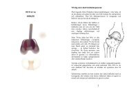
Fig. 1. The individually made molded rigid ‘‘Wessmark’’<br />
cup, which was used in the study.<br />
48<br />
Pilot group<br />
n = 5<br />
Untreated patients<br />
n = 15<br />
Randomised group<br />
n = 30<br />
Heel cup treated patients<br />
n = 15<br />
age was 11 years (range 9<strong>–</strong>14 years). Twenty-four children had<br />
bilateral pain and 11 children had unilateral heel pain (eight<br />
boys, three girls, five with pain in the right and six in the left<br />
foot). The painless children (n 5 15) were individually matched<br />
(same gender, age, activity level and playing in the same sports<br />
team) as the children in group I (Fig. 2). The children were<br />
recruited during a 6-month period.<br />
Inclusion and exclusion criteria<br />
Inclusion criteria were both gender, age 9<strong>–</strong>15 years, and the<br />
diagnosis of Sever’s injury, except for the children without<br />
symptoms who did not suffer from Sever’s injury. Findings<br />
confirming the diagnosis were tenderness over the lower onethird<br />
of the posterior calcaneus and a positive calcaneus<br />
compression test, the squeeze test, where all participants<br />
were examined by the first author. Heel pain had to be present<br />
for more than 2 weeks when examined. The most painful<br />
activity had to be a ball sport of some kind (soccer, floor ball,<br />
handball, etc.) with a pain level of 4 or more on the Borg CR-<br />
10 scale during the last week activities. The Borg scale is a self<br />
assessment VAS scale with anchors of verbal explanations<br />
added to the numbers in the pain scale (Neely et al., 1992).<br />
Zero represents absence of pain and 10 represents maximal<br />
pain. All participants had to be high-level athletes, measured<br />
with Engstrom’s five level activity score index (Engstro¨ m,<br />
2004). They had to master Swedish, and together with their<br />
parents they had to agree to participate.<br />
Exclusion criteria were intermittent pain, specified disease<br />
that could interfere with the heel pain, dominant pain from the<br />
Achilles tendon, earlier fracture in the area, a poorly defined<br />
ache in the lower extremities and participation in another<br />
study including any kind of treatment for pain.<br />
Radiographic examination <strong>–</strong> heel pad thickness<br />
The pilot group was only recruited to test the radiographic<br />
examination. In the true radiographic examination the thickness<br />
of the heel pad was measured (n 5 45) not including the<br />
pilot group, and the examinations were analyzed by the last<br />
author. The lower vertex of calcaneus was the bone reference<br />
mark, and a steel band fixated on the skin under the center<br />
portion of the heel was the skin reference mark. A 5 mm steel<br />
bullet marker was fixed on the medial side of the ankle as a<br />
reference, in order to calibrate each radiograph for each child.<br />
The heel pad thickness is defined as the distance between the<br />
lower vertex of the calcaneus and the steel band fixed under<br />
the heel. The measured error within the computer analysis was<br />
estimated to 0.2 mm. The radiographic investigator was partly<br />
blinded. The reliability test for heel pad thickness measure-<br />
Matched control group<br />
n = 15<br />
Fig. 2. Study design with two randomized groups with Sever’s injury. There was a 4 weeks intervention with heel cup in the<br />
treated group (group I). The untreated randomized control group (group II) is supposed to represent the natural course.<br />
2