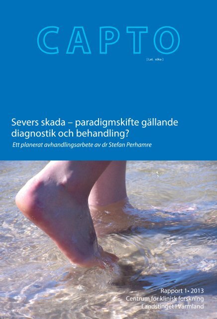
Severs skada – paradigmskifte gällande diagnostik och behandling?
Severs skada – paradigmskifte gällande diagnostik och behandling?
Severs skada – paradigmskifte gällande diagnostik och behandling?
You also want an ePaper? Increase the reach of your titles
YUMPU automatically turns print PDFs into web optimized ePapers that Google loves.
<strong>Severs</strong> <strong>skada</strong> <strong>–</strong> <strong>paradigmskifte</strong> <strong>gällande</strong><br />
<strong>diagnostik</strong> <strong>och</strong> <strong>behandling</strong>?<br />
Ett planerat avhandlingsarbete av dr Stefan Perhamre<br />
[ Lat. söka ]<br />
Rapport 1• 2013<br />
Centrum för klinisk forskning<br />
Landstinget i Värmland
<strong>Severs</strong> <strong>skada</strong> <strong>–</strong> <strong>paradigmskifte</strong><br />
<strong>gällande</strong> <strong>diagnostik</strong> <strong>och</strong> <strong>behandling</strong>?<br />
Ett planerat avhandlingsarbete av dr Stefan Perhamre<br />
Maria Klässbo 1 , Rolf Norlin 2<br />
1 medicine doktor, sjukgymnast, forskningsledare,<br />
Centrum för klinisk forskning<br />
Landstinget i Värmland, maria.klassbo@liv.se<br />
2 professor i ortopedi, Institutionen för hälsovetenskap <strong>och</strong> medicin<br />
Örebro universitet, rolf.norlin@orebroll.se<br />
Vår forskargrupp:<br />
Stefan Perhamre, leg läk, doktorand, Karlstad†.<br />
Maria Klässbo, med dr, Karlstad.<br />
Dagmara Lazowska, leg läk, Karlstad.<br />
Sofia Papageorgiou, leg läk, Karlstad.<br />
Fredrik Lundin, tekn dr, Karlstad.<br />
Staffan Janson, professor, Karlstad.<br />
Anders Lundin, med dr, Örebro.<br />
Rolf Norlin, professor, Örebro.<br />
Nyckelord:<br />
<strong>Severs</strong> <strong>skada</strong>, apofysit, calcaneus, inlägg, hälkopp<br />
Karlstad 2013
Rapporten kan beställas från<br />
Centrum för klinisk forskning<br />
Gamla Spinneriet - Älvgatan 49<br />
651 85 Karlstad<br />
054-61 70 75<br />
pia.tekin@liv.se<br />
Tryckt hos: Arkitektkopia, Karlstad 2013<br />
ISSN 1652-9324
Till Ola Wessmark, mister Idrottsmedicin i Värmland<br />
”Teorier vi tror på kallar vi fakta,<br />
fakta vi misstror kallar vi teorier.”<br />
okänd<br />
5
Innehåll<br />
SAMMANFATTNING ....................................................................................7<br />
ARTIKLAR .......................................................................................................8<br />
Definitioner <strong>och</strong> förkortningar .......................................................................9<br />
INLEDNING .................................................................................................10<br />
BAKGRUND .................................................................................................12<br />
SYFTE ............................................................................................................14<br />
MATERIAL ....................................................................................................15<br />
Deltagare delarbete I, IV <strong>och</strong> V ....................................................................16<br />
Deltagare delarbete II ...................................................................................17<br />
Deltagare delarbete III .................................................................................17<br />
METOD OCH RESULTAT ...........................................................................18<br />
Metod <strong>och</strong> resultat delarbete I ......................................................................18<br />
Metod <strong>och</strong> resultat delarbete II ....................................................................19<br />
Metod <strong>och</strong> resultat delarbete III ...................................................................23<br />
Hård hälkopp är bättre än kilklack .............................................................24<br />
Metod <strong>och</strong> resultat delarbete IV ...................................................................24<br />
Metod <strong>och</strong> resultat delarbete V ....................................................................26<br />
SLUTSATS .....................................................................................................27<br />
TACK .............................................................................................................28<br />
STEFAN PERHAMRES EGNA TACK I HANS<br />
PLANERADE AVHANDLING .....................................................................29<br />
REFERENSER ...............................................................................................32<br />
BILAGOR ......................................................................................................33<br />
Bilaga 1 ........................................................................................................33<br />
Bilaga 2 ........................................................................................................34<br />
Bilaga 3 ........................................................................................................35<br />
Bilaga 4 ........................................................................................................36<br />
Bilaga 5 ........................................................................................................41<br />
Bilaga 6 ........................................................................................................47
Sammanfattning<br />
Stefan Perhamre, idrottsläkare i Karlstad samarbetade tidigare med dr<br />
Ola Wessmark, <strong>och</strong> ortopedtekniker Ingemar Hansson vid Idrottshälsan,<br />
Landstinget i Värmland. Till Idrottshälsan sökte sig många barn <strong>och</strong> ungdomar<br />
med hälsmärta <strong>och</strong> de tyckte att en hård hälkopp fungerade påfallande<br />
bra som smärtlindring.<br />
Stefan Perhamre startade ett forskningsprojekt, tillsammans med Maria<br />
Klässbo, för att utvärdera hur bra hälkoppen verkligen var. När det blev<br />
klart att Stefan ville skriva en doktorsavhandling bildades vår forskargrupp<br />
kring hans avhandlingsprojekt.<br />
Med Stefans arbeten som grund har vi kunnat visa att diagnosen <strong>Severs</strong><br />
<strong>skada</strong> (hälsmärta hos unga) kan fastställas med vanliga kliniska test.<br />
Slätröntgen har inget diagnostiskt värde. Studierna har visat att den hälkopp<br />
Ola Wessmark tidigare designat, fungerar mycket bra som <strong>behandling</strong><br />
vid <strong>Severs</strong> <strong>skada</strong> <strong>och</strong> att orsaken till besvären rimligen är en överbelastningsreaktion<br />
i hälbenet. Vi har vidare kunnat visa att hälkoppen bidrar till<br />
att hälkudden trycks ihop från sidorna <strong>och</strong> blir lite tjockare under hälbenet<br />
<strong>och</strong> att toppbelastningen på hälen minskar.<br />
Stefan avled 2010, efter en tids sjukdom, men forskargruppen har kunnat<br />
gå vidare med Stefans artiklar så att de har publicerats.<br />
7
Artiklar<br />
Denna rapport är baserad på följande artiklar:<br />
I. Perhamre S, Lazowska D, Papageorgiou S, Lundin F, Klässbo M, Norlin<br />
R. Sever’s injury; a clinical diagnosis. J Am Podiatr Med Assoc, accepterad<br />
II. Perhamre S, Janson S, Norlin R, Klässbo M. Sever's injury: treatment<br />
with insoles provides effective pain relief. Scand J Med Sci Sports.<br />
2011;21(6):819-23<br />
III. Perhamre S, Lundin F, Norlin R, Klässbo M. Sever's injury; treat it<br />
with a heel cup: a randomized, crossover study with two insole alternatives.<br />
Scand J Med Sci Sports. 2011;21(6):e42-7<br />
IV. Perhamre S, Lundin F, Klässbo M, Norlin R. A heel cup improves the<br />
function of the heel pad in Sever's injury: effects on heel pad thickness,<br />
peak pressure and pain. Scand J Med Sci Sports. 2012;22(4):516-22<br />
V. Norlin R, Perhamre S, Lazowska D, Papageorgiou S, Klässbo M.<br />
Lundin A. MRI-findings in Sever’s injury are reduced during treatment:<br />
A randomised case-control study. Manuskript.<br />
Norlin R. <strong>Severs</strong> <strong>skada</strong> - lättbehandlad hälsmärta hos ungdomar. Svensk<br />
Idrottsforskning 2011;4:43-6.<br />
<strong>och</strong> Stefan Perhamres eget utkast till sin planerade avhandling.<br />
8
Definitioner <strong>och</strong> förkortningar<br />
VAS Visuell analog skala 0-10.<br />
Borg CR-10 VAS 0-10 med tillagda verbala ankare.<br />
IQR Kvartilsavstånd (Interquartile range), skillnaden mellan den<br />
tredje <strong>och</strong> första kvartilen. Kvartilavståndet anger inom vil<br />
k-et avstånd de 50% mittersta observationerna ligger (Q3-<br />
Q1).<br />
MRI/MRT Magnet Resonance Image/Tomography <strong>och</strong> kallas på sven-<br />
s ka för magnetkameraundersökning. Radiologisk metod<br />
som ger en avbildning av både skelett <strong>och</strong> mjukdelar.<br />
Diafys Skaftet, den mellersta delen, av ett rörben.<br />
Metafys Den del av ett långt rörben där övergången sker mellan<br />
skaft et <strong>och</strong> den förtjockade änden av benet.<br />
Fysen Den zon av änden på ett ben där längdtillväxten sker <strong>och</strong><br />
som ligger mellan metafys <strong>och</strong> epifys/apofys.<br />
Epifys De förtjockade ändarna av ett rörben, klädda med led-<br />
brosk, som ligger i anslutning till en led.<br />
Apofys De förtjockade ändarna av ett rörben som inte ligger i an-<br />
slutning till en led, exempelvis hälbenets bakersta del.<br />
9
Inledning<br />
Idrottshälsan startade år 1986 <strong>och</strong> var från 1989 organiserad i Friskvården i<br />
Värmland, men finansierad direkt av Landstinget i Värmland. År 2003 tog<br />
landstinget över ansvaret för Idrottshälsan. Idrottshälsan bemannades inledningsvis<br />
med idrottsläkare, sekreterare <strong>och</strong> assistent. År 2009 anställdes<br />
också en sjukgymnast. Till Idrottshälsan kan den som drabbats av skador i<br />
leder <strong>och</strong> muskler, som har aktiva motionsvanor eller är aktiv inom idrotten,<br />
vända sig utan remiss för att få hjälp med bedömning, <strong>behandling</strong>,<br />
träning <strong>och</strong> förslag på förebyggande åtgärder. Man kan också söka om man<br />
har allmänna medicinska frågeställningar, om det har relevans för den fysiska<br />
aktiviteten, t ex luftrörsbesvär, hjärt-, allergi- eller infektionsproblem.<br />
Barn <strong>och</strong> ungdomar som har hälsohinder som påverkar deras förmåga att<br />
vara fysiskt aktiva i skolan eller inom sina idrotter är en stor målgrupp för<br />
Idrottshälsan.<br />
Ola Wessmark, specialist i ortopedi, var den förste idrottsläkaren på<br />
Idrottshälsan. Många barn <strong>och</strong> ungdomar sökte Idrottshälsan för ömmande<br />
hälar. Ola beskriver själv att han ”av en slump” 1979 kom på att göra<br />
en avlastning av en ömmande häl i Ortoplastmaterial för att hjälpa en<br />
fotbollsspelande son, bilaga 1 1 . Resultatet var sensationellt skriver Ola vidare.<br />
Sonen kunde följande dag återuppta sin träning <strong>och</strong> behövde sen<br />
aldrig avstå träning eller match på grund av besvär från hälen. Wessmark<br />
gick sen vidare <strong>och</strong> gjorde, med hjälp av Ingemar Hansson som också<br />
var anställd på Idrottshälsan, många inlägg som kom att kallas hälkoppar.<br />
Något år senare gjorde Wessmark en telefonuppföljning av 46 barn, varav<br />
sju flickor. Medianåldern för flickorna var 11,6 år <strong>och</strong> för pojkarna 12,6<br />
år. Hela 39/46 hade kunnat återgå till träning/tävling utan fortsatta besvär<br />
då inläggen användes. Sex kunde delta i aktiviteter trots besvär, men med<br />
besvär i mindre grad. Endast en hade så stora besvär att han inte kunde<br />
delta i fysisk aktivitet.<br />
10
Idrottsläkarna vid Idrottshälsan, Landstinget i Värmland, Ola Wessmark <strong>och</strong> Stefan Perhamre.<br />
År 1993 började Stefan Perhamre arbeta på Idrottshälsan. Perhamre var<br />
specialist i allmänmedicin <strong>och</strong> hade ett mycket stort intresse för idrottsmedicin.<br />
Också Stefan slogs av den goda effekten av de hårda individgjutna<br />
hälkopparna. År 2000 gjorde Stefan en enklare utvärdering då han skickade<br />
en enkät med fem frågor <strong>och</strong> fick in svar från 83/115 barn (tabell 1).<br />
Tabell 1:<br />
Utvärdering av <strong>behandling</strong> med hälkopp hos barn med hälsmärta år 2000 (n=83).<br />
11
Bakgrund<br />
<strong>Severs</strong> <strong>skada</strong>, smärta i hälen som känns vid varje hälisättning <strong>och</strong> ökar med<br />
högre aktivitetsnivå, drabbar växande ungdomar, vanligen i åldern 8 <strong>–</strong> 15<br />
år. <strong>Severs</strong> <strong>skada</strong> är vanligast bland dem som är aktiva inom löpning <strong>och</strong><br />
fotboll.<br />
Redan 1907 beskrev den svenske röntgenläkaren Patrik Haglund hälsmärta<br />
av liknande karaktär 2 . Numera är tillståndet namngivet efter amerikanen<br />
James W Sever 3 .<br />
Genom åren har många teorier framförts både kring smärtorsak <strong>och</strong><br />
kring bästa <strong>behandling</strong>. Inflammation i hälbenets tillväxtzon (apophysitis<br />
calcanei) är en spridd förklaring 3 , men också ökat drag i hälsenan. Felställning<br />
i fot <strong>och</strong> underben har framförts som möjliga förklaringar. Ogden har<br />
lanserat synsättet att tillståndet är en överbelastnings<strong>skada</strong> i apofysen i det<br />
icke mogna hälen, därav benämningen <strong>Severs</strong> <strong>skada</strong> 4 .<br />
Smärta vid vissa aktiviteter är symptomet vid <strong>Severs</strong> <strong>skada</strong>. Ett tioårigt<br />
barn har oftast begränsad erfarenhet av smärta <strong>och</strong> att tänka abstrakt.<br />
Smärta hos barn har framför allt studerats när det gäller kroniska sjukdomar<br />
5,6,7 . Man har funnit att redan vid femårs ålder kan barn kvantifiera<br />
smärta <strong>och</strong> vid åtta års ålder värdera dessa kvaliteter. Eftersom det hos barn<br />
<strong>och</strong> ungdom kan finnas könsskillnader <strong>gällande</strong> smärta kan det vara enklare<br />
att välja att i studier inkludera enbart ett kön. Om man ska mäta smärta<br />
med hjälp av en enkät är det avgörande om mätmetoden har god validitet,<br />
mäter rätt sak. Visuell analog skala (VAS) anses vara ”gold standard” att<br />
använda för barn <strong>och</strong> ungdom. Borgs CR-10 skala är en vidareutveckling<br />
av VAS skalan med verbala ankare där noll representerar ingen smärta <strong>och</strong><br />
10 extrem smärta (bilaga 2) 8 . Den kliniska erfarenheten är att barn <strong>och</strong><br />
ungdomar med hälsmärta inte har några som helst svårigheter att använda<br />
VAS-skalan för att visa hur ont de har vid en viss aktivitet.<br />
Många olika <strong>behandling</strong>ar har förespråkats <strong>och</strong> använts genom åren.<br />
Då man misstänkt inflammation som grundorsak till besvären har allt från<br />
olika fysikaliska <strong>behandling</strong>ar till läkemedel <strong>och</strong> till <strong>och</strong> med gipsning använts.<br />
Man har även spekulerat i att felställning i foten kan utlösa besvären<br />
varför olika korrigerande inlägg har använts som <strong>behandling</strong>. Inte sällan<br />
behandlas <strong>Severs</strong> <strong>skada</strong> på samma sätt som man tidigare behandlat hälsenesmärta<br />
hos vuxna, till exempel med allt från töjningar till avlastande<br />
kilinlägg.<br />
12
Vila <strong>och</strong> olika sjukgymnastiska <strong>behandling</strong>ar har testats genom åren. För<br />
fysiskt aktiva barn <strong>och</strong> ungdomar kan rekommendationen vila <strong>och</strong> uppehåll<br />
med fysisk aktivitet upplevas mycket negativ.<br />
Åldern nio till 15 år anses vara den viktigaste åldern när det gäller att<br />
bygga upp skelett <strong>och</strong> neuromuskulär funktion med betydelse för resten<br />
av livet. Sportaktiviteter är också en källa till livsglädje <strong>och</strong> en kulturform<br />
med speciella traditioner <strong>och</strong> normer. Att förlora ett år eller mer på grund<br />
av hälsmärta kan ödelägga senare sportande. Lars-Magnus Engström har<br />
utvecklat ett aktivitetsindex för att kunna följa barn <strong>och</strong> ungdomars fysiska<br />
aktivitet (se bilaga 3) 9 .<br />
Om inte förr så försvinner problemet med <strong>Severs</strong> <strong>skada</strong> när hälbenets<br />
tillväxtzon växer samman med resten av hälbenet, alltså när längdtillväxten<br />
avstannar. Därför är ibland effekten av <strong>behandling</strong> svår att bedöma, särskilt<br />
hos de äldre barnen som är nära att avsluta sin längdtillväxt.<br />
Det finns ingen konsensus i litteraturen när det gäller <strong>diagnostik</strong> av<br />
<strong>Severs</strong> <strong>skada</strong> vid hälsmärta, inte hur man behandlar eller vad som orsakar<br />
smärtan.<br />
13
Syfte<br />
Syftet med studierna var att ge förslag på hur <strong>Severs</strong> <strong>skada</strong> kan diagnosticeras<br />
med kliniska test (delarbete I), behandlas med skoinlägg (delarbete<br />
II, III <strong>och</strong> bilagorna 4,5) <strong>och</strong> att ge tänkbara förklaringar till varför det ena<br />
alternativet, hälkoppen, hade överlägsna smärtlindrande effekter jämfört<br />
med hälkil (delarbete IV, V <strong>och</strong> bilaga 6).<br />
14
Material<br />
Delarbetena I, IV <strong>och</strong> V är baserade på studie 2 med datainsamling åren<br />
2008-2009 <strong>och</strong> delarbetena II <strong>och</strong> III är baserade på studie 1 med datainsamling<br />
2004-2005.<br />
Deltagare<br />
Tabell 2: Översikt över deltagarna i de olika delarbetena.<br />
15
Deltagare delarbete I, IV <strong>och</strong> V<br />
Deltagarna i studie 2, delarbetena I, IV <strong>och</strong> V, (n=45 sammanlagt) var dels<br />
barn med hälsmärta som randomiserades till <strong>behandling</strong>ar med två olika<br />
inlägg (n=30) <strong>och</strong> femton smärtfria matchade kontroller (tabell 2). De<br />
femton matchade kontrollerna hade samma kön, ålder, fysisk aktivitetsnivå<br />
<strong>och</strong> var aktiva i samma idrottsklubb som de med hälsmärta. Inga deltagare<br />
föll bort.<br />
Inklusionskriterier för alla 45 deltagare var:<br />
- pojkar eller flickor 9-15 år<br />
- nivå D eller E på Engströms aktivitetsindex (se bilaga 3) 9<br />
- kan göra sig förstådd på <strong>och</strong> förstår själv svenska utan problem<br />
- har förälders/föräldrars skriftliga samtycke till att delta<br />
För de 30 barn som skulle prova inlägg krävdes också att<br />
- de hade haft hälsmärta i distala delen av hälen i mer än två veckor<br />
- den mest smärtsamma aktiviteten måste vara bollsport i någon form (fotboll,<br />
innebandy, handboll eller liknande) med en smärtnivå på fyra eller<br />
mer, den senaste veckan, registrerat med Borgs CR-10 skala 8<br />
- de blivit medicinskt undersökta <strong>och</strong> konstaterats ha aktuellt problem<br />
Exklusionskriterier var:<br />
- smärta lokaliserad till Akillessenan eller hälbenet som helhet<br />
- smärta på natten<br />
- intermittent smärta<br />
- spridd smärta eller svullnad<br />
- annan specificerad sjukdom som kunde interferera med hälsmärtan<br />
- tidigare frakturer i området<br />
- svårdefinierad värk i nedre extremitet<br />
- medverkan i annan studie som innefattade någon typ av smärt<strong>behandling</strong><br />
Deltagarna rekryterades från olika idrottsklubbar dit information om studien<br />
skickades ut.<br />
I delarbete IV deltog, förutom de ovan nämnda 45 barnen, också en<br />
pilotgrupp av barn med hälsmärta (n=5), där man testade den röntgenologiska<br />
undersökningsmetodiken.<br />
16
Deltagare delarbete II<br />
Trettioåtta pojkar med hälsmärta uppfyllde inklusionskriternan, men tre<br />
ville inte delta i studien. I denna prospektiva randomiserade studie accepterade<br />
inledningsvis 35 pojkar (två fullföljde inte (n= 33)), med en medianålder<br />
på 12 år (range 9-14) med <strong>Severs</strong> <strong>skada</strong>, att delta (tabell 2). Arton<br />
randomiserades till att använda hälkil <strong>och</strong> 17 till hälkopp. Fem pojkar,<br />
medianålder 12 år (range 10-13 år), rekryterades konsekutivt till en kontrollgrupp<br />
som inte fick några inlägg. Alla deltagare var höggradigt fysiskt<br />
aktiva i olika idrotter.<br />
Inklusionskriterier var:<br />
- pojkar 9-15 år<br />
- hälsmärta i mer än två veckor <strong>och</strong>
Metod <strong>och</strong> resultat<br />
Alla barn som ingick i studierna fick inledningsvis ange ålder, längd, vikt<br />
<strong>och</strong> registrera sin fysiska aktivitetsnivå från A-E med Engströms aktivitetsindex<br />
(se bilaga 3) 9 . Den fysiska aktivitetsnivån registrerades sedan vid<br />
varje datainsamlingstillfälle.<br />
Studierna har godkänts av etikprövningsnämnden i Uppsala, dnr 2004<br />
M-377 <strong>och</strong> 2008/094. Alla deltagare gav sitt informerade samtyckte till<br />
att delta i studierna. I delarbete IV användes radiologi för att analysera<br />
hälkuddens tjocklek. Av etiska skäl undersöktes först en pilotgrupp (n=5)<br />
för att tillse att undersökningsmetodiken var tillfredställande.<br />
Metod <strong>och</strong> resultat delarbete I<br />
Syftet med delarbete I var att undersöka diagnostisk precision av olika<br />
kliniska test <strong>gällande</strong> hälsmärta vid <strong>Severs</strong> <strong>skada</strong>. Barnen i delarbete I, 30<br />
ungdomar med symtom <strong>och</strong> 15 symtomfria, fick svara på frågor om ålder,<br />
längd, vikt, vilken sport de utövade <strong>och</strong> fysisk aktivitetsnivå från A-E med<br />
Engströms aktivitetsindex (se bilaga 3) 9 . Barnen med hälsmärta fick ange<br />
hur länge de haft hälsmärta <strong>och</strong> smärta i höger/vänster/bilteralt.<br />
Tre kliniska test utfördes av Stefan Perhamre på alla barn (n=45). I de<br />
fall smärtan var bilateral fick barnet själva avgöra vilken häl som var mest<br />
smärtande <strong>och</strong> på den utfördes testerna. Test ett (A) var barfota hälstående,<br />
test två (B) tuberkelpalpation (tryck på hälbenets bakersta topp) <strong>och</strong> test<br />
tre (C) sidokompression (eng. squeeze test), figur 1. Upplevd smärta vid<br />
testerna registrerades på Borgs CR-10 skala 8 . Testaren (SP) tränade trycket<br />
vid sidokompression (test C) på en tryckkänslig gummiblåsa (vigorimeter)<br />
för att standardisera tryckets storlek.<br />
Figur 1: De tre kliniska testen som användes för att diagnosticera <strong>Severs</strong> <strong>skada</strong>.<br />
18
En slätröntgenbild med lateral projektion togs på alla barn (n=45). För<br />
barnen med hälsmärta avbildades den mest smärtande hälen. Två röntgenologer<br />
analyserade, oberoende av varandra <strong>och</strong> blindade för hälsmärta<br />
eller ej, graden av skleros (ökad täthet) på en fyrgradig-skala; ingen skleros,<br />
lätt, måttlig <strong>och</strong> höggradig skleros. De bedömde också graden av fragmentering<br />
av tillväxtzonen (apofysen) också det på en fyrgradig-skala; ingen<br />
fragmentering, två, tre eller minst fyra fragment. Förekomst av distala fragment<br />
eller inte noterades också.<br />
De matchade kontrollbarnen testades på samma sätt både med de kliniska<br />
testen <strong>och</strong> med slätröntgenundersökning.<br />
I delarbete I beräknades såväl sensitivitet (testets förmåga att skilja ut<br />
de sjuka individerna), specificitet (testets förmåga att skilja ut de friska<br />
individerna), negativt <strong>och</strong> positivt prediktivt värde för de tre kliniska testen<br />
<strong>och</strong> för de röntgenologiska (skleros, fragmentering <strong>och</strong> förekomst av<br />
distala fragment). För de röntgenologiska testerna beräknades sensitivitet<br />
<strong>och</strong> specificitet med olika cut-off värden där den kombination som gav<br />
bäst värden för båda valdes, där sensitivitet ansågs vara viktigare än specificitet.<br />
Inledningsvisa skillnader mellan grupperna analyserades med Fishers<br />
exakta test för data på nominal- <strong>och</strong> ordinalskalenivå <strong>och</strong> med Students<br />
t-test för övriga.<br />
Tre bra kliniska test vid <strong>Severs</strong> <strong>skada</strong> medan röntgenförändringar är<br />
svårtolkade<br />
Alla tre testen; barfota hälstående, tuberpalpation <strong>och</strong> sidokompression<br />
(eng. squeeze test) (figur 1) uppvisade 100 procents specificitet vilket<br />
innebär att alla tre testen kunde identifiera alla de friska ungdomarna som<br />
friska. När det gäller sensitivitet hade hälstående 100 procent, tuberpalpation<br />
80 procent <strong>och</strong> sidokompression 97 procents träffsäkerhet.<br />
Röntgen av hälen påvisade att skleros fanns hos alla ungdomar, både<br />
symtomatiska <strong>och</strong> asymtomatiska i kontrollgruppen. Även vid gradering<br />
av sklerosen noterades samma mönster i båda grupperna. Fragmentering<br />
var vanligare i gruppen med besvär (87 procent) jämfört med kontrollgruppen<br />
(53 procent). Denna skillnad var signifikant (p=0,01). Samstämmighet<br />
mellan de båda radiologernas bedömning (reliabilitet) var 100 procent<br />
för både skleros <strong>och</strong> fragmentering.<br />
Metod <strong>och</strong> resultat delarbete II<br />
Syftet med delarbete II var att undersöka om hälinlägg, hälkopp <strong>och</strong><br />
hälkil, kunde minska smärtan hos pojkar med hälsmärta, <strong>Severs</strong> <strong>skada</strong><br />
(bilaga 4) 12 . Trettioåtta pojkar uppfyllde inklusionskriterierna. Tre föll bort<br />
av olika skäl. Resterande 35 pojkar, mellan 9 <strong>och</strong> 14 år, fick slumpmässigt<br />
19
ehandling med antingen hälkopp (n=17) eller en avlastande hälkil (n=18)<br />
(figur 2). De slumpandes genom att ta en lapp i en låda försedd med lock.<br />
Deltagarna fick välja två fysiska aktiviteter; aktivitet A <strong>och</strong> B, där hälen<br />
smärtade <strong>och</strong> som de utövade minst 3 gånger per vecka. Aktivitet A skulle<br />
vara den som gav mest smärta. Under de två första veckorna efter randomisering<br />
gavs ingen <strong>behandling</strong>. Ungdomarna registrerade själva smärta<br />
vid aktivitet A <strong>och</strong> B (Borgs CR-10 skala) <strong>och</strong> aktivitetsnivå (Engströms<br />
aktivitetsindex), se bilaga 3 <strong>och</strong> figur 3. Därefter använde hälften av ungdomarna<br />
hälkopp <strong>och</strong> hälften hälkil under 4 veckor. Slutligen följde en ny<br />
två-veckorsperiod utan <strong>behandling</strong>.<br />
För att få en uppfattning om naturalförloppet rekryterades konsekutivt<br />
fem patienter med <strong>Severs</strong> <strong>skada</strong> som inte fick några inlägg. De fick registrera<br />
smärta <strong>och</strong> aktivitetsnivå vid två tillfällen vid start <strong>och</strong> efter två månader.<br />
Figur 2: Hälkopp eller hälkil som ska användas i en stadig <strong>och</strong> knuten idrottssko.<br />
20
Figur 3. Studiedesign för delarbete II.<br />
21
I delarbete II beräknades mediansmärta för varje pojke i alla tre faser av<br />
studien, före, under <strong>och</strong> efter <strong>behandling</strong>, <strong>och</strong> separat för aktivitet A <strong>och</strong><br />
B. Mediansmärta <strong>och</strong> kvartilsavvikelse för hela gruppen (n=33) beräknades<br />
sen för varje fas <strong>och</strong> aktivitet. Wilcoxons signed-rank test användes för att<br />
räkna ut förändring över tid separat för aktivitet A <strong>och</strong> B, med jämförelser<br />
mellan faserna, se figur 5. Holms sekventiella Bonferroni test användes för<br />
att korrigera för massignifikans.<br />
Figur 5: Staplar som visar smärta (median) i faserna före, under <strong>och</strong> efter <strong>behandling</strong> för de två självvalda<br />
smärtande fysiska aktiviteterna i studiegruppen (n=33) där A är den mest smärtande, delarbete II. Jämförelser<br />
mellan mediansmärta i faserna presenteras med p-värden.<br />
Inlägg reducerar smärtan påtagligt<br />
Resultatet blev att, trots att alla ungdomar bibehöll en hög fysisk aktivitetsnivå<br />
(Engströms aktivitetsnivå D <strong>och</strong> E), besvären reducerades signifikant<br />
under den period de använde något av inläggen. Mätt med Borgs CR-10<br />
skala (visuell analog skala mellan 0 <strong>och</strong> 10) sjönk smärtan från cirka 4,5 till<br />
2. Efter <strong>behandling</strong>sperioden återkom smärtan, om än på en något lägre<br />
nivå än före <strong>behandling</strong>en, cirka 3,5.<br />
I den obehandlade kontrollgruppen skedde ingen påtaglig förändring<br />
av smärtnivån under de 8 veckornas observation. En eventuell skillnad i<br />
<strong>behandling</strong>seffekt mellan inläggen kunde inte säkert bedömas.<br />
22
Metod <strong>och</strong> resultat delarbete III<br />
Syftet med delarbete III var att analysera vilket inlägg, hälkil eller hälkopp,<br />
som gav mest smärtlindring hos ungdomar med hälsmärta, <strong>Severs</strong> <strong>skada</strong><br />
(bilaga 5) 13 .<br />
Fyrtiofyra ungdomar med typiska hälsmärtor randomiserades till två<br />
olika <strong>behandling</strong>sgrupper. Ungdomarna följdes under totalt 26 veckor.<br />
Efter en två veckor lång basal bedömningsperiod utan <strong>behandling</strong> (B1)<br />
randomiserades de antingen att få hälkopp (n=20) eller hälkil (n=24) att<br />
använda i fyra veckor i sina sportskor (vecka 3-6). Därefter följde en två<br />
veckor lång basperiod utan <strong>behandling</strong> (B2), varefter de fick använda den<br />
andra typen av inlägget under fyra veckor (vecka 9-12) i en cross-over design.<br />
Slutligen fick de efter en tredje basperiod (B3) utan <strong>behandling</strong>, fritt<br />
välja det inlägg de tyckte var bäst eller välja att vara utan inlägg. Denna<br />
tredje, valfria <strong>behandling</strong>speriod var 12 veckor lång (figur 4). Efter ett år<br />
fick deltagarna svara på frågor om grad av smärtlindring under studiens<br />
26 veckor, typ av inlägg de använt <strong>och</strong> smärtlindringsgrad under året. Tre<br />
pojkar svarade inte (n=41).<br />
Figur 4: Design av jämförande studie mellan hälkopp <strong>och</strong> hälkil, delarbete III.<br />
En ordinal mixad regression, där den initiala smärtan modellerades med<br />
tid <strong>och</strong> <strong>behandling</strong> som förklarande variabler, utfördes. Effekten av varje<br />
förklarande variabel var en oddskvot (OR), som representerade den multiplicerade<br />
effekten av förändring i varje variabel. Självrapporterad smärta<br />
(Borgs CR-10, 0-10) kategoriserades till fem nivåer (0-2, 3-4, 5-6, 7-8<br />
<strong>och</strong> 9-10) <strong>och</strong> smärtnivån vid varje mättillfälle modellerades genom att<br />
använda initial smärta <strong>och</strong> <strong>behandling</strong> som fixed effekt, antal dagar sedan<br />
baslinje 1-3 som en fixed <strong>och</strong> random effekt <strong>och</strong> patient som en random<br />
intercept. Låga oddskvoter tranformerades till mindre smärta <strong>och</strong> höga<br />
oddskvoter till mer smärta.<br />
23
Som ett beskrivande komplement till regressionsanalyserna konstruerades<br />
ett diagram med smärta (median med kvartilsavstånd) för varje fas<br />
(figur 6). Smärtlindringen för hälkil <strong>och</strong> hälkopp jämfördes inkluderande<br />
båda de valda fysiska aktiviteterna.<br />
Figur 6: Separerade resultat, median <strong>och</strong> kvartilsavstånd, för grupperna som randomiserats till att använda<br />
hälkopp <strong>och</strong> hälkil i delarbete III. Figuren visar de tre baslinjefaserna före <strong>behandling</strong>, efter första randomiserade<br />
alternativet <strong>och</strong> efter det andra då inlägg byttes (cross-over). Grupp I (vita staplar) startade med hälkopp<br />
<strong>och</strong> bytte sen till hälkil. Grupp II (gråa staplar) startade med hälkil. Resultatet innefattar smärtskattningar<br />
från både den mest smärtande aktiviteten, A, <strong>och</strong> den näst mest smärtande aktiviteten, B.<br />
Hård hälkopp är bättre än kilklack<br />
Utvärdering visade att de som använde hälkopp upplevde signifikant mindre<br />
smärta än de som använde kil. Detta visades också i att 77 procent av<br />
ungdomarna valde att fortsätta med hälkoppen. Ingen valde, vid det fria<br />
valet, att avstå från <strong>behandling</strong>.<br />
Metod <strong>och</strong> resultat delarbete IV<br />
Syftet med delarbete IV var att studera två tänkbara orsaker till varför<br />
hälkoppen ger så bra smärtlindring omedelbart när ungdomarna börjar<br />
använda den 14 . Vi ville studera om hälkudden ökade i tjocklek när man använde<br />
hälkoppen, vilket borde reducera de direkta tryckkrafterna på hälbenet<br />
vid belastning med kroppsvikten. Dessutom ville vi mäta fördelningen<br />
av tryckkrafterna under foten vid löpning. Bland 30 ungdomar med <strong>Severs</strong><br />
24
<strong>skada</strong> randomiserades 15 till <strong>behandling</strong> med hälkopp <strong>och</strong> 15 fick ingen<br />
<strong>behandling</strong>. Ytterligare 15 ungdomar utan hälsmärta var kontrollgrupp.<br />
Hälkuddens tjocklek mättes på en röntgenbild (sidobild tagen stående<br />
på ett ben, hälstående, på den mest smärtande hälen på de med hälsmärta<br />
(n=30) <strong>och</strong> på samma häl för de matchade kontrollerna de utan hälsmärta<br />
(n=15)). Tre bilder togs; barfota (alla 45 ungdomar), med idrottssko (alla<br />
45 ungdomar)<strong>och</strong> med hälkopp i idrottssko (enbart <strong>behandling</strong>sgruppen,<br />
n=30).<br />
Med mätutrustning (F-Scan från Tek scan) för tryckmätning kunde av<br />
ekonomiska skäl endast 10 ungdomars belastningsmönster under foten<br />
analyseras. Dessa 10 valdes ur <strong>behandling</strong>sgruppen. En tryckkänslig film<br />
registrerade trycket inom varje kvadratcentimeter under hela foten. Tre<br />
mätningar gjorde under hälstående. Dessutom mättes belastningsmönstret<br />
under foten under nio löpsteg med löphastighet 100 (metronom). Dessa<br />
mätningar gjordes både med enbart idrottssko <strong>och</strong> med hälkopp i idrottsskon.<br />
Maximal belastning noterades under häl, framtrampet <strong>och</strong> under<br />
stortån.<br />
De röntgenologiska mätningarna, tjocklek i millimeter (medelvärde),<br />
konfidensintervall <strong>och</strong> p-värden, analyserades med Students t-test. En linjär<br />
mixad regressionsmodell användes för att analysera toppbelastning som<br />
utfallsvariabel, där varje barn användes som random effekt <strong>och</strong> löpning 1,<br />
2, 3 <strong>och</strong> hälkopp eller ej som fixed effekt. I båda fallen användes de logaritmerade<br />
värdena av toppbelastning.<br />
Hälkoppen ger tjockare hälkudde <strong>och</strong> lägre maximal tryck under foten<br />
I stående skiljde inte hälkuddens tjocklek mellan symtomatiska ungdomar<br />
<strong>och</strong> de i den symtomfria kontrollgruppen. Hälkuddens tjocklek ligger i<br />
denna grupp ungdomar mellan cirka 6,5 <strong>och</strong> 8 millimeter. Med enbart<br />
idrottssko på foten ökade hälkuddens tjocklek med drygt 2 millimeter,<br />
vilket kan förklara att många ungdomar upplever mindre besvär när de<br />
använder bra skor. Med hälkopp i idrottsskon ökade hälkuddens tjocklek<br />
ytterligare drygt 1,5 millimeter vilket kan förklara varför ungdomarna<br />
upplever ännu mindre smärta när de också har hälkoppen på plats (Tabell<br />
3).<br />
Tabell 3: Hälkuddens tjocklek vid stående på ett ben, hälstående, delarbete IV.<br />
25
Dataanalys visade att maximal tryckbelastning under hälstående minskade<br />
med 25% när hälkopp användes, jämfört med tryckvärdet när enbart<br />
idrottssko användes. Under löpning minskade maximal belastning med<br />
21% om hälkopp användes. Även denna signifikanta minskning av det<br />
maximala trycket kan förklara den minskning av belastningssmärta som<br />
ungdomarna noterar när de använder hälkoppen.<br />
Metod <strong>och</strong> resultat delarbete V<br />
Syftet med delarbete V var att, med magnetkamera (MR), undersöka hälarna<br />
på ungdomar med <strong>och</strong> utan hälsmärta <strong>och</strong> även undersöka de med<br />
hälsmärta efter att de behandlats med hälkopp. Samma 45 ungdomar<br />
som i studie I <strong>och</strong> IV, där trettio av dem hade hälsmärta, ingick i studien.<br />
Femton av de 30 med besvär hade fått <strong>behandling</strong> med hälkopp <strong>och</strong> 15<br />
hade varit utan <strong>behandling</strong>. Alla genomgick undersökning med MR både<br />
före <strong>behandling</strong>sstart <strong>och</strong> en månad senare. Även kontrollgruppen (n=15)<br />
genomgick MR.<br />
Icke parametriska metoder, Wilcoxons rank sum test <strong>och</strong> Kruskal-Wallis<br />
test användes för att jämföra MR fynden mellan patienter <strong>och</strong> kontroller<br />
<strong>och</strong> före <strong>och</strong> efter <strong>behandling</strong> med hälkopp.<br />
Magnetkameraundersökning (MR) påvisar tecken på skaderelaterad skelettpåverkan<br />
Preliminära data, från denna ännu ej publicerad studie, visar att alla<br />
ungdomar, alltså även symtomfria, hade viss grad av ödem i hälbenet. Dock<br />
har de med <strong>Severs</strong> <strong>skada</strong> signifikant mer ödem. Detta ödem reduceras signifikant<br />
efter en månads <strong>behandling</strong> (figur 7) då också belastningssmärtan<br />
minskat påtagligt.<br />
Figur 7: Magnetkamerabilder, delarbete V.<br />
26
Slutsats<br />
Diagnosen <strong>Severs</strong> <strong>skada</strong> kan fastställas vid ett vanligt besök hos läkare eller<br />
sjukgymnast där belastningssmärta strikt lokaliserad till hälen, tydlig smärta<br />
vid hälstående, smärta vid sidokompression av hälen <strong>och</strong> vanligen också<br />
tryckömhet över tuber (mitt bak på hälknölen) är typiska fynd. Dessa tre<br />
test kan mycket väl skilja sjuka från friska ungdomar när det gäller <strong>Severs</strong><br />
<strong>skada</strong>. Trots skillnader i graden av fragmentering av hälbenet är det inte<br />
särskilt meningsfullt att använda vanlig röntgen för att säkerställa diagnosen<br />
<strong>Severs</strong> <strong>skada</strong>.<br />
Inläggs<strong>behandling</strong>, hälkil eller hälkopp, reducerar hälsmärta påtagligt<br />
så att en hög fysisk aktivitetsnivå kan bibehållas. Behandling med styv hälkopp,<br />
som formas direkt på hälen, ger snabb symtomlindring <strong>och</strong> bättre<br />
smärtlindring för de flesta jämfört med hälkil.<br />
Hälkoppen ökar hälkuddens tjocklek <strong>och</strong> minskar det maximala trycket<br />
på hälen. Besvären försvinner alltid när hälbenets tillväxt avstannar, men<br />
med <strong>behandling</strong> med hälkopp minskar eller försvinner besvären mycket<br />
snabbare utan att begränsa barnens fysiska aktivitet.<br />
<strong>Severs</strong> <strong>skada</strong> kan mycket väl vara en ren belastningsutlöst <strong>skada</strong> på hälbenet<br />
hos växande ungdomar som idrottar. Magnetkameraundersökning<br />
visar ett klart ökat ödem i hälbenet <strong>och</strong> detta ödem minskar under <strong>behandling</strong>,<br />
parallellt med att smärtan minskar.<br />
27
Tack<br />
Tack för finansiellt stöd till:<br />
Landstinget i Värmland; Primärvårdens FoU-enhet, Division hälsa, habilitering<br />
<strong>och</strong> rehabilitering, Centrum för klinisk forskning <strong>och</strong> Fonden för<br />
ortopedtekniska hjälpmedel. Tack också till Karlstad universitet, Örebro<br />
universitet <strong>och</strong> Centrum för idrottsforskning.<br />
28
Stefan Perhamres egna tack i<br />
hans planerade avhandling<br />
I would like to express my sincere and warm gratitude to:<br />
Rolf Norlin, my tutor and co-author who accepted me as a doctoral student<br />
when he started as professor at Örebro University. Thank you for<br />
guiding me through the project with lots of wisdom, support and patience.<br />
Maria Klässbo, my co-tutor and co-author who alone had to carry the<br />
burden guiding a scientific blueberry in the beginning of my scientific<br />
journey. Your work capacity, patience and accuracy will take you far.<br />
Staffan Janson, my co-author in the first paper, professor in two disciplines,<br />
and the visionary humanist who started up the local scientific and progressive<br />
group in Värmland County Council, where I later was included.<br />
You showed me the way.<br />
Fredrik Lundin, the appreciated statistician and co-author, who has been<br />
the guarantee for correct data analyses.<br />
Ingemar Hansson, who has produced all the heel cups in the studies. We<br />
have had a close relationship through all these years. Your loyalty has been<br />
unbeatable.<br />
Christer Carlmark, art responsible at the Centre for Public Health Research,<br />
Värmland County Council and Klara Perhamre, my daughter, for<br />
help with the lay-out of some figures.<br />
Ola Wessmark, the former chief doctor at our clinic, who was an intelligent<br />
clinical model for me during our first years together at the clinic. You<br />
made the first moulded Orfit heel cup. Without this prototype there would<br />
have been no scientific work by me on this item.<br />
Dagmara Makowska (Lazowska) and Sofia Papageorgiou, radiologists<br />
at the Central Hospital Karlstad, who did a professional job examining the<br />
radiograph pictures in Paper IV.<br />
29
Lars Hynsjö, M. Sc at the department of Chemistry, Sahlgrenska University<br />
Hospital for his stimulating input with multi-variat analyses of the<br />
topics.<br />
Freddy Gustavsen, former chief of the company Adapt AB, who has been<br />
an important enthusiast and discussion partner. Your great network and<br />
generosity has been much worth.<br />
Carola Dimitrov¸ product manager CA Mätsystem AB for initiated support<br />
in the peak pressure measurements and analyses.<br />
Mikael Hasselgren, medical chief of the Centre for Clinical Science, for<br />
his faith in this project and decisions of financial support from Värmland<br />
County Council.<br />
Eva Stjernström, divisional chief of the Värmland County Council for<br />
her faith in this project and financial support despite a difficult economic<br />
situation in the Division of Health, Habilitation and Rehabilitation,<br />
Värmland County Council .<br />
Göran Ejlertsson, professor in Public Human Science, who helped me in<br />
my academic statistical studies.<br />
Ulf Tidefelt and Gunilla Ahlsén, prefect and study headmistress of Örebro<br />
University, for solid support all the way through.<br />
Kerstin Granström and Margaretha Warnqvist, two excellent secretaries<br />
at the Sports Clinic and the Scientific Unit for friendship, help and support.<br />
We have been “tight-knit families” at work.<br />
Gordon Riemersma, GP in Karlstad and the examiner of our English.<br />
Håkan Alfredson, professor in Sports Medicine Umeå University for a<br />
solid friendship and active support in this project.<br />
Peter Nilsson, chief of the Orthopaedic device Company in association<br />
with Värmland County Council for your interest in developing science in<br />
this field.<br />
30
Lars Magnus Engström, former professor Dep. of Social and Cultural<br />
Studies in Education, Stockholm Institute of Education for support with<br />
the questionnaires.<br />
Volunteers and patients who participated in this study.<br />
My family, Anneli, Klara, Axel and Maja, for everything.<br />
31
Referenser<br />
1 Wessmark O. Apofysitis calcanei, Haglund-<strong>Severs</strong> sjukdom. Idrottsme-<br />
dicin 1987;3:10<br />
2 Haglund P. Archiv für klinische Chirurgie 1907;82:922-30<br />
3 Sever JW. Apophysitis of the os calcis. New York Medical Journal<br />
1912;95:1025-9<br />
4 Ogden JA, Ganey TM, Hill JD, Jaakkola JI. Sever’s injury: A stress fracture<br />
of the immature calcaneal metaphysis. J Pediatr Orthop 2004;24:488<strong>–</strong>92<br />
5 McGrath P, Finley GA. Measurement of Pain in Infants and Children.<br />
Seattle: IASP Press 1998: 210<br />
6 McGrath P, Finley GA. Chronic and Recurrent Pain in Children and<br />
Adolescents. Seattle: IASP Press 1999: 288<br />
7 O´Rourke D. The measurement of pain in infants, children, and adolescents:<br />
From policy to practice. Phys Ther. 2004:84:560-70<br />
8 Neely G, Ljunggren G, Sylén C, Borg G. Comparison between the<br />
Visual Analogue Scale (VAS) and the Category Ratio Scale (CR-10) for<br />
evaluation of leg exertion. Int J Sports Med 1992;13:133-6<br />
9 Engström LM. Barns <strong>och</strong> ungdomars idrottsvanor i förändring. Svensk<br />
Idrottsforskning 2004;4:1-6<br />
10 Perhamre S, Lazowska D, Papageorgiou S, Lundin F, Klässbo M, Norlin<br />
R. Sever’s injury; a clinical diagnosis. Accepted J Am Podiatr Med Assoc<br />
11 Perhamre S, Janson S, Norlin R, Klässbo M. Sever's injury: treatment<br />
with insoles provides effective pain relief. Scand J Med Sci Sports<br />
2011;21(6):819-23<br />
12 Perhamre S, Lundin F, Norlin R, Klässbo M. Sever's injury; treat it with a<br />
heel cup: a randomized, crossover study with two insole alternatives. Scand<br />
J Med Sci Sports 2011;21(6):e42-7<br />
13 Perhamre S, Lundin F, Klässbo M, Norlin R. A heel cup improves the<br />
function of the heel pad in Sever's injury: effects on heel pad thickness,<br />
peak pressure and pain. Scand J Med Sci Sports 2012;22(4):516-22<br />
14 Norlin R, Perhamre S, Lazowska D, Papageorgiou S, Klässbo M, Lundin<br />
A. MRI-findings in Sever’s injury are reduced during treatment: A randomised<br />
case-control study. Submitted<br />
32
Bilagor<br />
Att göra en hälkopp <strong>–</strong><br />
Bilaga 1<br />
bästa <strong>behandling</strong>en vid <strong>Severs</strong> <strong>skada</strong> (apofysit i calcaneus).<br />
Plastmaterialet ORFIT Classic Soft 3,2 mm tjock (ej perforerat) används. En skiva (60 x 90<br />
cm) brukar räcka till 10 inlägg <strong>och</strong> köps i Sverige via Össur (art.nr. 671 48 88).<br />
Barnet får ställa foten på plattan <strong>och</strong> lämpligt område ritas ut. Därefter klipps detta ut. Plattan<br />
läggs i varmt vattenbad (45 grader) under 1 minut (t.ex. Hydrokollator, Össur), varefter den<br />
direkt läggs på den obelastade foten. Plasten formas manuellt med särskild tillklämning kring<br />
hälen för att hålla hälkudden på plats. När plasten är modellerad fixeras den med elastisk linda<br />
mot foten <strong>och</strong> barnet får sitta stilla med obelastad fot i 6 minuter. Därefter klipps inlägget till<br />
så att det passar foten.<br />
Efter detta är inlägget färdigt att användas. Barnet får gå med inlägget för att kontrollera att<br />
det inte skaver. Inlägg görs alltid till båda fötter, även vid ensidiga besvär.<br />
Total tillverkningstid cirka 30 min/par.<br />
Särskilda synpunkter:<br />
Undvik det perforerade materialet eftersom dessa ofta spricker.<br />
För tunga personer kan detta material vara för tunt <strong>och</strong> spricka.<br />
Inlägget kan även användas vid hälsporre.<br />
Ingemar Hansson lär gärna ut hur detta går till i praktiken.<br />
Hans kontaktuppgifter är:<br />
Idrottshalsan.karlstad@liv.se<br />
070-340 94 07<br />
Ingemar Hansson/Rolf Norlin<br />
28<br />
33
Bilaga 2<br />
34<br />
Namn: Testnummer: v 1<br />
Dessa frågor skall du besvara för de aktiviteter du valt varje vecka under hela undersökningen. Du tar med det<br />
ifyllda protokollet när du kommer till Idrottshälsan <strong>och</strong> behåller det sedan för nästa period. Två siffror för smärta<br />
<strong>och</strong> två siffror för hur foten fungerar skall fyllas i varje vecka. ( Fyll i dina två valda aktiviteter på de streckade<br />
raderna).<br />
Hur ont gör det?<br />
För att kunna beskriva smärtan för aktivitet 1 För att kunna beskriva smärtan för aktivitet 2<br />
......................................................skall du ......................................................skall du<br />
ange en siffra från 0 till 10 eller om det är ange en siffra från 0 till 10 eller om det är<br />
maximalt anges det med ett +. Ringa in en maximalt anges det med ett +. Ringa in en<br />
siffra som passar bäst för aktiviteten denna vecka. siffra som passar bäst för aktiviteten denna<br />
vecka.<br />
0 Ingen alls 0 Ingen alls<br />
0.5 Extremt svag (knappt kännbar) 0.5 Extremt svag (knappt kännbar)<br />
1 Mycket svag 1 Mycket svag<br />
2 Svag (lätt) 2 Svag (lätt)<br />
3 Måttlig 3 Måttlig<br />
4 4<br />
5 Stark (kraftig) 5 Stark (kraftig)<br />
6 6<br />
7 Mycket stark 7 Mycket stark<br />
8 8<br />
9 9<br />
10 Extremt stark ( nästan maximal) 10 Extremt stark ( nästan maximal)<br />
+ Maximal + Maximal<br />
Hur stora problem har du?<br />
Om foten / fötterna fungerar utan problem Om foten / fötterna fungerar utan problem<br />
betyder det 100 poäng. Hur många poäng betyder det 100 poäng. Hur många poäng<br />
får din fot / dina fötter på en skala från får din fot / dina fötter på en skala från<br />
0 till 100 för aktivitet 1............................... 0 till 100 för aktivitet 2...............................<br />
denna vecka ? denna vecka ?<br />
...................................poäng ...................................poäng<br />
29
Namn: Testnummer:<br />
Hur fysiskt aktiv är du? (v 0 )<br />
Bilaga 3<br />
Vilken person stämmer bäst in på dig?<br />
Person A: Rör sig ganska lite.<br />
Person B: Rör sig en hel del men aldrig så att han/hon blir andfådd <strong>och</strong><br />
svettig.<br />
Person C: Rör sig en hel del <strong>och</strong> blir svettig <strong>och</strong> andfådd någon gång ibland.<br />
Person D: Rör sig så att han/hon blir svettig <strong>och</strong> andfådd flera gånger i<br />
veckan.<br />
Person E: Rör sig så att han/hon blir svettig <strong>och</strong> andfådd varje dag eller<br />
nästan varje dag.<br />
Hur länge har besvären funnits?<br />
Hur lång tid har du haft ont i hälen/hälarna i sådan omfattning att det begränsat<br />
dig fysiskt?<br />
I ...................veckor<br />
30<br />
35
Bilaga 4<br />
Scand J Med Sci Sports 2011: 21: 819<strong>–</strong>823<br />
doi: 10.1111/j.1600-0838.2010.01051.x<br />
Sever’s injury: treatment with insoles provides effective pain relief<br />
S. Perhamre 1 , S. Janson 2,3 , R. Norlin 4 , M. Kla¨ ssbo 5<br />
1 Centre of Sports Medicine in Va¨rmland, Va¨rmland County Council, Karlstad, Sweden, 2 Department of Paediatrics, Va¨rmland<br />
County Council, Karlstad, Sweden, 3 Department of Public Health, Karlstad University, Karlstad, Sweden, 4 Department of<br />
Orthopaedics, University Hospital, O¨rebro University, O¨rebro, Sweden, 5 Centre for Clinical Research, Va¨rmland County Council,<br />
Karlstad, Sweden<br />
Corresponding author: Stefan Perhamre, Idrottsha¨lsan i Va¨rmland, Kolvgatan 1 B, S-653 41 Karlstad, Sweden. Tel: 146 54 15 34 12,<br />
Fax: 146 54 15 58 98, E-mail: stefan.perhamre@liv.se<br />
Accepted for publication 7 October 2009<br />
Sever’s injury (apophysitis calcanei) is considered to be the<br />
dominant cause of heel pain among children between 8 and<br />
15 years. The traditional advice is to reduce and modify the<br />
level of physical activity. Recommended treatment in general<br />
is the same as for adults with Achilles tendon pain. The<br />
purpose of the study was to find out if insoles, of two<br />
different types, were effective in relieving heel pain in a<br />
group of boys (n 5 38) attending a Sports Medicine Clinic<br />
for heel pain diagnosed as Sever’s injury. The type of insole<br />
was randomized, and self-assessed pain during physical<br />
activity in the treatment phase with insoles was compared<br />
Sever’s injury (apophysitis calcanei, Sever’s disease)<br />
is considered to be the dominant cause of heel pain<br />
among growing children between 8 and 15 years of<br />
age, but epidemiologic data on its incidence are still<br />
lacking (McKenzie et al., 1981; Orava & Virtanen,<br />
1982; Staheli, 1998; Ogden et al., 2004; Hendrix,<br />
2005). The complaint is benign, and disappears without<br />
exception after puberty when the apophyseal<br />
growth plate is closed. The natural course is poorly<br />
studied, but the condition appears to settle within 6<strong>–</strong><br />
12 months. Occasionally, symptoms will persist for 2<br />
years (Cyriax, 1982; Brukner & Khan, 2007).<br />
The condition includes a spectrum of pain from<br />
recurrent light pain to strong pain. Boys constitute<br />
two-thirds of the patients, probably due to different<br />
physical activity preferences compared with girls of<br />
the same age (Lutter, 1992). The physical activity<br />
that appears to produce the highest levels of pain<br />
includes frequent running and jumping, and the sport<br />
that tends to dominate is soccer (Micheli et al., 1987;<br />
Madden & Mellion, 1996). Winter sports are only<br />
represented in low frequencies (Orava & Virtanen,<br />
1982; Micheli et al., 1987). The average age at onset is<br />
11<strong>–</strong>12 years (Micheli et al., 1987; Lutter, 1992).<br />
Modern literature describes Sever’s injury in terms<br />
of mechanical overuse with repetitive microtrauma<br />
36<br />
& 2010 John Wiley & Sons A/S<br />
with pain in the corresponding pre- and post-treatment<br />
phases without insoles. There were no other treatments<br />
added and the recommendations were to stay on the same<br />
activity level. All patients maintained their high level of<br />
physical activity throughout the study period. Significant<br />
pain reduction during physical activity when using insoles<br />
was found. Application of two different types of insoles<br />
without any immobilization, other treatment, or modification<br />
of sport activities results in significant pain relief in boys<br />
with Sever’s injury.<br />
to the calcaneal apophysis and its growth plate. Some<br />
authors relate the injury to forces from the Achilles<br />
tendon and the calf muscle complex, causing shear<br />
stress that compromises the apophysis (Adams &<br />
Hamblen, 1990; Volpon & de Carvalho Filho, 2002;<br />
Hendrix, 2005). Others suggest the repetitive impact<br />
force at heel strike as an alternative or complementary<br />
explanation (McKenzie et al., 1981; Orava &<br />
Virtanen, 1982; Ogden et al., 2004).<br />
The clinical findings are tenderness over the calcaneal<br />
tubercle and a positive calcaneus compression test<br />
(Micheli et al., 1987; Adams & Hamblen, 1990; Madden<br />
& Mellion, 1996; Kasser, 2006). In 40% of the<br />
cases, only one heel is affected (Micheli et al., 1987).<br />
The traditional advice is to diminish or eliminate<br />
provoking activities for a period of time. This is<br />
similar to the recommendations for treating Mb<br />
Osgood Schlatter (Hendrix, 2005). In addition, patients<br />
are recommended treatment similar to that<br />
for adults with Achilles tendon pain, most frequently<br />
including stretching, strengthening exercises for<br />
calf and extensor muscles and also NSAID treatment<br />
(Micheli et al., 1987; Lutter, 1992; Ishikawa,<br />
2005; Kasser, 2006). The importance of correcting<br />
malalignment, dominated by pronation, is stressed<br />
by some authors (Szames et al., 1990; Madden &<br />
819
Perhamre et al.<br />
Mellion, 1996). Insoles of a molded plastizote orthosis,<br />
a heel cup to align pronation, or a heel wedge to<br />
reduce shear stress to the apophysis, are included in<br />
most recommendations (Micheli et al., 1987; Lutter,<br />
1992; Peck, 1995; Staheli, 1998; Kasser, 2006). Nonweightbearing<br />
with the lower limb immobilized in a<br />
short leg cast or a removable ankle orthosis worn<br />
during daytime is sometimes recommended (Lutter,<br />
1992; Ogden et al., 2004; Kasser, 2006). All studies<br />
are, without exception, observational studies.<br />
The purpose of this study was to find out if insoles,<br />
of two different types, without any other treatments<br />
or recommendations were effective in relieving heel<br />
pain in a group of 35 boys, 9<strong>–</strong>15 years of age,<br />
attending a Sports Medicine Clinic.<br />
Materials and methods<br />
The design in this experimental study was an intervention phase<br />
(4 weeks) with two randomized insoles, heel cup or heel wedge,<br />
and two non-treatment phases without insoles, one before and<br />
one after treatment, of 2 weeks, respectively (Fig. 1). Thirty-five<br />
boys were consecutively included with a median age of 12<br />
Intervention group, n=33<br />
Inclusion<br />
Nonintervention group, n=5<br />
Inclusion<br />
2 w<br />
4 w<br />
Heel cup<br />
Heel wedge<br />
2 w<br />
Randomisation End of<br />
study<br />
7-8 w<br />
Fig. 1. An experimental study in a group of boys with heel<br />
pain (n 5 33), with randomized types of insoles (heel wedge<br />
n 5 18, heel cup n 5 17). The cohort was followed for 2<br />
weeks in a pre-treatment phase, for 4 weeks in an intervention<br />
phase with insoles and for 2 weeks in a post-treatment<br />
phase, with no insoles. A separate consecutive series of<br />
patients in a ‘‘natural course group’’ was followed in 7<strong>–</strong>8<br />
weeks without insoles (n 5 5).<br />
820<br />
years (range 9<strong>–</strong>14 years). The patients were examined at the<br />
Sports Medicine Clinic for heel pain and were recruited during<br />
10 months. All the examinations were performed by the first<br />
author.<br />
Inclusion criteria were male gender, age 9<strong>–</strong>15 years, and a<br />
diagnosis of Sever’s injury. Findings confirming the diagnosis<br />
were tenderness over the calcaneal tuberosity and a positive<br />
compression test. Additional negative test results from the<br />
Achilles tendon and other parts of the hind foot were<br />
compulsory. Heel pain had to be present for more than 2<br />
weeks, but o26 weeks when examined. All participants were<br />
high-level athletes. Each patient had to specify two physical<br />
activities giving heel pain (here, named activity A and activity<br />
B), where the most painful activity was A. These two activities<br />
must be performed at least three times a week during the study<br />
period. Soccer was the dominating sports activity (n 5 28) in<br />
which the boys registered their pain, followed by running<br />
(n 5 22). In all cases where soccer was one of the choices, it<br />
was reported as the most painful activity. The median duration<br />
of pain on examination was 10 weeks (range 4<strong>–</strong>20 weeks).<br />
Eighteen of the boys were diagnosed with unilateral Sever’s<br />
injury, and 17 with bilateral. No previous treatment was given.<br />
Three boys fulfilling the inclusion criteria were not willing<br />
to participate in the study. Two boys dropped out of the study,<br />
one of them because he missed filling in the questionnaire, and<br />
the other because of a calf muscle strain (n 5 38).<br />
Borg’s CR-10 scale was used for measuring pain during<br />
activity A and B (Neely et al., 1992). Borg’s CR-10 scale is a<br />
self-assessment visual analog scale (VAS) with anchors of<br />
verbal explanations added to the numbers of the pain scale.<br />
Zero represents absence of pain and 10 represent maximal<br />
pain.<br />
Activity level was measured with Engstrom’s activity index<br />
(Engstrom, 2004) with five levels of activity, A<strong>–</strong>E. When<br />
included in the study, eight boys reported the second highest<br />
level D (strained to shortness of breath and sweating many<br />
times a week) and 27 boys reported the highest level of<br />
activity, E, (strained to shortness of breath and sweating every<br />
day or most days of the week) (Fig. 4).<br />
To study the natural course, we added a second inclusion<br />
period of 2 months with a consecutive series of patients with<br />
Sever’s injury, not using insoles, with the same inclusion and<br />
exclusion criteria (Fig. 1). The patients were examined at the<br />
Clinic, by the same person, in the same way as those in the<br />
cohort. Self-assessed pain recordings and activity levels were<br />
collected twice, at the start and at the end of the period. This<br />
group included five boys (median age 12 years, range 10<strong>–</strong>13<br />
years). One boy fulfilling the inclusion criteria was not willing<br />
to participate. Four of the boys were diagnosed with unilateral,<br />
and one with bilateral pain. The median time with<br />
symptoms was 14 weeks (range 6<strong>–</strong>26 weeks).<br />
In the experimental study, two alternative insoles, a heel<br />
wedge and heel cup, were used (Fig. 2). The heel wedge was a<br />
5-mm-cork wedge covered with a thin elastic surface. The<br />
purpose of the heel wedge was to lift the heel in the loading<br />
phase and thereby limit the shear stresses of the apophysis.<br />
The heel cup was a rigid plastic cup applied directly on the<br />
bare heel, with a moulded long arch support. It encloses the<br />
heel with a 2<strong>–</strong>3 cm brim. The purpose of the heel cup was to fix<br />
the heel pad directly below the calcaneal tuberosity during heel<br />
strike. Bilateral insoles were always used.<br />
After inclusion in the study, the boys were randomized by<br />
picking one ticket from a concealed box. Randomization<br />
resulted in a heel wedge group (n 5 18) and a heel cup group<br />
(n 5 17). During 2 weeks of pre-treatment, the pain questionnaire<br />
for pain related to the chosen sport activity was filled in<br />
six times for the two chosen activities A and B. No insoles<br />
were used. In the treatment phase of 4 weeks, when the insoles<br />
37
Fig. 2. The heel cup, the foot (in the study always supported<br />
by a sports shoe) and the heel wedge.<br />
were used, the questionnaire was filled in eight times per<br />
person for each activity. A post-treatment phase of 2 weeks<br />
finally followed, without any insoles, when again the pain<br />
questionnaire for pain related to the chosen sport activity was<br />
filled in six times each for the two chosen activities A and B<br />
(Fig. 1). Activity levels were assessed at the start of the study<br />
and in the two phase shifts, three times in total (Fig. 4).<br />
Measurement for the non-treatment group (n 5 5) was<br />
activity level, median and range for the self-assessed pain at<br />
the start and end.<br />
The study was approved by the Regional Ethics Board of<br />
Uppsala (dnr 2004 M-377).<br />
Statistics<br />
Median pain for each boy was calculated for each of the three<br />
phases, pre-treatment, treatment and post-treatment, separately<br />
for activities A and B, and termed the pain variable.<br />
Median pain for the whole study group (n 5 33) was thereafter<br />
calculated from these pain variables, for each phase and<br />
activity. Interquartile range (IQR) (i.e. the numerical difference<br />
between the 25th and 75th centiles) was also calculated.<br />
To analyze the change over time between the different<br />
phases, for activity A and activity B separately, Wilcoxon’s<br />
signed-rank test was used. The pain variables for the group for<br />
activity A and activity B in the pre-treatment phase were<br />
compared with the pain variables in the treatment phase. The<br />
pain variables in the treatment phase were also compared with<br />
the pain variables in the post-treatment phase, and finally the<br />
pain variables in the pre-treatment phase were compared with<br />
the variables in the post-treatment phase (Fig. 3).<br />
To correct for mass significance, Holm’s sequential Bonferroni’ssss<br />
test was used.<br />
Results<br />
There was a significant reduction in pain level during<br />
activity with insoles, when comparing pain levels in<br />
the pre-treatment and treatment phase, for both<br />
activities A and B (Po0.001) (Fig. 3). Median pain<br />
level in the pre-treatment phase was 4.5 in activity A<br />
(IQR 2.5) and 4.0 in activity B (IQR 2.5). During<br />
treatment, pain was reduced to 2.0 (IQR 3.50) in<br />
both activities A and B. After discontinuation of the<br />
38<br />
Median pain values (VAS)<br />
10.00<br />
9.00<br />
8.00<br />
7.00<br />
6.00<br />
5.00<br />
4.00<br />
3.00<br />
2.00<br />
1.00<br />
Sever’s injury: insole treatment<br />
p
Perhamre et al.<br />
Number of patients<br />
25<br />
20<br />
15<br />
10<br />
5<br />
Activity level<br />
D<br />
E<br />
Pre-treatment Treatment<br />
Phase<br />
Fig. 4. Level of activity measured with Engstrom’s activity<br />
index (n 5 33) in the pre-treatment and treatment phase. One<br />
person changed from level D (strained to shortness of breath<br />
and sweating many times a week) to E (strained to shortness<br />
of breath and sweating every day or most days of the week)<br />
during treatment, and the others were on the same activity<br />
level in both phases.<br />
beneficial in the CNS forever (Hanrahan & Carlson,<br />
2006). This period is also crucial to build up the<br />
skeleton (Schwinghandl et al., 1999; Linde´n et al.,<br />
2006, 2007). Sport activities in general, before and<br />
during puberty, are also very important from mental<br />
aspects. The joy of being an athlete is of great value<br />
(Engstrom, 1999). To lose 1 year or more because of<br />
heel pain can be devastating for participation in<br />
sports later on.<br />
Ogden et al. (2004) challenges the view on this<br />
disease as a concept of apophysitis. Instead, he found<br />
a traumatic stress fracture of the immature calcaneal<br />
trabecular bone, visualized with MRI. Ogden therefore<br />
singled out the repeated and intense impact forces<br />
as being responsible for the damage of the trabecular<br />
bone. Our findings with excellent pain relief during the<br />
use of insoles support this concept. The wedge lifting<br />
the heel 5 mm probably reduces shear forces on the<br />
apophysis and the cup probably reduces heel strike<br />
impact forces on the calcaneus due to its effect of<br />
fixing the heel pad under the calcaneal tuberosity.<br />
The results in this study are valid for several<br />
reasons. It is a consecutive study, which includes<br />
more than 90% of all boys seeking help for Sever’s<br />
injury at our department during the study period.<br />
The study design focuses on a single treatment and<br />
uses an internal control with a pre- and post-treatment<br />
phase. None of the boys used any other type of<br />
treatment during the study period, neither drugs, nor<br />
physiotherapy, or other orthoses. Pain was possible<br />
to quantify from time to time during defined, often<br />
822<br />
performed activities, which excluded recall bias. The<br />
self-assessed questionnaire was filled in at home, with<br />
no influence from the investigators.<br />
There were two dropouts during the study period<br />
because of two specific reasons, with no relation to<br />
the study design. Because of this, we have analyzed<br />
those who completed the study, instead of using the<br />
intention-to-treat analysis.<br />
From pre-treatment to treatment phase, there was<br />
significant pain relief in both activities. After removing<br />
the insole, pain returned. This proves that pain<br />
reduction is an effect of the treatment and not due to<br />
spontaneous healing, as part of the natural course.<br />
The results in the non-treatment group also indicate<br />
this. The natural course in Sever’s injury, among<br />
physically active boys in general, lasts for longer than<br />
8 weeks (Cyriax, 1982; Brukner & Khan, 2007).<br />
Recurrence rate is estimated to be 20% (Kvist et<br />
al., 1984). Our non-treatment group is small and is<br />
not a strict control group because the boys were<br />
included separately during a stipulated period. On a<br />
group level, they had some characteristics that were<br />
different compared with the studied cohorts. The pain<br />
at start was worse than in the treatment group (VAS 7<br />
vs VAS 4<strong>–</strong>4.5), the duration of pain was longer (14 vs<br />
10 weeks) and there were unilateral symptoms in 4/5<br />
of the cases, compared with less than half in the<br />
cohort groups, where bilateral pain dominated.<br />
It might be considered a weakness that the study<br />
has no ‘‘proper’’ control group. From an ethical<br />
point of view, we found it questionable to leave a<br />
group without any treatment, as a third study group,<br />
and have them continue at the same high activity<br />
level, with considerable pain. It had also been more<br />
troublesome to recruit study patients with this design.<br />
Another weakness in the study is that we do not<br />
compare the benefit of the two insole alternatives.<br />
The number of patients was too small to obtain<br />
enough statistical power to answer this question.<br />
Treatments suggested previously in Sever’s injury<br />
often include a reduction of activity level and physiotherapy<br />
(Micheli et al., 1987; Lutter, 1992; Madden<br />
& Mellion, 1996; Ishikawa, 2005; Kasser, 2006).<br />
Limitation in activity is the dominating recommendation.<br />
However, the scientific support for these<br />
treatment regimens is lacking. Our study is designed<br />
as a single treatment study without any other therapies<br />
included. In literature, in general (except in those<br />
studies that treat patients with a cast for a stipulated<br />
time), there is a number of treatments offered as a<br />
mix. This study has therefore no opportunity to<br />
discuss whether, for instance, physical therapy provides<br />
additional benefit in Sever’s injury or not.<br />
The present study shows that two insoles provided<br />
significant pain relief in highly active boys with<br />
Sever’s injury. This treatment is effective without<br />
reducing their level of activity. Further studies are<br />
39
equired to determine the etiology of the pain and<br />
which insole is the better one.<br />
Perspectives<br />
Treating Sever’s injury with insoles makes it possible<br />
for the boys studied, 9<strong>–</strong>15 years old, to be active in<br />
sports at a high level, instead of being passive,<br />
waiting for the symptoms to fade away. Sever’s<br />
injury is common in this age, and for many reasons<br />
it is important to develop adequate treatments.<br />
References<br />
Adams JC, Hamblen DL. The leg, ankle<br />
and foot. Outline of orthopaedics, 11th<br />
edn. Edinburgh: Churchill Livingstone,<br />
1990, 433pp.<br />
Brukner P, Khan K. Clinical sports<br />
medicine, 3rd edn. North Ryde NWT:<br />
McGraw-Hill, 2007, xxxix, 737pp.<br />
Cyriax J. The foot. In: Cyriax J, ed.<br />
Textbook of orthopaedic medicine vol<br />
1, diagnosis of soft tissue lesions, 8th<br />
edn. London: Baillie´ re Tindall, 1982:<br />
435pp.<br />
Engstrom L-M. Sports as a social marker<br />
[in Swedish]. Stockholm: HLS, 1999,<br />
143pp.<br />
Engstrom L-M. School <strong>–</strong> sports <strong>–</strong> health:<br />
studies of the subject sports and health,<br />
and young people’s physical activity,<br />
physical capacity and health condition:<br />
basis, purposes and methods [in<br />
Swedish]. Stockholm:<br />
Idrottshogskolan, 2004.<br />
Falk B, Tenenbaum G. The effectiveness<br />
of resistance training in children. A<br />
meta-analysis. Sports Med 1996: 22:<br />
176<strong>–</strong>186.<br />
Hanrahan SJ, Carlson TB. Game skills: a<br />
fun approach to learning sport skills.<br />
Champaign 2006: 175.<br />
Hendrix CL. Calcaneal apophysitis (Sever<br />
disease). Clin Podiatr Med Surg 2005:<br />
22: 55<strong>–</strong>62.<br />
Ishikawa SN. Conditions of the calcaneus<br />
in skeletally immature patients. Foot<br />
Ankle Clin 2005: 10: 503<strong>–</strong>513.<br />
Kasser JR. The foot <strong>–</strong> acquired<br />
conditions. In: Weinstein SL, Lovell<br />
WW, Winter RB, Morrissy RT, eds.<br />
Lovell and winter’s pediatric<br />
orthopaedics 2, 6th edn. Philadelphia:<br />
40<br />
Lippincott Williams & Wilkins, 2006:<br />
1257<strong>–</strong>1329.<br />
Kvist M, Kujala U, Heinonen O, Kolu T.<br />
Osgood Schlatter and Sever’s disease in<br />
young athletes. Duodecim. 1984: 100:<br />
142<strong>–</strong>150. [in Finnish].<br />
Linde´ n C, Ahlborg H, Besjakov J,<br />
Gardsell P, Karlsson M. A school<br />
curriculum-based exercise program<br />
increases bone mineral accrual and<br />
bone size in prepubertal girls: two-year<br />
prospective data from the Pediatric<br />
Osteoporosis Prevention (POP) study.<br />
J Bone Miner Res 2006: 21: 829<strong>–</strong>835.<br />
Linde´ n C, Alwis G, Ahlborg H, Gardsell<br />
P, Valdimarsson Ö, Stenevi-Lundgren<br />
S, Besjakov J, Karlsson M. Exercise,<br />
bone mass and bone size in prepubertal<br />
boys: one-year data from the pediatric<br />
osteoporosis prevention study. Scand J<br />
Med Sci Sports 2007: 17: 340<strong>–</strong>347.<br />
Lutter LD. Sports related injuries. In:<br />
Drennan JC, ed. The child’s foot and<br />
ankle. New York: Raven Press, 1992:<br />
407<strong>–</strong>416.<br />
Madden CC, Mellion MB. Sever’s disease<br />
and other causes of heel pain in<br />
adolescents. Am Fam Physician 1996:<br />
54: 1995<strong>–</strong>2000.<br />
McKenzie DC, Taunton JE, Clement DB,<br />
Smart GW, McNicol KL. Calcaneal<br />
epiphysitis in adolescent athletes. Can J<br />
Appl Sport Sci 1981: 6: 123<strong>–</strong>125.<br />
Micheli LJ, Ireland ML, Micheli LJ,<br />
Ireland ML. Prevention and<br />
management of calcaneal apophysitis<br />
in children: an overuse syndrome. J<br />
Pediatr Orthop 1987: 7: 34<strong>–</strong>38.<br />
Neely G, Ljunggren G, Sylén C, Borg G.<br />
Comparison between the Visual<br />
Sever’s injury: insole treatment<br />
Key words: calcaneus, sever’s injury, apophysitis calcanei,<br />
insoles, heel cup, heel wedge.<br />
Acknowledgements<br />
Thanks to Mikael Hasselgren Ph.D. from the Primary Care<br />
Research Unit and statistician Katarina Wiberg Hedman and<br />
Fredrik Lundin, Centre for Clinical Research, Va¨ rmland<br />
County Council, for support. Financial support was provided<br />
by the Primary Care Research Unit and by the foundation for<br />
Orthopaedic Devices, Va¨ rmland County Council.<br />
Analogue Scale (VAS) and the<br />
Category Ratio Scale (CR-10) for<br />
evaluation of leg exertion. Int J Sports<br />
Med 1992: 13: 133<strong>–</strong>136.<br />
Ogden JA, Ganey TM, Hill JD, Jaakkola<br />
JI. Sever’s injury: a stress fracture of<br />
the immature calcaneal metaphysis. J<br />
Pediatr Orthop 2004: 24: 488<strong>–</strong>492.<br />
Orava S, Virtanen K. Oste<strong>och</strong>ondroses in<br />
athletes. Br J Sports Med 1982: 16:<br />
161<strong>–</strong>168.<br />
Ozmun JC, Mikesky AE, Surburg PR.<br />
Neuromuscular adaptations following<br />
prepubescent strength training. Med<br />
Sci Sports Exerc 1994: 26: 510<strong>–</strong>514.<br />
Peck DM. Apophyseal injuries in the<br />
young athlete. Am Fam Physician<br />
1995: 51: 1891<strong>–</strong>1895.<br />
Schwinghandl JK, Sudi B, Eibl B,<br />
Wallner S, Borkenstein M. Effects of<br />
an individualised training program<br />
during weight reduction on body<br />
composition. A randomised trial. Arch<br />
Dis Child 1999: 81: 426<strong>–</strong>428.<br />
Staheli LT. Sports/foot and ankle.<br />
Fundamentals of pediatric orthopedics,<br />
2nd edn. Philadelphia: Lippincott-<br />
Raven, 1998: 111<strong>–</strong>128.<br />
Szames SE, Forman WM, Oster J, Eleff<br />
JC, Woodward P. Sever’s disease and<br />
its relationship to equinus: a statistical<br />
analysis. Clin Podiatr Med Surg 1990:<br />
7: 377<strong>–</strong>384.<br />
Volpon JB, de Carvalho Filho G.<br />
Calcaneal apophysitis: a quantitative<br />
radiographic evaluation of the<br />
secondary ossification center. Arch<br />
Orthop Trauma Surg 2002: 122: 338<strong>–</strong><br />
341.<br />
823
Scand J Med Sci Sports 2011: 21: e42<strong>–</strong>e47<br />
doi: 10.1111/j.1600-0838.2010.01140.x<br />
Sever’s injury; treat it with a heel cup: a randomized, crossover study<br />
with two insole alternatives<br />
S. Perhamre 1{ , F. Lundin 2 , R. Norlin 3 , M. Kla¨ ssbo 2<br />
1 2 3<br />
Centre of Sports Medicine in Va¨rmland, Karlstad, Sweden, Centre for Clinical Research, Karlstad, Sweden, Department of<br />
Orthopaedics, University Hospital, O¨rebro University, O¨rebro, Sweden<br />
Corresponding author: Rolf Norlin MD, Department of Orthopaedic Surgery, University Hospital, 70185 O¨rebro, Sweden.<br />
Tel: 146 19 60 25 366, Fax: 146 19 60 25 337 E-mail: rolf.norlin@orebroll.se<br />
{<br />
Deceased.<br />
Accepted for publication 24 March 2010<br />
Sever’s injury (apophysitis calcanei) is considered to be the<br />
dominant cause of heel pain among children. Common<br />
advice is to reduce physical activity. However, our previous<br />
study showed that application of insoles reduced pain in<br />
Sever’s injury without having to reduce physical activity.<br />
The purpose of this study was to test which of the two<br />
insoles, the heel wedge or the heel cup, provided best pain<br />
relief during sport activity in boys with Sever’s injury<br />
(n 5 51). There was a crossover design in the first rando-<br />
Sever’s injury (apophysitis calcanei, Sever’s disease)<br />
is considered to be the dominant cause of heel pain<br />
among growing children between 8 and 15 years of<br />
age (Staheli, 1998; Ogden et al., 2004; Hendrix,<br />
2005). The heel complaint is benign, but includes a<br />
wide spectrum of pain intensity, and duration. It<br />
disappears without exception after puberty (Cyriax,<br />
1982; Brukner & Khan, 2007).<br />
Boys constitute two-thirds of the patients, probably<br />
due to different physical activity preferences<br />
compared with girls at the same age (Lutter, 1992).<br />
The physical activity that produces the highest levels<br />
of pain includes frequent running and jumping, and<br />
the sport that tends to dominate is soccer (Madden &<br />
Mellion, 1996). Outdoor winter sports are represented<br />
at a low frequency (Orava & Virtanen, 1982;<br />
Micheli & Ireland, 1987). The average age at onset is<br />
11<strong>–</strong>12 years (Lutter, 1992). In 35<strong>–</strong>40% of the cases,<br />
only one heel is affected (Micheli & Ireland, 1987;<br />
Perhamre et al., 2009)<br />
Sever’s injury is in modern literature described as<br />
induced by mechanical overuse with repetitive microtrauma<br />
to the calcaneal apophysis and its growth<br />
plate <strong>–</strong> the physis. The injury is reported to be related<br />
to forces from the Achilles tendon and calf complex<br />
due to shear stresses that compromise the apophysis<br />
(Peck, 1995; Volpon & de Carvalho Filho, 2002).<br />
Some authors suggest the repetitive impact forces at<br />
e42<br />
Bilaga 5<br />
& 2010 John Wiley & Sons A/S<br />
mized part of the study. In the second part, the boys, 9<strong>–</strong>14<br />
years, chose which insole they preferred. There was a<br />
reduction in odds score for pain to a fifth (a reduction of<br />
80%) for the cup compared with the wedge (Po0.001).<br />
When an active choice was made, the heel cup was preferred<br />
by 475% of the boys. All boys maintained their high level<br />
of physical activity throughout. At 1-year follow-up, 22 boys<br />
still used an insole and 19 of them reported its effect on pain<br />
as excellent or good (n 5 41).<br />
heel strike as an alternative or a complementary<br />
explanation (McKenzie et al., 1981; Orava & Virtanen,<br />
1982), including a microtraumatically induced<br />
calcaneal stress fracture of trabecular bone in refractory<br />
cases (Ogden et al., 2004).<br />
The clinical findings include tenderness over the<br />
lower part of the tubercle of calcanei and a positive<br />
calcaneal compression test, the squeeze test (Adams<br />
& Hamblen, 1990; Madden & Mellion, 1996; Kasser,<br />
2006).<br />
The traditional advice is to diminish or eliminate<br />
provoking activities for a prescribed period of time.<br />
This is similar to the recommendations for Mb<br />
Osgood Schlatter (Hendrix, 2005). The proposed<br />
treatment consists of a traditional concept for adults<br />
with Achilles tendon pain, most frequently including<br />
stretching, strengthening exercises for calf and extensor<br />
muscles, ice and NSAID treatment (Ishikawa,<br />
2005; Kasser, 2006). The importance of correcting<br />
malalignment, dominated by pronation, is stressed<br />
(Madden & Mellion, 1996) as well as the relation<br />
between pain and a tight heel cord (Szames et al.,<br />
1990). Insoles, either a molded plastizote orthosis to<br />
align pronation, or a heel wedge to directly reduce<br />
shear stresses to the apophysis, are the major insole<br />
proposals (Staheli, 1998; Kasser, 2006). Non-weightbearing<br />
with the lower limb immobilized in a short<br />
leg cast or a removable ankle orthosis worn during<br />
41
daytime is sometimes recommended (Ogden et al.,<br />
2004; Kasser, 2006). Our previous study showed that<br />
two specific types of insoles, with different approaches<br />
to microtraumatic stress, were effective in<br />
relieving pain among physically active boys (Perhamre<br />
et al., 2009). By lifting the heel 5 mm with<br />
the wedge, the tension forces are expected to be<br />
reduced in mid stance and in push off. The heel cup<br />
is thought to act as an internal tight box in the shoe,<br />
fixating the heel pad in order to reduce the direct<br />
impact force in heel strike.<br />
The purpose of this study was to test which of the<br />
two insoles, the heel wedge or the heel cup provided<br />
best pain relief in boys with Sever’s injury, without<br />
reducing their level of physical activity.<br />
Materials and methods<br />
The study was a prospective crossover designed, with alternation<br />
between baseline phases and intervention phases with the<br />
two different insole alternatives (Fig. 1). The first randomized<br />
part of the study period included 12 weeks. In the second nonrandomized<br />
part of the study (14 weeks), each boy chose one<br />
treatment alternative (Fig. 2).<br />
Inclusion criteria were male between 9 and 15 years with a<br />
history of Sever’s injury, and heel pain present for 42 weeks,<br />
but o26 weeks when examined. Findings confirming the<br />
diagnosis were tenderness over the calcaneal tubercle, and a<br />
positive compression test (Kasser, 2006). Additional negative<br />
test results from the Achilles tendon were compulsory. The<br />
level of physical activity must be high, at least the second<br />
highest level of sport activity (D 5 strained to shortness of<br />
breath and sweating many times a week or E 5 strained to<br />
Fig. 1. The heel wedge used in the study, lifting the heel<br />
5 mm and the heel cup (of 3 mm. thickness) fixating the heel<br />
pad. The cup offers no wedge effect, due to equal lift of the<br />
rear and fore foot.<br />
42<br />
shortness of breath and sweating every day or most days of the<br />
week) according to Engstro¨ m’s activity index (Engstro¨ m,<br />
2004).<br />
Two alternative insoles, the heel wedge and the heel cup,<br />
were used (Fig. 1). Bilateral treatment was always performed.<br />
The heel wedge was a 5 mm cork wedge covered with a thin<br />
elastic surface. It lifted the heel 5 mm in mid stance and push<br />
off. The heel cup was a rigid thermoplastic cup (of 3 mm height<br />
but with no wedge effect) molded directly on the bare heel with<br />
a three-fourth length arch support. It enclosed the heel with a<br />
2<strong>–</strong>3 cm high brim in order to stop the heel pad from excessive<br />
deformation. The high brim made it different from earlier<br />
described molded heel insoles with their purpose to align the<br />
foot in running. There are some similar rigid alternatives, like<br />
the prefabricated Whitman<strong>–</strong>Roberts and the semi-molded<br />
Lange insoles in hard plaster.<br />
All boys were examined by the first author at the Sports<br />
Medicine Clinic for heel pain and recruited during February<strong>–</strong><br />
June during 3 following years, totally 15 months. A total of 51<br />
boys were invited of which four did not accept. During the<br />
study, three boys dropped out, one due to social complications,<br />
one due to difficulties filling in the questionnaire and one<br />
due to a calf muscle strain injury, leaving 44 boys. The<br />
analyses below were performed on data supplied by these 44<br />
boys who completed the study. Nineteen (43%) of the boys<br />
were diagnosed with unilateral Sever’s injury (10 right foot,<br />
nine left foot) and 25 with bilateral.<br />
After inclusion, the boys were randomized into two groups<br />
with tickets concealed in a box. No stratification was made.<br />
Thereafter, the pain questionnaire was filled in for the chosen<br />
activities during the baseline phase (weeks 1<strong>–</strong>2) preceding the<br />
interventions, including six registrations for each of the two<br />
activities. During the first intervention phase (weeks 3<strong>–</strong>6), the<br />
boys were using either heel cup (n 5 20, group I) or heel wedge<br />
(n 5 24, group II) including eight pain registrations for each of<br />
the two activities A and B. A second baseline phase (wash-out)<br />
followed (weeks 7<strong>–</strong>8) without any treatment. During the<br />
second intervention phase (weeks 9<strong>–</strong>12), there was a shift<br />
between insoles (crossover). In the second and final part of the<br />
study (after 12 weeks), the boys started with another baseline<br />
phase (wash-out) of 2 weeks without insoles followed by a last<br />
intervention period of 12 weeks. Here every boy, instead of<br />
being randomized, chose a treatment alternative with respect<br />
to his overall preferences, and was asked to focus on pain<br />
relief. He could also stay active without the insole. The<br />
intervention phases were concluded after 26 weeks (Fig. 2).<br />
There was a follow-up questionnaire added 1 year later<br />
concerning pain relief during the study period. The continuing<br />
need of insoles and pain relief with their used insoles the<br />
Following<br />
Randomisation Cross-over<br />
Choice<br />
choice:<br />
Group 1<br />
n=20<br />
Cup Cup<br />
19<br />
Cup<br />
n=34<br />
Group 2<br />
n=24<br />
Wedge Wedge Wedge<br />
5<br />
Time line: 0w 2w 6w 8w 12w 14w<br />
Period: B1 I1 B2 I2 B3<br />
Sever’s injury; treat it with heel cup<br />
Fig. 2. Study design with alternating periods of baseline ( 5 no treatment, B1, B2 and B3) and intervention phases<br />
( 5 treatment with insoles, I1, I2 and I3). The first part, including intervention phases (I1 and I2) were randomized with a<br />
crossover design, and the second part (I3) represented each boy’s personal choice (n 5 44).<br />
I3<br />
26w<br />
n=10<br />
e43
Perhamre et al.<br />
following year were also asked about. Three boys did not<br />
answer, leaving n 5 41.<br />
Pain was assessed with Borg’s CR-10 scale (Neely et al.,<br />
1992). It is a VAS scale with anchors of verbal explanations<br />
added to the numbers in the pain scale, and was used for<br />
measuring pain associated with the two sports activities<br />
chosen by each boy. Zero was absence of pain and 10 was<br />
maximal pain. These self-assessed pain recordings were<br />
made in direct relation to training and sport events, and<br />
were answered with no influence from the investigators.<br />
Activity level was measured with Engstro¨ m’s activity index<br />
(Engstro¨ m, 2004), which includes five levels of physical<br />
activity, A<strong>–</strong>E and assessed at start, at phase shifts and at the<br />
end of week 26.<br />
When soccer was one of the chosen activities (n 5 33), it was<br />
always chosen as the first activity A, producing the highest<br />
levels of heel pain. Running was the dominating activity<br />
chosen as activity B (n 5 29). A majority of the boys (n 5 26)<br />
had soccer as activity A, and running as activity B.<br />
The study has been approved by the Regional Ethics Board<br />
of Uppsala, Sweden (dnr 2004 M-377).<br />
Neither the Public Sports Medicine Clinic nor the authors<br />
have any relation to, profit or other benefits from the<br />
companies that manufacture the products in this study.<br />
Table 1. Baseline characteristics for the primary heel wedge group and<br />
the primary heel cup group at study start<br />
Primary heel Primary heel<br />
wedge cohort cup cohort<br />
Patients, numbers 24 20<br />
Median age, years (range) 12 (10<strong>–</strong>14) 12 (9<strong>–</strong>14) P 5 0.844*<br />
Median pain level (IQR) 4 (2.75) 5 (3.75) P 5 0.149*<br />
Pain history, weeks (range) 8 (4<strong>–</strong>25) 12 (4<strong>–</strong>24) P 5 0.102*<br />
*P-values computed using the two-tailed asymptotic Mann<strong>–</strong>Whitney<br />
U-test.<br />
IQR, interquartile range.<br />
Table 2. The results of the first randomized 12 weeks of the study, using<br />
a mixed ordinal logistic regression model to accommodate the longitudinal<br />
design of the study (n 5 44)<br />
Variable Odds ratio P-value 95% confidence interval<br />
Activity A<br />
Time 0.93 0.001 [0.89, 0.97]<br />
Wedge 1 <strong>–</strong> <strong>–</strong><br />
No treatment 2.32 o0.001 [1.69, 3.19]<br />
Cup 0.22 o0.001 [0.15, 0.34]<br />
Initial pain 1.75 o0.001 [1.37, 2.23]<br />
Activity B<br />
Time 0.93 0.002 [0.89, 0.97]<br />
Wedge 1 <strong>–</strong> <strong>–</strong><br />
No treatment 2.29 o0.001 [1.67, 3.16]<br />
Cup 0.18 o0.001 [0.12, 0.27]<br />
Initial pain 1.81 o0.001 [1.51, 2.17]<br />
The wedge was chosen as base category (OR 1) for the treatment<br />
variable, because our primary target was the comparison between the<br />
wedge and the cup. The hypothesis was that the heel cup provided<br />
significant better pain relief than the heel wedge. This regression analysis<br />
included all measured data, and was corrected for time.<br />
e44<br />
Pain value (median), IQR<br />
10.00<br />
Borg CR-10<br />
7.50<br />
5.00<br />
2.50<br />
0.00<br />
Cup<br />
Wedge<br />
Wedge<br />
B1 I1 B2<br />
Phase<br />
I2 B3<br />
Fig. 3. Separated cup and wedge group showing median<br />
pain values and interquartile range (IQR) for the randomized<br />
first part of the study and the corresponding two<br />
baseline phases. Group I (white boxes) started with heel cup<br />
(with crossover to heel wedge in the second part 9 group II<br />
(gray boxes) started with heel wedge. The statistics includes<br />
both activity A and B.<br />
Statistics<br />
In an ordinal mixed regression, the estimated pain was<br />
modeled using initial pain, time and treatment as explanatory<br />
variables. The effect of each explanatory variable was an odds<br />
ratio (OR) representing the multiplicative effect of a change in<br />
this variable. We categorized the self-reported pain (VAS,<br />
score 0<strong>–</strong>10) into five levels (0<strong>–</strong>2, 3<strong>–</strong>4, 5<strong>–</strong>6, 7<strong>–</strong>8 and 9<strong>–</strong>10) and<br />
modeled the pain level at each occasion using initial pain and<br />
treatment as fixed effects, number of days since baseline 1<strong>–</strong>3 as<br />
a fixed and random effect and patient as a random intercept.<br />
Low odds scores translated into less pain and large odds into<br />
more pain.<br />
Modeling time since baseline 1<strong>–</strong>3 as both random and fixed<br />
effect takes into account a possible healing effect over time<br />
because this is a known property of the etiology of Sever’s<br />
injury. The regression analyses were based on baseline phase 1,<br />
intervention 1, baseline phase 2 and intervention 2 as these<br />
were the phases included in the randomized part of the study.<br />
The treatment variable had three levels where the wedge was<br />
chosen as the base category as our primary target was the<br />
comparison between the wedge and the cup. We analyzed the<br />
two activities A and B separately. All regression analyses were<br />
made using the statistical package gllamm (Rabe-Hesketh et<br />
al., 2002; Stata, 2007) with adaptive quadrature in STATA/IC<br />
10.0 (Stata Corp. LP, College Station, Texas, USA), (Table 2).<br />
As a descriptive supplement to these regression analyses, a<br />
diagram was added with pain medians and interquartile<br />
ranges (IQR) for each phase. The pain relief for the wedge<br />
and the cup was compared including both activities (Fig. 3).<br />
Results<br />
A total of 44 boys completed the study. There were<br />
no significant differences in age, median pain level<br />
Cup<br />
43
and pain history in weeks before the randomization<br />
between the two groups, starting with either heel<br />
wedges or heel cups (Table 1). All boys continued<br />
their high levels of physical activity during the study<br />
(Engstro¨ m’s activity index level E n 5 38, and D<br />
n 5 6).<br />
Pain was significantly lower in the heel cup group<br />
compared with the wedge group. For activity A and<br />
B, the effect of the cup was OR 5 0.22 (Po0.001<br />
[0.15, 0.34]) and OR 5 0.18 (Po0.001 [0.12, 0.27]),<br />
respectively, thus reducing the odds score for pain to<br />
a fifth compared with the wedge (ordinal data). There<br />
was also a significant effect of time for activity A<br />
OR 5 0.93 (P 5 0.001) and for activity B OR 5 0.93<br />
(P 5 0.002), which means that pain was reduced with<br />
time as an independent variable (Table 2). We did<br />
check duration of pain at baseline and age as<br />
explanatory variables, but none of them had any<br />
significant effect in the model.<br />
When able to freely choose in the final second part<br />
(intervention 3), 34 boys (77%) chose the heel cup<br />
and 10 (23%) chose the heel wedge (Fig. 2). No one<br />
preferred to stop using insoles.<br />
There was a continuous reduction of pain between<br />
the three intervention phases for both groups. After<br />
each intervention phase, the pain reoccurred in the<br />
next baseline phase (Fig. 3). During the prolonged<br />
phase with the free choice of insole (8 weeks compared<br />
with 4 weeks), the median pain was 0.5. After<br />
26 weeks when the study was concluded, the median<br />
value for pain was 0 (IQR 1).<br />
The 1-year follow-up questionnaire showed that<br />
pain relief during the study period was good or<br />
excellent in 40 answers (n 5 41) for the chosen insole,<br />
and in 14 (n 5 40) for the non-chosen alternative.<br />
During the following year, 14 were pain free throughout,<br />
12 still had pain when being active on their level<br />
and 14 had pain that disappeared in 3 months<br />
(median), where six boys had relapses thereafter<br />
(n 5 40). The number of boys that used insoles after<br />
1 year was 22. Nineteen of the boys assessed its effect<br />
on pain as excellent or good. Five boys found the<br />
insoles (all cups) inconvenient in the post-study<br />
phase.<br />
Discussion<br />
This study shows that treatment with heel cup was<br />
superior to treatment with heel wedge for relieving<br />
pain during sport activities in Sever’s injury without<br />
reducing physical activity, indicating that the reduction<br />
of repetitive impact forces in heel strike is the<br />
most important factor for pain relief.<br />
The result is valid for several reasons. Our randomized<br />
study was focused on a single treatment regime<br />
for heel pain: insole treatment (Figs 1 and 2). There<br />
44<br />
Sever’s injury; treat it with heel cup<br />
was no advertisement or active recruitment made and<br />
the boys were included in the same season during 3<br />
following years. There were two parallel groups with<br />
the same overall experiences during the randomized<br />
first half of the study with baseline phases and<br />
crossover treatment (Fig. 2). All pain recordings<br />
were self<strong>–</strong>assessed with no influence from the investigators.<br />
All boys continued being physically active at<br />
the same high level throughout the study period. The<br />
studied group appears to be typical for physically<br />
active boys, 9<strong>–</strong>15 years old, seeking Health Care for<br />
heel pain (Table 1). We put the upper inclusion limit<br />
in pain history before start to 26 weeks to avoid the<br />
most refractory cases, where 14% had a pain history<br />
between 4 and 6 months at start. No one had a pain<br />
history shorter than 4 weeks (mean 11 weeks). At the<br />
end of phase B2, 39 boys still had pain (n 5 44) (Fig.<br />
3). Less than 10% (n 5 3) of all the boys fulfilling the<br />
inclusion criteria at our clinic did not want to<br />
participate. Hence, missing cases were few and the<br />
reasons for dropping out were not related to the<br />
study design.<br />
We chose to include only boys with Sever’s injury,<br />
because boys totally have dominated in frequency,<br />
probably due to different gender preferences and<br />
training patterns (Lutter, 1992).<br />
Previous guidelines for treating Sever’s injury are<br />
built on principles for adults suffering from heel pain<br />
(Micheli & Ireland, 1987; Ishikawa, 2005; Kasser,<br />
2006). Scientific evidence for the effect of these<br />
treatment regimens are lacking. Insole treatment<br />
together with limitations in activity are the most<br />
frequent advice, and the wedge is the most frequent<br />
insole advice in the literature (Peck, 1995; Staheli,<br />
1998; Kasser, 2006). The approach is to limit the<br />
stress to the apophysis <strong>–</strong> Achilles tendon complex by<br />
placing the foot in slight equinus. It points out the<br />
apophysis and its growth plate, the physis, as the<br />
morphologic origin of pain. The second proposed<br />
alternative in the literature is to correct malalignment<br />
particularly pronation or flat-footed running, by a<br />
molded plastizote orthosis or a heel cup (Micheli &<br />
Ireland, 1987; Madden & Mellion, 1996).<br />
Ogden et al. (2004) questioned the concept of<br />
apophysitis. Instead, he found a traumatic stress<br />
fracture of the immature calcaneal trabecular bone,<br />
visualized with MRI. Ogden therefore singled out the<br />
repeated and intense impact forces as responsible for<br />
the damage of the trabecular bone close to the physis,<br />
the metaphysis and not to the apophysis. His arguments<br />
are supported by the fact that sports like ice<br />
hockey and cross country skiing, with very little<br />
impact forces compared with soccer and running,<br />
but with great shear stresses to the apophysis in pull<br />
off, are very uncommon as exacerbating sports for<br />
heel pain (Orava & Virtanen, 1982; Micheli & Ireland,<br />
1987). The thesis by Jo¨ rgensen (1989) (includ-<br />
e45
Perhamre et al.<br />
ing adults), found that a tight heel box in the sports<br />
shoe in order to minimize the deformation of the heel<br />
pad, gave less overuse injuries from foot, lower leg<br />
and knee compared with optimal external shockabsorbing<br />
material that was put under the heel<br />
without the box support. The rigid heel cup with its<br />
brim, produced unloaded, directly on the bare heel<br />
that was used in our study, appears to be optimal to<br />
minimize the deformation of the heel pad. This cup is<br />
different from the other insoles that are described as<br />
heel cups, where the ‘‘cups’’ are made like spoons<br />
without the brim, often in flexible material.<br />
The present study showed that the heel cup fixating<br />
the heel pad provided better pain relief in young boys<br />
with Sever’s injury compared with the traditional<br />
heel wedge. This indicates that the reduction of<br />
impact forces, rather than reduction of shear stresses,<br />
is crucial for optimal pain relief. We agree with the<br />
demand to change the terminology from Sever’s<br />
disease and apophysitis calcanei to Sever’s injury,<br />
as proposed by Ogden et al. (2004). This is supported<br />
by a histological analysis of the region that corroborates<br />
a primary compression, rather than a tension<br />
cytoarchitecture (Ogden, 2000). Cortical bone (80%<br />
of the total bone mass) is primarily found in the<br />
diaphyses of the long bones. Trabecular bone is<br />
mainly found in the metaphyseal regions, the vertebral<br />
bones and pelvic bones (Alfredson, 1997). In<br />
adulthood, 95% of calcaneus is built up with trabecular<br />
bone, the biggest part among all body bones<br />
(Apurva et al., 1997).<br />
There was an 80% reduction of the odds score for<br />
pain when using a heel cup compared with a wedge.<br />
The cup was also preferred by 475% in the second<br />
part of the study. It although appears obvious that<br />
both insoles offered pain relief, supported by the pain<br />
relapses when the insoles were removed in every<br />
following baseline phase for both activities (Table<br />
2, Fig. 3). After 26 weeks, when the intervention was<br />
concluded the pain level in general was very low<br />
(median 5 0), but at 1-year follow-up half of the boys<br />
still used insoles in their physical activities where half<br />
of the group used it for pain relief (n 5 12) and half of<br />
the group for prevention (n 5 10). The chosen insole<br />
still offered adequate pain relief, and appeared to<br />
protect the heel from repetitive traumatic stress, but<br />
did not in all cases appear to improve healing of the<br />
injury. The inconvenience with the cup (n 5 5) that<br />
first occurred after the study, can possibly be ex-<br />
References<br />
Adams JC, Hamblen DL, eds. The leg,<br />
ankle and foot. In: Outline of<br />
orthopaedics, 11th edn. Edinburgh:<br />
Churchill Livingstone, 1990: 359<strong>–</strong>402.<br />
e46<br />
Alfredson H. Exercise and bone<br />
in females and in athletes with<br />
chronic Achilles tendinosis. Umea˚ :<br />
Umea˚ University, 1997.<br />
plained by the fact that they can be grown out within<br />
a period of 18 months. The small group (n 5 10) who<br />
preferred the wedge had as good effect on pain with<br />
their choice, during the study period and at followup,<br />
as the cup group. A hypothesis is that there can<br />
be more than one morphologic origin in Sever’s<br />
injury.<br />
Another interesting finding was that 43% of the<br />
boys in the study had only unilateral pain, and were<br />
symptom free in the other foot. This big number of<br />
boys with unilateral pain is also earlier presented<br />
(Micheli & Ireland, 1987; Perhamre et al., 2009). This<br />
indicates that there may be internal unknown factors<br />
involved.<br />
Perspectives<br />
Heel pain is a common condition in an important<br />
period of development, between 8 and 15 years of<br />
age, and should be named Sever’s injury, not Sever’s<br />
disease. An individually made molded rigid heel cup<br />
with a fixating brim should primarily be offered in<br />
Sever’s injury, in order to protect the heel from a<br />
continuing overload with repetitive stress. If this does<br />
not work the next offer, with respect to the minor<br />
group in this study, can be a pair of 5 mm wedges.<br />
Sport activities in general, before and during puberty,<br />
are very important especially from physiological,<br />
neuromuscular and mental aspects. To lose a<br />
long period of time because of heel pain can be<br />
devastating for participation in sports later on, and<br />
is according to the results in this study, not necessary.<br />
Key words: calcaneus, Sever’s injury, apophysitis calcanei,<br />
insoles, heel cup, heel wedge.<br />
Acknowledgements<br />
Thanks to Ingemar Hansson, who made the insoles, Mikael<br />
Hasselgren PhD medical chief Centre for Clinical Science, Eva<br />
Stjernstro¨ m RPT, division chief Va¨ rmland County Council<br />
and Staffan Janson professor, Department of Paediatrics,<br />
University of O¨ rebro, and professor Department of Public<br />
Health, Karlstad University, for support. Financial support<br />
was provided by the Primary Care Research Unit, by the<br />
division Health, Habilitation and Rehabilitation and by the<br />
foundation for Orthopaedic Devices, Va¨ rmland County<br />
Council.<br />
Apurva S, Vliet E, Lewiecki M, Maricic M,<br />
Abdelmalek A, Gluck O, Baylink D.<br />
Bone density measurement <strong>–</strong> a systematic<br />
review. JIM 1997: 241: 12<strong>–</strong>22.<br />
45
Brukner P, Khan K. Clinical sports<br />
medicine, 3rd edn. North Ryde:<br />
McGraw-Hill, 2007, p. 737.<br />
Cyriax J. The foot. In: Cyriax J, ed.<br />
Textbook of orthopaedic medicine Vol<br />
1, Diagnosis of soft tissue lesions, 8th<br />
edn. London: Baillie´ re Tindall, 1982:<br />
435.<br />
Engstro¨ m L-M. School <strong>–</strong> sports <strong>–</strong> health:<br />
studies of the subject Sports and<br />
Health, and young people’s physical<br />
activity, physical capacity and health<br />
condition: basis, purposes and methods<br />
[in Swedish]. Stockholm:<br />
Idrottsho¨ gskolan, 2004: 142.<br />
Hendrix CL. Calcaneal apophysitis (Sever<br />
disease). Clin Podiatr Med Surg 2005:<br />
22: 55<strong>–</strong>62.<br />
Ishikawa SN. Conditions of the calcaneus<br />
in skeletally immature patients. Foot<br />
Ankle Clin 2005: 10: 503<strong>–</strong>513.<br />
Jo¨ rgensen U. Implication of heel strike:<br />
an anatomical, biomechanical,<br />
physiological and clinical study with<br />
focus on the heel pad. Link o¨ ping:<br />
Linko¨ ping University, 1989.<br />
Kasser JR. The Foot <strong>–</strong> Aquired<br />
conditions. In: Weinstein SL, Lovell<br />
WW, Winter RB, Morrissy RT, eds.<br />
Lovell and winter’s pediatric<br />
orthopaedics 2, 6th edn. Philadelphia:<br />
46<br />
Lippincott Williams & Wilkins, 2006:<br />
1257<strong>–</strong>1329.<br />
Lutter LD. Sports related injuries. In:<br />
Drennan JC, ed. The child’s foot and<br />
ankle. New York: Raven Press, 1992:<br />
407<strong>–</strong>416.<br />
Madden CC, Mellion MB. Sever’s disease<br />
and other causes of heel pain in<br />
adolescents. Am Fam Physician 1996:<br />
54: 1995<strong>–</strong>2000.<br />
McKenzie DC, Taunton JE, Clement DB,<br />
Smart GW, McNicol KL. Calcaneal<br />
epiphysitis in adolescent athletes. Can<br />
J Appl Sport Sci 1981: 6: 123<strong>–</strong>125.<br />
Micheli LJ, Ireland ML. Prevention and<br />
management of calcaneal apophysitis<br />
in children: an overuse syndrome.<br />
J Pediatr Orthop 1987: 7: 34<strong>–</strong>38.<br />
Neely GL, Sylén G, Borg CG.<br />
Comparison between the Visual<br />
Analogue Scale (VAS) and the<br />
Category Ratio Scale (CR-10) for<br />
evaluation of leg exertion. Int J Sports<br />
Med 1992: 13: 133<strong>–</strong>136.<br />
Ogden JA. Foot. In: Ogden JA, ed.<br />
Skeletal injury in the child, 3rd edn.<br />
New York: Springer, 2000: 1091<strong>–</strong>1159.<br />
Ogden JA, Ganey TM, Hill JD, Jaakkola<br />
JI. Sever’s injury: a stress fracture of<br />
the immature calcaneal metaphysis.<br />
J Pediatr Orthop 2004: 24: 488<strong>–</strong>492.<br />
Sever’s injury; treat it with heel cup<br />
Orava S, Virtanen K. Oste<strong>och</strong>ondroses in<br />
athletes. Br J Sports Med 1982: 16:<br />
161<strong>–</strong>168.<br />
Peck DM. Apophyseal injuries in the<br />
young athlete. Am Fam Physician<br />
1995: 51: 1891<strong>–</strong>1895.<br />
Perhamre S, Janson S, Norlin R, Klässbo<br />
M. Sever’s injury: treatment with<br />
insoles provides effective pain relief.<br />
Scand J Med Sci Sports 2009, in press.<br />
Rabe-Hesketh S, Skrondal A, Pickles A.<br />
Reliable estimation of generalized linear<br />
mixed models using adaptive quadrature.<br />
The Stata Journal 2002: 2: 1<strong>–</strong>21.<br />
Staheli LT, ed. Sports/Foot and ankle. In:<br />
Fundamentals of pediatric orthopedics,<br />
2nd edn. Philadelphia: Lippincott-<br />
Raven, 1998: 111<strong>–</strong>128.<br />
Stata P. STATA/IC release 10 user<br />
manual [XT] <strong>–</strong> longitudinal/panel data.<br />
College Station: Stata Press, 2007.<br />
Szames SE, Forman WM, Oster J, Eleff<br />
JC, Woodward P. Sever’s disease and<br />
its relationship to equinus: a statistical<br />
analysis. Clin Podiatr Med Surg 1990:<br />
7: 377<strong>–</strong>384.<br />
Volpon JB, de Carvalho Filho G. Calcaneal<br />
apophysitis: a quantitative radiographic<br />
evaluation of the secondary ossification<br />
center. Arch Orthop Trauma Surg<br />
2002: 122: 338<strong>–</strong>341.<br />
e47
Scand J Med Sci Sports 2011<br />
doi: 10.1111/j.1600-0838.2010.01266.x<br />
A heel cup improves the function of the heel pad in Sever’s injury:<br />
effects on heel pad thickness, peak pressure and pain<br />
S. Perhamre 1 , F. Lundin 2 , M. Kla¨ ssbo 2 , R. Norlin 3<br />
1 2<br />
Centre of Sports Medicine in Va¨rmland, Va¨rmland County Council, Karlstad, Sweden, Centre for Clinical Research, Va¨rmland<br />
County Council, Karlstad, Sweden, 3 Department of Orthopaedics, University Hospital, O¨rebro University, O¨rebro, Sweden<br />
Corresponding author: Rolf Norlin, MD, Department of Orthopaedics, University Hospital, 70185 O¨rebro, Sweden. E-mail:<br />
rolf.norlin@orebroll.se<br />
Accepted for publication 9 November 2010<br />
Sever’s injury (apophysitis calcanei) is considered to be the<br />
dominant cause of heel pain among children between 8 and<br />
15 years. Treating Sever’s injury with insoles is often<br />
proposed as a part of a traditional mix of recommendations.<br />
Using a custom-molded rigid heel cup with a brim enclosing<br />
the heel pad resulted in effective pain relief without reducing<br />
the physical activity level in our previous two studies. The<br />
purpose of this study was to assess the effect of the heel<br />
cup on heel pad thickness and heel peak pressure (n 5 50).<br />
The difference in heel pad thickness and in heel peak<br />
Sever’s injury (apophysitis calcanei, Sever’s disease)<br />
is considered to be the dominant cause of heel pain<br />
among children between 8 and 15 years of age<br />
(Staheli, 1998; Ogden et al., 2004; Hendrix, 2005).<br />
The condition was described by Haglund 1907 and<br />
named by Sever (1912) as a disease, later described as<br />
an oste<strong>och</strong>ondrosis. Focus has thereafter been concentrated<br />
on the apophysis. The dominating theory<br />
at present suggests that the cause of heel pain in<br />
Sever’s injury is a mechanical overuse due to repetitive<br />
impact pressure and shear stresses, most noticeable<br />
in activities including much running and<br />
jumping (Micheli & Ireland, 1987; Madden & Mellion,<br />
1996). Ogden et al.(2004) therefore proposed a<br />
change in terminology from disease to injury.<br />
The heel pad with its sum of cooperating compartments<br />
provides cushioning, and thereby reduces the<br />
peak force at the instance of heel strike. Weight<br />
bearing activities strongly stimulate its development<br />
and function. The heel pad develops from birth to<br />
adulthood and is composed of defined fat vesicles<br />
rich in elastic fibers and encapsulated with a thick<br />
fibrous outer coat, with a capability to resist the<br />
compressive and shear forces of ambulation. A<br />
healthy heel pad is highly resilient, returning about<br />
70% of the energy used for deformation in its elastic<br />
recoil. Atrophy due to inactivity results in fragmen-<br />
Bilaga 6<br />
& 2011 John Wiley & Sons A/S<br />
pressure using a sports shoe without and with a heel cup<br />
was compared. With the heel cup the heel pad thickness<br />
improved significantly and the heel peak pressure was<br />
significantly reduced. These effects correlated with a significant<br />
reduction in pain when using the heel cup in a sports<br />
shoe, compared with using a sports shoe without the heel<br />
cup. A heel cup, providing an effective heel pad support in the<br />
sports shoe, improved the heel pad thickness and reduced<br />
heel peak pressure in Sever’s injury with corresponding pain<br />
relief.<br />
tation of the elastic components, affecting the cellular<br />
biomechanics (Ogden, 2000). About 60% of the<br />
weight-bearing load is carried in the rear foot in<br />
static standing. This pressure is approximately 30%<br />
of the peak pressure produced in walking and 16% of<br />
that in running (Cavanagh et al., 1987; Donatelli,<br />
1990).<br />
Treating Sever’s injury with insoles is often a part<br />
of a traditional mix of recommendations. Different<br />
etiological theories result in two different main insole<br />
strategies. The dominating advice in literature is to<br />
choose a heel wedge, in order to unload the Achilles<br />
tendon and thereby reduce shear stresses to the<br />
apophysis/physis (Staheli, 1998; Brukner & Khan,<br />
2007). The importance of correcting malalignment,<br />
dominated by pronation, and a short heel cord, is<br />
stressed by other authors as the other main strategy<br />
(Szames et al., 1990; Madden & Mellion, 1996).<br />
In a recent study by our group we compared<br />
treatment with insoles of two different types (a cup<br />
with a brim and a heel wedge of 5 mm), with pre- and<br />
posttreatment phases without insoles. Both insoles<br />
effectively relieved heel pain in Sever’s injury (Perhamre<br />
et al., 2010a). The pain returned swiftly when<br />
the insoles were removed. In a following study, we<br />
found that the heel cup was significantly better than<br />
the heel wedge in relieving pain (Perhamre et al.,<br />
1<br />
47
Perhamre et al.<br />
2010b). More than 75% of the boys preferred the<br />
heel cup (Fig. 1).<br />
The purpose of this study was to analyze if thickness<br />
and function of the heel pad was affected by the<br />
use of a heel cup. Our two main research questions:<br />
Does the thickness of the heel pad differ between<br />
standing bare feet, in the sport shoe, and in the same<br />
sport shoe but with the heel cup?<br />
Is there a difference in local peak pressure in standing<br />
and running with vs without the heel cup in the shoe?<br />
Materials and methods<br />
Design<br />
The study included a pilot study (n 5 5) and a prospective<br />
intervention study (n 5 30). Children attending our clinic and<br />
diagnosed with Sever’s injury were randomized to either group<br />
I, heel cup treatment or to group II, without heel cup or other<br />
insole treatment during the study period of 4 weeks (Fig. 2). A<br />
third matched group of painless children (n 5 15) were included<br />
as a control group.<br />
Sample<br />
A total of 53 children fulfilling the inclusion criteria were<br />
offered to participate in the study. Three children (all girls) did<br />
not accept. No one dropped out of the study, leaving 50 who<br />
finished the study. Forty were boys and 10 were girls. Thirtyfive<br />
children with Sever’s injury were consecutively included,<br />
seeking for heel pain at a Sports Medicine Clinic. The median<br />
Fig. 1. The individually made molded rigid ‘‘Wessmark’’<br />
cup, which was used in the study.<br />
48<br />
Pilot group<br />
n = 5<br />
Untreated patients<br />
n = 15<br />
Randomised group<br />
n = 30<br />
Heel cup treated patients<br />
n = 15<br />
age was 11 years (range 9<strong>–</strong>14 years). Twenty-four children had<br />
bilateral pain and 11 children had unilateral heel pain (eight<br />
boys, three girls, five with pain in the right and six in the left<br />
foot). The painless children (n 5 15) were individually matched<br />
(same gender, age, activity level and playing in the same sports<br />
team) as the children in group I (Fig. 2). The children were<br />
recruited during a 6-month period.<br />
Inclusion and exclusion criteria<br />
Inclusion criteria were both gender, age 9<strong>–</strong>15 years, and the<br />
diagnosis of Sever’s injury, except for the children without<br />
symptoms who did not suffer from Sever’s injury. Findings<br />
confirming the diagnosis were tenderness over the lower onethird<br />
of the posterior calcaneus and a positive calcaneus<br />
compression test, the squeeze test, where all participants<br />
were examined by the first author. Heel pain had to be present<br />
for more than 2 weeks when examined. The most painful<br />
activity had to be a ball sport of some kind (soccer, floor ball,<br />
handball, etc.) with a pain level of 4 or more on the Borg CR-<br />
10 scale during the last week activities. The Borg scale is a self<br />
assessment VAS scale with anchors of verbal explanations<br />
added to the numbers in the pain scale (Neely et al., 1992).<br />
Zero represents absence of pain and 10 represents maximal<br />
pain. All participants had to be high-level athletes, measured<br />
with Engstrom’s five level activity score index (Engstro¨ m,<br />
2004). They had to master Swedish, and together with their<br />
parents they had to agree to participate.<br />
Exclusion criteria were intermittent pain, specified disease<br />
that could interfere with the heel pain, dominant pain from the<br />
Achilles tendon, earlier fracture in the area, a poorly defined<br />
ache in the lower extremities and participation in another<br />
study including any kind of treatment for pain.<br />
Radiographic examination <strong>–</strong> heel pad thickness<br />
The pilot group was only recruited to test the radiographic<br />
examination. In the true radiographic examination the thickness<br />
of the heel pad was measured (n 5 45) not including the<br />
pilot group, and the examinations were analyzed by the last<br />
author. The lower vertex of calcaneus was the bone reference<br />
mark, and a steel band fixated on the skin under the center<br />
portion of the heel was the skin reference mark. A 5 mm steel<br />
bullet marker was fixed on the medial side of the ankle as a<br />
reference, in order to calibrate each radiograph for each child.<br />
The heel pad thickness is defined as the distance between the<br />
lower vertex of the calcaneus and the steel band fixed under<br />
the heel. The measured error within the computer analysis was<br />
estimated to 0.2 mm. The radiographic investigator was partly<br />
blinded. The reliability test for heel pad thickness measure-<br />
Matched control group<br />
n = 15<br />
Fig. 2. Study design with two randomized groups with Sever’s injury. There was a 4 weeks intervention with heel cup in the<br />
treated group (group I). The untreated randomized control group (group II) is supposed to represent the natural course.<br />
2
ments on radiographs (n 5 30) had a difference between the<br />
two series of 1.8%, (mean error 0.15 mm, SD 0.31). In the<br />
reliability test the investigator was blinded, analyzing single<br />
radiographs.<br />
The thickness of the heel pad in the child’s most painful heel<br />
was estimated on a lateral view, with the matched painless<br />
child’s same heel as a control.<br />
In all three groups two radiographs were taken as follows:<br />
Barefoot standing on one heel.<br />
In the sports shoe standing on one heel.<br />
In the treatment group one additional radiograph was taken<br />
as follows:<br />
In the sports shoe standing on one heel with the heel cup in<br />
the shoe.<br />
Peak pressure measurements<br />
Ten study subjects were included, randomized from the<br />
intervention group with insoles, group I. Each participant<br />
used a pair of indoor sports shoes. The test included three<br />
measurements standing with shoes alone and three with heel<br />
cups in the sports shoes, nine-step measurements of running<br />
with shoes alone and finally nine step measurements with heel<br />
cups in the sports shoes.<br />
A plantar pressure measuring system was used (F-Scan;<br />
Tekscan, Boston, Massachusetts, USA), which is a system<br />
tested for validity and reliability, recommended for relative<br />
comparisons of plantar pressure distribution (Ahroni et al.,<br />
1998; Randolph et al., 2000; Tsung et al., 2004). The F-Scan<br />
sensor consisted of two thin, flexible polyester sheets that had<br />
electrically conductive electrodes deposited in stripes perpendicular<br />
to the stripes on the other sheet. By measuring the<br />
changes in current flow at each intersection point (sampling<br />
frequency 100 Hz), the applied pressure distribution could be<br />
measured by a computer and displayed as an image with a<br />
resolution of 10 10 mm (see Fig. 4). The peak pressure is<br />
defined as the maximum pressure measurement within a<br />
defined area under the heel (the area surrounded by a green<br />
line in Fig. 4). The area and location of maximum pressure<br />
and the measured value is automatically determined by the<br />
software. The F-Scan sensor was calibrated according to the<br />
procedure proposed by the manufacturer.<br />
The first part of the test was standing on their heels, in the<br />
same way as in the radiographic examination. Three snap shot<br />
measurements were made in each trial. The second part was<br />
in-door running at a modest tempo. The children did three<br />
short trials with shoe and three with shoe with heel cup each.<br />
Peak pressure for the three steps in the middle of each trial<br />
were measured.<br />
To estimate the reliability there was a test made for the peak<br />
pressure tests using the mixed regression analysis Cronbach’s<br />
a. The coefficient varies over the interval [0,1] where 0 means<br />
that the included variables have no reliability at all and 1<br />
means perfect reliability. All four coefficients showed significant<br />
reliability ( 0.97).<br />
Questionnaire<br />
All children answered questionnaires at the study start<br />
(n 5 45). Everyone answered specific questions about heel<br />
pain in their chosen ball sport (Borg CR-10) during the last<br />
week before study start (week 0). The insole groups filled in the<br />
same questions immediately after treatment. All randomized<br />
A heel cup improves the function of the heel pad<br />
children (n 5 30) answered the same questions at the end of the<br />
study.<br />
Statistics<br />
Radiographic measurements were analyzed with Student’s ttest<br />
with registration of millimeter thickness (mean), confidence<br />
interval and P-value. From the questionnaire measurement<br />
of pain level at start and follow-up with Borg CR-10 was<br />
performed.<br />
A linear mixed regression model was used for the analysis<br />
of peak pressure as response variable, using the child as a<br />
random effect and step 1, 2, 3 in running and treatment (heel<br />
cup yes/no) as fixed effects. In both cases the logarithm of the<br />
peak pressure was used resulting in a multiplicative model.<br />
The effect of the heel cup was given as the peak pressure ratio<br />
with values o1 meaning decrement and values 41 meaning<br />
increment. All analyses were made using the xtreg procedure<br />
(Stata, 2007). The normal distribution approximation fitted<br />
well for the logarithm of the running peak pressure. The<br />
approximation for the logarithm of the standing peak pressure<br />
showed over-dispersion. To remedy this we tried a model<br />
based fitted with procedure gllamm (Skrondal & Rabe-Hesketh,<br />
2004) using adaptive quadrature (Rabe-Hesketh &<br />
Skrondal, 2008) on pressure levels (six levels each 200 mmHg<br />
wide) of peak pressure yielding approximately the same fit for<br />
the predicted levels as we did using the previously described<br />
normal error model. Using this extra analysis we addressed the<br />
potential problem of the poor normal approximation.<br />
All data were calculated in commercially available software<br />
(SPSS, Stata Corp.).<br />
The study has been approved by the Regional Ethics Board<br />
of Uppsala, Sweden (dnr 2008/094).<br />
Neither the Public Sports Medicine Clinic nor the authors<br />
have any relation to, profit or other benefits from the<br />
companies that manufacture the products in this study.<br />
Results<br />
Activity level<br />
All children in the two symptomatic groups continued<br />
their high levels of physical activity during the<br />
study period. Three children, two from the intervention<br />
group and one from the control group, increased<br />
their activity level from D to E from start of the study<br />
to follow-up. There was no decrease to a lower<br />
activity level.<br />
Heel pad thickness<br />
Heel pad thickness was significantly increased by<br />
2.19 mm (mean) when using sports shoe without<br />
heel cup compared with standing barefoot<br />
(Po0.001). Adding the heel cup in the shoe again<br />
significantly increased the heel pad thickness by a<br />
further 1.43 mm (P 5 0.002). Hence, heel pad thickness<br />
was increased by 3.57 mm (mean) when using a<br />
sports shoe with heel cup compared with barefoot<br />
standing (Po0.001), Table 1. An example of the<br />
assessment is shown in Fig. 3. One radiograph in the<br />
treatment group with heel cup in the shoe was<br />
missing, because of a mistake by the staff. Age and<br />
3<br />
49
Perhamre et al.<br />
Table 1. Analysis of heel pad thickness during standing bare foot vs in shoe, and without heel cup in shoe vs with heel cup<br />
body weight did not seem to affect the thickness of<br />
the heel pad standing barefoot or in shoes with or<br />
without a heel cup, even though there was a total age<br />
range of 4 years of age and a weight range of 32 kg<br />
(Table 2). There was no difference in heel pad<br />
thickness when comparing studied soccer shoes<br />
with indoor sports shoes. Comparing the heel cup<br />
group with pain with their matched controls without<br />
pain, the heel cup group showed a non-significant<br />
tendency toward thinner heel pads both bare foot<br />
and in shoe, Table 2.<br />
Peak pressure<br />
Maximal peak pressure comparing sports shoe with<br />
and without heel cup was reduced during running<br />
and standing in favor of the sports shoe with heel<br />
cup. In the statistical analysis during running the<br />
effect of heel cup treatment was significant. The<br />
multiplicative effect of heel cup was 0.79 [0.75,<br />
0.83], resulting in a reduction of heel pad peak<br />
pressure with 21% compared with the shoe without<br />
insole. Data from one child was missing, because of<br />
damage to the F-Scan sensor (n 5 9), and the last<br />
trial running including heel cup treatment was deleted<br />
due to sensor errors for one child. In the<br />
corresponding analysis of standing peak pressure,<br />
the effect of the heel cup was significant, with a<br />
multiplicative effect of 0.75 [0.72, 0.78], resulting in<br />
a reduction with 25%. There were no data missing.<br />
50<br />
n Increase in heel pad<br />
thickness, mean (mm)<br />
95 % confidence interval P-value<br />
Bare foot vs Shoe only 45 2.19 1.69<strong>–</strong>2.69 o0.001<br />
Bare foot vs Shoe with cup* 15 3.57 2.56<strong>–</strong>4.29 o0.001<br />
Shoe only vs Shoe with cup* 15 1.53 0.63<strong>–</strong>2.23 5 0.002<br />
*Treatment group only.<br />
Fig. 3. Radiographic assessment of heel pad thickness when standing on heel in shoe without cup (left) and with heel cup<br />
(right). The most painful heel was investigated and the distance from vertex calcanei and the skin reference mark was measured.<br />
Note how the cup restores soft tissue thickness. The difference in heel pad thickness was in this case 1.95 mm.<br />
4<br />
Table 2. Heel pad thickness in different subgroups<br />
Heel pad thickness during standing,<br />
mean (mm)<br />
Bare<br />
foot<br />
Sports<br />
shoe<br />
Shoe with<br />
cup*<br />
Age<br />
9<strong>–</strong>10 years (n 5 10) 6.86 8.55 10.66<br />
11<strong>–</strong>12 years (n 5 29) 6.35 8.47 9.77<br />
13<strong>–</strong>14 years (n 5 2) 8.02 11.52 11.94<br />
Body weight<br />
30<strong>–</strong>37 kg (n 5 15) 6.30 8.31 9.43<br />
38<strong>–</strong>47 kg (n 5 15) 6.82 8.88 11.09<br />
48<strong>–</strong>62 kg (n 5 15) 6.85 9.47 10.4<br />
Type of sports shoe<br />
Soccer shoes (n 5 23) 6.66 8.93 10.12<br />
Indoor shoes (n 5 22) 6.79 8.90 10.18<br />
Cup treated (group I) 6.51 8.83 10.37<br />
No treatment (group II) 7.13 9.22 <strong>–</strong><br />
Controls (group III) 7.00 9.42 <strong>–</strong><br />
*Treatment group only.<br />
Table 3 shows the mean values in mmHg in running<br />
and standing. Figure 4 shows one typical example of<br />
maximal peak pressure measurement.<br />
Pain relief<br />
Table 4 shows a significant pain reduction in sport<br />
activities in the treated group during the study<br />
period. There was an immediate reduction of pain
Table 3. Peak pressure during standing and running in tested subgroup<br />
Peak pressure, mean (mmHg)<br />
n Shoe<br />
only<br />
Shoe with<br />
cup<br />
Difference<br />
(%)<br />
Standing on heel 10 1407 1056 25<br />
Running 9 1412 1109 21<br />
Fig. 4. Maximal peak pressure during running (three steps)<br />
without cup (upper row) and with cup (lower row) for one<br />
child. Square show area of maximal pressure under forefoot<br />
and hindfoot, respectively. Red color represents the highest<br />
pressure (41385 mmHg).<br />
when using heel cup, with further improvement at<br />
follow-up. The persisting pain with the cup was close<br />
to 0 ( 5 0.5). The reduction in pain level in the non-<br />
A heel cup improves the function of the heel pad<br />
treatment group is thought to reflect the natural<br />
course.<br />
Discussion<br />
This study showed that the use of a heel cup resulted<br />
in increased heel pad thickness and decreased heel<br />
pad peak pressure with corresponding pain relief.<br />
The findings indicate improved heel pad function,<br />
that probably protects the injured heel from further<br />
micro trauma. The very low level of persisting pain<br />
after 4-week treatment with the cup was close to 0<br />
( 5 0.5). These results confirm our results in previous<br />
studies.<br />
The importance of a functional heel box to avoid<br />
overuse injuries in adults was presented by Jo¨ rgensen(1989).<br />
Inspired by the thesis by Jo¨ rgensen, the<br />
former chief at our clinic, Dr. Wessmark, designed<br />
and produced a thermoplastic rigid heel cup with a<br />
2<strong>–</strong>3 cm brim, instead of the traditional cup with its<br />
spoon shape. The ‘‘Wessmark cup’’ has thereafter<br />
been used on this indication. There is no need to<br />
change shoe size because of the added heel cup.<br />
Earlier studies proposing insoles have focused on<br />
the relationship between Sever’s injury, pes equinus<br />
and malalignment. Szames pointed out that equinus<br />
had a definite impact on predisposing a child to<br />
Sever’s disease (Szames et al., 1990). In a retrospective<br />
analysis from his clinic, 482% with Sever’s<br />
injury also had equinus. The primary ambition has<br />
been to normalize the gait/running pattern (Madden<br />
& Mellion, 1996). An excessive pronating foot type is<br />
thought to be associated with poor shock absorption<br />
and therefore contributes to micro trauma of the<br />
heel. This malalignment also may contribute to overactivity<br />
of the calf muscle complex, because of the<br />
necessity to stabilize the talocrural joint and/or to<br />
substitute the eversion of calcaneus. A heel wedge, a<br />
molded plastizote orthosis or a spoon-shaped heel<br />
cup to align the running cycle, have been recommended<br />
from these points of view (Micheli & Ireland<br />
1987; Staheli, 1998; Kasser, 2006). All our children<br />
have been able to walk and stand on their heels with<br />
dorsiflected feet and do not seem to have tight<br />
Achilles tendons, defined as having o101 of dorsiflexion<br />
in the ankle joint, with the knee fully extended.<br />
In an often cited retrospective study by Micheli<br />
and Ireland (1987), the frequency of pronation (without<br />
definition) in a group of 85 patients was 27%.<br />
Almost everyone (98%) was treated with insoles<br />
despite malalignment or not. The insoles used were<br />
soft plastizote orthotic 75%, viscoelastic heel cup<br />
18%, or heel wedge 5%. Approximately 30% in our<br />
studies had unilateral pain. The fact that unilateral<br />
pronation and unilateral short calf muscle seem to be<br />
5<br />
51
Perhamre et al.<br />
Table 4. Pain level measured with Borgs CR-10 scale<br />
unusual, are two reasons to question these treatment<br />
strategies in Sever’s injury. The golden age of tissue<br />
development peaks at 11<strong>–</strong>12 years. The physiological<br />
remodelling to develop strong and lasting tissues<br />
with enough capacity to resist injuries (Wolff’s law)<br />
requires physical activity with repeated impact force<br />
to the hind foot. This relation between stimulating<br />
load and developing capacity seems to be disturbed<br />
in Sever’s injury. Histological analysis of the region<br />
shows a primary compression, rather than a tension<br />
cytoarchitecture (Ogden, 2000). The heel pad with its<br />
sum of cooperating compartments provides cushioning,<br />
and thereby reduces the impact force on calcaneus<br />
in heel strike (Ogden, 2000). An effective<br />
support to the heel pad therefore seems to be crucial,<br />
and this is supported by the findings in our previous<br />
study were the heel cup was shown to be superior to<br />
the wedge in giving pain relief (Perhamre et al.,<br />
2010a).<br />
Soccer is the predominating sport among our<br />
patients. Walter & Ng(2002) showed that the studded<br />
soccer shoe placed the foot in a dorsiflexed position,<br />
increasing the amount of pull from the Achilles<br />
tendon, exceeding the pressure placed upon the<br />
calcaneal apophysis. The conclusions from our previous<br />
studies are that pain in Sever’s injury primarily<br />
is an effect of impact, not shear forces. The reason<br />
why soccer dominates is probably simply because it is<br />
the dominating sport in this age, including both<br />
genders. It includes much running and jumping and<br />
the shoes offer little heel support.<br />
Our two previous studies have shown immediate<br />
and lasting pain relief with the heel cup, confirmed in<br />
the present study (Perhamre et al., 2010a, b). The<br />
purpose with this study was to assess the possible<br />
mechanisms behind the rapid and impressive pain<br />
relief. There was a significant increase of heel cup<br />
thickness from barefoot to shoe and from shoe to<br />
heel cup (Table 1, Fig. 3). There was also a significant<br />
reduction of the heel peak pressure using heel cups<br />
(Fig. 4). We believe that both these effects strongly<br />
support Ogden’s theory that Sever’s injury is a<br />
traumatic condition due to repetitive impact forces<br />
during heel strike with corresponding pathology in<br />
52<br />
Pain at start (median values) Pain at 4 weeks (median values)<br />
Shoes only With cup Shoes only With cup<br />
Group I (treatment) 7.0 2.0 3.0 0.5<br />
Group II (no treatment) 7.0 <strong>–</strong> 5.0 <strong>–</strong><br />
6<br />
the rear foot (Ogden et al., 2004). An effective<br />
treatment seems to reduce these forces. The cup<br />
seems to protect the heel, significantly reducing<br />
pain, compared with our non-treatment group (Table<br />
3). The sample in the peak pressure test was small<br />
(n 5 10) with one child missing in the running trials.<br />
This reduces the power of our conclusions. An<br />
interesting finding was that children with Sever’s<br />
injury show a tendency toward thinner heel pads<br />
compared with the painless control group (Table 2).<br />
This could possibly be a predisposing factor to<br />
Sever’s injury.<br />
Perspectives<br />
Sever’s injury seems to be a result of high impact<br />
forces during an intense puberty period with an<br />
immature heel. The traditional recommendation to<br />
stop or diminish the sports activity for an unspecified<br />
period of time seems inappropriate and we suggest<br />
that this should be replaced by the general advice to<br />
use heel cups and continue with the activities. The<br />
effect of using the rigid heel cup can be explained<br />
both by less deformation, preserving the heel pad<br />
thickness when loaded, and by the reduction of heel<br />
pad peak pressure during heel strike.<br />
Key words: Sever’s injury, apophysitis calcanei, insoles,<br />
heel cup, heel pad thickness, heel peak pressure.<br />
Acknowledgements<br />
Thanks to Ingemar Hansson, who made the heel cups, Klara<br />
Perhamre for help with the figure layout, Mikael Hasselgren,<br />
PhD, medical chief Centre for Clinical Science, Eva Stjernstro¨<br />
m RPT, division chief Va¨ rmland County Council for<br />
support and Ola Wessmark, former chief doctor at our Clinic,<br />
who made the prototype of this heel cup.<br />
Financial support was provided by the Primary Care<br />
Research Unit, by the division Health, Habilitation and<br />
Rehabilitation, the foundation for Orthopaedic Devices,<br />
Va¨ rmland County Council, O¨ rebro University Hospital Research<br />
Committee and Swedish National Centre for Research<br />
in Sports.
References<br />
Ahroni JH, Boyko EJ, Forsberg R.<br />
Reliability of F-scan in-shoe<br />
measurements of plantar pressure.<br />
Foot Ankle Int 1998: 19: 668<strong>–</strong>673.<br />
Brukner P, Khan K. Clinical sports<br />
medicine, 3rd edn. New York:<br />
McGraw-Hill, 2007: 737pp.<br />
Cavanagh P, Rogers MM, Iiboshi A.<br />
Pressure distribution under symptomfree<br />
feet during barefoot standing. Foot<br />
Ankle 1987: 7: 262<strong>–</strong>276.<br />
Donatelli R. The biomehanics of the foot<br />
and ankle. Philadelphia: F.A. Davis<br />
Company, 1990: 284pp.<br />
Engstro¨ m LM. School<strong>–</strong>sports<strong>–</strong>health:<br />
studies of the subject Sports and<br />
Health, and young people’s physical<br />
activity, physical capacity and health<br />
condition: basis, purposes and methods<br />
[in Swedish]. Stockholm:<br />
Idrottsho¨ gskolan, 2004: 142pp.<br />
Hendrix CL. Calcaneal apophysitis (Sever<br />
disease). Clin Podiatr Med Surg 2005:<br />
22: 55<strong>–</strong>62.<br />
Jo¨ rgensen U. Implication of heel strike:<br />
an anatomical, biomechanical,<br />
physiological and clinical study with<br />
focus on the heel pad. Link o¨ ping:<br />
Linko¨ ping University, 1989.<br />
Kasser JR The foot-aquired conditions.<br />
In: Weinstein SL, Lovell WW, Winter<br />
RB, Morrissy RT, eds. Lovell and<br />
Winter’s pediatric orthopaedics 2, 6th<br />
edn. Philadelphia: Lippincott Williams<br />
& Wilkins, 2006: 1257<strong>–</strong>1329.<br />
Madden CC, Mellion MB. Sever’s disease<br />
and other causes of heel pain in<br />
adolescents. Am Fam Physician 1996:<br />
54: 1995<strong>–</strong>2000.<br />
Micheli LJ, Ireland ML. Prevention and<br />
management of calcaneal apophysitis<br />
in children: an overuse syndrome. J<br />
Pediatr Orthop 1987: 7: 34<strong>–</strong>38.<br />
Neely GLG., Syle´ n C, Borg G.<br />
Comparison between the visual<br />
analogue scale (VAS) and the category<br />
ratio scale (CR-10) for evaluation of leg<br />
exertion. Int J Sports Med 1992: 13:<br />
133<strong>–</strong>136.<br />
Ogden JA. Foot. In: Ogden JA, ed.<br />
Skeletal injury in the child, 3rd edn.<br />
New York: Springer, 2000: 1091<strong>–</strong>1159.<br />
Ogden JA, Ganey TM, Hill JD, Jaakkola<br />
JI. Sever’s injury: a stress fracture of<br />
the immature calcaneal metaphysis. J<br />
Pediatr Orthop 2004: 24: 488<strong>–</strong>492.<br />
Perhamre S, Janson S, Norlin R, Kla¨ ssbo<br />
M. Sever’s injury: treatment with<br />
insoles provides effective pain relief.<br />
Scand J Med Sci Sports 2010a May<br />
(Epub ahead of print).<br />
Perhamre S, Lundin F, Norlin R, Kla¨ ssbo<br />
M. Sever’s injury; treat it with heel cup <strong>–</strong><br />
a randomised, crossover study with two<br />
insole alternatives. Scand J Med Sci<br />
Sports 2010b , (Epub ahead of print).<br />
Rabe-Hesketh S, Skrondal A. Multilevel<br />
and longitudinal modeling using stata,<br />
2nd edn. College Station: Stata Press,<br />
2008.<br />
A heel cup improves the function of the heel pad<br />
Randolph AL, Nelson M, Akkapeddi S,<br />
Levin A, Alexandrescu R. Reliability<br />
of measurements of pressures applied<br />
on the foot during walking by a<br />
computerized insole sensor system.<br />
Arch Phys Med Rehabil 2000: 81: 573<strong>–</strong><br />
578.<br />
Sever JW. Apophysitis of the os calcis.<br />
NY State J Med 1912: 95: 1025<strong>–</strong>1029.<br />
Skrondal A, Rabe-Hesketh S.<br />
Generalized latent variable modeling,<br />
1st edn. Boca Raton: Chapman and<br />
Hall/CRC, 2004: 508pp.<br />
Staheli LT. Sports/foot and ankle. In:<br />
Staheli LT, ed. Fundamentals of<br />
pediatric orthopedics, 2nd edn.<br />
Philadelphia: Lippincott-Raven, 1998:<br />
111<strong>–</strong>128.<br />
Stata P. STATA/IC release 10 user<br />
manual [XT] <strong>–</strong> longitudinal/panel<br />
data. College Station: Stata Press,<br />
2007.<br />
Szames SE, Forman WM, Oster J, Eleff<br />
JC, Woodward P. Sever’s disease and<br />
its relationship to equinus: a statistical<br />
analysis. Clin Podiatr Med Surg 1990:<br />
7: 377<strong>–</strong>384.<br />
Tsung BYS, Zhang M, Mak AFT, Wong<br />
MWN. Effectiveness of insoles on<br />
plantar pressure redistribution. JRRD<br />
2004: 41: 767<strong>–</strong>774.<br />
Walter JH, Ng GK. The evaluation of<br />
cleated shoes with the adolescent<br />
athlete in soccer. The Foot 2002: 12:<br />
158<strong>–</strong>165.<br />
7<br />
53
Stefan Perhamre, specialist i allmänmedicin arbetade sen 1993 på Idrottshälsan i Karlstad.<br />
<strong>Severs</strong> <strong>skada</strong> <strong>–</strong> <strong>paradigmskifte</strong> <strong>gällande</strong><br />
<strong>diagnostik</strong> <strong>och</strong> <strong>behandling</strong>?<br />
Stefan Perhamre, idrottsläkare i Karlstad samarbetade tidigare med dr Ola Wessmark,<br />
<strong>och</strong> ortopedtekniker Ingemar Hansson vid Idrottshälsan, Landstinget i Värmland. Till<br />
Idrottshälsan sökte sig många barn <strong>och</strong> ungdomar med hälsmärta <strong>och</strong> de tyckte att en<br />
hård hälkopp fungerade påfallande bra som smärtlindring.<br />
Stefan Perhamre startade ett forskningsprojekt, tillsammans med Maria Klässbo, för att<br />
utvärdera hur bra hälkoppen verkligen var. När det blev klart att Stefan ville skriva en<br />
doktorsavhandling bildades en forskargrupp kring hans avhandlingsprojekt.<br />
Med Stefans arbeten som grund har vi kunnat visa att diagnosen <strong>Severs</strong> <strong>skada</strong> (hälsmärta<br />
hos unga) kan fastställas med vanliga kliniska test. Slätröntgen har inget diagnostiskt värde.<br />
Studierna har visat att den hälkopp Ola Wessmark tidigare designat, fungerar mycket<br />
bra som <strong>behandling</strong> vid <strong>Severs</strong> <strong>skada</strong> <strong>och</strong> att orsaken till besvären rimligen är en överbelastningsreaktion<br />
i hälbenet. Vi har vidare kunnat visa att hälkoppen bidrar till att hälkudden<br />
trycks ihop från sidorna <strong>och</strong> blir lite tjockare under hälbenet <strong>och</strong> att toppbelastningen<br />
på hälen minskar.


