VISEP Brunkhorst NEMJ 2008
VISEP Brunkhorst NEMJ 2008
VISEP Brunkhorst NEMJ 2008

Create successful ePaper yourself
Turn your PDF publications into a flip-book with our unique Google optimized e-Paper software.
T h e n e w e ng l a nd j o u r na l o f m e dic i n e<br />
original article<br />
Intensive Insulin Therapy and Pentastarch<br />
Resuscitation in Severe Sepsis<br />
Frank M. <strong>Brunkhorst</strong>, M.D., Christoph Engel, M.D., Frank Bloos, M.D., Ph.D.,<br />
Andreas Meier-Hellmann, M.D., Max Ragaller, M.D., Norbert Weiler, M.D.,<br />
Onnen Moerer, M.D., Matthias Gruendling, M.D., Michael Oppert, M.D.,<br />
Stefan Grond, M.D., Derk Olthoff, M.D., Ulrich Jaschinski, M.D., Stefan John, M.D.,<br />
Rolf Rossaint, M.D., Tobias Welte, M.D., Martin Schaefer, M.D., Peter Kern, M.D.,<br />
Evelyn Kuhnt, M.Sc., Michael Kiehntopf, M.D., Christiane Hartog, M.D.,<br />
Charles Natanson, M.D., Markus Loeffler, M.D., Ph.D., and Konrad Reinhart, M.D.,<br />
for the German Competence Network Sepsis (SepNet)<br />
A bs tr ac t<br />
Background<br />
The role of intensive insulin therapy in patients with severe sepsis is uncertain. Fluid<br />
resuscitation improves survival among patients with septic shock, but evidence is<br />
lacking to support the choice of either crystalloids or colloids.<br />
Methods<br />
In a multicenter, two-by-two factorial trial, we randomly assigned patients with severe<br />
sepsis to receive either intensive insulin therapy to maintain euglycemia or<br />
conventional insulin therapy and either 10% pentastarch, a low-molecular-weight<br />
hydroxyethyl starch (HES 200/0.5), or modified Ringer’s lactate for fluid resuscitation.<br />
The rate of death at 28 days and the mean score for organ failure were coprimary<br />
end points.<br />
Results<br />
The trial was stopped early for safety reasons. Among 537 patients who could be<br />
evaluated, the mean morning blood glucose level was lower in the intensive-therapy<br />
group (112 mg per deciliter [6.2 mmol per liter]) than in the conventional-therapy<br />
group (151 mg per deciliter [8.4 mmol per liter], P
126<br />
In a study by van den berghe et al. involving<br />
critically ill surgical patients, intensive<br />
insulin therapy to maintain euglycemia<br />
(glucose level, 80 to 110 mg per deciliter [4.4 to<br />
6.1 mmol per liter]) lowered in-hospital mortality<br />
from 10.9% to 7.2%, mostly by reducing deaths<br />
from multiple organ failure with a proven septic<br />
focus. 1 This beneficial effect occurred predominantly<br />
in cardiac surgical patients who received<br />
high glucose challenges immediately after surgery<br />
(8 to 12 g of glucose intravenously per hour) and<br />
was associated with an unusually high rate of<br />
death (5.1%) among controls.<br />
Furthermore, in a follow-up study by Van den<br />
Berghe et al., involving critically ill patients who<br />
had not undergone surgery and had not received<br />
a high glucose challenge, intensive insulin therapy<br />
had no beneficial effect on survival rates. However,<br />
such therapy was associated with an increase<br />
in hypoglycemic events (mean glucose level, 31 mg<br />
per deciliter [1.7 mmol per liter]) by a factor of<br />
5 to 6. 2 Although it is unknown whether intensive<br />
insulin therapy improves the outcome during<br />
critical illness with severe sepsis, such therapy<br />
has been widely advocated. 3<br />
Few data are available to guide the choice of<br />
either colloid or crystalloid for fluid resuscitation<br />
in patients with septic shock. 4 In animal models,<br />
hydroxyethyl starch (HES), as compared with<br />
crystalloids, improved microcirculation during<br />
endotoxemia 5 and lessened tissue damage. 6 On<br />
the other hand, HES was associated with serious<br />
side effects, including coagulopathy and acute<br />
renal failure. 7,8 We assessed the safety and efficacy<br />
of intensive insulin therapy as compared<br />
with conventional insulin therapy (on the basis of<br />
the Leuven titration protocol) as well as the safety<br />
and efficacy of HES as compared with Ringer’s<br />
lactate in patients with severe sepsis or septic<br />
shock.<br />
Me thods<br />
Study Design<br />
In this multicenter, randomized study, called the<br />
Efficacy of Volume Substitution and Insulin Therapy<br />
in Severe Sepsis (<strong>VISEP</strong>) study, we compared<br />
intensive insulin therapy with conventional insulin<br />
therapy and HES with Ringer’s lactate, using a<br />
two-by-two factorial, open-label design. There was<br />
no a priori reason to expect interactions between<br />
the two types of treatment.<br />
T h e n e w e ng l a nd j o u r na l o f m e dic i n e<br />
n engl j med 358;2 www.nejm.org january 10, <strong>2008</strong><br />
Study Patients<br />
From April 2003 to June 2005, we recruited patients<br />
in multidisciplinary intensive care units<br />
(ICUs) at 18 academic tertiary hospitals in Germany.<br />
Patients with severe sepsis or septic shock<br />
who were at least 18 years of age were eligible to<br />
enroll in the study. Severe sepsis and septic shock<br />
were defined according to criteria reported previously<br />
(for details, see the Supplementary Appendix,<br />
available with the full text of this article at<br />
www.nejm.org). 9 Patients were deemed to be eligible<br />
if the onset of the syndrome was less than<br />
24 hours before admission to the ICU or less than<br />
12 hours after admission if the condition developed<br />
in the ICU. The treatment period was ended<br />
at 21 days after randomization or at discharge<br />
from the ICU or at the time of death (see the<br />
Supplementary Appendix).<br />
The trial was approved by the ethics committee<br />
at each participating institution. Written informed<br />
consent was obtained from all patients or<br />
their legal representatives. In cases in which previous<br />
consent could not be obtained from the<br />
patient because of critical illness or the use of<br />
sedatives or anesthetic drugs and in order to permit<br />
early resuscitation, the ethics committee approved<br />
a provision for delayed consent. In such<br />
cases, a surrogate decision maker was fully informed<br />
as soon as possible. Consent was then<br />
obtained or the patient was removed from the<br />
study and all study procedures were ended.<br />
The study’s sponsors — B. Braun, Novo Nordisk,<br />
and HemoCue — provided drugs and glucometers<br />
but had no role in the design of the<br />
study, the gathering or analysis of data, or the<br />
preparation of the manuscript. The sponsors also<br />
had no responsibility for the conduct of the trial,<br />
had no access to the data, and did not control<br />
the decision to publish the results. The authors<br />
accept full responsibility for the conduct of the<br />
trial, had complete and unrestricted access to the<br />
data, and vouch for the completeness and accuracy<br />
of the data.<br />
Insulin Therapy<br />
In the conventional-therapy group, a continuous<br />
insulin infusion (50 IU of Actrapid HM, Novo<br />
Nordisk) in 50 ml of 0.9% saline solution was<br />
delivered through a perfusion pump when the<br />
blood glucose level exceeded 200 mg per deciliter<br />
(11.1 mmol per liter); the insulin level was then<br />
adjusted to maintain a blood glucose level of<br />
The New England Journal of Medicine<br />
Downloaded from nejm.org on February 22, 2013. For personal use only. No other uses without permission.<br />
Copyright © <strong>2008</strong> Massachusetts Medical Society. All rights reserved.
Intensive Insulin and Pentastarch Resuscitation in Sepsis<br />
180 mg per deciliter (10.0 mmol per liter) to 200 mg<br />
per deciliter. In the intensive-therapy group, infusion<br />
of insulin was started when blood glucose<br />
levels exceeded 110 mg per deciliter; the insulin<br />
level was then adjusted to maintain euglycemia<br />
(80 to 110 mg per deciliter).<br />
The insulin dose was adjusted to whole-blood<br />
glucose levels, which were measured at intervals<br />
of 1 to 4 hours with the use of either arterial or<br />
capillary blood samples and a glucometer (Hemo-<br />
Cue). ICU nurses calculated insulin adjustments<br />
with the use of the Leuven titration guidelines. 10<br />
Fluid Resuscitation<br />
Patients were not eligible to participate in the<br />
study if they had received more than 1000 ml of<br />
HES in the 24 hours before randomization. (For<br />
details on fluid composition and hemodynamic<br />
management, see the Supplementary Appendix.)<br />
Renal-replacement therapy was instituted, regardless<br />
of the study-group assignment, in the case of<br />
acute renal failure or in the presence of another<br />
indication, such as volume overload or hyperkalemia.<br />
11<br />
Outcome Measures and Safety End Points<br />
The coprimary end points were the rate of death<br />
from any cause at 28 days and morbidity, as measured<br />
during the intervention by the mean score<br />
on the Sequential Organ Failure Assessment<br />
(SOFA), on a scale ranging from 0 to 4 for each<br />
of six organ systems, with an aggregate score of<br />
0 to 24 and higher scores indicating more severe<br />
organ dysfunction. Secondary end points were<br />
the rate of acute renal failure (defined as a doubling<br />
of the baseline serum creatinine level or the<br />
need for renal-replacement therapy), the time to<br />
hemodynamic stabilization, the frequency of vasopressor<br />
therapy, mean SOFA subscores, the need<br />
for red-cell transfusion, the duration of mechanical<br />
ventilation, the length of stay in the ICU, and<br />
mortality at 90 days. The occurrence of severe<br />
hypoglycemia (≤40 mg of glucose per deciliter<br />
[2.2 mmol per liter]) was defined as a safety end<br />
point. Serious adverse events were reported according<br />
to standard definitions. 12 One safety<br />
analysis was planned and performed before the<br />
first interim analysis.<br />
Statistical Analysis<br />
The study was designed to detect a reduction in<br />
mortality from 40% to 30% at 28 days. Such an<br />
effect was expected to reduce the mean SOFA<br />
score by 1.2 points. 13 To permit early termination<br />
of the study in case of futility or unexpectedly<br />
large effects, as well as modifications of the sample<br />
size and end points on the basis of interim<br />
results, we used a two-stage adaptive design with<br />
mortality and the mean SOFA score as coprimary<br />
end points. 14 To detect a difference of 1.2 in the<br />
mean SOFA score with a power of 80%, we needed<br />
to enroll 600 patients in the first stage of the<br />
adaptive study design. Therefore, the first interim<br />
efficacy analysis was performed after inclusion<br />
of 600 patients. We used the chi-square test and<br />
the t-test to assess differences in mortality at 28<br />
days and the mean SOFA score, respectively, in the<br />
intention-to-treat population. Details on the stopping<br />
strategy, as well as the analyses of secondary<br />
end points, are described in the Supplementary<br />
Appendix. Cox regression analysis with timedependent<br />
covariates was used to identify risk factors<br />
for the time to death. All reported P values<br />
are two-sided. Statistical analyses were performed<br />
with the use of SAS software, version 9.13.<br />
R esult s<br />
Trial Suspension<br />
After the first safety analysis, involving 488 patients,<br />
15 intensive insulin therapy was terminated<br />
early by the data and safety monitoring board,<br />
owing to an increased number of hypoglycemic<br />
events, as compared with conventional insulin<br />
therapy; hypoglycemia was reported in 30 of 247<br />
patients in the intensive-therapy group (12.1%)<br />
and in 5 of 241 patients in the conventional-therapy<br />
group (2.1%, P
128<br />
Enrollment and outcomes are shown in Figure 1<br />
of the Supplementary Appendix.<br />
Analyses of Interaction<br />
There were no significant interactions between<br />
the two study interventions with respect to the<br />
rate of death at 28 days (P = 0.55) and the rate at<br />
90 days (P = 0.71). However, we found a sugges-<br />
Table 1. Baseline Characteristics of the Patients.*<br />
T h e n e w e ng l a nd j o u r na l o f m e dic i n e<br />
n engl j med 358;2 www.nejm.org january 10, <strong>2008</strong><br />
tion of an interaction for the mean SOFA score<br />
(P = 0.07) and the development of acute renal failure<br />
(P = 0.06). There was no interaction for the<br />
mean SOFA score if the renal subscore was excluded<br />
(P = 0.11). Comparisons between single study<br />
groups suggested that the risk of acute renal failure<br />
in the intensive-therapy group was higher<br />
among patients who received HES than among<br />
Variable Insulin Therapy Fluid Resuscitation<br />
All Patients<br />
(N = 537)<br />
Conventional<br />
(N = 290)<br />
Intensive<br />
(N = 247) P Value†<br />
Ringer’s<br />
Lactate<br />
(N = 275)<br />
HES<br />
(N = 262) P Value‡<br />
Age — yr 64.6±13.7 65.2±13.2 64.0±14.3 0.35 64.9±14.1 64.4±13.3 0.72<br />
Male sex — no. (%) 322 (60.0) 171 (59.0) 151 (61.1) 0.61 164 (59.6) 158 (60.3) 0.87<br />
Body-mass index§ 27.3±5.5 27.5±5.3 26.9±5.8 0.22 27.2±5.5 27.3±5.6 0.74<br />
APACHE II score<br />
Preexisting condition — no. (%)‖<br />
20.2±6.7 20.3±6.8 20.2±6.6 0.84 20.3±6.7 20.1±6.7 0.72<br />
Hypertension<br />
Diabetes mellitus<br />
249 (46.4) 144 (49.7) 105 (42.5) 0.10 134 (48.7) 115 (43.9) 0.26<br />
Either type 163 (30.4) 91 (31.4) 72 (29.1) 0.58 83 (30.2) 80 (30.5) 0.93<br />
Type 1 73 (13.6) 41 (14.1) 32 (13.0) 0.69 37 (13.5) 36 (13.7) 0.92<br />
Type 2 90 (16.8) 50 (17.2) 40 (16.2) 0.75 46 (16.7) 44 (16.8) 0.98<br />
Heart failure 80 (14.9) 44 (15.2) 36 (14.6) 0.85 34 (12.4) 46 (17.6) 0.09<br />
Renal dysfunction 44 (8.2) 23 (7.9) 21 (8.5) 0.81 30 (10.9) 14 (5.3) 0.02<br />
COPD 82 (15.3) 44 (15.2) 38 (15.4) 0.95 46 (16.7) 36 (13.7) 0.34<br />
Liver cirrhosis<br />
Cancer<br />
12 (2.2) 7 (2.4) 5 (2.0) 0.76 6 (2.2) 6 (2.3) 0.93<br />
Previous disease 49 (9.1) 27 (9.3) 22 (8.9) 0.87 26 (9.5) 23 (8.8) 0.79<br />
Current disease 34 (6.3) 23 (7.9) 11 (4.5) 0.10 23 (8.4) 11 (4.2) 0.05<br />
Immunosuppression<br />
Site of infection — no. (%)‖<br />
10 (1.9) 7 (2.4) 3 (1.2) 0.36 5 (1.8) 5 (1.9) 1.00<br />
Lung 221 (41.2) 123 (42.4) 98 (39.7) 0.58 124 (45.1) 97 (37.0) 0.04<br />
Abdomen 207 (38.5) 112 (38.6) 95 (38.5) 0.93 103 (37.5) 104 (39.7) 0.64<br />
Bone or soft tissue 61 (11.4) 34 (11.7) 27 (10.9) 0.79 29 (10.5) 32 (12.2) 0.55<br />
Surgical wound 42 (7.8) 21 (7.2) 21 (8.5) 0.58 23 (8.4) 19 (7.3) 0.62<br />
Urogenital 47 (8.8) 29 (10.0) 18 (7.3) 0.27 18 (6.5) 29 (11.1) 0.07<br />
Primary bacteremia 22 (4.1) 10 (3.4) 12 (4.9) 0.41 11 (4.0) 11 (4.2) 0.92<br />
Other 23 (4.3) 10 (3.4) 13 (5.3) 0.29 10 (3.6) 13 (5.0) 0.45<br />
Recent surgical history — no. (%) 0.47 0.04<br />
Elective surgery 86 (16.0) 49 (16.9) 37 (15.0) 50 (18.2) 36 (13.7)<br />
Emergency surgery 198 (36.9) 100 (34.5) 98 (39.7) 88 (32.0) 110 (42.0)<br />
No history of surgery 252 (46.9) 140 (48.3) 112 (45.3) 137 (49.8) 115 (43.9)<br />
Missing data 1 (0.2) 1 (0.3) 0 0 1 (0.4)<br />
The New England Journal of Medicine<br />
Downloaded from nejm.org on February 22, 2013. For personal use only. No other uses without permission.<br />
Copyright © <strong>2008</strong> Massachusetts Medical Society. All rights reserved.
Table 1. (Continued.)<br />
Intensive Insulin and Pentastarch Resuscitation in Sepsis<br />
Variable Insulin Therapy Fluid Resuscitation<br />
Laboratory values<br />
All Patients<br />
(N = 537)<br />
Conventional<br />
(N = 290)<br />
Intensive<br />
(N = 247) P Value†<br />
Ringer’s<br />
Lactate<br />
(N = 275)<br />
HES<br />
(N = 262) P Value‡<br />
Blood glucose — mg/dl 0.05 0.13<br />
Median 134 138 130 136 133<br />
Interquartile range 110–178 111–184 108–167 112–184 106–168<br />
Glycated hemoglobin — % 0.04 0.58<br />
Median 5.9 6.0 5.9 6.0 5.8<br />
Interquartile range 5.3–6.3 5.4–6.4 5.2–6.2 5.3–6.3 5.3–6.3<br />
Plasma C-reactive protein — mg/liter 0.99 0.97<br />
Median 200 204 198 199 203<br />
Interquartile range 127–290 126–289 131–290 127–307 127–280<br />
Serum creatinine — mg/dl 0.45 0.68<br />
Median 1.43 1.44 1.40 1.39 1.47<br />
Interquartile range 0.96–2.13 0.95–2.20 0.96–2.07 0.94–2.20 0.96–2.07<br />
Creatinine clearance — ml/min 0.72 0.77<br />
Median 51.8 51.7 51.9 52.3 51.7<br />
Interquartile range 32.8–83.3 31.0–84.6 35.3–81.0 30.7–86.5 34.7–76.7<br />
Lactate — mmol/liter 0.14 0.93<br />
Median 2.2 2.4 2.1 2.2 2.2<br />
Interquartile range 1.5–4.0 1.6–4.0 1.4–3.8 1.5–4.3 1.5–3.8<br />
Hemodynamic variables<br />
Heart rate — bpm 0.95 0.82<br />
Median 104 104 104 104 103<br />
Interquartile range 90–118 90–118 90–118 90–117 90–118<br />
Central venous pressure — mm Hg 0.30 0.32<br />
Median 12.0 12.0 12.0 12.0 12.0<br />
Interquartile range 8.0–15.0 8.0–15.0 8.0–15.0 8.0–14.5 8.0–15.0<br />
Mean arterial pressure — mm Hg 0.82 0.64<br />
Median 75.0 75.0 77.0 75.0 75.5<br />
Interquartile range 68.0–85.0 68.0–84.0 67.0–85.0 68.0–85.0 67.0–85.0<br />
Central venous oxygen saturation — % 0.88 0.20<br />
Median 75.0 75.0 75.0 74.0 75.0<br />
Interquartile range 68.0–80.0 68.0–80.0 68.0–81.0 68.0–79.0 69.0–81.0<br />
* Plus–minus values are means ±SD. P values were calculated with the t-test or the Mann–Whitney test and the chi-square test or Fisher’s exact<br />
test, as appropriate. To convert the values for glucose to millimoles per liter, multiply by 0.05551. To convert the values for creatinine to<br />
micromoles per liter, multiply by 88.4. COPD denotes chronic pulmonary obstructive disease, and HES hydroxyethyl starch (pentastarch).<br />
† P values are for the comparison between conventional insulin therapy and intensive insulin therapy.<br />
‡ P values are for the comparison between Ringer’s lactate and HES.<br />
§ The body-mass index is the weight in kilograms divided by the square of the height in meters.<br />
Missing subscores on the Acute Physiology and Chronic Health Evaluation (APACHE II) were counted as 0. This scale ranges from 0 to 71,<br />
with higher scores indicating a greater severity of illness.<br />
‖ Multiple responses per patient were possible.<br />
n engl j med 358;2 www.nejm.org january 10, <strong>2008</strong> 129<br />
The New England Journal of Medicine<br />
Downloaded from nejm.org on February 22, 2013. For personal use only. No other uses without permission.<br />
Copyright © <strong>2008</strong> Massachusetts Medical Society. All rights reserved.
130<br />
those who received Ringer’s lactate (odds ratio,<br />
2.65; 95% confidence interval [CI], 1.51 to 4.68).<br />
However, the risk was also increased among patients<br />
in the HES group who received intensive<br />
insulin therapy, as compared with those who received<br />
conventional therapy (odds ratio, 1.69; 95%<br />
CI, 1.01 to 2.83).<br />
Insulin Therapy<br />
The characteristics of the patients and indicators<br />
of the severity of disease were well balanced between<br />
the intensive-therapy group and the conventional-therapy<br />
group (Table 1, and Table 1 of<br />
the Supplementary Appendix). The numbers of patients<br />
were also well balanced with respect to the<br />
receipt of concomitant medications relevant to<br />
hyperglycemia (Table 2 of the Supplementary Appendix).<br />
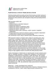
Nutrition and Blood Glucose Control<br />
Data regarding nutritional intake and blood glucose<br />
levels are shown in Figure 1 and in Table 4<br />
of the Supplementary Appendix. In the intensivetherapy<br />
group, 243 of 247 patients (98.4%) received<br />
insulin on at least one study day for glucose values<br />
above the target range (>110 mg per deciliter),<br />
whereas only 215 of 290 patients (74.1%) in<br />
the conventional-therapy group needed insulin<br />
because glucose values were outside the target<br />
range (≥200 mg per deciliter) (P
A<br />
Mean Kilocalories (no./day)<br />
Proportion (%)<br />
Mean Blood Glucose (mg/dl)<br />
1500<br />
1000<br />
500<br />
0<br />
60<br />
40<br />
20<br />
0<br />
200<br />
150<br />
100<br />
50<br />
0<br />
Intensive Insulin and Pentastarch Resuscitation in Sepsis<br />
Total Caloric Intake<br />
0 1 2 3 4 5 6 7 8 9 10 11 12 13 14<br />
Kilocalories Administered Enterally<br />
0 1 2 3 4 5 6 7 8 9 10 11 12 13 14<br />
Blood Glucose<br />
Conventional therapy<br />
Intensive therapy<br />
Conventional therapy<br />
Intensive therapy<br />
Conventional therapy<br />
Intensive therapy<br />
0 1 2 3 4 5 6 7 8 9 10 11 12 13 14<br />
Days<br />
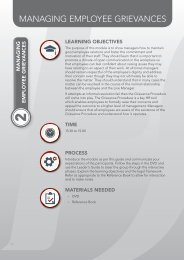
Figure 1. Nutrition, Blood Glucose, Systemic Pressures, and Central Venous Oxygen Saturation, According to the Type<br />
of Insulin and Fluid Therapy.<br />
AUTHOR:<br />
RETAKE 1st<br />
Panel A shows caloric intake and daily ICM morning blood <strong>Brunkhorst</strong> glucose (Reinhart) levels in all 537 patients during the first 14 days of the<br />
REG F FIGURE: 1 of 3<br />
2nd<br />
study, according to whether patients received intensive insulin therapy or conventional insulin therapy. Day 0 represents<br />
3rd<br />
the time at randomization until the CASE start of the next full 24-hour study day; Revised I bars denote 95% confidence intervals.<br />
The mean daily caloric intake (both EMail parenteral and enteral) Line and the 4-C fraction of SIZE kilocalories administered by the en-<br />
ARTIST: ts<br />
teral route, respectively, were calculated H/T H/T<br />
Enon only for days on which nutrition was given. 33p9 The type of nutrition was similar<br />
Combo<br />
in the two study groups. The mean morning blood glucose level in both study groups was calculated only for patients<br />
receiving insulin therapy on the respective study day AUTHOR, (P
132<br />
Fluid Resuscitation<br />
Before randomization, the characteristics of patients<br />
were well balanced between the group that<br />
received HES and the group that received Ringer’s<br />
lactate (Table 1, and Table 1 of the Supplementary<br />
Appendix). Patients in the two study groups<br />
received similar fluids in the 12 hours before<br />
randomization (Table 5 of the Supplementary<br />
Appendix).<br />
Patients in the Ringer’s lactate group received<br />
significantly more total resuscitation fluid than<br />
Table 2. Primary and Secondary Outcomes.*<br />
T h e n e w e ng l a nd j o u r na l o f m e dic i n e<br />
n engl j med 358;2 www.nejm.org january 10, <strong>2008</strong><br />
did patients in the HES group. The ratio of total<br />
fluid in the Ringer’s lactate group to that in the<br />
HES group was 1.32 for the entire study period<br />
(1.58 on day 1 and 1.44 on days 1 to 4). Patients<br />
in the HES group received a median cumulative<br />
dose of 70.4 ml per kilogram of body weight<br />
(interquartile range, 33.4 to 144.2). The median<br />
central venous pressure was 11.8 mm Hg (interquartile<br />
range, 9.5 to 14.2) in the HES group and<br />
10.7 mm Hg (interquartile range, 8.6 to 12.7) in<br />
the Ringer’s lactate group (P
Table 2. (Continued.)<br />
Intensive Insulin and Pentastarch Resuscitation in Sepsis<br />
Variable Insulin Therapy Fluid Resuscitation<br />
All Patients<br />
(N = 537)<br />
Conventional<br />
(N = 290)<br />
central venous oxygen saturation was 73.6% (interquartile<br />
range, 70.0 to 76.9) in the HES group<br />
and 72.4% (interquartile range, 69.3 to 75.9) in<br />
the Ringer’s lactate group (P = 0.04). The use of<br />
nonstudy colloid fluids is discussed in the Supplementary<br />
Appendix.<br />
Among patients who entered the study with<br />
values for central venous pressure that were below<br />
the hemodynamic target values (≥8 mm Hg),<br />
Intensive<br />
(N = 247) P Value†<br />
Ringer’s<br />
Lactate<br />
(N = 275)<br />
the target values were achieved faster in patients<br />
receiving HES than in those receiving Ringer’s<br />
lactate (P = 0.003) (Fig. 1B).<br />
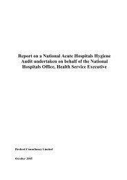
Mortality<br />
The rate of death at 28 days did not differ significantly<br />
between the HES group and the Ringer’s<br />
lactate group (26.7% and 24.1%, respectively;<br />
P = 0.48). However, there was a trend toward a<br />
HES<br />
(N = 262) P Value‡<br />
Hypoglycemia (≤40 mg/dl)
134<br />
A<br />
Probability of Survival (%)<br />
B<br />
Probability of Survival (%)<br />
C<br />
Probability of Survival (%)<br />
rate of death at 90 days that was higher in the<br />
AUTHOR: <strong>Brunkhorst</strong> (Reinhart) RETAKE 1st<br />
ICM<br />
HES group than in the Ringer’s lactate group<br />
REG F FIGURE: 2 of 3<br />
2nd<br />
3rd<br />
(41.0% vs. 33.9%, P = 0.09) (Table 2 and Fig. 2B).<br />
Morbidity Enon<br />
16p6<br />
Combo<br />
The mean SOFA scores did not differ significant-<br />
AUTHOR, PLEASE NOTE:<br />
ly between Figure the has been HES redrawn group and and type the has been Ringer’s reset. lac-<br />
T h e n e w e ng l a nd j o u r na l o f m e dic i n e<br />
100<br />
90<br />
80<br />
70<br />
Conventional therapy (N=290)<br />
60<br />
50<br />
40<br />
30<br />
20<br />
10<br />
0<br />
Intensive therapy (N=247)<br />
0 10 20 30 40 50<br />
Days<br />
60 70 80 90 100<br />
100<br />
90<br />
80<br />
70<br />
60<br />
Ringer’s lactate (N=275)<br />
50<br />
40<br />
30<br />
20<br />
10<br />
0<br />
HES (N=262)<br />
0 10 20 30 40 50<br />
Days<br />
60 70 80 90 100<br />
100<br />
90<br />
80<br />
70<br />
60<br />
50<br />
Low-dose HES (N=162)<br />
40<br />
30<br />
20<br />
10<br />
0<br />
High-dose HES (N=100)<br />
0 10 20 30 40 50<br />
Days<br />
60 70 80 90 100<br />
CASE<br />
EMail<br />
ARTIST: ts<br />
Line<br />
H/T<br />
Please check carefully.<br />
4-C<br />
H/T<br />
Revised<br />
SIZE<br />
JOB: 35802<br />
ISSUE: 01-10-07<br />
n engl j med 358;2 www.nejm.org january 10, <strong>2008</strong><br />
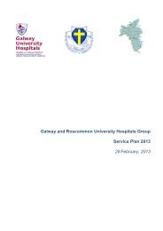
Figure 2. Kaplan–Meier Curves for Overall Survival.<br />
Panel A shows the comparison of overall survival between<br />
patients receiving intensive insulin therapy and<br />
those receiving conventional insulin therapy (P = 0.36<br />
by the log-rank test). Panel B shows the comparison<br />
between patients receiving pentastarch (HES) for volume<br />
resuscitation and those receiving Ringer’s lactate<br />
(P = 0.14 by the log-rank test). Panel C shows the comparison<br />
between patients in the low-dose HES subgroup<br />
(≤22 ml per kilogram of body weight per day),<br />
who received a median cumulative dose of 48.3 ml per<br />
kilogram (interquartile range, 21.9 to 96.2), and those<br />
in the high-dose subgroup (>22 ml per kilogram for at<br />
least 1 day during the study period), who received a median<br />
cumulative dose of 136.0 ml per kilogram (interquartile<br />
range, 79.0 to 180.0) (P
Intensive Insulin and Pentastarch Resuscitation in Sepsis<br />
Table 3. Adverse and Serious Adverse Events.*<br />
Variable Insulin Therapy Fluid Resuscitation<br />
All Patients<br />
(N = 537)<br />
in the 12 hours preceding study entry (median,<br />
2400 ml; interquartile range, 1000 to 3500) than<br />
did those who did not receive a dose escalation<br />
(median, 1135 ml; interquartile range, 500 to<br />
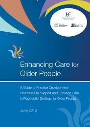
2560; P = 0.002). The rate of death at 90 days was<br />
Conventional<br />
(N = 290)<br />
Intensive<br />
Ringer’s Lactate<br />
(N = 247) P Value† (N = 275)<br />
significantly increased among patients who received<br />
a higher dose of HES, as compared with<br />
those who received a lower dose (57.6% vs. 30.9%,<br />
P
A Renal-Replacement Therapy<br />
100<br />
90<br />
80<br />
70<br />
60<br />
50<br />
40<br />
30<br />
20<br />
10<br />
0<br />
Ringer’s lactate HES<br />
0–40 >40–80 >80–150 >150–250 >250<br />
Cumulative Dose of Study Fluid (ml/kg)<br />
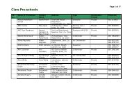
Figure 3. Cumulative Effect of Volume Resuscitation on the Need for Renal-<br />
Replacement Therapy AUTHOR:<br />
RETAKE<br />
ICM and the <strong>Brunkhorst</strong> Rate of Death (Reinhart) at 90 Days. 1st<br />
REG F FIGURE:<br />
2nd<br />
Panel A shows the relationship 3 of between 3 the cumulative dose of either penta-<br />
3rd<br />
starch (HES) or CASE Ringer’s lactate and the percentage of patients Revised who needed<br />
renal-replacement EMailtherapy<br />
(Panel A) and Line the rate 4-C of death at SIZE 90 days (Panel B).<br />
ARTIST: ts<br />
The need for renal-replacement H/T H/T<br />
Enon<br />
therapy and 90-day mortality 22p6 were signifi-<br />
Combo<br />
cantly correlated with the cumulative dose of HES (P40–80 >80–150 >150–250 >250<br />
Cumulative Dose of Study Fluid (ml/kg)<br />
Proportion of Patients (%)<br />
N=38<br />
N=70<br />
N=38<br />
N=70<br />
N=34<br />
N=61<br />
N=35<br />
N=61<br />
N=45<br />
N=59<br />
N=46<br />
N=58<br />
JOB: 35802<br />
ISSUE: 01-10-07<br />
easily explain these study results (Table 3B and<br />
Table 7 of the Supplementary Appendix).<br />
In a multivariate post hoc logistic-regression<br />
model that was adjusted for insulin therapy, the<br />
total dose of Ringer’s lactate, baseline creatinine<br />
clearance, mean arterial pressure, and total dose<br />
of colloids administered 12 hours before the start<br />
of therapy, the total dose of HES was a significant<br />
independent predictor of both the need for<br />
renal-replacement therapy and the rate of death<br />
at 90 days (Table 6 of the Supplementary Appendix).<br />
At 90 days, patients who had received a<br />
lower dose of HES were more likely to have renal<br />
failure than those who had received Ringer’s lac-<br />
T h e n e w e ng l a nd j o u r na l o f m e dic i n e<br />
N=48<br />
N=45<br />
N=48<br />
N=46<br />
N=94<br />
N=14<br />
N=94<br />
N=14<br />
n engl j med 358;2 www.nejm.org january 10, <strong>2008</strong><br />
tate (30.9% vs. 21.7%, P = 0.04) and were more<br />
likely to need renal-replacement therapy (25.9%<br />
vs. 17.3%, P = 0.03).<br />
Discussion<br />
In 537 patients with septic shock, we found no<br />
beneficial effect of intensive insulin treatment (administered<br />
according to the Leuven protocol) with<br />
respect to the rate of death at 28 days and the<br />
mean SOFA score; we also found no benefit with<br />
respect to any of the secondary end points. Moreover,<br />
our study was stopped early, at the first<br />
planned safety analysis, because intensive insulin<br />
therapy was associated with a significantly increased<br />
rate of severe hypoglycemic events and a<br />
trend toward a prolonged stay in the ICU.<br />
Cox regression analysis identified the occurrence<br />
of hypoglycemia as an independent risk<br />
factor for death from any cause. Hypoglycemia<br />
may be only a marker of a poor outcome, independently<br />
of insulin therapy. On the other hand,<br />
it is possible that unrecognized adverse effects<br />
of hypoglycemia on the brain or heart offset potential<br />
beneficial effects of intensive insulin therapy.<br />
16 The full extent of hypoglycemic events in<br />
our study is unknown, since the usual clinical<br />
warning signs and symptoms of hypoglycemia<br />
in the patients we studied may have been masked<br />
by critical illness and sedation.<br />
Our findings are similar to those of the second<br />
study by Van den Berghe et al., 2 which assessed<br />
the use of intensive insulin therapy in<br />
maintaining euglycemia in critically ill patients<br />
in a medical ICU. In our study, the nonsignificant<br />
differences in the rates of death at 28 days<br />
and at 90 days in the intensive-therapy group<br />
and the conventional-therapy group were similar<br />
to those in the study by Van den Berghe et al., as<br />
was the magnitude of the significant increase in<br />
hypoglycemic episodes in the intensive-therapy<br />
group, as compared with the conventional-therapy<br />
group (18.7% vs. 3.1% in the study by Van<br />
den Berghe et al. and 17.0% vs. 4.1% in our<br />
study). The mean blood glucose levels during<br />
hypoglycemia in the intensive-therapy group and<br />
the conventional-therapy group were also similar<br />
in the study by Van den Berghe et al. (32 mg<br />
and 31 mg per deciliter, respectively; P = 0.50) and<br />
in our study (31 mg and 28 mg per deciliter, respectively;<br />
P = 0.30). Moreover, in the study by Van<br />
den Berghe et al., mean morning blood glucose<br />
The New England Journal of Medicine<br />
Downloaded from nejm.org on February 22, 2013. For personal use only. No other uses without permission.<br />
Copyright © <strong>2008</strong> Massachusetts Medical Society. All rights reserved.
Intensive Insulin and Pentastarch Resuscitation in Sepsis<br />
levels in the intensive-therapy group and in the<br />
conventional-therapy group (111±29 mg and<br />
153±31 mg per deciliter, respectively) were similar<br />
to the levels in our study (112±18 mg and<br />
151±33 mg per deciliter, respectively). In their<br />
second study of medical ICU patients, Van den<br />
Berghe et al. performed exploratory subgroup<br />
analyses regarding the length of the ICU stay and<br />
the resolution of organ injury. The beneficial effects<br />
that were shown in these subgroup analyses<br />
were not confirmed in our study.<br />
Taken together, our study and the medical ICU<br />
study by Van den Berghe et al. establish that intensive<br />
insulin therapy has no measurable, consistent<br />
benefit in critically ill patients in a medical<br />
ICU, regardless of whether the patients have severe<br />
sepsis, and that such therapy increases the<br />
risk of hypoglycemic episodes. The results of these<br />
two studies are in marked contrast to the results<br />
of the first study by Van den Berghe et al., 1 which<br />
showed a beneficial effect of intensive insulin<br />
therapy on postoperative survival rates among<br />
critically ill surgical patients. In that study, the<br />
beneficial effect was predominantly seen in cardiac<br />
surgical patients (accounting for 62% of the<br />
study population) who were given intravenous<br />
glucose loads (200 to 300 g per 24 hours) on<br />
admission to the ICU. It is possible that intensive<br />
insulin therapy was beneficial in these patients<br />
because it decreased the adverse effect of this<br />
high glucose load.<br />
In sedated, severely ill patients with sepsis,<br />
the benefits of intensive insulin therapy (administered<br />
according to the Leuven protocol) are unproven,<br />
but the risk of hypoglycemia is increased<br />
by a factor of 5 to 6. We cannot exclude the possibility<br />
that patients with sepsis may benefit<br />
from other less strict insulin protocols, 17 given<br />
that variability in the glucose level was a stronger<br />
independent predictor of death in the ICU<br />
than was the mean glucose concentration. 18<br />
After the first planned interim analysis, our<br />
trial was suspended because of increased rates<br />
of renal failure and death at 90 days in the group<br />
receiving HES. Adverse effects of HES on renal<br />
function have been reported in patients who have<br />
undergone renal transplantation and in critically<br />
ill patients. 19,20 Schortgen et al. 21 reported adverse<br />
renal effects associated with a starch solution<br />
that had a higher degree of molar substitution<br />
(0.6) than that used in our study (0.5). Other<br />
studies did not detect adverse effects except for<br />
impaired coagulation, even with large doses of<br />
starch solutions; however, these studies were limited<br />
by their design, small size, and short observation<br />
periods. 22-27 Even though we used a “modern”<br />
HES solution 28 that was designed to have<br />
fewer side effects, we found an even higher incidence<br />
of acute renal failure than that reported<br />
by Schortgen et al. Our study showed that HES<br />
was associated with an increased need for renalreplacement<br />
therapy in patients with sepsis, even<br />
when it was administered at recommended daily<br />
doses, and that higher cumulative doses were<br />
associated with an increased rate of death at 90<br />
days. Our results should not be used to address<br />
the effect of rapid volume expansion on the outcome<br />
in patients with sepsis, nor should our findings<br />
be extrapolated to other volume expanders.<br />
The differences between the hemodynamic effects<br />
of HES and those of Ringer’s lactate were<br />
minor (e.g., a more rapid return to normal central<br />
venous pressure in the HES group). However,<br />
we observed marked adverse effects of HES therapy<br />
on kidney function, coagulation, transfusion<br />
requirements, and survival. The ability of HES to<br />
interfere with coagulation has already prompted<br />
warning labels and dose limitations. 29,30 Furthermore,<br />
long-term storage of the colloid is potentially<br />
toxic and may be responsible (beyond the<br />
adverse effects on renal function) for the observed<br />
increase in the rate of death at 90 days, particularly<br />
with higher doses. 21,31-36<br />
Fluid resuscitation with 10% HES 200/0.5 is<br />
harmful in patients with severe sepsis. At recommended<br />
doses, it causes renal impairment, and<br />
at high doses, it impairs long-term survival. Since<br />
adverse effects have been attributed to various<br />
HES solutions, 37 until long-term studies with adequate<br />
numbers of patients show that a particular<br />
HES solution is safe in critically ill patients,<br />
HES solutions should be avoided.<br />
Supported by a grant (01 KI 0106) from the German Federal<br />
Ministry of Education and Research and by unrestricted grants<br />
from B. Braun, HemoCue, and Novo Nordisk.<br />
Dr. Bloos reports receiving lecture fees from B. Braun, and<br />
Dr. Reinhart reports receiving lecture and consulting fees from<br />
B. Braun. No other potential conflict of interest relevant to this<br />
article was reported.<br />
We thank the members of the data and safety monitoring<br />
board: Charles L. Sprung, M.D., Hadassah Hebrew University<br />
Medical Center, Jerusalem; Waheedullah Karzai, M.D., Zentralklinik<br />
Bad Berka, Bad Berka, Germany; and Herbert Witte,<br />
Ph.D., Institute of Medical Statistics, Informatics and Documentation,<br />
University of Jena, Germany.<br />
n engl j med 358;2 www.nejm.org january 10, <strong>2008</strong> 137<br />
The New England Journal of Medicine<br />
Downloaded from nejm.org on February 22, 2013. For personal use only. No other uses without permission.<br />
Copyright © <strong>2008</strong> Massachusetts Medical Society. All rights reserved.
138<br />
T h e n e w e ng l a nd j o u r na l o f m e dic i n e<br />
Appendix<br />
The authors’ affiliations are as follows: the Department of Anesthesiology and Intensive Care Medicine (F.M.B., F.B., C.H., K.R.) and<br />
the Institute of Clinical Chemistry and Laboratory Medicine (M.K.), Friedrich Schiller University, Jena; the Institute of Medical Informatics,<br />
Statistics and Epidemiology (C.E., M.L.) and the Coordination Center for Clinical Trials (E.K.), University of Leipzig, Leipzig; the<br />
Department of Anesthesiology and Intensive Care Medicine, Helios Klinikum, Erfurt (A.M.-H.); the Department of Anesthesiology and<br />
Intensive Care Medicine, University Hospital of the Technical University of Dresden, Dresden (M.R.); the Department of Anesthesiology<br />
and Intensive Care Medicine, University Hospital Schleswig-Holstein, Campus Kiel, Kiel (N.W.); the Department of Anesthesiology and<br />
Intensive Care Medicine, University of Goettingen, Goettingen (O.M.); the Department of Anesthesiology and Intensive Care Medicine,<br />
Ernst Moritz Arndt University, Greifswald (M.G.); the Department of Nephrology and Medical Intensive Care, Charite, Campus Virchow-<br />
Klinikum, University Medical Center, Berlin (M.O.); the Department of Anesthesiology and Intensive Care Medicine, Martin Luther University,<br />
Halle-Wittenberg (S.G.); the Department of Anesthesiology and Intensive Care Medicine, University Hospital, Leipzig (D.O.); the<br />
Department of Anesthesiology and Critical Care Medicine, Klinikum Augsburg, Augsburg (U.J.); the Department of Nephrology and<br />
Hypertension, University of Erlangen-Nuremberg, Erlangen (S.J.); the Department of Anesthesiology and Intensive Care Medicine, University<br />
Hospital Aachen, Rheinisch-Westfaelische Technische Hochschule, Aachen (R.R.); the Department of Pulmonary and Critical Care<br />
Medicine, University Otto von Guericke, Magdeburg, and the Department of Pulmonary and Critical Care Medicine, Medizinische Hochschule<br />
Hannover, Hannover (T.W.); the Department of Anesthesiology and Intensive Care Medicine, Staedtisches Klinikum Brandenburg,<br />
Brandenburg (M.S.); and the Department of Anesthesiology and Intensive Care Medicine, Staedtisches Krankenhaus Dresden-Friedrichstadt,<br />
Dresden (P.K.) — all in Germany; and the Critical Care Medicine Department, National Institutes of Health, Bethesda, MD (C.N.).<br />
References<br />
1. Van den Berghe G, Wouters P, Week- 11. Thadhani R, Pascual M, Bonventre JV. a multicentre randomised study. Lancet<br />
ers F, et al. Intensive insulin therapy in Acute renal failure. N Engl J Med 1996;334: 2001;357:911-6.<br />
critically ill patients. N Engl J Med 2001; 1448-60.<br />
22. Wiesen P, Canivet JL, Ledoux D, Roedi-<br />
345:1359-67.<br />
12. What is a serious adverse event? Rockger L, Damas P. Effect of hydroxyethyl-<br />
2. Van den Berghe G, Wilmer A, Hermans ville, MD: MedWatch, FDA Safety Inforstarch on renal function in cardiac surgery:<br />
G, et al. Intensive insulin therapy in the mation and Adverse Event Reporting Pro- a large scale retrospective study. Acta An-<br />
medical ICU. N Engl J Med 2006;354:449- gram, 2004. (Accessed December 14, 2007, aesthesiol Belg 2005;56:257-63.<br />
61.<br />
at http://www.fda.gov/medwatch/report/ 23. Liet JM, Bellouin AS, Boscher C, Lejus<br />
3. Dellinger RP, Carlet JM, Masur H, et al. DESK/advevnt.htm.)<br />
C, Rozé JC. Plasma volume expansion by<br />
Surviving Sepsis Campaign guidelines for 13. Moreno R, Vincent JL, Matos R, et al. medium molecular weight hydroxyethyl<br />
management of severe sepsis and septic The use of maximum SOFA score to quan- starch in neonates: a pilot study. Pediatr<br />
shock. Crit Care Med 2004;32:858-73. [Ertify organ dysfunction/failure in intensive Crit Care Med 2003;4:305-7.<br />
rata, Crit Care Med 2004;32:1448, 2169- care: results of a prospective, multicentre 24. Beyer R, Harmening U, Rittmeyer O,<br />
70.]<br />
study. Intensive Care Med 1999;25:686-96. et al. Use of modified fluid gelatin and<br />
4. Roberts I, Alderson P, Bunn F, Chin- 14. Bauer P, Köhne K. Evaluation of ex- hydroxyethyl starch for colloidal volume<br />
nock P, Ker K, Schierhout G. Colloids verperiments with adaptive interim analyses. replacement in major orthopaedic surgery.<br />
sus crystalloids for fluid resuscitation in Biometrics 1994;50:1029-41.<br />
Br J Anaesth 1997;78:44-50.<br />
critically ill patients. Cochrane Database 15. <strong>Brunkhorst</strong> FM, Kuhnt E, Engel C, et 25. Vogt N, Bothner U, Brinkmann A, de<br />
Syst Rev 2004;4:CD000567.<br />
al. Intensive insulin therapy in patients Petriconi R, Georgieff M. Peri-operative<br />
5. Hoffmann JN, Vollmar B, Laschke with severe sepsis and septic shock is as- tolerance to large-dose 6% HES 200/0.5 in<br />
MW, Inthorn D, Schildberg FW, Menger sociated with an increased rate of hypo- major urological procedures compared<br />
MD. Hydroxyethyl starch (130 kD), but not glycemia: results from a randomized mul- with 5% human albumin. Anaesthesia<br />
crystalloid volume support, improves miticenter study (<strong>VISEP</strong>). Infection 2005;33: 1999;54:121-7.<br />
crocirculation during normotensive endo- Suppl 1:19.<br />
26. Arellano R, Gan BS, Salpeter MJ, et al.<br />
toxemia. Anesthesiology 2002;97:460-70. 16. Cryer PE. Diverse causes of hypoglyce- A triple-blinded randomized trial compar-<br />
6. Morisaki H, Bloos F, Keys J, Martin C, mia-associated autonomic failure in diaing the hemostatic effects of large-dose<br />
Neal A, Sibbald WJ. Compared with crysbetes. N Engl J Med 2004;350:2272-9. 10% hydroxyethyl starch 264/0.45 versus<br />
talloid, colloid therapy slows progression 17. Wilson M, Weinreb J, Hoo GW. Inten- 5% albumin during major reconstructive<br />
of extrapulmonary tissue injury in septic sive insulin therapy in critical care: a re- surgery. Anesth Analg 2005;100:1846-53.<br />
sheep. J Appl Physiol 1994;77:1507-18. view of 12 protocols. Diabetes Care 2007; 27. Sakr Y, Payen D, Reinhart K, et al. Ef-<br />
7. Barron ME, Wilkes MM, Navickis RJ. 30:1005-11.<br />
fects of hydroxyethyl starch administra-<br />
A systematic review of the comparative safe- 18. Egi M, Bellomo R, Stachowski E, tion on renal function in critically ill paty<br />
of colloids. Arch Surg 2004;139:552-63. French CJ, Hart G. Variability of blood glutients. Br J Anaesth 2007;98:216-24.<br />
8. Wilkes MM, Navickis RJ, Sibbald WJ. cose concentration and short-term mor- 28. Perazella MA. Drug-induced renal<br />
Albumin versus hydroxyethyl starch in tality in critically ill patients. Anesthesiol- failure: update on new medications and<br />
cardiopulmonary bypass surgery: a metaogy 2006;105:244-52.<br />
unique mechanisms of nephrotoxicity. Am<br />
analysis of postoperative bleeding. Ann 19. Winkelmayer WC, Glynn RJ, Levin R, J Med Sci 2003;325:349-62.<br />
Thorac Surg 2001;72:527-33.<br />
Avorn J. Hydroxyethyl starch and change 29. Haynes GR, Havidich JE, Payne KJ.<br />
9. American College of Chest Physicians/ in renal function in patients undergoing Why the Food and Drug Administration<br />
Society of Critical Care Medicine Consen- coronary artery bypass graft surgery. Kid- changed the warning label for hetastarch.<br />
sus Conference: definitions for sepsis and ney Int 2003;64:1046-9.<br />
Anesthesiology 2004;101:560-1.<br />
organ failure and guidelines for the use of 20. Cittanova ML, Leblanc I, Legendre C, 30. Jonville-Béra AP, Autret-Leca E, Gruel<br />
innovative therapies in sepsis. Crit Care Mouquet C, Riou B, Coriat P. Effect of hy- Y. Acquired type I von Willebrand’s dis-<br />
Med 1992;20:864-74.<br />
droxyethylstarch in brain-dead kidney doease associated with highly substituted<br />
10. Van den Berghe G, Wouters PJ, Bouilnors on renal function in kidney-trans- hydroxyethyl starch. N Engl J Med 2001;<br />
lon R, et al. Outcome benefit of intensive plant recipients. Lancet 1996;348:1620-2. 345:622-3.<br />
insulin therapy in the critically ill: insulin 21. Schortgen F, Lacherade JC, Bruneel F, 31. Legendre C, Thervet E, Page B, Per-<br />
dose versus glycemic control. Crit Care et al. Effects of hydroxyethylstarch and cheron A, Noël LH, Kreis H. Hydroxyethyl-<br />
Med 2003;31:359-66.<br />
gelatin on renal function in severe sepsis: starch and osmotic-nephrosis-like lesions<br />
n engl j med 358;2 www.nejm.org january 10, <strong>2008</strong><br />
The New England Journal of Medicine<br />
Downloaded from nejm.org on February 22, 2013. For personal use only. No other uses without permission.<br />
Copyright © <strong>2008</strong> Massachusetts Medical Society. All rights reserved.
Intensive Insulin and Pentastarch Resuscitation in Sepsis<br />
in kidney transplantation. Lancet 1993; 34. Christidis C, Mal F, Ramos J, et al.<br />
342:248-9.<br />
Worsening of hepatic dysfunction as a con-<br />
32. Pillebout E, Nochy D, Hill G, et al. Resequence of repeated hydroxyethylstarch<br />
nal histopathological lesions after ortho- infusions. J Hepatol 2001;35:726-32.<br />
topic liver transplantation (OLT). Am J 35. Auwerda JJ, Wilson JH, Sonneveld P.<br />
Transplant 2005;5:1120-9.<br />
Foamy macrophage syndrome due to hy-<br />
33. van Rijen EA, Ward JJ, Little RA. Efdroxyethyl starch replacement: a severe<br />
fects of colloidal resuscitation fluids on side effect in plasmapheresis. Ann Intern<br />
reticuloendothelial function and resistance Med 2002;137:1013-4.<br />
to infection after hemorrhage. Clin Diagn 36. Schmidt-Hieber M, Loddenkemper C,<br />
Lab Immunol 1998;5:543-9.<br />
Schwartz S, Arntz G, Thiel E, Notter M.<br />
Hydrops lysosomalis generalisatus — an<br />
underestimated side effect of hydroxyethyl<br />
starch therapy? Eur J Haematol 2006;<br />
77:83-5.<br />
37. Wiedermann CJ. Hydroxyethyl starch<br />
— can the safety problems be ignored?<br />
Wien Klin Wochenschr 2004;116:583-94.<br />
Copyright © <strong>2008</strong> Massachusetts Medical Society.<br />
n engl j med 358;2 www.nejm.org january 10, <strong>2008</strong> 139<br />
The New England Journal of Medicine<br />
Downloaded from nejm.org on February 22, 2013. For personal use only. No other uses without permission.<br />
Copyright © <strong>2008</strong> Massachusetts Medical Society. All rights reserved.