Dis Aquat Org - Inter Research
Dis Aquat Org - Inter Research
Dis Aquat Org - Inter Research

Create successful ePaper yourself
Turn your PDF publications into a flip-book with our unique Google optimized e-Paper software.
Vol. 88: 189–198, 2010<br />
doi: 10.3354/dao02169<br />
INTRODUCTION<br />
Viral haemorrhagic septicaemia (VHS) is a severe<br />
viral fish disease that causes significant losses in<br />
farmed rainbow trout Oncorhynchus mykiss across<br />
Europe. VHS virus (VHSV) has been isolated from a<br />
DISEASES OF AQUATIC ORGANISMS<br />
<strong>Dis</strong> <strong>Aquat</strong> <strong>Org</strong><br />
Published February 17<br />
Viral haemorrhagic septicaemia virus (VHSV)<br />
genotype II isolated from European river lamprey<br />
Lampetra fluviatilis in Finland during surveillance<br />
from 1999 to 2008<br />
Tuija Gadd1, *, Miia Jakava-Viljanen1 , Katja Einer-Jensen2 , Ellen Ariel2, 5 ,<br />
Perttu Koski3 1, 4<br />
, Liisa Sihvonen<br />
1 Finnish Food Safety Authority Evira, Mustialankatu 3, 00790 Helsinki, Finland<br />
2 National Veterinary Institute, Technical University of Denmark, Hangøvej 2, 8200 Århus N, Denmark<br />
3 Finnish Food Safety Authority Evira, PO Box 517, 90101 Oulu, Finland<br />
4 Department of Basic Veterinary Sciences, Faculty of Veterinary Medicine, PO Box 66, 00014 University of Helsinki, Finland<br />
5 Present address: School of Veterinary and Biomedical Sciences, James Cook University, Douglas Campus, Townsville 4811,<br />
Queensland, Australia<br />
ABSTRACT: We examined the occurrence of viral haemorrhagic septicaemia virus (VHSV) in the<br />
main spawning stocks of wild European river lamprey Lampetra fluviatilis in the rivers of Finland<br />
from 1999 to 2008. Pooled samples of internal organs (kidney, liver and heart or brain) from 2621 lampreys<br />
were examined for the presence of VHSV by standard virological techniques. VHSV was isolated<br />
from 5 samples from the rivers Lestijoki and Kalajoki, which flow from Finland into the Bothnian<br />
Bay of the Baltic Sea. The presence of VHSV was confirmed by immunofluorescent antibody<br />
technique (IFAT), ELISA and RT-PCR. Phylogenetic analysis based on the full-length VHSV glycoprotein<br />
(G) gene sequence revealed that the isolates were most closely related to the VHSV strain<br />
isolated in 1996 from herring Clupea harengus and sprat Sprattus sprattus in the Eastern Gotland<br />
Basin of the Baltic Sea, and were therefore assigned to VHSV genotype II. The partial G gene<br />
sequences obtained (nt 1 to 672–1129) of all 5 lamprey VHSV isolates were identical, and so were the<br />
entire G genes (nt 1 to 1524) of 2 isolates sequenced. The virulence of one of the lamprey isolates was<br />
evaluated by an experimental infection trial in rainbow trout Oncorhynchus mykiss fry. No mortality<br />
was induced postinfection by waterborne and intraperitoneal challenge, respectively, while 2 genotype<br />
Id isolates originating from Finnish rainbow trout caused marked mortality under the same conditions.<br />
The infection in the European river lamprey is thought to be independent from the epidemic<br />
in farmed rainbow trout in Finnish brackish waters, because the isolates from rainbow trout were of<br />
a different genotype. This is the first report of VHSV found in the European river lamprey. The role<br />
of wild river lampreys in maintaining the infection in the marine environment remains unclear.<br />
KEY WORDS: Vector of VHSV · Lamprey · Apathogenic · Risk · Epidemiology · Brackish water<br />
*Email: tuija.gadd@evira.fi<br />
Resale or republication not permitted without written consent of the publisher<br />
large number of free-living marine fish species in<br />
Europe, Japan and North America (Smail 1995, 2000,<br />
Dixon et al. 1997, Mortensen et al. 1999, Takano et al.<br />
2000, King et al. 2001, Brudeseth & Evensen 2002,<br />
Hedrick et al. 2003, Skall et al. 2005a). VHSV isolates<br />
originating from wild marine fish generally exhibit no<br />
© <strong>Inter</strong>-<strong>Research</strong> 2010 · www.int-res.com
190<br />
or very low pathogenicity to rainbow trout (Dixon et al.<br />
1997, Skall et al. 2004).<br />
VHSV is an enveloped negative-strand RNA virus<br />
belonging to the Novirhabdovirus genus of the family<br />
Rhabdoviridae. The VHSV genome is a nonsegmented,<br />
single-stranded RNA molecule with a length<br />
of approximately 11 200 nucleotides. The genome consists<br />
of 6 genes in the order 3’-N-P-M-G-NV-L-5’,<br />
encoding 5 structural proteins: nucleocapsid (N) protein,<br />
phosphoprotein (P), matrix protein (M), glycoprotein<br />
(G) and RNA polymerase protein (L), and one nonstructural<br />
protein (NV) (Schütze et al. 1999).<br />
VHSV isolates cluster into 4 genotypes (I to IV) with<br />
several sublineages within genotypes I (minimum Ia to<br />
Ie) and IV (IVa to IVb), which seems to correlate<br />
with the geographical regions of isolations<br />
rather than with pathogenicity<br />
(Snow et al. 1999, Einer-Jensen et al. 2004,<br />
Lumsden et al. 2007). Genotypes Ia, Ic and<br />
Id include a wide range of virus strains originating<br />
from brackish water and freshwater<br />
rainbow trout farms in Europe (Benmansour<br />
et al. 1997, Stone et al. 1997, Snow et al.<br />
1999, Thiery et al. 2002, Einer-Jensen et al.<br />
2004, Raja-Halli et al. 2006). In addition, 28<br />
isolates from marine fish species found in<br />
the Baltic Sea, Skagerrak and Kattegat, and<br />
one isolate from the English Channel appear<br />
to have the same ancestral source as<br />
these strains from rainbow trout and belong<br />
to genotype Ib (Einer-Jensen et al. 2004).<br />
Genotype Ie includes isolates from the Black<br />
Sea (Einer-Jensen et al. 2004, Nishizawa et<br />
al. 2006). Genotype II so far only includes<br />
some marine isolates found in the eastern<br />
Gotland Basin of the Baltic Sea (Snow et al.<br />
1999, Einer-Jensen et al. 2004). The third<br />
genotype (III) consists of isolates originating<br />
from outbreaks of VHS in turbot Scophthalmus<br />
maximus farms in the British Isles (Ross<br />
et al. 1994, Einer-Jensen et al. 2004), as well<br />
as isolates from a variety of marine species<br />
caught in Scottish waters and the Skagerrak<br />
(Snow et al. 1999, Einer-Jensen et al. 2004),<br />
one isolate from an eel Anguilla anguilla<br />
captured in the River Loire estuary in<br />
France (Thiery et al. 2002, Einer-Jensen et<br />
al. 2004), isolates from Greenland halibut<br />
Reinhardtius hippoglossoides caught at the<br />
Flemish Cap in the North Atlantic (López-<br />
Vázquez et al. 2006) and in 2007 to 2008<br />
from seawater-reared rainbow trout in Storfjorden<br />
in western Norway (Dale et al.<br />
2009). Genotype IVa includes isolates from<br />
wild marine fish in the North Pacific Ocean<br />
<strong>Dis</strong> <strong>Aquat</strong> <strong>Org</strong> 88: 189–198, 2010<br />
including the coast of the North America (Benmansour et<br />
al. 1997, Stone et al. 1997, Snow et al. 1999, Nishizawa et<br />
al. 2002), as well as isolates from Japanese flounder<br />
Paralichthys olivaceus (Nishizawa et al. 2002) and Pacific<br />
sand eel Ammodytes personatus around Japan<br />
(Watanabe et al. 2002). Isolates from the Great Lakes region<br />
of North America (Elsayed et al. 2006, Lumsden et<br />
al. 2007) and from 4 species of wild fish from the eastern<br />
Atlantic coast of Canada (Gagné et al. 2007) belong to<br />
genotype IVb.<br />
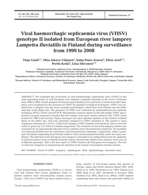
In Finland, VHSV was diagnosed for the first time in<br />
spring 2000 from 4 rainbow trout farms in brackish<br />
water (Raja-Halli et al. 2006) around Pyhtää (P) and the<br />
Åland Islands (A) (Fig. 1). There were 4 positive farms<br />
Fig. 1. Wild European river lampreys captured from the estuary of the rivers<br />
Iijoki, Oulujoki, Kalajoki (VHSV genotype II was isolated from 1 sample in<br />
2003), Lestijoki (VHSV genotype II was isolated from 4 samples in 2003), Perhojoki<br />
and Kokemäenjoki. Restriction areas of VHSV are A: Åland Islands 2000<br />
(whole area since 2001); P: Pyhtää 2000 to 2007; U: Uusikaupunki–<br />
Pyhäranta–Rauma 2003. All virus isolates are genotype Id
in Åland waters and 2 close to Pyhtää in 2001. In 2003,<br />
VHSV was diagnosed for the first time in Uusikaupunki<br />
(U) and later again in 2008. These isolates<br />
from rainbow trout were shown to be members of<br />
genotype Id (Raja-Halli et al. 2006), and since 2000,<br />
VHSV isolates of genotype Id have been recovered in<br />
fish from 2 to 14 farms in the waters around in the<br />
Åland Islands every year. Continental Finland is considered<br />
free of VHS apart from coastal restriction areas<br />
A, P and U as shown in Fig. 1. The transportation and<br />
movement of farmed fish and equipment is not allowed<br />
from the restriction area. Neither is it permitted to sell<br />
fish from facilities subject to restrictions to free areas.<br />
The management of lamprey stocks in rivers closed<br />
by hydroelectric power stations is carried out by capturing<br />
lampreys from the estuary and transferring<br />
them above the dam wall. This requires permission<br />
from the Ministry of Agriculture and Forestry (Decision<br />
1087/1998, MAFF 1998) and a survey of at least 60<br />
lampreys per river each year for viral and bacterial diseases<br />
such as VHSV, infectious haemotopoietic necrosis<br />
virus (IHNV) and bacterial kidney disease (BKD,<br />
data not shown) before transferring them upstream<br />
from the migration barrier.<br />
The aim of this study was to clarify the role of the<br />
European river lamprey Lampetra fluviatilis in the epidemiology<br />
of VHSV in Finland.<br />
MATERIALS AND METHODS<br />
Virus isolation and identification. Healthy adult<br />
river lampreys were caught during the spawning<br />
migration from the mouth of the spawning in rivers on<br />
Gadd et al.: VHSV genotype II isolated from lamprey<br />
191<br />
the Finnish coast of the Bothnian Bay. In total, 2621<br />
samples were collected during 1999 to 2008. Details of<br />
the sample distribution are summarised in Table 1.<br />
Pooled heart or brain, anterior kidney and liver tissue<br />
from a maximum of 10 lampreys (= 1 sample) were<br />
processed according to standard virological procedures<br />
as described by Raja-Halli et al. (2006) and inoculated<br />
onto subconfluent monolayer cell cultures of<br />
bluegill Lepomis macrochirus fry (BF-2) and epithelioma<br />
papulosum cyprini (EPC) cell lines (Wolf et al.<br />
1966, Fijan et al. 1983). The samples were incubated in<br />
a CO 2 incubator at 16°C according to methods described<br />
by Raja-Halli et al. (2006) and inspected regularly<br />
with a microscope for the occurrence of cytopathic<br />
effects (CPE) at 40× magnification. After 7 d<br />
incubation at 16°C, supernatant from samples without<br />
CPE was diluted to 1:100 and 1:1000, subcultured onto<br />
fresh cells and incubated for a further 7 d. In cases<br />
where total CPE was evident, the cell culture medium<br />
was collected and stored at –20°C for future studies.<br />
Staining of cells for immunofluorescence. The indirect<br />
fluorescent antibody technique (IFAT) was used<br />
for VHSV and IHNV identification with minor modification<br />
of the method described by Raja-Halli et al.<br />
(2006). Cover glass (diameter 13 mm, Menzel-Glaser)<br />
cultures of BF-2 cells grown in 24-well plates were<br />
inoculated at dilutions of 10 –1 and 10 –2 . Uninfected<br />
cells served as negative controls. The infected cultures<br />
were fixed with acetone on Days 1 and 2 postinfection<br />
and the IFAT was performed as described by Lorenzen<br />
et al. (1988). Briefly, the monoclonal antibody (MAb)<br />
1P5B11 (DTU) against the nucleocapsid (N) protein of<br />
the VHSV-F1 strain or the MAb Hyb 136-3 (DTU)<br />
against the N protein of IHNV were diluted in phos-<br />
Table 1. The number of European river lampreys and number of pooled samples investigated by standard virological procedures<br />
from Finnish rivers draining into the Gulf of Bothnia. Data are presented as no./pool where no. = number of lampreys examined<br />
and pool = number of pooled samples examined. Wild lampreys are caught in the estuaries of rivers dammed for hydroelectric<br />
power and transferred upstream from the artificial migration barriers. Subsets of 60 lampreys per river were virologically examined<br />
in pooled samples of up to 10 fish per pool and also analysed for the presence of bacterial kidney disease (BKD) (results not<br />
shown). nd: none detected<br />
Year Kemijoki Iijoki Oulujoki Kalajoki Lestijoki Perhonjoki Kokemäenjoki All All rivers<br />
rivers VHSV<br />
1999 nd nd nd nd nd nd 60/6 60/6<br />
2000 nd nd nd nd nd nd nd nd<br />
2001 nd nd 60/6 nd nd nd nd 60/6<br />
2002 nd nd 60/6 nd nd 60/6 nd 120/12<br />
2003 75/8 60/6 60/6 60/6 120/12 130/13 nd 505/51 50/5 a<br />
2004 nd 60/6 60/6 60/6 160/16 160/15 90/9 590/58<br />
2005 nd 60/6 60/6 60/6 60/6 60/6 60/6 360/36<br />
2006 nd 60/6 60/6 nd 60/6 80/8 260/26<br />
2007 60/6 nd 60/6 60/6 60/6 60/6 60/6 360/36<br />
2008 60/6 60/6 60/6 nd nd 66/7 60/6 306/31<br />
Total 195/20 300/30 480/48 240/24 460/46 616/61 330/33 2621/262 50/5<br />
a Four positive pooled samples from the River Lestijoki and one positive pool from the River Kalajoki
192<br />
phate-buffered saline and applied as the first antibody<br />
on the monolayer. After 30 min incubation at 37°C,<br />
diluted fluorescein isothiocyanate (FITC)-labelled rabbit<br />
anti-mouse immunoglobulin was added as the second<br />
antibody (DAKO A/S), and incubated for 30 min at<br />
37°C. The monolayer was washed and examined<br />
under an epifluorescence microscope. VHSV or IHNV<br />
were used as a positive controls and uninfected cells as<br />
negative controls.<br />
ELISA. Aliquots of 50 µl of culture medium from cell<br />
cultures showing evidence of CPE were analysed with<br />
a commercial ELISA kit according to the manufacturer’s<br />
instructions (Test-Line) to test for the presence<br />
of VHSV.<br />
RNA extraction, RT-PCR and sequencing. For nucleotide<br />
sequence analysis, total RNA was extracted<br />
from 140 µl of viral supernatant from VHSV-infected<br />
cells using QIAamp® Viral RNA Mini Kit (QIAGEN),<br />
according to the manufacturer’s instructions. Purified<br />
RNA samples were stored at –70°C until use. The G<br />
gene sequences of the 5 isolates were RT-PCR amplified<br />
and sequenced using various primer sets (Table 2)<br />
as described by Raja-Halli et al. (2006). The generated<br />
PCR products were purified using Microspin S-400 HR<br />
Columns (Amersham Biosciences) according to the<br />
manufacturer’s instructions. Sequencing was performed<br />
with an ABI PRISM 3100 Avant Genetic Analyzer<br />
(Applied Biosystems) using the PRISM Big Dye v.<br />
3.1 Cycle Sequencing kit (Applied Biosystems).<br />
Sequences were analysed using Sequencing Analysis<br />
Software v. 5.1 (Applied Biosystems).<br />
Analysis of sequence data. The consensus sequences<br />
of each isolate were initially compared using<br />
ClustalW2 (Larkin et al., 2007), and later edited manually.<br />
Subsequently, a nucleotide similarity search was<br />
performed with the basic local alignment search tool<br />
(BLAST, available at www.ncbi.nlm.nih.gov) and a<br />
number of representative VHSV isolates from each<br />
genotype were selected to further analyse the genetic<br />
<strong>Dis</strong> <strong>Aquat</strong> <strong>Org</strong> 88: 189–198, 2010<br />
Table 2. Primers used for RT-PCR amplification and sequence analysis. The relative<br />
nt position is given with respect to the open reading frame of each gene.<br />
Primer sets G1+ and NAH– were used for the initial RT-PCR amplification, and<br />
the sequencing included additional primers as described by Raja-Halli et al.<br />
(2006). Forward primers are marked + and reverse primers –<br />
Primer Relative position (nt) Sequence (5’–3’)<br />
G1+ 501–518 in M gene CGGGCAGGCGAAGGACTA<br />
G2+ 1–21 in G gene ATGGAATGGAATACTTTTTTC<br />
G3+ 578–600 in G gene CAACCTCGCCCTGTCAAACTCAT<br />
GLF+ 1023–1042 in G gene TGGACCCGGCAAGGCACACT<br />
G1– 260–282 in G gene CGGAGACGCTGGTGACTGATA<br />
G2– 678–697 in G gene TGTGATCATGGGTCCTGGTG<br />
G3– 1180–1203 in G gene GTCCCCAAATATCATCCCATCGTA<br />
NAH– 216–235 in NV gene CTAGGAGACTTATCCTCATGTC<br />
relationships (summarised in Table 3). The multiple<br />
sequence alignments were performed with the MegAlign<br />
program using the Clustal Method (DNASTAR)<br />
and ClustalW2 software (Larkin et al. 2007). Phylogenetic<br />
and molecular evolutionary analyses were conducted<br />
using MEGA v. 4 (Tamura et al. 2007). A phylogenetic<br />
tree was constructed from the entire G gene<br />
alignment with the maximum parsimony DNA distance<br />
method determined by 1000 data set bootstrap<br />
resampling within the MEGA program. The pairwise<br />
sequence divergences were calculated using the<br />
MegAlign program of LASERGENE, with default settings.<br />
The GeneBank/EMBL accession numbers of the<br />
sequences obtained in this study are GQ504013 and<br />
GQ504014.<br />
Challenge experiments. VHSV isolates used in the<br />
challenge experiments were of a low (usually 3 to 7)<br />
passage number, and were inoculated at low multiplicity<br />
on monolayers of BF-2 cells (Lorenzen et al. 1988).<br />
At full CPE the supernatants were harvested, titrated<br />
and stored at –20°C until used in trials. Isolates used in<br />
the challenge experiment are listed in Table 4.<br />
<strong>Dis</strong>infected eggs from an IPN- and VHS-free Danish<br />
rainbow trout hatchery were brought to the quarantine<br />
area of the challenge facilities. After hatching, the fry<br />
were maintained in partly deionised, chlorine-free,<br />
running tap water at 9 to 12°C. The challenge experiments<br />
were performed on individual fry, mean size<br />
7.1 g, in the biosecure challenge facility. Each challenge<br />
group consisted of 40 fish divided between two<br />
8 l aquaria, except for the bath challenge group for<br />
DK-3592B, which consisted of 60 fish divided among<br />
3 aquaria.<br />
Bath challenge: The water flow was turned off for<br />
2 h, and 4 ml (2 × 10 6 TCID 50 ml –1 ) of the challenge or<br />
cell culture medium (negative control) was added to<br />
each aquarium (see Table 5).<br />
Injection challenge: Fish were anaesthetized by<br />
immersion in 0.01% benzocaine and injected intraperitoneally<br />
(i.p.) with 50 µl virus isolates<br />
mixed with 0.9% NaCl to a final concentration<br />
of 1.6 to 2 × 10 4 TCID 50 ml –1<br />
or cell culture medium (negative control)<br />
(Table 5).<br />
During the follow-up period of 28 d,<br />
dead fish were collected daily and<br />
examined for clinical signs of VHS.<br />
Virological examination was performed<br />
on 5 dead fish from each<br />
aquarium; fish were either dead<br />
postchallenge or killed by a benzocaine<br />
overdose at the end of the trial.<br />
Virological examination was performed<br />
according to EU Commission<br />
Decision 2001/183/EC.
RESULTS<br />
Virus isolation and identification<br />
VHSV was isolated from 5 pooled samples originating<br />
from the River Lestijoki (4 samples) and the River<br />
Kalajoki (1 sample). CPE were only detected in the BF-<br />
2 cell line. The 5 isolated viruses were identified as<br />
VHSV by ELISA, IFAT and RT-PCR (Table 6).<br />
Gadd et al.: VHSV genotype II isolated from lamprey<br />
Table 3. Representative VHSV isolates used for phylogenetic analysis, including the accession number representing the<br />
sequenced isolates<br />
Isolate code Year of isolation Origin Host species Genotype Accession no.<br />
DK-F1 1962 Denmark Rainbow trout I AF345857<br />
DK-Hededam 1972 Denmark Rainbow trout I Z93412<br />
DK-M.Rhabdo 1979 Denmark Cod I (b) Z93414<br />
DK-1p40 1996 Baltic Sea Rockling I (b) AY546575<br />
DK-1p52 1996 Baltic Sea Sprat II AY546576<br />
DK-1p53 1996 Baltic Sea Herring II AY546577<br />
DK-1p55 1996 Baltic Sea Sprat II AY546578<br />
DK-4p168 1997 Skagerrak Herring III AY546582<br />
DK-5131 1988 Denmark Rainbow trout I (c) AF345859<br />
DK-5151 1988 Denmark Rainbow trout I (a) AF345858<br />
DK-200070-4 2000 Denmark Rainbow trout I (a) AY546612<br />
FI-ka-66 a 2000 Åland, Finland Rainbow trout I (d) AY546614<br />
FI-FiP01.00 2000 Pyhtää, Finland Rainbow trout I (d) AM086355<br />
FI-FiU01.03 2003 Uusikaupunki, Finland Rainbow trout I (d) AM086381<br />
FI-lamprey-743.03 2003 Finland, Lestijoki Lamprey II GQ504013<br />
FR-02-84 1984 France Rainbow trout I (a) U28800<br />
FR-07-71 1971 France Rainbow trout I (a) AY546616<br />
FR-L59X 1987 France Eel III AY546618<br />
GE-1.2 1981 Georgia Rainbow trout I (e) AY546619<br />
IR-F13.02.97 1997 Ireland Turbot III AY546620<br />
JP-KRRV9822 2000 Japan Japanese flounder IV AB179621<br />
NO-A163-68 EG46 1968 Norway b Rainbow trout I (d) AY546621<br />
SE-SVA-1033 2000 Kattegat Rainbow trout I (b) AY546623<br />
UK-860/94 1994 Gigha, W Scotland Turbot III AY546628<br />
UK-96-43 1996 English Channel Herring I (b) AF143862<br />
UK-MLA98/6 PT11 1998 North Sea Norway pout III AY546632<br />
US-Makah 1988 Washington, USA Coho salmon IV U28747<br />
a FI-ka-66 is the same isolate as FiA01.00 (Raja-Halli et al. 2006)<br />
b Presumably imported with fish from Denmark<br />
Table 4. List of VHSV isolates used in the challenge experiment for intraperitoneal (i.p.) injection or bath challenge<br />
Isolate name Year isolated Region Genotype Host species Accession no.<br />
DK-3592 1986 Denmark Ia a Rainbow trout b X66134<br />
DK-1p53 1996 Baltic Sea II a Herring AY546577<br />
FI-ka-66 c 2000 Finland, Åland Islands Id a Rainbow trout d AY546614<br />
FI-lamprey-743.03 2003 Finland, River Kalajoki II Lamprey GQ504013<br />
a The indicated genotypes are according to Einer-Jensen et al. (2004)<br />
b Isolated from a VHSV outbreak in freshwater<br />
c FI-ka-66 is the same strain as FiA01.00 (Raja-Halli et al. 2006)<br />
d Rainbow trout were certified VHS free before transfer from freshwater<br />
Genetic analysis<br />
193<br />
Sequences of the entire G gene (1524 nucleotides)<br />
were determined for a VHS strain isolated from wild<br />
river lampreys from the River Kalajoki (Fi-lamprey-<br />
739.03) and for 1 strain isolated from the River Lestijoki<br />
(FI-lamprey-743.03). For the 3 other lamprey isolates<br />
from the River Lestijoki, only the partial G gene<br />
sequences were determined from Fi-lamprey-737.03,
194<br />
1129 nucleotides (nt positions 1 to 1129), Fi-lamprey-<br />
738.03, 672 nucleotides (nt positions 1 to 672), and Filamprey-740.03,<br />
673 nucleotides (nt positions 1 to 673),<br />
respectively (Table 6).<br />
Pairwise comparisons of the sequenced regions revealed<br />
that all isolates were 100% identical (data not<br />
shown). The genetic relationships of the entire G genes<br />
between the Finnish lamprey isolate (FI-lamprey-<br />
743.03) and some representative isolates from the other<br />
known genotypes (I to IV, Snow et al. 1999, 2004, Einer-<br />
Jensen et al. 2004) are illustrated in Fig. 2. Within the<br />
genetically and geographically heterogeneous VHSV<br />
genotypes, the Finnish lamprey isolate was grouped in<br />
genotype II, together with strains isolated in 1996 from<br />
herring Clupea harengus (DK-1p53) and sprat Sprattus<br />
sprattus (DK-1p52) in Gotland, both of which had boot-<br />
<strong>Dis</strong> <strong>Aquat</strong> <strong>Org</strong> 88: 189–198, 2010<br />
Table 5. Summary of the rainbow trout challenge experiment. Virus titres were<br />
adjusted to 1.6–5 × 10 4 or 2 × 10 6 and used in intraperitoneal (i.p.) injection or<br />
bath challenge, respectively. The average cumulative mortality per challenge<br />
group as well as the recorded mortality within individual tanks is listed along<br />
with the results obtained from virological examination<br />
Isolate name Challenge TCID 50 Average % Proportion of<br />
method ml –1 mortality VHSV-positive<br />
(% mortality within fish in virological<br />
individual tanks) examination<br />
DK-3592 Injection 1.6 × 10 4 100 10/10<br />
(19/19, 20/20)<br />
Bath 2 × 10 6 98 15/15<br />
(20/20, 19/20, 20/20)<br />
DK-1p53 Injection 5 × 10 4 0 0/10<br />
(0/20, 0/20)<br />
Bath 2 × 10 6 0 0/10<br />
(0/20, 0/21)<br />
FI-ka-66 Injection 2.5 × 10 4 66 8/10<br />
(12/19, 13/19)<br />
Bath 2 × 10 6 13 5/10<br />
(5/20, 0/20)<br />
FI-lamprey- Injection 5 × 10 4 0 0/10<br />
743.03 (0/20, 0/20)<br />
Bath 2 × 10 6 0 0/10<br />
(0/20, 0/20)<br />
Medium only Injection 0 0/10<br />
(0/19, 0/20)<br />
Bath 0 0/10<br />
(0/20, 0/20)<br />
Table 6. VHSV genotype II isolates from European river lampreys in Finland<br />
Isolate code Virus isolation ELISA IFAT Length of<br />
BF-2 EPC obtained<br />
sequences (bp)<br />
FI-lamprey-737.03 pos neg pos pos 1129<br />
FI-lamprey-738.03 pos neg pos pos 672<br />
FI-lamprey-739.03 pos neg pos pos 1524<br />
FI-lamprey-740.03 pos neg pos pos 673<br />
FI-lamprey-743.03 pos neg pos pos 1524<br />
strap values that were 99% identical<br />
to lamprey. The Finnish consensus<br />
sequence was subsequently compared<br />
with the representative gene sequences<br />
summarised in Table 3. The genetic<br />
analysis revealed that the VHSV isolates<br />
from the geographically closest<br />
areas, represented by a Swedish<br />
marine isolate (SE-SVA-1033) and the<br />
Finnish rainbow trout isolates FiP01.00,<br />
FiA01.00 and FiU01.03, were of genotype<br />
Id and shared only 89 to 90% identity<br />
with the lamprey isolate.<br />
Infection trial<br />
No mortality was observed in the<br />
groups of rainbow trout challenged<br />
either with the marine FI-lamprey<br />
743.03 or the DK-1p53 genotype II isolates<br />
during the 28 d trial period. However,<br />
98 to 100% of fish exposed to the<br />
genotype Id isolate DK-3592B from<br />
freshwater-reared rainbow trout died<br />
within 14 d postchallenge. These results<br />
were identical for both routes of<br />
infection. In the case of the FI-ka-66<br />
isolate from trout (brackish water),<br />
66% of fish died after the i.p. injection<br />
challenge, whereas only 13% died<br />
after the bath challenge. The mortality<br />
curves are illustrated in Fig. 3. The<br />
variation between the duplicate tanks<br />
was minimal (Table 5). No mortality<br />
was observed in the mock-infected<br />
groups (cell culture medium).<br />
DISCUSSION<br />
This study provided unique survey data information<br />
on the VHSV status of river lampreys in Finland over a<br />
period of 10 yr. In total 2621 individuals represented by<br />
262 pooled samples were examined. Only 5 VHSVpositive<br />
pools were detected during 2003 at a rate of<br />
4 out of 6 (4/6) pooled samples in the River Lestijoki<br />
and 1 out of 6 (1/6) pooled samples in the River Kalajoki.<br />
In our laboratory, the VHSV genotype II isolates<br />
from lamprey only grew in the BF-2 cell line, whereas<br />
VHSV genotype I isolates from rainbow trout grew in<br />
both BF-2 and EPC cell lines. Further estimation of the<br />
prevalence of infection in the rivers Lestijoki and Kalajoki<br />
proved to be difficult because the sample sizes<br />
were limited.
20<br />
87<br />
We demostrated that river lampreys harbour active<br />
VHSV infections, like many other wild marine fish species<br />
(Smail 1995, 2000, Dixon et al. 1997, Mortensen et<br />
al. 1999, Takano et al. 2000, King et al. 2001, Brudeseth<br />
& Evensen 2002, Hedrick et al. 2003, Skall et al.<br />
2005a). Horizontal transmission from infected fish to<br />
naive fish via excreta is thought to be the main mode of<br />
transmitting VHS (Nilson & Fänge 1970), but lampreys<br />
are also potential candidates for mechanical transfer of<br />
VHSV due to their parasitic feeding habits on live fish<br />
and their ability to move from host to host. Lampreys<br />
are cyclostomes (superclass: Agnatha), which do not<br />
have a stomach and do not secrete hydrochloric acid<br />
(Hardisty 1979). The pH in the alimentary canal of<br />
cyclostomes is not in the range needed for the inactivation<br />
of VHSV (Neukirch 1985), which may aid in their<br />
role as potential mechanical vectors of VHSV.<br />
99<br />
99<br />
92<br />
Gadd et al.: VHSV genotype II isolated from lamprey<br />
94<br />
98<br />
99<br />
FR-07-71<br />
DK-M.Rhabdo<br />
DK-1p40<br />
DK-5151<br />
DK-200070-4<br />
FR-02-84<br />
SE-SVA-1033<br />
UK-96-43<br />
NO-A163-68 EG46<br />
FiP01.00<br />
FiA01.00<br />
96<br />
FiU01.03<br />
94<br />
96<br />
88<br />
DK-F1<br />
DK-5131<br />
DK-Hededam<br />
GE-1.2<br />
UK-860/94<br />
FR-L59X<br />
IR-F13.02.97<br />
DK-4p168<br />
UK-MLA98/6PT11<br />
FI-lamprey-743.03<br />
DK-1p52<br />
99<br />
DK-1p55<br />
98<br />
DK-1p53<br />
99<br />
US-Makah<br />
JP-KRRV9822<br />
Fig. 2. Maximum parsimony phylogenetic tree obtained using the entire G<br />
gene of VHSV. The scale bars indicate the phylogenetic distances. Bootstrap<br />
values are indicated at the nodes for a total of 1000 replicates. Sequence<br />
alignments were performed using ClustalW2. Phylogenetic trees were constructed<br />
using the Maximum Parsimony DNA distance method within the<br />
MEGA program. The origin of the representative VHSV isolates used for<br />
phylogenetic analysis is shown in Table 3<br />
99<br />
I (a)<br />
I (b)<br />
I (d)<br />
I (c)<br />
I (e)<br />
III<br />
II<br />
IV<br />
195<br />
The feeding migration of river lampreys<br />
in the Baltic Sea has been described<br />
by Tuunainen et al. (1980) based<br />
on the observations of fishers. Lampreys<br />
are pelagic, often attaching themselves<br />
to herring or sprat used as bait on salmon<br />
drift lines. Lamprey feeding scars have<br />
been found on herring and cod Gadus<br />
morhua along the Finnish coast, but scarring<br />
caused by lampreys has not been<br />
reported on Atlantic salmon Salmo salar,<br />
sea trout Salmo trutta trutta or whitefish<br />
Coregonus lavareti. According to fish<br />
farmers in the region where the most important<br />
lamprey spawning rivers of Finland<br />
enter the Bothnian Bay, lamprey<br />
scars or attacks on farmed rainbow trout<br />
have not been observed by local fish<br />
farmers (J. Väätäjä pers. comm.).<br />
It is not known whether lampreys pose<br />
a serious threat of transmitting VHS to<br />
farmed fish. However, as the attachment<br />
of lampreys to farmed salmonids in net<br />
cages appears to be a rare phenomenon,<br />
it is unlikely that they act as mechanical<br />
vectors of the disease among farmed<br />
salmonids. The results of our study support<br />
this view in that VHSV isolates from<br />
European river lampreys were of genotype<br />
II, while all isolates from farmed<br />
fish — mainly rainbow trout — in the Baltic<br />
Sea have been of type Ib or Id (Einer-<br />
Jensen et al. 2004, Raja-Halli et al. 2006).<br />
Thus, there is no apparent connection<br />
between the VHSV epidemic in brackish<br />
water trout farms and the findings of<br />
VHSV in lampreys reported here. However,<br />
like other infected fish in the waters<br />
surrounding cages, they could be presumed to excrete<br />
the virus into the environment and thereby potentially<br />
transmit VHS to farmed fish. It is also possible that they<br />
could play a role in the transmission of VHS between<br />
individual herring and sprat, species that also appear<br />
to be their natural hosts and are known to harbour the<br />
VHS virus (Hershberger et al. 1999, Skall et al. 2005b).<br />
In our study, about 8900 lampreys caught in the estuary<br />
of the rivers Lestijoki and Kalajoki had already<br />
been transferred to the neighbouring River Perhonjoki,<br />
where the first obstacle to their migration is situated<br />
about 25 km upstream. In the River Perhonjoki the<br />
power station has a compensatory obligation to transfer<br />
about 12 500 brood river lampreys over the dam as<br />
well as to stock 10 million larvae to suitable areas.<br />
Lampreys do not feed during the winter, but they<br />
adhere to stones and other substrata and lay eggs or
Cumulative mortality (%)<br />
196<br />
100<br />
90<br />
80<br />
70<br />
60<br />
50<br />
40<br />
30<br />
20<br />
10<br />
0<br />
release sperm in the following spring, after which they<br />
all die. Hatched larvae burrow into the bottom sediment<br />
and remain there for several years until they<br />
migrate to the sea.<br />
No fish farms are situated in the water courses of the<br />
rivers Perhonjoki or Kalajoki, but on River Lestijoki<br />
there is one farm growing both brown trout Salmo<br />
trutta fario and whitefish. This farm has been examined<br />
for the presence of VHSV since 1995 using methods<br />
specified in EU Commission Decision 2001/183/EC<br />
(Anonymous 2001), and all examined samples have<br />
been negative. The VHSV isolates from the wild lampreys<br />
captured from the estuary of the rivers Lestijoki<br />
and Kalajoki belong to genotype II. Both rivers flow<br />
into the Bothnian Bay of the Baltic Sea, more than<br />
300 km from the nearest VHS-positive farm. Before<br />
isolates from European river lamprey were identified,<br />
genotype II only included isolates from fish caught in<br />
the narrow region referred to as the eastern Gotland<br />
Basin (Baltic Sea). It is possible that genotype II is more<br />
widespread than the known distribution or that European<br />
river lampreys migrate to the eastern Gotland<br />
Basin and become infected there.<br />
Since 2000, VHSV has mainly been found in Finnish<br />
rainbow trout farms located near the Åland Islands<br />
(restriction areas in Fig. 1). The rainbow trout isolates<br />
<strong>Dis</strong> <strong>Aquat</strong> <strong>Org</strong> 88: 189–198, 2010<br />
DK-3592B – Injection<br />
DK-3592B – Bath<br />
DK-1p53 – Injection<br />
DK-1p53 – Bath<br />
FI-ka-66/2000 – Injection<br />
FI-ka-66/2000 – Bath<br />
FI-ka 743/03 – Injection<br />
FI-ka 743/03 – Bath<br />
Medium – Injection<br />
Medium – Bath<br />
1 3 5 7 9 11 13 15 17 19 21 23 25 27<br />
Days post challenge<br />
Fig. 3. Development of mortality in rainbow trout after being challenged with<br />
the various VHSV isolates. Each curve represents the cumulative percentage<br />
mortality of 2 aquaria, each with 20 fish. The only exception was the bath<br />
challenge with isolate DK-3592B, which was carried out using 3 aquaria (60 fish<br />
in total)<br />
all belong to genotype Id and are phylogenetically<br />
positioned close to the<br />
common ancestor of marine and freshwater<br />
isolates. The observed presence<br />
of 2 relatively different genotypes<br />
(identity level 87 to 90%) within the<br />
Baltic Sea could, therefore, be a result<br />
of differences in genetic evolution<br />
within the biotopes or of 2 separate<br />
introduction events in this area (Einer-<br />
Jensen et al. 2004). However, within<br />
the last 10 yr, several events have illustrated<br />
that the host spectrum and virulence<br />
of VHSV can evolve rapidly. Phylogenetic<br />
analyses have indicated that<br />
the European genotype I lineages have<br />
a common ancestral source. Outbreaks<br />
of VHSV without physical linkage to<br />
freshwater VHSV have occurred in<br />
Scandinavian marine-reared rainbow<br />
trout (Einer-Jensen et al. 2004), indicating<br />
that the virulence in rainbow trout<br />
is not geno-subtype restricted. This theory<br />
was further supported by the finding<br />
that genotype Ib isolates from outbreaks<br />
of VHS in Swedish net pens in<br />
the Kattegat were almost identical to<br />
isolates found in the surrounding marine<br />
environment (99% identity within<br />
the G genes) (Einer-Jensen et al. 2004). In addition,<br />
Dale et al. (2009) recently diagnosed pathogenic<br />
VHSV genotype III isolates in a Norwegian rainbow<br />
trout farm for the first time.<br />
Selected genetically similar VHSV isolates (>99.4%<br />
G-protein identity) showed different virulence for<br />
juvenile rainbow trout in both i.p. injection and immersion<br />
challenges (75.0 and 15.4%, respectively, as<br />
opposed to
isolates belong to the only sublineage outside of<br />
Europe that has been associated with significant mortality<br />
in freshwater species.<br />
According to Skall et al. (2004), VHSV isolates from<br />
wild marine fish were generally nonpathogenic or of<br />
very low virulence by immersion challenge for rainbow<br />
trout, but significant mortality with clinical signs<br />
of VHS was observed when wild marine fish isolates<br />
were injected intraperitoneally. Finnish VHSV genotype<br />
Id strains induced a mortality of approximately<br />
40% when infected by immersion (Raja-Halli et al.<br />
2006, this study), which is lower than mortality caused<br />
by VHSV isolates from freshwater rainbow trout farms<br />
(Skall et al. 2004). There is evidence that a nonpathogenic<br />
form of VHSV may become more virulent after<br />
passage through rainbow trout (Snow & Cunningham<br />
2000, Skall et al. 2004).<br />
Based on our findings, it seems that the infections of<br />
European river lamprey with VHSV are not linked to<br />
the rainbow trout infections in Finland. No mortality<br />
was observed during the infection trial in which rainbow<br />
trout were challenged with the lamprey isolate<br />
FI-lamprey 743.03. Nevertheless, due to the lack of<br />
knowledge of the molecular basis of VHSV pathogenicity,<br />
the potential threat that lamprey strains of<br />
VHSV may pose to the environment and the fish farming<br />
industry cannot be ignored.<br />
Acknowledgements. This study was partly financially supported<br />
by the Ministry of Agriculture in Finland. Staff at the<br />
<strong>Research</strong> Department at Evira, especially C. Ek-Kommonen,<br />
E. Rimaila-Pärnänen, M. Raja-Halli, M. Eriksson-Kallio,<br />
P. Anttila, K. Inkinen and M. Tyllilä, are acknowledged for<br />
helping in the diagnosis of fish samples. Special thanks to<br />
L. Nuotio for help in preparing the map. L. Kaartinen is<br />
acknowledged for a critical review and fruitful criticism of the<br />
manuscript. Warm thanks to T. E. Kjær, N. Nicolaijsen,<br />
S. Madsen, M. Eliassen, H. Frank Skall, H. Buchholtz and<br />
L. Troels for their excellent assistance in connection with the<br />
infection trial at DTU.<br />
LITERATURE CITED<br />
Anonymous (2001) Commission Decision 2001/183/EC of 22<br />
February 2001 laying down the sampling plans and diagnostic<br />
methods for the detection and confirmation of certain<br />
fish diseases and repealing Decision 92/532/EEC. Off<br />
J Euro Commun L 067:0065–0076<br />
Anonymous (2008) Viral hemorrhagic septicemia virus<br />
(VHSv) isolated from sea lamprey. US Fish Wildl Serv.<br />
www.fws.gov/midwest/LaCrosseFishHealthCenter/<br />
Documents/Winter 2008Newstr.pdf<br />
Benmansour A, Basurco B, Monnier AF, Vende P, Winton JR,<br />
de Kinkelin P (1997) Sequence variation of the glycoprotein<br />
gene identifies three distinct lineages within field<br />
isolates of viral haemorrhagic septicaemia virus, a fish<br />
rhabdovirus. J Gen Virol 78:2837–2846<br />
Brudeseth BE, Evensen Ø (2002) Occurrence of viral haemorrhagic<br />
septicaemia virus (VHSV) in wild marine fish species<br />
in the coastal regions of Norway. <strong>Dis</strong> <strong>Aquat</strong> <strong>Org</strong> 52:<br />
21–28<br />
Gadd et al.: VHSV genotype II isolated from lamprey<br />
197<br />
Campbell S, Collet B, Einer-Jensen K, Secombes CJ, Snow M<br />
(2009) Identifying potential virulence determinants in<br />
viral haemorrhagic septicaemia virus (VHSV) for rainbow<br />
trout. <strong>Dis</strong> <strong>Aquat</strong> <strong>Org</strong> 86:205–212<br />
Dale OB, Ørpetveit I, Lyngstad TM, Kahns S, Skall HF, Olesen<br />
NJ, Dannevig BH (2009) Outbreak of viral haemorrhagic<br />
septicaemia (VHS) in seawater-farmed rainbow trout in<br />
Norway caused by VHS virus Genotype III. <strong>Dis</strong> <strong>Aquat</strong> <strong>Org</strong><br />
85:93–103<br />
Dixon PF, Feist S, Kehoe E, Parry L, Stone DM, Way K (1997)<br />
Isolation of viral haemorrhagic septicaemia virus from<br />
Atlantic herring Clupea harengus from the English Channel.<br />
<strong>Dis</strong> <strong>Aquat</strong> <strong>Org</strong> 30:81–89<br />
Einer-Jensen K, Ahrens P, Forsberg R, Lorenzen N (2004)<br />
Evolution of the fish rhabdovirus viral haemorrhagic septicaemia<br />
virus. J Gen Virol 85:1167–1179<br />
Elsayed E, Faisal M, Thomas M, Whelan G, Batts W, Winton J<br />
(2006) Isolation of viral hemorrhagic septicemia virus from<br />
muskellunge, Esox masquinongy (Mitchill), in Lake St.<br />
Clair, Michigan, USA reveals a new sub-lineage of the<br />
North American genotype. J Fish <strong>Dis</strong> 29:611–619<br />
Fijan N, Sulimanović D, Bearzotti M, Muzinić D and others<br />
(1983) Some properties of the Epithelioma papulosum<br />
cyprini (EPC) cell line from carp (Cyprinus carpio). Ann<br />
Inst Pasteur Virol 134:207–220<br />
Gagné N, Mackinnon AM, Boston L, Souter B, Cook-Versloot<br />
M, Griffiths S, Olivier G (2007) Isolation of viral haemorrhagic<br />
septicaemia virus from mummichog, stickleback,<br />
striped bass and brown trout in eastern Canada. J Fish <strong>Dis</strong><br />
30:213–223<br />
Groocock GH, Getchell RG, Wooster GA, Britt KL and others<br />
(2007) Detection of viral hemorrhagic septicemia in round<br />
gobies in New York State (USA) waters of Lake Ontario<br />
and the St. Lawrence River. <strong>Dis</strong> <strong>Aquat</strong> <strong>Org</strong> 76:187–192<br />
Hardisty MW (1979) Biology of the cyclostomes. Chapman &<br />
Hall, London<br />
Hedrick RP, Batts WN, Yun S, Traxler GS, Kaufman J, Winton<br />
JR (2003) Host and geographic range extensions of the<br />
North American strain of viral hemorrhagic septicaemia<br />
virus. <strong>Dis</strong> <strong>Aquat</strong> <strong>Org</strong> 55:211–220<br />
Hershberger PK, Kocan RM, Elder NE, Meyers TR, Winton JR<br />
(1999) Epizootiology of viral hemorrhagic septicemia virus<br />
in Pacific herring from the spawn-on-kelp fishery in Prince<br />
William Sound, Alaska, USA. <strong>Dis</strong> <strong>Aquat</strong> <strong>Org</strong> 37:23–31<br />
King JA, Snow M, Smail DA, Raynard RS (2001) <strong>Dis</strong>tribution<br />
of viral haemorrhagic septicaemia virus in wild fish species<br />
of the North Sea, northeast Atlantic Ocean and Irish<br />
Sea. <strong>Dis</strong> <strong>Aquat</strong> <strong>Org</strong> 47:81–86<br />
Larkin MA, Blackshields G, Brown NP, Chenna R and others<br />
(2007) ClustalW and ClustalX version 2. Bioinformatics 23:<br />
2947–2948<br />
López-Vázquez C, Raynard RS, Bain N, Snow M, Bandín I,<br />
Dopazo CP (2006) Genotyping of marine viral haemorrhagic<br />
septicaemia virus isolated from the Flemish Cap by<br />
nucleotide sequence analysis and restriction fragment<br />
length polymorphism patterns. <strong>Dis</strong> <strong>Aquat</strong> <strong>Org</strong> 73:23–31<br />
Lorenzen N, Olesen NJ, Vestergard Jorgensen PE (1988) Production<br />
and characterization of monoclonal antibodies to<br />
four Egtved virus structural proteins. <strong>Dis</strong> <strong>Aquat</strong> <strong>Org</strong><br />
4:35–42<br />
Lumsden JS, Morrison B, Yason C, Russell S and others (2007)<br />
Mortality event in freshwater drum Aplodinotus grunniens<br />
from Lake Ontario, Canada, associated with viral<br />
haemorrhagic septicemia virus, Type IV. <strong>Dis</strong> <strong>Aquat</strong> <strong>Org</strong><br />
76:99–111<br />
MAFF (Ministry of Agriculture and Forestry of Finland)<br />
(1998) Permission for the management of lamprey
198<br />
stocks in rivers closed by weir/weirs carried out by capturing<br />
lampreys from the estuary and transferring them<br />
across the migration barrier. Decision of the Ministry of<br />
Agriculture and Forestry of Finland (1087/1998). www.<br />
finlex.fi/fi/laki/alkup/2008/20080470<br />
Mortensen HF, Heuer OE, Lorenzen N, Otte L, Olesen NJ<br />
(1999) Isolation of viral haemorrhagic septicaemia virus<br />
(VHSV) from wild marine species in the Baltic Sea, Kattegat,<br />
Skagerrak and the North Sea. Virus Res 63:<br />
95–106<br />
Neukirch M (1985) Uptake, multiplication and excretion of<br />
viral haemorrhagic septicaemia virus in rainbow trout<br />
(Salmo gairdneri). In: Ellis AE (ed) Fish and shellfish<br />
pathology. Academic Press, London, p 295–300<br />
Nilson A, Fänge R (1970) Digestive proteases in the cyclostome<br />
Myxine glutinosa (L.). Comp Biochem Physiol 32: 237–250<br />
Nishizawa T, Iida H, Takano R, Isshiki T, Nakajima K,<br />
Muroga K (2002) Genetic relatedness among Japanese,<br />
American and European isolates of viral hemorrhagic septicaemia<br />
virus (VHSV) based on partial G and P genes. <strong>Dis</strong><br />
<strong>Aquat</strong> <strong>Org</strong> 48:143–148<br />
Nishizawa T, Savas H, Isidan H, Ustünda C, Iwamoto H,<br />
Yoshimizu M (2006) Genotyping and pathogenicity of viral<br />
hemorrhagic septicemia virus from free-living turbot<br />
(Psetta maxima) in a Turkish coastal area of the Black Sea.<br />
Appl Environ Microbiol 72:2373–2378<br />
Raja-Halli M, Vehmas TK, Rimaila-Pärnänen E, Sainmaa S,<br />
Skall HF, Olesen NJ, Tapiovaara H (2006) Viral haemorrhagic<br />
septicaemia (VHS) outbreaks in Finnish rainbow<br />
trout farms. <strong>Dis</strong> <strong>Aquat</strong> <strong>Org</strong> 72:201–211<br />
Ross K, McCarthy U, Huntly PJ, Wood BP and others (1994)<br />
An outbreak of viral haemorrhagic septicaemia (VHS) in<br />
turbot (Scophthalmus maximus) in Scotland. Bull Eur<br />
Assoc Fish Pathol 14:213–214<br />
Schütze H, Mundt E, Mettenleiter TC (1999) Complete<br />
genomic sequence of viral hemorrhagic septicaemia virus,<br />
a fish rhabdovirus. Virus Genes 19:59–65<br />
Skall HF, Slierendrecht WJ, King JA, Olesen NJ (2004) Experimental<br />
infection of rainbow trout Oncorhynchus mykiss<br />
with viral haemorrhagic septicaemia virus isolates from<br />
European marine and farmed fishes. <strong>Dis</strong> <strong>Aquat</strong> <strong>Org</strong> 58:<br />
99–110<br />
Skall HF, Olesen NJ, Mellergaard S (2005a) Prevalence of<br />
viral haemorrhagic septicaemia virus in Danish marine<br />
fishes and its occurrence in new host species. <strong>Dis</strong> <strong>Aquat</strong><br />
<strong>Org</strong> 66:145–151<br />
Skall HF, Olesen NJ, Mellergaard S (2005b) Viral haemorrhagic<br />
septicaemia virus in marine fish and its implications<br />
Editorial responsibility: Gregory Chinchar,<br />
Jackson, Mississippi, USA<br />
<strong>Dis</strong> <strong>Aquat</strong> <strong>Org</strong> 88: 189–198, 2010<br />
for fish farming — a review. J Fish <strong>Dis</strong> 28:509–529<br />
Smail DA (1995) Isolation and identification of viral haemorrhagic<br />
septicaemia (VHS) virus from North Sea cod<br />
(Gadus morhua L.). ICES Maricult Comm Rep CM 1995/<br />
F:15<br />
Smail DA (2000) Isolation and identification of viral haemorrhagic<br />
septicaemia (VHS) viruses from cod Gadus morhua<br />
with the ulcus syndrome and from haddock Melanogrammus<br />
aeglefinus having skin haemorrhages in the North<br />
Sea. <strong>Dis</strong> <strong>Aquat</strong> <strong>Org</strong> 41:231–235<br />
Snow M, Cunningham CO (2000) Virulence and nucleotide<br />
sequence analysis of marine viral haemorrhagic septicaemia<br />
virus following in vivo passage in rainbow trout<br />
Oncorhynchus mykiss. <strong>Dis</strong> <strong>Aquat</strong> <strong>Org</strong> 42:17–26<br />
Snow M, Cunningham CO, Melvin WT, Kurath G (1999)<br />
Analysis of the nucleoprotein gene identifies distinct lineages<br />
of viral haemorrhagic septicaemia virus within the<br />
European marine environment. Virus Res 63:35–44<br />
Snow M, Bain N, Black J, Taupin V and others (2004) Genetic<br />
population structure of marine viral haemorrhagic septicaemia<br />
virus (VHSV). <strong>Dis</strong> <strong>Aquat</strong> <strong>Org</strong> 61:11–21<br />
Stone DM, Way K, Dixon PF (1997) Nucleotide sequence of<br />
the glycoprotein gene of viral haemorrhagic septicaemia<br />
(VHS) viruses from different geographical areas: a link<br />
between VHS in farmed fish species and viruses isolated<br />
from North Sea cod (Gadus morhua L.). J Gen Virol 78:<br />
1319–1326<br />
Takano R, Nishizawa T, Aritmoto M, Muroga K (2000) Isolation<br />
of viral haemorrhagic septicaemia virus (VHSV) from<br />
wild Japanese flounder, Paralichthys olivaceus. Bull Eur<br />
Assoc Fish Pathol 20:186–192<br />
Tamura K, Dudley J, Nei M, Kumar S (2007) MEGA4: molecular<br />
evolutionary genetics analysis (MEGA) software<br />
version 4.0. Mol Biol Evol 24:1596–1599<br />
Thiéry R, de Boisséson C, Jeffroy J, Castric J, de Kinkelin P,<br />
Benmansour A (2002) Phylogenetic analysis of viral haemorrhagic<br />
septicaemia virus (VHSV) isolates from France<br />
(1971–1999). <strong>Dis</strong> <strong>Aquat</strong> <strong>Org</strong> 52:29–37<br />
Tuunainen P, Ikonen E, Auvinen H (1980) Lamprey and lamprey<br />
fisheries in Finland. Can J Fish <strong>Aquat</strong> Sci 37:<br />
1953–1959<br />
Watanabe L, Pakingking RJ, Iida H, Nishizawa T, Iida Y, Arimoto<br />
M, Muroga K (2002) Isolation of aquabirnavirus and<br />
viral hemorrhagic septicemia virus (VHSV) from wild<br />
marine fishes. Fish Pathol 37:189–191<br />
Wolf K, Gravell M, Malsberger RG (1966) Lymphocystis virus:<br />
isolation and propagation in centrarchid fish cell lines.<br />
Science 151:1004–1005<br />
Submitted: August 31, 2009; Accepted: October 16, 2009<br />
Proofs received from author(s): February 1, 2010