Vocal cord paralysis secondary to a benign parathyroid cyst: a case ...
Vocal cord paralysis secondary to a benign parathyroid cyst: a case ...
Vocal cord paralysis secondary to a benign parathyroid cyst: a case ...

You also want an ePaper? Increase the reach of your titles
YUMPU automatically turns print PDFs into web optimized ePapers that Google loves.
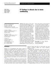
Eur Radiol (2008) 18: 2015–2018<br />
DOI 10.1007/s00330-008-0919-8 INTERPRETATION CORNER<br />
E. K. Woo<br />
R. Simo<br />
B. Conn<br />
S. E. J. Connor<br />
Received: 6 Oc<strong>to</strong>ber 2007<br />
Revised: 2 January 2008<br />
Accepted: 11 January 2008<br />
# European Society of Radiology 2008<br />
E. K. Woo (*)<br />
Department of Radiology, S<strong>to</strong>ke<br />
Mandeville Hospital,<br />
Aylesbury, Buckinghamshire, UK,<br />
HP21 8AL<br />
e-mail: eric.woo@buckshosp.nhs.uk<br />
Tel.: +44-1296-316917<br />
R. Simo<br />
Department of O<strong>to</strong>laryngology,<br />
Guy’s Hospital,<br />
London, UK<br />
Case report<br />
<strong>Vocal</strong> <strong>cord</strong> <strong>paralysis</strong> <strong>secondary</strong> <strong>to</strong> a <strong>benign</strong><br />
<strong>parathyroid</strong> <strong>cyst</strong>: a <strong>case</strong> report with clinical,<br />
imaging and pathological findings (2008:6b)<br />
B. Conn<br />
Department of Dental His<strong>to</strong>pathology,<br />
Guy’s Hospital,<br />
London, UK<br />
S. E. J. Connor<br />
Department of Radiology,<br />
Guy’s Hospital,<br />
London, UK<br />
An 83-year-old man presented with a his<strong>to</strong>ry of hoarseness,<br />
neck swelling and shortness of breath. There was no his<strong>to</strong>ry<br />
of dysphagia, voice strain or neck pain. Physical examination<br />
revealed a slight smooth fullness of the right neck,<br />
but no nodularity <strong>to</strong> suggest cervical lymphadenopathy.<br />
Fibreoptic endoscopy revealed right vocal <strong>cord</strong> immobility.<br />
No intrinsic vocal <strong>cord</strong> lesion was identified. This was<br />
consistent with right recurrent laryngeal nerve palsy.<br />
CT was performed through the neck and chest. Signs of a<br />
right vocal <strong>cord</strong> <strong>paralysis</strong> were seen. There was thickening<br />
and medial positioning of the right aryepiglottic fold and<br />
dilatation of the pyriform sinus (Fig. 1). The arytenoid<br />
cartilage was anteromedially positioned (Fig. 2). There was<br />
also atrophy of the thyroarytenoid (Fig. 3) and cricoarytenoid<br />
muscles (Fig. 4). A large 3.3-cm (transverse)×3.9cm<br />
(AP)×3.8-cm (craniocaudal) well-defined low-density<br />
lesion was also seen posterior <strong>to</strong> the right lobe of the<br />
thyroid gland. This extended <strong>to</strong> the superior mediastinum<br />
and was associated with slight displacement of the trachea<br />
Abstract Parathyroid <strong>cyst</strong>s are uncommon<br />
entities. Symp<strong>to</strong>matic <strong>parathyroid</strong><br />
<strong>cyst</strong>s are extremely rare with<br />
approximately only 200 <strong>case</strong>s reported<br />
in the literature. Only ten <strong>case</strong>s have<br />
been reported with recurrent laryngeal<br />
nerve <strong>paralysis</strong> and none in the radiological<br />
literature. We present a <strong>case</strong><br />
of <strong>parathyroid</strong> <strong>cyst</strong> and recurrent<br />
laryngeal nerve <strong>paralysis</strong> with illustrations<br />
of the clinical, radiological<br />
and pathological appearances as well<br />
as discussion on the management of<br />
this condition.<br />
Keywords Parathyroid . Cyst . CT<br />
<strong>to</strong> the left (Fig. 5). The appearance was in keeping with a<br />
<strong>parathyroid</strong> <strong>cyst</strong>.<br />
Excision of the presumed <strong>parathyroid</strong> <strong>cyst</strong> was planned.<br />
Preoperative labora<strong>to</strong>ry tests were all within normal limits<br />
(including serum calcium and <strong>parathyroid</strong> hormone levels).<br />
Intra-operatively, a large <strong>cyst</strong>ic lesion was identified<br />
separate from and posterior <strong>to</strong> the right lobe of the thyroid<br />
gland consistent with a <strong>parathyroid</strong> <strong>cyst</strong> (Fig. 6). The<br />
recurrent laryngeal nerve was seen <strong>to</strong> enter the <strong>cyst</strong> wall<br />
and therefore sacrificed with excision of the <strong>cyst</strong>. Pathological<br />
examination revealed a <strong>parathyroid</strong> <strong>cyst</strong> with no<br />
malignant features (Fig. 7). Post-operatively, there was<br />
improvement in the patient’s pressure symp<strong>to</strong>ms, but the<br />
vocal <strong>cord</strong> <strong>paralysis</strong> remained.<br />
Discussion<br />
Parathyroid <strong>cyst</strong>s are uncommon entities. Symp<strong>to</strong>matic<br />
<strong>parathyroid</strong> <strong>cyst</strong>s are extremely rare with approximately<br />
only 200 <strong>case</strong>s reported in the literature [1]. Only ten <strong>case</strong>s
2016<br />
Fig. 1 Axial CT scan shows thickening and medial positioning of<br />
the right aryepiglottic fold (arrow) and dilatation of the ipsilateral<br />
pyriform sinus<br />
have been reported with recurrent laryngeal nerve <strong>paralysis</strong><br />
[2–6] and none in the radiological literature. From<br />
embryological developments, the <strong>parathyroid</strong> glands and<br />
therefore <strong>parathyroid</strong> <strong>cyst</strong>s can be found related <strong>to</strong> the<br />
thyroid gland (upper <strong>parathyroid</strong> glands) and the thymus<br />
Fig. 2 Axial CT scan shows anteromedial positioning of the right<br />
arytenoid cartilage (arrow)<br />
Fig. 3 Axial CT scan shows atrophy of the right thyroarytenoid<br />
muscle (arrow)<br />
(lower <strong>parathyroid</strong> glands). A <strong>parathyroid</strong> <strong>cyst</strong> may be<br />
found in the neck or the mediastinum and may mimic a<br />
thyroid or mediastinal lesion [7]. Most are presumed <strong>to</strong> be<br />
asymp<strong>to</strong>matic and are occasionally described as incidental<br />
findings in postmortem studies [8]. If they present<br />
clinically, symp<strong>to</strong>ms include dysphagia, odynophagia and<br />
very rarely recurrent laryngeal nerve palsy [5, 6].<br />
Approximately 10% of patients with <strong>parathyroid</strong> <strong>cyst</strong>s<br />
have hyper<strong>parathyroid</strong>ism [9].<br />
Fig. 4 Axial CT scan shows atrophy of the right cricoarytenoid<br />
muscle (arrowhead)
Fig. 5 Axial CT scan shows a large well-defined <strong>cyst</strong>ic lesion<br />
posterior <strong>to</strong> the right lobe of the thyroid gland extending <strong>to</strong> the<br />
superior mediastinum and causing minor mass effect. The appearance<br />
is consistent with a <strong>parathyroid</strong> <strong>cyst</strong><br />
Theories of the pathogenesis of <strong>parathyroid</strong> <strong>cyst</strong>s are<br />
varied and include: development from epithelial remnants<br />
of the third and fourth branchial pouches, fusion of<br />
micro<strong>cyst</strong>s present in the normal gland and as <strong>parathyroid</strong><br />
hormone retention <strong>cyst</strong>s due <strong>to</strong> gland hyperfunction. It has<br />
also been proposed that <strong>cyst</strong>s may arise due <strong>to</strong> the<br />
persistence of Kurtsteiner canals associated with the<br />
developing <strong>parathyroid</strong> gland [10]. Microscopically, a<br />
<strong>parathyroid</strong> <strong>cyst</strong> is composed of a fibrous connective tissue<br />
wall with occasional entrapped islands of <strong>parathyroid</strong> chief<br />
cells. Cysts may be lined by chief cells that can secrete<br />
<strong>parathyroid</strong> hormone in<strong>to</strong> the <strong>cyst</strong> cavity.<br />
The imaging features of <strong>parathyroid</strong> <strong>cyst</strong>s can be nonspecific.<br />
Ultrasound would show a low echogenic lesion<br />
Fig. 6 Operative view of the <strong>parathyroid</strong> <strong>cyst</strong> being delivered <strong>to</strong> the<br />
neck<br />
2017<br />
Fig. 7 Right picture: (H&E ×100) fibrous <strong>cyst</strong> wall with incomplete<br />
single-cell thick epithelial lining (arrow). Top left: (H&E ×200) <strong>cyst</strong><br />
wall and lining (arrow). Bot<strong>to</strong>m left: (H&E ×20) <strong>parathyroid</strong> gland<br />
in fat adjacent <strong>to</strong> <strong>cyst</strong><br />
closely related <strong>to</strong> the posterior aspect of the thyroid gland,<br />
but is difficult <strong>to</strong> distinguish from a thyroid nodule. A fineneedle<br />
aspiration often contains brownish fluid with<br />
elevated <strong>parathyroid</strong> hormone level [11, 12]. Technetium<br />
radionuclide scintigraphy may complicate matters by<br />
demonstrating a cold nodule [6].<br />
In our patient, CT was performed <strong>to</strong> evaluate the cause of<br />
the recurrent laryngeal nerve palsy. Generally, MRI is used<br />
<strong>to</strong> evaluate a proximal vagal lesion, and CT of the skull<br />
base through <strong>to</strong> the upper mediastinum is used <strong>to</strong> evaluate a<br />
recurrent laryngeal nerve (or distal vagal) lesion. CT clearly<br />
demonstrated the signs of a vocal <strong>cord</strong> <strong>paralysis</strong>. The <strong>cyst</strong>ic<br />
nature as well as its inferoposterior relation <strong>to</strong> the thyroid<br />
suggested a <strong>parathyroid</strong> <strong>cyst</strong>. Less likely differential<br />
diagnoses would include thyroid <strong>cyst</strong>, <strong>cyst</strong>ic hygroma,<br />
thymopharyngeal <strong>cyst</strong>, thymic <strong>cyst</strong> and <strong>cyst</strong>ic adenopathy.<br />
The lack of circumferential thyroid tissue around the lesion<br />
would not favour a thyroid <strong>cyst</strong>. Thymopharyngeal and<br />
thymic <strong>cyst</strong>s as well as <strong>cyst</strong>ic hygromas are usually<br />
congenital and usually found in the paediatric group.<br />
Cystic hygromas are typically transspatial. Cystic adenopathy<br />
is not found in the visceral space and would be<br />
located lateral <strong>to</strong> the thyroid and related <strong>to</strong> the internal<br />
jugular vein and carotid sheath.<br />
Uncomplicated non-functioning <strong>parathyroid</strong> <strong>cyst</strong>s may<br />
be treated by ultrasound-guided aspiration as there are no<br />
reports of malignancy [5, 6]. Sclerotherapy has been used,<br />
but there is a risk of neuro<strong>to</strong>xicity [13]. Surgical excision is<br />
recommended for recurrence, functioning <strong>parathyroid</strong> <strong>cyst</strong>s<br />
or when there are symp<strong>to</strong>ms of dysphagia, dyspnoea or<br />
recurrent laryngeal nerve palsy.<br />
In conclusion, <strong>parathyroid</strong> <strong>cyst</strong> is a rare lesion that<br />
should be considered in the differential diagnosis of a<br />
recurrent laryngeal nerve palsy. The imaging findings of a<br />
<strong>cyst</strong>ic lesion inferoposteriorly related <strong>to</strong> the thyroid gland<br />
should strongly suggest the diagnosis.
2018<br />
References<br />
1. Alvi A, Myssiorek D, Wasserman P<br />
(1996) Parathyroid <strong>cyst</strong>: current diagnostic<br />
and management principles.<br />
Head Neck 18:370–373<br />
2. Grey AB, Shaw JHF, Anderson NE,<br />
Holdaway IM (1993) Parathyroid <strong>cyst</strong><br />
with recurrent vocal <strong>cord</strong> paresis. Aust<br />
NZ Surg 63:561–562<br />
3. Clark OH (1978) Parathyroid <strong>cyst</strong>s. Am<br />
J Surg 135:395–405<br />
4. Coates G, Pearman K, Holl-Allen RTJ<br />
(1991) Recurrent nerve palsy due <strong>to</strong><br />
<strong>parathyroid</strong> <strong>cyst</strong>s. Int Surg 76:192–193<br />
Precisely correct answer was received by closing date from:<br />
Shinichi Kan, Sagamihara, Kanagawa, Japan<br />
Manabu Minami, Ibaraki, Japan<br />
Annemie Snoeckx, Zandhoven, Belgium<br />
Vassilios Maniatis, Palini, Greece<br />
Thaworn Dendumrongsup, Songkhla, Thailand<br />
N.B.S. Mani, Miami, USA<br />
Luc Vanstraelen, Mol, Belgium<br />
5. Sen P, Flower N, Papesch M, Davis A,<br />
Spedding AV (2000) A <strong>benign</strong> <strong>parathyroid</strong><br />
<strong>cyst</strong> presenting with hoarse<br />
voice. J Laryngol O<strong>to</strong>l 114:147–148<br />
6. Coelho DH, Boey HP (2006) Benign<br />
<strong>parathyroid</strong> <strong>cyst</strong> causing vocal fold<br />
<strong>paralysis</strong>: a <strong>case</strong> report and review of<br />
the literature. Head Neck 28:564–566<br />
7. Landau O, Chamberlain DW, Kennedy<br />
RS, Pearson FG, Keshavjee S (1997)<br />
Mediastinal <strong>parathyroid</strong> <strong>cyst</strong>s. Ann<br />
Thorac Surg 63:951–953<br />
8. Katz A, Drunkleman D (1984) Needle<br />
aspiration of non functioning <strong>parathyroid</strong><br />
<strong>cyst</strong>s. Arch Surg 119:307–308<br />
9. Mitmaker B, Lehman S, Lamoureux E,<br />
Begin L (1991) Parathyroid <strong>cyst</strong>: diagnosis<br />
and treatment of an unusual<br />
surgical problem. Can J Surg 34:59–61<br />
10. Lloyd RV, Douglas BR, Young WF<br />
(2002) AFIP Atlas of nontumor pathology.<br />
Endocrine diseases. Chapter 2,<br />
page 62<br />
11. Layfield LJ (1991) Fine needle aspiration<br />
cy<strong>to</strong>logy of <strong>cyst</strong>ic <strong>parathyroid</strong><br />
lesions. Acta Cy<strong>to</strong>l 1001 35:447–450<br />
12. Ihm PS, Dray T, Sofferman RA, Nathan<br />
M, Hardin NJ (2001) Parathyroid <strong>cyst</strong>s:<br />
diagnosis and management. Laryngoscope<br />
111:1576–1578<br />
13. Sanchez A, Carret<strong>to</strong> H (1993) Treatment<br />
of non-functioning <strong>parathyroid</strong><br />
<strong>cyst</strong> with tetracycline injection. Head<br />
Neck 15:263–265