CT radiation dose in children - European Radiology
CT radiation dose in children - European Radiology
CT radiation dose in children - European Radiology

Create successful ePaper yourself
Turn your PDF publications into a flip-book with our unique Google optimized e-Paper software.
Eur Radiol (2008) 18: 1980–1986<br />
DOI 10.1007/s00330-008-0963-4 PEDIATRIC<br />
Francis R. Verdun<br />
Daniel Gutierrez<br />
John Paul Vader<br />
Abbas Aroua<br />
Leonor Tr<strong>in</strong>idad Alamo-Maestre<br />
François Bochud<br />
François Gud<strong>in</strong>chet<br />
Received: 20 July 2007<br />
Revised: 27 February 2008<br />
Accepted: 2 March 2008<br />
Published onl<strong>in</strong>e: 4 April 2008<br />
# <strong>European</strong> Society of <strong>Radiology</strong> 2008<br />
F. R. Verdun (*) . D. Gutierrez .<br />
A. Aroua . F. Bochud<br />
University Institute for Radiation<br />
Physics (IRA–DUMSC),<br />
CHUV and University of Lausanne,<br />
Grand-Pré 1,<br />
1007 Lausanne, Switzerland<br />
e-mail: Francis.Verdun@chuv.ch<br />
Tel.: +41-21-6233434<br />
Fax: +41-21-6233435<br />
J. P. Vader<br />
University Institute of Social<br />
and Preventive Medic<strong>in</strong>e<br />
(IUMSP–DUMSC),<br />
CHUV and University of Lausanne,<br />
Bugnon 17,<br />
1005 Lausanne, Switzerland<br />
Introduction<br />
S<strong>in</strong>ce its first applications <strong>in</strong> the early 1970s the use of<br />
computed tomography (<strong>CT</strong>) has been cont<strong>in</strong>uously<br />
grow<strong>in</strong>g. Accord<strong>in</strong>g to the 2000 report [1] of the United<br />
Nations Scientific Committee on the Effects of Atomic<br />
Radiation (UNSCEAR), the frequency of <strong>CT</strong> exam<strong>in</strong>ations<br />
<strong>in</strong> countries of high healthcare level <strong>in</strong>creased on<br />
average from 6.1 per year per 1,000 population <strong>in</strong> the<br />
1970s to 48 per year per 1,000 population <strong>in</strong> the period<br />
1991-1996, while the average effective <strong>dose</strong> per <strong>CT</strong><br />
exam<strong>in</strong>ation <strong>in</strong>creased from 1.3 mSv <strong>in</strong> the 1970s to<br />
8.8 mSv <strong>in</strong> the period 1991-1996. This <strong>in</strong>crease went on<br />
steadily dur<strong>in</strong>g the last decade. In Switzerland, the results<br />
of the 2003 survey on the exposure of the population by<br />
<strong>CT</strong> <strong>radiation</strong> <strong>dose</strong> <strong>in</strong> <strong>children</strong>: a survey<br />
to establish age-based diagnostic reference<br />
levels <strong>in</strong> Switzerland<br />
L. T. Alamo-Maestre . F. Gud<strong>in</strong>chet<br />
University Hospital of Lausanne (CHUV),<br />
Department of Radiodiagnostic<br />
and Interventional <strong>Radiology</strong>,<br />
CHUV and University of Lausanne,<br />
1011 Lausanne, Switzerland<br />
Abstract This work aimed at assess<strong>in</strong>g<br />
the <strong>dose</strong>s delivered <strong>in</strong> Switzerland<br />
to paediatric patients dur<strong>in</strong>g<br />
computed tomography (<strong>CT</strong>) exam<strong>in</strong>ations<br />
of the bra<strong>in</strong>, chest and abdomen,<br />
and at establish<strong>in</strong>g diagnostic<br />
reference levels (DRLs) for various<br />
age groups. Forms were sent to the ten<br />
centres perform<strong>in</strong>g <strong>CT</strong> on <strong>children</strong>,<br />
address<strong>in</strong>g the demographics, the <strong>in</strong>dication<br />
and the scann<strong>in</strong>g parameters:<br />
number of series, kilovoltage, tube<br />
current, rotation time, reconstruction<br />
slice thickness and pitch, volume <strong>CT</strong><br />
<strong>dose</strong> <strong>in</strong>dex (<strong>CT</strong>DIvol) and <strong>dose</strong> length<br />
product (DLP). Per age group, the<br />
proposed DRLs for bra<strong>in</strong>, chest and<br />
abdomen are, respectively, <strong>in</strong> terms of<br />
<strong>CT</strong>DIvol: 20, 30, 40, 60 mGy; 5, 8, 10,<br />
12 mGy; 7, 9, 13, 16 mGy; and <strong>in</strong><br />
terms of DLP: 270, 420, 560,<br />
1,000 mGy cm; 110, 200, 220,<br />
460 mGy cm; 130, 300, 380, 500 mGy<br />
cm. An optimisation process should<br />
be <strong>in</strong>itiated to reduce the spread <strong>in</strong><br />
<strong>dose</strong> recorded <strong>in</strong> this study. A major<br />
element of this process should be the<br />
use of DRLs.<br />
Keywords Computed tomography .<br />
Paediatrics . Radiation dosimetry .<br />
Radiation protection .<br />
Diagnostic reference levels<br />
medical X-ray imag<strong>in</strong>g [2] <strong>in</strong>dicated a 70% <strong>in</strong>crease of<br />
<strong>CT</strong> exam<strong>in</strong>ations <strong>in</strong> a 5-year period (1998-2003), which,<br />
comb<strong>in</strong>ed with a 20% <strong>in</strong>crease <strong>in</strong> the average effective<br />
<strong>dose</strong> per <strong>CT</strong> procedure, led to an <strong>in</strong>crease by a factor of<br />
two <strong>in</strong> the <strong>CT</strong> contribution to the collective <strong>dose</strong> due to<br />
medical X-rays reach<strong>in</strong>g 47%. The same trend was<br />
registered <strong>in</strong> other <strong>European</strong> countries. In Norway, the<br />
frequency of <strong>CT</strong> exam<strong>in</strong>ations <strong>in</strong>creased by a factor of<br />
2.2 <strong>in</strong> a decade and <strong>CT</strong> contribution to collective<br />
effective <strong>dose</strong> was estimated to account for 59% of the<br />
total, as opposed to 30% <strong>in</strong> the previous survey [3]. In<br />
Germany, the <strong>in</strong>crease <strong>in</strong> the collective <strong>dose</strong> due to <strong>CT</strong><br />
rose by about 50% between 1996 and 2002, and <strong>in</strong> the<br />
UK the frequency of <strong>CT</strong> exam<strong>in</strong>ations <strong>in</strong>creased by 39%<br />
between 1998 and 2002.
The proportion of all <strong>CT</strong> exam<strong>in</strong>ations which are<br />
performed <strong>in</strong> <strong>children</strong> does not exceed a few per cent. In<br />
Switzerland, the most recent nationwide survey on the<br />
exposure of the population by diagnostic and <strong>in</strong>terventional<br />
radiology that provided patient age data <strong>in</strong>dicated a <strong>CT</strong><br />
paediatric fraction of 1% <strong>in</strong> 1998 [4, 5]. The same figure<br />
was reported <strong>in</strong> Germany for the period 2005-2006 [6].<br />
Other countries reported higher values. Recent surveys<br />
cover<strong>in</strong>g the year 2000 revealed a paediatric fraction of<br />
2.7% <strong>in</strong> Japan [7], and of 6.5% <strong>in</strong> the USA [8]. The latter<br />
figure is comparable with that given by UNSCEAR for the<br />
period 1991-1996 [1]. Only general guidel<strong>in</strong>es are<br />
available for the use of <strong>CT</strong> <strong>in</strong> the paediatric population<br />
[9-13] and the need for size-based <strong>CT</strong> protocols has only<br />
recently been emphasised [6, 14-18]. Given the recent<br />
attention to <strong>radiation</strong> risks and <strong>CT</strong> <strong>in</strong> <strong>children</strong> [19-24] there<br />
is an urgent need for optimisation of the present practice by<br />
means of the <strong>in</strong>troduction of the Diagnostic Reference<br />
Level (DRL) by the International Commission on Radiological<br />
Protection (ICRP) <strong>in</strong> 1997 [25]. For these reasons,<br />
we sought to assess the practice of paediatric <strong>CT</strong> <strong>in</strong><br />
Switzerland us<strong>in</strong>g forms sent to the ten ma<strong>in</strong> centres<br />
(<strong>in</strong>clud<strong>in</strong>g regional and university hospitals) that perform<br />
<strong>CT</strong> exam<strong>in</strong>ations <strong>in</strong> <strong>children</strong>. The results of this survey<br />
allow the development of a set of DRL values for the most<br />
common paediatric <strong>CT</strong> exam<strong>in</strong>ations.<br />
Materials and method<br />
The survey conducted between January and December<br />
2005 consisted of a questionnaire addressed to the ten<br />
centres deal<strong>in</strong>g with paediatric <strong>CT</strong> <strong>in</strong> Switzerland (Aarau,<br />
Bell<strong>in</strong>zona, Bern, Fribourg, Geneva, Lausanne, St Gallen,<br />
Sion, W<strong>in</strong>terthur, and Zurich). The data were treated <strong>in</strong> an<br />
anonymous way, with the ten centres be<strong>in</strong>g coded by<br />
alphabetical letters from A to J.<br />
In this survey, the paediatric population was separated<br />
<strong>in</strong>to four age/weight groups (
1982<br />
Table 2 Number of exam<strong>in</strong>ations performed <strong>in</strong> the various centres<br />
Centre 0–5 years 5–10 years 10–15 years Total<br />
(0–20 kg) (20–35 kg) (>35 kg)<br />
208 126 186 520<br />
B 10 b<br />
7 b<br />
8 b<br />
25<br />
D a<br />
304 219 290 813<br />
E 29 35 33 97<br />
F 69 76 145 290<br />
G a<br />
261 212 217 690<br />
I a<br />
563 273 203 1039<br />
J 26 38 86 150<br />
All 1,470 986 1,168 3,624<br />
a<br />
University or paediatric hospital<br />
b<br />
Based on the total number and us<strong>in</strong>g the average distribution of all the other centres<br />
A a<br />
DRL is obta<strong>in</strong>ed by multiply<strong>in</strong>g the mean value, obta<strong>in</strong>ed<br />
by averag<strong>in</strong>g the typical values provided by the participat<strong>in</strong>g<br />
centres, by an appropriate factor (1.25 <strong>in</strong> the present<br />
work). Due to the typical shape of the <strong>dose</strong> distributions,<br />
this method provides a figure comparable with the third<br />
quartile. Each centre provided for each anatomical region<br />
and cl<strong>in</strong>ical <strong>in</strong>dication typical values for the various age<br />
groups, tak<strong>in</strong>g <strong>in</strong>to account the variability due to differences<br />
<strong>in</strong> the protocols used (number of sequences, technical<br />
parameters, etc.). Therefore, the average values over the<br />
participat<strong>in</strong>g centres expressed the national variability for a<br />
given exam<strong>in</strong>ation/<strong>in</strong>dication.<br />
Results<br />
Information was obta<strong>in</strong>ed from eight out of the ten centres.<br />
Two centres (C and H), which are small regional hospitals,<br />
could not provide it due to the limitation of their account<strong>in</strong>g<br />
systems. Three centres were not able to provide detailed<br />
<strong>in</strong>formation and gave either the number of all exam<strong>in</strong>ations<br />
ventilated by age, but merg<strong>in</strong>g the first two age categories<br />
(
Table 4 Means values averaged over participat<strong>in</strong>g centres and ranges of <strong>CT</strong>DIvol (mGy) and the DLP (mGy cm) values<br />
Age group Quantity Bra<strong>in</strong> Chest Abdomen<br />
n Mean Range n Mean Range n Mean Range<br />
1984<br />
DLP (mGy.cm)<br />
DLP (mGy.cm)<br />
DLP (mGy.cm)<br />
700<br />
600<br />
500<br />
400<br />
300<br />
200<br />
100<br />
700<br />
600<br />
500<br />
400<br />
F (2 phases)<br />
C<br />
B<br />
J<br />
E (2 phases)<br />
D<br />
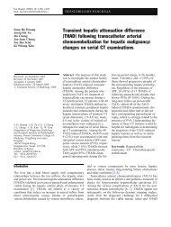
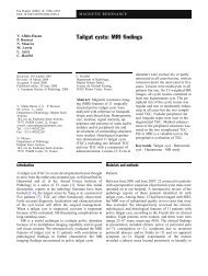
that displays the plot of the DLP as a function of the<br />
<strong>CT</strong>DI vol and the DLP. For bra<strong>in</strong> exam<strong>in</strong>ation, almost all<br />
data po<strong>in</strong>ts can be aligned along a straight l<strong>in</strong>e that has a<br />
slope of 11.7 (R 2 =0.86), mean<strong>in</strong>g that the average length of<br />
coverage is 11.7 cm for this particular exam<strong>in</strong>ation and age<br />
category. This exam<strong>in</strong>ation length is fully compatible with<br />
the data obta<strong>in</strong>ed <strong>in</strong> the UK <strong>in</strong> 2003 (10.9-11.7 cm) [26].<br />
For both chest and abdomen exam<strong>in</strong>ations, the slope is 22<br />
(R 2 =0.94 and R 2 =0.97, respectively), i.e. an average scan<br />
length of 22 cm. It is of note that the value obta<strong>in</strong>ed for the<br />
A<br />
This study<br />
H<br />
G<br />
I<br />
UK 2003 [26]<br />
DRL [31]<br />
0<br />
0 10 20 30 40 50 60 70<br />
<strong>CT</strong>DIw (mGy)<br />
<strong>CT</strong>DI vol (mGy)<br />
<strong>CT</strong>DI vol (mGy)<br />
E (2 phases)<br />
L= 11.7 cm<br />
L = 22 cm<br />
DRL [31]<br />
300<br />
D<br />
UK 2003 [26]<br />
200<br />
100<br />
This study<br />
F (2 phases)<br />
I<br />
B<br />
C J G<br />
A<br />
0<br />
0<br />
H<br />
5 10 15 20 25 30 35<br />
700<br />
600<br />
500<br />
400<br />
E (2 phases)<br />
D (2 phases)<br />
L= 22 cm<br />
DRL [26]<br />
(upper + lower<br />
abdomen)<br />
300<br />
200 F (2 phases)<br />
H<br />
This study<br />
G I<br />
DRL [26]<br />
(upper abdomen only)<br />
100 C<br />
A J<br />
B<br />
0<br />
0 5 10 15 20 25 30<br />
Fig. 1 Bra<strong>in</strong> (top), chest (middle) and abdom<strong>in</strong>al (bottom) exam<strong>in</strong>ation<br />
for <strong>children</strong> <strong>in</strong> age group 1 to 5 years<br />
chest <strong>in</strong> this survey for a similar age category is slightly<br />
higher than the UK data (16.5–18.6 cm) [26]. Table 4<br />
shows that the <strong>in</strong>ter-centre variation for bra<strong>in</strong> exam<strong>in</strong>ation<br />
and for the different age groups ranges from a factor of 3.2–<br />
6.2 for the <strong>CT</strong>DI vol (3.6–6.9 for the DLP) the highest<br />
variation be<strong>in</strong>g for age group 10–15 years where the<br />
<strong>CT</strong>DI vol delivered varies from 14 to 85 mGy and the DLP<br />
varies from 216 to 1,485 mGy cm. For chest exam<strong>in</strong>ation<br />
the <strong>in</strong>ter-centre variations for the different age groups range<br />
from a factor of 9.5–25 for the <strong>CT</strong>DI vol (11–51.4 for the<br />
DLP), the highest variation be<strong>in</strong>g for age group 1–5 years<br />
where the <strong>CT</strong>DI vol delivered varies from 0.8 to 20 mGy and<br />
the DLP varies from 12 to 617 mGy cm. For abdomen<br />
exam<strong>in</strong>ation, the <strong>in</strong>ter-centre variations for the different age<br />
groups ranges from a factor of 4.0–12.1 for the <strong>CT</strong>DI vol<br />
(8.2–22.4 for the DLP), the highest variation be<strong>in</strong>g for age<br />
group 1-5 years where the <strong>CT</strong>DI vol delivered varies from<br />
1.4 to 16 mGy and the DLP varies from 29 to 650 mGy cm.<br />
It is worth mention<strong>in</strong>g that only one centre (I) that<br />
performs more than 1,000 <strong>CT</strong> exam<strong>in</strong>ations with a SS<strong>CT</strong><br />
system does not adapt the protocols to the age of the<br />
patient. Thus, there is great potential for optimisation. The<br />
small centres (E and F) recourse systematically to a biphase<br />
acquisition. This practice is not adopted by the other<br />
centres for bra<strong>in</strong> and chest exam<strong>in</strong>ations. Centre D (large)<br />
performs also a bi-phase acquisition dur<strong>in</strong>g abdomen<br />
exam<strong>in</strong>ations, which leads to high <strong>dose</strong> values. Here aga<strong>in</strong><br />
there is a potential for optimisation by specify<strong>in</strong>g<br />
accurately the <strong>in</strong>stances for which bi-phase acquisition is<br />
<strong>in</strong>dicated.<br />
The great dispersion of the <strong>CT</strong>DI vol reflects a great<br />
dispersion of the associated image quality. The majority of<br />
the big centres produce images of homogeneous quality<br />
s<strong>in</strong>ce they are well grouped. Centre I only, where no<br />
adaptation of protocols is used, the images produced are of<br />
excellent quality for a very young population of patients.<br />
On the other hand, <strong>in</strong> certa<strong>in</strong> centres, where only a few<br />
exam<strong>in</strong>ations are carried out, such low <strong>CT</strong>DI values are<br />
used (0.8 mGy for chest and patients between 0 and<br />
5 years; 1.4 mGy for abdomen and patients from 0 and<br />
10 years) that the diagnostic quality of the image may be<br />
questioned.<br />
As shown <strong>in</strong> Table 5, the DRLs <strong>in</strong> terms of <strong>CT</strong>DI vol and<br />
DRLs are compatible with the values reported <strong>in</strong> Germany<br />
and the UK. In most cases they are lower than the UK data.<br />
Compared with the German data, the values established <strong>in</strong><br />
this work are lower for the bra<strong>in</strong> exam<strong>in</strong>ation, but higher<br />
for chest and abdomen for some age groups.<br />
The DRLs obta<strong>in</strong>ed <strong>in</strong> this <strong>in</strong>vestigation were established<br />
by consider<strong>in</strong>g typical <strong>dose</strong> values associated with<br />
standard protocols used <strong>in</strong> a given centre for the various<br />
types of exam<strong>in</strong>ations and age groups, and then averag<strong>in</strong>g<br />
over the ten participat<strong>in</strong>g centres and multiply<strong>in</strong>g by an<br />
appropriate factor. These DRLs should be considered as<br />
provisional only, to be replaced <strong>in</strong> the future by more<br />
robust data established through surveys of measured <strong>dose</strong>s
on a big sample of patients lead<strong>in</strong>g to empirical <strong>dose</strong><br />
distributions. The collection of such data is time and<br />
resource consum<strong>in</strong>g, but the new Swiss legislation<br />
concern<strong>in</strong>g the implementation of DRLs and the sett<strong>in</strong>gup<br />
of a national <strong>dose</strong> database, as well as the standardisation<br />
of the way the <strong>dose</strong> descriptors (<strong>CT</strong>DI vol and DLP<br />
parameters) will be stored <strong>in</strong> the DICOM header, which is<br />
under discussion, will all facilitate such an endeavour.<br />
Conclusion<br />
The frequency of paediatric <strong>CT</strong> exam<strong>in</strong>ations and the<br />
typical values of the related <strong>dose</strong> quantities (<strong>CT</strong>DI vol<br />
and DRLs) were surveyed <strong>in</strong> the ten Swiss centres<br />
perform<strong>in</strong>g paediatric <strong>CT</strong>. Mean values averaged over<br />
the participat<strong>in</strong>g centres were calculated and the<br />
correspond<strong>in</strong>g DRLs were established by multiply<strong>in</strong>g<br />
the mean values by 1.25. This <strong>in</strong>vestigation revealed<br />
that 4,000–5,000 <strong>CT</strong> exam<strong>in</strong>ations are carried out on<br />
<strong>children</strong> <strong>in</strong> Switzerland, with an average of 453 per<br />
centre perform<strong>in</strong>g paediatric <strong>CT</strong>. Significant variations<br />
of the <strong>radiation</strong> <strong>dose</strong> delivered to the paediatric popu-<br />
References<br />
1. United Nations Scientific Committee<br />
on the Effects of Atomic Radiation<br />
(2000) Report to the general assembly,<br />
annex D: Medical <strong>radiation</strong> exposures.<br />
United Nations, New York<br />
2. Aroua A, Vader JP, Valley JF, Verdun<br />
FR (2007) Exposure of the Swiss<br />
population by radiodiagnostics : 2003<br />
Review. Health Phys 92:442–448<br />
3. Børretzen I, Lysdahl KB, Olerud HM<br />
(2007) Diagnostic radiology <strong>in</strong><br />
Norway—trends <strong>in</strong> exam<strong>in</strong>ation<br />
frequency and collective effective <strong>dose</strong>.<br />
Radiat Prot Dosim 124:339–347<br />
4. Aroua A, Burnand B, Decka I, Vader<br />
JP, Valley JF (2002) Nation-wide<br />
survey on <strong>radiation</strong> <strong>dose</strong>s <strong>in</strong> diagnostic<br />
and <strong>in</strong>terventional radiology <strong>in</strong> Switzerland<br />
<strong>in</strong> 1998. Health Phys 83:46–55<br />
5. Aroua A, Decka I, Burnand B, Vader<br />
JP, Valley JF (2002) Dosimetric aspects<br />
of a national survey of diagnostic and<br />
<strong>in</strong>terventional radiology <strong>in</strong> Switzerland.<br />
Medical Phys 29:2247–2259<br />
6. Galanski M, Nagel HD, Stamm G<br />
(2007) Paediatric <strong>CT</strong> exposure practice<br />
<strong>in</strong> the federal republic of Germany:<br />
results of a nationwide survey<br />
<strong>in</strong> 2005-2006. Mediz<strong>in</strong>ische<br />
Hochschule, Hannover<br />
7. Kanae N, Masaki M, Kazuo I, Takashi<br />
M (2004) Survey of <strong>CT</strong> practice <strong>in</strong><br />
Japan and collective effective <strong>dose</strong><br />
estimation. Nippon Acta Radiologica<br />
64:151–158<br />
8. Nationwide Evaluation of X-Ray<br />
Trends (NEXT): 2000 Survey of Computed<br />
Tomography (2006), CRCPD<br />
publication NEXT_2000<strong>CT</strong>-T<br />
9. Donnelly LF, Emery KH, Brody AS,<br />
Laor T, Gylys-Mor<strong>in</strong> VM, Anton CG,<br />
Thomas SR, Frush DP (2001) M<strong>in</strong>imiz<strong>in</strong>g<br />
<strong>radiation</strong> <strong>dose</strong> for paediatric<br />
body applications of s<strong>in</strong>gle-detector<br />
helical <strong>CT</strong>: strategies at a large <strong>children</strong>’s<br />
hospital. AJR Am J Roentgenol<br />
176:303–306<br />
10. Cody DD, Moxley DM, Krugh KT,<br />
O’Daniel JC, Wagner LK, Eftekhari F<br />
(2004) Strategies for formulat<strong>in</strong>g appropriate<br />
MD<strong>CT</strong> techniques when imag<strong>in</strong>g<br />
the chest, abdomen, and pelvis <strong>in</strong><br />
pediatric patients. AJR Am J<br />
Roentgenol 182:849–859<br />
1985<br />
lation were found. An optimisation process should be<br />
<strong>in</strong>itiated <strong>in</strong> order to reduce this spread <strong>in</strong> <strong>dose</strong><br />
(appropriate image quality requirements for a given<br />
<strong>in</strong>dication, number of acquisition phases that are<br />
cl<strong>in</strong>ically relevant, etc.). A major element of the<br />
optimisation process is a consensus on the DRLs that<br />
need to be used. This becomes a priority <strong>in</strong> the light of<br />
contributions such as described <strong>in</strong> a recent article<br />
published <strong>in</strong> the Lancet [32]. A set of DRL values for<br />
<strong>CT</strong> exam<strong>in</strong>ations of the bra<strong>in</strong>, the chest and the<br />
abdomen and for the various age groups are proposed<br />
here for temporary use <strong>in</strong> paediatrics until a more<br />
extensive survey is organised to collect <strong>dose</strong> data on a<br />
large sample of patients and to establish empirical <strong>dose</strong><br />
distributions.<br />
Acknowledgements The authors gratefully acknowledge f<strong>in</strong>ancial<br />
support from the Swiss National Science Foundation (Grant no.<br />
3200B0-105951) and are thankful to the members of SSPR, <strong>in</strong><br />
particular: Drs G. Eich, S. Hanqu<strong>in</strong>et, J.-M. Girard, A. Racle, G.<br />
Remsei, T. Schraner, P. Waibel, R. Wolf, Mme M. Wyttenbach, M. P.<br />
Brégis, and A. Devaud)<br />
11. Holl<strong>in</strong>gsworth C, Frush DP, Cross M,<br />
Lucaya J (2003) Helical <strong>CT</strong> of the<br />
body: a survey of techniques used for<br />
pediatric patients. AJR Am J Roentgenol<br />
180:401–406<br />
12. Vock P (2005) <strong>CT</strong> <strong>dose</strong> reduction <strong>in</strong><br />
<strong>children</strong>. Eur Radiol 15:2330–2340<br />
13. Boone JM, Geraghty EM, Sielbert JA,<br />
Wootton-George SL (2003) Dose reduction<br />
<strong>in</strong> pediatric <strong>CT</strong>: a rational<br />
approach. <strong>Radiology</strong> 228:352–360<br />
14. Paterson A, Frush DP, Donnelly LF<br />
(2001) Helical <strong>CT</strong> of the body: are<br />
sett<strong>in</strong>gs adjusted for pediatric patients?<br />
AJR Am J Roentgenol 176:297–301<br />
15. Huda W, Lieberman KA, Chang J,<br />
Roskopf ML (2004) Patient size and xray<br />
technique factors <strong>in</strong> head computed<br />
tomography exam<strong>in</strong>ations. II. Image<br />
quality. Med Phys 31:595–601<br />
16. Siegel MJ, Schmidt B, Bradley D,<br />
Suess C, Hildebolt C (2004) Radiation<br />
<strong>dose</strong> and image quality <strong>in</strong> pediatric <strong>CT</strong>:<br />
effect of technical factors and phantom<br />
size and shape. <strong>Radiology</strong> 233:515–<br />
522
1986<br />
17. Verdun FR, Lepori D, Monn<strong>in</strong> P, Valley<br />
JF, Schnyder P, Gud<strong>in</strong>chet F (2004)<br />
Management of patient <strong>dose</strong> and image<br />
noise <strong>in</strong> rout<strong>in</strong>e pediatric <strong>CT</strong> abdom<strong>in</strong>al<br />
exam<strong>in</strong>ations. Eur Radiol 14:835–841<br />
18. Wilt<strong>in</strong>g JE, Zwartkruis A, van Leeuwen<br />
MS, Timmer J, Kamphuis AG,<br />
Feldberg M (2001) A rational approach<br />
to <strong>dose</strong> reduction <strong>in</strong> <strong>CT</strong>: <strong>in</strong>dividualized<br />
scan protocols. Eur Radiol 11:2627–<br />
2632<br />
19. Pages J, Buls N, Osteuax M (2003) <strong>CT</strong><br />
<strong>dose</strong>s <strong>in</strong> <strong>children</strong>: a multicentre study.<br />
Br J Radiol 76:803–811<br />
20. L<strong>in</strong>ton OA, Mettler FA (2003) National<br />
conference on <strong>dose</strong> reduction <strong>in</strong> <strong>CT</strong><br />
with an emphasis on pediatric patients.<br />
AJR Am J Roentgenol 181:321–329<br />
21. Hall EJ (2002) Lessons we have<br />
learned from our <strong>children</strong>: cancer risks<br />
from diagnostic radiology. Pediatr<br />
Radiol 32:700–706<br />
22. Ron E (2002) Let’s not relive the<br />
past: a review of cancer risk after<br />
diagnostic or therapeutic ir<strong>radiation</strong>.<br />
Pediatric <strong>Radiology</strong> 32:739–744 discussion<br />
751–754<br />
23. Brenner DJ, Elliston CD, Hall EJ,<br />
Berdon WE (2001) Estimated risks of<br />
<strong>radiation</strong>-<strong>in</strong>duced fatal cancer from<br />
paediatric <strong>CT</strong>. AJR Am J Roentgenol<br />
176:289–296<br />
24. Frush DP, Donnelly LF, Rosen NS<br />
(2003) Computed tomography and <strong>radiation</strong><br />
risks: what pediatric health care<br />
providers should know. Pediatrics<br />
112:951–957<br />
25. International Commission on Radiological<br />
Protection (1997) Radiological<br />
protection and safety <strong>in</strong> medic<strong>in</strong>e.<br />
ICRP Publication 73, Pergamon, Oxford<br />
New York<br />
26. Schrimpton PC, Hillier MC, Lewis<br />
MA, Dunn M (2005) Dose from <strong>CT</strong><br />
exam<strong>in</strong>ation <strong>in</strong> the UK–2003 review.<br />
NRPB–W67 Report. National Radiological<br />
Protection Board, Chilton<br />
27. International Electrotechnical Committee<br />
(2002) Medical diagnostic X-ray<br />
equipment-Particular requirements for<br />
the safety of X-ray equipment for <strong>CT</strong>;<br />
Standard IEC #60601-2-44<br />
28. http://www.impactscan.org<br />
29. Neofotistou V (2001) Review of patient<br />
dosimetry <strong>in</strong> cardiology. Radiat Prot<br />
Dosim 94:177–182<br />
30. Aroua A, Besançon A, Buchillier-<br />
Decka I, Trueb P, Valley JF, Verdun FR,<br />
Zeller W (2004) Adult reference levels<br />
<strong>in</strong> diagnostic and <strong>in</strong>terventional radiology<br />
for temporary use <strong>in</strong> Switzerland.<br />
Radiat Prot Dosim 111:289–295<br />
31. Shrimpton PC, Wall BF (2000) Reference<br />
<strong>dose</strong>s for paediatric computed<br />
tomography. Radiat Prot Dosim<br />
90:249–252<br />
32. Mart<strong>in</strong> DR, Semelka RC (2006) Health<br />
effects of ionis<strong>in</strong>g <strong>radiation</strong> from diagnostic<br />
<strong>CT</strong>. Lancet 367:1712–1714