Masticator space abscess derived from odontogenic infection ...
Masticator space abscess derived from odontogenic infection ...
Masticator space abscess derived from odontogenic infection ...
Create successful ePaper yourself
Turn your PDF publications into a flip-book with our unique Google optimized e-Paper software.
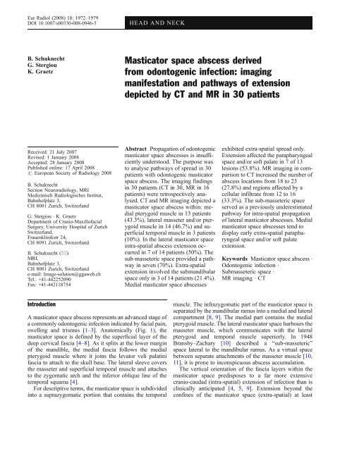
Eur Radiol (2008) 18: 1972–1979<br />
DOI 10.1007/s00330-008-0946-5 HEAD AND NECK<br />
B. Schuknecht<br />
G. Stergiou<br />
K. Graetz<br />
Received: 21 July 2007<br />
Revised: 1 January 2008<br />
Accepted: 28 January 2008<br />
Published online: 17 April 2008<br />
# European Society of Radiology 2008<br />
B. Schuknecht<br />
Section Neuroradiology, MRI<br />
Medizinisch Radiologisches Institut,<br />
Bahnhofplatz 3,<br />
CH 8001 Zurich, Switzerland<br />
G. Stergiou . K. Graetz<br />
Department of Cranio-Maxillofacial<br />
Surgery, University Hospital of Zurich<br />
Switzerland,<br />
Frauenklinikstr 24,<br />
CH 8091 Zurich, Switzerland<br />
B. Schuknecht (*)<br />
MRI,<br />
Bahnhofplatz 3,<br />
CH 8001 Zurich, Switzerland<br />
e-mail: Image-solution@ggaweb.ch<br />
Tel.: +41-442252090<br />
Fax: +41-442118754<br />
Introduction<br />
A masticator <strong>space</strong> <strong>abscess</strong> represents an advanced stage of<br />
a commonly <strong>odontogenic</strong> <strong>infection</strong> indicated by facial pain,<br />
swelling and trismus [1–3]. Anatomically (Fig. 1), the<br />
masticator <strong>space</strong> is defined by the superficial layer of the<br />
deep cervical fascia [4–8]. As it splits at the lower margin<br />
of the mandible, the medial fascia follows the medial<br />
pterygoid muscle where it joins the levator veli palatini<br />
fascia to attach to the skull base. The lateral sleeve covers<br />
the masseter and superficial temporal muscle and attaches<br />
to the zygomatic arch and the inferior oblique line of the<br />
temporal squama [4].<br />
For descriptive terms, the masticator <strong>space</strong> is subdivided<br />
into a suprazygomatic portion that contains the temporal<br />
<strong>Masticator</strong> <strong>space</strong> <strong>abscess</strong> <strong>derived</strong><br />
<strong>from</strong> <strong>odontogenic</strong> <strong>infection</strong>: imaging<br />
manifestation and pathways of extension<br />
depicted by CT and MR in 30 patients<br />
Abstract Propagation of <strong>odontogenic</strong><br />
masticator <strong>space</strong> <strong>abscess</strong>es is insufficiently<br />
understood. The purpose was<br />
to analyse pathways of spread in 30<br />
patients with <strong>odontogenic</strong> masticator<br />
<strong>space</strong> <strong>abscess</strong>. The imaging findings<br />
in 30 patients (CT in 30, MR in 16<br />
patients) were retrospectively analysed.<br />
CT and MR imaging depicted a<br />
masticator <strong>space</strong> <strong>abscess</strong> within: medial<br />
pterygoid muscle in 13 patients<br />
(43.3%), lateral masseter and/or pterygoid<br />
muscle in 14 (46.7%) and superficial<br />
temporal muscle in 3 patients<br />
(10%). In the lateral masticator <strong>space</strong><br />
intra-spatial <strong>abscess</strong> extension occurred<br />
in 7 of 14 patients (50%). The<br />
sub-masseteric <strong>space</strong> provided a pathway<br />
in seven (70%). Extra-spatial<br />
extension involved the submandibular<br />
<strong>space</strong> only in 3 of 14 patients (21.4%).<br />
Medial masticator <strong>space</strong> <strong>abscess</strong>es<br />
exhibited extra-spatial spread only.<br />
Extension affected the parapharyngeal<br />
<strong>space</strong> and/or soft palate in 7 of 13<br />
lesions (53.8%). MR imaging in comparison<br />
to CT increased the number of<br />
<strong>abscess</strong> locations <strong>from</strong> 18 to 23<br />
(27.8%) and regions affected by a<br />
cellular infiltrate <strong>from</strong> 12 to 16<br />
(33.3%). The sub-masseteric <strong>space</strong><br />
served as a previously underestimated<br />
pathway for intra-spatial propagation<br />
of lateral masticator <strong>abscess</strong>es. Medial<br />
masticator <strong>space</strong> <strong>abscess</strong>es tend to<br />
display early extra-spatial parapharyngeal<br />
<strong>space</strong> and/or soft palate<br />
extension.<br />
Keywords <strong>Masticator</strong> <strong>space</strong> <strong>abscess</strong> .<br />
Odontogenic <strong>infection</strong> .<br />
Submasseteric <strong>space</strong> .<br />
MR imaging . CT<br />
muscle. The infrazygomatic part of the masticator <strong>space</strong> is<br />
separated by the mandibular ramus into a medial and lateral<br />
compartment [8, 9]. The medial part contains the medial<br />
pterygoid muscle. The lateral masticator <strong>space</strong> harbours the<br />
masseter muscle, which communicates with the lateral<br />
pterygoid and temporal muscle superiorly. In 1948<br />
Bransby–Zachary [10] described a “sub-masseteric”<br />
<strong>space</strong> lateral to the mandibular ramus. As a virtual <strong>space</strong><br />
between separate attachments of the masseter muscle [10,<br />
11], it is prone to inconspicuous <strong>abscess</strong> accumulation.<br />
The vertical orientation of the fascia layers within the<br />
masticator <strong>space</strong> predisposes to a far more extensive<br />
cranio-caudad (intra-spatial) extension of <strong>infection</strong> than is<br />
clinically anticipated [4, 5, 9]. Extension beyond the<br />
confines of the masticator <strong>space</strong> (extra-spatial) at least
Fig. 1 a, b Line drawing of the<br />
coronal (a) and axial anatomy<br />
(b) with schematic delineation<br />
of the preferred pathways of<br />
intra- and extraspatial extension<br />
of <strong>infection</strong><br />
initially is considered rare [4]. It subsequently may affect<br />
the parapharyngeal <strong>space</strong> medially, the submandibular<br />
<strong>space</strong> inferiorly, the buccal <strong>space</strong> anteriorly, or the parotid<br />
<strong>space</strong> posteriorly [5].<br />
The precise pathways of intra-spatial <strong>abscess</strong> propagation<br />
remained undetermined [1, 2, 12] or were assumed to<br />
be via the parotid and parapharyngeal <strong>space</strong> [3]. In the<br />
present series of 30 patients, the imaging manifestations<br />
were assessed in order to support understanding of intraand<br />
extramasticator <strong>space</strong> extension of <strong>odontogenic</strong><br />
<strong>abscess</strong>es.<br />
Patients and methods The imaging findings of 30 patients<br />
with a masticator <strong>space</strong> <strong>abscess</strong> confirmed by surgery in<br />
28 patients and follow-up imaging in 2 patients were<br />
retrospectively reviewed. The patients had been included<br />
into the study prospectively with the clinical diagnosis of<br />
<strong>odontogenic</strong> infratemporal fossa <strong>abscess</strong>.<br />
The patients had been treated between 2000 and 2006 at<br />
the Department of Cranio-Maxillo-Facial Surgery in<br />
Zurich under the surveillance of the senior author and<br />
head of the department (K.G.). The patients had given<br />
consent for retrospective evaluation of the imaging and<br />
surgical findings.<br />
Imaging consisted of contrast-enhanced CT (100 ml at<br />
2 ml/s, 40-s data acquisition delay) performed in every of<br />
30 patients using a 4- or 64-multi-detector CT (MDCT)<br />
with a slice collimation of 1 mm reconstructed to<br />
1.25/0.7 mm increment for the 4 MDCT, a slice collimation<br />
of 0.6 mm reconstructed with 0.5 mm increment for the 64<br />
MDCT. Matrix size was 1,024×1,024, field of view 15 cm.<br />
Multi-planar reconstruction (MPR) images were obtained<br />
in the axial and coronal plane with a slice thickness of<br />
3 mm for soft tissue images. Window/level setting was<br />
300/100. High-resolution images were obtained with a<br />
uniform kernel of H70h and window/level setting of<br />
3,200/700.<br />
MR had additionally been performed in 16 patients<br />
based on urgency of surgical drainage and the availability<br />
of MR examination time. The MR examination was done<br />
on the same day as CT in 12 of 16 patients, on the<br />
following day in 4 cases. MR was performed with a 1.5-T<br />
1973<br />
MR system with an eight-channel phased array head<br />
coil and a field of view of 180 mm. The sequences<br />
employed were axial and coronal T2 fast spin echo<br />
sequences (TR 4,000–4,200, TE 90ms, three excitations,<br />
3.5-mm-thick sections, matrix 448×224, ETL 13) and<br />
axial/coronal T1 sequences (TR 400–450, TE 10–14 ms,<br />
two excitations, 3.5-mm slice thickness, matrix 448×<br />
224) obtained before and after intravenous GD administration<br />
(20 ml 0.1 mmol/l). A fat saturation pulse was<br />
added to the axial and coronal contrast-enhanced T1weighted<br />
sequences.<br />
The CT and MR images were retrospectively and<br />
independently reviewed by a neuro-radiologist (B.S), and<br />
a maxillo-facial surgeon with particular experience in<br />
maxillo-facial and dental radiology (G.S). In each<br />
individual patient the CT images were analysed first<br />
followed by the MR examination when available. The<br />
review was performed blinded to the results of surgery.<br />
Images were assessed with respect to the presence of<br />
<strong>abscess</strong> (A), sub-masseter <strong>abscess</strong> (smA) and cellulitis (x)<br />
and bone changes.<br />
Based on the nomenclature and descriptions of the fascia<br />
lined <strong>space</strong>s in previous publications [4, 5], including<br />
definition of the sub-masseteric <strong>space</strong> [11], each observer<br />
attributed the location of an <strong>abscess</strong> or cellulitis to the<br />
different components of the masticator <strong>space</strong>: medial<br />
pterygoid muscle (MPTM), masseter muscle (MM)/<br />
submasseteric <strong>space</strong> (sm), lateral pterygoid muscle<br />
(LPTM) and temporal muscle (TM). Extension affected<br />
the aforementioned locations (intra-spatial extension) or<br />
spread towards adjacent <strong>space</strong>s (extra-spatial extension):<br />
parapharyngeal <strong>space</strong> (PPS), buccal <strong>space</strong> (BS), parotid<br />
<strong>space</strong> (PaS), submandibular <strong>space</strong> (SMS) and sublingual<br />
<strong>space</strong> (SLS).<br />
The standard of reference was confirmation of pus<br />
during incision or drainage or persistence of an <strong>abscess</strong><br />
compartment not reached by previous drainage depicted on<br />
follow-up CT imaging. With the surgical report as<br />
reference for <strong>abscess</strong> location, a final consensus reading<br />
was performed recording differences between the observers<br />
and between CT and MR examinations. For cellulitis<br />
consensus was reached based on the second reading.
1974<br />
Results<br />
Clinical findings<br />
The findings are summarised in Table 1. The mean age of<br />
patients (16 male, 14 female) at presentation was<br />
Table 1 Clinical and imaging findings<br />
Patient Sex Age Cause Intervall/<br />
d<br />
Drainage/<br />
treatment<br />
45.0 years, the age range 12 to 77 years. In 27 patients<br />
(90.0%) a definitive <strong>odontogenic</strong> source was identified, in<br />
another 2 patients (5%) a dental origin was likely. An<br />
infected recurrent keratocystic <strong>odontogenic</strong> tumour gave<br />
rise to an <strong>abscess</strong> in one patient. In 13 patients, tooth<br />
extraction for severe caries disease had preceded hospita-<br />
Imaging <strong>Masticator</strong> <strong>space</strong> Soft Additional <strong>space</strong>s Bone<br />
CT MR MPTM (s)<br />
MM<br />
LPTM TM palate PPS BS PaS SMS Involvement<br />
1 m 43 38 13 Intraoral x x A –<br />
2 f 39 Pericoronitis 48 6 Intraoral x A –<br />
3 m 47 Extraction 48 14 Intraoral x A –<br />
4 m 51 37 n. a. Intraoral x x A x –<br />
5 m 48 Extraction 46 20 Extraoral x x A x –<br />
6 f 42 Extraction 48 7 Extraoral x A A –<br />
7 f 42 Extraction 38 11 Intra-extraoral x A A –<br />
8 m 29 47 n. a. Intraoral x x A A –<br />
9 m 38 Extraction 38 6 Intra-extraoral x A x A –<br />
10 f 55 Extraction 28 4 Extraoral x x A A A x –<br />
11 f 28 Extraction 38 6 Extraoral x A A A –<br />
12 m 67 38 <strong>abscess</strong> n. a. Extraoral x A x A A x x Erosion 38<br />
13 m 32 Extraction 48 5 Extraoral x A x x x –<br />
14 m 25 Extraction 48 7 weeks Intra-extraoral x x A Osteomyelitis<br />
15 f 47 37 pus n. a. Extraoral x x smA x x x x A –<br />
16 m 58 Root remnant<br />
27<br />
5 Extraoral x x A x x –<br />
17 m 73 37 n. a. Intraoral x smA –<br />
18 m 28 Odontogenic<br />
28?<br />
6 weeks Intra-extraoral x x A A –<br />
19 f 12 Infected 48<br />
follicle<br />
42 Extraoral x x smA x –<br />
20 m 41 Abcsess 48 n.a. Extraoral x x smA A x –<br />
21 f 28 Extraction 18 7 Intraoral x smA A –<br />
22 f 47 Odontogenic<br />
37?<br />
25 Extraoral x x smA A x x –<br />
23 m 56 47 pus 20 Intraoral x x x smA A x –<br />
24 f 77 37 pus 11 Extraoral x x x smA A A –<br />
25 f 33 Extraction 18<br />
(48)<br />
14 Antibiotics x x x A x –<br />
26 f 43 Extraction 27 11 Intra-extraoral x x smA A x x –<br />
27 m 66 28 4 Extraoral x x smA x A x Erosion 28<br />
28 f 53 Extraction16/17 7 Intra-extraoral x x A Sequester 17<br />
29 f 58 18 8 Antibiotics x x A –<br />
30 m 45 Infected<br />
keratocyst<br />
7 Cystostomie x x A Keratocyst<br />
MPTM=medial pterygoid muscle, PPS=parapharyngeal <strong>space</strong>, (s) MM=(sub) masseter muscle, BS=buccal <strong>space</strong>, LPTM=lateral pterygoid<br />
muscle, PaS=parotid <strong>space</strong>, TM=temporal muscle, SMS=submandibular <strong>space</strong>, A=<strong>abscess</strong>, smA=sub-masseter <strong>abscess</strong>, x=cellular<br />
infiltrate
Fig. 2 a, b A 47-year-old male<br />
presenting 14 days following<br />
extraction 48 with progressive<br />
painful trismus. Coronal (a) and<br />
axial CT (b) images depict an<br />
intramuscular fluid-dense lesion<br />
with rim enhancement corresponding<br />
to an <strong>abscess</strong> that is<br />
confined to the medial pterygoid<br />
muscle<br />
lisation on average by an interval of 7 days (range 5–<br />
20 days).<br />
Infection of the second or third mandibular molar gave<br />
rise to a medial pterygoid <strong>abscess</strong> in 12 of 13 (92.8%)<br />
patients. The second or third maxillary molar was the cause<br />
of a temporal muscle <strong>abscess</strong> in every of three patients<br />
(cases 28–30). Lateral infrazygomatic <strong>space</strong> <strong>abscess</strong>es in<br />
14 patients (cases 14–27) originated <strong>from</strong> a maxillary focus<br />
in six (42.8%) and a mandibular source of <strong>infection</strong> in eight<br />
patients (57.2%).<br />
Imaging findings<br />
According to the inclusion criterion every patient harboured<br />
an <strong>abscess</strong> within the masticator <strong>space</strong> (Fig. 1). A<br />
total of 50 <strong>abscess</strong> locations were encountered as detailed<br />
in Table 1. Different compartments of the masticator<br />
<strong>space</strong> were affected by 37 <strong>abscess</strong>es (74%); adjacent<br />
<strong>space</strong>s were additionally involved by 13 <strong>abscess</strong>es (26%).<br />
Cellular infiltration (marked x in Table 1) affected the<br />
masticator <strong>space</strong> with 16 and adjacent <strong>space</strong>s with 17<br />
sites.<br />
<strong>Masticator</strong> <strong>space</strong> location and intra-spatial extension<br />
Imaging depicted an <strong>abscess</strong> within the medial masticator<br />
<strong>space</strong> corresponding to the medial pterygoid<br />
muscle (Figs. 2 and 3) in 13 patients (43.3%). A lateral<br />
masticator <strong>space</strong> <strong>abscess</strong> (Figs. 4 and 5) occurredin14<br />
instances (46.7%) and affected the masseter muscle in 3,<br />
the sub-masseteric <strong>space</strong> in 10 and/or lateral pterygoid<br />
muscle in 1 instance. Intra-spatial upward extension<br />
occurred via the sub-masseteric <strong>space</strong> in seven of ten<br />
patients and involved the lateral pterygoid muscle in five<br />
and the temporal muscle in two instances. The<br />
suprazygomatic masticator <strong>space</strong> was the location of an<br />
<strong>abscess</strong> confined to the temporal muscle (Fig. 5) in three<br />
patients (10%).<br />
<strong>Masticator</strong> <strong>space</strong> location and extra-spatial extension<br />
In 10 out of 30 patients (33.3%), imaging depicted<br />
extension of a masticator <strong>abscess</strong> into adjacent <strong>space</strong>s<br />
(Table 1). Extra-spatial <strong>abscess</strong> propagation <strong>from</strong> the lateral<br />
masticator <strong>space</strong> was related to the submandibular <strong>space</strong><br />
only and was noticed in 3 of 14 patients (21.4%).<br />
Extra-spatial extension originating <strong>from</strong> the medial<br />
masticator <strong>space</strong> was depicted in 7 of 13 patients<br />
(53.8%). In five out of seven instances, the parapharyngeal<br />
<strong>space</strong> was affected, including three patients with concomitant<br />
soft palate <strong>abscess</strong>. Abscess extension in another two<br />
patients solely involved the soft palate.<br />
Image analysis<br />
1975<br />
The results of image analysis are detailed in Table 2. With<br />
the surgical report as standard of reference, agreement<br />
between the two observers amounted to 44 out of a total<br />
number of 50 <strong>abscess</strong> locations (88%). Discrepancies<br />
consisted of three (sub)-masseteric <strong>abscess</strong>es, one lateral<br />
pterygoid, temporal muscle and one parapharyngeal<br />
<strong>abscess</strong> missed by one observer. The observers disagreed<br />
in 8 out of 33 regions (24.2%) affected by a cellular<br />
infiltrate. Discrepancies were related to the masticator<br />
<strong>space</strong> in 9 out of 14 instances (64.3%): in 5 of 6 <strong>abscess</strong>es<br />
and in 4 out of 8 regions with cellular infiltrate.<br />
Superiority of MR over CT imaging (MR>CT) was<br />
found in ten and consisted of improved recognition of an<br />
<strong>abscess</strong> in six and cellular infiltration in four locations.<br />
Seven of these ten problems were related to the masticator<br />
<strong>space</strong> (70%). In five patients MR was able to identify an<br />
<strong>abscess</strong> that on CT had been judged as muscular infiltrate<br />
or swelling related to the masseter muscle in two cases, the<br />
medial and lateral pterygoid and temporal muscle in one<br />
patient each. In those patients investigated by MR, the<br />
number of correct <strong>abscess</strong> locations rose <strong>from</strong> 18 recognised<br />
on CT to 23 on MR (27.8%). The regions affected by<br />
a cellular infiltrate increased <strong>from</strong> 12 to 16 (33.3%).
1976<br />
Fig. 3 a–d A 55-year-old<br />
female 4 days following<br />
extraction 28 with reduced<br />
mouth opening (7 mm), pain on<br />
swallowing and highly febrile<br />
status. Medial masticator <strong>space</strong><br />
<strong>abscess</strong> with parapharyngeal<br />
and soft palate extension: Axial<br />
CT (a), axial T2 (b), axial T1<br />
Gd (c) and coronal Gd-enhanced<br />
MR image (d) depict a fluid<br />
collection within the medial<br />
pterygoid muscle (single arrow)<br />
extending with loculations into<br />
the parapharyngeal <strong>space</strong><br />
medially (*) and into the soft<br />
palate (double arrow)<br />
Discussion<br />
Odontogenic <strong>infection</strong>s most commonly present with an<br />
intra-oral manifestation. Severe <strong>infection</strong>s leading to<br />
<strong>abscess</strong> formation within the suprahyoid <strong>space</strong>s of the<br />
neck are rare [2, 4, 7]. An <strong>odontogenic</strong> <strong>abscess</strong> arising <strong>from</strong><br />
a mandibular molar tends to perforate the cortical bone on<br />
the lingual side [13], while in the maxilla the bone is<br />
thinner on the buccal aspect [14]. In a surgical series [15],<br />
Fig. 4 a–d A 56-year-old male<br />
with <strong>abscess</strong> (47) and 3 weeks<br />
progressive swelling of right<br />
cheek. Submasseteric <strong>abscess</strong><br />
with upward extension into<br />
the lateral pterygoid muscle:<br />
Coronal T2 (a), T1 Gdenhanced<br />
fat-suppressed (b),<br />
axial T2 (c) and coronal<br />
contrast-enhanced CT images<br />
(d) reveal sub-masseteric fluid<br />
collection extending upwards<br />
into the lateral pterygoid muscle<br />
via the semilunar incisure<br />
(arrow) of the mandible. Notice<br />
displacement and lack of<br />
involvement of the parotid<br />
<strong>space</strong> on image (a) and (c)<br />
the submandibular <strong>space</strong> was the most frequent site in<br />
single <strong>space</strong> (23.9%) and multiple <strong>space</strong> <strong>abscess</strong> locations<br />
(35.6%). As the submandibular <strong>space</strong> is readily amenable<br />
to physical examination, CT or MR are only rarely<br />
required. Primary submandibular <strong>abscess</strong> location therefore<br />
was not included in this study.<br />
Due to the deep transverse and extensive vertical<br />
orientation of the masticator <strong>space</strong>, imaging plays a central<br />
role in the detection, delineation and treatment planning of
Fig. 5 a–d A 66-year-old male<br />
(case 27) with rapid onset<br />
swelling of left cheek and temporal<br />
region, painful trismus and<br />
fever following extraction 28.<br />
Submasseteric <strong>abscess</strong> with<br />
temporal muscle extension<br />
and lateral pterygoid cellulitis.<br />
Axial T2 (a) and T1 Gdenhanced<br />
images (b) reveal a<br />
submasseteric and temporal<br />
muscle <strong>abscess</strong>. Coronal T1<br />
fat-suppressed image (c)<br />
delineates suprazygomatic<br />
extension into the superficial<br />
temporal muscle, while the<br />
lateral pterygoid muscle is<br />
involved by cellulitis only. The<br />
corresponding coronal CT<br />
image (d) is more difficult to<br />
interpret with respect to involvement<br />
of the submasseteric <strong>space</strong><br />
(arrow), temporal muscle extension<br />
(double arrow) and lateral<br />
pterygoid muscle swelling (*)<br />
<strong>abscess</strong>es involving this location. The masticator <strong>space</strong> was<br />
the site of single- and multi-<strong>space</strong> <strong>infection</strong>s in 4.2% and<br />
7% of 150 patients treated surgically over 2 decades [15].<br />
Limited amenability to physical examination and the<br />
frequent occurrence of trismus contributed to draw attention<br />
to CT [1, 3–5, 12, 16–19] as the primary imaging<br />
modality. CT was considered sufficiently sensitive to<br />
distinguish cellulitis <strong>from</strong> <strong>abscess</strong> [1, 5], to recognise<br />
multi-<strong>space</strong> involvement and to indicate the source of<br />
<strong>infection</strong> or the presence of osteomyelitis [5, 18, 19]. The<br />
primary location of an <strong>abscess</strong> is determined by the origin<br />
of <strong>infection</strong> [3, 13, 15]. In our series, <strong>infection</strong> of the<br />
second or third mandibular molar led to a medial pterygoid<br />
muscle <strong>abscess</strong> in 12 of 13 instances (92.8%). Lateral<br />
masticator <strong>space</strong> <strong>abscess</strong>es were caused by a maxillary and<br />
mandibular focus in six and eight patients, respectively,<br />
while temporal muscle <strong>abscess</strong> location was related to an<br />
upper jaw focus in four of five patients in this series and in<br />
every of seven patients reported by Yonetsu et al. [3].<br />
Secondary extension of masticator <strong>space</strong> <strong>abscess</strong>es<br />
appears to reflect the anatomic subdivision of the<br />
masticator <strong>space</strong> and the integrity of fascia to adjacent<br />
<strong>space</strong>s. Propagation of <strong>infection</strong> within the masticator<br />
<strong>space</strong> has been noticed to be frequently directed superiorly<br />
[5, 8, 17, 18]. The pathway of upward extension, however,<br />
is not understood [1, 2, 5, 12]. It has been suggested that<br />
<strong>infection</strong>s <strong>from</strong> “the medial pterygoid muscle spread into<br />
the parapharyngeal <strong>space</strong> to involve the lateral pterygoid<br />
muscle” [3]. However, taking concomitant involvement of<br />
lateral masticator muscles into account, the pathway<br />
Table 2 Interobserver agreement and comparison of MR and CT<br />
Abscess Infiltrate Interobserver agreement Abscess Infiltrate<br />
Spaces n CT n MR n CT n MR Abscess Infiltrate MR>CT MR>CT<br />
MPTM 13 5 5 2 13/13 100% 4/5 80% 1/5 1/2<br />
(s) MM 13 8 4 1 10/13 76.9% 2/4 50% 2/8 –<br />
LPTM 6 4 2 2 5/6 83.4% 3/3 75% 1/4 –<br />
TM 5 4 4 3 4/5 80% 3/4 75% 1/4 1/3<br />
Soft palate 5 1 5 2 5/5 100% 4/5 80% – 1/2<br />
PPS 5 2 – – 4/5 80% – – 1/2 –<br />
BS – – 2 1 – – 2/2 100% – –<br />
PaS – – 4 4 – – 4/7 57.2% – 1/4<br />
SMS 3 2 3 1 3/3 100% 3/3 100% – –<br />
1977
1978<br />
proposed appeared likely in only 1 out of 26 patients<br />
(3.8%). In the present series, the medial pterygoid muscle or<br />
parapharyngeal <strong>space</strong> did not serve as a pathway to lateral<br />
pterygoid or suprazygomatic extension in any patient.<br />
Anatomic observations also render extension via the<br />
medial masticator <strong>space</strong> unlikely [4, 20]. Curtin [4] found<br />
the interpterygoid aponeurosis to be attached to the lateral<br />
surface of the medial pterygoid muscle and to the medial<br />
surface of the mandibular ramus. With an oblique ascending<br />
course between the medial and lateral pterygoid<br />
muscle, the interpterygoid aponeurosis separates both<br />
muscles despite their proximity. The vertical orientation<br />
of fascia that has been suggested to promote upward<br />
propagation of <strong>infection</strong> [5, 9, 12, 18, 19] holds true,<br />
therefore, for the lateral masticator <strong>space</strong> only.<br />
Lateral masticator <strong>space</strong> <strong>abscess</strong>es in this series<br />
displayed intra-spatial upward extension in seven patients<br />
(50%). Accordingly Ariji et al. [12] report lateral pterygoid<br />
and temporal muscle involvement in 52.9% and 64.7% of<br />
17 patients without specifying the exact extension. The<br />
parotid <strong>space</strong> has been postulated as pathway to lateral<br />
pterygoid and temporal muscle involvement [3]. Recruitment<br />
of an external <strong>space</strong> for intra-spatial extension renders<br />
this pathway unlikely as well as lack of parotid involvement<br />
in 50% of our patients with lateral pterygoid and in<br />
66.7% with temporal muscle <strong>abscess</strong>.<br />
The sub-masseteric <strong>space</strong> has only recently gained<br />
attention based on the observation that MR defined a submasseteric<br />
<strong>abscess</strong> better than did CT in two cases [11].<br />
Due to common failure to preoperatively identify these<br />
lesions on CT, the dental and otorhinologic and not<br />
radiologic literature primarily dealt with <strong>abscess</strong>es in this<br />
location [10, 21–23]. In the present series the submasseteric<br />
<strong>space</strong> was the site of an <strong>abscess</strong> in ten cases<br />
and dealt as a pathway to upwards extension in seven<br />
patients. The relevance of the sub-masseteric “pathway” is<br />
further reinforced by the observation that it coincided with<br />
a lateral pterygoid muscle <strong>abscess</strong> in five of six patients<br />
(83.3%) and with a temporal muscle <strong>abscess</strong> in two and<br />
cellular infiltrate in another three instances. Concomitant<br />
involvement of both muscles is common and may be<br />
explained on anatomic grounds. The temporal muscle is<br />
not separated by a distinct fascia <strong>from</strong> the medial surface of<br />
the lateral pterygoid muscle and fibres of both muscles<br />
frequently interlace [24]. The sub-masseteric pathway also<br />
provides an explanation for <strong>abscess</strong> extension against<br />
gravity, which is exerted by mastication forces. Caudal<br />
extension is limited by insertion of facial layers at the<br />
inferior mandibular border [4].<br />
Medial masticator <strong>space</strong> <strong>abscess</strong>es in 5 of 13 patients<br />
(38.5%) in our series concomitantly demonstrated extraspatial<br />
extension into the parapharyngeal <strong>space</strong>. Parapharyngeal<br />
propagation, cellulitis included, has previously been<br />
reported in 27.8% [12], 43% [2] and 50% (3) of patients.<br />
“Openings in the superior part of the interpterygoid fascia”<br />
[20] orsmall“gaps” disclosedoncadavericdissections[4]<br />
serve as an explanation. Integrity of the medial layer of the<br />
masticator <strong>space</strong> is an insufficient criterion to distinguish<br />
malignancy <strong>from</strong> <strong>infection</strong>. In a study by Som and coworkers<br />
[7], the medial masticator <strong>space</strong> fascia was grossly intact in<br />
76.7% of aggressive masticator <strong>space</strong> tumours.<br />
Despite the fact that the parapharyngeal <strong>space</strong> is linked<br />
to the submandibular <strong>space</strong> inferiorly, submandibular<br />
extension of a parapharyngeal <strong>space</strong> <strong>abscess</strong> was not<br />
encountered. Instead a soft palate <strong>abscess</strong> was found in 5 of<br />
13 cases (38.5%) in conjunction with a parapharyngeal<br />
<strong>abscess</strong> location in 3 instances. Infection is postulated to<br />
track posteriorly <strong>from</strong> the retromolar trigone to become<br />
sequestered as an <strong>abscess</strong> within the pharyngeal wall [9].<br />
The pterygo-mandibular raphe may act as a watershed<br />
structure enabling simultaneous submucosal dissection into<br />
the soft palate anteriorly and extension into the parapharyngeal<br />
<strong>space</strong> posteriorly.<br />
Ready availability of CT pre-operatively and a short<br />
examination time are factors that favoured CT as imaging<br />
technique of first choice. With 16 patients available for<br />
comparison between CT and MR the largest series of<br />
patients with <strong>infection</strong> of the masticator <strong>space</strong> is presented<br />
limited by the intention not to postpone surgery when an<br />
instantaneous MR examination was not feasible. This factor<br />
may have led to some selection bias excluding the more<br />
seriously ill patients. CT enabled the diagnosis of a masticator<br />
<strong>space</strong> <strong>abscess</strong> in every patient. The severity of<br />
<strong>infection</strong>, however, was underestimated in 9 patients substantiated<br />
by an increase in <strong>abscess</strong> locations <strong>from</strong> 18 on<br />
CT to 23 on MR (27.8%) and a rise in the regions affected by<br />
a cellular infiltrate <strong>from</strong> 12 to 16 (33.3%). Recognition of<br />
intra-spatial masticator <strong>space</strong> extension was observed to<br />
particularly benefit <strong>from</strong> MR imaging. Fat as a natural<br />
contrast tissue is sparse compared to the parapharyngeal and<br />
buccal <strong>space</strong>. The distinction of muscle, <strong>abscess</strong> and<br />
cellulitis therefore was facilitated by MR in this location.<br />
When signs of <strong>infection</strong> are not as prominent as in the<br />
present series, MR may aid in narrowing the differential<br />
diagnosis [25]. Alternative lesions that may cause facial<br />
swelling include benign masseteric hypertrophy, venous<br />
intramuscular angioma, buccal lymphangioma, superficial<br />
parotid tumour [18] or chronic osteomyelitis [26, 27].<br />
Conclusion<br />
CT and MR enable recognition of the location and extent of<br />
masticator <strong>space</strong> <strong>abscess</strong>es <strong>derived</strong> <strong>from</strong> severe <strong>odontogenic</strong><br />
<strong>infection</strong>. Imaging lends support to identification of<br />
the sub-masseteric <strong>space</strong> as an important pathway for intraspatial<br />
propagation of lateral masticator <strong>space</strong> <strong>abscess</strong>es.<br />
Medial masticator <strong>space</strong> <strong>abscess</strong>es tend to display early<br />
extra-spatial extension into the parapharyngeal <strong>space</strong> and/<br />
or soft palate. The severity of <strong>infection</strong> defined by the<br />
number of locations identified is recognised to better<br />
advantage by MR imaging.
References<br />
1. Kim HJ, Park ED, Kim JH et al (1997)<br />
Odontogenic versus non<strong>odontogenic</strong><br />
deep neck <strong>space</strong> <strong>infection</strong>s. J Comput<br />
Assist Tomogr 21:202–208<br />
2. Flynn TR, Shanti RM, Levi MH et al<br />
(2006) Severe <strong>odontogenic</strong> <strong>infection</strong>s,<br />
part 1: prospective report. J Oral<br />
Maxillofac Surg 64:1093–1103<br />
3. Yonetsu K, Izumi M, Nakamura T<br />
(1998) Deep facial <strong>infection</strong>s of <strong>odontogenic</strong><br />
origin: CT assessment of pathways<br />
of <strong>space</strong> involvement. AJNR Am<br />
J Neuroradiol 107:807–809<br />
4. Curtin HD (1987) Separation of the<br />
masticator <strong>space</strong> <strong>from</strong> the parapharyngeal<br />
<strong>space</strong>. Radiology 163:195–204<br />
5. Hardin CW, Harnsberger HR, Osborn<br />
AG et al (1985) Infection and tumor of<br />
the masticator <strong>space</strong>: CT evaluation.<br />
Radiology 157:413–417<br />
6. Tryhus MR, Smoker WR, Harnsberger<br />
HR (1990) The normal and diseased<br />
masticator <strong>space</strong>. Semin Ultrasound CT<br />
MR 11:476–485<br />
7. Som PM, Curtin HD, Silvers AR<br />
(1997) A re-evaluation of imaging<br />
criteria to assess aggressive masticator<br />
<strong>space</strong> tumors. Head Neck 19:335–341<br />
8. Connor SEJ, Davitt SM (2004) <strong>Masticator</strong><br />
<strong>space</strong> masses and pseudomasses.<br />
Clin Radiol 59:237–245<br />
9. Chong VFH, Fan YF (1996) Pictorial<br />
Review: radiology of the masticator<br />
<strong>space</strong>. Clinical Radiology 51:457–465<br />
10. Bransby-Zachary GM (1948) The submasseteric<br />
<strong>space</strong>. Br Dent J 84:10–13<br />
11. Jones KC, Silver J, Millar WS et al<br />
(2003) Chronic submasseteric <strong>abscess</strong>:<br />
anatomic, radiologic and pathologic<br />
features. AJNR Am J Neuroradiol<br />
24:1119–1163<br />
12. Ariji E, Moriguchi S, Kuroki T et al<br />
(1991) Computed tomography of maxillofacial<br />
<strong>infection</strong>. Dentomaxillofac<br />
radiol 20:147–151<br />
13. Oshima A, Ariji Y, Goto M et al (2004)<br />
Anatomical considerations for the<br />
spread of <strong>odontogenic</strong> <strong>infection</strong> originating<br />
<strong>from</strong> the pericoronitis of<br />
impacted mandibular third molar:<br />
Computed tomographic analyses. Oral<br />
Surg Oral Med Oral Pathol Oral Radiol<br />
Endod 98:589–597<br />
14. Megran DW, Scheifele DW, Chow AW<br />
(1984) Odontogenic <strong>infection</strong>s. Pediatr<br />
Infect Dis 3:258–265<br />
15. Storoe W, Haugg RH, Lillich TT<br />
(2001) The changing face of <strong>odontogenic</strong><br />
<strong>infection</strong>s. J Oral Maxillofac Surg<br />
59:739–748<br />
16. Schwimmer AM, Roth SE, Morrison Sl<br />
(1986) The use of computerized tomography<br />
in the diagnosis and management<br />
of temporal and infratemporal<br />
<strong>space</strong> <strong>abscess</strong>. Oral Surg Oral Med Oral<br />
Pathol 160:207–212<br />
17. Akst LM, Albani BJ, Strome M (2005)<br />
Subacute infratemporal fossa cellulitis<br />
with subsequent <strong>abscess</strong> formation in<br />
an immunocompromised patient. Am J<br />
Otolaryngol 26:35–38<br />
18. Braun IF, Hoffman JC, Reede D et al<br />
(1984) Computed tomography of the<br />
buccomasseteric region. II Pathology.<br />
AJNR Am J Neuroradiol 5:611–616<br />
1979<br />
19. Doxey GP, Harnsberger HR, Hardin<br />
DW et al (1985) The masticator <strong>space</strong>:<br />
the influence of CT scanning on therapy.<br />
Laryngoscope 95:1444–1447<br />
20. Wolfram-Gabel R, Kahn JL, Bourjat P<br />
(1997) The parapharyngeal adipose<br />
corpus: morphologic study. Surg Radiol<br />
Anat 19:249–255<br />
21. Leu YS, Lee JC, Cahn KC (2000)<br />
Submasseteric <strong>abscess</strong>: report of two<br />
cases. Am J Otolaryngol 21:281–283<br />
22. Mandel L, Barmash H (1958) Submasseteric<br />
<strong>abscess</strong>. Oral Surg Oral Med<br />
Oral Pathol 11:1210–1219<br />
23. Balatsouras DG, Kloutsos GM,<br />
Protopapas D et al (2001) Submasseteric<br />
<strong>abscess</strong>. J Laryngol Otol 115:<br />
68–70<br />
24. Geers C, Nyssen-Behets C, Cosnard G<br />
et al (2005) The deep belly of the<br />
temporalis muscle: an anatomical, histological<br />
and MRI study. Surg Radiol<br />
Anat 27:184–191<br />
25. Fielding AF, Reck SF, Barker JW<br />
(1987) Use of magnetic resonance<br />
imaging for localization of a maxillofacial<br />
<strong>infection</strong>: report of a case. J Oral<br />
Maxillofac Surg 45:548–50<br />
26. Newman MH Jr, Emley WE (1974)<br />
Chronic masticator <strong>space</strong> <strong>infection</strong>.<br />
Arch Otolaryngol 99:128–131<br />
27. Schuknecht B, Valavanis A (2003)<br />
Osteomyelitis of the mandible. Neuroimaging<br />
Clin N Am 13:605–618


