Tailgut cysts: MRI findings - Springer
Tailgut cysts: MRI findings - Springer
Tailgut cysts: MRI findings - Springer

You also want an ePaper? Increase the reach of your titles
YUMPU automatically turns print PDFs into web optimized ePapers that Google loves.
Eur Radiol (2008) 18: 2586–2593<br />
DOI 10.1007/s00330-008-1028-4 MAGNETIC RESONANCE<br />
V. Aflalo-Hazan<br />
P. Rousset<br />
N. Mourra<br />
M. Lewin<br />
L. Azizi<br />
C. Hoeffel<br />
Received: 30 October 2007<br />
Revised: 18 March 2008<br />
Accepted: 9 April 2008<br />
Published online: 20 June 2008<br />
# European Society of Radiology 2008<br />
V. Aflalo-Hazan (*) . P. Rousset .<br />
M. Lewin . L. Azizi<br />
Department of Radiology, Hôpital Saint<br />
Antoine,<br />
184, rue du Faubourg Saint-Antoine,<br />
75571 PARIS Cedex 12, France<br />
e-mail: vanessahazan@hotmail.com<br />
Tel.: +33-1-49282256<br />
Fax: +33-1-49282259<br />
N. Mourra<br />
Department of Pathology,<br />
Hôpital Saint Antoine,<br />
184, rue du Faubourg Saint-Antoine,<br />
75571 PARIS Cedex 12, France<br />
Introduction<br />
<strong>Tailgut</strong> <strong>cysts</strong>: <strong>MRI</strong> <strong>findings</strong><br />
C. Hoeffel<br />
Department of Radiology,<br />
Hôpital Robert Debré,<br />
Avenue du Général Koenig,<br />
51092 Reims Cedex, France<br />
A tailgut cyst (TGC) is a rare developmental lesion thought<br />
to arise from the embryonic postanal gut, well described by<br />
Hjermstad and al. at the Armed Forces Institute of<br />
Pathology in a series of 53 cases [1]. Since this initial<br />
description, reports in the radiology literature have been<br />
limited [2–6], the largest series by Yang et al. consisted of<br />
five patients [7]. TGC are often fortuitously discovered.<br />
Early accurate diagnosis, localization and characterization<br />
of the cyst lesion will affect the surgical management.<br />
When a tailgut cyst is suspected, excision is important<br />
because of the unwanted, life-threatening complications in<br />
long-standing cases.<br />
MR is a useful technique to evaluate pelvic disorders<br />
because of its multiplanar imaging capability and its good<br />
soft tissue contrast. We retrospectively evaluated the <strong>MRI</strong><br />
features of tailgut cyst in 11 patients with correlation to<br />
pathological <strong>findings</strong> to further characterize the MR<br />
imaging <strong>findings</strong> encountered in pelvic tailgut <strong>cysts</strong>.<br />
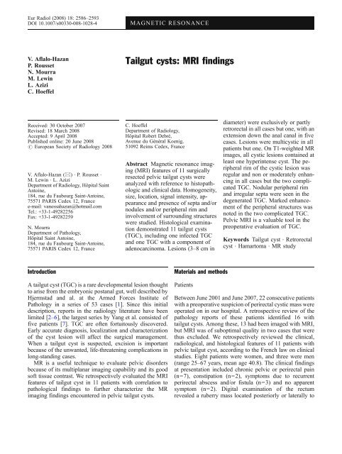
Abstract Magnetic resonance imaging<br />
(<strong>MRI</strong>) features of 11 surgically<br />
resected pelvic tailgut <strong>cysts</strong> were<br />
analyzed with reference to histopathologic<br />
and clinical data. Homogeneity,<br />
size, location, signal intensity, appearance<br />
and presence of septa and/or<br />
nodules and/or peripheral rim and<br />
involvement of surrounding structures<br />
were studied. Histological examination<br />
demonstrated 11 tailgut <strong>cysts</strong><br />
(TGC), including one infected TGC<br />
and one TGC with a component of<br />
adenocarcinoma. Lesions (3–8 cmin<br />
Materials and methods<br />
Patients<br />
diameter) were exclusively or partly<br />
retrorectal in all cases but one, with an<br />
extension down the anal canal in five<br />
cases. Lesions were multicystic in all<br />
patients but one. On T1-weighted MR<br />
images, all cystic lesions contained at<br />
least one hyperintense cyst. The peripheral<br />
rim of the cystic lesion was<br />
regular and non or moderately enhancing<br />
in all cases but the two complicated<br />
TGC. Nodular peripheral rim<br />
and irregular septa were seen in the<br />
degenerated TGC. Marked enhancement<br />
of the peripheral structures was<br />
noted in the two complicated TGC.<br />
Pelvic <strong>MRI</strong> is a valuable tool in the<br />
preoperative evaluation of TGC.<br />
Keywords <strong>Tailgut</strong> cyst . Retrorectal<br />
cyst . Hamartoma . MR study<br />
Between June 2001 and June 2007, 22 consecutive patients<br />
with a preoperative suspicion of perirectal cystic mass were<br />
operated on in our hospital. A retrospective review of the<br />
pathology reports of these patients identified 16 with<br />
tailgut <strong>cysts</strong>. Among these, 13 had been imaged with <strong>MRI</strong>,<br />
but <strong>MRI</strong> was of suboptimal quality in two cases that were<br />
thus excluded. We retrospectively reviewed the clinical,<br />
radiological, and histological features of 11 patients with<br />
pelvic tailgut cyst, according to the French law on clinical<br />
studies. Eight patients were women, and three were men<br />
(range 25–67 years, mean age 40.8). The clinical <strong>findings</strong><br />
at presentation included chronic pelvic or perirectal pain<br />
(n=7), constipation (n=2), symptoms due to recurrent<br />
perirectal abscess and/or fistula (n=3) and no apparent<br />
symptom (n=2). Digital examination of the rectum<br />
revealed a ruberry mass located posteriorly or laterally to
the rectum in ten patients (91%). Only two patients had an<br />
abnormal anorectal region with a perianal draining sinus<br />
(n=1) or a posterior sacral dimple (n=1). Laboratory<br />
studies demonstrated elevated C-reactive protein level in<br />
one patient.<br />
Imaging<br />
All 11 patients underwent preoperative <strong>MRI</strong>; postoperative<br />
<strong>MRI</strong> was performed in one case. All MR examinations were<br />
achieved on a 1.5-T unit using a phased-array pelvic coil,<br />
thin-sections, either in our department (Siemens, MR<br />
Magnetom, Erlangen, Germany, n=8) or in other institutions<br />
(n=3). No rectal preparation nor rectal distension was<br />
performed. MR examination protocol included sagittal<br />
(n=9), axial (n=10) and coronal (n=3) T2-weighted images.<br />
Imaging parameters for T2-weighted sequences on our MR<br />
unit were: TR/TE, 5940/122; echo-train length 16, field of<br />
view 25 cm, 4 mm slice thickness, no interslice gap, 240×<br />
256 matrix, three excitations. Unenhanced gradient-echo T1weighted<br />
(TR/TE, 636/13; field of view 25 cm, 4 mm slice<br />
thickness, no interslice gap, 230×256 matrix, three excitations)<br />
were performed in the axial plane (n=11) and coronal<br />
plane (n=2) and followed by axial gradient-echo T1weighted<br />
images either with fat-suppression (n=9) or not<br />
(n=2), after intravenous administration of 0.1 mmol/kg of<br />
gadodiamide (Omniscan, Nycomed Imaging, Oslo, Norway).<br />
Image review<br />
Radiological images were analyzed by consensus of two<br />
experienced abdominal radiologists. They were not aware<br />
of the pathological reports, but were aware of the diagnosis<br />
of tailgut cyst. The following features were analyzed:<br />
lesion location and extent, lesion size, presence of<br />
numerous <strong>cysts</strong> grouping together in the same location<br />
(multicystic lesion) or presence of one cyst, signal<br />
characteristics intensity of the main cyst with regard to<br />
muscle on both unenhanced T1-weighted images (T1WI)<br />
and T2-weighted images (T2WI), appearance of lesion<br />
surface and peripheral rim, homogeneous or heterogeneous<br />
appearance of the lesion, presence of internal nodules or<br />
vegetations or enhancing septa. The appearance of the<br />
surrounding fat, muscles and bones was also recorded.<br />
These features were selected from the previous case reports<br />
and our personal experience [2–8].<br />
All tumors had been surgically resected, either through a<br />
posterior surgical approach (n=8) or through a combined<br />
posterior approach and anterior diverting colostomy (n=2),<br />
or during abdominoperineal resection in the case of the<br />
TGC with the component of adenocarcinoma. The MR<br />
imaging <strong>findings</strong> were compared to the histopathological<br />
features of the lesions. The lesions could not always be<br />
analyzed precisely for the characterization of the internal<br />
fluid as the <strong>cysts</strong> had been at least partly emptied before<br />
their examination in the department of pathology. Microscopically,<br />
lesions were diagnosed as tailgut <strong>cysts</strong> accordingly<br />
to the literature [1] by experienced pathologists<br />
subspecialized in the GI field.<br />
Results<br />
2587<br />
The clinical, <strong>MRI</strong> and pathological <strong>findings</strong> in patients<br />
with tailgut <strong>cysts</strong> are summarized in Table 1.<br />
Histopathological study demonstrated 11 tailgut <strong>cysts</strong>,<br />
including 1 infected cyst and 1 complicated by a welldifferentiated<br />
mucinous adenocarcinoma. The latter occured<br />
in a context of chronic fistula in ano. Cysts were filled with a<br />
content that varied from clear to yellowish pasty substance<br />
and at least some evidence of mucinous internal component<br />
was recorded in the main cyst in eight cases.<br />
<strong>MRI</strong> finding study demonstrated that lesions were<br />
primarily located in the retrorectal space in ten cases with<br />
a downward and lateral extension to the ischioanal fossa in<br />
five cases (Fig. 3) and to the anal canal in five cases. TGCs<br />
were developped primarily in the ischio-anal fossa in one<br />
case. The rectum was displaced anteriorly or laterally in six<br />
patients, owing to a mass effect from the cystic lesion.The<br />
mean largest diameter of the lesions was of 5.7 cm (range 3–<br />
8 cm). Cysts were numerous (more than two) in all patients<br />
but one. Multicystic lesions consisted of a large main cyst,<br />
most often multiloculated (7/10) and associated with other<br />
smaller <strong>cysts</strong> grouping together in the same location, in an<br />
adjacent fibrous tissue, hypointense on both T1-weighted<br />
and T2-weighted images (Fig. 1). In eight patients (73%),<br />
some of the adjacent smaller <strong>cysts</strong> were adherent to the main<br />
one, resulting in a honeycomb pattern (Figs. 2 and 3).<br />
As far, as non-complicated <strong>cysts</strong> are concerned, the<br />
cystic mass was either well-circumscribed (n=9/9) by a<br />
thin regular peripheral rim (7/9) or a thick (>1 cm) smoothbordered<br />
wall (2/9). The rim was hypointense on all<br />
sequences apart from three cases in which a moderate<br />
enhancement after gadolinium administration was seen<br />
(Fig. 4). This peripheral rim corresponded to a fibrous wall<br />
at pathology. The borders of the cystic lesions were<br />
irregular in the two complicated <strong>cysts</strong>, with a strong<br />
enhancement of the wall and of the peritumoral adipose and<br />
muscular tissues (Fig. 5). Septa of multiloculated noncomplicated<br />
<strong>cysts</strong> were always thin and regular, either non<br />
or mildly enhancing, whereas they were thick but regular in<br />
the infected cyst and irregularly thickened and markedly<br />
enhancing in the TGC complicated by an adenocarcinoma<br />
(Fig. 6). The signal of the tailgut <strong>cysts</strong> was mostly<br />
heterogeneous (8/11), either because of a difference in the<br />
signal intensity of the <strong>cysts</strong> content (2/11) or because of a<br />
heterogeneous content of the main <strong>cysts</strong> (6/11). Main cyst<br />
displayed high signal intensity on T1-weighted images in<br />
8/11 patients (73%) and high signal intensity on T2weighted<br />
images in all cases. No evidence of fat was found
2588<br />
Table 1 Clinical <strong>MRI</strong> and pathologic <strong>findings</strong> of tailgut <strong>cysts</strong><br />
Patient/ age<br />
(year)sex<br />
Symptoms Localization MR features Pathology<br />
1) 25/F Lower abdominal pain,<br />
posterior sacral dimple<br />
Retrorectal space extension<br />
to ischioanal fossa and<br />
anal canal<br />
2) 29/F Lower abdominal pain Retrorectal space, extension<br />
to anal canal<br />
3) 32/F Lower abdominal pain,<br />
constipation<br />
4) 33/F Chronic right sciatica,<br />
recurrrent perirectal<br />
abscess (three previous<br />
drainages)<br />
5) 34/F Lower abdominal pain,<br />
pain during defecation<br />
Multicystic and honeycomb pattern,<br />
well-defined hyperintense on T1W1,<br />
hyperintense on T2WI, nonenhancing<br />
wall<br />
Multicystic and honeycomb pattern,<br />
well-defined, hyperintense on T1W1,<br />
hyperintense on T2WI, moderately<br />
enhancing wall<br />
Retrorectal space Multicystic and honeycomb pattern,<br />
well-defined, hyperintense on T1W1,<br />
hyperintense on T2WI, unenhanced<br />
after gadolinium<br />
Retrorectal space, extension<br />
to ischio anal fossa<br />
Retrorectal space, extension<br />
to ischio anal fossa<br />
6) 36/F Lower abdominal pain Retrorectal space, extension<br />
to anal canal<br />
7) 37/F Constipation Retrorectal space, extension<br />
to anal canal<br />
8) 39/M Anal pain, recurrent<br />
perirectal abscesses<br />
Retrorectal space, extension<br />
to anal canal<br />
9) 56/F Incidental detection Retrorectal space, extension<br />
to ischioanal fossa<br />
<strong>Tailgut</strong> cyst<br />
<strong>Tailgut</strong> cyst with<br />
abundant inflammatory<br />
reaction within the wall<br />
<strong>Tailgut</strong> cyst<br />
Multicystic and honeycomb pattern, Infected tailgut cyst<br />
irregular and thickened wall, (Escherichia Coli)<br />
isointense on T1W1, hyperintense<br />
on T2WI, enhancement of the wall and<br />
perilesional fat planes and<br />
adjacent structures<br />
Multicystic and honeycomb pattern, <strong>Tailgut</strong> cyst<br />
well-defined, isointense on T1W1,<br />
hyperintense on T2WI, moderately<br />
enhancing wall<br />
Multicystic and honeycomb pattern,<br />
well-defined, hyperintense on T1W1,<br />
hyperintense on T2WI, moderately<br />
enhancing wall<br />
Multicystic and honeycomb pattern,<br />
well-defined, hyperintense on T1W1,<br />
hyperintense on T2WI, no<br />
enhancement<br />
Multicystic, well-defined,<br />
hyperintense on T1W1,<br />
hyperintense on T2WI, smaller<br />
para anal “distant cyst,”<br />
no enhancement<br />
Multicystic, well-defined, hyperintense<br />
on T1W1, hyperintense on T2WI, no<br />
enhancement<br />
10) 61/M Incidental detection Right ischioanal fossa Unilocular, well-defined, hyperintense<br />
on T1W1, hyperintense on T2WI, no<br />
enhancement<br />
11) 67/M Perirectal abcess, anal fistula,<br />
anal pain<br />
Retrorectal space, extension<br />
to ischioanal fossa<br />
Multicystic and honeycomb pattern,<br />
intracystic vegetations, irregular and<br />
thick wall, hypointense on T1W1,<br />
hyperintense on T2WI, important peripheral<br />
and internal enhancement,<br />
enhancement of the wall and perilesional<br />
fat planes and adjacent<br />
structures<br />
<strong>Tailgut</strong> cyst with<br />
abundant inflammatory<br />
reaction within the wall<br />
<strong>Tailgut</strong> cyst<br />
<strong>Tailgut</strong> cyst<br />
<strong>Tailgut</strong> cyst<br />
<strong>Tailgut</strong> cyst<br />
<strong>Tailgut</strong> cyst with component<br />
of mucinous<br />
adenocarcinoma
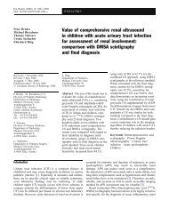
Fig. 1 Axial T2-weighted MR image in a 25-year-old female<br />
(patient 1). The multicystic retrorectal lesion consists of numerous<br />
<strong>cysts</strong> interspersed in a fibrous tissue (arrowheads). Note that the<br />
main cystic lesion displays heterogeneous signal<br />
whenever fat-saturated images were available. Presence of<br />
intracystic enhancing vegetations was observed in the case<br />
of tailgut cyst with a component of adenocarcinoma<br />
(Fig. 6), with a good correlation with the pathological<br />
features. No evidence of calcified deposits was found,<br />
although in one case pathologic examination revealed<br />
small areas of calcification in the cyst wall.<br />
Discussion<br />
<strong>Tailgut</strong> <strong>cysts</strong> are rare developmental lesions known to<br />
occur more frequently in middle-aged women (3/1),<br />
although cases at any age, including neonates, have been<br />
reported [1]. In our series, the average age of patients was<br />
40.8 years and the majority of them were women (with a<br />
mean age of 35.2 years). The clinical symptoms in our<br />
patients were consistent with the current knowledge. TGCs<br />
are characterized as multicystic lesions lined by a variety of<br />
epithelial cell types. Hjermstad and Helwig in their review<br />
[1] required the presence of columnar or transitional<br />
epithelium to exclude dermoid or epidermoid <strong>cysts</strong>, and<br />
required the absence of myenteric plexus and serosa to<br />
exclude bowel duplication <strong>cysts</strong>. They are thought to be<br />
congenital lesions due to some developmental error during<br />
embryogenesis. TGCs are believed to be vestigial remnants<br />
of the tailgut [1, 9]. TGC are located predominantly in the<br />
retrorectal area, but cases in perirenal and subcutaneous<br />
areas have also been reported [10–14]. One tailgut cyst in<br />
our series was located in the ischioanal fossa. The only<br />
2589<br />
TGCs involving the ischioanal fossa reported in the<br />
literature so far were large TGCs primarily developed in<br />
the retrorectal space with a downward and lateral extension<br />
to the ischioanal fossa as in four cases in our series [4, 5,<br />
12, 15, 16]. However, the development of tailgut vestiges<br />
that may reside in the ischioanal fossa [13] can possibly<br />
lead to the formation of TGC in this space. A feature of<br />
clinical relevance was the rather frequent presence of small<br />
<strong>cysts</strong> extending down the anal canal, distant from the main<br />
retrorectal cyst. We indeed observed in one case for which<br />
postoperative MR imaging had been performed that the<br />
main cyst had been resected along with its neighbor smaller<br />
<strong>cysts</strong>, whereas the small <strong>cysts</strong> down the canal anal had been<br />
left in place. Even though the surgical removal of such<br />
associated <strong>cysts</strong> may be impossible, they have to be<br />
preoperatively identified with <strong>MRI</strong> because their persistence<br />
may explain recurrence or infection [17, 18].<br />
The present series supports that <strong>MRI</strong> is suitable for<br />
preoperative evaluation of tailgut cyst. In agreement with a<br />
previous report [7], our lesions were rather large in size<br />
(average 5.7 cm). <strong>MRI</strong> improves tissue characterization<br />
because of its high contrast resolution between different<br />
tissue compartments. It allows detection of mucin and<br />
blood in a cystic lesion. However, signal intensity of<br />
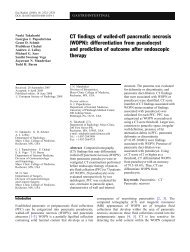
Fig. 2 Coronal T2-weighted MR image in a 37-year-old woman<br />
(patient 7) with a retrorectal TGC. Small <strong>cysts</strong> are grouped together<br />
in a honeycomb pattern (arrow) and are adjacent to a main larger<br />
cyst (arrowhead)
2590<br />
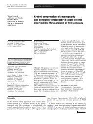
Fig. 3 Coronal T2-weighted MR image in a 34-year-old woman<br />
(patient 5) demonstrates the lateral extension to the ischio-anal fossa<br />
of a retrorectal tailgut cyst (arrows). Note the honeycomb pattern<br />
(arrowhead)<br />
mucinous fluid may vary depending on protein concentration.<br />
On T1-weighted images, the signal intensity may<br />
change from hypointense to hyperintense as protein<br />
concentration increases. On T2-weighted images, signal<br />
intensity of mucinous fluids can decrease from highly<br />
hyperintense to hypointense with increasing protein concentration<br />
and viscosity. This explains the variety of the<br />
signal intensity displayed by the <strong>cysts</strong> in our series, even in<br />
the same patient. However, a rather constant feature was<br />
the presence of high signal intensity relative to muscle in<br />
most of the main <strong>cysts</strong> (n=9). Mucinous or proteinaceous<br />
component of the cyst fluid demonstrated in most of the<br />
cases on histological examination was assumed to be the<br />
cause of this high signal intensity, and no blood product nor<br />
fat was found either radiologically or histologically within<br />
the lesions. An intermediate to high signal on T1-weighted<br />
images with or without hyperintense foci has already been<br />
reported in TGC [2, 3, 5, 6, 8, 18, 19]. Yang et al. [7]<br />
reported hypointense TGCs on T1-weighted images, but<br />
their standard of reference was the signal intensity of fat,<br />
whereas ours was that of muscle.The cystic lesion<br />
consisted of a unilocular cyst in only one case. Several<br />
reports [3, 19, 7, 6] have highlighted the common<br />
multicystic appearance of tailgut <strong>cysts</strong>. Less commonly,<br />
cases of unilocular cyst have been reported [1, 2]. A<br />
common feature in our series was the frequent presence of<br />
small <strong>cysts</strong> clustered together and adjacent to the main cyst,<br />
thus resulting in a honeycomb pattern, an imaging finding<br />
confirmed at pathological examination. This finding of a<br />
large dominant cyst surrounded by a number of small<br />
irregular <strong>cysts</strong> with a diameter up to 15 mm has also been<br />
described at pathology in one case report by Kripocavic<br />
[20]. Kim et al. [3] describe in their report of one retrorectal<br />
TGC imaged with <strong>MRI</strong> a cystic lesion formed by small<br />
<strong>cysts</strong> grouped together in a honeycomb pattern. We<br />
presume that this feature may be suggestive of a tailgut<br />
cyst, although we did not compare TGCs to other<br />
developmental cystic lesions in this area.<br />
Septas in multiloculated non-complicated TGCs were<br />
thin and presented a mild enhancement after IV administration<br />
of contrast material, in accordance with the<br />
literature [3, 5, 8]. The presence of a peripheral rim<br />
circumscribing the cystic lesions corresponded to a fibrotic<br />
Fig. 4 <strong>Tailgut</strong> cyst in a 29-year-old woman (patient 2). Axial<br />
unenhanced (a) and fat-saturated gadolinium enhanced (b) T1weighted<br />
MR images show a thin regular peripheral rim<br />
circumscribing a retrorectal cyst (arrows, a), displaying moderate<br />
enhancement (arrows, b)
Fig. 5 Infected TGC in a 33-year-old woman (patient 4). Axial fatsuppressed<br />
gadolinium enhanced T1-weighted MR image demonstrates<br />
a cystic lesion in the right ischioanal fossa with a thick<br />
enhancing border (arrowheads) as well as marked enhancement of<br />
the peripheral fat planes (black arrow) and muscular structures<br />
(white arrow)<br />
rim with chronic inflammatory reaction at pathology. When<br />
the cyst wall was thick, <strong>cysts</strong> were interspersed in a dense<br />
fibrous tissue. Although peripheral inflammatory reaction<br />
was present in all cases, the peripheral rim did not enhance<br />
in six out of nine non-complicated TGCs. [8, 10, 21]. As far<br />
as non-complicated <strong>cysts</strong> are concerned, preservation of a<br />
fat plane between the adjacent tissue and/or the rectum was<br />
present in all cases but one. Although most TGCs are easy<br />
to resect because of a good cleavage plane, surgical<br />
resection may be difficult in cases of mutiloculated large<br />
lesions, infected cyst or rarely when the wall of the cyst is<br />
intimately related to the rectal wall muscle fibers, as in one<br />
non-complicated cyst in our series. Only four cases of such<br />
<strong>cysts</strong> involving the muscular layers of the rectal wall have<br />
been reported in the literature, and two of them were<br />
degenerated TGCs [2, 4, 12, 22]. Surgical approach<br />
depends on the location, size of the lesion and on the<br />
presence of complication. For most lesions, a posterior<br />
approach with removal of a portion of the coccyx will<br />
allow the best visualization and removal of the multiloculated<br />
cyst [1, 23, 24]. In the case of larger tumor, or<br />
when malignancy is suspected, a combined abdominalsacral<br />
approach may be used [23, 24, 25].<br />
Differential diagnosis with other cystic lesions of the<br />
retrorectal space or of the ischioanal fossa is often difficult.<br />
Other developmental <strong>cysts</strong> (epidermal cyst, dermoid cyst,<br />
anal gland <strong>cysts</strong> and rectal cystic duplication) may have<br />
similar imaging characteristics although they are more<br />
often unilocular. The presence of fat content on fatsaturated<br />
images is suggestive of a dermoid cyst. Rectal<br />
duplication <strong>cysts</strong> are often communicating with the rectal<br />
lumen and anterior to the rectum [9]. Anal gland <strong>cysts</strong> have<br />
a lower location than tailgut cyst and are typically located<br />
in close approximation to the anal sphincter [8, 26].<br />
Anterior meningocele is a well-defined unilocular thinwalled,<br />
fluid-filled lesion of the retrorectal space with a<br />
2591<br />
stalk that may be seen communicating with the thecal sac.<br />
<strong>MRI</strong> is limited in detecting calcification, particularly when<br />
it would be helpful to exclude dermoid cyst or teratoma.<br />
Indeed, although peripheral calcifications have already<br />
been reported in tailgut cyst [3, 6, 8, 20], they are more<br />
frequently seen in teratoma, dermoid cyst and other tumors<br />
arising in the retrorectal space.Although our series is<br />
limited by the fact that we did not compare the MR <strong>findings</strong><br />
of TGCs with those of other lesions that may occur in the<br />
same location, we believe that the diagnosis of TGC may<br />
be suggested if a multicystic lesion, with numerous small<br />
<strong>cysts</strong> adherent to the main lesion, displaying high signal<br />
intensity on T1-weighted images and with a fibrous wall, is<br />
present. However, this putative discrimination is of little<br />
practical importance because the treatment and diagnosis<br />
for developmental lesions is thorough complete surgical<br />
excision. Simple cyst excision or drainage would indeed<br />
lead to recurrence or infection.<br />
The potential complications of TGC include infection<br />
with recurrent fistula, which can misdiagnose as a recurrent<br />
perineal abscess, and rarely, a malignant transformation [4,<br />
25, 27].<br />
Our series included one case of acutely infected cyst.<br />
Acute infection was suspected on the basis of the clinical,<br />
biological <strong>findings</strong> and of some <strong>MRI</strong> criteria, such as a<br />
markedly enhancing irregular peripheral wall and enhancing<br />
infiltration of the perilesional fat and muscles. Our<br />
description of an infected tailgut cyst imaged with <strong>MRI</strong> is<br />
the first detailed one in the literature. When the complicated<br />
lesion is discovered without any knowledge of an<br />
underlying tailgut cyst, differential diagnosis with an<br />
abscess is difficult. We assume it is possible mainly on<br />
the basis of the location of the lesion (either retrorectal or in<br />
the ischioanal fossa) and of its multicystic appearance<br />
particularly if small <strong>cysts</strong> are clustered around a main cystic<br />
lesion. A history of recurring pelvic abcesses, repeated<br />
operations for anal fistula with no underlying obvious<br />
disease should also raise suspicion for a retrorectal cyst [4,<br />
19, 25, 27].<br />
Differentiation between an infected tailgut cyst from an<br />
abscess is of importance because an abscess must be treated<br />
by a combination of drainage and antibiotherapy, whereas<br />
TGC needs to be surgically excised. Another possible<br />
complication of TGC is malignant degeneration. So far,<br />
fewer than 30 cases of degenerated tailgut <strong>cysts</strong> have been<br />
documented in the literature. Among them, few have been<br />
imaged and described with <strong>MRI</strong> [4, 5, 18, 28, 29]. Most of<br />
the degenerated TGCs are associated with mucinous<br />
adenocarcinoma, although some cases of carcinoid associated<br />
with tailgut <strong>cysts</strong> have also been reported [12, 21,<br />
30, 31], as well as exceptional cases of sarcoma, endometrioid<br />
carcinoma and clear cell carcinoma. Our <strong>findings</strong> of a<br />
cystic lesion with markedly enhancing irregular wall,<br />
nodular thickening of the wall, enhancing cystic vegetations<br />
and markedly enhancing irregular septal walls are<br />
consistent with what has been described in the literature
2592<br />
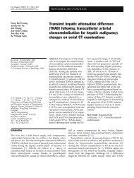
Fig. 6 TGC associated with mucinous adenocarcinoma (patient 11).<br />
Sagittal T2-weighted (a) and fat-supressed gadolinium-enhanced<br />
T1-weighted MR images (b) in a 67-year-old man. The cystic lesion<br />
displays heterogeneous high signal intensity on T2-weighted MR<br />
images (a), with irregular borders and septas (arrows), intracystic<br />
enhancing vegetations (arrowheads) and enhancement of the<br />
[4, 5, 18, 29]. In addition, we observed marked enhancement<br />
of the surrounding fat planes and muscular structures. In our<br />
series, the TGC with a component of adenocarcinoma was<br />
associated with a fistula. Mucinous adenocarcinomas have<br />
been reported to arise from fistula in ano [32]. However, in<br />
our case, adenocarcinoma was clearly identified at pathological<br />
examination as arising from the TGC. Marked<br />
enhancement of the perilesional areas and of the lesion wall,<br />
nodular thickening of the wall as well as the presence of<br />
intracystic vegetations are not features of non-complicated<br />
TGC and are thus likely to suggest complication of a tailgut<br />
peripheral planes (b). Photomicrographs of the <strong>cysts</strong> (c and d);<br />
original magnification ×25 (c) and ×100 (d), stain: hematoxylin and<br />
eosin, show a cyst containing some mucus (arrow) and lined with<br />
mucus secreting epithelium (arrowhead, c). The epithelium is partly<br />
dysplastic, and cancerous tubes are visible around the cyst. Papilla at<br />
higher magnification (d) is lined by mucus secreting cells (arrowhead)<br />
cyst. In such cases, biopsy is not advisable because it may<br />
not show the potential malignancy and because it carries a<br />
significant risk of spillage of tumor cells.<br />
Conclusion<br />
Our series supports that <strong>MRI</strong> is a useful imaging<br />
investigation when a retro and/or peri anorectal cyst is<br />
suspected. If a multicystic mass with small <strong>cysts</strong> adherent to<br />
a large cyst and without any evidence of sacrococcygeal
involvement is present, either in the retrorectal space or in<br />
the ischioanal fossa in a middle-aged woman, the diagnosis<br />
of tailgut cyst has to be considered. However, final diagnosis<br />
is based on pathological examination after surgical resection.<br />
References<br />
1. Hjermstad BM, Helwig EB (1988)<br />
<strong>Tailgut</strong> <strong>cysts</strong>. Report of 53 cases. Am J<br />
Clin Pathol 89:139–147<br />
2. Liessi G, Cesari S, Pavanello M, Butini<br />
R (1995) <strong>Tailgut</strong> <strong>cysts</strong>: CT and MR<br />
<strong>findings</strong>. Abdom Imaging 20:256–258<br />
3. Kim MJ, Kim WH, Kim NK, Yun MJ,<br />
Park YN, Lee JT, Yoo HS (1997)<br />
<strong>Tailgut</strong> cyst: Multilocular cystic appearance<br />
on <strong>MRI</strong>. J Comput Assist<br />
Tomogr 21:731–732<br />
4. Lim KE, Hsu WC, Wang CR (1998)<br />
<strong>Tailgut</strong> cyst with malignancy: MR<br />
imaging <strong>findings</strong>. AJR Am J Roentgenol<br />
170:1488–1490<br />
5. Moulopoulos LA, Karvouni E, Kehagias<br />
D, Dimopoulos MA, Gouliamos A,<br />
Vlahos L (1999) MR imaging of complex<br />
tail-gut <strong>cysts</strong>. Clin Radiol 54:118–<br />
122<br />
6. Podberesky DJ, Falcone RA, Emery<br />
KH, Care MM, Anton CG, Miles L,<br />
Ryckman FC (2005) <strong>Tailgut</strong> cyst in a<br />
child. Pediatr Radiol 35:194–197<br />
7. Yang DM, Park CH, Jin W, Chang SK,<br />
Kim JE, Choi SJ, Jung DH (2005)<br />
<strong>Tailgut</strong> cyst: <strong>MRI</strong> evaluation. AJR Am<br />
J Roentgenol 184:1519–1523<br />
8. Menassa-Moussa L, Kanso H,<br />
Checrallah A, Abboud J, Ghossain M<br />
(2005) CT and MR <strong>findings</strong> of a<br />
retrorectal cystic hamartoma confused<br />
with an adnexal mass on ultrasound.<br />
Eur Radiol 15:263–266<br />
9. Johnson AR, Ros PR, Hjermstad BM<br />
(1986) <strong>Tailgut</strong> cyst: diagnosis with CT<br />
and sonography. AJR Am J Roentgenol<br />
147:1309–1311<br />
10. Kang JW, Kim SH, Kim KW, Moon<br />
SK, Kim CJ, Chi JG (2002) Unusual<br />
perirenal location of a tailgut cyst.<br />
Korean J Radiol 3:267–270<br />
11. Sung MT, Ko SF, Niu CK, Hsieh CS,<br />
Huang HY (2003) Perirenal tailgut cyst<br />
(cystic hamartoma). J Pediatr Surg<br />
38:1404–1406<br />
12. Song DE, Park JK, Hur B, Ro JY<br />
(2004) Carcinoid tumor arising in a<br />
tailgut cyst of the anorectal junction<br />
with distant metastasis: a case report<br />
and review of the literature. Arch<br />
Pathol Lab Med 128:578–580<br />
13. Mills SE, Walker AN, Stallings RG,<br />
Allen MS Jr (1984) Retrorectal cystic<br />
hamartoma. Report of three cases,<br />
including one with a perirenal component.<br />
Arch Pathol Lab Med 108:737–<br />
740<br />
14. Murao K, Fukui Y, Numoto S, Urano Y<br />
(2003) <strong>Tailgut</strong> cyst as a subcutaneous<br />
tumor at the coccygeal region. Am J<br />
Dermatopathol. 25:275–277<br />
15. Llauger J, Palmer J, Perez C, Monill J,<br />
Ribe J, Moreno A (1998) The normal<br />
and pathologic ischiorectal fossa at CT<br />
and MR imaging. Radiographics<br />
18:61–82<br />
16. Karcaaltincaba M, Karcaaltincaba D,<br />
Ayhan A (2005) Diagnosis of tailgut<br />
<strong>cysts</strong>. AJR Am J Roentgenol<br />
185:1369–1370<br />
17. Costello D, Schofield A, Stirling R,<br />
Theodorou N (2000) Extrarectal mass:<br />
a tailgut cyst. J R Soc Med<br />
93:85–86<br />
18. Graadt van Roggen JF, Welvaart K, de<br />
Roos A, Offerhaus GJ, Hogendoorn PC<br />
(1999) Adenocarcinoma arising within<br />
a tailgut cyst: clinicopathological description<br />
and follow up of an unusual<br />
case. J Clin Pathol 52:310–322<br />
19. Dahan H, Arrive L, Wendum D, Docou<br />
le Pointe H, Djouhri H, Tubiana JM<br />
(2001) Retrorectal developmental <strong>cysts</strong><br />
in adults:clinical and radiologic-histopathologic<br />
review, differential diagnosis,<br />
and treatment. Radiographics<br />
21:575–584<br />
20. Krivokapic Z, Dimitrijevic I, Barisic G,<br />
Markovic V, Krstic M (2005) Adenosquamous<br />
carcinoma arising within a<br />
retrorectal tailgut cyst: report of a case.<br />
World J Gastroenterol 11:6225–6227<br />
21. Mathieu A, Chamlou R, Le Moine F,<br />
Maris C, Van de Stadt J, Salmon I<br />
(2005) <strong>Tailgut</strong> cyst associated with a<br />
carcinoid tumor: case report and review<br />
of the literature. Histol Histopathol<br />
20:1065–1069<br />
2593<br />
<strong>MRI</strong> is a helpful technique to define the extent of the cystic<br />
mass, its relationship to the surrounding structures and also<br />
to demonstrate possible complications in order to choose the<br />
best surgical approach.<br />
22. Levert LM, Van Rooyen W, Van Den<br />
Bergen HA (1996) Cysts of the tailgut.<br />
Eur J Surg 162:149–152<br />
23. Pyo DJ (1990) <strong>Tailgut</strong> cyst (retrorectal<br />
cyst hamartoma): case report and review.<br />
Mt Sinai J Med 57:249–252<br />
24. Hannon J, Subramony C, Scott-Conner<br />
CE (1994) Benign retrorectal tumors in<br />
adults: the choice of operative approach.<br />
Am Surg 60:267–272<br />
25. Killingsworth C, Gadacz TR (2005)<br />
<strong>Tailgut</strong> cyst (retrorectal cystic hamartoma):<br />
report of a case and review of<br />
the literature. Am Surg 71:666–673<br />
26. Kulaylat MN, Doerr RJ, Neuwirth M,<br />
Satchidanand SK (1998) Anal duct/gland<br />
cyst: report of a case and review of the<br />
literature. Dis Colon Rectum 41:103–110<br />
27. Jao SW, Beart RW Jr, Spencer RJ, Reiman<br />
HM, Ilstrup DM (1985) Retrorectal tumors.<br />
Mayo Clinic experience, 1960–<br />
1979. Dis Colon Rectum 28:644–652<br />
28. Moreira AL, Scholes JV, Boppana S,<br />
Melamed J (2001) p53 Mutation in<br />
adenocarcinoma arising in retrorectal<br />
cyst hamartoma (tailgut cyst): report of<br />
2 cases-an immunohistochemistry/immunoperoxidase<br />
study. Arch Pathol<br />
Lab Med 125:1361–1364<br />
29. Cho BC, Kim NK, Lim BJ, Kang SO,<br />
Sohn JH, Roh JK, Choi ST, Kim SA,<br />
Park SE (2005) A carcinoembryonic<br />
antigen-secreting adenocarcinoma arising<br />
in tailgut cyst: clinical implications<br />
of carcinoembryonic antigen. Yonsei<br />
Med J 46:555–561<br />
30. Mourra N, Caplin S, Parc R, Flejou JF<br />
(2003) Presacral neuroendocrine carcinoma<br />
developed in a tailgut cyst: report<br />
of a case. Dis Colon Rectum 46:411–413<br />
31. Jacob S, Dewan Y, Joseph S (2004)<br />
Presacral carcinoid tumour arising in a<br />
tailgut cyst-a case report. Indian J<br />
Pathol Microbiol 47:32–33<br />
32. Hama Y, Makita K, Yamana T,<br />
Dodanuki K (2006) Mucinous adenocarcinoma<br />
arising from fistula in ano:<br />
<strong>MRI</strong> <strong>findings</strong>. AJR Am J Roentgenol<br />
187:517–521