Guideline: the use of electrosurgical units - ESGE
Guideline: the use of electrosurgical units - ESGE
Guideline: the use of electrosurgical units - ESGE
You also want an ePaper? Increase the reach of your titles
YUMPU automatically turns print PDFs into web optimized ePapers that Google loves.
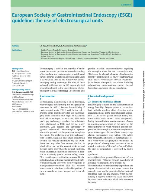
European Society <strong>of</strong> Gastrointestinal Endoscopy (<strong>ESGE</strong>)<br />
guideline: <strong>the</strong> <strong>use</strong> <strong>of</strong> <strong>electrosurgical</strong> <strong>units</strong><br />
Authors J. F. Rey 1 , U. Beilenh<strong>of</strong>f 2 , C. S. Neumann 3 , J. M. Dumonceau 4<br />
Institutions<br />
Bibliography<br />
DOI http://dx.doi.org/<br />
10.1055/s-0030-1255594<br />
Published ahead <strong>of</strong> print<br />
Endoscopy<br />
© Georg Thieme Verlag KG<br />
Stuttgart · New York<br />
ISSN 0013-726X<br />
Corresponding author<br />
J. M. Dumonceau, MD, PhD<br />
Division <strong>of</strong> Gastroenterology<br />
and Hepatology<br />
Geneva University Hospitals<br />
Micheli-du-Crest Street 24<br />
1205 Geneva<br />
Switzerland<br />
Fax: +41-22-3729366<br />
jmdumonceau@hotmail.com<br />
1 Institut Arnault Tzanck, St. Laurent du Var, France<br />
2 European Society <strong>of</strong> Gastroenterology and Endoscopy Nurses and Associates (President), Ulm, Germany<br />
3 European Society <strong>of</strong> Gastroenterology and Endoscopy Nurses and Associates (Past President), Birmingham,<br />
United Kingdom<br />
4 Division <strong>of</strong> Gastroenterology and Hepatology, University Hospital <strong>of</strong> Geneva, Geneva, Switzerland<br />
Electrosurgery is <strong>use</strong>d in <strong>the</strong> majority <strong>of</strong> endoscopic<br />
<strong>the</strong>rapeutic procedures. An understanding<br />
<strong>of</strong> <strong>the</strong> fundamental <strong>electrosurgical</strong> principles and<br />
various settings available on <strong>electrosurgical</strong> <strong>units</strong><br />
is essential for <strong>the</strong> safe and effective <strong>use</strong> <strong>of</strong> electrosurgery<br />
during endoscopy. The aims <strong>of</strong> <strong>the</strong>se<br />
technical guidelines are to: (1) expose physical<br />
principles relevant to <strong>the</strong> understanding <strong>of</strong> electrosurgery<br />
during endoscopy; (2) describe and<br />
1 Introduction<br />
!<br />
Electrosurgery in endoscopy is an old technique,<br />
with urologists already using it in an aqueous environment<br />
in 1926 [1]. Despite <strong>the</strong> availability <strong>of</strong><br />
<strong>electrosurgical</strong> <strong>units</strong> (ESUs) with better safety<br />
pr<strong>of</strong>iles, some practitioners still <strong>use</strong> electrosurgery<br />
under conditions that might be hazardous<br />
with old technologies. In particular, ESUs with<br />
spark gap technology pre-date <strong>the</strong> solid-state<br />
<strong>units</strong> introduced in 1968, and are no longer<br />
acceptable. Also obsolete are “grounded” or<br />
“ground referenced” <strong>electrosurgical</strong> systems<br />
where <strong>the</strong> ground, not <strong>the</strong> generator, completes<br />
<strong>the</strong> circuit. The replacement <strong>of</strong> this technology<br />
with isolated, balanced, and return monitoring<br />
systems has virtually eliminated any risk to patients<br />
that may arise from current division, in<br />
which all or part <strong>of</strong> <strong>the</strong> current seeks ground<br />
through paths o<strong>the</strong>r than <strong>the</strong> neutral electrode,<br />
which could result in alternate site burns. In addition<br />
to markedly greater patient safety, newer<br />
ESUs provide opportunities for enhanced bipolar<br />
outputs and sophisticated neutral electrode safety<br />
monitoring [2]. Moreover, <strong>the</strong> development <strong>of</strong><br />
microprocessor-controlled ESUs has enabled<br />
more constant, reliable or predictable qualities <strong>of</strong><br />
current waveform, power output, and tissue effects.<br />
<strong>Guideline</strong>s<br />
provide practical recommendations regarding<br />
<strong>electrosurgical</strong> <strong>units</strong> that are commonly in <strong>use</strong>;<br />
(3) discuss <strong>the</strong> clinical relevance <strong>of</strong> technologies<br />
recently implemented in newer <strong>electrosurgical</strong><br />
<strong>units</strong>; and (4) review factors relevant to commonly<br />
performed <strong>the</strong>rapeutic procedures, including<br />
polypectomy, sphincterotomy, contact <strong>the</strong>rmal<br />
hemostasis, and argon plasma coagulation.<br />
2 Technical background<br />
!<br />
2.1 Electricity and tissue effects<br />
Electrosurgery is based on <strong>the</strong> transformation <strong>of</strong><br />
energy from high frequency electric current into<br />
heat, with <strong>the</strong> resulting effect <strong>of</strong> cutting and/or<br />
coagulating tissue at <strong>the</strong> point <strong>of</strong> current application<br />
[3]. As current passes through tissue, electrons<br />
collide with various tissue components.<br />
During <strong>the</strong>se collisions, a certain amount <strong>of</strong> energy<br />
is dissipated depending on <strong>the</strong> nature <strong>of</strong> <strong>the</strong><br />
material traversed, which results in a rise in temperature.<br />
Electrosurgical waveforms may be set to<br />
promote two types <strong>of</strong> tissue effects, namely coagulation<br />
(temperature rises within cells, which<br />
<strong>the</strong>n dehydrate and shrink) or cut (heating <strong>of</strong> cellular<br />
water occurs so rapidly that cells burst). The<br />
proportion <strong>of</strong> cells coagulated to those cut can be<br />
varied, resulting in a “blended” or “mixed” effect.<br />
The rise in temperature is governed by Joule’s<br />
law:<br />
Q=I 2 ×R×t<br />
where Q is <strong>the</strong> heat generated by a current <strong>of</strong> constant<br />
intensity (I) flowing through a conductor <strong>of</strong><br />
electrical resistance (R), for a time (t) [3].<br />
Tissue electrical resistance mostly depends on <strong>the</strong><br />
degree <strong>of</strong> vascularization and water content. For<br />
example, bone and fat present a higher electrical<br />
resistance than skin and muscles. When electrosurgery<br />
is applied, progressive tissue desiccation<br />
increases tissue resistance, which reduces current<br />
intensity.<br />
Rey J F. et al. Technical guidelines for electrosurgery… Endoscopy<br />
Downloaded by: Thieme Verlagsgruppe. Copyrighted material.
<strong>Guideline</strong>s<br />
Key points and recommendations<br />
!<br />
a) Protection measures for <strong>the</strong> patient<br />
The <strong>electrosurgical</strong> unit (ESU) should only be <strong>use</strong>d by medical<br />
personnel after appropriate training.<br />
" Inspect <strong>the</strong> ESU for damage, including insulation <strong>of</strong> all cables<br />
and electrodes, missing components, and working lights and<br />
sounds (set on an audible level) prior to <strong>use</strong>.<br />
" The ESU should stand firm; no fluids should be placed on top<br />
<strong>of</strong> <strong>the</strong> ESU.<br />
" Do not <strong>use</strong> worn out or defective active electrodes, forceps<br />
or scissors.<br />
" Do not repair active electrodes, forceps or scissors.<br />
" Do not <strong>use</strong> <strong>the</strong> ESU in <strong>the</strong> presence <strong>of</strong> flammable material<br />
or substances (e.g. alcohol or nitrous oxide).<br />
" The patient must be insulated against all electrically conductive<br />
parts. Make sure that <strong>the</strong> patient does not come<br />
into contact with o<strong>the</strong>r metal parts not insulated from <strong>the</strong><br />
ground (e.g. operating table), although this concern is<br />
greatly diminished by <strong>the</strong> <strong>use</strong> <strong>of</strong> isolated and/or balanced<br />
ESUs.<br />
" Place <strong>the</strong> patient on a dry, electrically insulating layer.<br />
" Pacemaker or defibrillator (all types): seek advice from<br />
competent authority prior to endoscopy. Permanent ECG<br />
monitoring is recommended in <strong>the</strong>se patients during electrosurgery.<br />
The <strong>use</strong> <strong>of</strong> bipolar applications might minimize<br />
possible complications. If a monopolar <strong>electrosurgical</strong> system<br />
is <strong>use</strong>d, position <strong>the</strong> neutral electrode as close as<br />
possible to <strong>the</strong> active electrode. Direct contact with <strong>the</strong><br />
implanted device and <strong>the</strong> leads should be avoided.<br />
" The power settings should be adapted to <strong>the</strong> type <strong>of</strong> procedure,<br />
tissue structure, patient body mass index, endo<strong>the</strong>rapy<br />
instrument <strong>use</strong>d, and manufacturers’ recommendations.<br />
Always <strong>use</strong> <strong>the</strong> lowest power setting possible that will<br />
accomplish <strong>the</strong> desired tissue effect.<br />
" Before activating <strong>the</strong> ESU, <strong>the</strong> power settings should be<br />
rechecked and verbally confirmed between <strong>the</strong> endoscopist<br />
and <strong>the</strong> assistant.<br />
" If current is not required, keep <strong>the</strong> foot away from <strong>the</strong> pedal<br />
to prevent accidental pressing, or disconnect <strong>the</strong> electrode<br />
from <strong>the</strong> ESU.<br />
" If inadequate current output is observed, stop <strong>the</strong> procedure<br />
immediately. Use <strong>the</strong> power switch as emergency stop for<br />
malfunctions. Leakage <strong>of</strong> current may be <strong>the</strong> result <strong>of</strong> a<br />
malfunctioning endo<strong>the</strong>rapy instrument, ESU or broken<br />
insulation <strong>of</strong> <strong>the</strong> endoscope and may ca<strong>use</strong> <strong>use</strong>r and/or<br />
patient burns. Electrosurgical <strong>units</strong> that constantly monitor<br />
leakage <strong>of</strong> current may help to ensure <strong>use</strong>r and patient<br />
safety.<br />
Rey J F. et al. Technical guidelines for electrosurgery … Endoscopy<br />
b) Staff safety<br />
" Avoid contact with <strong>the</strong> neutral electrode.<br />
" When applying current, always be sure to wear gloves and<br />
touch <strong>the</strong> equipment or patient body with <strong>the</strong> entire palm<br />
<strong>of</strong> <strong>the</strong> hand, not with a single finger.<br />
" Electrosurgical equipment needs to be grounded in order<br />
to minimize interference with videoendoscopic systems.<br />
" Smoke generated during <strong>electrosurgical</strong> procedures can be<br />
irritating and potentially harmful to personnel; surgical<br />
masks and adequate ventilation <strong>of</strong> smoke may be <strong>use</strong>ful.<br />
c) Neutral electrode<br />
" Only patient neutral electrodes (plates or grounding pads)<br />
recommended by <strong>the</strong> ESU manufacturer should be <strong>use</strong>d. For<br />
example, some ESUs require split type plates to monitor <strong>the</strong><br />
quality <strong>of</strong> contact between <strong>the</strong> plate and <strong>the</strong> patient; single<strong>use</strong><br />
plates should not be re<strong>use</strong>d.<br />
" Check expiration date (if expired patient plates are <strong>use</strong>d, <strong>the</strong><br />
adhesive may fail to maintain contact with <strong>the</strong> patient’s skin<br />
and burns may result).<br />
" Check <strong>the</strong> patient plate for any damage/modification or<br />
sharp edges.<br />
" The neutral electrode should not be attached over some<br />
structures, including bony protuberances, metal implants or<br />
pros<strong>the</strong>sis, skin folds, scar tissue, hairy areas, any form <strong>of</strong><br />
skin discoloration/injury, limbs with a restricted blood supply,<br />
adjacent to ECG electrodes or onto pressure areas/points.<br />
" The neutral electrode should be attached over well perf<strong>use</strong>d<br />
muscle tissue; <strong>the</strong> skin must be clean, dry, and free <strong>of</strong> hair to<br />
avoid loss <strong>of</strong> contact between <strong>the</strong> plate and <strong>the</strong> skin. The<br />
electrode should not be completely wrapped around a limb.<br />
Overlapping needs to be avoided. Ensure that <strong>the</strong> neutral<br />
electrode has full patient-skin contact.<br />
" The patient plate should be <strong>of</strong> appropriate size for <strong>the</strong> patient<br />
weight and should never be cut to size.<br />
" Patient plates that have once been removed from <strong>the</strong> patient<br />
skin have to be replaced by new ones.<br />
d) Special situation: polypectomy or EMR<br />
" Adjust settings according to particular conditions<br />
(e.g. low power settings for small bowel and cecum).<br />
" If polypectomy snare sticks in a polyp, increase cutting<br />
(see text above).<br />
" Do not touch metal parts, such as clips, with snare when<br />
applying current.<br />
" Do not touch <strong>the</strong> scope with metal parts <strong>of</strong> endo<strong>the</strong>rapy<br />
instruments.<br />
" Watch out that snare tip does not accidentally touch <strong>the</strong><br />
bowel wall opposite to mucosectomy.<br />
" Avoid deep coagulation <strong>of</strong> muscle layer (risk <strong>of</strong> late perforation).<br />
" Before applying current, make sure that <strong>the</strong> muscularis<br />
propria is not entrapped in <strong>the</strong> snare loop.<br />
Downloaded by: Thieme Verlagsgruppe. Copyrighted material.
2.2 Electrical circuit<br />
Depending on <strong>the</strong> clinical indication and type <strong>of</strong> device <strong>use</strong>d, energy<br />
may be transmitted by ESUs in monopolar or bipolar mode.<br />
" In monopolar mode, <strong>the</strong> electrical current passes from <strong>the</strong> active<br />
electrode to <strong>the</strong> target tissue, through <strong>the</strong> patient’s body,<br />
to finally exit <strong>the</strong> patient through a large dispersive neutral<br />
electrode (also called a patient plate or return electrode)<br />
(● " Fig. 1).<br />
" In bipolar mode, <strong>the</strong> endoscopic device contains both <strong>the</strong> active<br />
and <strong>the</strong> neutral electrodes in close proximity to each o<strong>the</strong>r.<br />
The electrical current passes directly from one electrode to <strong>the</strong><br />
o<strong>the</strong>r through a small amount <strong>of</strong> tissue in contact with both<br />
electrodes. The current does not pass through <strong>the</strong> rest <strong>of</strong> <strong>the</strong><br />
patient’s body and no patient plate is required (● " Fig. 2).<br />
2.3 Current density and rise in temperature<br />
Current density is <strong>the</strong> amount <strong>of</strong> electrical current per unit area<br />
<strong>of</strong> cross section. In a monopolar circuit, current density is relatively<br />
high at <strong>the</strong> point <strong>of</strong> tissue contact with <strong>the</strong> active electrode<br />
due to <strong>the</strong> small contact area; current density is considerably<br />
lower at <strong>the</strong> point <strong>of</strong> contact between <strong>the</strong> patient and <strong>the</strong> neutral<br />
electrode beca<strong>use</strong> <strong>the</strong> larger surface area <strong>of</strong> <strong>the</strong> latter disperses<br />
<strong>the</strong> current returning to <strong>the</strong> ESU.<br />
E S U<br />
Neutral electrode<br />
Active electrode<br />
Fig. 1 Monopolar electrosurgery. Electricity flows from <strong>the</strong> active electrode<br />
(usually inserted through <strong>the</strong> working channel <strong>of</strong> <strong>the</strong> endoscope) to<br />
<strong>the</strong> neutral electrode placed on patient skin.<br />
E S U<br />
Bipolar forceps<br />
Fig. 2 Bipolar electrosurgery. Electricity flows from <strong>the</strong> active to <strong>the</strong> neutral<br />
electrode, both <strong>of</strong> <strong>the</strong>m being located in close proximity on a single<br />
endoscopic device.<br />
<strong>Guideline</strong>s<br />
If we assume that tissue electrical resistance is uniform and heat<br />
conduction is negligible, <strong>the</strong> increase in tissue temperature is directly<br />
related to <strong>the</strong> amount <strong>of</strong> electrical energy absorbed by <strong>the</strong><br />
tissue. This amount (QE) can be expressed by <strong>the</strong> equation:<br />
QE = (I 2 × R)/S<br />
where R represents tissue electrical resistance, I <strong>the</strong> current intensity,<br />
and S <strong>the</strong> surface <strong>of</strong> tissue in contact with <strong>the</strong> electrode.<br />
When electrical current is applied for 1 second, tissue temperature<br />
increases according to <strong>the</strong> following equation (assuming<br />
that all <strong>the</strong> electrical energy is transformed into heat):<br />
ΔT = (K × QE)/S = (I 2 × K × R)/S 2<br />
where K is a tissue-specific heat parameter.<br />
Tissue temperature increases with <strong>the</strong> square <strong>of</strong> <strong>the</strong> current intensity.<br />
QE may be adjusted to ca<strong>use</strong> an increase in tissue temperature<br />
at <strong>the</strong> level <strong>of</strong> contact with <strong>the</strong> active electrode while<br />
<strong>the</strong>rmal effect at <strong>the</strong> level <strong>of</strong> <strong>the</strong> neutral electrode, which is considerably<br />
larger, is negligible. Conversely, if <strong>the</strong> patient is in contact<br />
with a small portion <strong>of</strong> <strong>the</strong> neutral electrode, or if <strong>the</strong> neutral<br />
electrode is not correctly oriented, <strong>the</strong> temperature at this level<br />
may rise and ca<strong>use</strong> skin burn (● " Fig. 3).<br />
Therefore, it is important to ensure that <strong>the</strong> patient skin is in<br />
good contact with <strong>the</strong> entire surface <strong>of</strong> <strong>the</strong> neutral electrode<br />
and avoid any interfering matter (e.g. cream, hair, scar) that<br />
could decrease conductivity and ca<strong>use</strong> skin burns. A “contact<br />
quality monitor” (CQM) ESU system, combined with split patient<br />
plates, helps to prevent <strong>the</strong> development <strong>of</strong> such burns as <strong>the</strong><br />
CQM shuts down power delivery if <strong>the</strong> contact surface becomes<br />
too small. The neutral electrode should also be positioned as<br />
close as possible to <strong>the</strong> active electrode to reduce passage <strong>of</strong> electrical<br />
current through <strong>the</strong> patient’s body.<br />
2.4 Current frequency<br />
Human myocardium is sensitive to ho<strong>use</strong>hold alternating electrical<br />
currents <strong>of</strong> low frequency, with a risk <strong>of</strong> ventricular fibrillation<br />
and cardiac arrest. High frequency current (> 300 kHz) is<br />
<strong>use</strong>d in electrosurgery beca<strong>use</strong> myocardium sensitivity decreases<br />
with increasing current frequencies. However, <strong>the</strong> risk <strong>of</strong> electrostatic<br />
losses increases with increasing frequencies, thus reducing<br />
<strong>the</strong> efficiency <strong>of</strong> current application and increasing <strong>the</strong><br />
risk <strong>of</strong> burns to ei<strong>the</strong>r <strong>the</strong> operator or <strong>the</strong> patient. Therefore,<br />
high frequency currents in <strong>the</strong> range <strong>of</strong> 300 – 1000 kHz are usually<br />
employed during electrosurgery.<br />
2.5 Current waveforms<br />
High frequency currents generated by ESUs consist <strong>of</strong> one <strong>of</strong> <strong>the</strong><br />
two following types.<br />
" Pure sinusoidal current waveform (● " Fig. 4 a):<br />
– <strong>the</strong> “crest factor” (ratio <strong>of</strong> <strong>the</strong> peak to <strong>the</strong> root-mean-square<br />
voltage) is constant at 1.4 for every sinus waveform;<br />
– higher peak voltages provide more intense coagulation/<br />
hemostasis effects;<br />
– if <strong>the</strong> voltage peak (Vp) is below 200 V, it is insufficient for<br />
a cutting effect, but it is ideal for s<strong>of</strong>t coagulation;<br />
– Vp <strong>of</strong> approximately 300 V enable a “pure” cutting effect<br />
with <strong>the</strong> smallest possible coagulation effect.<br />
" Amplitude modulated current waveforms (● " Fig. 4 b):<br />
– <strong>the</strong> crest factor varies between 1.5 and 8, with increasing<br />
crest factors providing deeper coagulation effect;<br />
– in some ESUs, cutting and coagulation currents with <strong>the</strong>se<br />
characteristics are named “Blend Cut” or “Dry Cut” and<br />
“Fulgurate,”“Forced Coag” or “Spray Coag,” respectively.<br />
Rey J F. et al. Technical guidelines for electrosurgery… Endoscopy<br />
Downloaded by: Thieme Verlagsgruppe. Copyrighted material.
<strong>Guideline</strong>s<br />
a<br />
b<br />
c<br />
Fig. 3 Placement <strong>of</strong> <strong>the</strong> neutral electrode (“patient plate”) for monopolar<br />
electrosurgery. a Correct plate positioning, with its “long edge” directed<br />
towards <strong>the</strong> operative field. b Wrong plate positioning, with <strong>the</strong> “short<br />
edge” directed towards <strong>the</strong> operative field (high current density at <strong>the</strong><br />
plate corner might ca<strong>use</strong> extended heating or “asymmetry alarm”).<br />
c Neutral electrode equipped with an equipotential ring requiring no<br />
particular plate orientation.<br />
Some ESUs combine cutting and coagulation waveforms in one<br />
mode that alternates each type <strong>of</strong> waveform (e.g. Endocut, Pulsecut)<br />
(● " Fig. 5).<br />
3 Electrosurgical <strong>units</strong><br />
!<br />
All ESUs available for endoscopic <strong>use</strong> in Europe provide basic features<br />
including current waveforms for cutting and coagulation <strong>of</strong><br />
several characteristics, blended currents, as well as CQM (provided<br />
that an adequate neutral electrode is <strong>use</strong>d). In addition to this,<br />
some ESUs provide modes that alternate cutting and coagulation<br />
phases (and, in some <strong>units</strong>, adaptation <strong>of</strong> <strong>the</strong> current according to<br />
continuous measurements made during <strong>the</strong> procedure, making a<br />
closed-loop feedback) or a bipolar cutting mode. Some ESUs allow<br />
<strong>the</strong> <strong>use</strong>r to modify many parameters <strong>of</strong> <strong>the</strong> current selected<br />
Rey J F. et al. Technical guidelines for electrosurgery … Endoscopy<br />
a<br />
b<br />
Fig. 4 Current waveforms <strong>use</strong>d in electrosurgery. a Pure sinus current<br />
waveform: continuous sinusoidal waveform with a crest factor (ratio <strong>of</strong> <strong>the</strong><br />
peak to <strong>the</strong> root-mean-square voltage) <strong>of</strong> 1.4; b Modulated current waveform:<br />
crest factor may vary between 1.5 and 8.<br />
(● " Fig. 6), to connect optional modules that provide argon plasma<br />
coagulation (APC) or irrigation through ancillary devices.<br />
Some ESUs also support <strong>the</strong> <strong>use</strong> <strong>of</strong> a single device that allows<br />
both injection and dissection during endoscopic submucosal dissection<br />
(ESD). The size <strong>of</strong> <strong>the</strong> ESU may also be an important factor<br />
in small endoscopy rooms. Additional information on ESUs is<br />
available from Morris et al. [4].<br />
4 Interference with o<strong>the</strong>r electrical equipment<br />
!<br />
Modern endoscopy <strong>units</strong> are equipped with several electrical devices<br />
and appliances that may potentially give rise to electrical<br />
interferences. For example, <strong>the</strong> electrocardiograph (ECG) can create<br />
a false ground and skin burns can develop at points <strong>of</strong> contact<br />
with <strong>the</strong> electrodes if <strong>the</strong> electrode cables are placed close to, or<br />
entwined with, cables leading to <strong>the</strong> active electrode.<br />
In a patient with a cardiac pacemaker or an implantable cardioverter-defibrillator,<br />
constant supervision must be maintained<br />
when applying high frequency current, particularly in monopolar<br />
mode, as it can result in cardiac arrest in a truly pacemaker-dependent<br />
patient. Monitoring <strong>of</strong> <strong>the</strong> patient ECG must include<br />
<strong>the</strong> possibility to detect pacemaker discharges (artifact filter<br />
should be disabled) and peripheral pulse should be monitored<br />
using a pulse oximeter [5]. Defibrillator equipment should be<br />
readily accessible. A preprocedure electrocardiogram should be<br />
obtained and, if necessary, <strong>the</strong> device should be reset in ventricular<br />
asynchronous (VOO) mode. This type <strong>of</strong> adjustment should<br />
be made by <strong>the</strong> appropriate personnel (e.g. cardiologist or pacemaker<br />
technician), and once <strong>the</strong> endoscopic procedure is terminated,<br />
<strong>the</strong> device is interrogated and reset to <strong>the</strong> preprocedure<br />
mode. During <strong>the</strong> procedure, bipolar electrosurgery is preferred,<br />
if feasible. If <strong>the</strong> monopolar mode is <strong>use</strong>d, pure cut current may<br />
be preferable and <strong>the</strong> neutral electrode is positioned in a location<br />
Downloaded by: Thieme Verlagsgruppe. Copyrighted material.
V<br />
V<br />
Cut duration<br />
Cutting cycle<br />
Effect<br />
Coagulation cycle<br />
Cut interval<br />
that minimizes <strong>the</strong> current crossing <strong>the</strong> pacemaker-heart circuit.<br />
In emergent situations, placing a magnet close to <strong>the</strong> device temporarily<br />
“reprograms” <strong>the</strong> pacer into asynchronous mode if <strong>the</strong><br />
device has a magnet mode (most have). A magnet marketed by<br />
one pacemaker manufacturer is usually effective with o<strong>the</strong>r<br />
brands <strong>of</strong> pacemaker.<br />
5 Electrosurgical settings<br />
!<br />
Heat generated in <strong>the</strong> digestive mucosa is directly proportional to<br />
<strong>the</strong> square <strong>of</strong> current intensity (e.g. if current intensity is doubled,<br />
<strong>the</strong> heat generated increases by a factor <strong>of</strong> 4). Understanding<br />
this relationship between current intensity, density, and heat<br />
is essential for adequate ESU operation. As an example, <strong>the</strong> <strong>the</strong>rmal<br />
effect <strong>of</strong> current application to <strong>the</strong> base <strong>of</strong> a polyp is shown in<br />
● " Fig. 7.<br />
All o<strong>the</strong>r things being equal, <strong>the</strong> larger <strong>the</strong> base <strong>of</strong> <strong>the</strong> polyp or<br />
<strong>the</strong> snare wire, <strong>the</strong> more energy will be required to section it.<br />
Manufacturer-recommended settings for various applications<br />
are presented in <strong>the</strong> Appendices available on line. Settings can<br />
be adjusted before and during <strong>the</strong> procedure according to <strong>the</strong> endoscopist<br />
preferences and desirable <strong>the</strong>rapeutic response. For example,<br />
in <strong>the</strong> Endocut mode, no coagulation is applied between<br />
cutting cycles if <strong>the</strong> lowest “effect” level is selected, which may<br />
be <strong>use</strong>ful at limiting coagulation-related damage in <strong>the</strong> cecum.<br />
During ESD using a Flush knife, coagulation may be switched<br />
from “S<strong>of</strong>t Coag” to “Forced Coag” to treat a bleeding vessel.<br />
t<br />
t<br />
Fig. 5 Endocut and<br />
Pulsecut modes, consisting<br />
<strong>of</strong> alternating<br />
cutting and coagulation<br />
currents.<br />
Fig. 6 Endocut settings<br />
that can be adjusted<br />
by <strong>the</strong> operator<br />
include <strong>the</strong> duration <strong>of</strong><br />
<strong>the</strong> cutting phase, <strong>the</strong><br />
interval duration between<br />
cutting phases,<br />
and <strong>the</strong> coagulation<br />
effect.<br />
High current<br />
density<br />
Low current density<br />
1 mm 2<br />
2 mm 2<br />
10 mm 2<br />
<strong>Guideline</strong>s<br />
Fig. 7 Current densities<br />
during polypectomy<br />
at various levels <strong>of</strong> a<br />
polyp stalk. The density<br />
<strong>of</strong> <strong>the</strong> monopolar<br />
current administered<br />
through <strong>the</strong> snare (and,<br />
hence, <strong>the</strong> rise in temperature)<br />
varies according<br />
to cross-sectional<br />
areas.<br />
6 Practical applications<br />
!<br />
6.1 Polypectomy<br />
Although this section deals with colon polypectomy, <strong>the</strong> principles<br />
described here are applicable to o<strong>the</strong>r polypectomy sites.<br />
Bleeding is <strong>the</strong> most frequent complication <strong>of</strong> polypectomy, occurring<br />
in 1%–4% <strong>of</strong> cases, compared with a 0.5 % risk for perforation<br />
[6].<br />
6.1.1 Polypectomy snares<br />
Polypectomy snares <strong>of</strong> different size, shape, and wire characteristics<br />
are commercially available. A snare with a particular diameter<br />
and shape is selected according to <strong>the</strong> size <strong>of</strong> <strong>the</strong> polyp and<br />
bowel features (e.g. a small oval snare is preferable for removing<br />
a small polyp in a diverticular-laden, narrow, colonic segment). It<br />
is important to have a snare design that facilitates capture <strong>of</strong> <strong>the</strong><br />
lesion; rotatable snares may facilitate polyp grasping. Features <strong>of</strong><br />
<strong>the</strong> snare wire may also influence <strong>the</strong> <strong>electrosurgical</strong> outcome, as<br />
a thin or mon<strong>of</strong>ilament wire will favor cutting whereas a thick or<br />
braided wire will favor coagulation. As manufacturer-recommended<br />
settings are not uniformly based on a particular type <strong>of</strong><br />
snare and wire characteristic, ESU settings may need to be adjusted<br />
accordingly, depending on <strong>the</strong> type <strong>of</strong> snare <strong>use</strong>d (e.g. by testing<br />
on a piece <strong>of</strong> meat ex vivo). Thicker snares require higher<br />
power settings (powerful cut with limited coagulation). Any<br />
alteration to <strong>the</strong> snare wire (especially fraying) may lead to an<br />
increase in <strong>the</strong> contact area between <strong>the</strong> polyp and <strong>the</strong> snare,<br />
resulting in decreased efficiency. Also, <strong>the</strong> speed <strong>of</strong> snare closure<br />
directly affects <strong>the</strong> efficacy <strong>of</strong> <strong>the</strong> current applied and outcome.<br />
For example, closing <strong>the</strong> snare too fast might result in insufficient<br />
tissue coagulation, resulting in bleeding.<br />
6.1.2 Current application<br />
Prototype bipolar snares have been assessed in experimental<br />
studies, but monopolar snare polypectomy is currently <strong>the</strong><br />
standard <strong>of</strong> practice [7]. The <strong>use</strong> <strong>of</strong> blended or coagulation current,<br />
ra<strong>the</strong>r than pure cut waveforms, is recommended for polypectomy.<br />
In a large multicenter study encompassing more than<br />
9000 polypectomies, <strong>the</strong> <strong>use</strong> <strong>of</strong> pure cut current was associated<br />
with <strong>the</strong> highest odds ratio for immediate postpolypectomy<br />
bleeding along with inadvertent cold polypectomy (odds ratio <strong>of</strong><br />
6.95 and 71.15, respectively) [8]. In a single-center report <strong>of</strong> a<br />
large series <strong>of</strong> polypectomies using pure cut current, <strong>the</strong> bleeding<br />
Rey J F. et al. Technical guidelines for electrosurgery… Endoscopy<br />
Downloaded by: Thieme Verlagsgruppe. Copyrighted material.
<strong>Guideline</strong>s<br />
rate was acceptable (3.1 % <strong>of</strong> patients) but <strong>the</strong> authors insisted on<br />
<strong>the</strong> need to have hemoclips readily available as <strong>the</strong>se were <strong>use</strong>d<br />
in 12 % <strong>of</strong> <strong>the</strong>ir patients [6].<br />
In a retrospective study that compared blended with coagulation<br />
current, <strong>the</strong> incidence <strong>of</strong> postpolypectomy bleeding was similar<br />
in both groups <strong>of</strong> patients but <strong>the</strong> time interval to bleeding was<br />
different. Immediate postpolypectomy bleeding was more frequent<br />
with blended current, whereas delayed bleeding (2 – 8<br />
days postpolypectomy) occurred more frequently with coagulation<br />
current [9]. Patient circumstances may influence <strong>the</strong> selection<br />
<strong>of</strong> a particular current waveform, as it may be more practical<br />
to treat immediate ra<strong>the</strong>r than delayed bleeding.<br />
The type <strong>of</strong> current <strong>use</strong>d may also affect <strong>the</strong> adequacy <strong>of</strong> resected<br />
polyps for histopathological examination. A retrospective study<br />
compared polypectomy specimens obtained using two different<br />
current settings: “blended 2” current at a setting <strong>of</strong> 30 W (model<br />
not specified; ValleyLab, Boulder, Colorado, USA) and Endocut<br />
with cutting and coagulation settings <strong>of</strong> 120 W (ICC 200, Erbe,<br />
Tübingen, Germany). A dedicated pathologist blinded to <strong>the</strong> resection<br />
technique found that resection margins were evaluable<br />
in 60.3% vs. 75.7 % <strong>of</strong> <strong>the</strong> polyps resected using blended 2 vs. Endocut<br />
current, respectively (P = 0.046) [10].<br />
In <strong>the</strong> absence <strong>of</strong> firm evidence-based data, no specific recommendation<br />
can be made regarding <strong>the</strong> selection <strong>of</strong> a particular<br />
<strong>electrosurgical</strong> current for polypectomy except for <strong>the</strong> avoidance<br />
<strong>of</strong> pure cut current. A recent survey found that identical proportions<br />
<strong>of</strong> endoscopists <strong>use</strong>d coagulation current (46 %) and blended<br />
current (46 %) for polypectomy, whereas only a small number<br />
<strong>of</strong> responders <strong>use</strong>d pure cut (3 %) or modified <strong>the</strong> current during<br />
polypectomy (4 %) [11].<br />
Practical recommendations regarding <strong>the</strong> technique <strong>of</strong> snare polypectomy<br />
include <strong>the</strong> following.<br />
" Once seized with a snare, <strong>the</strong> polyp should be tented away<br />
from <strong>the</strong> colon wall in order to decrease <strong>the</strong> risk <strong>of</strong> perforation.<br />
" During polypectomy, tissue resistance will increase as desiccation<br />
occurs, and sectioning <strong>the</strong> central part <strong>of</strong> <strong>the</strong> polyp base<br />
is <strong>the</strong>refore slower.<br />
" Contact between <strong>the</strong> surface <strong>of</strong> <strong>the</strong> polyp and <strong>the</strong> bowel wall<br />
should be avoided to minimize contralateral burn. This may be<br />
difficult with a very large polyp, but wiggling <strong>the</strong> polyp during<br />
current application will minimize damage <strong>of</strong> normal tissue in<br />
contact with <strong>the</strong> polyp being resected.<br />
" A risk <strong>of</strong> snare entrapment in desiccated tissue exists for very<br />
large polyps. If <strong>the</strong> area <strong>of</strong> contact between <strong>the</strong> snare and <strong>the</strong><br />
tissue is large, current density may be too low to efficiently<br />
section <strong>the</strong> polyp base. The natural tendency is to increase<br />
snare tightening around <strong>the</strong> polyp but this may section <strong>the</strong><br />
tissue without providing enough coagulation or increase <strong>the</strong><br />
area <strong>of</strong> contact between <strong>the</strong> snare and <strong>the</strong> tissue surrounding<br />
<strong>the</strong> metal wire, fur<strong>the</strong>r decreasing cutting efficiency. This situation<br />
can best be avoided by not overly tightening <strong>the</strong> snare<br />
(strangulation) or attempting polypectomy on a large pedicle<br />
with too low a power setting. Some <strong>the</strong>orize that using a cut<br />
mode that provides automatic power adjustment (e.g. Endocut)<br />
may be <strong>of</strong> benefit, but this has not been studied in a randomized<br />
trial. O<strong>the</strong>rwise, if entrapment occurs, it may be<br />
beneficial to pa<strong>use</strong> to allow <strong>the</strong> tissue to reabsorb some water<br />
and <strong>the</strong>n to adjust <strong>the</strong> power settings <strong>of</strong> <strong>the</strong> ESU in a stepwise<br />
manner.<br />
Rey J F. et al. Technical guidelines for electrosurgery … Endoscopy<br />
6.1.3 Resection <strong>of</strong> small polyps with a hot biopsy forceps<br />
Polypectomy using a hot biopsy forceps should be avoided as this<br />
technique leaves residual neoplastic tissue in situ in 15% <strong>of</strong> cases<br />
and <strong>the</strong> pathological examination <strong>of</strong> resected specimens is severely<br />
hampered by <strong>the</strong>rmal artifacts [12]. If <strong>the</strong> technique is<br />
never<strong>the</strong>less <strong>use</strong>d to sample a large polyp or to resect a polyp <strong>of</strong><br />
less than 5 mm, <strong>the</strong> forceps should be tented away from <strong>the</strong> bowel<br />
wall to allow for current concentration to a relatively small area<br />
and limit <strong>the</strong>rmal damage to <strong>the</strong> submucosa. As <strong>the</strong> area <strong>of</strong> contact<br />
between <strong>the</strong> metal cups <strong>of</strong> <strong>the</strong> forceps is relatively large, <strong>the</strong><br />
current in <strong>the</strong> tissue can be sizeable, resulting in a significant<br />
<strong>the</strong>rmal effect. A deep <strong>the</strong>rmal effect can ca<strong>use</strong> delayed wall necrosis<br />
and perforation, particularly in areas where <strong>the</strong> digestive<br />
wall is thin, such as <strong>the</strong> cecum and ascending colon.<br />
6.1.4 Risk <strong>of</strong> explosion during colon polypectomy<br />
Several case reports have described explosions occurring at <strong>the</strong><br />
time <strong>of</strong> electrosurgery in <strong>the</strong> colon. The risk <strong>of</strong> explosion exists<br />
when combustible gas concentrations are elevated in <strong>the</strong> colon<br />
(methane > 5%, hydrogen > 4%, and oxygen > 5%) [13]. These conditions<br />
may be present in poorly prepped or unprepped colons,<br />
or when mannitol-based bowel preparations are <strong>use</strong>d [14]. With<br />
<strong>the</strong> advent <strong>of</strong> polyethylene glycol or sodium phosphate-based<br />
preparations for bowel cleansing, this concern has markedly decreased.<br />
Using carbon dioxide for gut distension during endoscopy<br />
can also minimize <strong>the</strong> explosion risk.<br />
6.2 Sphincterotomy<br />
6.2.1 Sphincterotomes<br />
Sphincterotomes consisting <strong>of</strong> mon<strong>of</strong>ilament or braided cutting<br />
wires are commercially available. Mon<strong>of</strong>ilament sphincterotomes<br />
may provide a more clear-cut incision with less risk <strong>of</strong><br />
heat injury at <strong>the</strong> sphincterotomy edges and <strong>the</strong> ampullary area<br />
but, to our knowledge, a prospective randomized trial comparing<br />
<strong>the</strong> two devices has yet to be performed. One <strong>of</strong> <strong>the</strong> available<br />
sphincterotomes (CleverCut, Olympus Inc., Tokyo, Japan) has <strong>the</strong><br />
proximal part <strong>of</strong> <strong>the</strong> exposed wire covered with insulation material<br />
to decrease contact between <strong>the</strong> wire and <strong>the</strong> endoscope or<br />
surrounding tissues. Sphincterotomes may have one, two or<br />
three independent lumens to accept <strong>the</strong> cutting wire, a guide<br />
wire, and contrast medium. Single-lumen sphincterotomes are<br />
rarely <strong>use</strong>d as <strong>the</strong> guide wire should be removed during sphincterotomy<br />
(electric short circuits may develop between <strong>the</strong> cutting<br />
wire and <strong>the</strong> guide wire if guide wire insulation is defective,<br />
inducing lesions at <strong>the</strong> distal end <strong>of</strong> <strong>the</strong> guide wire) [15]. A double-lumen<br />
sphincterotome equipped with an angled Tuohy-Borst<br />
valve (DASH, Wilson Cook Inc., Winston-Salem, North Carolina,<br />
USA) allows injection <strong>of</strong> contrast medium when a guide wire has<br />
been inserted into <strong>the</strong> sphincterotome beca<strong>use</strong> <strong>the</strong> valve prevents<br />
spillage <strong>of</strong> contrast medium during injection. Rotatable<br />
sphincterotomes (e.g. Autotome, Boston Scientific Inc., Natick,<br />
Massach<strong>use</strong>tts, USA) may be <strong>use</strong>ful, particularly in difficult situations<br />
such as Billroth II anatomy.<br />
6.2.2 Current application<br />
Factors associated with a higher effectiveness for cut initiation<br />
and propagation during biliary sphincterotomy in an experimental<br />
setting include a smaller diameter <strong>of</strong> <strong>the</strong> electrode wire, a<br />
shorter length <strong>of</strong> wire in contact with <strong>the</strong> tissue, a higher force<br />
applied with <strong>the</strong> wire onto tissue, and higher power settings<br />
[16]. Practical issues related to sphincterotomy are as follows.<br />
Downloaded by: Thieme Verlagsgruppe. Copyrighted material.
" If no effect is observed within one or two seconds after current<br />
application, it may be <strong>use</strong>ful to reduce <strong>the</strong> length <strong>of</strong> wire<br />
in contact with <strong>the</strong> surrounding tissue.<br />
" Uncontrolled rapid cutting (“zipper effect”), a potentially<br />
dangerous event, may develop as <strong>the</strong> length <strong>of</strong> wire in contact<br />
with <strong>the</strong> tissue decreases (e.g. once tissue has been cut) or if<br />
<strong>the</strong> force applied with <strong>the</strong> sphincterotome is high.<br />
The type <strong>of</strong> current <strong>use</strong>d for sphincterotomy (pure cut vs. blended)<br />
does not influence <strong>the</strong> incidence or severity <strong>of</strong> pancreatitis,<br />
<strong>the</strong> most frequent complication <strong>of</strong> sphincterotomy, as shown in<br />
a meta-analysis <strong>of</strong> randomized trials [17]. One <strong>of</strong> <strong>the</strong> trials in <strong>the</strong><br />
meta-analysis, in which <strong>the</strong> Endocut mode was <strong>use</strong>d to provide<br />
blended current, failed to show any significant difference in<br />
terms <strong>of</strong> postsphincterotomy pancreatitis with Endocut vs. pure<br />
cut current [18]. However, pure cut current was associated with a<br />
higher incidence <strong>of</strong> bleeding. Blended current is recommended<br />
for biliary sphincterotomy, particularly in patients at high risk <strong>of</strong><br />
bleeding [19]. Current modes that provide alternating cutting<br />
and coagulation phases (e.g. Endocut, Pulsecut) are increasingly<br />
being <strong>use</strong>d, in part due to <strong>the</strong> impression that <strong>the</strong>y provide better<br />
control <strong>of</strong> <strong>the</strong> cut. The Endocut I mode has been specifically designed<br />
for sphincterotomy but has not been validated in controlled<br />
trials.<br />
As heating related to coagulation is thought to favor <strong>the</strong> development<br />
<strong>of</strong> local edema that might obstruct pancreatic outflow, <strong>the</strong><br />
combination <strong>of</strong> current waveforms has been explored as a potential<br />
means to reduce postsphincterotomy pancreatitis (e. g. pure<br />
cut current for sphincterotomy initiation followed by blended<br />
current after having cut 3 – 5 mm <strong>of</strong> tissue). However, comparative<br />
studies <strong>of</strong> combined current waveforms vs. pure cut or blended<br />
current <strong>use</strong>d throughout sphincterotomy have found no difference<br />
in <strong>the</strong> outcome <strong>of</strong> postsphincterotomy pancreatitis<br />
[20, 21].<br />
6.3 Hemostasis<br />
All ESUs <strong>of</strong>fer coagulation modes that can be applied immediately<br />
during a cutting procedure using a nonspecific device (e.g. applying<br />
coagulation current at <strong>the</strong> bleeding edge <strong>of</strong> a biliary sphincterotomy<br />
using <strong>the</strong> sphincterotome wire) or to treat a spontaneously<br />
bleeding lesion with a specific device (e.g. bipolar coagulation<br />
probe).<br />
A range <strong>of</strong> devices are available for ei<strong>the</strong>r monopolar or bipolar<br />
coagulation. These devices include APC probes, coagulating forceps,<br />
and bipolar probes. These latter devices require contact<br />
with tissue and allow for immediate hemostasis by compressing<br />
<strong>the</strong> bleeding vessel (tamponade). APC is particularly <strong>use</strong>ful in<br />
treating large surface areas; it should be <strong>use</strong>d with caution in<br />
<strong>the</strong> vicinity <strong>of</strong> hemostatic endoclips, as <strong>the</strong> latter are current conductors.<br />
APC is a noncontact, monopolar, electrocoagulation technique<br />
that works by applying high frequency current to <strong>the</strong> tissues<br />
through ionized argon (argon is an inert gas that requires to be<br />
ionized to conduct current). As <strong>the</strong> current exiting <strong>the</strong> tip <strong>of</strong> <strong>the</strong><br />
APC probe follows <strong>the</strong> path <strong>of</strong> least resistance, tissues that are already<br />
coagulated (and, hence, <strong>of</strong> increased electrical resistance)<br />
receive less current than surrounding tissues. The high frequency<br />
current is applied while attempting to avoid direct contact between<br />
<strong>the</strong> probe and <strong>the</strong> bowel wall in order to avoid gas injection<br />
into tissue (pneumatosis). Compared with contact methods<br />
<strong>of</strong> coagulation, APC provides a more homogeneous tissue effect<br />
and can treat large surface areas more rapidly. Also, it may harbor<br />
a lower perforation risk beca<strong>use</strong> <strong>the</strong> penetration depth is limited<br />
<strong>Guideline</strong>s<br />
to 1–3 mm [22]. However, APC lacks <strong>the</strong> mechanical (tamponade)<br />
hemostatic effect <strong>of</strong> contact techniques. APC is mainly<br />
<strong>use</strong>d for:<br />
" coagulating superficial vascular lesions (e.g. sporadic angiodysplasias,<br />
gastric antral vascular ectasia, radiation proctitis);<br />
" palliative ablation <strong>of</strong> tumors, in particular tissue ingrowth/<br />
overgrowth <strong>of</strong> esophageal stents;<br />
" ablation <strong>of</strong> residual polyp tissue after piecemeal resection <strong>of</strong><br />
large sessile polyps [23];<br />
" transection <strong>of</strong> metal stents at high power settings (e.g. migrated<br />
biliary stents impinging onto <strong>the</strong> contralateral duodenal<br />
wall, causing obstruction and ulcers) [24].<br />
Practical recommendations regarding APC <strong>use</strong> include adequate<br />
bowel cleansing (for colonic applications), avoidance <strong>of</strong> APC in<br />
<strong>the</strong> vicinity <strong>of</strong> metallic devices (e. g. clips), and intermittent suctioning<br />
<strong>of</strong> <strong>the</strong> argon gas to minimize gut distension.<br />
6.4 Endoscopic mucosal resection and submucosal<br />
dissection<br />
Endocut and Pulsecut are commonly <strong>use</strong>d settings for endoscopic<br />
mucosal resection (EMR) and ESD, although some endoscopists<br />
<strong>use</strong> pure cut waveforms. Compared with EMR, ESD is technically<br />
more challenging and time consuming but enables en bloc resection<br />
in most cases. The instruments (knives) <strong>use</strong>d for dissection<br />
and <strong>the</strong> type <strong>of</strong> current waveforms and power settings selected<br />
are influenced by various parameters, such as lesion location, tissue<br />
characteristics, and operator preferences. As <strong>the</strong> technique <strong>of</strong><br />
ESD is not yet standardized, endoscopists mainly rely on personal<br />
or reported experience thus far.<br />
7 Monopolar vs. bipolar accessories<br />
!<br />
All ESUs <strong>use</strong>d in digestive endoscopy provide monopolar and bipolar<br />
modes; few provide bipolar cutting current. This is due to<br />
<strong>the</strong> limited availability <strong>of</strong> devices adapted for bipolar <strong>use</strong> apart<br />
from coagulation probes. Prototype bipolar polypectomy snares<br />
[7], hot biopsy forceps [25], sphincterotomes, and needle knife<br />
for ESD [26, 27] have been developed to pr<strong>of</strong>it from <strong>the</strong>ir <strong>the</strong>oretical<br />
advantages over monopolar devices (i. e. less power to<br />
achieve similar effects and more superficial delivery <strong>of</strong> energy<br />
with decreased risk <strong>of</strong> perforation) [25, 28]. However, <strong>the</strong>se bipolar<br />
devices are not widely available, in part due to <strong>the</strong>ir more<br />
complex design and fabrication costs relative to <strong>the</strong>ir monopolar<br />
counterparts.<br />
Bipolar mode is currently <strong>use</strong>d almost exclusively with ca<strong>the</strong>ter<br />
probes designed for hemostasis <strong>of</strong> nonvariceal bleeding lesions.<br />
This type <strong>of</strong> probe provides treatment outcomes similar to o<strong>the</strong>r<br />
effective modalities [29]. Bipolar probes usually permit irrigation,<br />
which is particularly <strong>use</strong>ful if an endoscope with a single<br />
working channel is <strong>use</strong>d.<br />
8 Conclusion<br />
!<br />
Digestive endoscopy has been developing as a <strong>the</strong>rapeutic tool<br />
for more than 30 years, with electrosurgery being an essential<br />
part <strong>of</strong> this development. In electrosurgery, high frequency current<br />
is <strong>use</strong>d to produce heat in tissue. Various parameters, including<br />
current waveform, power setting, type <strong>of</strong> active electrode,<br />
and duration <strong>of</strong> current delivery allow control <strong>of</strong> tissue heating<br />
and <strong>the</strong> resulting outcome <strong>of</strong> cut and/or coagulation <strong>of</strong> tissue.<br />
Rey J F. et al. Technical guidelines for electrosurgery… Endoscopy<br />
Downloaded by: Thieme Verlagsgruppe. Copyrighted material.
<strong>Guideline</strong>s<br />
The safe and effective performance <strong>of</strong> electrosurgery during endoscopy<br />
requires an understanding <strong>of</strong> <strong>the</strong>se parameters.<br />
Acknowledgment<br />
!<br />
The authors thank P. Dumonceau for assistance with generating<br />
<strong>the</strong> figures.<br />
Competing interests: Representatives from <strong>the</strong> following companies<br />
participated in an advisory capacity: J. Raiser (Erbe Elektromedizin,<br />
Tübingen, Germany), I. Mesecke-von Rheinbaben<br />
(Olympus Europa, Hamburg, Germany), T. Carter (Boston Scientific,<br />
Hemel Hempstead, UK), and C. Tockweller (Gebruder Martin<br />
GmbH, Tuttlingen, Germany).<br />
References<br />
1 Cushing H. Electro-surgery as an aid to <strong>the</strong> removal <strong>of</strong> intracranial tumors.<br />
Surg Gynecol Obstet 1928; 47: 751 – 784<br />
2 Wright VC. Contemporary electrosurgery: physics for physicians. J Fam<br />
Pract 1994; 39: 119 – 122<br />
3 Curtiss LE. High frequency currents in endoscopy: a review <strong>of</strong> principles<br />
and precautions. Gastrointest Endosc 1973; 20: 9 – 12<br />
4 Morris ML, Tucker RD, Baron TH, Song LMWK. Electrosurgery in gastrointestinal<br />
endoscopy: principles to practice. Am J Gastroenterol 2009;<br />
104: 1563 – 1574<br />
5 American Society <strong>of</strong> Anes<strong>the</strong>siologists Task Force on Perioperative Management<br />
<strong>of</strong> Patients with Cardiac Rhythm Management Devices. Practice<br />
advisory for <strong>the</strong> perioperative management <strong>of</strong> patients with cardiac<br />
rhythm management devices: pacemakers and implantable cardioverter-defibrillators.<br />
Anes<strong>the</strong>siology 2005; 103: 186 – 198<br />
6 Parra-Blanco A, Kaminaga N, Kojima T et al. Colonoscopic polypectomy<br />
with cutting current: is it safe? Gastrointest Endosc 2000; 51: 676 –<br />
681<br />
7 Tucker RD, Platz CE, Sievert CE et al. In vivo evaluation <strong>of</strong> monopolar<br />
versus bipolar <strong>electrosurgical</strong> polypectomy snares. Am J Gastroenterol<br />
1990; 85: 1386 – 1390<br />
8 Kim HS, Kim TI, Kim WH et al. Risk factors for immediate postpolypectomy<br />
bleeding <strong>of</strong> <strong>the</strong> colon: a multicenter study. Am J Gastroenterol<br />
2006; 101: 1333 – 1341<br />
9 Van Gossum A, Cozzoli A, Adler M et al. Colonoscopic snare polypectomy:<br />
analysis <strong>of</strong> 1485 resections comparing two types <strong>of</strong> current. Gastrointest<br />
Endosc 1992; 38: 472 – 475<br />
10 Fry LC, Lazenby AJ, Mikolaenko I et al. Diagnostic quality <strong>of</strong> polyps resected<br />
by snare polypectomy: does <strong>the</strong> type <strong>of</strong> <strong>electrosurgical</strong> current<br />
<strong>use</strong>d matter? Am J Gastroenterol 2006; 101: 2123 – 2127<br />
11 Singh N, Harrison M, Rex DK. A survey <strong>of</strong> colonoscopic polypectomy<br />
practices among clinical gastroenterologists. Gastrointest Endosc<br />
2004; 60: 414 – 418<br />
12 Vanagunas A, Jacob P, Vakil N. Adequacy <strong>of</strong> “hot biopsy” for <strong>the</strong> treatment<br />
<strong>of</strong> diminutive polyps: a prospective randomized trial. Am J Gastroenterol<br />
1989; 84: 383 – 385<br />
Rey J F. et al. Technical guidelines for electrosurgery … Endoscopy<br />
13 Monahan DW, Peluso FE, Goldner F. Combustible colonic gas levels during<br />
flexible sigmoidoscopy and colonoscopy. Gastrointest Endosc<br />
1992; 38: 40 – 43<br />
14 Avgerinos A, Kalantzis N, Rekoumis G et al. Bowel preparation and <strong>the</strong><br />
risk <strong>of</strong> explosion during colonoscopic polypectomy. Gut 1984; 25:<br />
361 – 364<br />
15 Johlin FC, Tucker RD, Ferguson S. The effect <strong>of</strong> guidewires during <strong>electrosurgical</strong><br />
sphincterotomy. Gastrointest Endosc 1992; 38: 536 – 540<br />
16 Ratani RS, Mills TN, Ainley CC, Swain CP. Electrophysical factors influencing<br />
endoscopic sphincterotomy. Gastrointest Endosc 1999; 49: 43 – 52<br />
17 Verma D, Kapadia A, Adler DG. Pure versus mixed <strong>electrosurgical</strong> current<br />
for endoscopic biliary sphincterotomy: a meta-analysis <strong>of</strong> adverse<br />
outcomes. Gastrointest Endosc 2007; 66: 283 – 290<br />
18 Norton ID, Petersen BT, Bosco J et al. A randomized trial <strong>of</strong> endoscopic<br />
biliary sphincterotomy using pure-cut versus combined cut and coagulation<br />
waveforms. Clin Gastroenterol Hepatol 2005; 3: 1029 – 1033<br />
19 Dumonceau JM, Andriulli A, Deviere J et al. European Society <strong>of</strong> Gastrointestinal<br />
Endoscopy (<strong>ESGE</strong>) <strong>Guideline</strong>: prophylaxis <strong>of</strong> post-ERCP pancreatitis.<br />
Endoscopy 2010; 42: 503 – 515<br />
20 Gorelick A, Cannon M, Barnett J et al. First cut, <strong>the</strong>n blend: an electrocautery<br />
technique affecting bleeding at sphincterotomy. Endoscopy<br />
2001; 33: 976 – 980<br />
21 Stefanidis G, Karamanolis G, Viazis N et al. A comparative study <strong>of</strong> postendoscopic<br />
sphincterotomy complications with various types <strong>of</strong> <strong>electrosurgical</strong><br />
current in patients with choledocholithiasis. Gastrointest<br />
Endosc 2003; 57: 192 – 197<br />
22 Farin G, Grund KE. Technology <strong>of</strong> argon plasma coagulation with<br />
particular regard to endoscopic applications. Endosc Surg Allied Technol<br />
1994; 2: 71 – 77<br />
23 Brooker JC, Saunders BP, Shah SG et al. Treatment with argon plasma coagulation<br />
reduces recurrence after piecemeal resection <strong>of</strong> large sessile<br />
colonic polyps: a randomized trial and recommendations. Gastrointest<br />
Endosc 2002; 55: 371 – 375<br />
24 Manner H, Enderle MD, Pech O et al. Second-generation argon plasma<br />
coagulation: two-center experience with 600 patients. J Gastroenterol<br />
Hepatol 2008; 23: 872 – 878<br />
25 Savides TJ, See JA, Jensen DM et al. Randomized controlled study <strong>of</strong> injury<br />
in <strong>the</strong> canine right colon from simultaneous biopsy and coagulation<br />
with different hot biopsy forceps. Gastrointest Endosc 1995; 42:<br />
573 – 578<br />
26 Saito Y, Uraoka T, Matsuda T et al. Endoscopic treatment <strong>of</strong> large superficial<br />
colorectal tumors: a case series <strong>of</strong> 200 endoscopic submucosal<br />
dissections (with video). Gastrointest Endosc 2007; 66: 966 – 973<br />
27 Siegel JH, Veerappan A, Tucker R. Bipolar versus monopolar sphincterotomy:<br />
a prospective trial. Am J Gastroenterol 1994; 89: 1827 – 1830<br />
28 Tucker RD, Sievert CE, Platz CE et al. Bipolar <strong>electrosurgical</strong> sphincterotomy.<br />
Gastrointest Endosc 1992; 38: 113 – 117<br />
29 Laine L, McQuaid KR. Endoscopic <strong>the</strong>rapy for bleeding ulcers: an evidence-based<br />
approach based on meta-analyses <strong>of</strong> randomized controlled<br />
trials. Clin Gastroenterol Hepatol 2009; 7: 33 – 47; quiz 31 – 32<br />
Appendices 1 and 2 are available online:<br />
www.thieme-connect.com/media/endoscopy/supmat/en146.pdf<br />
Downloaded by: Thieme Verlagsgruppe. Copyrighted material.


