Treatment of Bleeding Gastroesophageal Varices - Medical Journal ...
Treatment of Bleeding Gastroesophageal Varices - Medical Journal ...
Treatment of Bleeding Gastroesophageal Varices - Medical Journal ...
Create successful ePaper yourself
Turn your PDF publications into a flip-book with our unique Google optimized e-Paper software.
Med. J. Malaysia Vol. 47 NoA December 1992<br />
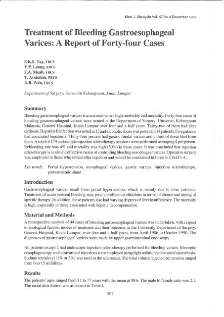
<strong>Treatment</strong> <strong>of</strong> <strong>Bleeding</strong> <strong>Gastroesophageal</strong><br />
<strong>Varices</strong>: A Report <strong>of</strong> Forty-four Cases<br />
S.K.S. Tay, FRCS<br />
Y.P. Leong, FRCS<br />
F.A. Meah, FRCS<br />
T. Abdullah, FRCS<br />
A.R. Zain, FRCS<br />
Department <strong>of</strong> Surgery, Universiti Kebangsaan, Kuala Lumpur<br />
Summary<br />
<strong>Bleeding</strong> gastroesophageal varices is associated with a high morbidity and mortality. Forty-four cases <strong>of</strong><br />
bleeding gastroesophageal varices were treated at the Department <strong>of</strong> Surgery, Universiti Kebangsaan<br />
Malaysia, General Hospital, Kuala Lumpur over four and a half years. Thirty-two <strong>of</strong> them had liver<br />
cirrhosis. Hepatitis B'infection was noted in 13 anda1coholic abuse was present in 14 patients. Five patients<br />
had associated hepatoma. Thirty-four percent had gastric fundal varices and a third <strong>of</strong> these bled from<br />
them. A total <strong>of</strong> 179 endoscopic injection sclerotherapy sessions were performed averaging 4 per person.<br />
Rebleeding rate was 4% and mortality was high (50%) in these cases. It was concluded that injection<br />
sclerotherapy is a safe and effective means <strong>of</strong> controlling bleeding oesophageal varices. Operative surgery<br />
was employed in those who rebled after injection and would be considered in those in Child's A.<br />
Key words: Portal hypertension, oesophageal varices, gastric varices, injection sclerotherapy,<br />
portasystemic shunt<br />
Introduction<br />
<strong>Gastroesophageal</strong> varices result from portal hypertension, which is mainly due to liver cirrhosis.<br />
<strong>Treatment</strong> <strong>of</strong> acute variceal bleeding may pose a problem to clinicians in terms <strong>of</strong> choice and timing <strong>of</strong><br />
specific therapy. In addition, these patients also had varying degrees <strong>of</strong>liver insufficiency. The mortality<br />
is high, especially in those associated with hepatic decompensation.<br />
Material and Methods<br />
A retrospective analysis <strong>of</strong> 44 cases <strong>of</strong> bleeding gastroesophageal varices was undertaken, with respect<br />
to etiological factors, modes <strong>of</strong> treatment and their outcome, at the University Department <strong>of</strong> Surgery,<br />
General Hospital, Kuala Lumpur, over four and a half years, from April 1986 to October 1990. The<br />
diagnoses <strong>of</strong> gastroesophageal varices were made by upper gastrointestinal endoscopy.<br />
All patients except 2 had endoscopic injection sclerotherapy performed for bleeding varices. fibreoptic<br />
oesophagoscopy and intravariceal injections were employed using light sedation with topical anaesthesia.<br />
Sodium tetradecyl (1 % or 3%) was used as the sclerosant. The total volume injected per session ranged<br />
from 6 to 15 millilitres.<br />
Results<br />
The patients' ages ranged from 11 to 77 years with the mean at 49.6. The male to female ratio was 2: 1.<br />
The racial distribution was as shown in Table 1.<br />
267
Table I<br />
Racial distribution<br />
<strong>Gastroesophageal</strong> varices Gastric varices<br />
n (%) n (%)<br />
Malays 19 (43.2%) 4 (21%)<br />
Chinese 12 (27.3%) 8 (67%)<br />
Indians 10 (22.7%) 2 (67%)<br />
Others 3 (6.8%) (33%)<br />
Total 44 (100%) 15<br />
Fourteen patients imbibed alcohol regularly while 24 <strong>of</strong>them never did. It was noted that 70% <strong>of</strong> the Indian<br />
patients did so while 90% <strong>of</strong> the Malay patients did not. Thirteen patients tested positive for hepatitis B<br />
surface antigen while 19 <strong>of</strong> them were negative. The virology status <strong>of</strong> the remaining 12 patients could<br />
not be ascertained. Two patients fell into Child's classification j A, 21 into Band 9 into C. In 2 <strong>of</strong>the patients,<br />
their liver status could not be established.<br />
Thirty-two patients had liver cirrhosis based on clinical criteria while 10 <strong>of</strong> them were not cirrhotic. The<br />
liver status <strong>of</strong> the remaining 2 patients could not be ascertained. Five <strong>of</strong> the cirrhotic patients also had<br />
hepatoma. Only 2 <strong>of</strong> these were histologically proven while the others had very suggestive clinical,<br />
radiological and biochemical features. Two <strong>of</strong> the patients with hepatoma tested positive for hepatitis B<br />
surface antigen while the remaining 3 tested negative. Of the patients without cirrhosis, 1 had carcinoma<br />
<strong>of</strong> the pancreas and 2 had portal vein thrombosis. The remaining 7 did not have sufficient clinical<br />
information available.<br />
Seven cases <strong>of</strong> malignancies were recorded: hepatoma in 5 and 1 each for rectal carcinoma and carcinoma<br />
<strong>of</strong> the pancreas. About 20% <strong>of</strong> the cases had associated peptic diseases <strong>of</strong> the stomach or duodenum.<br />
TableI also shows the racial distribution <strong>of</strong> patients with gastricfundal varices. Thirty-four percent (n:=:15)<br />
<strong>of</strong> the patients had gastric fundal varices. These were present initially (documented at endoscopy or at<br />
operation) or were developed subsequently. One third (n:=:5) <strong>of</strong> them had bled before. The proportion <strong>of</strong><br />
Chinese patients with gastric fundal varices is significantly higher than that <strong>of</strong>the other races (p:=:0.031).<br />
A total <strong>of</strong> 179 injection sclerotherapy episodes were performed. Each patient had an average <strong>of</strong> 4 injection<br />
sessions. The maximum number <strong>of</strong> injection sessions per person was 15. The complications included<br />
rebleeding (n:=:8), ulceration (n:=:5) and stricture (n:=:I). Ofthose who rebled, 3 had successful reinjection<br />
sclerotherapy, 2 had emergency operations, 2 developed encephalopathy and the remaining 1 aspirated.<br />
Only I <strong>of</strong> the 2 cases who had emergency operations survived, together with the 3 who had successful<br />
reinjection sclerotherapy (Tables II and III ).<br />
268
Rebleeding<br />
Ulceration<br />
Stricture<br />
Successful reinjection<br />
Emergency operation<br />
Encephalopathy<br />
Aspiration<br />
Table II<br />
Complications <strong>of</strong> injection<br />
Table:m<br />
Outcome <strong>of</strong> rebleeding<br />
8 (4.5%)<br />
5 (2.8%)<br />
1 (0.6%)<br />
3<br />
2 (one died)<br />
2 (both died)<br />
(died)<br />
Ulcerations secondary to injection sclerotherapy healed with time. The 1 case <strong>of</strong> stricture was the result<br />
<strong>of</strong> injection sclerotherapy followed by stapled oesophageal transection with devascularisation <strong>of</strong> the lower<br />
oesophagus. However, the patient was symptom-free after only 1 dilatation.<br />
Oesophageal transection<br />
Shunt<br />
Underunning <strong>of</strong> varix<br />
Distal pancreatectomy<br />
Splenectomy<br />
Table IV<br />
Operations<br />
Eleven patients were operated upon. Three <strong>of</strong> them were for problems not related to oesophageal varices.<br />
Table N shows the types <strong>of</strong> operations perfonned on the remaining 8 patients. Stapled oesophageal<br />
transection was combined with devascularisation <strong>of</strong> the lower oesophagus and upper stomach. Subsequently,<br />
the patient had endoscopic surveillance and had reinjection sclerotherapy if varices were noted<br />
to have recurred. The shunt performed was a lienorenal shunt for portal vein thrombosis. Underunning <strong>of</strong><br />
varix was performed for bleeding gastric fundal varix as an emergency procedure. Both patients remain<br />
well to this day. Splenectomy was performed as part <strong>of</strong> another operation.<br />
There were a total <strong>of</strong> 9 deaths. Three were due to unrelated causes (i.e., 2 from hepatoma and 1 from<br />
carcinoma sigmoid ). Four deaths were directly related to rebleeding. The other 2 died <strong>of</strong> aspiration and<br />
encephalopathy respectively.<br />
Discussion<br />
<strong>Gastroesophageal</strong> varices tend to bleed from the lower end <strong>of</strong> the oesophagus probably because <strong>of</strong> the<br />
unique venous anatomy <strong>of</strong> that area. The veins in the lower oesophagus (2 to 5 cm from the<br />
269<br />
4<br />
2<br />
4
oesophagogastric junction) were located in the lamina propria subepithelially. These veins increase in<br />
number and in the total area covered in cases <strong>of</strong> portal hypertension2 making them more likely to bleed.<br />
Other factors like high oesophageal pressures in that area and gastric reflux have been implicated but the<br />
exact causes have not been proven3.<br />
The incidence <strong>of</strong> bleeding in cirrhotic patients has been reported to be 28.6% and almost all <strong>of</strong> those who<br />
bled did so within 2 years <strong>of</strong> the diagnosis4. Two-thirds survived the first bleed but less than 10% were<br />
still alive at the end <strong>of</strong> the 6 year studT. In patients with hepatic decompensation, the cause for their<br />
bleeding upper GIT was usually bleeding oesophageal varices. They also tended to bleed more severely<br />
and had a higher rate <strong>of</strong> mortalityS.<br />
Gastric varices were noted to be not problematic in at least 1 study6. However in our series, one-third <strong>of</strong><br />
our patients had gastric fundal varices and a third <strong>of</strong> these bled. Operative underunning <strong>of</strong> the bleeding<br />
point was required for 2 <strong>of</strong> these patients.<br />
Oesophageal varices that did not bleed did not need treatmentl. Prophylactic shunt surgery7,8 had been<br />
abandoned because <strong>of</strong> associated high mortality and morbidity. <strong>Treatment</strong> <strong>of</strong> acute variceal bleeding<br />
entailed initial resuscitation, early endoscopic diagnosis, arrest <strong>of</strong> bleeding point and appropriate support<br />
for underlying liver insufficiency. Lowering <strong>of</strong> portal hypertension could be achieved by medical or<br />
operative means. Arrest <strong>of</strong> the bleeding source could be non operative, operative or a combination <strong>of</strong> these.<br />
Nonoperative treatment9 includes medical therapy, balloon tamponade, injection sclerotherapy, electrocautery,<br />
laser photocoagulation and percutaneous transhepatic obliteration <strong>of</strong> varices. Operative treatment<br />
could take the form <strong>of</strong> direct attack on the varices or a portasystemic shunting to reduce the portal pressure.<br />
Direct attack on the bleeding varices is performed via oesophageal transection and devascularisation<br />
<strong>of</strong> oesophagogastric area. This was proposed by Sugiura and Futagawa lO in 1973 and modified by<br />
otherslI.12,13. Good results with regards to haemostasis were encountered. Rebleeding ranged from<br />
none to 15% in the follow-up period <strong>of</strong> up to 13 years. Mortality ranged from 12% to 56%. Higher<br />
mortality was encountered in those undergoing emergency operation and/or those with severe liver<br />
disease (Child's C).<br />
Portasystemic shunts had been established to be effective treatment for bleeding oesophageal varices8.<br />
Various shunts14 had been performed, i.e., portacaval, lienorenal, distal splenorenal and mesocaval with<br />
its various modifications. Overall operative mortality ranged from 12% to 42%, shunt failure ranged from<br />
5 % to 50% and the incidence <strong>of</strong> encephalopathy was 34 %15,31. A 5 year survival rate <strong>of</strong> 60% had been found<br />
in those who received a shunt procedure as compared to 10% in those who did not17 but no significant<br />
difference was encountered in another study18.<br />
Rebleeding after portacaval shunt was 7% as compared to 65% in nonshunted patientsl6. Two-thirds <strong>of</strong><br />
this rebleeding occurred within 12 months <strong>of</strong> the surgery. Emergency portacaval shuntingl9,20 resulted in<br />
greater long-term survival compared to one performed electively. Major side-effects <strong>of</strong> portacaval shunt<br />
were hepatic encephalopathy and accelerated hepatic failure. To prevent these, hepatic arterialisation21 or<br />
a selective distal splenorenal shunt22,23 could be performed. Selective shunt was contraindicated in the<br />
presence <strong>of</strong> massive ascites. Child's criteria1 staged the degree <strong>of</strong> hepatic dysfunction and was used in the<br />
selection <strong>of</strong> patients for surgery. Operative mortality was 6% for Child's A, 24% for Band 50% for C24.<br />
Endoscopic injection sclerotherapy was initially practised in 193625 but renewed interest only came about<br />
in 197326 when the results <strong>of</strong> 15 years <strong>of</strong> sclerotherapy were reviewed. The bleeding varices were<br />
controlled in 93% <strong>of</strong> admissions. The total admission mortality was 18% and mortality per injection was<br />
270
6. Terblanche J, Northover JMA, Bornman P et al. A prospective<br />
controlled trial <strong>of</strong> sclerotherapy in the long-term management <strong>of</strong><br />
patients after oesophageal variceal bleeding. Surg Gynaecol<br />
Obstet 1979; 148 : 323-33.<br />
7. Resnick RH, Chalmers TC, Ishira AM et al. A controlled<br />
study <strong>of</strong> the prophylactic portacaval shunt. Ann Intern Med<br />
1969;70 : 675-88.<br />
8. Malt RA. Portasystemic venous shunts. (first <strong>of</strong> two parts). New<br />
Engl J Med 1976;295 : 24-9.<br />
9. J<strong>of</strong>fe SN. Non-operative management <strong>of</strong> variceal bleeding. Br J<br />
Surg 1984;71 : 85-91.<br />
10. Sugiura M and Futagawa S. A new technique for treating oesophageal<br />
varices. J Thorac Cardiovasc Surg 1973;66: 677-85.<br />
11. Ginsberg RJ, Waters PF, Zeldin RA et al. A modified Sugiura<br />
procedure. Ann Thorac Surg 1982;34: 258-64.<br />
12. Mir J, Ponce J, Morena E et al. Oesophagela transection and<br />
paraoesophagogastric devascularisation performed as emergency<br />
measureforuncontrolled variceal bleeding. Surg Gynecol Obstet<br />
1982;155: 868-72.<br />
13. UmeyamaK, Yoshikawa K, Yamashita T, Todo Tand Satakc K.<br />
Transabdominal oesophageal transection for oesophageal varices:<br />
experience in 101 patients. Br J Surg 1983;70: 419-22.<br />
14. Malt RA. Portasystemic venous shunts (second <strong>of</strong> two parIs).<br />
New Engl J Med 1976;295 : 80-6.<br />
15. Voorhees Jr AB, Price Jr JB and Britton RC. Portasystemic<br />
shunting procedures for portal hypertension. Am J Surg<br />
1970;119: 501-5.<br />
16. Jackson FC, Penrin EB, Felix WR and Smith AG. A clinical<br />
investigation <strong>of</strong> the portacaval shunt. Survival analysis <strong>of</strong> the<br />
therapeutic operation. Ann Surg 1971;174: 672-701.<br />
17. Mikkelsen WP. Therapeutic portacaval shunt. Arch Surg<br />
1974;108: 302-5.<br />
18. Resnick RH, Ther FL, Ishihara AM et a1. A controlled study <strong>of</strong> the<br />
therapeutic portacaval shunt. Gastroenterology 1974;67 : 843-57.<br />
19. Orl<strong>of</strong>fMJ, Charters III AC, Chandler, JG et a1. Portacaval shunt<br />
as emergency procedure in unselected patients with alcoholic<br />
cirrhosis. Surg Gynecol Obstet 1975; 141 : 59-68.<br />
20. Orl<strong>of</strong>fMJ, Charters 111 AC, ChandlerJG et a1. Portacaval shunt<br />
as emergency procedure in unselected patients with alcoholic<br />
cirrhosis. Surg Gynecol Obstet 1975;141: 59-68.<br />
21. MaillardJN, France C, RueffB, PrandiD, SicotC and France C.<br />
Hepatic arterialization and portacaval shunt in hepatic cirrhosis.<br />
Arch Surg 1974;108: 315-20.<br />
22. WalTen WD, SalamAA, HutsonD andZeppaR. Selectivedistal<br />
splenorenal shunt. Arch Surg 1974;108: 306-14.<br />
23. Galambos JT, Warren WD, Rudman D, Smith III RE and Salam<br />
AA. Selective and total shunts in the treatment <strong>of</strong> bleeding<br />
varices. New Engl J Med 1976;295 : 1089-95.<br />
24. Campbell DP, Parker DE and Anagnostopoulos CB. Survival<br />
prediction in portacaval shunts: a computerized statistical analysis.<br />
AmJ Surg 1973;126: 748-51.<br />
25. Crafoord C and Frenckner P. New surgical treatment <strong>of</strong> varicose<br />
veins <strong>of</strong> the oesophagus. Acta OtolaryngoI1939;27: 422-29.<br />
26. Johnston GW and Rodgers HW. A review <strong>of</strong> IS years' experience<br />
in the use <strong>of</strong> sclerotherapy in the control <strong>of</strong> acute haemorrhage<br />
from oesophageal varices. Br J Surg 1973;60: 797-800.<br />
272<br />
27. Mc Kee RF, Garder OJ and Carter DC. Injection sclerotherapy<br />
for bleeding varices: risk factors and complications. Br J Surg<br />
1991;78: 1098-101.<br />
28. Carter DC. Somatostatin and its analogues in acute variceal<br />
bleeding. New Perspectives in Gastroenterology 1990.<br />
29. Jenkis S. Somatostatin and its analogues in upper GI bleeding. New<br />
Perspectives in Gastroenterology 1991.<br />
30. Westaby D, Melia WM, MacDougall BRD, Hegarty JE and<br />
Williams R. Injection sclerotherapy for oesophageal varices: a<br />
prospective randomised trial <strong>of</strong> different treatment schedules.<br />
GU11984;25 : 129-32.<br />
31. Orl<strong>of</strong>f MJ, Bell RH, Hyde PV and Skivolocki WP. Long-telTll<br />
results <strong>of</strong> emergency portacaval shunt for bleeding oesophageal<br />
varices in unselected patients with alcoholic cirrhosis. Ann Surg<br />
1980;192: 325-37.


