

How to measure intraocular pressure: applanation tonometry
How to measure intraocular pressure: applanation tonometry
How to measure intraocular pressure: applanation tonometry

Create successful ePaper yourself
Turn your PDF publications into a flip-book with our unique Google optimized e-Paper software.
hOW TO<br />
<strong>How</strong> <strong>to</strong> <strong>measure</strong> <strong>intraocular</strong><br />
<strong>pressure</strong>: <strong>applanation</strong> <strong>to</strong>nometry<br />
sue stevens<br />
Nurse Advisor <strong>to</strong> the Community Eye<br />
Health Journal, International Centre<br />
for Eye Health, London School of<br />
Hygiene and Tropical Medicine, Keppel<br />
Street, London WC1E 7HT, UK.<br />
Clare Gilbert<br />
Reader, International Centre for Eye<br />
Health; Chief Medical Advisor,<br />
Sightsavers International, UK.<br />
nick Astbury<br />
Consultant Ophthalmic Surgeon,<br />
Norfolk and Norwich University<br />
Hospital NHS Trust, Colney Lane,<br />
Norwich NR4 7UY, UK.<br />
All adults attending an eye unit should have<br />
their <strong>intraocular</strong> <strong>pressure</strong> (IOP) <strong>measure</strong>d,<br />
unless there is a contraindication (e.g.<br />
trauma or corneal ulcer). Many people with<br />
glaucoma have no symp<strong>to</strong>ms and do not<br />
know they have the condition. All children who<br />
have had cataract surgery should also have<br />
their IOP <strong>measure</strong>d at every follow-up visit, if<br />
possible. Finding glaucoma early allows<br />
treatment <strong>to</strong> be given which will preserve<br />
sight. Although elevated IOP is not the only<br />
sign of glaucoma, measuring it is simple and<br />
quick <strong>to</strong> do. It should therefore be done<br />
routinely on all adults attending eye care<br />
facilities. Applanation <strong>to</strong>nometry, described<br />
in this article, is the preferred method (the<br />
‘gold standard’). Schiötz <strong>to</strong>nometry, which<br />
will be described in a future issue, is a useful<br />
screening test which can be performed by<br />
nurses or ophthalmic technicians.<br />
equipment<br />
• Tonometer, either Goldmann (used on slit<br />
lamps) or Perkins (hand-held)<br />
• Applanation prism<br />
• Local anaesthetic drops<br />
• Fluorescein strips<br />
• Clean cot<strong>to</strong>n wool or gauze swabs.<br />
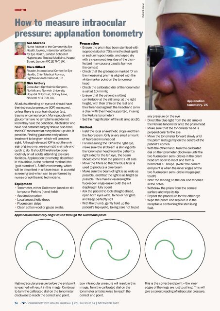
Applanation <strong>to</strong>nometry rings viewed through the Goldmann prism<br />
High <strong>intraocular</strong> <strong>pressure</strong> before the end point<br />
is reached will result in this image. Continue<br />
<strong>to</strong> turn the calibrated dial on the <strong>to</strong>nometer<br />
clockwise <strong>to</strong> reach the correct end point.<br />
Preparation<br />
• Ensure the prism has been sterilised with<br />
isopropyl alcohol 70% (methylated spirit)<br />
or sodium hypochlorite, and wiped dry<br />
with a clean swab (residue of the disinfectant<br />
may cause a caustic burn on<br />
the cornea)<br />
• Check that the graduation marked ‘0’ on<br />
the measuring prism is aligned with the<br />
white marker point on the <strong>to</strong>nometer<br />
head<br />
• Check the calibrated dial of the <strong>to</strong>nometer<br />
is set at 10 mmHg<br />
• Ensure that the patient is sitting<br />
comfortably at the slit lamp: at the right<br />
height, with their chin on the rest and<br />
their forehead against the headband (or in<br />
a chair with their head supported, if using<br />
the Perkins <strong>to</strong>nometer)<br />
• Set the magnification of the slit lamp at x10.<br />
Method<br />
• Instil the local anaesthetic drops and then<br />
the fluorescein. Only a very small amount<br />
of fluorescein is needed<br />
• For measuring the IOP in the right eye,<br />
make sure the slit beam is shining on<strong>to</strong><br />
the <strong>to</strong>nometer head from the patient’s<br />
right side; for the left eye, the beam<br />
should come from the patient’s left side<br />
• Move the filters so that the blue filter is<br />
used <strong>to</strong> produce a blue beam<br />
• Make sure the beam of light is as wide as<br />
possible, and that the light is as bright as<br />
possible. This makes visualising the<br />
fluorescein rings easier (with the slit<br />
diaphragm fully open)<br />
• Ask the patient <strong>to</strong> look straight ahead,<br />
open both eyes wide, fix his or her gaze<br />
and keep perfectly still<br />
• With the thumb, gently hold up the<br />
patient’s <strong>to</strong>p eyelid, taking care not <strong>to</strong> put<br />
Low <strong>intraocular</strong> <strong>pressure</strong> will result in this<br />
image. Turn the calibrated dial on the<br />
<strong>to</strong>nometer anticlockwise <strong>to</strong> reach the<br />
correct end point.<br />
74 Community EyE HEaltH Journal | Vol 20 iSSuE 64 | DECEmBEr 2007<br />
Richard Scawn<br />
<strong>applanation</strong><br />
<strong>to</strong>nometry. uK<br />
any <strong>pressure</strong> on the eye<br />
• Direct the blue light from the slit lamp or<br />
the Perkins <strong>to</strong>nometer on<strong>to</strong> the prism head<br />
• Make sure that the <strong>to</strong>nometer head is<br />
perpendicular <strong>to</strong> the eye<br />
• Move the <strong>to</strong>nometer forward slowly until<br />
the prism rests gently on the centre of the<br />
patient’s cornea<br />
• With the other hand, turn the calibrated<br />
dial on the <strong>to</strong>nometer clockwise until the<br />
two fluorescein semi-circles in the prism<br />
head are seen <strong>to</strong> meet and form a<br />
horizontal ‘S’ shape. (Note: the correct<br />
end point is when the inner edges of the<br />
two fluorescein semi-circle images just<br />
<strong>to</strong>uch)<br />
• Note the reading on the dial and record it<br />
in the notes<br />
• Withdraw the prism from the corneal<br />
surface and wipe its tip<br />
• Repeat the procedure for the other eye<br />
• Wipe the prism and replace it in the<br />
receptacle containing the sterilising<br />
fluid.<br />
This is the correct end point – the inner<br />
edges of the rings are just <strong>to</strong>uching. This will<br />
give a correct reading of <strong>intraocular</strong> <strong>pressure</strong>.
Calibration of the<br />
Goldmann <strong>to</strong>nometer<br />
• It is possible <strong>to</strong> check the calibration<br />
of the <strong>to</strong>nometer; this should be<br />
done every six months. Calibration is<br />
done at dial positions 0, 2, and 6<br />
(equivalent <strong>to</strong> 0, 20, and 60 mmHg)<br />
• Insert the prism in the holder and<br />
place the <strong>to</strong>nometer on the slit<br />
lamp<br />
• At dial position 0, the feeler arm<br />
should be in free movement. If the<br />
dial is turned backwards a small way<br />
(<strong>to</strong> the equivalent of position -0.05),<br />
the arm should fall <strong>to</strong>wards the<br />
examiner. If the dial is turned<br />
forwards a small way (<strong>to</strong> the equivalent<br />
of position +0.05) the arm<br />
should fall <strong>to</strong>wards the patient<br />
• If the arm doesn’t respond in the<br />
above way, the <strong>to</strong>nometer is<br />
inaccurate at dial position 1<br />
• To check dial positions 2 and 6, the<br />
check weight is used (this is normally<br />
found in the case with the <strong>to</strong>nometer<br />
prisms or in the drawer of the slit<br />
lamp). There are five markings<br />
engraved on the bar. These<br />
represent 0 centrally, then 2 on<br />
either side, and 6 <strong>to</strong>wards the<br />
edges<br />
• Line up the adjustable holder with<br />
index mark 2 on the weight. With the<br />
longer end of the bar facing you, put<br />
it in<strong>to</strong> the slot on the side of the<br />
<strong>to</strong>nometer and push it all the way in<br />
• Repeat the above steps (for dial<br />
position 0), with the dial now at<br />
position 2. This time, turn the dial<br />
backwards <strong>to</strong> the equivalent of 1.95<br />
and forwards <strong>to</strong> the equivalent of<br />
2.05<br />
• To check dial position 6, move<br />
the weight bar <strong>to</strong> the<br />
end position. Repeat<br />
the steps at dial<br />
position 6, turning the<br />
dial backwards <strong>to</strong> the<br />
equivalent of 5.9 and<br />
forwards <strong>to</strong> the equivalent<br />
of 6.1<br />
• If the <strong>to</strong>nometer is<br />
inaccurate at any of these<br />
dial positions, it should<br />
be returned <strong>to</strong> the<br />
manufacturer<br />
for<br />
recalibration.<br />
a Goldmann<br />
<strong>to</strong>nometer<br />
neWs<br />
Better vision for<br />
safer roads: an<br />
instance of<br />
accidental<br />
advocacy in Nigeria<br />
While former MSc student<br />
Barka David Lass was testing<br />
the vision of commercial drivers<br />
at a minibus station (or ‘park’)<br />
in Jos, Nigeria, in July 2007,<br />
he was spotted by a television<br />
crew, there <strong>to</strong> film a news insert<br />
on lost property. The producer<br />
was so taken by Lass’s research<br />
(conducted for his MSc dissertation,<br />
summarised on page<br />
71) – that an interview with him<br />
was broadcast on national news the very same day.<br />
Nigeria has seen a five-fold increase in the number of deaths due <strong>to</strong> traffic accidents over<br />
the last twenty years. The interview with Lass highlighted the fact that, although drivers have<br />
<strong>to</strong> satisfy a minimum legal requirement for visual acuity in Nigeria, not even the vision of<br />
commercial drivers is routinely tested before licences are issued.<br />
After the news item, Lass was invited <strong>to</strong> participate in a television programme on vision<br />
and safe driving, broadcast by the Plateau Radio Television Corporation. Other participants<br />
included representatives of the Vehicle Inspection Office and of the Federal Road Safety<br />
Commission.<br />
The participants talked about the definition of vision, how <strong>to</strong> assess it, and what constitutes<br />
good vision for driving. Other <strong>to</strong>pics included the high number of road traffic accidents<br />
in Nigeria, the relationship between vision and accidents, and the various laws and penalties<br />
related <strong>to</strong> vision and driving. The programme was watched in over six states and in the<br />
federal capital terri<strong>to</strong>ry of Nigeria.<br />
The following recommendations were put forward at the end of the discussion:<br />
• all commercial vehicle drivers applying for a licence should undergo a comprehensive eye<br />
exam, conducted by an ophthalmologist<br />
• all commercial vehicle drivers should have their vision tested before their driving licences<br />
can be renewed (every four years)<br />
• staff of the Federal Road Safety Commission and of the Vehicle Inspection Office should<br />
be trained <strong>to</strong> assess vision, so that they can carry out periodic checks on drivers.<br />
Since his return <strong>to</strong> Nigeria after completing his MSc at the International Centre for Eye Health<br />
in London, Lass has met with the producer of the programme; <strong>to</strong>gether, they are working on<br />
ways <strong>to</strong> ensure that these recommendations are carried out.<br />
Million-dollar advocacy success for<br />
VISION 2020 Australia<br />
The fierce competition between political parties during<br />
the run-up <strong>to</strong> the 2007 elections provided an opportunity<br />
for VISION 2020 Australia <strong>to</strong> secure more than<br />
AUS $100 million in funding for eye health and vision care.<br />
VISION 2020 Australia had worked with its global<br />
partners <strong>to</strong> produce a comprehensive proposal <strong>to</strong><br />
eliminate blindness and visual impairment in the<br />
Southeast Asia and Pacific region. In July 2007, this<br />
was presented <strong>to</strong> the minister and shadow (opposition)<br />
minister responsible for overseas aid.<br />
On World Sight Day, 11 Oc<strong>to</strong>ber, Shadow Minister for<br />
International Development Assistance Bob McMullan<br />
pledged AUS $45 million over two years <strong>to</strong> fund the proposal<br />
<strong>to</strong> prevent blindness and improve vision care in the region.<br />
Two weeks later, Foreign Minister Alexander Downer<br />
pledged funding of AUS $60 million over ten years <strong>to</strong><br />
implement part of the proposal.<br />
More information on VISION 2020 Australia’s<br />
advocacy work is available on the VISION 2020<br />
Australia website: www.vision2020australia.org.au<br />
the television interview with Barka David lass and<br />
representatives of the federal road safety<br />
commission and vehicle inspection office. niGEria<br />
australia’s shadow minister for<br />
international development<br />
assistance on World Sight Day.<br />
auStralia<br />
Community EyE HEaltH Journal | Vol 20 iSSuE 64 | DECEmBEr 2007<br />
75<br />
Barka David Lass<br />
VISION 2020 Australia


