SYPRO Orange and Red protein gel stains
SYPRO Orange and Red protein gel stains
SYPRO Orange and Red protein gel stains

Create successful ePaper yourself
Turn your PDF publications into a flip-book with our unique Google optimized e-Paper software.
instructions<br />
product code<br />
RPN 5801/2/3/4<br />
Warning<br />
For research use only.<br />
Not recommended or intended<br />
for diagnosis of disease in<br />
humans or animals.<br />
Do not use internally or<br />
externally in humans or animals.<br />
Contains DMSO. See safety data<br />
sheet supplied.<br />
Harmful<br />
Components<br />
RPN 5801<br />
<strong>SYPRO</strong> <strong>Orange</strong> <strong>gel</strong> stain<br />
5000x concentrate in<br />
DMSO, 500µl<br />
RPN 5802<br />
<strong>SYPRO</strong> <strong>Orange</strong> <strong>gel</strong> stain<br />
5000x concentrate in<br />
DMSO, 10x50µl<br />
RPN 5803<br />
<strong>SYPRO</strong> <strong>Red</strong> <strong>gel</strong> stain<br />
5000x concentrate in<br />
DMSO, 500µl<br />
RPN 5804<br />
<strong>SYPRO</strong> <strong>Red</strong> <strong>gel</strong> stain<br />
5000x concentrate in<br />
DMSO, 10x50µl<br />
Sufficient material in each<br />
to prepare 2.5 litres of<br />
working stain solution<br />
which is sufficient to stain<br />
~50 polyacrylamide<br />
mini<strong>gel</strong>s.<br />
<strong>SYPRO</strong> <strong>Orange</strong> <strong>and</strong><br />
<strong>Red</strong> <strong>protein</strong> <strong>gel</strong> <strong>stains</strong><br />
<strong>SYPRO</strong> <strong>Orange</strong> <strong>and</strong> <strong>Red</strong> <strong>protein</strong> <strong>gel</strong> <strong>stains</strong><br />
enabling fast, simple <strong>and</strong> sensitive staining<br />
of <strong>protein</strong>s in electrophoretic <strong>gel</strong>s.<br />
Description<br />
i RPN5801/2/3/4 Rev-A, 2000 " p1<br />
<strong>SYPRO</strong> <strong>Orange</strong> <strong>and</strong> <strong>Red</strong> <strong>protein</strong> <strong>gel</strong> <strong>stains</strong><br />
are designed for fast, simple, sensitive<br />
staining of <strong>protein</strong>s in electrophoretic <strong>gel</strong>s.<br />
The staining properties of the two <strong>SYPRO</strong><br />
dyes are similar, <strong>and</strong> both are equally<br />
suitable for use in most procedures. Our<br />
scientists have noted that the <strong>SYPRO</strong><br />
<strong>Orange</strong> <strong>gel</strong> stain is slightly brighter,<br />
whereas the <strong>SYPRO</strong> <strong>Red</strong> <strong>gel</strong> stain has<br />
somewhat lower background fluorescence.<br />
For those using a laser-excited <strong>gel</strong> scanner,<br />
we recommend the <strong>SYPRO</strong> <strong>Orange</strong> stain<br />
for argon laser-based instruments <strong>and</strong> the<br />
<strong>SYPRO</strong> <strong>Red</strong> stain for instruments that<br />
employ green He-Ne or Nd:YAG lasers<br />
(Figure 2). Both dyes are efficiently excited<br />
by UV or broadb<strong>and</strong> illumination (Figure<br />
2) <strong>and</strong>, with the correct filters, work well<br />
with CCD camera archiving systems.<br />
<strong>SYPRO</strong> <strong>Orange</strong> <strong>and</strong> <strong>Red</strong> <strong>protein</strong> <strong>gel</strong> <strong>stains</strong><br />
are not suitable for staining <strong>protein</strong>s on<br />
blotting membrane or in IEF <strong>gel</strong>s <strong>and</strong> they<br />
show reduced sensitivity when staining<br />
<strong>protein</strong>s on 2-D <strong>gel</strong>s.<br />
Other materials<br />
required<br />
(not supplied with these<br />
products)<br />
" St<strong>and</strong>ard <strong>gel</strong> electrophoresis<br />
equipment<br />
<strong>and</strong> solutions(eg from<br />
Hoefer)<br />
" UV transilluminator<br />
" Small plastic box lids<br />
or sealable plastic<br />
bags<br />
H<strong>and</strong>ling<br />
Packaging: Screw cap<br />
plastic vials contained<br />
within foil bag.<br />
Storage: Store in the dark<br />
at room temperature,<br />
2-8°C or -15°C to -30°C<br />
Stored as above, stock<br />
solutions are stable for 6-<br />
12 months.<br />
Diluted staining reagent<br />
(in buffer or acetic acid)<br />
is stable for 3 months<br />
when stored in sterile<br />
detergent free glass or<br />
plastic bottles at 2-8°C in<br />
the dark.<br />
<strong>SYPRO</strong> <strong>Orange</strong> <strong>and</strong> <strong>Red</strong> <strong>protein</strong> <strong>gel</strong> <strong>stains</strong><br />
provide the following advantages over<br />
conventional colorimetric <strong>stains</strong>:<br />
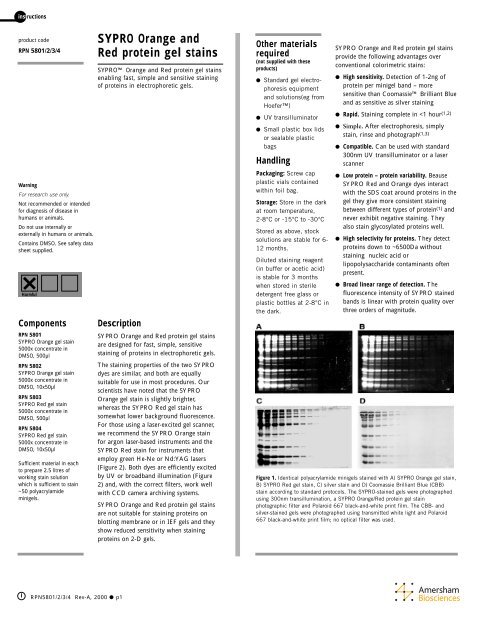
" High sensitivity. Detection of 1-2ng of<br />
<strong>protein</strong> per mini<strong>gel</strong> b<strong>and</strong> – more<br />
sensitive than Coomassie Brilliant Blue<br />
<strong>and</strong> as sensitive as silver staining<br />
" Rapid. Staining complete in
Critical parameters<br />
The following points are<br />
critical to the performance of<br />
this protocol <strong>and</strong> should be<br />
strictly observed<br />
• Store <strong>SYPRO</strong> <strong>gel</strong> <strong>stains</strong><br />
protected from light.<br />
• Allow vials to equilibrate to<br />
RT, sonicate to redissolve<br />
any dye particles <strong>and</strong><br />
centrifuge briefly before<br />
opening.<br />
• When running SDSpolyacrylamide<br />
<strong>gel</strong>s use<br />
0.05% SDS in the running<br />
buffer.<br />
• Use the staining reagent at<br />
the dilution stated for<br />
optimum results.<br />
• Do not fix the <strong>protein</strong>s in<br />
the <strong>gel</strong> with methanol as<br />
this will result in a reduced<br />
signal with <strong>SYPRO</strong> <strong>Red</strong> <strong>and</strong><br />
<strong>Orange</strong> <strong>stains</strong>.<br />
Protocol<br />
#<br />
SDS-polyacrylamide <strong>gel</strong> electrophoresis<br />
Prepare fresh running buffer <strong>and</strong> run SDS-polyacrylamide <strong>gel</strong> according to<br />
st<strong>and</strong>ard protocols, (4,5) with the following exception. We recommend<br />
using 0.05% SDS in the running buffer (instead of the usual 0.1% SDS).<br />
This has been shown to be the optimal concentration of SDS for<br />
sensitivity <strong>and</strong> speed. Higher concentrations require extended washing to<br />
reduce background fluorescence <strong>and</strong> lower concentrations of SDS (or old<br />
running buffer) result in poor resolution of b<strong>and</strong>s.<br />
$<br />
Staining <strong>protein</strong>s in the <strong>gel</strong><br />
Staining <strong>protein</strong>s after electrophoresis<br />
1. Prepare the staining solution by diluting the stock <strong>SYPRO</strong> reagent<br />
1:5000 in 7.5%(v/v) acetic acid <strong>and</strong> mixing vigorously.<br />
i RPN5801/2/3/4 " p2<br />
Safety warnings <strong>and</strong><br />
precautions<br />
Warning: For research use only.<br />
Not recommended or intended for<br />
diagnosis of disease in humans or<br />
animals. Do not use internally or<br />
externally in humans or animals.<br />
Warning: Contains DMSO. See safety<br />
data sheet supplied.<br />
The toxicity of the <strong>SYPRO</strong> <strong>protein</strong> <strong>gel</strong><br />
<strong>stains</strong> has not been fully evaluated <strong>and</strong><br />
no data is currently available. Please<br />
h<strong>and</strong>le with care.<br />
Waste solutions of <strong>SYPRO</strong> <strong>stains</strong> should<br />
be poured through activated charcoal<br />
before disposal. The charcoal must then<br />
be incinerated to destroy the dye. We<br />
have found that 1g of activated charcoal<br />
binds at least 98% of the <strong>SYPRO</strong><br />
<strong>Orange</strong> or <strong>SYPRO</strong> <strong>Red</strong> dye present in<br />
2.5l of 1x staining solution prepared in<br />
7.5% acetic acid, which is equivalent to<br />
the amount of dye in 500µl of the 5000x<br />
concentrated DMSO solution.<br />
All chemicals should be considered as<br />
potentially hazardous. We therefore<br />
recommend that this product is h<strong>and</strong>led<br />
only by those persons who have been<br />
trained in laboratory techniques <strong>and</strong> that<br />
it is used in accordance with the<br />
principles of good laboratory practice.<br />
Wear suitable protective clothing such as<br />
laboratory overalls, safety glasses <strong>and</strong><br />
gloves. Care should be taken to avoid<br />
contact with skin or eyes. In the case of<br />
contact with skin or eyes wash<br />
immediately with water (see safety data<br />
sheet for specific advice).<br />
• Diluting the stain below the recommended concentration will result in<br />
reduced staining sensitivity.<br />
• Using higher staining concentrations than recommended will not<br />
result in better detection, but will instead result in increased<br />
background in the <strong>gel</strong> <strong>and</strong> quenching of the fluorescence from dye<br />
molecules crowded around the <strong>protein</strong>s.<br />
• The staining solution may be reused up to four times. However,<br />
sensitivity is greatly reduced after the second use.<br />
• For low percentage <strong>gel</strong>s <strong>and</strong> for very small <strong>protein</strong>s, 10% acetic acid<br />
solution will result in better retention of the <strong>protein</strong> in the <strong>gel</strong><br />
without compromising sensitivity.<br />
• Acetic acid will interfere with transfer of the <strong>protein</strong>s to a blot. For<br />
Western blotting <strong>and</strong> other blotting techniques, you may dilute the<br />
<strong>SYPRO</strong> <strong>Orange</strong> or <strong>Red</strong> stain in st<strong>and</strong>ard transfer buffer. However, as<br />
this results in lower sensitivity staining we recommend staining with<br />
<strong>SYPRO</strong> Tangerine, RPN 5805 for blotting techniques.<br />
• Do not fix the <strong>protein</strong>s in the <strong>gel</strong> with methanol-containing solutions.<br />
Methanol removes the SDS coat from <strong>protein</strong>s, strongly reducing the<br />
signal from <strong>SYPRO</strong> <strong>Orange</strong> or <strong>Red</strong> <strong>stains</strong>.<br />
2. Pour the staining solution into a small plastic dish<br />
• For one or two st<strong>and</strong>ard-size mini<strong>gel</strong>s, use about 50ml of staining<br />
solution. For larger <strong>gel</strong>s, use between 500 <strong>and</strong> 750ml of staining<br />
solution.<br />
• Clean <strong>and</strong> rinse the staining dishes well before use as detergent will<br />
interfere with staining.<br />
• Gels may also be stained in sealable plastic bags, ensuring the correct<br />
volume of staining solution is used.<br />
3. Place the <strong>gel</strong> into the staining solution<br />
• Cover the container with aluminium foil to protect the dye from<br />
bright light.<br />
4. Gently agitate the <strong>gel</strong> at room temperature<br />
• The staining time is 10 to 60 minutes, depending on the thickness or<br />
percentage of the <strong>gel</strong>. For 1mm thick 15% polyacrylamide <strong>gel</strong>s,<br />
optimal signal is achieved after 40 to 60 minutes staining.<br />
• Additional staining time (several hours to overnight) does not<br />
enhance or degrade the signal. Gels can be left in stain for up to a<br />
week with only a small loss in sensitivity; our detection limits under<br />
these conditions are approximately 2-4ng/b<strong>and</strong>.<br />
5. Rinse briefly with 7.5% acetic acid<br />
• This brief rinse (less than a minute) removes excess stain from the <strong>gel</strong><br />
surface to reduce background fluorescence on the surface of the<br />
transilluminator or <strong>gel</strong> scanner.<br />
• 30 minutes of destaining in 7.5% acetic acid has been shown to<br />
improve background <strong>and</strong> signal detection in a <strong>gel</strong> scanner. However,<br />
testing has shown that, for Polaroid 667 black-<strong>and</strong>-white<br />
photography, even a 10 minute destaining results in lower sensitivity.<br />
Staining <strong>protein</strong>s during electrophoresis<br />
• <strong>SYPRO</strong> <strong>protein</strong> <strong>gel</strong> <strong>stains</strong> can be dissolved in the cathode (top)<br />
running buffer to stain <strong>protein</strong>s as the <strong>gel</strong> runs. The <strong>SYPRO</strong> stock<br />
solution can be diluted 5000-fold into the cathode running buffer.<br />
The dye moves through the <strong>gel</strong> with the SDS front, so that all sizes of<br />
<strong>protein</strong> are stained. Staining does not influence relative migration of<br />
<strong>protein</strong>s
through the <strong>gel</strong>. However this results in poorer <strong>protein</strong> staining <strong>and</strong><br />
higher background fluorescence.<br />
• <strong>SYPRO</strong> <strong>Red</strong> <strong>and</strong> <strong>Orange</strong> <strong>stains</strong> cannot be used as pre<strong>stains</strong> for SDS<br />
<strong>gel</strong>s due to their affinity for the SDS.<br />
Triton X-100 Gels<br />
Triton X-100 at 0.1% or greater will interfere with <strong>SYPRO</strong> dye<br />
staining. If Triton X-100 is used with your <strong>gel</strong>, we recommend soaking<br />
the <strong>gel</strong> in two to three changes of buffer to be sure the Triton X-100 is<br />
diluted out, <strong>and</strong> then incubating the <strong>gel</strong> in 0.05% SDS for 30 minutes<br />
before staining as usual.<br />
2-D Gels <strong>and</strong> IEF Gels<br />
<strong>SYPRO</strong> <strong>Orange</strong> <strong>and</strong> <strong>Red</strong> <strong>protein</strong> <strong>gel</strong> <strong>stains</strong> are not suitable for staining<br />
<strong>protein</strong>s on IEF <strong>gel</strong>s, <strong>and</strong> they show reduced sensitivity when staining<br />
<strong>protein</strong>s on 2-D <strong>gel</strong>s.<br />
Nondenaturing Gels<br />
Protein can be stained after native <strong>gel</strong> electrophoresis by dissolving<br />
<strong>SYPRO</strong> dyes in water <strong>and</strong> then following the protocol above.<br />
• Staining <strong>protein</strong>s in nondenaturing <strong>gel</strong>s is highly <strong>protein</strong>-selective <strong>and</strong><br />
will generally be less sensitive than staining <strong>protein</strong>s in SDS <strong>gel</strong>s;<br />
however, because there is essentially no background fluorescence,<br />
photographic exposures can be very long.<br />
• If it is not necessary to maintain the <strong>protein</strong> in a native state after<br />
electrophoresis, the best sensitivity can be achieved if the <strong>gel</strong> is soaked<br />
in 0.05% SDS for about 30 minutes <strong>and</strong> then stained with a solution<br />
of <strong>SYPRO</strong> dye diluted in 7.5% acetic acid. (3)<br />
%<br />
Viewing the Gel<br />
Gels may be left in staining solution overnight without losing sensitivity.<br />
However, the fixation in acetic acid is relatively mild, so for low<br />
percentage <strong>gel</strong>s or very small <strong>protein</strong>s, photographs should be taken as<br />
soon as possible after staining, before the <strong>protein</strong>s begin to diffuse.<br />
• Gels may be visualized on a st<strong>and</strong>ard 300nm UV transilluminator (eg<br />
from Hoefer). We recommend cleaning the surface of the<br />
transilluminator with water <strong>and</strong> a soft cloth after using to minimize<br />
the build up of fluorescent dyes on the surface.<br />
• Place the <strong>gel</strong> directly on the transilluminator. Plastic wraps, such as<br />
SaranWrap , fluoresce on their own <strong>and</strong> even more when exposed to<br />
<strong>SYPRO</strong> <strong>Orange</strong> or <strong>Red</strong> stain. This gives a large background signal if<br />
the <strong>gel</strong> is sitting on a piece of plastic wrap on a UV transilluminator<br />
<strong>and</strong> makes it impossible to get good sensitivity.<br />
• Amersham Biosciences PhastGel has polyester backing<br />
material Gelbond which is not only highly autofluorescent, but also<br />
binds the <strong>SYPRO</strong> <strong>stains</strong>, producing additional background<br />
fluorescence. Consequently, the plastic backing should be removed<br />
before trying to visualize your results. Amersham Biosciences<br />
markets a <strong>gel</strong> backing remover for use with the PhastTransfer<br />
system.<br />
• For those using a laser-excited <strong>gel</strong> scanner, we recommend the <strong>SYPRO</strong><br />
<strong>Orange</strong> stain for argon-laser based instruments <strong>and</strong> the <strong>SYPRO</strong> <strong>Red</strong><br />
stain for instruments that employ green He-Ne or Nd:YAG lasers<br />
(Figure 2).<br />
i RPN 5801/2/3/4 " p3<br />
Figure 2. The fluorescence excitation <strong>and</strong> emission spectra of A) <strong>SYPRO</strong> <strong>Orange</strong><br />
<strong>and</strong> B) <strong>SYPRO</strong> <strong>Red</strong> <strong>protein</strong> <strong>gel</strong> <strong>stains</strong> diluted 1:10,000 in water containing 0.05%<br />
SDS <strong>and</strong> 150µg/mL bovine serum albumin.<br />
&<br />
Photographing the <strong>gel</strong><br />
Photography of the <strong>gel</strong> is essential to obtain high sensitivity. The<br />
camera's integrating effect can make b<strong>and</strong>s visible that are not visible<br />
to the eye.<br />
Photography with a Polaroid Camera<br />
The highest sensitivity with a Polaroid camera will be obtained using<br />
Polaroid 667 black-<strong>and</strong>-white print film <strong>and</strong> the <strong>SYPRO</strong> <strong>protein</strong> <strong>gel</strong><br />
stain photographic filter RPN5810 (7)<br />
• St<strong>and</strong>ard ethidium bromide filters should not be used as they will<br />
block much of the light <strong>and</strong> lead to lower sensitivity. Supplemental<br />
UV blocking filters are not usually required.<br />
• Polaroid 667 film is a fast film with an ISO rating of ASA3000. The<br />
use of different film types may require longer exposure times or<br />
different filters.<br />
• Exposure time will vary with the intensity of the illumination source:<br />
with an f-stop of 4.5, typically 2-5 seconds for <strong>SYPRO</strong> <strong>Orange</strong> stain<br />
<strong>and</strong> 3-8 seconds for <strong>SYPRO</strong> <strong>Red</strong> stain.<br />
• We generally observe detection limits of ~50ng <strong>protein</strong>/b<strong>and</strong> with<br />
300nm transillumination <strong>and</strong> ~1–2ng/b<strong>and</strong> in a photograph taken<br />
with a Polaroid 667 black <strong>and</strong> white print film. Our detection limits<br />
of 1-2ng/b<strong>and</strong> are obtained using an Ultraviolet Transilluminator,<br />
which has six 15-watt bulbs that provide peak illumination at<br />
312nm. When using weaker illumination sources, exposures must be<br />
correspondingly longer.<br />
• Although our detection limits are 1-2ng/b<strong>and</strong> for most <strong>protein</strong>s, we<br />
would like to emphasize that b<strong>and</strong>s containing 5-10ng/ <strong>protein</strong> are<br />
more readily detected. B<strong>and</strong>s containing less than 5-10ng <strong>protein</strong><br />
require longer exposures <strong>and</strong> sharp b<strong>and</strong>s for good visualization.<br />
Longer exposures can result in higher background.<br />
• Noticeable photobleaching can occur after several minutes of<br />
exposure to ultraviolet light. If a <strong>gel</strong> becomes photobleached, it can<br />
be restained by simply returning it to the staining solution.<br />
Photography with a CCD Camera<br />
CCD Cameras also provide good sensitivity, however the <strong>SYPRO</strong><br />
photographic filter may not be optimal. Contact the manufacturer of<br />
your camera system for the optimal filter sets to use.
Storing the Stained Gel<br />
Gels may be stored by keeping them protected from light in the staining<br />
solution. The signal does decrease somewhat after several days, but,<br />
depending on the amount of <strong>protein</strong> in your b<strong>and</strong>s, your <strong>gel</strong>s may retain<br />
a usable signal for many weeks.<br />
Gels may be dried between sheets of cellophane, although there is<br />
sometimes a slight decrease in sensitivity. Store the dried <strong>gel</strong> in the dark<br />
to prevent photobleaching.<br />
• If the <strong>gel</strong>s are dried on to paper, the light will scatter <strong>and</strong> the<br />
sensitivity will decrease.<br />
• If the <strong>gel</strong> is dried between sheets of other plastic, the plastic typically<br />
used is not transparent to UV light.<br />
'<br />
Destaining the Gel<br />
Gels may be mostly destained by incubation overnight in 0.1% Tween 20. Alternatively, incubation in several changes of 7.5% acetic acid will<br />
eventually remove all of the stain. Incubation in methanol will strip off<br />
dye <strong>and</strong> SDS, but will also precipitate <strong>protein</strong>s.<br />
(<br />
Tips<br />
• The SDS front at the bottom of the <strong>gel</strong> <strong>stains</strong> very heavily with<br />
<strong>SYPRO</strong> <strong>stains</strong>. Unless the <strong>protein</strong>s of interest are co-migrating with<br />
the SDS front, it will be advantageous to run the SDS front off the<br />
<strong>gel</strong>.<br />
• Coloured <strong>stains</strong> <strong>and</strong> marker dyes, as well as commercially prestained<br />
<strong>protein</strong> markers, interfere with <strong>SYPRO</strong> dye staining <strong>and</strong> quench<br />
fluorescence.<br />
• Highly-coloured prosthetic groups (e.g. heme) that remain bound in<br />
native <strong>gel</strong>s will quench fluorescence of the <strong>SYPRO</strong> <strong>Orange</strong> <strong>and</strong> <strong>Red</strong><br />
<strong>stains</strong>.<br />
• Odd marks on stained <strong>gel</strong>s can be caused by several factors. If the <strong>gel</strong><br />
is squeezed, a mark appears that <strong>stains</strong> heavily with the <strong>SYPRO</strong> dyes.<br />
This is probably a localized high concentration of SDS that has<br />
difficulty diffusing out. Glove powder can also give background<br />
markings, so we recommend rinsing or washing gloves prior to<br />
h<strong>and</strong>ling <strong>gel</strong>s.<br />
• Staining with the <strong>SYPRO</strong> <strong>Orange</strong> dye occasionlly results in <strong>gel</strong>s with<br />
scattered fluorescent speckles. However, they do not reduce the dye's<br />
sensitivity.<br />
• <strong>SYPRO</strong> dye stained <strong>gel</strong>s can be restained with either Coomassie<br />
Brilliant Blue or with silver stain procedures. In fact, for some silver<br />
staining methods, we have found that prestaining with <strong>SYPRO</strong> dyes<br />
actually increases the rate of staining <strong>and</strong> the sensitivity for detection.<br />
• To stain <strong>gel</strong>s previously stained with Coomassie Brilliant Blue stain,<br />
the stain must be completely removed as it will quench the<br />
fluorescence of <strong>SYPRO</strong> dyes. Soaking the <strong>gel</strong> in either 30% methanol<br />
or 7.5% acetic acid with several changes of the destaining solution<br />
will be effective at removing the Coomassie stain. Once the<br />
Coomassie dye has been removed, the <strong>gel</strong> should be incubated in<br />
0.05% SDS for 30 minutes before staining with the <strong>SYPRO</strong> stain as<br />
usual.<br />
i<br />
RPN 5801/2/3/4 " p4<br />
References<br />
1. Anal. Biochem., 239, p.223, 1996.<br />
2. J.NIH Res., 7, p.82, 1995.<br />
3. Anal. Biochem., 239, p.238, 1996.<br />
4. Short Protocols in Molecular Biology, second edition, Ausubel et al.,<br />
John Wiley & Sons, 1992.<br />
5. Nature, 227, p680, 1970.<br />
6. Electrophoresis, 19, p.2169, 1998.<br />
7. Anal. Biochem., 248, p.168, 1997.<br />
Product information<br />
Product name code<br />
<strong>SYPRO</strong> <strong>Orange</strong> <strong>protein</strong> <strong>gel</strong> stain<br />
500µl RPN 5801<br />
10x50µl RPN 5802<br />
<strong>SYPRO</strong> <strong>Red</strong> <strong>protein</strong> <strong>gel</strong> stain<br />
500µl RPN 5803<br />
100x50µl RPN 5804<br />
Related products<br />
<strong>SYPRO</strong> Tangerine <strong>protein</strong> <strong>gel</strong> stain RPN 5805<br />
<strong>SYPRO</strong> <strong>protein</strong> <strong>gel</strong> stain starter kit RPN 5811<br />
Protein molecular weight markers<br />
Broad range MW 6500-205000 RPN 5800<br />
<strong>SYPRO</strong> <strong>protein</strong> <strong>gel</strong> stain photographic filter RPN 5810<br />
Hoefer, PhastGel <strong>and</strong> PhastTransfer<br />
are trademarks of Amersham<br />
Biosciences Limited or its<br />
subsidiaries<br />
Amersham is a trademark of<br />
Amersham plc<br />
<strong>SYPRO</strong> is a trademark of Molecular<br />
Probes Inc<br />
Coomassie is a trademark of ICI plc<br />
Polaroid is a trademark of Polaroid<br />
(UK) Ltd<br />
Triton is a trademark of Union<br />
Carbide Chemical <strong>and</strong> Plastics Co<br />
SaranWrap is a trademark of Dow<br />
Chemical Corporation<br />
Gelbond is a trademark of FMC Corp<br />
Tween is a trademark of ICI Americas<br />
Inc<br />
© Amersham Biosciences<br />
UK Limited 2000 – All rights reserved<br />
All goods <strong>and</strong> services are sold<br />
subject to the terms <strong>and</strong> condition of<br />
sale of the company within the<br />
Amersham Biosciences Goup<br />
which supplies them. A copy of these<br />
terms <strong>and</strong> conditions is available on<br />
request.<br />
http://www.amershambiosciences.com<br />
Amersham Biosciences UK Limited<br />
Amersham Place Little Chalfont Buckinghamshire Engl<strong>and</strong> HP7<br />
9NA<br />
Amersham Biosciences<br />
SE-751 84 Uppsala Sweden<br />
Amersham Biosciences<br />
800 Centennial Avenue PO Box 1327 Piscataway NJ 08855 USA<br />
Amersham Biosciences Europe GmbH<br />
Munzinger Strasse 9 D-79111 Freiburg Germany