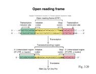
ppt slides
ppt slides
ppt slides

Create successful ePaper yourself
Turn your PDF publications into a flip-book with our unique Google optimized e-Paper software.
Neurons migrate “inside-out” to form cortical layers:<br />
• deep layer cells are born first<br />
• superficial cells are born later
Bohner, Akers and McConnell 1997<br />
Isolate ventricular<br />
zone cells from early<br />
stage brain (E29)<br />
Label with 3H<br />
Thymidine during<br />
dissociation<br />
Let sit in culture for<br />
6 hours (about the<br />
time observed in vivo<br />
for completion of<br />
final mitosis)<br />
Little purple dots are<br />
3H-T label
Once cells removed, they varied the culture conditions<br />
1. Low density 2. Pellet 3. Explant<br />
No contacts Pellet culturedifferent<br />
contacts<br />
Cells in S phase<br />
Mitotic cells<br />
Normal contacts<br />
After 6 hours in culture, dissociate cells, inject cell (transplant)<br />
into P1 (late stage) ventricular zone.<br />
What’s your prediction? The transplanted E29 cells:<br />
a. Should go deep: P1 environment does not matter<br />
b. Should go superficial: P1 environment does not matter<br />
c. Should go deep, but in-culture conditions might affect fate<br />
d. Should go superficial, but in-culture conditions might affect fate
The data:<br />
superficial<br />
deep<br />
What do you conclude?<br />
a. Signaling is required for blockage of deep layer fate<br />
b. Signaling is required for blockage of superficial layer fate<br />
c. Signaling is required to induce superficial layer fate
Possible Mechanism?<br />
Different levels of<br />
β-catenin activation at different stages<br />
Signaling between the cells affects amount of b-catenin activation<br />
Early stage: high b-catenin activation; later stage: no b-catenin activ
MCDB 4650 Class 11<br />
Examples of cell fate commitment:<br />
Eye development and Neural Crest Cells<br />
Next exam is coming up on Monday Feb 25<br />
Review session this Friday at 2:30 PM
What about other regions of the nervous system?
Sensory placodes are formed early<br />
and are initially not committed<br />
9.31<br />
Eye itself is an outpocketing<br />
of the Diencephalon<br />
Vertebrate eye development<br />
Source of Shh<br />
Optic vesicle<br />
(eye induction)
The eye field, and molecules expressed<br />
Shh<br />
(from notochord)
Shh splits the forebrain into two fields
Cell types of<br />
the vertebrate<br />
retina<br />
9.34<br />
RPE
To determine if retinal progenitors were multipotent,<br />
they were labeled with a retrovirus that could be<br />
visualized<br />
All cells shown in the section of retina are likely descended<br />
from a single progenitor – lineage tracing experiment<br />
Is there any potential caveat?<br />
So, progenitor cells are multipotent: ie, a single dividing cell<br />
can give rise to several different kinds of cell types
Retinal cells differentiate in a certain order, with<br />
considerable overlap in what is produced at what time<br />
Retinal ganglion cells<br />
Amacrines<br />
Rods<br />
Bipolar cells<br />
Cook 2003
What about the environment of the retinal precursors (ie, signaling<br />
ligands)? How can we test if it is involved in commitment of fate?<br />
a. Isolate cells in culture from different stages of retinal<br />
development<br />
b. Do heterochronic transplants<br />
c. Combine cell types from different stages of development in<br />
culture<br />
d. Isolate secreted proteins present in retina at different stages of<br />
development and test their effect on cell fate<br />
e. I want to do two of the above experiments
To test whether the fates could be<br />
influenced, Cepko and colleagues (1999):<br />
1. Verified what progenitor cells from<br />
different stages made in isolated culture,<br />
and<br />
2. mixed the earlier stage progenitor cells<br />
(E16) with later stage cells (P0), then<br />
categorized what cell types were made.<br />
Normal sequence: cones and amacrines, then rods
Retinal progenitor cells from two different stages were isolated and<br />
put in culture:<br />
• E16 (early) - labeled<br />
• P0 (late)<br />
• E16:P0 (two stages of cells mixed together with 20 fold excess<br />
P0 )<br />
THEN, just the E16 cells were followed to see what they<br />
differentiated into
(from Belliveau and Cepko, 1999)<br />
amacrines<br />
1:20 ratio<br />
Cones (GREY)<br />
Rods (WHITE)<br />
Cones (GREY)<br />
Rods (WHITE)<br />
A. % labeled cells that differentiated into<br />
amacrine cells after 5 days in culture (5DIV)<br />
B. % labeled cells that differentiated into<br />
cone photoreceptors (grey) or rod<br />
photoreceptors (white) after 5 days in culture<br />
C. Same as B, but after 15 days in culture
Are the differentiated cells in the P0 extracts<br />
influencing the fates of the progenitors?<br />
E16 +P0<br />
cells with<br />
lots<br />
amacrines<br />
E16+P0 E16 + P0<br />
cells with cells<br />
few<br />
amacrines<br />
What can you conclude from this series of experiments?<br />
% heavily labeled cells that<br />
differentiated into<br />
amacrines or photoreceptors<br />
amacrine<br />
photoreceptors
One possible model: there are two different sets of<br />
progenitor cells that are not committed, but “biased”.<br />
The signals from differentiated or even post-mitotic<br />
cells changes the progenitor cells over time<br />
Progenitors<br />
Differentiated cells
Looking at markers for photoreceptors and bipolar cells in the retina:<br />
Photoreceptor-<br />
specific marker<br />
Bipolar-specific<br />
marker<br />
early<br />
late<br />
From this data, the control over which cell type is produced could be<br />
a. Transcriptional<br />
b. Translational<br />
c. Either
The microRNA stops being expressed over time<br />
This micro RNA<br />
specifically inhibits<br />
the bipolarspecific<br />
transcripts<br />
from being<br />
translated<br />
(bright red label)
Take home:<br />
Both intrinsic changes (decreasing levels of a<br />
microRNA) and extrinsic changes (molecules secreted<br />
by already differentiated cells) impact the<br />
commitment of retinal progenitor cells at a given time<br />
point.
Neural crest cells: unique cells of ectodermal origin that<br />
ultimately populate many different locations and tissues
Neural crest cells: unique cells of ectodermal origin that<br />
ultimately populate many different locations and tissues<br />
Determination of<br />
these cells as neural<br />
crest requires BMP<br />
which in turn<br />
activates molecules<br />
required for neural<br />
crest specification<br />
as well as migration
Different regions of<br />
endoderm determine<br />
different kinds of cranial<br />
neural crest derivatives<br />
(signals from endoderm to<br />
crest cells):<br />
transplant pharyngeal<br />
endoderm that normally lies<br />
right under the jaw to a host:<br />
extra jaw is induced from<br />
neural crest cells near the<br />
transplant
On the other hand, there<br />
are also signals from the<br />
neural crest to the<br />
pharyngeal endoderm:<br />
if you transplant duck<br />
neural crest cells into a<br />
quail, the quail makes a<br />
duck’s beak
This led to another interesting set of experiments:<br />
Why don’t birds have teeth? (or conversely, why<br />
do we have teeth rather than a beak?)<br />
Teeth form from neural crest cells and adjacent<br />
epidermis. The epidermis “coats” the tooth or the<br />
beak, while the main part of each structure is<br />
derived from neural crest cells.
teeth form from head neural crest cells and adjacent<br />
epidermis: In mouse, the epidermis “coats” the tooth, while<br />
the main part of the tooth is derived from neural crest cells<br />
birds, who have these tissue types, the combination does not<br />
lead to teeth.<br />
what is the most likely explanation for why birds have lost the<br />
ability to make teeth? Think about how things could have<br />
changed on the molecular level.<br />
mouse neural crest cells in isolation: teeth are not formed<br />
bird neural crest cells in isolation: teeth are not formed<br />
mouse jaw epidermis combined with bird neural crest:<br />
teeth are not formed.<br />
bird jaw epidermis combined with mouse neural crest:<br />
teeth are formed.
The bird epidermis could be producing a signaling<br />
molecule that can induce the neural crest cells to make<br />
teeth, but the neural crest cells in the bird no longer<br />
produce a functional receptor (or transcription factor<br />
downstream) that allows them to respond. This could be<br />
due to a mutation in the coding region or multiple<br />
regulatory regions of the DNA that codes for the receptor<br />
or transcription factor. Thus, when you put the bird<br />
epidermis with mouse neural crest, since the receptor is<br />
still made, teeth can form. The opposite could also be<br />
true: that a signaling molecule released from the neural<br />
crest is no longer present to induce the epidermis.
Summary: Neural crest potency<br />
• Crest cells start out multipotent<br />
• Some restriction exists initially (trunk cannot<br />
make cardiac and cranial fates)<br />
• Potency is restricted over time<br />
• Fate largely determined by molecules in the<br />
environment (primarily growth factors)<br />
• Crest cells also influence the differentiation of<br />
surrounding tissues (reciprocal induction)