Wnt Signaling Polarizes an Early C. elegans ... - MCD Biology
Wnt Signaling Polarizes an Early C. elegans ... - MCD Biology
Wnt Signaling Polarizes an Early C. elegans ... - MCD Biology

You also want an ePaper? Increase the reach of your titles
YUMPU automatically turns print PDFs into web optimized ePapers that Google loves.
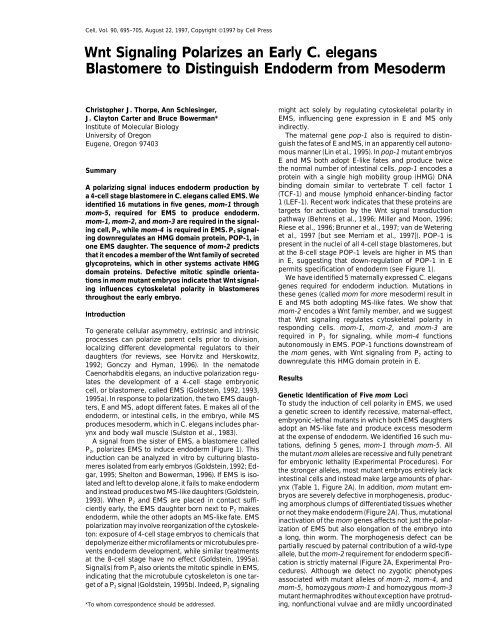
Cell, Vol. 90, 695–705, August 22, 1997, Copyright ©1997 by Cell Press<br />
<strong>Wnt</strong> <strong>Signaling</strong> <strong>Polarizes</strong> <strong>an</strong> <strong>Early</strong> C. eleg<strong>an</strong>s<br />
Blastomere to Distinguish Endoderm from Mesoderm<br />
Christopher J. Thorpe, Ann Schlesinger, might act solely by regulating cytoskeletal polarity in<br />
J. Clayton Carter <strong>an</strong>d Bruce Bowerm<strong>an</strong>*<br />
EMS, influencing gene expression in E <strong>an</strong>d MS only<br />
Institute of Molecular <strong>Biology</strong><br />
indirectly.<br />
University of Oregon<br />
The maternal gene pop-1 also is required to distin-<br />
Eugene, Oregon 97403<br />
guish the fates of E <strong>an</strong>d MS, in <strong>an</strong> apparently cell autonomous<br />
m<strong>an</strong>ner (Lin et al., 1995). In pop-1 mut<strong>an</strong>t embryos<br />
E <strong>an</strong>d MS both adopt E-like fates <strong>an</strong>d produce twice<br />
Summary<br />
the normal number of intestinal cells. pop-1 encodes a<br />
protein with a single high mobility group (HMG) DNA<br />
A polarizing signal induces endoderm production by binding domain similar to vertebrate T cell factor 1<br />
a 4-cell stage blastomere in C. eleg<strong>an</strong>s called EMS. We (TCF-1) <strong>an</strong>d mouse lymphoid enh<strong>an</strong>cer-binding factor<br />
identified 16 mutations in five genes, mom-1 through 1 (LEF-1). Recent work indicates that these proteins are<br />
mom-5, required for EMS to produce endoderm. targets for activation by the <strong>Wnt</strong> signal tr<strong>an</strong>sduction<br />
mom-1, mom-2, <strong>an</strong>d mom-3 are required in the signal- pathway (Behrens et al., 1996; Miller <strong>an</strong>d Moon, 1996;<br />
ing cell, P2, while mom-4 is required in EMS. P2 signal-<br />
ing downregulates <strong>an</strong> HMG domain protein, POP-1, in<br />
one EMS daughter. The sequence of mom-2 predicts<br />
that it encodes a member of the <strong>Wnt</strong> family of secreted<br />
glycoproteins, which in other systems activate HMG<br />
domain proteins. Defective mitotic spindle orientations<br />
in mom mut<strong>an</strong>t embryos indicate that <strong>Wnt</strong> signal-<br />
ing influences cytoskeletal polarity in blastomeres<br />
throughout the early embryo.<br />
Riese et al., 1996; Brunner et al., 1997; v<strong>an</strong> de Wetering<br />
et al., 1997 [but see Merriam et al., 1997]). POP-1 is<br />
present in the nuclei of all 4-cell stage blastomeres, but<br />
at the 8-cell stage POP-1 levels are higher in MS th<strong>an</strong><br />
in E, suggesting that down-regulation of POP-1 in E<br />
permits specification of endoderm (see Figure 1).<br />
We have identified 5 maternally expressed C. eleg<strong>an</strong>s<br />
genes required for endoderm induction. Mutations in<br />
these genes (called mom for more mesoderm) result in<br />
E <strong>an</strong>d MS both adopting MS-like fates. We show that<br />
Introduction<br />
mom-2 encodes a <strong>Wnt</strong> family member, <strong>an</strong>d we suggest<br />
that <strong>Wnt</strong> signaling regulates cytoskeletal polarity in<br />
To generate cellular asymmetry, extrinsic <strong>an</strong>d intrinsic<br />
processes c<strong>an</strong> polarize parent cells prior to division,<br />
localizing different developmental regulators to their<br />
daughters (for reviews, see Horvitz <strong>an</strong>d Herskowitz,<br />
1992; Gonczy <strong>an</strong>d Hym<strong>an</strong>, 1996). In the nematode<br />
responding cells. mom-1, mom-2, <strong>an</strong>d mom-3 are<br />
required in P2 for signaling, while mom-4 functions<br />
autonomously in EMS. POP-1 functions downstream of<br />
the mom genes, with <strong>Wnt</strong> signaling from P2 acting to<br />
downregulate this HMG domain protein in E.<br />
Caenorhabditis eleg<strong>an</strong>s, <strong>an</strong> inductive polarization regulates<br />
the development of a 4-cell stage embryonic<br />
Results<br />
cell, or blastomere, called EMS (Goldstein, 1992, 1993,<br />
1995a). In response to polarization, the two EMS daughters,<br />
E <strong>an</strong>d MS, adopt different fates. E makes all of the<br />
endoderm, or intestinal cells, in the embryo, while MS<br />
produces mesoderm, which in C. eleg<strong>an</strong>s includes phar-<br />
ynx <strong>an</strong>d body wall muscle (Sulston et al., 1983).<br />
A signal from the sister of EMS, a blastomere called<br />
P2, polarizes EMS to induce endoderm (Figure 1). This<br />
induction c<strong>an</strong> be <strong>an</strong>alyzed in vitro by culturing blasto-<br />
meres isolated from early embryos (Goldstein, 1992; Ed-<br />
gar, 1995; Shelton <strong>an</strong>d Bowerm<strong>an</strong>, 1996). If EMS is isolated<br />
<strong>an</strong>d left to develop alone, it fails to make endoderm<br />
<strong>an</strong>d instead producestwo MS-like daughters (Goldstein,<br />
1993). When P2 <strong>an</strong>d EMS are placed in contact suffi-<br />
ciently early, the EMS daughter born next to P2 makes<br />
endoderm, while the other adopts <strong>an</strong> MS-like fate. EMS<br />
polarization may involve reorg<strong>an</strong>ization of the cytoskele-<br />
ton: exposure of 4-cell stage embryos to chemicals that<br />
depolymerize either microfilaments or microtubules pre-<br />
vents endoderm development, while similar treatments<br />
at the 8-cell stage have no effect (Goldstein, 1995a).<br />
Signal(s) from P2 also orients the mitotic spindle in EMS,<br />
indicating that the microtubule cytoskeleton is one tar-<br />
get of a P2 signal (Goldstein, 1995b). Indeed, P2 signaling<br />
Genetic Identification of Five mom Loci<br />
To study the induction of cell polarity in EMS, we used<br />
a genetic screen to identify recessive, maternal-effect,<br />
embryonic-lethal mut<strong>an</strong>ts in which both EMS daughters<br />
adopt <strong>an</strong> MS-like fate <strong>an</strong>d produce excess mesoderm<br />
at the expense of endoderm. We identified 16 such mutations,<br />
defining 5 genes, mom-1 through mom-5. All<br />
the mut<strong>an</strong>t mom alleles are recessive <strong>an</strong>d fully penetr<strong>an</strong>t<br />
for embryonic lethality (Experimental Procedures). For<br />
the stronger alleles, most mut<strong>an</strong>t embryos entirely lack<br />
intestinal cells <strong>an</strong>d instead make large amounts of phar-<br />
ynx (Table 1, Figure 2A). In addition, mom mut<strong>an</strong>t em-<br />
bryos are severely defective in morphogenesis, producing<br />
amorphous clumps of differentiated tissues whether<br />
or not they make endoderm (Figure 2A). Thus, mutational<br />
inactivation of the mom genes affects not just the polarization<br />
of EMS but also elongation of the embryo into<br />
a long, thin worm. The morphogenesis defect c<strong>an</strong> be<br />
partially rescued by paternal contribution of a wild-type<br />
allele, but the mom-2 requirement for endoderm specifi-<br />
cation is strictly maternal (Figure 2A, Experimental Procedures).<br />
Although we detect no zygotic phenotypes<br />
associated with mut<strong>an</strong>t alleles of mom-2, mom-4, <strong>an</strong>d<br />
mom-5, homozygous mom-1 <strong>an</strong>d homozygous mom-3<br />
mut<strong>an</strong>t hermaphrodites withoutexception have protrud-<br />
*To whom correspondence should be addressed.<br />
ing, nonfunctional vulvae <strong>an</strong>d are mildly uncoordinated
Cell<br />
696<br />
E <strong>an</strong>d MS Both Adopt MS-like Fates in mom<br />
Mut<strong>an</strong>t Embryos<br />
Terminally differentiated mom mut<strong>an</strong>t embryos that lack<br />
intestinal cells also appear to make excess pharynx (Figure<br />
2A), suggesting that E might produce <strong>an</strong> MS-like<br />
pattern of cell fate. We tested this possibility in three<br />
ways. We used tissue-specific <strong>an</strong>tibodies to determine<br />
if E in mom-2 mut<strong>an</strong>t embryos produces mesodermal<br />
cell types normally made by MS, we tested the genetic<br />
requirements for ectopic pharyngeal cell production by<br />
E, <strong>an</strong>d we <strong>an</strong>alyzed the cell lineage of some E descend<strong>an</strong>ts<br />
to determine if they adopt cell cycle times <strong>an</strong>d<br />
cleavage patterns similar to those made by a wild-type<br />
MS blastomere. As described below, our results indicate<br />
that E adopts <strong>an</strong> MS-like fate in mom-2 mut<strong>an</strong>t embryos.<br />
To examine the fate of E, we first used a laser microbeam<br />
focused through a microscope to kill every cell<br />
in early mom-2 mut<strong>an</strong>t embryos except for E. These<br />
partial embryos produced differentiated descend<strong>an</strong>ts<br />
that were fixed <strong>an</strong>d stained with cell type–specific monoclonal<br />
<strong>an</strong>tibodies (Table 2, Figure 2B). Similar laser ablation<br />
data were obtained for mut<strong>an</strong>t embryos lacking<br />
mom-1, mom-3 <strong>an</strong>d mom-4 function (A. S., C. J. T.,<br />
Figure 1. EMS Development Requires Both Autonomously Acting M. Meneghini, <strong>an</strong>d B. B., unpublished data). In control<br />
POP-1 <strong>an</strong>d a Signal from P2 experiments using wild-type embryos, E always pro-<br />
POP-1 protein is found in all nuclei at the 4-cell stage (dark shading). duced intestinal cells but not two cell types made by<br />
A signal from P2 to EMS early in the 4-cell stage (curved arrow)<br />
MS: pharyngeal muscle <strong>an</strong>d body wall muscle. For two<br />
polarizes EMS such that the potential to produce endoderm is acstrong<br />
mom2 alleles, E in most operated embryos<br />
quired by only the E daughter of EMS. E contains relatively low levels<br />
of POP-1<strong>an</strong>d makes only intestinal cells, whileMS has relativelyhigh failed to produce <strong>an</strong>y intestinal cells. Instead, when E<br />
levels of POP-1 <strong>an</strong>d produces mesoderm. Sister cells are indicated failed to make gut, it produced four cell types normally<br />
by short connecting lines. made by MS: pharyngeal <strong>an</strong>d body wall muscle cells,<br />
pharyngeal gl<strong>an</strong>d cells, <strong>an</strong>d pharyngeal marginal cells<br />
(Table 2). MS in mom mut<strong>an</strong>t embryos also produces<br />
(C. J. T., A. S., <strong>an</strong>d B. B., unpublished data). Here we pharynx <strong>an</strong>d body wall muscle (Table 2). As no other<br />
focus on the maternal-effect phenotypes caused by mu- early blastomere besides MS in wild-type embryos pro-<br />
tations in the mom genes. We note that because some duces both pharynx <strong>an</strong>d body wall muscle (Sulston et al.,<br />
mom genes have zygotic functions, the maternal-effect 1983), E <strong>an</strong>d MS in mom mut<strong>an</strong>t embryos both appear to<br />
alleles we have isolated may not be null. Indeed, the adopt MS-like fates.<br />
different penetr<strong>an</strong>ce of the gut phenotypes associated<br />
If E in mom-2 embryos develops like MS, then it should<br />
with our different mom alleles suggests that most alleles<br />
require skn-1 but not glp-1 to produce pharyngeal mus-<br />
are not null (Table 1). However, double mut<strong>an</strong>t embryos<br />
cle cells (Priess et al., 1987; Bowerm<strong>an</strong> et al., 1992). MS<br />
from mothers homozygous for both mom-2 <strong>an</strong>d mom-4<br />
makes about half the pharyngeal cells produced during<br />
have a completely penetr<strong>an</strong>t gut defect (Table 1). Thus,<br />
embryogenesis, with the remainder made by two gr<strong>an</strong>d-<br />
daughters of the 4-cell stage blastomere ABa (Sulston<br />
the mom gene products, whether they function in a<br />
et al., 1983). The production of pharyngeal cells by ABa<br />
single pathway or in parallel, are essential for the specifidescend<strong>an</strong>ts<br />
requires <strong>an</strong> inductive signal from MS at<br />
cation of endoderm in early C. eleg<strong>an</strong>s embryos.<br />
about the 12-cell stage (Priess et al., 1987; Hutter <strong>an</strong>d<br />
In addition to their lack of endoderm, mom-1, mom-2,<br />
Schnabel, 1994; M<strong>an</strong>go et al., 1994a); glp-1 encodes<br />
mom-3, <strong>an</strong>d mom-5 mut<strong>an</strong>t embryos also are defective<br />
a putative receptor required for ABa descend<strong>an</strong>ts to<br />
in orienting the mitotic spindle of <strong>an</strong> 8-cell stage blastoreceive<br />
the MS signal (Priess et al., 1987; Austin <strong>an</strong>d<br />
mere called ABar (Table 1, Figure 3). In wild-type em-<br />
Kimble, 1989; Yochem <strong>an</strong>d Greenwald, 1989). Mutations<br />
bryos, ABar divides along a largely left–right (l/r) axis,<br />
in glp-1 result in <strong>an</strong> absence of ABa-derived pharyngeal<br />
roughly orthogonal to the three other AB descend<strong>an</strong>ts<br />
cells but do not affect the production of pharyngeal cells<br />
that all divide along the <strong>an</strong>terior–posterior (a/p) axis with<br />
by MS (Priess et al., 1987). Mutations in skn-1 result in<br />
a pronounced dorsal–ventral (d/v) tilt (Figure 3D). In all a loss of all pharyngeal cells, as skn-1 is required both<br />
but mom-4 mut<strong>an</strong>ts, ABar divides roughly parallel to the to specify MS fate <strong>an</strong>d to activate the MS signal that<br />
other AB descend<strong>an</strong>ts (Table 1, Figure 3H). Because induces ABa descend<strong>an</strong>ts to make pharyngeal cells<br />
the paternal rescue of morphogenesis described above (Bowerm<strong>an</strong> et al., 1992; Mello et al., 1992; Shelton <strong>an</strong>d<br />
does not correlate with rescue of the ABar spindle orien- Bowerm<strong>an</strong>, 1996). By staining fixed,terminally differentitation<br />
(Experimental Procedures), the abnormal axis of ated embryos from double mut<strong>an</strong>t mothers with cellthe<br />
mitotic spindle in ABar c<strong>an</strong>not fully account for the type specific <strong>an</strong>tibodies (Experimental Procedures), we<br />
highly penetr<strong>an</strong>t morphogenesis defect observed in found that skn-1; mom-2 embryos produce few or no<br />
mom mut<strong>an</strong>t embryos.<br />
pharyngeal cells: 17 of 46 double mut<strong>an</strong>t embryos made
Cell Polarity <strong>an</strong>d Endoderm Specification<br />
697<br />
Table 1. Penetr<strong>an</strong>ce of Endoderm Defect <strong>an</strong>d ABar Cleavage Abnormality in mom Mut<strong>an</strong>t Embryos<br />
Fraction of Embryos<br />
% Embryos with Aberr<strong>an</strong>t ABar<br />
Gene Allele Lacking Gut (n) a Cleavage b<br />
mom-1 or10 85 (215) 10/10<br />
or46 52 (160) 7/7<br />
or65 50 (275)<br />
or83 46 (153)<br />
or70 39 (321) 13/13<br />
mom-2 or85 88 (485)<br />
or9 77 (226)<br />
or48 74 (380)<br />
or42 72 (194) 28/31<br />
or33 50 (210)<br />
or77 8 (65) 15/15<br />
mom-3 or78 65 (361) 30/30<br />
mom-4 or39 40 (100) 0/12<br />
or49 24 (181) 0/12<br />
or11 1 (228) 2/17<br />
mom-5 or57 5 (167) 9/9<br />
skn-1; mom-2 zu67; or42 100 (241) —<br />
mom-4; mom-2 or39; or42 100 (124) —<br />
a To assay production of intestinal cells, embryos were collected from mom mut<strong>an</strong>t hermaphrodites <strong>an</strong>d allowed to develop at least 10 hr at<br />
20C. The presence of intestinal cells was scored using polarizing light microscopy to detect intestine-specific birefringent gut gr<strong>an</strong>ules.<br />
b The orientation of the mitotic spindle of the ABar blastomere was examined in lateral views of 8-cell stage embryos using Nomarski optics.<br />
In wild-type embryos, the <strong>an</strong>terior daughter of ABar contacts the MS blastomere (Figure 3D). ABar cleavage was scored as defective if its<br />
posterior daughter contacted MS (Figure 3H).<br />
between 1 <strong>an</strong>d 5 pharyngeal muscle cells while the re- genetic interval, we obtained rescue of the mut<strong>an</strong>t phemainder<br />
made none, compared to the 39 made in wild notype by germline tr<strong>an</strong>sformation with the cosmids<br />
type (Sulston et al., 1983). In contrast, both E <strong>an</strong>d MS F52E1 <strong>an</strong>d ZK427. Sequence data from the C. eleg<strong>an</strong>s<br />
from glp-1; mom-2 double mut<strong>an</strong>t embryos produce Genome Center predicts a gene, F38E1.7, on ZK427 that<br />
pharynx: 9 of 10 embryos in which E was isolated, <strong>an</strong>d would encode a member of the <strong>Wnt</strong> family of secreted<br />
6 of 6 in which MS was isolated, made large numbers signaling molecules (Experimental Procedures). To deof<br />
pharyngeal muscle cells. Because E <strong>an</strong>d MS in mom-2 termine if F38E1.7 is the mom-2 locus, we microinjected<br />
mut<strong>an</strong>t embryos require skn-1 but not glp-1 function to <strong>an</strong>tisense RNA from one exon of the predicted gene into<br />
produce pharyngeal cells, we conclude that both EMS<br />
daughters adopt <strong>an</strong> MS-like fate.<br />
To define their fates more precisely, we compared<br />
the cell lineages E <strong>an</strong>d MS produce in mom-2 mut<strong>an</strong>t<br />
embryos to the wild-type MS lineage (Figure 2C). C.<br />
eleg<strong>an</strong>s embryos produce a nearly invari<strong>an</strong>t cell lineage,<br />
as determined by scoring the relative positions of<br />
daughter cells following each division <strong>an</strong>d observing<br />
their eventual fates (Sulston et al., 1983). In wild-type<br />
embryos, E <strong>an</strong>d MS become different shortly after their<br />
birth: Ea <strong>an</strong>d Ep are the first cells to migrate inside the<br />
embryo during gastrulation, <strong>an</strong>d they divide about 15<br />
min later th<strong>an</strong> do MSa <strong>an</strong>d MSp. In mom-2 embryos, Ea<br />
<strong>an</strong>d Ep fail to gastrulate, <strong>an</strong>d they divide nearly synchronously<br />
with MSa <strong>an</strong>d MSp. Subsequent divisions of the<br />
E descend<strong>an</strong>ts also occur with approximately MS-like<br />
timing, <strong>an</strong>d several cell deaths that occur in a wild-type<br />
the syncitial gonad of wild-type <strong>an</strong>imals. All injected<br />
<strong>an</strong>imals produced dead embryos with a Mom mut<strong>an</strong>t<br />
phenotype (Experimental Procedures). To confirm fur-<br />
ther the gene identity, we sequenced six mut<strong>an</strong>t alleles<br />
of mom-2. Each allele contains a single lesion within<br />
the coding sequence, except for one mutation at a splice<br />
junction (Figure 4). We conclude that mom-2 is F38E1.7<br />
<strong>an</strong>d that it encodes a member of the <strong>Wnt</strong> family of secreted<br />
signaling glycoproteins. The predicted MOM-2/<strong>Wnt</strong> pro-<br />
tein is 363 amino acids in length <strong>an</strong>d includes 24 highly<br />
conserved cysteine residues. BLAST searches indicated<br />
that the predicted MOM-2 protein is most closely related<br />
to mammali<strong>an</strong> <strong>Wnt</strong>-2, sharing approximately 40% amino<br />
acid identity with mouse <strong>an</strong>d hum<strong>an</strong> <strong>Wnt</strong>-2. We con-<br />
clude that the polarization of gut potential in EMS requires<br />
the <strong>Wnt</strong> signal tr<strong>an</strong>sduction pathway.<br />
MS lineage also occur at the corresponding points in<br />
the lineage of E in some mom-2 mut<strong>an</strong>t embryos. How- mom-1, mom-2, <strong>an</strong>d mom-3 Are Required<br />
ever, we often do not observe characteristic cell deaths in P2 for Endoderm Induction, while<br />
in both the E <strong>an</strong>d MS lineages (Figure 2C). We conclude mom-4 Is Required in EMS<br />
that E <strong>an</strong>d MS in mom-2 mut<strong>an</strong>t embryos both adopt To determine which mom genes are required in P2 for<br />
fates similar but not identical to a wild-type MS.<br />
signaling <strong>an</strong>d which are required in EMS for responding,<br />
we assembled genetically mosaic partial embryos in<br />
mom-2 Encodes a <strong>Wnt</strong> Family Member<br />
vitro (Figure 5 <strong>an</strong>d Experimental Procedures). Using<br />
We used positional cloning to identify the wild-type blastomeres isolated from wild-type <strong>an</strong>d mom mut<strong>an</strong>t<br />
mom-2 gene (Figure 4). After mapping mom-2 to a small embryos, we found that mom-1, mom-2, <strong>an</strong>d mom-3
Cell<br />
698<br />
Figure 2. E Adopts <strong>an</strong> MS-like Identity in mom-2 Embryos<br />
(A) Immunofluorescence <strong>an</strong>d light micrographs of wild-type (left column), mom-2 (middle column), <strong>an</strong>d mom-2/ embryos (right column) from<br />
mom-2/mom-2 mothers. Embryos were allowed to develop 15 hr (a, b, e, <strong>an</strong>d f), or 8 hr (c <strong>an</strong>d d) at 20C. (a, b, <strong>an</strong>d c) Living embryos viewed<br />
with Nomarski optics. Pharyngeal tissue is surrounded by a prominent basement membr<strong>an</strong>e (arrowheads), <strong>an</strong>d contains a secreted cuticle<br />
(wide arrows). Visible in wild-type embryos, but not in mom-2 or mom-2/ embryos, are intestinal cells with characteristically large nuclei<br />
containing a single large nucleolus (thin arrows in [a]). While homozygous mom-2 embryos fail to undergo <strong>an</strong>y morphogenesis <strong>an</strong>d invariably<br />
arrest as unelongated clumps of differentiated tissue (b), mom-2/ embryos, even those lacking intestine, often elongate into short, stubby<br />
worms (c <strong>an</strong>d i). Paternal contribution of a wild-type copy of mom-2 does not rescue the intestine defect (Experimental Procedures). (d, e,<br />
<strong>an</strong>d f) Intact embryos stained with J126, a monoclonal <strong>an</strong>tibody (MAb) that recognizes intestinal cells. (g, h, <strong>an</strong>d i) Intact embryos stained<br />
with 9.2.1, a MAb that recognizes pharyngeal muscle cells.<br />
(B) Immunofluorescence micrographs of operated mom-2 embryos in which all blastomeres in the embryos except E were killed with a laser<br />
microbeam. Embryos were allowed to develop 8 hours (a <strong>an</strong>d c), or 16 hours (b) at 20C before fixing <strong>an</strong>d staining. MAb J126 (a) recognizes<br />
intestinal cells, MAb 9.2.1 (b) recognizes pharyngeal muscle cells, <strong>an</strong>d MAb 5.6 (c) recognizes body-wall muscle.<br />
(C) Cell deaths scored in E <strong>an</strong>d MS lineages from mom-2 mut<strong>an</strong>t embryos (below) compared to the wild-type MS lineage (above). Vertical<br />
bars represent time; horizontal bars represent cell division. Programmed cell deaths are indicated by Xs. The fraction of embryos in which<br />
the corresponding E <strong>an</strong>d MS lineages in mom-2 mut<strong>an</strong>t embryos produced cell deaths are indicated below each X.
Cell Polarity <strong>an</strong>d Endoderm Specification<br />
699<br />
Figure 3. Mitotic Spindle Axes Are Misoriented<br />
in mom-2 <strong>an</strong>d mom-2; mom-1 Mut<strong>an</strong>t<br />
Embryos<br />
All embryos are shown in lateral views with<br />
<strong>an</strong>terior to the left <strong>an</strong>d ventral up; maternal<br />
genotypes are indicated at the top of each<br />
column. (A, E, <strong>an</strong>d I ) Wild-type, mom-2, <strong>an</strong>d<br />
mom-2; mom-1 embryos at the 4-cell stage.<br />
(B, F, <strong>an</strong>d J) EMS cleavage. In wild-type <strong>an</strong>d<br />
mom-2 embryos, the EMS spindle aligns on<br />
the a/p axis, while in some mom-2; mom-1<br />
embryos, it is tilted along the d/v axis, as<br />
shown here. (C, G, <strong>an</strong>d K) 8-cell stage. E <strong>an</strong>d<br />
MS are positioned normally in the mom-2;<br />
mom-1 embryo following cytokinesis. (D, H,<br />
<strong>an</strong>d L) 12-cell stage. In wild-type embryos,<br />
ABar divides along a mostly l/r axis, tr<strong>an</strong>sverse<br />
to that of the other AB descend<strong>an</strong>ts; the<br />
division axes of ABal <strong>an</strong>d ABpr are marked for<br />
comparison. The <strong>an</strong>terior daughter of ABar,<br />
ABara, touches MS. In mom-2 mut<strong>an</strong>t embryos,<br />
ABar divides parallel to the other AB<br />
descend<strong>an</strong>ts <strong>an</strong>d ABarp instead of ABara<br />
contacts MS. In a small fraction of mom-2;<br />
mom-1 embryos (2 of 21 embryos scored, <strong>an</strong><br />
example of which is shown here), we observe<br />
dramatic rearr<strong>an</strong>gements of blastomeres, beginning<br />
at the 8-cell stage. In both cases, posterior<br />
blastomeres moved <strong>an</strong>teriorly, such<br />
that E <strong>an</strong>d MS became the <strong>an</strong>teriormost cells<br />
in the embryo. Other mom-2; mom-1 embryos<br />
showed the same ABar cleavage axis defect observed in mom-2 mut<strong>an</strong>t embryos. Surprisingly, double mut<strong>an</strong>t embryos made intestinal<br />
cells more often th<strong>an</strong> either single mut<strong>an</strong>t: 52% of mom-2; mom-1 embryos (n197) compared to 15% for or10 <strong>an</strong>d 28% for or42 embryos<br />
(Table 1). Laser ablation experiments with double mut<strong>an</strong>t embryos indicate that E but not MS produces endoderm: Of 14, 7 isolated E’s made<br />
intestinal cells, compared to 0 of 15 isolated MS blastomeres.<br />
are required in P2, while mom-4 is required in EMS. mom-4 <strong>an</strong>d the Interpretation of Cell<br />
Because of the low penetr<strong>an</strong>ce of the endoderm pheno- Polarity in EMS<br />
type in mom-5 mut<strong>an</strong>t embryos (Table 1), we have not Our blastomere mosaic <strong>an</strong>alysis indicates that mom-4<br />
been able to determine which blastomere requires is required in EMS to respond to the P2 signal (Table 3).<br />
mom-5 function. The development of P2 appears normal To determine how mom-4 functions with respect to the<br />
in mom-1, mom-2, <strong>an</strong>d mom-3 mut<strong>an</strong>t embryos (Experi- establishment or interpretation of cell polarity, we exammental<br />
Procedures), suggesting that the wild-type ined mom-4 function in pie-1 mut<strong>an</strong>t embryos. Muta-<br />
genes function specifically in P2 signaling. Finally, our tions in pie-1 cause P2 to develop like EMS, producing<br />
identification of mom-2 as a <strong>Wnt</strong> gene is consistent with excess pharynx <strong>an</strong>d intestine (Mello et al., 1992). Unlike<br />
its function being required in P2 for signaling.<br />
EMS, P2 in pie-1 embryos does not require a signal from<br />
Table 2. Cell Types Produced by E <strong>an</strong>d MS in mom-2 Mut<strong>an</strong>t Embryos<br />
Body Pharyngeal Pharyngeal<br />
Blastomere Wall Pharyngeal Gl<strong>an</strong>d Marginal<br />
Genotype Isolated Intestinal Cells Muscle Muscle Cells Cells<br />
Wild type E 13/13 0/7 0/10 — —<br />
mom-2(or42) E 9/170 17/17 15/15 10/10 16/17<br />
mom-2(or9) E 2/16 — 8/10 — —<br />
Wild type MS 0/8 11/11 12/12 8/8 9/9<br />
mom-2(or42) MS 0/64 6/6 8/10 13/14 12/13<br />
mom-2(or9) MS 0/9 — 6/6 — —<br />
All blastomeres in the embryo except MS or E were killed using a laser microbeam. ABa, ABp, <strong>an</strong>d P2 were killed at the 4-cell stage, then<br />
either MS or E was killed following division of EMS. The operated embryos were allowed to develop overnight <strong>an</strong>d then scored for the presence<br />
of intestinal cells using polarizing optics. Operated embryos not making gut were fixed <strong>an</strong>d stained with MAbs to detect the tissues indicated<br />
(see Experimental Procedures). When E was isolated by laser ablation at the 8-cell stage, 9 of 22 operated embryos made intestinal cells.<br />
The nine that made intestine were double stained with J126 <strong>an</strong>d 3NB12; one made both pharyngeal <strong>an</strong>d intestinal cells while the remainder<br />
made only intestine.
Cell<br />
700<br />
Table 3. P2 <strong>an</strong>d EMS Blastomere Mosaics<br />
may be sufficient to specify E fate in P3, suggesting that<br />
Blastomeres Recombined<br />
Fraction of Recombin<strong>an</strong>t<br />
the intrinsic polarity of P2 <strong>an</strong>d the induced polarity of<br />
EMS share common properties.<br />
P2 EMS Embryos Making Gut To position mom-4 relative to the establishment of<br />
WT WT 46/47<br />
cell polarity during endoderm induction, we constructed<br />
WT<br />
mom-1<br />
WT<br />
mom-2<br />
mom-1<br />
WT<br />
mom-2<br />
WT<br />
7/7<br />
2/7<br />
10/12<br />
1/8<br />
a mom-4; pie-1 strain <strong>an</strong>d asked if mom-4 is required<br />
for P2 to produce endoderm in pie-1 mut<strong>an</strong>t embryos<br />
(Table 4). By two criteria, P2 in mom-4; pie-1 double<br />
mut<strong>an</strong>t embryos remains polarized. P2 divides asymmet-<br />
rically (24 of 24 embryos observed by light microscopy),<br />
WT<br />
mom-3<br />
mom-3<br />
WT<br />
11/11<br />
4/12<br />
<strong>an</strong>d P gr<strong>an</strong>ules are segregated to P3 (11 of 11 embryos;<br />
see Experimental Procedures). While intact pie-1 em-<br />
WT mom-4 2/10 bryos make extra intestinal cells, mom-4; pie-1 double<br />
mom-4 WT 7/7<br />
mut<strong>an</strong>t embryos usually make no endoderm (C. J. T.<br />
P2 <strong>an</strong>d EMS blastomeres were isolated from wild-type <strong>an</strong>d mom <strong>an</strong>d B. B., unpublisheddata). Using laser ablation experimut<strong>an</strong>t<br />
embryos <strong>an</strong>d recombined to form genetically mosaic partial ments to isolate the daughters of P2 <strong>an</strong>d EMS in double<br />
embryos. The genotypes of the recombined blastomeres are shown<br />
in the left column.The mut<strong>an</strong>t alleles used were: mom-1(or10), mom-<br />
2(or42), mom-3(or78), <strong>an</strong>d mom-4(or39). The fraction of partial em-<br />
bryos making intestinal cells in each experiment is shown in the<br />
right column.<br />
mut<strong>an</strong>t embryos, we found that both E <strong>an</strong>d P3 usually<br />
failed to produce intestinal cells (Table 4). Because<br />
mom-4 is required for P3 to make intestinal cells in pie-1<br />
mut<strong>an</strong>t embryos, even though P2 remains polarized,<br />
mom-4 may be required to interpret, not to establish,<br />
polarity in a wild-type EMS blastomere.<br />
<strong>an</strong>y neighboring blastomere to produce intestinal cells;<br />
in isolation it still divides into one E-like <strong>an</strong>d one MSlike<br />
daughter (Goldstein, 1995a). The ability of a pie-1<br />
mut<strong>an</strong>t P2 to produce endoderm autonomously raises<br />
the possibility that <strong>an</strong> intrinsic polarity within P2 is sufficient<br />
to specify endoderm.<br />
Several lines of evidence indicate that P2 possesses<br />
<strong>an</strong> intrinsic polarity that might specify endoderm in pie-1<br />
mut<strong>an</strong>t embryos. First, P2 isolated from wild-type or<br />
pie-1 embryos always divides asymmetrically, producing<br />
one larger <strong>an</strong>d one smaller daughter, called C <strong>an</strong>d P3 An alternative expl<strong>an</strong>ation for the lack of gut in pie-1;<br />
mom-4 double mut<strong>an</strong>t embryos is that autocrine signal-<br />
ing might polarize P2 in a pie-1 mut<strong>an</strong>t. If so, mom-4<br />
might play a role in tr<strong>an</strong>sducing <strong>an</strong> autocrine signal. To<br />
determine if P2 uses autocrine, mom-dependent signal-<br />
ing to specify endoderm in pie-1 mut<strong>an</strong>t embryos, we<br />
constructed double mut<strong>an</strong>ts of pie-1 with mom-1,<br />
mom-2, <strong>an</strong>d mom-3. In all cases, EMS but not P2 fails<br />
to produce endoderm (Table 4). We conclude that the<br />
production of endoderm by P2 in pie-1 mut<strong>an</strong>t embryos<br />
is largely independent of <strong>Wnt</strong> signaling, <strong>an</strong>d that mom-4<br />
may be unique among the mom genes in operating<br />
in wild type. Moreover, the smaller P3 daughter inherits downstream of, or parallel to, the establishment of cell<br />
cytoplasmic particles called P gr<strong>an</strong>ules, which are seg- polarity during endoderm induction. However, because<br />
regated to P3 as P2 divides (Strome <strong>an</strong>d Wood, 1983;<br />
Hird et al., 1996). Finally, although it is not known to<br />
both P2 <strong>an</strong>d EMS fail to make gut in 5% of intact pie-1;<br />
mom-2 mut<strong>an</strong>t embryos (10 of 191) <strong>an</strong>d in 1% of pie-1;<br />
have <strong>an</strong>y function in P2, the HMG domain protein POP-1 mom-3 embryos (4 of 471), <strong>Wnt</strong> signaling might have a<br />
accumulates asymmetrically in the daughters of P2, with<br />
C having higher levels th<strong>an</strong> P3 (Lin et al., 1995; Figure<br />
minor role in polarizing P2.<br />
1). In pie-1 mut<strong>an</strong>t embryos, C nearly always becomes<br />
MS-like, while P3 always adopts <strong>an</strong> E-like fate, consistent<br />
<strong>Wnt</strong>-Dependent P2 <strong>Signaling</strong> Restricts<br />
POP-1 Function to MS<br />
with lower levels of POP-1 permitting the specification As a final approach to positioning the mom genes in a<br />
of endoderm in both E <strong>an</strong>d P3 in pie-1 mut<strong>an</strong>t embryos. pathway of endoderm specification, we examined inter-<br />
To summarize, in pie-1 embryos P2’s intrinsic polarity actions of the mom genes with pop-1. Whereas the two<br />
Figure 4. Molecular Cloning of mom-2<br />
(A) mom-2 maps near pos-1, on LGV. mom-2<br />
mut<strong>an</strong>ts are rescued by a mix of cosmids<br />
ZK427 <strong>an</strong>d F52E1, but not F52E1 alone. Injection<br />
of <strong>an</strong>tisense RNA to exon 5 of F38E1.7,<br />
a predicted gene on the cosmid F38E1, which<br />
overlaps extensively with ZK427, into the gonads<br />
of wild-type <strong>an</strong>imals results in a phenotype<br />
indistinguishable from that of mom-2<br />
mut<strong>an</strong>t embryos. All six alleles of mom-2 have<br />
lesions in this gene, as depicted above <strong>an</strong>d<br />
below the exon/intron diagram.
Cell Polarity <strong>an</strong>d Endoderm Specification<br />
701<br />
permits the production of endoderm (Lin et al., 1995).<br />
Because inactivation of the P2 signal in mom mut<strong>an</strong>ts<br />
causes E to become like MS, P 2 signaling might be required<br />
to downregulate POP-1 in E. Consistent with this<br />
model, pop-1; mom-2 <strong>an</strong>d pop-1 mom-4 double mut<strong>an</strong>t<br />
embryos phenotypically resemble pop-1 single mut<strong>an</strong>ts<br />
(Figure 6A). We also found that most mom-2 mut<strong>an</strong>t<br />
embryos show equal levels of POP-1 in MS <strong>an</strong>d E (Figure<br />
6B). Therefore, one function of mom-2/<strong>Wnt</strong> signaling is<br />
to reduce the nuclear levels of POP-1 in the posterior<br />
daughter of EMS. We conclude that pop-1 may function<br />
downstream of all mom genes <strong>an</strong>d that down-regulation<br />
of POP-1 in E results from polarization of EMS by a <strong>Wnt</strong><br />
signal from P2.<br />
mom-2; mom-1 Double Mut<strong>an</strong>t Embryos Exhibit<br />
Widespread Defects in Mitotic Spindle Orientation<br />
<strong>an</strong>d Blastomere Positioning<br />
The misorientation of the ABar mitotic spindle in mom<br />
mut<strong>an</strong>t embryos (Table 1) <strong>an</strong>d the observation that a<br />
signal from P2 orients the mitotic spindle in EMS (Goldstein,<br />
1995b) prompted us to consider if the mom genes<br />
Figure 5. mom-2 Is Required in P2 <strong>an</strong>d Not in EMS for the Induction might be required for proper orientation of mitotic spinof<br />
Endoderm in C. eleg<strong>an</strong>s dles in other blastomeres. Normally, the EMS spindle<br />
P2 <strong>an</strong>d EMS blastomeres were isolated from wild-type <strong>an</strong>d mom-2 sets up along the l/r axis <strong>an</strong>d then rotates to lie along<br />
embryos, then recombined in culture medium to form genetically the a/p axis, pointing toward P2 (Hym<strong>an</strong> <strong>an</strong>d White,<br />
mosaic partial embryos (see Experimental Procedures). Partial em- 1987). We examined the orientation of the EMS spindle<br />
bryos were allowed to develop for 20 hr <strong>an</strong>d intestinal cell production<br />
in mom mut<strong>an</strong>t embryos <strong>an</strong>d observed subtle misalignassayed<br />
using polarizing optics. (A <strong>an</strong>d D) Recombined blastomeres<br />
viewed using Nomarski optics. The partial embryo in (A) is composed ment of the mitotic spindle in some mom-1, mom-3,<br />
<strong>an</strong>d mom-5 mut<strong>an</strong>t embryos (A. S., C. J. T., <strong>an</strong>d B. B.,<br />
of a wild-type P2 blastomere (smaller cell) <strong>an</strong>d a mom-2 EMS. The<br />
partial embryo in (D) was made by recombining a mom-2 P2 with a unpublished data).<br />
wild-type EMS. (B <strong>an</strong>d E) EMS division in the same partial embryos. To test more stringently if the mom genes are required<br />
P2 has been previously shown to orient the EMS mitotic spindle generally for mitotic spindle orientations in the early<br />
such that it “points” at P2. In both partial embryos shown here, the<br />
EMS spindle axis is oriented correctly. (C <strong>an</strong>d F) The differentiated<br />
descend<strong>an</strong>ts of these partial embryos viewed using polarized light<br />
optics to detect gut gr<strong>an</strong>ules, present only in (C).<br />
embryo, we constructed a mom-2(or42); mom-1(or10)<br />
double mut<strong>an</strong>t strain, combining two of our strongest<br />
mom alleles (Experimental Procedures, Table 1). We<br />
observed partially penetr<strong>an</strong>t but widespread defects in<br />
mitotic spindle orientation <strong>an</strong>d blastomere positioning<br />
daughters of EMS adopt MS-like fates in mom mut<strong>an</strong>t in mom-2; mom-1 double mut<strong>an</strong>ts (Figure 3). For exam-<br />
embryos, mutational inactivation of pop-1 results in both ple, in m<strong>an</strong>y mom-2; mom-1 embryos, the mitotic spin-<br />
EMS daughters adopting E-like fates (Lin et al., 1995). dle in EMS is misaligned (10 of 22 EMS cleavages<br />
POP-1 is present at higher levels in the nucleus of MS scored). EMS often divided more along a l/r or a d/v<br />
th<strong>an</strong> in the nucleus of E in most wild-type embryos, axis (Figure 3I). In addition to the relatively frequent EMS<br />
suggesting that down-regulation of nuclear POP-1 in E spindle orientation defects, a small fraction of mom-2;<br />
Table 4. P3 <strong>an</strong>d E Fate in pie-1; mom Double Mut<strong>an</strong>t Embryos<br />
Fraction of Operated<br />
Genotype Blastomere Isolated Embryos Making Gut<br />
pie-1(zu127) P3 14/16<br />
E 13/13<br />
mom-1(or10); pie-1(zu127) P3 3/3<br />
E 0/2<br />
mom-2(or42); pie-1(zu127) P3 7/7<br />
E 6/42<br />
mom-3(or78); pie-1(zu127) P3 7/7<br />
E 3/18<br />
mom-4(or39); pie-1(zu127) P3 0/13<br />
E 2/10<br />
All blastomeres in the embryo except P3 or E were killed using a laser microbeam. The operated embryos were allowed to develop overnight,<br />
<strong>an</strong>d the production of intestinal cells was assayed using polarizing optics to detect gut gr<strong>an</strong>ules.
Cell<br />
702<br />
Discussion<br />
We isolated mutations in 5 maternally expressed genes,<br />
mom-1 through mom-5, each required for induction of<br />
endoderm during embryogenesis in C. eleg<strong>an</strong>s. One<br />
gene required for endoderm induction, mom-2, is predicted<br />
to encode a member of the widely conserved<br />
<strong>Wnt</strong> family of secreted glycoproteins. These signaling<br />
proteins polarize cell fates across tissues <strong>an</strong>d in some<br />
cases are known to polarize individual cells (Nusse <strong>an</strong>d<br />
Varmus, 1992; Parr <strong>an</strong>d McMahon, 1994; Herm<strong>an</strong> et al.,<br />
1995; Miller <strong>an</strong>d Moon, 1996). mom-2/<strong>Wnt</strong> signaling<br />
specifies endoderm by posttr<strong>an</strong>scriptionally down-regulating<br />
the HMG domain protein POP-1 in the posterior<br />
daughter of <strong>an</strong> early blastomere. Widespread defects in<br />
mitotic spindle orientation in mom-2; mom-1 double<br />
mut<strong>an</strong>t embryos suggest that <strong>Wnt</strong> signaling influences<br />
the cytoskeleton to regulate polarity in blastomeres<br />
throughout the early embryo.<br />
Figure 6. mom-2 Acts to Negatively Regulate pop-1 in E <strong>Wnt</strong> <strong>Signaling</strong> Specifies Endoderm<br />
(A) Immunofluorescence micrographs of pop-1, mom-2, <strong>an</strong>dpop-1; in C. eleg<strong>an</strong>s Embryos<br />
mom-2 embryos fixed <strong>an</strong>d stained with J126 to detect intestinal Our genetic <strong>an</strong>d molecular <strong>an</strong>alyses of mom-2 indicate<br />
cells. pop-1 embryos produce twicethe normal amount of endoderm<br />
(a), while most mom-2(or42) embryos fail to make <strong>an</strong>y (b). pop-1;<br />
mom-2 embryos make twice the normal amount (c); laser ablation<br />
experiments show that both MS <strong>an</strong>d E make gut in pop-1; mom-2<br />
double mut<strong>an</strong>t embryos, as in pop-1 alone (see text). When pop-1<br />
that <strong>Wnt</strong> signaling from P2 at the 4-cell stage polarizes<br />
the ventralmost blastomere EMS, thereby distinguishing<br />
endoderm from mesoderm. The <strong>Wnt</strong> signal tr<strong>an</strong>sduction<br />
pathway has been conserved throughout <strong>an</strong>imal evolu-<br />
function was eliminated in homozygous mom-4 mut<strong>an</strong>t mothers tion, being found in metazo<strong>an</strong> org<strong>an</strong>isms as diverse as<br />
by RNA-mediated gene silencing (see Experimental Procedures), hum<strong>an</strong>s, fish, frogs, insects, mice, <strong>an</strong>d nematodes<br />
mut<strong>an</strong>t embryos produced intestinal cells comparable in number to<br />
those made by pop-1 mut<strong>an</strong>t embryos (n 43).<br />
(B) Immunofluorescence micrographs of wild-type <strong>an</strong>d mom-2(or42)<br />
embryos, stained with POP-1 <strong>an</strong>tiserum. The embryos are oriented<br />
with the <strong>an</strong>terior end to the left, <strong>an</strong>d the ventral surface toward the<br />
(Nusse <strong>an</strong>d Varmus, 1992; Parr <strong>an</strong>d McMahon, 1994;<br />
Herm<strong>an</strong> et al., 1995). Intriguingly, we have shown that<br />
three genes, mom-1, mom-2, <strong>an</strong>d mom-3, are required<br />
in P2 for signaling; only two genes, porcupine <strong>an</strong>d wing-<br />
lower part of the photo. In the wild-type embryo, MS (indicated by less, are known to be required in signaling cells for <strong>Wnt</strong><br />
the arrow) contains higher levels of POP-1 th<strong>an</strong> does E (arrowhead). pathway function in Drosophila (Kadowaki et al., 1996).<br />
We observed this pattern in 19 of 24 wild-type embryos, similar to<br />
the frequency reported in Lin et al., 1995. In 39 of 53 mom-2(or42)<br />
embryos, <strong>an</strong> example of which is shown here, E <strong>an</strong>d MS accumulate<br />
equal levels of POP-1. In both of the 7-cell stage embryos shown<br />
here, no staining is visible in P2, which is in mitosis. Equal levels of<br />
staining are visible in the four AB descend<strong>an</strong>ts.<br />
Homozygous mom-1 <strong>an</strong>d mom-3 hermaphrodites, in ad-<br />
dition to producing mom mut<strong>an</strong>t embryos, have protrud-<br />
ing vulvae, exhibit a highly penetr<strong>an</strong>t egg-laying defect,<br />
are mildly uncoordinated <strong>an</strong>d often rupture at the vulva<br />
upon reaching adulthood (C. J. T., A. S., <strong>an</strong>d B. B.,<br />
unpublished data). As mom-2 <strong>an</strong>imals do not show these<br />
zygotic defects, mom-1 <strong>an</strong>d mom-3 may regulate the<br />
mom-1 embryos exhibited widespread abnormalities in function of other C. eleg<strong>an</strong>s <strong>Wnt</strong>(s) during larval devel-<br />
mitotic spindle orientation <strong>an</strong>d blastomere positioning.<br />
In some embryos, C, a daughter of P2, divided d/v in-<br />
opment.<br />
stead of a/p, <strong>an</strong>d 16-cell stage AB descend<strong>an</strong>ts divided mom-2/<strong>Wnt</strong> <strong>Signaling</strong> Down-Regulates<br />
with r<strong>an</strong>dom orientations (data not shown). In a few the HMG Domain Protein POP-1<br />
cases, posterior <strong>an</strong>d ventral blastomeres moved to the Our <strong>an</strong>alysis of endoderm induction demonstrates that<br />
<strong>an</strong>terior end of the embryo, displacing the cells normally <strong>Wnt</strong>-mediated signaling downregulates POP-1 in the E<br />
there (compare Figures 3D <strong>an</strong>d 3I). No defects were daughter of EMS. For example, the opposite phenotypes<br />
observed in the orientation of the mitotic spindles in the of pop-1 <strong>an</strong>d mom-2 mut<strong>an</strong>ts <strong>an</strong>d the finding that pop-1;<br />
germline precursors P0,P1,P2, <strong>an</strong>d P3 (Figure 3K, data mom-2 double mut<strong>an</strong>ts resemble pop-1 single mut<strong>an</strong>ts<br />
not shown), lending support to the notion that germline suggest that mom-2 acts upstream to regulate pop-1<br />
precursors possess <strong>an</strong> intrinsic polarity (Schierenberg, negatively. Furthermore, in wild-type embryos POP-1 is<br />
1987). Whether the abnormal positioning of blastomeres present at high levels in the nucleus of MS but not of<br />
in some mom-2; mom-1 embryos results indirectly from E, <strong>an</strong>d only E makes endoderm (Lin et al., 1995). In<br />
spindle orientation defects or is caused by ch<strong>an</strong>ges in mom-2 mut<strong>an</strong>ts, nuclear POP-1 levels are high in both<br />
cell adhesion or migration is not clear. These data indi- EMS daughters, <strong>an</strong>d both adopt MS-like fates. Thus,<br />
cate that <strong>Wnt</strong> signaling influences cell polarity in somatic <strong>Wnt</strong> signaling from P2 polarizes EMS such that nuclear<br />
blastomeres throughout the early embryo <strong>an</strong>d suggest levels of POP-1 are down-regulated in E, permitting en-<br />
that <strong>Wnt</strong> signaling regulates cytoskeletal polarity.<br />
doderm fate. As pop-1 mRNA is maternally provided,
Cell Polarity <strong>an</strong>d Endoderm Specification<br />
703<br />
this regulation must occur posttr<strong>an</strong>scriptionally. It is not In other systems, <strong>Wnt</strong> signaling is thought to function,<br />
known whether the asymmetric accumulation of POP-1 at least in part, by regulating gene tr<strong>an</strong>scription in reinvolves<br />
<strong>an</strong> active localization of preexisting POP-1 to sponding nuclei. In C. eleg<strong>an</strong>s, the mom genes may<br />
MS, a degradation of POP-1 in E, or preferential tr<strong>an</strong>sla- control both endoderm induction <strong>an</strong>d mitotic spindle<br />
tion of pop-1 mRNA in MS. We note that POP-1 levels orientation solely by influencing polarity of the cytoskelcould<br />
be regulated solely by polarizing the cytoskeleton eton. In such a model, <strong>Wnt</strong> signaling causes a differential<br />
of EMS to localize factors that act posttr<strong>an</strong>scriptionally regulation of POP-1 in E <strong>an</strong>d MS due to their inherit<strong>an</strong>ce<br />
to restrict high levels of nuclear POP-1 to MS. of qualitatively distinct cytoplasms. Additional studies<br />
The negative regulation of pop-1 is surprising because in C. eleg<strong>an</strong>s promise to shed new light on how <strong>Wnt</strong><br />
POP-1 is related to tr<strong>an</strong>scription factors such as LEF-1 signaling c<strong>an</strong> influence the cytoskeleton in addition to<br />
<strong>an</strong>d TCF-1 that are activated, not down-regulated, by<br />
<strong>Wnt</strong> signaling (Lin <strong>an</strong>d Priess, 1995; Miller <strong>an</strong>d Moon,<br />
1996; but see Merriam et al., 1997). In both Xenopus<br />
regulating gene expression.<br />
<strong>an</strong>d Drosophila, LEF-1/TCF-1-like proteins bind -catenin<br />
<strong>an</strong>d appear to tr<strong>an</strong>slocate with it to the nucleus (Behrens<br />
Experimental Procedures<br />
et al., 1996; Riese et al., 1997; v<strong>an</strong> de Wetering et al., Strains <strong>an</strong>d Alleles<br />
1997). Indeed, LEF-1’s ability to induce axis duplication N2 Bristol was used as the wild-type strain; the basic methods of<br />
<strong>an</strong>d to activate target genes requires -catenin, suggesting<br />
that a complex of the two proteins tr<strong>an</strong>scription-<br />
ally activates target genes. Furthermore, mutational<br />
<strong>an</strong>alysis of the Drosophila TCF homolog p<strong>an</strong>golin indi-<br />
C. eleg<strong>an</strong>s culture, mutagenesis, <strong>an</strong>d genetics were as described<br />
(Brenner, 1974). The mutations <strong>an</strong>d bal<strong>an</strong>cer chromosomes used<br />
are listed by chromosome as follows: LGI: dpy-5(e61), him-1(e879),<br />
mom-4(or11, or39, or49), mom-5(or57), pop-1(zu189), hT1(I;V),<br />
hT2(I;III); szT1(I;X). LGII: mom-3(or78), rol-6(e187), mnC1(inversion<br />
cates that wingless signaling activates p<strong>an</strong>golin (Brun- bal<strong>an</strong>cer: dpy-10; unc-52). LGIII: dpy-18(e364), glp-1(e2141ts), piener<br />
et al., 1997; Riese et al., 1997). We conclude that 1(zu127), qC1(inversion bal<strong>an</strong>cer: dpy-19, glp-1, mog-1). LGIV: skn-<br />
although <strong>Wnt</strong> signaling activates HMG domain tr<strong>an</strong>scription<br />
factors in other systems, it c<strong>an</strong> regulate <strong>an</strong><br />
HMG domain protein negatively during endoderm<br />
induction in C. eleg<strong>an</strong>s. POP-1 is similar to P<strong>an</strong>golin<br />
1(zu67), DnT1(IV;V), nT1(IV;V). LGV: dpy-11(e224), mom-2(or9, or33,<br />
or42, or48, or77, or85), pos-1(zu148), rol-3(e754), unc-23(e324), unc-<br />
42(e270), unc-76(e911). LGX: dpy-6(e14), lin-2(e1309), lon-2(e678),<br />
mom-1(or10, or46, or65, or70, or83), unc-6(n102). The double mu-<br />
t<strong>an</strong>t strains used in this study have the following genotypes: dpy-5<br />
<strong>an</strong>d LEF-1 in the region required for interacting with mom-4(or39)/hT1; mom-2(or42)/hT1; dpy-11 mom-2(or42)/;<br />
-catenin (Riese et al., 1997), raising the interesting pos- unc-6 mom-1(or10)/, pop-1 dpy-5/hT1; mom-2(or42) unc-42/<br />
sibility that such interactions might be capable of both hT1, dpy-18 pie-1/qC1; mom-2(or42) unc-42/Dnt1, skn-1/nT1;<br />
activating <strong>an</strong>d repressing the function of HMG-domain mom-2(or42) unc-42/Dnt1, <strong>an</strong>d glp-1(ts); mom-2(or42)/DnT1. St<strong>an</strong>-<br />
tr<strong>an</strong>scription factors.<br />
dard linkage group <strong>an</strong>d three-factor <strong>an</strong>alysis were used to map the<br />
mom genes: mom-1 maps near the center of LGX (more precise<br />
<strong>Wnt</strong> <strong>Signaling</strong> Regulates Cytoskeletal Polarity<br />
data available via Acedb), mom-2 is located on the right arm of LGV<br />
(see below), mom-3 maps on the extreme right arm of LGII, <strong>an</strong>d<br />
throughout the <strong>Early</strong> Embryo<br />
The partially penetr<strong>an</strong>t spindle orientation defects ob-<br />
mom-4 <strong>an</strong>d mom-5 are on the right arm of LGI.<br />
served in mom-2; mom-1 double mut<strong>an</strong>t embryos suggest<br />
that <strong>Wnt</strong> signaling regulates polarity in somatic<br />
blastomeres throughout the early embryo. Consistent<br />
with a role for the mom genes in polarizing the fates of<br />
blastomeres other th<strong>an</strong> EMS, defects were observed<br />
Isolation of mom Alleles<br />
The mom-1 allele or46 <strong>an</strong>d the mom-5 allele or57 were isolated<br />
in a screen for Tc1 tr<strong>an</strong>sposon-induced mutations, as described<br />
elsewhere (Mello et al., 1994). All other alleles of mom genes were<br />
isolated by a screen for maternal effect embryonic lethal mutations<br />
also in the cell fate patterns produced by the 4-cell stage after meth<strong>an</strong>esulfonic acid ethyl ester mutagenesis (Kemphues et<br />
blastomere ABp in mom-1, mom-2 <strong>an</strong>d mom-3 embryos al., 1988). mom-2 is required maternally, as no viable self-progeny<br />
(C. J. T., A. S., <strong>an</strong>d B. B., unpublished data). Moreover, were produced by hermaphrodites homozygous for mom-2(or42)<br />
misorientation of the mitotic spindle in ABar is the mostly<br />
highly penetr<strong>an</strong>t defect we have observed in mom-1,<br />
mom-2, mom-3, <strong>an</strong>d mom-5 single mut<strong>an</strong>t embryos. It<br />
(0%, n 5000). mom-2 is not required zygotically; all embryos<br />
produced by a mom-2(or42)/ hermaphrodite are viable (100%, n <br />
1086). However, paternal contribution of a wild-type mom-2 allele<br />
c<strong>an</strong> weakly rescue the morphogenesis defect. Of 638 mom-2/<br />
is possible that the MOM-2 secreted by P2 is a diffusible embryos obtained by mating wild-type males into purged momlig<strong>an</strong>d<br />
that polarizes blastomeres throughout the em- 2(or42) hermaphrodites, 8 hatched, <strong>an</strong>d 5 others elongated subst<strong>an</strong>-<br />
bryo. Alternatively, mom-2 signaling from P2 might be tially but did not hatch. All of the hatchers died as very young larvae.<br />
required indirectly for orienting the ABar mitotic spindle, Visual inspection of unhatched embryos indicated that morphogen-<br />
or MOM-2 might be expressed by cells other th<strong>an</strong> P2. Finally, mom-1; mom-2 mut<strong>an</strong>t embryos show extensive<br />
abnormalities in orienting mitotic spindles in somatic<br />
blastomeres throughout the early embryo, while mom-1<br />
esis of the pharynx was more normal in the mom-2/ embryos: 30<br />
of 48 embryos scored had a well-elongated pharynx with a buccal<br />
cavity, compared with 5 of 98 mom-2/mom-2 embryos. The pene-<br />
tr<strong>an</strong>ce of the gut defect in unhatched mom-2/ embryos was only<br />
slightly ch<strong>an</strong>ged: 35% made gut, compared to 28% for mom-2/<br />
<strong>an</strong>d mom-2 single mut<strong>an</strong>ts do not. One possible expla- mom-2 (Table 1), suggesting that only the morphogenesis defect<br />
nation for these observations is that mom-2 acts primarwas rescued. In 61 of 63 mom-2/ embryos scored, ABar divided<br />
ily to polarize gut potential, while a second <strong>Wnt</strong> functions along <strong>an</strong> aberr<strong>an</strong>t axis as it does in mom-2/mom-2 embryos. The<br />
primarily to orient mitotic spindles. Such functional dis-<br />
tinctions among <strong>Wnt</strong>s may exist in Xenopus, as ectopic<br />
two embryos in which ABar divided along its normal axis showed<br />
no signs of elongation, while <strong>an</strong>other embryo in which ABar divided<br />
abnormally showed subst<strong>an</strong>tial morphogenesis, elongating to nearly<br />
expression of Xwnt-5A causes defects in cell–cell adhesion,<br />
while ectopic X<strong>Wnt</strong>8 causes duplication of the d/v<br />
the two-fold stage. Thus, the rescue of the elongation defect ob-<br />
served in mom-2/ embryos does not appear to correlate with<br />
axis (Du et al., 1995).<br />
rescue of the ABar cleavage axis defect.
Cell<br />
704<br />
Genetic Analysis pharyngeal muscle cells, respectively. An intermediate filament <strong>an</strong>ti-<br />
mom-2(or42) was mapped to the right arm of LGV, near rol-3, using body, TIB-131 (Pruss et al., 1981), was used to detect pharyngeal<br />
st<strong>an</strong>dard two- <strong>an</strong>d three-factor genetic <strong>an</strong>alysis. The other alleles marginal cells. Intestinal cells were detected using either polarizing<br />
were shown to be alleles of mom-2 by linkage group mapping <strong>an</strong>d light microscopy in living embryos to score birefringent gut gr<strong>an</strong>ules<br />
complementation tests. Rol, nonUnc <strong>an</strong>d Unc, nonRol recombin<strong>an</strong>ts or using the monoclonal Ab J126 (M<strong>an</strong>go et al., 1994b). P gr<strong>an</strong>ules<br />
were picked from a rol-3 mom-2(or42)/dpy-11 unc-42 strain. Of 71 were stained with MAb OIC1D4 from S. Strome. All P-gr<strong>an</strong>ule stain-<br />
Rol, nonUnc recombin<strong>an</strong>ts, 6 were mom-2(or42), <strong>an</strong>d 8 of 64 Unc, ing was done on mixed stage populations of embryos, which were<br />
nonRol recombin<strong>an</strong>ts were mom-2(or42). This data places mom-2 costained with DAPI to facilitate identification of early embryos in<br />
approximately 0.08 map units to the right of rol-3. Unc, nonPos-1 which the blastomere(s) containing P gr<strong>an</strong>ules could be unambigu-<br />
<strong>an</strong>d Dpy, nonMom-2 recombin<strong>an</strong>ts were examined from a dpy-11 ously determined. Staining of embryos with POP-1 <strong>an</strong>tiserum <strong>an</strong>d<br />
mom-2(or42)/pos-1 unc-42 strain. Of Unc, nonPos-1, 50 picked up time-lapse lineage <strong>an</strong>alysis were performed as described (Lin et al.,<br />
mom-2(or42), <strong>an</strong>d 52 of 52 Dpy, nonMom-2 <strong>an</strong>imalspicked up pos-1, 1995; Draper et al. 1996). The lineage data presented in Figure 2 is<br />
placing mom-2 extremely close to pos-1. mom-2(or42) was out- based on <strong>an</strong>alysis of 3 mom-2(or42) embryos in which E did not<br />
crossed 10 times to N2, <strong>an</strong>d both chromosomal arms were crossed<br />
off to ensure that no other mutations were responsible for the pheno-<br />
types described.<br />
make intestine.<br />
To determine if P2 develops normally in mom mut<strong>an</strong>t embryos,<br />
we examined the localization of germline-associated P gr<strong>an</strong>ules in<br />
P2 <strong>an</strong>d its descend<strong>an</strong>ts. At the 4-cell stage, normally only P2 contains<br />
Molecular Analysis of mom-2 <strong>an</strong>d RNA-Mediated<br />
Gene Silencing<br />
Cosmid rescue of mom-2(or42) was done as described (Mello et<br />
al., 1991). Cosmids were injected singly or in groups with a marker<br />
rol-6 plasmid into a mom-2 unc-23/dpy-11 unc-42 strain. Heterozygous<br />
F1 tr<strong>an</strong>sform<strong>an</strong>ts were scored for tr<strong>an</strong>smission of the array.<br />
From such lines, rolling unc-23 <strong>an</strong>imals were scored for rescue of<br />
the mom-2 phenotype. A mixture of cosmids F52E1 <strong>an</strong>d ZK427<br />
(injected at 5 ng/l each) rescued the mom-2 phenotype. All Unc<br />
rollers showed similar levels of rescue. While 100% of embryos<br />
from mom-2(or42) mothers fail to hatch (n 5000 embryos), in a<br />
P gr<strong>an</strong>ules. When P2 divides, the P gr<strong>an</strong>ules are segregated to P3,<br />
the next germline precursor, <strong>an</strong>d then into the germline daughter<br />
of P3,P4.Inmom-1(or10), mom-2(or42), mom-3(or78), mom-4(or39),<br />
<strong>an</strong>d mom-5(or57) mut<strong>an</strong>t embryos, Pgr<strong>an</strong>ules are localized normally<br />
at all stages (data not shown). Further, terminally differentiated mom<br />
mut<strong>an</strong>t embryos produce two germ cells that are normal in appear-<br />
<strong>an</strong>ce (data not shown). To further examine the fate of P2, we isolated<br />
P2 descend<strong>an</strong>ts in mom-2(or42) embryos by laser ablation <strong>an</strong>d then<br />
counted the number of muscle cells made by each after fixing <strong>an</strong>d<br />
staining with MAb 5.6. In wild-type embryos, two gr<strong>an</strong>ddaughters<br />
of P2, Ca <strong>an</strong>d Cp, each make 16 body wall muscle cells, while a<br />
third gr<strong>an</strong>ddaughter, D, makes 20 (Sulston et al., 1983). We observed<br />
typical rescued brood a few embryos (2% of the brood) did hatch, similar numbers of muscle cells produced by these blastomeres in<br />
<strong>an</strong>d several of the unhatched embryos showed signific<strong>an</strong>t rescue mom-2 embryos (Ca <strong>an</strong>d Cp each made 14–20 muscle cells, n <br />
of the elongation defect. Some hatched<strong>an</strong>imals grew to adulthood—<br />
those that still had the array showed continued rescue, while those<br />
that did not have the array laid broods of inviable mom embryos.<br />
11; D made 18–20, n 8). By these criteria, P2 develops normally<br />
in mom mut<strong>an</strong>t embryos.<br />
Injection of cosmid F52E1 alone at 10 ng/l did not rescue mom-2. Blastomere Isolation <strong>an</strong>d Culture<br />
Cosmid F38E1, sequenced by the C. eleg<strong>an</strong>s Genome Project, Blastomeres were isolated from early embryos as described (Edgar,<br />
overlaps with cosmid ZK427. Using PCR, we cloned exon 5 from 1995; Shelton <strong>an</strong>d Bowerm<strong>an</strong>, 1996). Wild-type <strong>an</strong>d mom mut<strong>an</strong>t<br />
the predicted gene F38E1.7 into pBluescript SK() (Stratagene, La <strong>an</strong>imals were cut open in separate watch glasses; 1-cell stage em-<br />
Jolla, CA). Antisense RNA was synthesized from this construct fol- bryos were selected <strong>an</strong>d processed in parallel. The P1 blastomere<br />
lowing the method of Guo <strong>an</strong>d Kemphues (1995), then injected into was separated from AB early in the 2-cell stage. The two daughters<br />
the gonads of wild-type hermaphrodites. We injected ten <strong>an</strong>imals of P1, EMS <strong>an</strong>d P2, were separated as quickly as possible to prevent<br />
<strong>an</strong>d tr<strong>an</strong>sferred them to fresh plates daily. Unhatched embryos were <strong>an</strong>y signaling from occurring. At this time, P2 <strong>an</strong>d EMS blastomeres<br />
removed from plates roughly 24 hr after fertilization <strong>an</strong>d tr<strong>an</strong>sferred of different genotypes were placed in contact with each other using<br />
to agar pads for microscopy. For all ten injected worms, some a stream of culture medium. The partial embryos were cultured<br />
embryonic lethality was visible on day 1 following injection, with the overnight in a humidity chamber <strong>an</strong>d then scored for the production<br />
unhatched embryos showing a morphogenesis defect but no gut<br />
defect. By day 2, 95% of embryos failed to hatch <strong>an</strong>d showed a<br />
of intestinal cells using polarizing optics.<br />
strong mom phenotype indistinguishable from mom-2(or42) embryos.<br />
Similar procedures were used to microinject pop-1 RNA into<br />
Acknowledgments<br />
eight homozygous mom-4(or39) mut<strong>an</strong>t mothers to produce pop-1 The authors th<strong>an</strong>k Bruce Draper, Sus<strong>an</strong> M<strong>an</strong>go, members of the<br />
mom-4 double mut<strong>an</strong>t embryos. All born more th<strong>an</strong> 12 hr after Bowerm<strong>an</strong> laboratory, Vivi<strong>an</strong> Siegel, <strong>an</strong>d <strong>an</strong>onymous reviewers for<br />
injection, 5–10 embryos/mother were examined for gut production helpful comments on this m<strong>an</strong>uscript. We are grateful to Russell<br />
by scoring morphology <strong>an</strong>d the presence of birefringent gut gr<strong>an</strong>- Hill for providing the pos-1 strain; to Rueyling Lin for providing the<br />
ules. pop-1 RNA was made in vitro using <strong>an</strong> RT–PCR cDNA product pop-1 strain <strong>an</strong>d POP-1 <strong>an</strong>tiserum; to David Miller for providing<br />
corresponding to a full-length pop-1 message with a T7 promoter MAbs 9.2.1 <strong>an</strong>d 5.6; to Sus<strong>an</strong> Strome for MAbs to P gr<strong>an</strong>ules; <strong>an</strong>d<br />
site present in the amplifying primers.<br />
to Will Downs, Jim Priess, Christi<strong>an</strong> Rocheleau, <strong>an</strong>d Craig Mello<br />
We used PCR to clone genomic mom-2 DNA from wild type, lin-2<br />
for valuable discussions <strong>an</strong>d for communication of data prior to<br />
(the strain in which we did the mutagenesis), <strong>an</strong>d all six of our mom-2<br />
alleles. Separate PCR reactions were used for sequencing each<br />
str<strong>an</strong>d, starting 500 bp upstream of the initiation codon, <strong>an</strong>d ending<br />
100 bp downstream of the stop codon. All sequencing was done at<br />
the University of Oregon DNA Sequencing Facility, using <strong>an</strong> ABI 377<br />
Prism automated fluorescent sequencer. Using RT-PCR (according<br />
to m<strong>an</strong>ufacturer’s directions, GIBCO–BRL Life Technologies), we<br />
cloned a nearly full-length cDNA from wild-type embryonic RNA.<br />
Both str<strong>an</strong>ds were sequenced; comparison to thewild-type genomic<br />
sequence allowed us to determine the gene structure shown in<br />
publication. Several strains were obtained from the C. eleg<strong>an</strong>s Genetic<br />
Center, funded by the NIH National Center for Research Re-<br />
sources (NCRR). Special th<strong>an</strong>ks to Aaron Severson for adv<strong>an</strong>ces in<br />
worm microinjection technique; to Chris Shelton for patient guid<strong>an</strong>ce<br />
with blastomere m<strong>an</strong>ipulations; <strong>an</strong>d to Chuck Carey, Craig<br />
Miller, David Rivers, <strong>an</strong>d Ari Sainz for constructing double mut<strong>an</strong>t<br />
strains. C. J. T. <strong>an</strong>d A. S. were supported by training gr<strong>an</strong>ts from<br />
the National Institutes of Health (HD07348 <strong>an</strong>d GM07413); J. C. C.<br />
<strong>an</strong>d B. B. were supported by gr<strong>an</strong>ts from the Americ<strong>an</strong> C<strong>an</strong>cer<br />
Society (DB-61) <strong>an</strong>d the National Institutes of Health (GM49869).<br />
Figure 4.<br />
Received May 16, 1997; revised July 2, 1997.<br />
Microscopy <strong>an</strong>d Laser Ablations<br />
Laser ablations, embryo fixation, <strong>an</strong>d <strong>an</strong>tibody staining were per-<br />
References<br />
formed as previously described (Avery <strong>an</strong>d Horvitz, 1989; Bowerm<strong>an</strong> Austin, J., <strong>an</strong>d Kimble, J. (1989). Tr<strong>an</strong>script <strong>an</strong>alysis of glp-1 <strong>an</strong>d<br />
et al., 1992). The monoclonal <strong>an</strong>tibodies 5.6 <strong>an</strong>d 9.2.1 (Miller et al., lin-12, homologous genes required for cell interactions during devel-<br />
1983) were used to detect the presence of body wall muscle <strong>an</strong>d<br />
opment of C. eleg<strong>an</strong>s. Cell 58, 565–571.
Cell Polarity <strong>an</strong>d Endoderm Specification<br />
705<br />
Avery, L., <strong>an</strong>d Horvitz, H.R. (1989). Pharyngeal pumping continues required to distinguish the fates of equivalent blastomeres in early<br />
after laser killing of the pharyngeal nervous system of C. eleg<strong>an</strong>s. C. eleg<strong>an</strong>s embryos. Development 120, 2305–2315.<br />
Neuron 3, 473–485. Mello, C.C., Kramer, J.M., Stinchcomb, D., <strong>an</strong>d Ambros, V. (1991).<br />
Behrens, J., von Kries, J.P., Kuhl, M., Bruhn, L., Wedlich, D., Efficient gene tr<strong>an</strong>sfer in C. eleg<strong>an</strong>s: extrachromosomal mainte-<br />
Grosschedl, R., <strong>an</strong>d Birchmeier, W. (1996). Functional interaction of n<strong>an</strong>ce <strong>an</strong>d integration of tr<strong>an</strong>sforming sequences. EMBO J. 10,<br />
-catenin with the tr<strong>an</strong>scription factor LEF-1. Nature 382, 638–642. 3959–3970.<br />
Bowerm<strong>an</strong>, B., Eaton, B.A., <strong>an</strong>d Priess, J.R. (1992). skn-1, a mater- Mello, C.C., Draper, B.W., Krause, M., Weintraub, H., <strong>an</strong>d Priess,<br />
nally expressed gene required to specify the fate of ventral blasto- J.R. (1992). The pie-1 <strong>an</strong>d mex-1 genes <strong>an</strong>d maternal control of<br />
meres in the early C. eleg<strong>an</strong>s embryo. Cell 68, 1061–1075. blastomere identity in early C. eleg<strong>an</strong>s embryos. Cell 70, 163–176.<br />
Brenner, S. (1974). The genetics of Caenorhabditis eleg<strong>an</strong>s. Genet- Mello, C.C., Draper, B.W., <strong>an</strong>d Priess, J.R. (1994). The maternal<br />
ics 77, 71–94. genes apx-1 <strong>an</strong>d glp-1 <strong>an</strong>d establishment of dorsal–ventral polarity<br />
Brunner, E., Peter, O., Schweizer, L., <strong>an</strong>d Basler, K. (1997). p<strong>an</strong>golin in the early C. eleg<strong>an</strong>s embryo. Cell 77, 95–106.<br />
encodes a Lef-1 homologue that acts downstream of armadillo to Merriam, J.M., Rubinstein, A.B., <strong>an</strong>d Klymkowsky, M.W. (1997). Cytr<strong>an</strong>sduce<br />
the wingless signal in Drosophila. Nature 385, 829–833. toplasmically <strong>an</strong>chored plakoglobin induces a WNT-like phenotype<br />
Draper, B.W., Mello, C.C., Bowerm<strong>an</strong>, B., Hardin, J., <strong>an</strong>d Priess, J.R. in Xenopus embryos. Dev. Biol. 185, 67–81.<br />
(1996). MEX-3 is a KH domain protein that regulates blastomere Miller, J.M., <strong>an</strong>d Moon, R.T. (1996). Signal tr<strong>an</strong>sduction through<br />
identity in early C. eleg<strong>an</strong>s embryos. Cell 87, 205–216. -catenin <strong>an</strong>d specification of cell fate during embryogenesis.<br />
Du, S.J., Purcell, S.M., Christi<strong>an</strong>, J.L., McGrew, L.L., <strong>an</strong>d Moon, R.T. Genes Dev. 10, 2527–2539.<br />
(1995). Identification of distinct classes <strong>an</strong>d functional domains of Miller, D.M.I., Ortiz, I., Berliner, G.C., <strong>an</strong>d Epstein, H.F. (1983). Differ-<br />
<strong>Wnt</strong>s through expression of wild-type <strong>an</strong>d chimeric proteins in Xeno- ential localization of two myosins within nematode thick filaments.<br />
pus embryos. Mol. Cell. Biol. 15, 2625–2634. Cell 34, 477–790.<br />
Edgar, L. (1995). Blastomere culture <strong>an</strong>d <strong>an</strong>alysis. In Caenorhabditis Nusse, R., <strong>an</strong>d Varmus, H.E. (1992). <strong>Wnt</strong> genes. Cell 69, 1073–1087.<br />
eleg<strong>an</strong>s: Modern Biological Analysis of <strong>an</strong> Org<strong>an</strong>ism. (S<strong>an</strong> Diego,<br />
CA: Academic Press), pp. 303–321.<br />
Goldstein, B. (1992). Induction of gut in Caenorhabditis eleg<strong>an</strong>s<br />
embryos. Nature 357, 255–257.<br />
Goldstein, B. (1993). Establishment of gut fate in the E lineage of<br />
C. eleg<strong>an</strong>s: the roles of lineage-dependent mech<strong>an</strong>isms <strong>an</strong>d cell<br />
interactions. Development 118, 1267–1277.<br />
Goldstein, B. (1995a). An <strong>an</strong>alysis of the response to gut induction<br />
in the C. eleg<strong>an</strong>s embryo. Development 121, 1227–1236.<br />
Parr, B.A., <strong>an</strong>d McMahon, A.P. (1994). <strong>Wnt</strong> genes <strong>an</strong>d vertebrate<br />
development. Curr. Opin. Genet. Dev. 4, 523–528.<br />
Priess, J.R., Schnabel, H. <strong>an</strong>d Schnabel, R. (1987). The glp-1 locus<br />
<strong>an</strong>d cellular interactions in early C. eleg<strong>an</strong>s embryos. Cell 51,<br />
601–611.<br />
Pruss, R.M., Mirsky, R., Raff, M.C., Thorpe, R., Dowding, A.J., <strong>an</strong>d<br />
Anderton, B.H. (1981). All classes of intermediate filaments share a<br />
common <strong>an</strong>tigenic determin<strong>an</strong>t defined by a monoclonal <strong>an</strong>tibody.<br />
Cell 27, 419–428.<br />
Goldstein, B. (1995b). Cell contacts orient some cell division axes<br />
in the Caenorhabditis eleg<strong>an</strong>s embryo. J. Cell Biol. 129, 1071–1080.<br />
Riese, J., Yu, X., Munnerlyn, A., Eresh, S., Hsu, S.C., Grosschedl, R.,<br />
<strong>an</strong>d Bienz, M. (1997). LEF-1, a nuclear factor coordinating signaling<br />
Gonczy, P., <strong>an</strong>d Hym<strong>an</strong>, A.A. (1996). Cortical domains <strong>an</strong>d the mech- inputs from wingless <strong>an</strong>d decapentaplegic. Cell 88, 777–787.<br />
<strong>an</strong>isms of asymmetric cell division. Trends Cell Biol. 6, 382–387.<br />
Schierenberg, E. (1987). Reversal of cellular polarity <strong>an</strong>d early cell–<br />
Herm<strong>an</strong>, M.A., <strong>an</strong>d Horvitz, H.R. (1994). The Caenorhabditis eleg<strong>an</strong>s cell interactions in the embryo of Caenorhabiditis eleg<strong>an</strong>s. Dev. Biol.<br />
gene lin-44 controls the polarity of asymmetric cell divisions. Devel- 122, 452–463.<br />
opment 120, 1035–1047.<br />
Shelton, C.A., <strong>an</strong>d Bowerm<strong>an</strong>, B. (1996). Time-dependent responses<br />
Herm<strong>an</strong>, M.A., Vassilieva, L.L., Horvitz, H.R., Shaw, J.E., <strong>an</strong>d Her- to glp-1-mediated inductions in early C. eleg<strong>an</strong>s embryos. Developm<strong>an</strong>,<br />
R.K. (1995). The C. eleg<strong>an</strong>s gene lin-44, which controls the ment 122, 2043–2050.<br />
polarity of certain asymmetric divisions, encodes a <strong>Wnt</strong> protein <strong>an</strong>d<br />
acts cell nonautonomously. Cell 83, 101–110.<br />
Hird, S.N., Paulsen, J.E., <strong>an</strong>d Strome, S. (1996). Segregation of germ<br />
gr<strong>an</strong>ules in living Caenorhabditis eleg<strong>an</strong>s embryos: cell-type-specific<br />
mech<strong>an</strong>isms for cytoplasmic localization. Development 122,<br />
1303–1312.<br />
Horvitz, H.R., <strong>an</strong>d Herskowitz, I. (1992). Mech<strong>an</strong>isms of asymmetric<br />
cell division: two Bs or not two Bs, that is the question. Cell 68,<br />
237–255.<br />
Hutter, H., <strong>an</strong>d Schnabel, R. (1994). glp-1<strong>an</strong>d inductions establishing<br />
embryonic axes in C. eleg<strong>an</strong>s. Development 120, 2051–2064.<br />
Hym<strong>an</strong>, A.A., <strong>an</strong>d White, J.G. (1987). Determination of cell division<br />
axes in the early embryogenesis of Caenorhabditis eleg<strong>an</strong>s. J. Cell<br />
Biol. 105, 2123–2135.<br />
Strome, S., <strong>an</strong>d Wood, W.B. (1983). Generation of asymmetry <strong>an</strong>d<br />
segregation of germ-line gr<strong>an</strong>ules in early C. eleg<strong>an</strong>s embryos. Cell<br />
35, 15–25.<br />
Sulston, J.E., Schierenberg, E., White, J.G., <strong>an</strong>d Thomson, J.N.<br />
(1983). The embryonic cell lineage of the nematode Caenorhabditis<br />
eleg<strong>an</strong>s. Developmental <strong>Biology</strong> 100, 64–119.<br />
v<strong>an</strong> den Heuvel, M., Harrym<strong>an</strong>-Samos, C., Klingensmith, J., Perri-<br />
mon, N., <strong>an</strong>d Nusse, R. (1993). Mutations in the segment polarity<br />
genes wingless <strong>an</strong>d porcupine impair secretion of the wingless protein.<br />
EMBO J. 12, 5293–5302.<br />
v<strong>an</strong> de Wetering, M., Cavallo, R., Dooijes, D., v<strong>an</strong> Beest, M., v<strong>an</strong> Es,<br />
J., Loureiro, J., Ypma, A., Hursh, D., Jones, T., Bejsovec, A., et al.<br />
(1997). Armadillo coactivates tr<strong>an</strong>scription driven by the product of<br />
the Drosophila segment polarity gene dTCF. Cell 88, 789–799.<br />
Kadowaki, T., Wilder, E., Klingensmith, J., Zachary, K.,<strong>an</strong>d Perrimon,<br />
N. (1996). The segment polarity gene porcupine encodes a putative<br />
multitr<strong>an</strong>smembr<strong>an</strong>e protein involved in Wingless processing.<br />
Genes Dev. 10, 3116–3128.<br />
Kemphues, K.J., Priess, J.R., Morton, D.G., <strong>an</strong>d Cheng, N.S. (1988).<br />
Identification of genes required for cytoplasmic localization in early<br />
C. eleg<strong>an</strong>s embryos. Cell 52, 311–320.<br />
Lin, R., Thompson, S., <strong>an</strong>d Priess, J.R. (1995). pop-1 encodes <strong>an</strong><br />
HMG box protein required for the specification of a mesoderm precursor<br />
in early C. eleg<strong>an</strong>s embryos. Cell 83, 599–609.<br />
M<strong>an</strong>go, S.E., Lambie, E.J., <strong>an</strong>d Kimble, J. (1994a). The pha-4 gene<br />
is required to generate thepharyngeal primordium of Caenorhabditis<br />
eleg<strong>an</strong>s. Development 120, 3019–3031.<br />
M<strong>an</strong>go, S.E., Thorpe, C.J., Martin, P.R., Chamberlain, S.H., <strong>an</strong>d<br />
Bowerm<strong>an</strong>, B. (1994b). Two maternal genes, apx-1 <strong>an</strong>d pie-1, are<br />
Yochem, J., <strong>an</strong>d Greenwald, I. (1989). glp-1 <strong>an</strong>d lin-12, genes implicated<br />
in distinct cell–cell interactions in C. eleg<strong>an</strong>s, encode similar<br />
tr<strong>an</strong>smembr<strong>an</strong>e proteins. Cell 58, 553–563.