A Molecular View of Microbial Diversity and the Biosphere - Science
A Molecular View of Microbial Diversity and the Biosphere - Science
A Molecular View of Microbial Diversity and the Biosphere - Science

Create successful ePaper yourself
Turn your PDF publications into a flip-book with our unique Google optimized e-Paper software.
A <strong>Molecular</strong> <strong>View</strong> <strong>of</strong> <strong>Microbial</strong><br />
<strong>Diversity</strong> <strong>and</strong> <strong>the</strong> <strong>Biosphere</strong><br />
Norman R. Pace<br />
Over three decades <strong>of</strong> molecular-phylogenetic studies, researchers have compiled an<br />
increasingly robust map <strong>of</strong> evolutionary diversification showing that <strong>the</strong> main diversity<br />
<strong>of</strong> life is microbial, distributed among three primary relatedness groups or domains:<br />
Archaea, Bacteria, <strong>and</strong> Eucarya. The general properties <strong>of</strong> representatives <strong>of</strong> <strong>the</strong> three<br />
domains indicate that <strong>the</strong> earliest life was based on inorganic nutrition <strong>and</strong> that photosyn<strong>the</strong>sis<br />
<strong>and</strong> use <strong>of</strong> organic compounds for carbon <strong>and</strong> energy metabolism came<br />
comparatively later. The application <strong>of</strong> molecular-phylogenetic methods to study natural<br />
microbial ecosystems without <strong>the</strong> traditional requirement for cultivation has resulted in<br />
<strong>the</strong> discovery <strong>of</strong> many unexpected evolutionary lineages; members <strong>of</strong> some <strong>of</strong> <strong>the</strong>se<br />
lineages are only distantly related to known organisms but are sufficiently abundant that<br />
<strong>the</strong>y are likely to have impact on <strong>the</strong> chemistry <strong>of</strong> <strong>the</strong> biosphere.<br />
<strong>Microbial</strong> organisms occupy a peculiar<br />
place in <strong>the</strong> human view <strong>of</strong> life. Microbes<br />
receive little attention in our general texts<br />
<strong>of</strong> biology. They are largely ignored by most<br />
pr<strong>of</strong>essional biologists <strong>and</strong> are virtually unknown<br />
to <strong>the</strong> public except in <strong>the</strong> contexts<br />
<strong>of</strong> disease <strong>and</strong> rot. Yet, <strong>the</strong> workings <strong>of</strong> <strong>the</strong><br />
biosphere depend absolutely on <strong>the</strong> activities<br />
<strong>of</strong> <strong>the</strong> microbial world (1). Our texts<br />
articulate biodiversity in terms <strong>of</strong> large organisms:<br />
insects usually top <strong>the</strong> count <strong>of</strong><br />
species. Yet, if we squeeze out any one <strong>of</strong><br />
<strong>the</strong>se insects <strong>and</strong> examine its contents under<br />
<strong>the</strong> microscope, we find hundreds or<br />
thous<strong>and</strong>s <strong>of</strong> distinct microbial species. A<br />
h<strong>and</strong>ful <strong>of</strong> soil contains billions <strong>of</strong> microbial<br />
organisms, so many different types that accurate<br />
numbers remain unknown. At most<br />
only a few <strong>of</strong> <strong>the</strong>se microbes would be<br />
known to us; only about 5000 noneukaryotic<br />
organisms have been formally described<br />
(2) (in contrast to <strong>the</strong> half-million described<br />
insect species). We know so little<br />
about microbial biology, despite it being a<br />
part <strong>of</strong> biology that looms so large in <strong>the</strong><br />
sustenance <strong>of</strong> life on this planet.<br />
The reason for our poor underst<strong>and</strong>ing<br />
<strong>of</strong> <strong>the</strong> microbial world lies, <strong>of</strong> course, in <strong>the</strong><br />
fact that microbes are tiny, individually invisible<br />
to <strong>the</strong> eye. The mere existence <strong>of</strong><br />
microbial life was recognized only relatively<br />
recently in history, about 300 years ago,<br />
with Leeuwenhoek’s invention <strong>of</strong> <strong>the</strong> microscope.<br />
Even under <strong>the</strong> microscope, however,<br />
<strong>the</strong> simple morphologies <strong>of</strong> most microbes,<br />
usually nondescript rods <strong>and</strong><br />
spheres, prevented <strong>the</strong>ir classification by<br />
morphology, <strong>the</strong> way that large organisms<br />
had always been related to one ano<strong>the</strong>r. It<br />
The author is in <strong>the</strong> Departments <strong>of</strong> Plant <strong>and</strong> <strong>Microbial</strong><br />
Biology <strong>and</strong> <strong>Molecular</strong> <strong>and</strong> Cell Biology, University <strong>of</strong><br />
California, Berkeley, CA 94720–3102, USA. E-mail:<br />
nrpace@nature.berkeley.edu<br />
734<br />
was not until <strong>the</strong> late 19th century <strong>and</strong> <strong>the</strong><br />
development <strong>of</strong> pure-culture techniques<br />
that microbial organisms could be studied as<br />
individual types <strong>and</strong> characterized to some<br />
extent, mainly by nutritional criteria. However,<br />
<strong>the</strong> pure-culture approach to <strong>the</strong> study<br />
<strong>of</strong> <strong>the</strong> microbial world seriously constrained<br />
<strong>the</strong> view <strong>of</strong> microbial diversity because most<br />
microbes defy cultivation by st<strong>and</strong>ard methods.<br />
Moreover, <strong>the</strong> morphological <strong>and</strong> nutritional<br />
criteria used to describe microbes<br />
failed to provide a natural taxonomy, ordered<br />
according to evolutionary relationships.<br />
<strong>Molecular</strong> tools <strong>and</strong> perspective based<br />
on gene sequences are now alleviating <strong>the</strong>se<br />
constraints to some extent. Even <strong>the</strong> early<br />
results are changing our perception <strong>of</strong> microbial<br />
diversity.<br />
A Sequence-Based Map<br />
<strong>of</strong> Biodiversity<br />
Before <strong>the</strong> development <strong>of</strong> sequence-based<br />
methods, it was impossible to know <strong>the</strong><br />
evolutionary relationships connecting all <strong>of</strong><br />
life <strong>and</strong> <strong>the</strong>reby to draw a universal evolutionary<br />
tree. Whittaker, in 1969, just as <strong>the</strong><br />
molecular methods began to develop, summarized<br />
evolutionary thought in <strong>the</strong> context<br />
<strong>of</strong> <strong>the</strong> “Five Kingdoms” <strong>of</strong> life: animals,<br />
plants, fungi, protists (“protozoa”), <strong>and</strong><br />
monera (bacteria) (3). There also was recognized<br />
a higher, seemingly more fundamental<br />
taxonomic distinction between eukaryotes,<br />
organisms that contain nuclear<br />
membranes, <strong>and</strong> prokaryotes, predecessors<br />
<strong>of</strong> eukaryotes that lack nuclear membranes<br />
(4). These two categories <strong>of</strong> organisms were<br />
considered independent <strong>and</strong> coherent relatedness<br />
groups. The main evolutionary diversity<br />
<strong>of</strong> life on Earth, four <strong>of</strong> <strong>the</strong><br />
five traditional taxonomic kingdoms, was<br />
thought to lie among <strong>the</strong> eukaryotes, par-<br />
SCIENCE VOL. 276 2 MAY 1997 www.sciencemag.org<br />
ticularly <strong>the</strong> multicellular forms.<br />
The breakthrough that called to question<br />
many previous beliefs <strong>and</strong> brought order<br />
to microbial, indeed biological, diversity<br />
emerged with <strong>the</strong> determination <strong>of</strong> molecular<br />
sequences <strong>and</strong> <strong>the</strong> concept that sequences<br />
could be used to relate organisms<br />
(5). The incisive formulation was reached<br />
by Carl Woese who, by comparison <strong>of</strong> ribosomal<br />
RNA (rRNA) sequences, established<br />
a molecular sequence–based phylogenetic<br />
tree that could be used to relate all organisms<br />
<strong>and</strong> reconstruct <strong>the</strong> history <strong>of</strong> life (6,<br />
7). Woese articulated <strong>the</strong> now-recognized<br />
three primary lines <strong>of</strong> evolutionary descent,<br />
termed “urkingdoms” or “domains”: Eucarya<br />
(eukaryotes), Bacteria (initially called eubacteria),<br />
<strong>and</strong> Archaea (initially called archaebacteria)<br />
(8).<br />
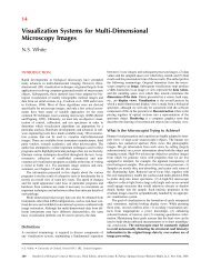
Figure 1 is a current phylogenetic tree<br />
based on small-subunit (SSU) rRNA sequences<br />
<strong>of</strong> <strong>the</strong> organisms represented. The<br />
construction <strong>of</strong> such a tree is conceptually<br />
simple (9). Pairs <strong>of</strong> rRNA sequences from<br />
different organisms are aligned, <strong>and</strong> <strong>the</strong> differences<br />
are counted <strong>and</strong> considered to be<br />
some measure <strong>of</strong> “evolutionary distance” between<br />
<strong>the</strong> organisms. There is no consideration<br />
<strong>of</strong> <strong>the</strong> passage <strong>of</strong> time, only <strong>of</strong> change<br />
in nucleotide sequence. Pair-wise differences<br />
between many organisms can <strong>the</strong>n be used<br />
to infer phylogenetic trees, maps that represent<br />
<strong>the</strong> evolutionary paths leading to <strong>the</strong><br />
modern-day sequences. The tree in Fig. 1 is<br />
largely congruent with trees made using any<br />
molecule in <strong>the</strong> nucleic acid–based, information-processing<br />
system <strong>of</strong> cells. On <strong>the</strong><br />
o<strong>the</strong>r h<strong>and</strong>, phylogenetic trees based on<br />
metabolic genes, those involved in <strong>the</strong> manipulation<br />
<strong>of</strong> small molecules <strong>and</strong> in interaction<br />
with <strong>the</strong> environment, commonly do<br />
not concur with <strong>the</strong> rRNA-based version<br />
[see (10, 11) for reviews <strong>and</strong> discussions <strong>of</strong><br />
phylogenetic results with different molecules].<br />
Incongruities in phylogenetic trees<br />
made with different molecules may reflect<br />
lateral transfers or even <strong>the</strong> intermixings <strong>of</strong><br />
genomes in <strong>the</strong> course <strong>of</strong> evolution. Some<br />
metabolic archaeal genes, for instance, appear<br />
much more highly related to specific<br />
bacterial versions than to <strong>the</strong>ir eucaryal homologs;<br />
o<strong>the</strong>r archaeal genes seem decidedly<br />
eukaryotic in nature; still o<strong>the</strong>r archaeal<br />
genes are unique. None<strong>the</strong>less, <strong>the</strong> recently<br />
determined sequence <strong>of</strong> <strong>the</strong> archaeon Methanococcus<br />
jannaschii shows that <strong>the</strong> evolutionary<br />
lineage Archaea is independent <strong>of</strong><br />
both Eucarya <strong>and</strong> Bacteria (12).<br />
Interpreting <strong>the</strong> <strong>Molecular</strong><br />
Tree <strong>of</strong> Life<br />
“Evolutionary distance” in this type <strong>of</strong> phylogenetic<br />
tree (Fig. 1), <strong>the</strong> extent <strong>of</strong> sequence<br />
change, is read along line segments.
The tree can be considered a rough map <strong>of</strong><br />
<strong>the</strong> evolution <strong>of</strong> <strong>the</strong> genetic core <strong>of</strong> <strong>the</strong><br />
cellular lineages that led to <strong>the</strong> modern<br />
organisms (sequences) included in <strong>the</strong> tree.<br />
The time <strong>of</strong> occurrence <strong>of</strong> evolutionary<br />
events cannot be extracted reliably from<br />
phylogenetic trees, despite common attempts<br />
to do so. Time cannot be accurately<br />
correlated with sequence change because<br />
<strong>the</strong> evolutionary clock is not constant in<br />
different lineages (7). This disparity is evidenced<br />
in Fig. 1 by <strong>the</strong> fact that lines<br />
leading to <strong>the</strong> different reference organisms<br />
are not all <strong>the</strong> same length; <strong>the</strong>se different<br />
lineages have experienced different extents<br />
<strong>of</strong> sequence change. None<strong>the</strong>less, <strong>the</strong> order<br />
<strong>of</strong> occurrence <strong>of</strong> branchings in <strong>the</strong> trees can<br />
be interpreted as a genealogy, <strong>and</strong> intriguing<br />
insights into <strong>the</strong> evolution <strong>of</strong> cells are<br />
emerging.<br />
A sobering aspect <strong>of</strong> large-scale phylogenetic<br />
trees such as that shown in Fig. 1 is<br />
<strong>the</strong> graphical realization that most <strong>of</strong> our<br />
legacy in biological science, historically<br />
based on large organisms, has focused on a<br />
narrow slice <strong>of</strong> biological diversity. Thus,<br />
we see that animals (represented in Fig. 1 by<br />
Homo), plants (Zea), <strong>and</strong> fungi (Coprinus)<br />
constitute small <strong>and</strong> peripheral branches <strong>of</strong><br />
even eukaryotic cellular diversity. If <strong>the</strong><br />
animals, plants, <strong>and</strong> fungi are taken to comprise<br />
taxonomic “kingdoms,” <strong>the</strong>n we must<br />
recognize as kingdoms at least a dozen o<strong>the</strong>r<br />
eucaryotic groups, all microbial, with as<br />
much or more independent evolutionary<br />
history than that which separates <strong>the</strong> three<br />
traditional eukaryotic kingdoms (13).<br />
The rRNA <strong>and</strong> o<strong>the</strong>r molecular data<br />
solidly confirm <strong>the</strong> notion stemming from<br />
<strong>the</strong> last century that <strong>the</strong> major organelles <strong>of</strong><br />
eukaryotes—mitochondria <strong>and</strong> chloroplasts—are<br />
derived from bacterial symbionts<br />
that have undergone specialization<br />
through coevolution with <strong>the</strong> host cell. Sequence<br />
comparisons establish mitochondria<br />
as representatives <strong>of</strong> Proteobacteria (<strong>the</strong><br />
group in Fig. 1 including Escherichia <strong>and</strong><br />
Agrobacterium) <strong>and</strong> chloroplasts as derived<br />
from cyanobacteria (Synechococcus <strong>and</strong><br />
Gloeobacter in Fig. 1) (14). Thus, all respiratory<br />
<strong>and</strong> photosyn<strong>the</strong>tic capacity <strong>of</strong> eukaryotic<br />
cells was obtained from bacterial<br />
symbionts; <strong>the</strong> “endosymbiont hypo<strong>the</strong>sis”<br />
for <strong>the</strong> origin <strong>of</strong> organelles is no longer<br />
hypo<strong>the</strong>sis but well-grounded fact. The nuclear<br />
component <strong>of</strong> <strong>the</strong> modern eukaryotic<br />
cell did not derive from one <strong>of</strong> <strong>the</strong> prokaroytic<br />
lineages, however. The rRNA <strong>and</strong><br />
o<strong>the</strong>r molecular trees show that <strong>the</strong> eukaryotic<br />
nuclear line <strong>of</strong> descent extends as deeply<br />
into <strong>the</strong> history <strong>of</strong> life as do <strong>the</strong> bacterial<br />
<strong>and</strong> archaeal lineages. The mitochondrion<br />
<strong>and</strong> chloroplast came in relatively late. This<br />
late evolution is evidenced by <strong>the</strong> fact that<br />
mitochondria <strong>and</strong> chloroplasts diverged<br />
from free-living organisms that branch peripherally<br />
in molecular trees. Moreover, <strong>the</strong><br />
most deeply divergent eukaryotes even lack<br />
mitochondria (15). These latter organisms,<br />
little studied but sometimes troublesome<br />
creatures such as Giardia, Trichomonas, <strong>and</strong><br />
Vairimorpha, none<strong>the</strong>less contain at least a<br />
few bacterial-type genes (16). These genes<br />
may be evidence <strong>of</strong> an earlier mitochondrial<br />
symbiosis with Eucarya that was lost (11)or<br />
perhaps o<strong>the</strong>r symbiotic or gene-transfer<br />
events between <strong>the</strong> evolutionary domains.<br />
The root <strong>of</strong> <strong>the</strong> universal tree in Fig. 1,<br />
<strong>the</strong> point <strong>of</strong> origin <strong>of</strong> <strong>the</strong> modern lineages,<br />
cannot be established using sequences <strong>of</strong><br />
only one type <strong>of</strong> molecule. However, recent<br />
phylogenetic studies <strong>of</strong> gene families that<br />
originated before <strong>the</strong> last common ancestor<br />
<strong>of</strong> <strong>the</strong> three domains have positioned <strong>the</strong><br />
root <strong>of</strong> <strong>the</strong> universal tree deep on <strong>the</strong> bacterial<br />
line (10). Therefore, Eucarya <strong>and</strong> Archaea<br />
had a common history that excluded<br />
<strong>the</strong> descendants <strong>of</strong> <strong>the</strong> bacterial line. This<br />
period <strong>of</strong> evolutionary history shared by<br />
Eucarya <strong>and</strong> Archaea was an important time<br />
in <strong>the</strong> evolution <strong>of</strong> cells, during which <strong>the</strong><br />
refinement <strong>of</strong> <strong>the</strong> primordial informationprocessing<br />
mechanisms occurred. Thus,<br />
modern representatives <strong>of</strong> Eucarya <strong>and</strong> Archaea<br />
share many properties that differ from<br />
bacterial cells in fundamental ways. One ex-<br />
www.sciencemag.org SCIENCE VOL. 276 <br />
ARTICLES<br />
ample <strong>of</strong> similarities <strong>and</strong> differences is in <strong>the</strong><br />
nature <strong>of</strong> <strong>the</strong> transcription machinery. The<br />
RNA polymerases <strong>of</strong> Eucarya <strong>and</strong> Archaea<br />
resemble each o<strong>the</strong>r in subunit composition<br />
<strong>and</strong> sequence far more than ei<strong>the</strong>r resembles<br />
<strong>the</strong> bacterial type <strong>of</strong> polymerase. Moreover,<br />
whereas all bacterial cells use sigma factors to<br />
regulate <strong>the</strong> initiation <strong>of</strong> transcription, eucaryal<br />
<strong>and</strong> archaeal cells use TATA-binding<br />
proteins (17, 18).<br />
Because <strong>of</strong> <strong>the</strong> shared history <strong>of</strong> Eucarya<br />
<strong>and</strong> Archaea, we should, perhaps, look to<br />
<strong>the</strong> Archaea to identify fundamental properties<br />
<strong>of</strong> far more complex cells such as our<br />
own. The eukaryotic nuclear membrane, for<br />
instance, is considered by cell biologists to<br />
be an intrinsic component <strong>of</strong> <strong>the</strong> nucleus,<br />
somehow responsible for its integrity. The<br />
fact that Archaea remained “prokaryotic,”<br />
that is, did not develop a nuclear membrane,<br />
indicates that a membrane is not<br />
required for nuclear function, which Archaea<br />
certainly achieve (as do Bacteria, for<br />
that matter). Indeed, <strong>the</strong> archaeal nuclear<br />
zone even seems to exclude ribosomes (19),<br />
<strong>and</strong> <strong>the</strong> genome <strong>of</strong> M. jannaschii is sprinkled<br />
with homologs <strong>of</strong> eucaryal nuclear <strong>and</strong> nucleolar<br />
structural genes (12). What constitutes<br />
a “nucleus?” Certainly <strong>the</strong> acquisition<br />
<strong>of</strong> <strong>the</strong> nuclear membrane was a relatively<br />
late event in <strong>the</strong> establishment <strong>of</strong> <strong>the</strong> eu-<br />
Fig. 1. Universal phylogenetic<br />
tree based on<br />
SSU rRNA sequences.<br />
Sixty-four rRNA sequences<br />
representative<br />
<strong>of</strong> all known phylogenetic<br />
domains were<br />
aligned, <strong>and</strong> a tree was<br />
produced using FASTD-<br />
NAML (43, 52). That tree<br />
was modified, resulting<br />
in <strong>the</strong> composite one<br />
shown, by trimming lineages<br />
<strong>and</strong> adjusting<br />
branch points to incorporate<br />
results <strong>of</strong> o<strong>the</strong>r<br />
analyses. The scale bar<br />
corresponds to 0.1<br />
changes per nucleotide.<br />
2 MAY 1997 735
caryal line <strong>of</strong> descent, occurring only after<br />
<strong>the</strong> separation from Archaea. Perhaps <strong>the</strong><br />
nuclear membrane is after all not fundamental<br />
to <strong>the</strong> function <strong>of</strong> <strong>the</strong> nucleus but<br />
ra<strong>the</strong>r is a relatively late-arriving embellishment.<br />
One hypo<strong>the</strong>sis would be that <strong>the</strong><br />
nuclear membrane was an invention derived<br />
from <strong>the</strong> Golgi apparatus to serve as a<br />
ga<strong>the</strong>ring basket for nuclear products, for<br />
distribution by <strong>the</strong> Golgi throughout <strong>the</strong><br />
cell. The properties <strong>of</strong> nuclear pores would<br />
be consistent with this hypo<strong>the</strong>sis; <strong>the</strong>y are<br />
large orifices, typically 10 nm in diameter,<br />
unlikely to gate anything except large molecules<br />
(20). The evolutionary record suggests,<br />
<strong>the</strong>n, that we look to something more<br />
fundamental than <strong>the</strong> nuclear membrane<br />
for <strong>the</strong> integrity <strong>of</strong> <strong>the</strong> nucleus <strong>and</strong> by<br />
which to define <strong>the</strong> essential quality <strong>of</strong> <strong>the</strong><br />
eukaryotic cell. The shared evolutionary<br />
history <strong>of</strong> Eucarya <strong>and</strong> Archaea suggests<br />
that we may be able to recognize <strong>the</strong> most<br />
fundamental elements <strong>of</strong> our own nucleus<br />
through study <strong>of</strong> <strong>the</strong> archaeal version.<br />
The Metabolic <strong>Diversity</strong> <strong>of</strong> Life<br />
The molecular-phylogenetic perspective<br />
(Fig. 1) is a reference framework within<br />
which to describe microbial diversity; <strong>the</strong><br />
sequences <strong>of</strong> genes can be used to identify<br />
organisms. This capability is an important<br />
concept for microbial biology. It is not possible<br />
to describe microorganisms as traditionally<br />
done with large organisms, through<br />
<strong>the</strong>ir morphological properties. To be sure,<br />
some microbes are intricate <strong>and</strong> beautiful in<br />
<strong>the</strong> microscope, but <strong>the</strong>y are mainly relatively<br />
unfeatured at <strong>the</strong> resolution <strong>of</strong> routine<br />
microscopy. Therefore, in order to distinguish<br />
different types <strong>of</strong> microbes, microbiologists<br />
early turned to metabolic properties<br />
such as utilizable sources <strong>of</strong> nutrition, for<br />
instance, sources <strong>of</strong> carbon, nitrogen, <strong>and</strong><br />
energy. <strong>Microbial</strong> taxonomy accumulated as<br />
anecdotal descriptions <strong>of</strong> metabolically <strong>and</strong><br />
morphologically distinct types <strong>of</strong> organisms<br />
that were essentially unrelatable. <strong>Molecular</strong><br />
phylogeny now provides a framework within<br />
which we can relate organisms objectively,<br />
<strong>and</strong> also through which we can interpret <strong>the</strong><br />
evolutionary flow <strong>of</strong> <strong>the</strong> metabolic machineries<br />
that constitute microbial diversity.<br />
Laboratory studies <strong>of</strong> microbial metabolism<br />
have focused mainly on organisms such<br />
as Escherichia coli <strong>and</strong> Bacillus subtilis. In<strong>the</strong><br />
broad sense, such organisms metabolize<br />
much as animals do; we are all “organotrophs,”<br />
using reduced organic compounds<br />
for energy <strong>and</strong> carbon. Organotrophy is not<br />
<strong>the</strong> prevalent form <strong>of</strong> metabolism in <strong>the</strong><br />
environment, however. Autotrophic metabolism,<br />
fixation <strong>of</strong> CO 2 to reduced organic<br />
compounds, must necessarily contribute to a<br />
greater biomass than organotrophic metabo-<br />
736<br />
lism, which it supports (a principle long<br />
appreciated by ecologists). Energy for fixing<br />
CO 2 is ga<strong>the</strong>red in two ways: by phototrophy<br />
(photosyn<strong>the</strong>sis) or lithotrophy (coupling<br />
<strong>the</strong> oxidation <strong>of</strong> reduced inorganic compounds<br />
such as hydrogen, hydrogen sulfide,<br />
or ferrous iron to <strong>the</strong> reduction <strong>of</strong> a chemical<br />
oxidant, a terminal electron acceptor such as<br />
oxygen, nitrate, sulfate, sulfur, or carbon dioxide).<br />
Thus, metabolic diversity can be<br />
generalized in terms <strong>of</strong> organotroph or autotroph,<br />
phototroph or lithotroph, <strong>and</strong> <strong>the</strong><br />
nature <strong>of</strong> <strong>the</strong> electron donor <strong>and</strong> acceptor.<br />
The phylogenetic distributions <strong>of</strong> different<br />
types <strong>of</strong> carbon <strong>and</strong> energy metabolism<br />
among different organisms do not necessarily<br />
follow <strong>the</strong> evolutionary pattern <strong>of</strong> rRNA<br />
(Fig. 1). Presumably, this lack <strong>of</strong> correspondence<br />
is because <strong>of</strong> past lateral transfers <strong>of</strong><br />
those metabolic genes <strong>and</strong> larger scale symbiotic<br />
fusions. None<strong>the</strong>less, <strong>the</strong>re are domain-level<br />
tendencies that may speak to<br />
<strong>the</strong> ancestral nature <strong>of</strong> <strong>the</strong> three domains <strong>of</strong><br />
life (21). The perspective here is currently<br />
limited mainly to Archaea <strong>and</strong> Bacteria.<br />
Such broad generalities cannot yet be assessed<br />
for <strong>the</strong> Eucarya because so little is<br />
known about <strong>the</strong> metabolic breadth <strong>of</strong> <strong>the</strong><br />
domain, <strong>the</strong> properties <strong>of</strong> <strong>the</strong> most deeply<br />
divergent lineages. There is considerable<br />
information about one pole <strong>of</strong> eukaryotic<br />
diversity, that represented by animals,<br />
plants, <strong>and</strong> fungi. We know little about <strong>the</strong><br />
o<strong>the</strong>r pole, <strong>the</strong> amitochondriate organisms<br />
that spun <strong>of</strong>f <strong>of</strong> <strong>the</strong> main eucaryal line early<br />
in evolution (22). The known instances <strong>of</strong><br />
such lineages, represented by Trichomonas,<br />
Giardia, <strong>and</strong> Vairimorpha in Fig. 1, are primarily<br />
pathogens. Pathogenicity to humans<br />
is a rare trait among <strong>the</strong> rest <strong>of</strong> eucaryotes<br />
<strong>and</strong> bacteria, <strong>and</strong> no archaeal pathogen is<br />
known. This correlation may indicate that<br />
nonpathogenic, deeply divergent eucaryotes<br />
are abundant in <strong>the</strong> environment but not<br />
yet detected. They should be sought in<br />
anaerobic ecosystems, possibly coupled<br />
metabolically to o<strong>the</strong>r organisms. A driving<br />
<strong>the</strong>me <strong>of</strong> <strong>the</strong> eucaryal line seems to be<br />
<strong>the</strong> establishment <strong>of</strong> physical symbiosis<br />
with o<strong>the</strong>r organisms. Beyond that, <strong>the</strong><br />
general metabolism <strong>of</strong> <strong>the</strong> rudimentary eukaryotic<br />
cell seems simple, based on fermentative<br />
organotrophy. By virtue <strong>of</strong> symbiotic<br />
partners, however, eukaryotes are<br />
able to take on phototrophic or lithotrophic<br />
life-styles <strong>and</strong> to use <strong>the</strong> electronacceptor<br />
oxygen (23).<br />
Symbiotic microbes commonly confer<br />
<strong>the</strong> lithotrophic way <strong>of</strong> life even on animals,<br />
although this was only recently recognized.<br />
The 2-m-long tubeworm Riftia<br />
pachyptila, for instance, lives in <strong>the</strong> vicinity<br />
<strong>of</strong> sea-floor hydro<strong>the</strong>rmal vents <strong>and</strong> metabolizes<br />
hydrogen sulfide <strong>and</strong> carbon dioxide<br />
by means <strong>of</strong> sulfide-oxidizing, carbon diox-<br />
SCIENCE VOL. 276 2 MAY 1997 www.sciencemag.org<br />
ide–fixing bacterial symbionts (24). This<br />
invertebrate <strong>and</strong> metabolically similar ones<br />
may contribute significantly to primary productivity<br />
in <strong>the</strong> ocean (25). It is not necessary<br />
to go to unusual (from our perspective)<br />
places such as ocean-floor vents to<br />
encounter o<strong>the</strong>r equally fascinating hydrogen<br />
sulfide–dependent eukaryotes (26).<br />
Underfoot at <strong>the</strong> ocean beach, for example,<br />
microbial respiration <strong>of</strong> seawater sulfate creates<br />
a hydrogen sulfide–rich ecosystem populated<br />
by little-known creatures such as<br />
Kentrophoros, a flat, gulletless ciliate that<br />
under <strong>the</strong> microscope appears fuzzy because<br />
it cultivates on its outer surface a crop <strong>of</strong><br />
sulfide-oxidizing bacteria (27). These bacteria<br />
are ingested by endocytosis <strong>and</strong> <strong>the</strong>reby<br />
provide nutrition for Kentrophoros. In<br />
o<strong>the</strong>r anaerobic environments, methanogens,<br />
members <strong>of</strong> Archaea, live intracellularly<br />
with eukaryotes <strong>and</strong> serve as metabolic<br />
hydrogen sinks (28). Still o<strong>the</strong>r symbioses<br />
based on inorganic energy sources are all<br />
around us <strong>and</strong> are little explored for <strong>the</strong>ir<br />
diversity <strong>of</strong> microbial life (26).<br />
Many lithotrophic, but comparatively few<br />
organotrophic, representatives <strong>of</strong> Archaea<br />
have been obtained in pure culture (29).<br />
There are primarily two metabolic <strong>the</strong>mes,<br />
both relying on hydrogen as a main source <strong>of</strong><br />
energy. Among <strong>the</strong> known Euryarchaeota—<br />
one <strong>of</strong> <strong>the</strong> two archaeal kingdoms known<br />
through cultivated organisms—<strong>the</strong> main<br />
electron acceptor is carbon dioxide, <strong>and</strong> <strong>the</strong><br />
product, methane—“natural gas.” Most <strong>of</strong><br />
<strong>the</strong> methane encountered in <strong>the</strong> outer few<br />
kilometers <strong>of</strong> Earth’s crust or on <strong>the</strong> surface is<br />
determined by isotopic analysis to be <strong>the</strong><br />
product <strong>of</strong> methanogenic Archaea, past <strong>and</strong><br />
present. Such organisms probably constitute<br />
a large component <strong>of</strong> global biomass. They<br />
certainly <strong>of</strong>fer an inexhaustible source <strong>of</strong><br />
renewable energy to humankind.<br />
The general metabolic <strong>the</strong>me <strong>of</strong> <strong>the</strong> o<strong>the</strong>r<br />
established kingdom <strong>of</strong> Archaea, Crenarchaeota,<br />
is also <strong>the</strong> oxidation <strong>of</strong> molecular<br />
hydrogen, but with a sulfur compound as <strong>the</strong><br />
terminal electron acceptor. All <strong>of</strong> <strong>the</strong> cultivated<br />
representatives <strong>of</strong> Crenarchaeota also<br />
are <strong>the</strong>rmophiles. Consequently, such organisms<br />
have been referred to as “<strong>the</strong>rmoacidophilic”<br />
or “hyper<strong>the</strong>rmophilic” Archaea;<br />
some grow at <strong>the</strong> highest known<br />
temperatures for life, up to 113°C in <strong>the</strong> case<br />
<strong>of</strong> Pyrolobus fumaris (30). These crenarchaeotes<br />
might seem bizarre, capable <strong>of</strong> thriving<br />
at temperatures above <strong>the</strong> usual boiling<br />
point <strong>of</strong> water on a diet <strong>of</strong> H 2 ,CO 2 , <strong>and</strong><br />
elemental sulfur <strong>and</strong> exhaling hydrogen sulfide.<br />
Yet, in terms <strong>of</strong> <strong>the</strong> molecular structures<br />
<strong>of</strong> <strong>the</strong> basic cellular machineries, <strong>the</strong>se<br />
creatures resemble eukaryotes far more<br />
closely than ei<strong>the</strong>r resembles <strong>the</strong> bacterium<br />
E. coli (17).<br />
The metabolic diversity <strong>of</strong> microbes is
usually couched in terms <strong>of</strong> <strong>the</strong> utilization<br />
<strong>of</strong> complex organic compounds. From that<br />
st<strong>and</strong>point, metabolic diversity seems, on<br />
<strong>the</strong> basis <strong>of</strong> cultivated instances <strong>of</strong> organisms,<br />
to have flowered mainly among <strong>the</strong><br />
Bacteria. Even here, however, reliance on<br />
organic nutrients probably was not ancestral.<br />
The most deeply branching <strong>of</strong> <strong>the</strong> cultured<br />
bacterial lineages, represented by<br />
Aquifex <strong>and</strong> Thermotoga in Fig. 1, are basically<br />
lithotrophs that use hydrogen as an<br />
energy source <strong>and</strong> electron acceptors such<br />
as sulfur compounds (Thermotoga) orlow<br />
levels <strong>of</strong> oxygen (Aquifex) (31). Cultivated<br />
instances <strong>of</strong> <strong>the</strong>se deeply branching bacterial<br />
lineages also are all <strong>the</strong>rmophilic <strong>and</strong><br />
thus share two important physiological attributes<br />
with <strong>the</strong> deeply branching <strong>and</strong><br />
slowly evolving Archaea: a hydrogen-based<br />
energy source <strong>and</strong> growth at high temperatures.<br />
This coincidence suggests that <strong>the</strong><br />
last common ancestor <strong>of</strong> all life also metabolized<br />
hydrogen for energy at high temperatures.<br />
This inference is consistent with<br />
current notions regarding <strong>the</strong> origin <strong>of</strong> life,<br />
that it came to be in a geo<strong>the</strong>rmal setting at<br />
high temperature (32).<br />
Chlorophyll-based photosyn<strong>the</strong>sis was a<br />
bacterial invention. It seems to have appeared<br />
well after <strong>the</strong> establishment <strong>of</strong> <strong>the</strong><br />
bacterial line <strong>of</strong> descent, at or before <strong>the</strong><br />
divergence <strong>of</strong> <strong>the</strong> line in Fig. 1 leading to<br />
Chlor<strong>of</strong>lexus, a photosyn<strong>the</strong>tic genus (33),<br />
<strong>and</strong> after <strong>the</strong> deeper divergences such as<br />
those leading to Aquifex <strong>and</strong> Thermotoga,<br />
genera that are not known to have photosyn<strong>the</strong>tic<br />
representatives. Most bacterial<br />
photosyn<strong>the</strong>sis is anaerobic, however. Oxygenic<br />
photosyn<strong>the</strong>sis, <strong>the</strong> water-based photosyn<strong>the</strong>tic<br />
mechanism that produces <strong>the</strong><br />
powerful electron acceptor oxygen, arose<br />
only in <strong>the</strong> kingdom-level lineage <strong>of</strong> cyanobacteria.<br />
This invention changed <strong>the</strong> surface<br />
<strong>of</strong> Earth pr<strong>of</strong>oundly <strong>and</strong> conventionally<br />
is thought to be <strong>the</strong> basis, directly or<br />
indirectly, <strong>of</strong> most present-day biomass.<br />
Anaerobic photosyn<strong>the</strong>sis is widely distributed<br />
in <strong>the</strong> late-branching bacterial<br />
kingdoms. The more ancient <strong>the</strong>me <strong>of</strong><br />
lithotrophy, metabolism <strong>of</strong> inorganic compounds,<br />
is also widely distributed phylogenetically,<br />
intermixed with organotrophic<br />
organisms. The pattern suggests that organotrophy<br />
arose many times from o<strong>the</strong>rwise<br />
photosyn<strong>the</strong>tic or lithotrophic organisms.<br />
Indeed, many instances <strong>of</strong> Bacteria can<br />
switch between <strong>the</strong>se modes <strong>of</strong> nutrition,<br />
carrying out photosyn<strong>the</strong>sis in <strong>the</strong> light <strong>and</strong><br />
lithotrophy or organotrophy in <strong>the</strong> dark.<br />
Particularly among Bacteria, this type <strong>of</strong><br />
energy metabolism seems highly volatile in<br />
evolution: Bacteria that are closely related<br />
by molecular criteria can display strikingly<br />
different phenotypes when assessed in <strong>the</strong><br />
laboratory through <strong>the</strong> nature <strong>of</strong> <strong>the</strong>ir car-<br />
bon <strong>and</strong> energy metabolism. In <strong>the</strong> relatively<br />
closely related “gamma subgroup” <strong>of</strong> <strong>the</strong><br />
kingdom <strong>of</strong> Proteobacteria (delineated by<br />
<strong>the</strong> genus Escherichia in Fig. 1), for instance,<br />
we find <strong>the</strong> phenotypically disparate organisms<br />
E. coli (organotroph), Chromatium<br />
vinosum (hydrogen sulfide–based phototroph),<br />
<strong>and</strong> <strong>the</strong> symbiont <strong>of</strong> <strong>the</strong> tubeworm<br />
R. pachyptila (hydrogen sulfide–based<br />
symbiont). The superficial metabolic diversity<br />
<strong>of</strong> <strong>the</strong>se types <strong>of</strong> Bacteria belies <strong>the</strong>ir<br />
underlying close evolutionary relatedness,<br />
giving no hint <strong>of</strong> <strong>the</strong> close similarities <strong>of</strong><br />
<strong>the</strong>ir basic machineries. The versatility <strong>of</strong><br />
Bacteria makes <strong>the</strong> metabolic machineries<br />
<strong>of</strong> Archaea <strong>and</strong> Eucarya seem comparatively<br />
more monotonous. As <strong>the</strong> sequences <strong>of</strong><br />
diverse genomes are compared, it will be<br />
possible to map <strong>the</strong> flow <strong>of</strong> metabolic genes<br />
onto <strong>the</strong> rRNA-based tree <strong>and</strong> <strong>the</strong>reby see<br />
how metabolic diversity has been molded<br />
through evolution.<br />
The molecular perspective gives us more<br />
than just a glimpse <strong>of</strong> <strong>the</strong> evolutionary past;<br />
it also brings a new future to <strong>the</strong> discipline<br />
<strong>of</strong> microbial biology. Because <strong>the</strong> molecular-phylogenetic<br />
identifications are based<br />
on sequence, as opposed to metabolic properties,<br />
microbes can be identified without<br />
being cultivated. Consequently, all <strong>the</strong> sequence-based<br />
techniques <strong>of</strong> molecular biology<br />
can be applied to <strong>the</strong> study <strong>of</strong> natural<br />
microbial ecosystems, heret<strong>of</strong>ore little<br />
known with regard to organismal makeup.<br />
A Sequence-Based Glimpse <strong>of</strong><br />
Biodiversity in <strong>the</strong> Environment<br />
Knowledge <strong>of</strong> microorganisms in <strong>the</strong> environment<br />
has depended in <strong>the</strong> past mainly<br />
on studies <strong>of</strong> pure cultures in <strong>the</strong> laboratory.<br />
Rarely are microbes so captured, however.<br />
Studies <strong>of</strong> several types <strong>of</strong> environments<br />
estimate that more than 99% <strong>of</strong> organisms<br />
seen microscopically are not cultivated by<br />
routine techniques (34). With <strong>the</strong> sequence-based<br />
taxonomic framework <strong>of</strong> molecular<br />
trees, only a gene sequence, not a<br />
functioning cell, is required to identify <strong>the</strong><br />
organism in terms <strong>of</strong> its phylogenetic type.<br />
The occurrence <strong>of</strong> phylogenetic types <strong>of</strong><br />
organisms, “phylotypes,” <strong>and</strong> <strong>the</strong>ir distributions<br />
in natural communities can be surveyed<br />
by sequencing rRNA genes obtained<br />
from DNA isolated directly from <strong>the</strong> environment.<br />
Analysis <strong>of</strong> microbial ecosystems<br />
in this way is more than a taxonomic exercise<br />
because <strong>the</strong> sequences provide experimental<br />
tools—for instance, molecular hybridization<br />
probes—that can be used to<br />
identify, monitor, <strong>and</strong> study <strong>the</strong> microbial<br />
inhabitants <strong>of</strong> natural ecosystems (35).<br />
Ribosomal RNA genes are obtained by<br />
cloning DNA isolated directly from <strong>the</strong> environment.<br />
“Shotgun libraries” <strong>of</strong> r<strong>and</strong>om<br />
www.sciencemag.org SCIENCE VOL. 276 <br />
ARTICLES<br />
DNA fragments are a source <strong>of</strong> rRNA, as<br />
well as o<strong>the</strong>r genes, but require sorting <strong>of</strong><br />
rRNA genes from <strong>the</strong> o<strong>the</strong>rs. The quickest<br />
way to survey <strong>the</strong> constituents <strong>of</strong> microbial<br />
ecosystems is through <strong>the</strong> use <strong>of</strong> <strong>the</strong> polymerase<br />
chain reaction (PCR) (36). The<br />
highly conserved nature <strong>of</strong> rRNA allows for<br />
<strong>the</strong> syn<strong>the</strong>sis <strong>of</strong> “universal” PCR primers<br />
that can anneal to sequences conserved in<br />
<strong>the</strong> rRNA genes from all three phylogenetic<br />
domains. In principle, PCR carried out with<br />
<strong>the</strong>se primers amplifies <strong>the</strong> rRNA genes <strong>of</strong><br />
all types <strong>of</strong> organisms present in an environmental<br />
sample. Individual types <strong>of</strong> genes<br />
in <strong>the</strong> mixture are separated by a cloning<br />
step <strong>and</strong> <strong>the</strong>n sequenced.<br />
A molecular-phylogenetic assessment <strong>of</strong><br />
an uncultivated organism can provide insight<br />
into many <strong>of</strong> <strong>the</strong> properties <strong>of</strong> <strong>the</strong><br />
organism through comparison with its studied<br />
relatives. One example <strong>of</strong> <strong>the</strong> perspective<br />
that phylogeny can <strong>of</strong>fer on an o<strong>the</strong>rwise<br />
unknown organism is seen with <strong>the</strong><br />
sulfur-oxidizing microorganisms that provide<br />
nutrition to symbiotic invertebrates<br />
such as <strong>the</strong> vent tubeworm R. pachyptila<br />
(24). Although many attempts to cultivate<br />
<strong>the</strong> symbionts for phenotypic characterization<br />
failed, rRNA analyses revealed that<br />
many <strong>of</strong> <strong>the</strong> basic cellular properties <strong>of</strong> <strong>the</strong><br />
symbionts were already familiar to us. The<br />
Riftia symbiont <strong>and</strong> a number <strong>of</strong> o<strong>the</strong>r sulfur-oxidizing<br />
symbionts associated with invertebrate<br />
animals all proved to be fairly<br />
closely related to one ano<strong>the</strong>r, close relatives<br />
also to <strong>the</strong> intensively studied organisms<br />
E. coli <strong>and</strong> Pseudomonas aeruginosa<br />
(37). Because <strong>of</strong> <strong>the</strong>ir phylogenetic proximity,<br />
many <strong>of</strong> <strong>the</strong> properties <strong>of</strong> <strong>the</strong> symbionts<br />
can be inferred from those <strong>of</strong> <strong>the</strong> wellstudied<br />
organisms. For instance, we can predict<br />
with good confidence <strong>the</strong> nature <strong>of</strong> <strong>the</strong><br />
ribosome <strong>and</strong> antibiotic-susceptibility patterns,<br />
<strong>the</strong> nature <strong>of</strong> <strong>the</strong> DNA-replicative<br />
machinery, <strong>the</strong> character <strong>of</strong> <strong>the</strong> RNA polymerase<br />
complex, <strong>the</strong> character <strong>of</strong> biosyn<strong>the</strong>tic<br />
pathways <strong>and</strong> <strong>the</strong>ir regulatory mechanisms,<br />
<strong>the</strong> nature <strong>of</strong> <strong>the</strong> cell envelope <strong>and</strong><br />
energy transduction schemes, <strong>and</strong> many<br />
o<strong>the</strong>r cellular properties <strong>of</strong> <strong>the</strong> symbionts.<br />
On <strong>the</strong> o<strong>the</strong>r h<strong>and</strong>, because E. coli <strong>and</strong> P.<br />
aeruginosa do not oxidize sulfur, <strong>the</strong>se relations<br />
cannot provide insight into <strong>the</strong> sulfuroxidative<br />
pathways <strong>of</strong> <strong>the</strong> symbionts. The<br />
rRNA sequence does, however, identify<br />
free-living <strong>and</strong> cultivated (but less-studied)<br />
close relatives <strong>of</strong> <strong>the</strong> symbionts—for example<br />
Thiomicrospira sp. L-12 ( a hydro<strong>the</strong>rmal-vent<br />
isolate) <strong>and</strong> Thiothrix sp.—that<br />
also rely on sulfur oxidation <strong>and</strong> so are<br />
likely to provide good models for this process<br />
in <strong>the</strong> symbionts.<br />
Every nucleic acids–based study <strong>of</strong> natural<br />
microbial ecosystems so far performed<br />
has uncovered novel types <strong>of</strong> rRNA se-<br />
2 MAY 1997 737
quences, <strong>of</strong>ten representing major new lineages<br />
only distantly related to known ones.<br />
The discovery <strong>of</strong> rRNA sequences in <strong>the</strong><br />
environment that diverge more deeply in<br />
phylogenetic trees than those <strong>of</strong> cultivated<br />
organisms is particularly noteworthy. It<br />
means that <strong>the</strong> divergent organisms recognized<br />
by rRNA sequence are potentially<br />
more different from known organisms in <strong>the</strong><br />
lineage than <strong>the</strong> known organisms are from<br />
one ano<strong>the</strong>r. The deepest divergences in<br />
both <strong>the</strong> Bacteria <strong>and</strong> Archaea were first<br />
discovered in rRNA-based surveys <strong>of</strong> hot<br />
spring–associated communities in Yellowstone<br />
National Park.<br />
The geo<strong>the</strong>rmal features <strong>of</strong> Yellowstone<br />
National Park have been favorite<br />
haunts <strong>of</strong> high-temperature biologists for<br />
decades (38). Currently, rRNA-based<br />
methods are being used to survey phylotypes<br />
present in a number <strong>of</strong> Yellowstone<br />
hot springs with disparate chemical settings.<br />
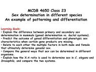
One <strong>of</strong> <strong>the</strong>se, Octopus Spring (Fig.<br />
2A)—a near-boiling, slightly alkaline, extremely<br />
low-nutrient flow near Old Faithful<br />
geyser containing an abundant community<br />
<strong>of</strong> pink filaments—yielded <strong>the</strong> first<br />
evidence for <strong>the</strong> lineage currently thought<br />
to be <strong>the</strong> most deeply divergent in <strong>the</strong><br />
Bacteria. When this lineage, represented<br />
by Aquifex <strong>and</strong> EM17 (pink filament<br />
clone) (39) in Fig. 1, was first encountered<br />
by 5S rRNA sequence (40), little could be<br />
inferred about <strong>the</strong> physiology <strong>of</strong> <strong>the</strong> associated<br />
organism because no cultivated specific<br />
relative had yet been described. Subsequent<br />
clues to <strong>the</strong> nature <strong>of</strong> <strong>the</strong> pink<br />
filaments came with <strong>the</strong> discovery <strong>of</strong> A.<br />
pyrophilus, cultured from an Icel<strong>and</strong>ic hot<br />
spring with a chemical character similar to<br />
that <strong>of</strong> Octopus Spring (41), <strong>and</strong> determination<br />
<strong>of</strong> <strong>the</strong> 16S rRNA sequence for <strong>the</strong><br />
pink filaments (39). The A. pyrophilus <strong>and</strong><br />
pink filament sequences are sufficiently<br />
closely related (Fig. 1) that many <strong>of</strong> <strong>the</strong>ir<br />
properties are likely to be shared. The<br />
mode <strong>of</strong> nutrition <strong>of</strong> <strong>the</strong> pink filaments,<br />
for instance, is predicted to be that <strong>of</strong><br />
Aquifex, consumption <strong>of</strong> hydrogen with<br />
low levels <strong>of</strong> oxygen <strong>and</strong> fixation <strong>of</strong> carbon<br />
dioxide. Many o<strong>the</strong>r representatives<br />
<strong>of</strong> <strong>the</strong> Aquifex-EM17 relatedness group<br />
(Aquificales) have now been cultured,<br />
mainly from high-temperature settings,<br />
<strong>and</strong> all are <strong>the</strong>rmophilic hydrogen oxidizers<br />
(31).<br />
Hot springs on <strong>the</strong> nor<strong>the</strong>rn flank <strong>of</strong><br />
<strong>the</strong> Yellowstone caldera usually have high<br />
concentrations <strong>of</strong> iron (II), hydrogen sulfide,<br />
hydrogen, <strong>and</strong> carbon dioxide—a<br />
wealth <strong>of</strong> foodstuffs for compatible physiologies.<br />
Ongoing sequence-based studies<br />
<strong>of</strong> <strong>the</strong> microbial inhabitants <strong>of</strong> one <strong>of</strong><br />
<strong>the</strong>se springs, Obsidian Pool (Fig. 2B),<br />
have radically revised our view <strong>of</strong> <strong>the</strong> phy-<br />
738<br />
logenetic diversity <strong>of</strong> Archaea. All cultivated<br />
Crenarchaeota branch in <strong>the</strong> cluster<br />
bracketed by Pyrodictium <strong>and</strong> Therm<strong>of</strong>ilum<br />
in Fig. 1. Discovery <strong>of</strong> a rich abundance <strong>of</strong><br />
diverse crenarchaeal rRNA genes in Obsidian<br />
Pool sediment (for example, pSL<br />
sequences in Fig. 1), scores <strong>of</strong> new genera,<br />
exp<strong>and</strong>ed <strong>the</strong> known phylogenetic diversity<br />
(estimated by specific line-segment<br />
lengths) <strong>of</strong> Crenarchaeota severalfold<br />
(42). More surprising, o<strong>the</strong>r sequences<br />
from Obsidian Pool (pJP27 <strong>and</strong> pJP28 in<br />
Fig. 1) seem to branch so deeply in <strong>the</strong><br />
overall archaeal tree that <strong>the</strong>y constitute a<br />
new kingdom-level branch <strong>of</strong> Archaea,<br />
recognized provisionally as “Korarchaeota”<br />
(43). It now will be interesting to<br />
study o<strong>the</strong>r genes from <strong>the</strong>se novel organisms.<br />
These genes, as well as information<br />
on <strong>the</strong> physiology <strong>and</strong> o<strong>the</strong>r properties <strong>of</strong><br />
<strong>the</strong> organisms, will be obtained most<br />
readily if <strong>the</strong>y can be cultured. Even without<br />
cultivation, however, cloning large<br />
fragments <strong>of</strong> environmental DNA <strong>and</strong><br />
<strong>the</strong>n “chromosome walking” to assemble<br />
contiguous clones <strong>of</strong>fers access to <strong>the</strong> genomes<br />
<strong>of</strong> <strong>the</strong>se or o<strong>the</strong>r uncultivated organisms<br />
(44).<br />
Continuing study <strong>of</strong> Obsidian Pool is<br />
exp<strong>and</strong>ing <strong>the</strong> known extent <strong>of</strong> bacterial,<br />
as well as archaeal, diversity. Obsidian<br />
Pool, judged extremely inhospitable from<br />
<strong>the</strong> human st<strong>and</strong>point, contains a rich<br />
diversity <strong>of</strong> sequence types representing<br />
most <strong>of</strong> <strong>the</strong> known bacterial kingdoms, as<br />
well as kingdom-level divergences never<br />
described by cultivation (45). Phylogenetic<br />
studies <strong>of</strong> cultured <strong>and</strong> environmental<br />
sequences have exp<strong>and</strong>ed substantially<br />
our appreciation <strong>of</strong> <strong>the</strong> scope <strong>of</strong> bacterial<br />
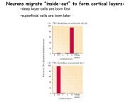
diversity: In 1987, only about 12 phylogenetic<br />
kingdoms (main phyla) <strong>of</strong> Bacteria<br />
were recognized (Fig. 3, inset) (7), but<br />
now, at least 25 to 30 distinct, kingdomlevel<br />
phylogenetic divergences are resolved<br />
(Fig. 3). The topology <strong>of</strong> <strong>the</strong> bacterial<br />
tree is remarkable. Bacterial diversity<br />
seems to have arisen mainly from an<br />
A B<br />
explosive radiation <strong>of</strong> lineages, ra<strong>the</strong>r<br />
than from <strong>the</strong> sequential divergence <strong>of</strong><br />
main lines seen, for instance, in <strong>the</strong> eucaryal<br />
domain (Fig. 1). Preliminary results<br />
from Obsidian Pool also call into question<br />
ano<strong>the</strong>r supposition based on culture studies,<br />
that Archaea dominate high-temperature<br />
environments. Quantitative hybridization<br />
<strong>of</strong> domain-specific oligonucleotide<br />
probes to rRNA genes obtained by PCR<br />
indicates that bacterial genes outnumber<br />
archaeal genes by 50:1 in this environment.<br />
Such conclusions, <strong>of</strong> course, are<br />
compromised to an unknown extent by<br />
considerations such as nonuniform amplification<br />
<strong>of</strong> different rRNA genes, but <strong>the</strong><br />
trend seems to indicate that bacteria dominate<br />
this environment.<br />
It is not necessary to go to extreme<br />
environments to encounter exotic diversity;<br />
it is all around us. Phylotypes that,<br />
because <strong>of</strong> <strong>the</strong>ir abundance, must be significant<br />
contributors to <strong>the</strong> biosphere<br />
have escaped detection until <strong>the</strong> sequence-based<br />
methods developed. One<br />
example <strong>of</strong> an arena for research opened<br />
by <strong>the</strong> molecular methods involves <strong>the</strong><br />
recently discovered mesophilic (low-temperature)<br />
Crenarchaeota (represented in<br />
Fig. 1 by pGrf <strong>and</strong> marSBAR). On <strong>the</strong><br />
basis <strong>of</strong> culture-studies, crenarchaeotes<br />
had been thought to be restricted to hightemperature<br />
environments. Cloned rRNA<br />
gene analysis shows, however, that lowtemperature<br />
versions <strong>of</strong> Crenarchaeota are<br />
abundant globally in marine (19, 46) <strong>and</strong><br />
terrestrial (47) environments, in typically<br />
30 to 50% <strong>of</strong> planktonic rRNA genes in<br />
limited samplings <strong>of</strong> Atlantic, Pacific, <strong>and</strong><br />
Antarctic waters (48). The physiologies <strong>of</strong><br />
<strong>the</strong> low-temperature crenarchaeotes are<br />
unknown; none has yet been cultivated.<br />
The properties <strong>of</strong> <strong>the</strong>ir remote relatives—<br />
<strong>the</strong> cultivated, high-temperature Crenarchaeota—hint<br />
that <strong>the</strong> mesophilic types<br />
might also engage in hydrogen metabolism,<br />
perhaps using some oxidation state <strong>of</strong><br />
sulfur as an electron acceptor.<br />
Fig. 2. Yellowstone National Park hot springs rich in microbial diversity. (A) Octopus Spring. The source<br />
pool <strong>of</strong> this hot spring is 90° to 93°C <strong>and</strong> extremely low in nutrients but contains abundant biomass <strong>and</strong><br />
<strong>the</strong> deepest known evolutionary divergences in <strong>the</strong> domain Bacteria. (B) Obsidian Pool. <strong>Molecular</strong><br />
studies find that <strong>the</strong> inhospitable environment <strong>of</strong> this hot spring, 75° to 95°C in temperature <strong>and</strong><br />
containing high concentrations <strong>of</strong> iron (II) <strong>and</strong> hydrogen sulfide, supports an extensive diversity <strong>of</strong><br />
previously unknown microbial life, both archaeal <strong>and</strong> bacterial.<br />
SCIENCE VOL. 276 2 MAY 1997 www.sciencemag.org
<strong>Microbial</strong> <strong>Diversity</strong> <strong>and</strong> <strong>the</strong> Limits<br />
<strong>of</strong> <strong>the</strong> <strong>Biosphere</strong><br />
Textbooks generally portray only a part <strong>of</strong><br />
<strong>the</strong> global distribution <strong>of</strong> life, <strong>the</strong> part that<br />
is immediately dependent on ei<strong>the</strong>r <strong>the</strong><br />
harvesting <strong>of</strong> sunlight or <strong>the</strong> metabolism<br />
<strong>of</strong> <strong>the</strong> decay products <strong>of</strong> photosyn<strong>the</strong>sis.<br />
The molecular phylogenetic record shows,<br />
however, that lithotrophic metabolism<br />
preceded <strong>and</strong> is more widespread phylogenetically<br />
<strong>and</strong> geographically than is ei<strong>the</strong>r<br />
phototrophy or organotrophy. The lithotrophic<br />
biosphere potentially extends kilometers<br />
into <strong>the</strong> crust <strong>of</strong> Earth, an essentially<br />
unknown realm (49). These considerations<br />
may indicate that lithotrophy<br />
contributes far more to <strong>the</strong> biomass <strong>of</strong><br />
Earth than currently thought.<br />
Part <strong>of</strong> that lithotrophic biomass is in<br />
microhabitats all around us, usually away<br />
from light <strong>and</strong> oxygen. It is not necessary<br />
to look far to find such environments: <strong>the</strong><br />
rumens <strong>of</strong> cattle <strong>and</strong> <strong>the</strong> guts <strong>of</strong> termites<br />
<strong>and</strong> humans, for example, are significant<br />
sources <strong>of</strong> methane, a signature <strong>of</strong> hydrogen<br />
metabolism. Most life that depends on<br />
inorganic energy metabolism, however,<br />
probably is in little-known environments,<br />
based on poorly understood geochemistries.<br />
The oceans, for instance, cover 70%<br />
<strong>of</strong> Earth’s surface to an average depth <strong>of</strong> 4<br />
km. Most life in <strong>the</strong> ocean is microbial,<br />
<strong>and</strong> <strong>the</strong> metabolic patterns <strong>of</strong> such organisms<br />
are not understood: Large st<strong>and</strong>ing<br />
crops <strong>of</strong> low-temperature crenarchaeotes,<br />
potential hydrogen oxidizers, may indicate<br />
an unsuspected, lithotrophy-based food<br />
chain in <strong>the</strong> oceans. Ano<strong>the</strong>r little-studied<br />
environment with global significance<br />
is <strong>the</strong> deep subsurface (50). There is increasing<br />
evidence that <strong>the</strong> crust <strong>of</strong> Earth is<br />
shot through with biomass, wherever <strong>the</strong><br />
physical conditions permit. Metabolism <strong>of</strong><br />
hydrogen is a dominant <strong>the</strong>me among organisms<br />
isolated from geo<strong>the</strong>rmal settings<br />
or deep aquifers (51). Hydrogen is generated<br />
readily by abiotic mechanisms such as<br />
interaction <strong>of</strong> water with iron-bearing basalt,<br />
<strong>the</strong> main stuff <strong>of</strong> Earth’s crust; consequently,<br />
a food source is unlikely to be<br />
limiting in most subterranean environments.<br />
Ra<strong>the</strong>r, it is likely to be <strong>the</strong> oxidant,<br />
<strong>the</strong> terminal electron acceptor, that<br />
limits growth. None<strong>the</strong>less, it seems possible<br />
that much, perhaps most, <strong>of</strong> <strong>the</strong> biomass<br />
on Earth is subterranean, a biological<br />
world based on lithotrophy. Although <strong>the</strong><br />
metabolic rate <strong>of</strong> this subterranean biosphere<br />
is likely to be far slower than in <strong>the</strong><br />
more dynamic, photic environment, life is<br />
likely to be as pervasive in occurrence, <strong>and</strong><br />
perhaps in cellular diversity, as we experience<br />
on <strong>the</strong> surface.<br />
Opportunity for an Environmental<br />
Genome Survey<br />
It is clear from even <strong>the</strong> small number <strong>of</strong><br />
environments so far studied with <strong>the</strong> molecular<br />
methods that our underst<strong>and</strong>ing <strong>of</strong><br />
<strong>the</strong> makeup <strong>of</strong> <strong>the</strong> natural microbial world<br />
is rudimentary. The sequence-based methods,<br />
however, now provide a way to survey<br />
biodiversity rapidly <strong>and</strong> comprehensively.<br />
Ribosomal RNA genes ga<strong>the</strong>red from <strong>the</strong><br />
environment are snapshots <strong>of</strong> organisms,<br />
representatives <strong>of</strong> different types <strong>of</strong> genomes,<br />
targets for fur<strong>the</strong>r characterization if<br />
<strong>the</strong>y seem interesting or useful. If we want<br />
to underst<strong>and</strong> <strong>the</strong> biosphere, I think it important,<br />
even essential, that we undertake a<br />
representative survey <strong>of</strong> microbial diversity<br />
in <strong>the</strong> environment. A complete cataloging<br />
www.sciencemag.org SCIENCE VOL. 276 <br />
Fig. 3. Diagrammatic representation<br />
<strong>of</strong> <strong>the</strong> known phylogenetic<br />
span <strong>of</strong> Bacteria in<br />
1987 (inset) <strong>and</strong> today. Phylogenetic<br />
trees containing<br />
sequences from <strong>the</strong> indicated<br />
organisms or groups <strong>of</strong><br />
organisms, chosen to represent<br />
<strong>the</strong> broadest diversity<br />
<strong>of</strong> Bacteria, were used as<br />
<strong>the</strong> basis for this diagram<br />
(compiled with P. Hugenholtz).<br />
Filled sectors indicate<br />
that several representative<br />
sequences fall within <strong>the</strong> indicated depth <strong>of</strong> branching. Lines<br />
designated by OP represent one or more phylotypes that were<br />
identified in Obsidian Pool by means <strong>of</strong> molecular methods but<br />
have not been not cultivated. The inset is an outline <strong>of</strong> <strong>the</strong><br />
bacterial tree compiled by Woese in 1987 (7).<br />
<strong>of</strong> Earth’s microbial biota is needless <strong>and</strong>, <strong>of</strong><br />
course, impossible. A representative survey,<br />
however, is worthwhile. A representative<br />
survey could be achieved with modest effort,<br />
with <strong>the</strong> use <strong>of</strong> automated sequencing<br />
technology. Analysis <strong>of</strong> 1000 clones (to<br />
detect <strong>the</strong> most abundant genome types)<br />
from each <strong>of</strong> 100 chemically different environments<br />
would be comparable to <strong>the</strong> effort<br />
to sequence a single microbial genome. The<br />
questions are large <strong>and</strong> many: What kinds<br />
<strong>of</strong> organisms do we share this planet with<br />
<strong>and</strong> depend on? What model systems should<br />
we choose for laboratory studies <strong>of</strong> environmental<br />
processes? How extensive is <strong>the</strong><br />
fund <strong>of</strong> biodiversity from which we can<br />
draw useful lessons <strong>and</strong> resources? Can we<br />
use <strong>the</strong> distribution <strong>of</strong> microbes as a biosensor<br />
array to map <strong>and</strong> monitor <strong>the</strong> chemistries<br />
<strong>of</strong> Planet Earth? Are <strong>the</strong>re deeper<br />
branchings in <strong>the</strong> tree <strong>of</strong> life than <strong>the</strong> lineages<br />
we know?<br />
The opportunities for <strong>the</strong> discovery <strong>of</strong><br />
new organisms <strong>and</strong> <strong>the</strong> development <strong>of</strong> resources<br />
based on microbial diversity are<br />
greater than ever before. <strong>Molecular</strong> sequences<br />
have finally given microbial biologists<br />
a way to define <strong>the</strong>ir subjects, through<br />
molecular phylogeny. The sequences also<br />
are <strong>the</strong> basis <strong>of</strong> <strong>the</strong> tools that will allow<br />
microbial biologists to explore <strong>the</strong> distribution<br />
<strong>and</strong> roles <strong>of</strong> <strong>the</strong> organisms in <strong>the</strong> environment.<br />
<strong>Microbial</strong> biology can now be a<br />
whole science; <strong>the</strong> organism can be studied<br />
in <strong>the</strong> ecosystem.<br />
REFERENCES<br />
______________<br />
ARTICLES<br />
1. M. T. Madigan, J. M. Martinko, J. Parker, Brock Biology<br />
<strong>of</strong> Microorganisms (Prentice-Hall, Upper Saddle<br />
River, NJ, ed. 8, 1996).<br />
2. A. T. Bull, M. Goodfellow, J. H. Slater, Annu. Rev.<br />
Microbiol. 46, 219 (1992).<br />
2 MAY 1997 739
3. R. H. Whittaker, <strong>Science</strong> 163, 150 (1969).<br />
4. E. Chatton, Titres et Travoux Scientifiques (Sete,<br />
Sottano, Italy, 1937).<br />
5. E. Zuckerk<strong>and</strong>l <strong>and</strong> L. Pauling, J. Theor. Biol. 8, 357<br />
(1965); R. M. Schwartz <strong>and</strong> M. O. Dayh<strong>of</strong>f, <strong>Science</strong><br />
199, 395 (1978).<br />
6. C. R. Woese <strong>and</strong> G. E. Fox, Proc. Natl. Acad. Sci.<br />
U.S.A. 74, 5088 (1977).<br />
7. C. R. Woese, Microbiol. Rev. 51, 221 (1987).<br />
8. , O. K<strong>and</strong>ler, M. L. Wheelis, Proc. Natl. Acad.<br />
Sci. U.S.A. 87, 4576 (1990).<br />
9. D. L. Sw<strong>of</strong>ford, G. J. Olsen, P. J. Waddell, D. M.<br />
Hillis, in <strong>Molecular</strong> Systematics, D. M. Hillis, C.<br />
Moritz, B. K. Mable, Eds. (Sinauer, Sunderl<strong>and</strong>, MA,<br />
1996), pp. 407–514.<br />
10. W. F. Doolittle <strong>and</strong> J. R. Brown, Proc. Natl. Acad.<br />
Sci. U.S.A. 91, 6721 (1994).<br />
11. J. D. Palmer, <strong>Science</strong> 275, 790 (1997).<br />
12. C. J. Bult et al., ibid. 273, 1058 (1996).<br />
13. The taxonomic term “kingdom” has no molecular<br />
definition. I use <strong>the</strong> term to indicate main lines <strong>of</strong><br />
radiation in <strong>the</strong> particular domain; 14 such “kingdom-level”<br />
lines are associated with <strong>the</strong> eucaryal line<br />
<strong>of</strong> descent in Fig. 1 [see also (22)].<br />
14. J. Sapp, Evolution by Association: A History <strong>of</strong> Symbiosis<br />
(Oxford Univ. Press, New York, 1994).<br />
15. T. Cavalier-Smith, Microbiol. Rev. 57, 953 (1993).<br />
16. E. T. N. Bui, P. J. Bradley, P. J. Johnson, Proc. Natl.<br />
Acad. Sci. U.S.A. 93, 9651 (1996); A. Germot, H.<br />
Philippe, H. le Guyader, ibid., p. 14614; A. J. Roger,<br />
C. G. Clark, W. F. Doolittle, ibid., p. 14618.<br />
17. T. L. Marsh, C. I. Reich, R. B. Whitelock, G. J. Olsen,<br />
ibid. 91, 4180 (1994).<br />
18. T. Rowl<strong>and</strong>s, P. Baumann, S. P. Jackson, <strong>Science</strong><br />
264, 1326 (1994).<br />
19. E. F. DeLong, Proc. Natl. Acad. Sci. U.S.A. 89, 5685<br />
(1992).<br />
20. L. I. Davis, Annu. Rev. Biochem. 64, 865 (1995).<br />
740<br />
21. O. K<strong>and</strong>ler, in Early Life on Earth, S. Bengtson, Ed.<br />
(Nobel Symp. 84, Columbia Univ. Press, New York,<br />
1993), pp. 152–160.<br />
22. M. L. Sogin, ibid., pp. 181–192.<br />
23. D. C. Smith <strong>and</strong> A. E. Douglas, The Biology <strong>of</strong> Symbiosis<br />
(Edward Arnold, London, 1987).<br />
24. V. Tunnicliffe, Am. Sci. 80, 336 (1992).<br />
25. R. A. Lutz et al., Nature 371, 663 (1994).<br />
26. T. Fenchel <strong>and</strong> B. J. Finlay, Ecology <strong>and</strong> Evolution in<br />
Anoxic Worlds, R. M. May <strong>and</strong> P. H. Harvey, Eds.<br />
(Oxford Ser. Ecol. Evol., Oxford Univ. Press, Oxford,<br />
1995).<br />
27. , Ophelia 30, 75 (1989).<br />
28. T. M. Embley <strong>and</strong> B. J. Finlay, Microbiology 140, 225<br />
(1994).<br />
29. M. Kates, D. J. Kushner, A. T. Ma<strong>the</strong>son, Eds., The<br />
Biochemistry <strong>of</strong> Archaea (Archaebacteria) (Elsevier,<br />
Amsterdam, 1993).<br />
30. K. O. Stetter, Am. Soc. Microbiol. News 61, 285<br />
(1995).<br />
31. C. Pitulle et al., Int. J. Syst. Bacteriol. 44, 620 (1994).<br />
32. N. R. Pace, Cell 65, 531 (1991).<br />
33. B. K. Pierson, in Early Life on Earth (Nobel Symp. 84,<br />
Columbia Univ. Press, New York, 1993), pp. 161–<br />
180.<br />
34. R. I. Amann, W. Ludwig, K. H. Schleifer, Microbiol.<br />
Rev. 59, 143 (1995).<br />
35. N. R. Pace, D. A. Stahl, D. J. Lane, G. J. Olsen, Am.<br />
Soc. Microbiol. News 51, 4 (1985); P. Hugenholtz<br />
<strong>and</strong> N. R. Pace, Trends Biotechnol. 14, 190 (1996).<br />
36. R. K. Saiki et al., <strong>Science</strong> 239, 487 (1988).<br />
37. D. L. Distel et al., J. Bacteriol. 170, 2506 (1988).<br />
38. T. D. Brock, Thermophilic Microorganisms <strong>and</strong> Life<br />
at High Temperatures (Springer-Verlag, New York,<br />
1978).<br />
39. A.-L. Reysenbach, G. S. Wickham, N. R. Pace, Appl.<br />
Environ. Microbiol. 60, 2113 (1994).<br />
40. D. A. Stahl, D. J. Lane, G. J. Olsen, N. R. Pace, ibid.<br />
SCIENCE VOL. 276 2 MAY 1997 www.sciencemag.org<br />
49, 1379 (1985).<br />
41. R. Huber et al., Syst. Appl. Microbiol. 15, 340 (1992).<br />
42. S. M. Barns, R. E. Fundyga, M. W. Jeffries, N. R.<br />
Pace, Proc. Natl. Acad. Sci. U.S.A. 91, 1609 (1994).<br />
43. S. M. Barns, C. F. Delwiche, J. D. Palmer, N. R.<br />
Pace, ibid. 93, 9188 (1996).<br />
44. J. L. Stein, T. L. Marsh, K. Y. Wu, H. Shizuya, E. F.<br />
DeLong, J. Bacteriol. 178, 591 (1996).<br />
45. P. Hugenholtz, C. Pitulle, K. L. Hershberger, N. R.<br />
Pace, in preparation.<br />
46. J. A. Fuhrman, K. McCallum, A. A. Davis, Nature<br />
356, 148 (1992).<br />
47. T. Ueda, Y. Suga, T. Matsuguchi, Eur. J. Soil Sci. 46,<br />
415 (1995); K. L. Hershberger, S. M. Barns, A.-L.<br />
Reysenbach, S. C. Dawson, N. R. Pace, Nature 384,<br />
420 (1996); S. B. Bintrim, T. J. Donohue, J. H<strong>and</strong>elsman,<br />
G. P. Roberts, R. M. Goodman, Proc. Natl.<br />
Acad. Sci. U.S.A. 94, 277 (1997); G. Jurgens, K.<br />
Lindstrom, A. Saano, Appl. Environ. Microbiol. 63,<br />
803 (1997).<br />
48. E. F. DeLong, K. Y. Wu, B. B. Prezelin, R. V. M.<br />
Jovine, Nature 371, 695 (1994).<br />
49. W. C. Ghiorse, <strong>Science</strong> 275, 789 (1997).<br />
50. T. Gold, Proc. Natl. Acad. Sci. U.S.A. 89, 6045<br />
(1992); J. K. Fredrickson <strong>and</strong> T. C. Onstott, Sci. Am.<br />
275, 68 (October 1996); D. Lovely <strong>and</strong> F. Chapelle,<br />
Rev. Geophys. 33, 365 (1995).<br />
51. T. O. Stevens <strong>and</strong> J. P. McKinley, <strong>Science</strong> 270, 450<br />
(1995); K. Pedersen, Earth Sci. Rev. 34, 243 (1993).<br />
52. B. L. Maidak et al., Nucleic Acids Res. 25, 109<br />
(1997).<br />
53. I thank S. Kustu, G. Olsen, <strong>and</strong> C. Woese for helpful<br />
comments on <strong>the</strong> manuscript, <strong>and</strong> S. Barns, S.<br />
Dawson, C. Delwiche, <strong>and</strong> P. Hugenholtz for assistance<br />
with <strong>the</strong> figures. Research in my laboratory is<br />
supported by grants from <strong>the</strong> U.S. Department <strong>of</strong><br />
Energy <strong>and</strong> NIH.