Factors affecting the clinical success of screw implants used as ...
Factors affecting the clinical success of screw implants used as ...
Factors affecting the clinical success of screw implants used as ...

Create successful ePaper yourself
Turn your PDF publications into a flip-book with our unique Google optimized e-Paper software.
ORIGINAL ARTICLE<br />
<strong>Factors</strong> <strong>affecting</strong> <strong>the</strong> <strong>clinical</strong> <strong>success</strong> <strong>of</strong> <strong>screw</strong><br />
<strong>implants</strong> <strong>used</strong> <strong>as</strong> orthodontic anchorage<br />
Hyo-Sang Park, a Seong-Hwa Jeong, b and Oh-Won Kwon c<br />
Daegu, Republic <strong>of</strong> South Korea<br />
Introduction: The purposes <strong>of</strong> this study were to examine <strong>the</strong> <strong>success</strong> rates and find factors <strong>affecting</strong> <strong>the</strong><br />
<strong>clinical</strong> <strong>success</strong> <strong>of</strong> <strong>screw</strong> <strong>implants</strong> <strong>used</strong> <strong>as</strong> orthodontic anchorage. Methods: Eighty-seven consecutive<br />
patients (35 male, 52 female; mean age, 15.5 years) with a total <strong>of</strong> 227 <strong>screw</strong> <strong>implants</strong> <strong>of</strong> 4 types were<br />
examined. Success rates during a 15-month period <strong>of</strong> force application were determined according to 18<br />
<strong>clinical</strong> variables. Results: The overall <strong>success</strong> rate w<strong>as</strong> 91.6%. The <strong>clinical</strong> variables <strong>of</strong> <strong>screw</strong>-implant<br />
factors (type, diameter, and length), local host factors (occlusogingival positioning), and management factors<br />
(angle <strong>of</strong> placement, onset and method <strong>of</strong> force application, ligature wire extension, exposure <strong>of</strong> <strong>screw</strong> head,<br />
and oral hygiene) did not show any statistical differences in <strong>success</strong> rates. General host factors (age, sex) had<br />
no statistical significance. Mobility, jaw (maxilla or mandible), and side <strong>of</strong> placement (right or left), and<br />
inflammation showed significant differences in <strong>success</strong> rates. Mobility, <strong>the</strong> right side <strong>of</strong> <strong>the</strong> jaw, and <strong>the</strong><br />
mandible were <strong>the</strong> relative risk factors in <strong>the</strong> logistic regression analysis when excluding mobility,<br />
inflammation around <strong>the</strong> <strong>screw</strong> <strong>implants</strong> w<strong>as</strong> added to <strong>the</strong> risk factors. Conclusions: To minimize <strong>the</strong> failure<br />
<strong>of</strong> <strong>screw</strong> <strong>implants</strong>, inflammation around <strong>the</strong> implant must be controlled, especially for <strong>screw</strong>s placed in <strong>the</strong><br />
right side <strong>of</strong> <strong>the</strong> mandible. (Am J Orthod Dent<strong>of</strong>acial Orthop 2006;130:18-25)<br />
Anchorage control is an important factor in <strong>the</strong><br />
<strong>success</strong> <strong>of</strong> orthodontic treatment. There have<br />
been many attempts to devise suitable anchorage<br />
methods, including intraoral and extraoral appliances.<br />
All intraoral appliances, however, show some<br />
loss <strong>of</strong> anchorage. Extraoral appliances do not provide<br />
reliable anchorage without patient compliance.<br />
When using skeletal anchorage such <strong>as</strong> osseous<br />
dental <strong>implants</strong>, miniplates,<br />
1 mini<strong>screw</strong>s, 2,3 or micro<strong>screw</strong>s,<br />
4-7 clinicians can expect reliable anchorage<br />
without patient compliance. Among <strong>the</strong>se anchorage<br />
devices, micro<strong>screw</strong> <strong>implants</strong> have incre<strong>as</strong>ingly been<br />
<strong>used</strong> for orthodontic anchorage because <strong>of</strong> <strong>the</strong>ir absolute<br />
anchorage, e<strong>as</strong>y placement and removal, and low<br />
cost. The small size <strong>of</strong> micro<strong>screw</strong> <strong>implants</strong> allows<br />
<strong>the</strong>m to be placed into bone between <strong>the</strong> teeth, thus<br />
4-7<br />
expanding <strong>the</strong>ir <strong>clinical</strong> applications. With more<br />
patients treated with <strong>screw</strong> <strong>implants</strong> <strong>as</strong> anchorage, <strong>the</strong>ir<br />
stability is ga<strong>the</strong>ring attention.<br />
From <strong>the</strong> Department <strong>of</strong> Orthodontics, School <strong>of</strong> Dentistry, Kyungpook<br />
National University, Daegu, Republic <strong>of</strong> Korea.<br />
aAssistant pr<strong>of</strong>essor.<br />
bResearch <strong>as</strong>sistant.<br />
cPr<strong>of</strong>essor. Reprint requests to: Dr Hyo-Sang Park, Department <strong>of</strong> Orthodontics, School <strong>of</strong><br />
Dentistry, Kyungpook National University, 101 Dongin-2-Ga, Daegu, Republic<br />
<strong>of</strong> Korea, 700-422; e-mail, parkhs@knu.ac.kr.<br />
Submitted, July 2004; revised and accepted, November 2004.<br />
0889-5406/$32.00<br />
Copyright © 2006 by <strong>the</strong> American Association <strong>of</strong> Orthodontists.<br />
doi:10.1016/j.ajodo.2004.11.032<br />
18<br />
The <strong>success</strong> <strong>of</strong> dental <strong>implants</strong> h<strong>as</strong> been studied<br />
extensively. Mini<strong>screw</strong> or micro<strong>screw</strong> <strong>implants</strong>, however,<br />
<strong>used</strong> <strong>as</strong> orthodontic anchorage should be loaded<br />
early to reduce treatment time and should be removed<br />
after treatment. In addition, micro<strong>screw</strong> <strong>implants</strong> are<br />
normally placed below or above <strong>the</strong> roots or between<br />
<strong>the</strong> roots <strong>of</strong> <strong>the</strong> teeth, or in <strong>the</strong> palatal or retromolar<br />
area, where<strong>as</strong> dental <strong>implants</strong> are placed in <strong>the</strong> edentulous<br />
ridges. Patients receiving dental <strong>implants</strong> are<br />
generally older than patients who have <strong>the</strong>m for orthodontic<br />
purposes. Therefore, factors <strong>affecting</strong> <strong>the</strong> <strong>clinical</strong><br />
<strong>success</strong> <strong>of</strong> dental <strong>implants</strong> might not be <strong>as</strong>sociated<br />
with mini<strong>screw</strong> or micro<strong>screw</strong> <strong>implants</strong> for orthodontic<br />
8 9<br />
anchorage. Miyawaki et aland<br />
Cheng et alstudied<br />
<strong>the</strong><br />
stability <strong>of</strong> <strong>screw</strong> <strong>implants</strong> for orthodontic purposes.<br />
These studies mostly dealt with factors <strong>affecting</strong> <strong>the</strong><br />
stability <strong>of</strong> mini<strong>screw</strong>s (over 1.5 mm in diameter) and<br />
miniplates. The use <strong>of</strong> micro<strong>screw</strong> <strong>implants</strong> h<strong>as</strong> now been<br />
expanded, but <strong>the</strong>re are still many unknown factors that<br />
could affect <strong>the</strong> <strong>clinical</strong> <strong>success</strong> <strong>of</strong> mini<strong>screw</strong> or micro<strong>screw</strong><br />
(less than 1.2 mm in diameter) <strong>implants</strong>.<br />
The purposes <strong>of</strong> this study were to find factors<br />
related to <strong>the</strong> <strong>clinical</strong> <strong>success</strong> <strong>of</strong> mini<strong>screw</strong> and micro<strong>screw</strong><br />
<strong>implants</strong> and to examine <strong>the</strong> <strong>success</strong> rates <strong>of</strong><br />
various types <strong>of</strong> micro<strong>screw</strong> and mini<strong>screw</strong> <strong>implants</strong>.<br />
MATERIAL AND METHODS<br />
The sample consisted <strong>of</strong> 87 consecutive patients (35<br />
male, 52 female; mean age, 15.5 years; SD, 8.3 years)<br />
who received mini<strong>screw</strong> or micro<strong>screw</strong> <strong>implants</strong> <strong>as</strong>
American Journal <strong>of</strong> Orthodontics and Dent<strong>of</strong>acial Orthopedics<br />
Volume 130, Number 1<br />
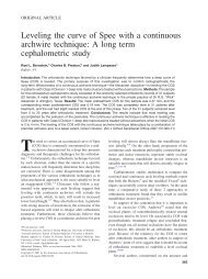
Fig 1. Four types <strong>of</strong> <strong>screw</strong> <strong>implants</strong> <strong>used</strong> in study.<br />
orthodontic anchorage. The patients were informed <strong>of</strong><br />
<strong>the</strong> advantages and disadvantages <strong>of</strong> this procedure.<br />
After collecting informed consent from <strong>the</strong> patients, <strong>the</strong><br />
<strong>implants</strong> were placed.<br />
Four types <strong>of</strong> <strong>screw</strong> <strong>implants</strong> (total, 227) were <strong>used</strong><br />
in this study: 19 type A micro<strong>screw</strong>s (Stryker Leibinger<br />
Inc, Kalamazoo, Mich) (diameter, 1.2 mm; length, 5<br />
mm); 157 type B micro<strong>screw</strong>s (Osteomed, Addison,<br />
Tex) (diameter, 1.2 mm; length, 6, 8, or 10 mm); 46<br />
type C micro<strong>screw</strong>s (Absoanchor, Dentos, Daegu, Korea)<br />
(diameter, 1.2 mm; length, 4, 6, 7, 8, or 10 mm),<br />
and 5 type D mini<strong>screw</strong>s (KLS-Martin, Jacksonville,<br />
Fla) (diameter, 2 mm; length, 10, 12, 14, or 15 mm)<br />
(Fig 1, Table I). The type C micro<strong>screw</strong> <strong>implants</strong> were<br />
developed for orthodontic purposes, with special features<br />
for attaching el<strong>as</strong>tic materials.<br />
The surgical procedure included local anes<strong>the</strong>sia, a<br />
small vertical stab incision (3-4 mm), reflection <strong>of</strong><br />
flaps, a pit made with a round bur, a hole made with a<br />
pilot drill, and placement <strong>of</strong> <strong>the</strong> <strong>screw</strong> <strong>implants</strong> with a<br />
<strong>screw</strong>driver. Surgical placement <strong>of</strong> <strong>the</strong> various <strong>screw</strong>s<br />
followed <strong>the</strong> same procedure according to previous<br />
reports. 5-7 The <strong>screw</strong>s were placed and checked by 1<br />
doctor (H-S.P.). The <strong>screw</strong> <strong>implants</strong> were placed at 30°<br />
to 40° angles to <strong>the</strong> long axes <strong>of</strong> <strong>the</strong> teeth in <strong>the</strong><br />
maxillary arch and at 10° to 20° angles in <strong>the</strong> mandibular<br />
posterior area. The <strong>screw</strong> <strong>implants</strong> in <strong>the</strong> retromolar<br />
area and <strong>the</strong> distobuccal bone to <strong>the</strong> mandibular<br />
second molars were placed at 90° to <strong>the</strong> bone surface.<br />
The re<strong>as</strong>on for placing <strong>the</strong> <strong>screw</strong> <strong>implants</strong> at those<br />
angulations w<strong>as</strong> to reduce root contact by <strong>the</strong> <strong>screw</strong><br />
<strong>implants</strong> without reducing <strong>the</strong> length <strong>of</strong> <strong>the</strong> <strong>screw</strong>. A<br />
long <strong>screw</strong> might have incre<strong>as</strong>ed stability, and an<br />
angled <strong>screw</strong> provides more bone contact than a <strong>screw</strong><br />
placed perpendicular to <strong>the</strong> bone. Just after placement,<br />
<strong>the</strong> initial stability <strong>of</strong> <strong>the</strong> <strong>screw</strong> implant w<strong>as</strong> checked;<br />
<strong>the</strong>re w<strong>as</strong> no sign <strong>of</strong> mobility.<br />
Table I. Success rate, number <strong>of</strong> patients and <strong>implants</strong>,<br />
and sizes <strong>of</strong> <strong>implants</strong><br />
Type <strong>of</strong> mini<strong>screw</strong> or micro<strong>screw</strong> implant<br />
A B C D<br />
Success rate (%) 84.2 93.6 89.1 80.0<br />
Patients (n) 10 67 16 4<br />
Screw <strong>implants</strong> (n) 19 157 46 5<br />
Success (n) 16 147 41 4<br />
Size <strong>of</strong> <strong>screw</strong>s (mm)<br />
Diameter 1.2 1.2 1.2 2.0<br />
Length (n) 5 (19) 10 (10) 10 (7) 15 (2)<br />
8 (77) 8 (4) 14 (1)<br />
6 (70) 7 (2) 12 (1)<br />
6 (15) 10 (1)<br />
4 (18)<br />
P .154, Fisher exact test.<br />
Park, Jeong, and Kwon 19<br />
Clinical variables<br />
To prevent examiner bi<strong>as</strong>, 18 <strong>clinical</strong> variables<br />
were investigated by <strong>the</strong> same doctor (H-S.P.). The<br />
variables were divided into 3 categories: <strong>screw</strong> implant<br />
factors, host factors, and management factors. Screw<br />
implant factors included type, length, and diameter <strong>of</strong><br />
<strong>the</strong> <strong>screw</strong> <strong>implants</strong>. Host factors were related to age and<br />
sex. Local host factors at recipient sites included jaw in<br />
which <strong>the</strong> <strong>screw</strong>s were placed, side <strong>of</strong> <strong>screw</strong> placement<br />
(right or left), sites <strong>of</strong> placement, and occlusogingival<br />
positioning <strong>of</strong> <strong>the</strong> <strong>screw</strong> <strong>implants</strong>. Procedure management<br />
factors referred to angle <strong>of</strong> placement, method <strong>of</strong><br />
force application, onset <strong>of</strong> force application, duration <strong>of</strong><br />
loading to <strong>screw</strong> <strong>implants</strong>, use <strong>of</strong> ligature extension,<br />
and exposure <strong>of</strong> <strong>the</strong> <strong>screw</strong> head. Environmental management<br />
factors were oral hygiene and inflammation<br />
around <strong>the</strong> <strong>screw</strong> <strong>implants</strong>. Mobility w<strong>as</strong> checked<br />
during use.<br />
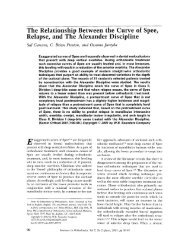
According to occlusogingival positioning <strong>of</strong> <strong>the</strong><br />
<strong>screw</strong>s, <strong>the</strong> sample w<strong>as</strong> divided into 4 groups: lower<br />
oral mucosa (<strong>screw</strong>s in <strong>the</strong> lower oral mucosa and deep<br />
in <strong>the</strong> vestibule), upper attached gingiva (placed in <strong>the</strong><br />
upper attached gingival zone), upper oral mucosa-low<br />
(placed in <strong>the</strong> upper oral mucosa up to 3 mm from<br />
<strong>the</strong> mucogingival line), and upper oral mucosa-high<br />
(placed high in <strong>the</strong> upper vestibule) (Fig 2). The lower<br />
oral mucosa and <strong>the</strong> upper oral mucosa-high groups had<br />
freely moving s<strong>of</strong>t tissues at <strong>the</strong> site <strong>of</strong> placement, and<br />
<strong>the</strong> upper oral mucosa-low group had partly moving<br />
s<strong>of</strong>t tissues around <strong>the</strong> <strong>screw</strong> <strong>implants</strong>. The upper<br />
attached gingiva group had firmly attached gingivae<br />
around <strong>the</strong> <strong>screw</strong> <strong>implants</strong>; this group included <strong>screw</strong><br />
<strong>implants</strong> in <strong>the</strong> palatal alveolar area.<br />
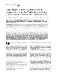
To <strong>as</strong>sess <strong>the</strong> effect <strong>of</strong> site <strong>of</strong> placement on <strong>success</strong>,<br />
because bone density and cortical bone thickness vary,
20 Park, Jeong, and Kwon<br />
placement sites were examined and divided into 5<br />
groups: (1) retromolar area, distobuccal to <strong>the</strong> lower<br />
second molar (LR and LD7), (2) buccal alveolar bone<br />
between <strong>the</strong> lower first and second molars (L67), (3)<br />
upper and lower anterior area (A), (4) buccal alveolar<br />
bone between <strong>the</strong> upper second premolar and <strong>the</strong> first<br />
molar, and between <strong>the</strong> upper first and second molars<br />
(U56 and U67), and (5) upper palatal alveolar bone<br />
between <strong>the</strong> first and second molars (UP) (Fig 3).<br />
Three angulations were <strong>used</strong>: 10° to 20 3 , 30° to 40°,<br />
and 90°. There were 4 methods <strong>of</strong> force application,<br />
with less than 200 g <strong>of</strong> force applied by (1) power<br />
chain, (2) Super thread (Rocky Mountain Orthodontics,<br />
Denver, Colo), (3) nickel-titanium coil spring, and<br />
(4) ligature tie-back. The sample w<strong>as</strong> divided into 2<br />
groups according to <strong>the</strong> ligature wire extension: yes or<br />
no. To attach <strong>the</strong> el<strong>as</strong>tic materials, <strong>the</strong> ligature extension<br />
w<strong>as</strong> connected to <strong>the</strong> neck <strong>of</strong> <strong>the</strong> <strong>screw</strong> <strong>implants</strong><br />
when <strong>the</strong> <strong>screw</strong> head w<strong>as</strong> expected to be covered by<br />
s<strong>of</strong>t tissue. The exposure <strong>of</strong> <strong>the</strong> <strong>screw</strong>s w<strong>as</strong> ei<strong>the</strong>r open<br />
or closed. If <strong>the</strong> head <strong>of</strong> <strong>screw</strong> w<strong>as</strong> exposed in <strong>the</strong><br />
mouth, <strong>the</strong> patients were included in <strong>the</strong> open group.<br />
O<strong>the</strong>rwise, <strong>the</strong>y were included in <strong>the</strong> closed group.<br />
To check <strong>the</strong> effect <strong>of</strong> oral hygiene on <strong>success</strong>, <strong>the</strong><br />
amount <strong>of</strong> food debris and plaque accumulation on <strong>the</strong><br />
tooth surfaces were <strong>as</strong>sessed; <strong>the</strong> sample w<strong>as</strong> divided<br />
into 3 groups: good, fair, or poor. Inflammation around<br />
<strong>the</strong> <strong>screw</strong> implant w<strong>as</strong> checked in <strong>the</strong> following categories:<br />
yes or no. Redness or swelling around <strong>the</strong> neck<br />
<strong>of</strong> <strong>the</strong> <strong>screw</strong>s w<strong>as</strong> a sign <strong>of</strong> inflammation. Each patient<br />
w<strong>as</strong> instructed to use a tooth brush to clean <strong>the</strong> teeth<br />
and a compressed water spray to clean <strong>the</strong> <strong>screw</strong><br />
American Journal <strong>of</strong> Orthodontics and Dent<strong>of</strong>acial Orthopedics<br />
July 2006<br />
Fig 2. Occlusogingival position <strong>of</strong> micro-implant: A, upper oral mucosa-high; B, upper oral<br />
mucosa-low; C, upper attached gingiva; D, lower oral mucosa.<br />
<strong>implants</strong>. If oral hygiene deteriorated, <strong>the</strong> patient w<strong>as</strong><br />
reinstructed to improve hygiene.<br />
Mobility w<strong>as</strong> checked with cotton tweezers at 5 to<br />
8 months after placement. There were 3 groups: yes<br />
(mobile), no (not mobile), and unknown (impossible to<br />
check because <strong>of</strong> overlying s<strong>of</strong>t tissue). If <strong>the</strong>re w<strong>as</strong> any<br />
discernible mobility, <strong>the</strong> <strong>screw</strong> implant w<strong>as</strong> counted in<br />
mobile group.<br />
Screw <strong>implants</strong> that were maintained in <strong>the</strong> bone to<br />
<strong>the</strong> end <strong>of</strong> treatment or to intentional removal regardless<br />
<strong>of</strong> mobility were considered <strong>success</strong>ful. If <strong>the</strong><br />
<strong>screw</strong> <strong>implants</strong> loosened during treatment, <strong>the</strong>y were<br />
considered to have failed.<br />
Statistics<br />
The overall <strong>success</strong> rate and <strong>the</strong> <strong>success</strong> rates for<br />
<strong>the</strong> type <strong>of</strong> <strong>screw</strong> implant and o<strong>the</strong>r <strong>clinical</strong> variables<br />
were calculated.<br />
To compare <strong>the</strong> differences <strong>of</strong> <strong>the</strong> levels <strong>of</strong> <strong>success</strong><br />
according to age, onset <strong>of</strong> force, duration <strong>of</strong> force<br />
application, and length <strong>of</strong> <strong>the</strong> <strong>screw</strong> <strong>implants</strong>, <strong>the</strong><br />
Student t test w<strong>as</strong> <strong>used</strong>. To compare <strong>the</strong> differences in<br />
<strong>the</strong> <strong>success</strong> rate according to <strong>the</strong> cl<strong>as</strong>sification <strong>of</strong> each<br />
<strong>clinical</strong> variable, <strong>the</strong> chi-square or Fisher exact test w<strong>as</strong><br />
performed with a statistical analysis program (version<br />
1.0, SPSS, Chicago, Ill). Logistic regression analysis<br />
w<strong>as</strong> performed to estimate <strong>the</strong> influence <strong>of</strong> each factor<br />
on failure. The odds ratio <strong>of</strong> each factor for failure <strong>of</strong><br />
<strong>the</strong> <strong>screw</strong> <strong>implants</strong> w<strong>as</strong> calculated. The odds ratio<br />
represents <strong>the</strong> proportionate risk for failure <strong>of</strong> <strong>screw</strong><br />
<strong>implants</strong>.
American Journal <strong>of</strong> Orthodontics and Dent<strong>of</strong>acial Orthopedics<br />
Volume 130, Number 1<br />
Park, Jeong, and Kwon 21<br />
Fig 3. Sites <strong>of</strong> placement: A, retromolar area distobuccal to mandibular second molar; B, buccal<br />
alveolar bone between mandibular first and second molars, and buccal alveolar bone between<br />
maxillary second premolar and first molar, and between maxillary first and second molars;<br />
C, maxillary and mandibular anterior area; D, maxillary palatal alveolar bone between first and<br />
second molars.<br />
RESULTS<br />
icance. There w<strong>as</strong> no significant correlation in <strong>success</strong><br />
The overall <strong>success</strong> rate w<strong>as</strong> 91.6% for all <strong>screw</strong> rate according to <strong>the</strong> method <strong>of</strong> force application or<br />
<strong>implants</strong> (208 <strong>of</strong> 227 <strong>screw</strong>s) with a mean period <strong>of</strong> placement angle. For environmental management fac-<br />
force application <strong>of</strong> 15 months. When <strong>the</strong> <strong>screw</strong> imtors, <strong>screw</strong> <strong>implants</strong> with inflammation showed signifplants<br />
failed, new ones were placed into a neighboring icantly less <strong>success</strong>.<br />
area. Eleven <strong>of</strong> 19 <strong>screw</strong>s that failed were replaced and Some <strong>screw</strong> <strong>implants</strong> showed fracturing during<br />
were <strong>success</strong>ful to <strong>the</strong> end <strong>of</strong> treatment.<br />
placement and removal. A total <strong>of</strong> 8 <strong>screw</strong>s were<br />
The <strong>success</strong> rates for <strong>the</strong> types <strong>of</strong> <strong>screw</strong> <strong>implants</strong> were broken, 3 during placement and 5 during removal.<br />
84.2 % for type A, 93.6% for type B, 89.1% for type C, Seven <strong>of</strong> <strong>the</strong> 8 fractured <strong>screw</strong>s were type B, and <strong>the</strong><br />
and 80% for type D. There were no significant differences o<strong>the</strong>r w<strong>as</strong> a type D mini<strong>screw</strong>.<br />
in <strong>the</strong> <strong>success</strong> rates between <strong>the</strong> types <strong>of</strong> <strong>screw</strong> <strong>implants</strong> Screw <strong>implants</strong> with mobility and unknown sam-<br />
(P .154), although <strong>the</strong> <strong>success</strong> rates for types B and C ples showed significantly less <strong>success</strong> than those with-<br />
were higher than for types A and D (Table I). Thereout<br />
mobility.<br />
were no statistically significant differences in <strong>the</strong> suc- The odds ratios (relative risk) for <strong>screw</strong> implant<br />
cess rates between diameter and length <strong>of</strong> <strong>the</strong> <strong>screw</strong>s. failure with mobility and unknown were 0.041 and<br />
For host factors, <strong>the</strong>re were no significant differ- 0.167, respectively. The odds ratios <strong>of</strong> failure in <strong>the</strong><br />
ences according to age and sex (Tables II and III). Forright<br />
side and in <strong>the</strong> mandible were 0.187 and 0.203,<br />
<strong>the</strong> local host factor, <strong>the</strong> <strong>screw</strong> <strong>implants</strong> placed in <strong>the</strong> respectively (Table IV). Excluding <strong>the</strong> mobility vari-<br />
maxilla showed a significantly higher <strong>success</strong> rate than ables in <strong>the</strong> logistic regression model, <strong>the</strong> odds ratios<br />
those placed in <strong>the</strong> mandible (Table III). The left side were 0.168 for <strong>screw</strong>s on <strong>the</strong> right side, 0.187 for<br />
had significantly higher <strong>success</strong> than <strong>the</strong> right side. <strong>screw</strong>s in <strong>the</strong> mandible, and 0.208 for <strong>implants</strong> with<br />
For procedure management factors, <strong>the</strong> <strong>screw</strong> heads inflammation around <strong>the</strong>m (Table V).<br />
covered by overlying s<strong>of</strong>t tissue showed higher <strong>success</strong><br />
than <strong>the</strong> exposed <strong>screw</strong> heads in <strong>the</strong> oral mucosa,<br />
DISCUSSION<br />
although it w<strong>as</strong> not statistically significant. The <strong>screw</strong> Screw <strong>implants</strong> can fail for various re<strong>as</strong>ons, <strong>as</strong> w<strong>as</strong><br />
<strong>implants</strong> in <strong>the</strong> UP showed higher <strong>success</strong> than those in<br />
10<br />
found with dental <strong>implants</strong>. The causes <strong>of</strong> dental<br />
o<strong>the</strong>r locations, although <strong>the</strong>re w<strong>as</strong> no statistical signif- implant failure include host factors (osteoporosis and
22 Park, Jeong, and Kwon<br />
Table II. Means and standard deviations <strong>of</strong> <strong>clinical</strong><br />
variables in <strong>success</strong> and failure groups<br />
Clinical variable<br />
Success<br />
(n 208)<br />
Failure<br />
(n 19)<br />
Mean SD Mean SD<br />
Significance<br />
(Student t test)<br />
Length <strong>of</strong> <strong>screw</strong><br />
(mm) 7.06 1.74 6.58 2.09 .257<br />
Age (y) 19.7 7.31 17.59 6.66 .227<br />
Onset <strong>of</strong> force<br />
(wk) 3.93 2.84 4.16 3.53 .741<br />
Duration <strong>of</strong> force<br />
(mo) 15.08 6.16 3.40 4.08 .00<br />
uncontrolled diabetes, smoking, and parafunctional<br />
habits), surgical factors <strong>of</strong> improper surgical technique,<br />
and management factors. Among <strong>the</strong>se factors, smoking<br />
and o<strong>the</strong>r host factors were not evaluated in this<br />
study because <strong>the</strong> sample comprised children and<br />
young adults. The effects <strong>of</strong> <strong>the</strong>se factors on failure <strong>of</strong><br />
<strong>screw</strong> <strong>implants</strong>, however, should be elucidated in a<br />
future study.<br />
Surgical factors include improper surgical techniques<br />
such <strong>as</strong> lack <strong>of</strong> initial stability, overheating<br />
during placement, and <strong>the</strong> fitness <strong>of</strong> <strong>the</strong> pilot hole to <strong>the</strong><br />
diameter <strong>of</strong> <strong>the</strong> <strong>screw</strong> implant. In this study, because all<br />
<strong>screw</strong> <strong>implants</strong> were placed by same doctor with <strong>the</strong><br />
same procedure, <strong>the</strong> effect <strong>of</strong> <strong>the</strong> surgical factors on <strong>the</strong><br />
<strong>clinical</strong> <strong>success</strong> <strong>of</strong> <strong>the</strong> <strong>screw</strong> <strong>implants</strong> w<strong>as</strong> not evaluated.<br />
However, by following this surgical procedure,<br />
clinicians might have acceptable <strong>success</strong> in practice.<br />
Management factors include poor home care, inflammation<br />
or infection, oral hygiene, and excessive<br />
load. An earlier study found that 6 <strong>of</strong> 12 failed <strong>screw</strong><br />
11 The<br />
<strong>implants</strong> failed within 2 months after placement.<br />
re<strong>as</strong>ons for failure might be errors during <strong>the</strong> surgical<br />
procedure. The remaining 6 <strong>screw</strong> <strong>implants</strong> failed<br />
between 2 and 10 months, and <strong>the</strong> cause might be<br />
management error. This might indicate that surgical<br />
and management procedures are both important for<br />
<strong>screw</strong> implant <strong>success</strong>.<br />
Because this study is a new field, we know little<br />
about factors that affect <strong>the</strong> rates <strong>of</strong> <strong>success</strong> <strong>of</strong> <strong>screw</strong><br />
<strong>implants</strong>. Therefore, in this study, we wanted to include<br />
<strong>as</strong> many factors <strong>as</strong> possible. Screw implant factors, host<br />
factors including local host factors at recipient sites,<br />
and procedure and environmental management factors<br />
were evaluated. Among <strong>the</strong>m, significant differences<br />
were found in local host and management factors.<br />
The factors <strong>as</strong>sociated with <strong>the</strong> failure <strong>of</strong> <strong>screw</strong><br />
<strong>implants</strong> were mobility, side, and jaw <strong>of</strong> placement.<br />
Screw <strong>implants</strong> on <strong>the</strong> right side <strong>of</strong> <strong>the</strong> jaw had a higher<br />
failure rate, and <strong>the</strong> mandible had a higher failure rate<br />
American Journal <strong>of</strong> Orthodontics and Dent<strong>of</strong>acial Orthopedics<br />
July 2006<br />
Table III. Success rate and number <strong>of</strong> <strong>screw</strong> <strong>implants</strong><br />
according to <strong>clinical</strong> variables<br />
Clinical variable<br />
Success<br />
rate<br />
(%)<br />
Success/total<br />
<strong>screw</strong><br />
<strong>implants</strong> (n)<br />
Significance<br />
(chi-square or<br />
Fisher exact)<br />
Diameter <strong>of</strong> <strong>screw</strong> .357<br />
1.2 mm 91.9 204/222<br />
2.0 mm 80 4/5<br />
Sex .21<br />
Male 88.76 79/89<br />
Female 93.48 129/138<br />
Jaw <strong>of</strong> placement .01<br />
Maxilla 96.0 119/124<br />
Mandible 86.4 89/103<br />
Side <strong>of</strong> placement .03<br />
Right 86.3 101/117<br />
Left 97.3 107/110<br />
Site <strong>of</strong> placement .059<br />
LR and LD7 81.8 27/33<br />
L67 90.5 57/63<br />
A 81.8 9/11<br />
U56 and U67 95.4 103/108<br />
UP 100 12/12<br />
Occlusogingival position .45<br />
Lower oral mucosa 88.4 76/86<br />
Upper attached<br />
gingiva 91.2 31/34<br />
Upper oral<br />
mucosa-low 91.5 65/71<br />
Upper oral<br />
mucosa-high 100 23/23<br />
Angle <strong>of</strong> placement .95<br />
10°-20° 91.0 61/67<br />
30°-40° 95.2 100/105<br />
90° 85.2 46/54<br />
Method <strong>of</strong> force<br />
application .26<br />
Power chain 83.3 5/6<br />
Super thread 88.8 95/107<br />
Nickel-titanium coil<br />
spring 94.6 106/112<br />
Ligature tie-back 100 2/2<br />
Ligature extension .77<br />
Yes 93.8 45/48<br />
No 91.1 163/179<br />
Exposure <strong>of</strong> <strong>screw</strong> head .06<br />
Closed 94.6 123/130<br />
Open 87.6 85/97<br />
Oral hygiene .40<br />
Good 100 18/18<br />
Fair 91.2 125/137<br />
Poor 90.3 65/72<br />
Inflammation .05<br />
Yes 84.4 65/77<br />
No 95.3 143/150<br />
Mobility .00<br />
Yes 75.6 34/45<br />
No 98.6 137/139<br />
Unknown 86.0 37/43<br />
Statistics: Chi-square or Fisher exact test.
American Journal <strong>of</strong> Orthodontics and Dent<strong>of</strong>acial Orthopedics<br />
Volume 130, Number 1<br />
Park, Jeong, and Kwon 23<br />
than <strong>the</strong> maxilla. Excluding mobility, inflammation<br />
around <strong>the</strong> <strong>screw</strong> <strong>implants</strong> w<strong>as</strong> added <strong>as</strong> a relative risk<br />
factor.<br />
In dental <strong>implants</strong>, mobility due to lack <strong>of</strong> osseointe-<br />
10<br />
gration is a sign <strong>of</strong> failure. For <strong>screw</strong> <strong>implants</strong> <strong>used</strong> <strong>as</strong><br />
orthodontic anchorage, however, mobility might not<br />
represent failure. We checked <strong>the</strong> mobility <strong>of</strong> <strong>the</strong> <strong>screw</strong><br />
<strong>implants</strong> 5 to 8 months after placement, during loading.<br />
Even though minimal mobility w<strong>as</strong> a risk factor <strong>of</strong><br />
failure, 34 <strong>of</strong> 45 minimally mobile <strong>screw</strong> <strong>implants</strong><br />
were <strong>success</strong>ful. By using comparatively low force<br />
(less than 200 g), <strong>the</strong> <strong>screw</strong> <strong>implants</strong> that showed<br />
minimal mobility could be set <strong>as</strong> anchorage. If heavy<br />
force is applied to <strong>screw</strong> <strong>implants</strong>, <strong>the</strong>ir mobility might<br />
be incre<strong>as</strong>ed, and <strong>the</strong>y can fail by not becoming sufficiently<br />
osseointegrated to <strong>the</strong> bone. In <strong>the</strong> animal studies<br />
12 13<br />
<strong>of</strong> Ohmae et al and Deguchi et al, stable <strong>screw</strong><br />
<strong>implants</strong> showed osseointegration from 25% to 40%.<br />
Deguchi et al<br />
13 lateral forces. Therefore, minimal mobility can be<br />
allowed in orthodontic <strong>screw</strong> <strong>implants</strong>. A study<br />
showing <strong>the</strong> reintegration <strong>of</strong> titanium <strong>implants</strong> after<br />
mechanical loosening<br />
postulated that less osseointegration<br />
does not necessarily indicate a negative finding. When<br />
an excessive load is applied, partly osseointegrated<br />
<strong>screw</strong> <strong>implants</strong> can become severely mobile and eventually<br />
fail. Screw <strong>implants</strong>, however, can be maintained<br />
with minimal mobility when applied force is light.<br />
Dental <strong>implants</strong> are usually loaded in all directions in<br />
addition to vertical occlusal forces, but orthodontic<br />
<strong>screw</strong> <strong>implants</strong> are usually loaded with unidirectional<br />
14 and <strong>the</strong> speculation <strong>of</strong> <strong>the</strong><br />
possible <strong>success</strong> <strong>of</strong> <strong>implants</strong> with rotational mobility<br />
15<br />
without bacterial infection after delayed loading<br />
might support this. The nature <strong>of</strong> <strong>screw</strong>-implant<br />
removal after treatment can expand <strong>the</strong> boundaries <strong>of</strong><br />
<strong>success</strong> to <strong>screw</strong> <strong>implants</strong> showing minimally discernible<br />
mobility.<br />
The left side had higher <strong>success</strong> than <strong>the</strong> right. This<br />
might be explained by better hygiene on <strong>the</strong> left side <strong>of</strong><br />
<strong>the</strong> dental arch by right-handed patients, who are most<br />
<strong>of</strong> <strong>the</strong> population.<br />
16 Better hygiene could reduce -in<br />
flammation around <strong>the</strong> <strong>screw</strong> <strong>implants</strong>.<br />
The mandible w<strong>as</strong> expected to have a higher <strong>success</strong><br />
rate because it h<strong>as</strong> a thicker and more dense<br />
17,18<br />
cortical bone than <strong>the</strong> maxilla. The results, however,<br />
were <strong>the</strong> opposite <strong>of</strong> our expectations. The <strong>as</strong>sumed<br />
re<strong>as</strong>ons might be overheating <strong>of</strong> <strong>the</strong> bone during<br />
drilling and irritation during chewing. Because <strong>the</strong><br />
mandible h<strong>as</strong> denser bone, <strong>the</strong>re is a greater chance <strong>of</strong><br />
generating heat greater than 47°C, which is <strong>the</strong> critical<br />
19,20<br />
temperature that can cause bone damage. In addition,<br />
<strong>screw</strong> <strong>implants</strong> placed in <strong>the</strong> posterior part <strong>of</strong> <strong>the</strong><br />
mandible can e<strong>as</strong>ily be irritated by food during chewing.<br />
These factors might negatively affect <strong>the</strong> <strong>clinical</strong><br />
<strong>success</strong> <strong>of</strong> <strong>screw</strong> <strong>implants</strong>. The reduced <strong>success</strong> <strong>of</strong> <strong>the</strong><br />
<strong>screw</strong> <strong>implants</strong> in <strong>the</strong> LR and LD7 group might support<br />
this <strong>as</strong>sumption. The mandibular posterior area w<strong>as</strong><br />
9<br />
also considered a risk site in a study by Cheng et Toal.<br />
reduce heat generation, copious irrigation with saline<br />
solution w<strong>as</strong> recommended.<br />
21 Excessive pressure <strong>of</strong> <strong>the</strong><br />
drill on <strong>the</strong> bone surface incre<strong>as</strong>ed heat. Worn drills<br />
21<br />
also produced more heat.<br />
As discussed in many previous studies <strong>of</strong> dental<br />
<strong>implants</strong>, peri-implantitis is an important factor in<br />
dental-implant failure. 22 Our results are similar to<br />
previous studies. Inflammation can damage <strong>the</strong> bone<br />
surrounding <strong>the</strong> neck <strong>of</strong> <strong>screw</strong> <strong>implants</strong>. With progressive<br />
damage <strong>of</strong> <strong>the</strong> cortical bone, <strong>screw</strong> <strong>implants</strong> can<br />
be endangered. 22,23 To ensure <strong>success</strong>, it is important to<br />
prevent inflammation around <strong>the</strong> <strong>screw</strong> <strong>implants</strong>. In this<br />
study, oral hygiene did not affect <strong>success</strong>, but local<br />
inflammation around <strong>the</strong> <strong>screw</strong> <strong>implants</strong> did. Local<br />
inflammation can be exaggerated not only by oral<br />
hygiene but also by weak nonkeratinized s<strong>of</strong>t tissue<br />
around <strong>the</strong> neck <strong>of</strong> <strong>the</strong> <strong>screw</strong> implant. A recent study<br />
showed that nonkeratinized mucosa w<strong>as</strong> a risk factor<br />
for mini<strong>screw</strong>s. 9 Table IV. Odds ratios for failure <strong>of</strong> 227 <strong>screw</strong> <strong>implants</strong><br />
including mobility<br />
Log odds<br />
Odds P<br />
Clinical variable Estimate SE ratio value 95% CI<br />
Mobility <strong>of</strong> <strong>screw</strong>s<br />
(mobile) 3.203 0.830 0.041 .000 0.008-0.207<br />
Mobility <strong>of</strong> <strong>screw</strong>s<br />
(unknown)<br />
Side <strong>of</strong> placement<br />
(right)<br />
Jaw <strong>of</strong> placement<br />
(mandible)<br />
1.792<br />
1.678<br />
1.596<br />
0.873<br />
0.681<br />
0.681<br />
0.167<br />
0.187<br />
0.203<br />
.040<br />
.014<br />
.019<br />
0.030-0.922<br />
0.049-0.709<br />
0.053-0.769<br />
Table V. Odds ratios for failure <strong>of</strong> 227 <strong>screw</strong> <strong>implants</strong><br />
excluding mobility<br />
Log odds<br />
Odds P<br />
Clinical variable Estimate SE ratio value 95% CI<br />
Side <strong>of</strong> placement<br />
(right) 1.783 0.666 0.168 .007 0.046-0.619<br />
Jaw <strong>of</strong> placement<br />
(mandible)<br />
Inflammation<br />
1.675<br />
1.572<br />
0.581<br />
0.540<br />
0.187<br />
0.208<br />
.004<br />
.004<br />
0.060-0.584<br />
0.072-0.598<br />
The highest <strong>success</strong> rate (100%) <strong>of</strong><br />
<strong>screw</strong> <strong>implants</strong> placed in <strong>the</strong> maxillary palatal area<br />
where <strong>the</strong>re is thick keratinized mucosa might support<br />
this. In addition, <strong>the</strong> <strong>screw</strong> <strong>implants</strong> in closed group, in
24 Park, Jeong, and Kwon<br />
American Journal <strong>of</strong> Orthodontics and Dent<strong>of</strong>acial Orthopedics<br />
July 2006<br />
which <strong>the</strong> head <strong>of</strong> <strong>screw</strong> w<strong>as</strong> covered by s<strong>of</strong>t tissue, had<br />
greater <strong>success</strong> than <strong>the</strong> open group, although it w<strong>as</strong> not<br />
statistically significant. The overlying s<strong>of</strong>t tissue on <strong>the</strong><br />
head <strong>of</strong> <strong>screw</strong> <strong>implants</strong> might be a barrier against<br />
inflammation. We instructed patients to clean <strong>the</strong> <strong>screw</strong><br />
<strong>implants</strong> with compressed water spray. Once inflammation<br />
arose, it tended to persist in nonkeratinized<br />
24<br />
mucosa are<strong>as</strong>. Francetti et alcompared<br />
<strong>the</strong> effects <strong>of</strong><br />
chlorhexidine spray and mouthw<strong>as</strong>h on controlling<br />
plaque after implant surgery. They found that <strong>the</strong><br />
plaque index improved, but <strong>the</strong>re w<strong>as</strong> no difference<br />
between <strong>the</strong> 2 methods. Therefore, water spray on<br />
<strong>screw</strong> <strong>implants</strong> might be an effective method to control<br />
inflammation.<br />
There w<strong>as</strong> no significant difference in <strong>the</strong> <strong>success</strong><br />
rate with respect to <strong>the</strong> onset <strong>of</strong> force application. This<br />
might indicate that immediate loading <strong>of</strong> <strong>screw</strong> <strong>implants</strong><br />
is possible. An animal experiment proved that<br />
<strong>the</strong>re w<strong>as</strong> osseointegration after immediate loading <strong>of</strong><br />
<strong>the</strong> <strong>screw</strong> <strong>implants</strong> and suggested immediate loading to<br />
25<br />
reduce <strong>the</strong> treatment time. Recent reports also recom-<br />
6<br />
mended immediate loading <strong>of</strong> <strong>screw</strong> <strong>implants</strong>. Therefore,<br />
<strong>screw</strong> <strong>implants</strong> can be loaded immediately after<br />
placement without a discernible deterioration <strong>of</strong> stability.<br />
There w<strong>as</strong> no significant difference in <strong>the</strong> failure<br />
rates between <strong>the</strong> 3 placement angles <strong>of</strong> <strong>the</strong> <strong>screw</strong><br />
<strong>implants</strong>. The re<strong>as</strong>on for placing <strong>the</strong> <strong>screw</strong> <strong>implants</strong> at<br />
angles to <strong>the</strong> bone surface w<strong>as</strong> to allow for use <strong>of</strong> long<br />
<strong>screw</strong> <strong>implants</strong> without damaging roots. The contact<br />
surface <strong>of</strong> <strong>the</strong> <strong>screw</strong> <strong>implants</strong> to <strong>the</strong> cortical bone w<strong>as</strong><br />
incre<strong>as</strong>ed by placing <strong>the</strong>m at angles. A study to elucidate<br />
<strong>the</strong> effects <strong>of</strong> <strong>the</strong> <strong>screw</strong> angle on <strong>the</strong> stability <strong>of</strong> <strong>the</strong><br />
mandibular sagittal split osseotomy showed no difference<br />
in resistance to segment movement between <strong>the</strong><br />
26<br />
60° and 90° angle groups. Therefore, clinicians can<br />
place long <strong>screw</strong> <strong>implants</strong> with angulations to bone<br />
surface without decre<strong>as</strong>ing stability, and <strong>the</strong> capability<br />
<strong>of</strong> using long <strong>screw</strong>s might influence <strong>success</strong> positively.<br />
The length <strong>of</strong> dental <strong>implants</strong> w<strong>as</strong> reported to<br />
27<br />
have a positive effect on stability. In our study,<br />
however, <strong>the</strong> length <strong>of</strong> <strong>the</strong> <strong>screw</strong> <strong>implants</strong> did not<br />
significantly affect <strong>the</strong>ir <strong>clinical</strong> <strong>success</strong>. Also, <strong>the</strong>ir<br />
diameter did not affect <strong>success</strong> rates, in contr<strong>as</strong>t to<br />
ano<strong>the</strong>r study. 8 seemed to be insufficient to evaluate <strong>the</strong> effect <strong>of</strong> each<br />
factor with statistical significance.<br />
As mentioned earlier, <strong>the</strong> <strong>success</strong> rate for <strong>screw</strong><br />
<strong>implants</strong> in previous studies varied between 83.9% and<br />
93.3%.<br />
This w<strong>as</strong> ca<strong>used</strong> by a small sample<br />
using 2.0-mm diameter <strong>screw</strong>s.<br />
This study w<strong>as</strong> performed to screen every possible<br />
factor that could affect <strong>the</strong> <strong>success</strong> <strong>of</strong> <strong>screw</strong> <strong>implants</strong>.<br />
The sample w<strong>as</strong> collected consecutively in 1 clinic, so<br />
<strong>the</strong> study design might not have been appropriate to<br />
<strong>as</strong>sess <strong>the</strong> effect <strong>of</strong> <strong>screw</strong> diameter, length, and type on<br />
<strong>success</strong>. The o<strong>the</strong>r problem in this study w<strong>as</strong> a small<br />
failure rate. This small number <strong>of</strong> failures (19 <strong>of</strong> 227)<br />
8,9,11 These rates might be explained by <strong>the</strong><br />
various types <strong>of</strong> <strong>screw</strong> <strong>implants</strong>, different surgical<br />
techniques, and varying management protocols. Therefore,<br />
a direct comparison <strong>of</strong> <strong>success</strong> rates might not be<br />
possible. An important <strong>as</strong>pect, however, is that by<br />
removing every possible cause <strong>of</strong> failure that w<strong>as</strong><br />
discussed in each study, clinicians might be able to<br />
incre<strong>as</strong>e <strong>the</strong> chances <strong>of</strong> <strong>success</strong>.<br />
In this study, <strong>the</strong> overall <strong>success</strong> rate <strong>of</strong> <strong>screw</strong><br />
<strong>implants</strong> <strong>used</strong> <strong>as</strong> anchorage w<strong>as</strong> 91.6% with a mean<br />
time <strong>of</strong> 15 months <strong>of</strong> force application. Including <strong>the</strong><br />
replaced <strong>the</strong> <strong>screw</strong> <strong>implants</strong>, <strong>the</strong> <strong>success</strong> rate would be<br />
8<br />
almost 96.5%. In a study by Miyawaki et all al,<br />
1.0-mm diameter <strong>screw</strong>s failed, but <strong>the</strong> 1.5-mm and<br />
2.3-mm diameter <strong>screw</strong>s showed no significant difference<br />
with <strong>success</strong> rates <strong>of</strong> 83.9% and 85%, respectively.<br />
Our results, in conjunction with <strong>the</strong> study by<br />
Miyawaki et al,<br />
8 indicate that <strong>screw</strong>s with diameters <strong>of</strong><br />
1.2, 1.5, and 2.3 mm have acceptable levels <strong>of</strong> <strong>success</strong>.<br />
The 1.0-mm diameter <strong>screw</strong>, however, had too much<br />
failure <strong>clinical</strong>ly even though animal studies showed<br />
osseointegration. 12,13 From a <strong>clinical</strong> point <strong>of</strong> view,<br />
smaller diameter <strong>screw</strong>s are e<strong>as</strong>ier and less traumatic to<br />
place and use. Screw <strong>implants</strong> with a diameter over 1.2<br />
mm can be recommended <strong>as</strong> orthodontic anchor <strong>screw</strong><br />
<strong>implants</strong>.<br />
The mean period <strong>of</strong> force application to <strong>the</strong> mini<strong>screw</strong><br />
or micro<strong>screw</strong> <strong>implants</strong> w<strong>as</strong> 15 months, which is<br />
sufficient to provide proper anchorage in most orthodontic<br />
patients. The most critical time period demanding<br />
anchorage control for <strong>success</strong>ful orthodontic treatment<br />
is for anterior tooth retraction in extraction<br />
patients. This usually takes 10 to 12 months <strong>of</strong> micro-<br />
6<br />
<strong>screw</strong> implant anchorage sliding mechanics. In nonextraction<br />
treatment, <strong>the</strong> distal movement <strong>of</strong> <strong>the</strong> posterior<br />
segment can be obtained within 10 months. This is<br />
because <strong>the</strong> posterior segment can be distalized to-<br />
7<br />
ge<strong>the</strong>r, and not 1 tooth at a time. Therefore, micro<strong>screw</strong><br />
<strong>implants</strong> seem to cover <strong>the</strong> critical time period<br />
requiring absolute anchorage.<br />
Five <strong>screw</strong> <strong>implants</strong> were fractured during <strong>the</strong><br />
removal procedure. If <strong>the</strong>re is too much osseointegration,<br />
clinicians might have difficulty in removing <strong>the</strong><br />
<strong>screw</strong>s, or <strong>the</strong>y can fracture. There h<strong>as</strong> been no study <strong>of</strong><br />
how much osseointegration is needed for orthodontic<br />
<strong>screw</strong> <strong>implants</strong> when considering <strong>the</strong> need for both<br />
stability and e<strong>as</strong>y removal. This should be elucidated in<br />
a well-designed experimental model.
American Journal <strong>of</strong> Orthodontics and Dent<strong>of</strong>acial Orthopedics<br />
Volume 130, Number 1<br />
CONCLUSIONS<br />
The overall <strong>success</strong> rate w<strong>as</strong> 91.6%, with a mean<br />
period <strong>of</strong> force application <strong>of</strong> 15 months. Therefore,<br />
<strong>screw</strong> <strong>implants</strong> can be <strong>used</strong> for orthodontic anchorage<br />
predictably and consistently in routine orthodontic<br />
practice. Mobility, <strong>the</strong> patient’s right side, mandibular<br />
implant sites, and inflammation were <strong>as</strong>sociated with<br />
<strong>screw</strong> implant failure in this study. To minimize failure,<br />
clinicians should attempt to reduce inflammation around<br />
<strong>the</strong> <strong>screw</strong> <strong>implants</strong>, especially for <strong>screw</strong>s placed on <strong>the</strong><br />
right side in <strong>the</strong> mandible.<br />
REFERENCES<br />
1. Umemori M, Sugawara J, Mitani H, Nag<strong>as</strong>aka H, Kawamura H.<br />
Skeletal anchorage system for open bite correction. Am J Orthod<br />
Dent<strong>of</strong>acial Orthop 1999;115:166-74.<br />
2. Creekmore TD, Eklund MK. The possibility <strong>of</strong> skeletal anchorage.<br />
J Clin Orthod 1983;17:266-9.<br />
3. Costa A, Raffaini M, Melsen B. Mini<strong>screw</strong>s <strong>as</strong> orthodontic<br />
anchorage: a preliminary report. Int J Adult Orthod Orthognath<br />
Surg 1998;13:201-9.<br />
4. Kanomi R. Mini-implant for orthodontic anchorage. J Clin<br />
Orthod 1997;31:763-7.<br />
5. Park HS. The skeletal cortical anchorage using titanium micro<strong>screw</strong><br />
<strong>implants</strong>. Korean J Orthod 1999;29:699-706.<br />
6. Park HS. The use <strong>of</strong> micro-implant <strong>as</strong> orthodontic anchorage.<br />
Seoul, Korea: Narae; 2001.<br />
7. Park HS, Kwon TG, Sung JH. Nonextraction treatment with<br />
micro<strong>screw</strong> <strong>implants</strong>. Angle Orthod 2004;74:539-49.<br />
8. Miyawaki S, Koyama I, Inoue M, Mishima K, Sugahara T,<br />
Takano-Yamamoto T. <strong>Factors</strong> <strong>as</strong>sociated with <strong>the</strong> stability <strong>of</strong><br />
titanium <strong>screw</strong>s placed in <strong>the</strong> posterior region for orthodontic<br />
anchorage. Am J Orthod Dent<strong>of</strong>acial Orthop 2003;124:373-8.<br />
9. Cheng SJ, Tseng IY, Lee JJ, Kok SH. A prospective study <strong>of</strong> <strong>the</strong><br />
risk factors <strong>as</strong>sociated with failure <strong>of</strong> mini-<strong>implants</strong> <strong>used</strong> for<br />
orthodontic anchorage. Int J Oral Maxill<strong>of</strong>ac Implants 2004;19:<br />
100-6.<br />
10. Ashley ET, Covington LL, Bishop BG, Breault LG. Ailing and<br />
failing endosseous dental <strong>implants</strong>: a literature review. J Contemp<br />
Dent Pract 2003;4:35-50.<br />
11. Park HS. Clinical study on <strong>success</strong> rate <strong>of</strong> micro<strong>screw</strong> <strong>implants</strong><br />
for orthodontic anchorage. Korean J Orthod 2003;33:151-6.<br />
12. Ohmae M, Saito S, Moroh<strong>as</strong>hi T, Seki K, Qu H, Kanomi R, et al.<br />
A <strong>clinical</strong> and histological evaluation <strong>of</strong> titanium mini-<strong>implants</strong><br />
Park, Jeong, and Kwon 25<br />
<strong>as</strong> anchors for orthodontic intrusion in <strong>the</strong> beagle dog. Am J<br />
Orthod Dent<strong>of</strong>acial Orthop 2001;119:489-97.<br />
13. Deguchi T, Takano-Yamamoto T, Kanomi R, Hartsfield JK Jr,<br />
Roberts WE, Garetto LP. The use <strong>of</strong> small titanium <strong>screw</strong>s for<br />
orthodontic anchorage. J Dent Res 2003;82:377-81.<br />
14. Ivan<strong>of</strong>f CJ, Sennerby L, Lekholm U. Reintegration <strong>of</strong> mobilized<br />
titanium <strong>implants</strong>. An experimental study in rabbit tibia. Int<br />
J Oral Maxill<strong>of</strong>ac Surg 1997;26:310-5.<br />
15. Esposito M, Hirsch J, Lekholm U, Thomsen P. Differential<br />
diagnosis and treatment strategies for biologic complications<br />
and failing oral <strong>implants</strong>: a review <strong>of</strong> <strong>the</strong> literature. Int J Oral<br />
Maxill<strong>of</strong>ac Implants 1999;14:473-90.<br />
16. Tezel A, Orbak R, Canakci V. The effect <strong>of</strong> right or lefthandedness<br />
on oral hygiene. Int J Neurosci 2001;109:1-9.<br />
17. Champy M, Pape H, Gerlach KL, Lodde JP. Mandibular fracture.<br />
In: Kruger E, Schilli W, editors. Oral and maxill<strong>of</strong>acial traumatology.<br />
Vol. 2, Chicago: Quintessence; 1986. p. 19-43.<br />
18. Park HS. An anatomical study using CT images for <strong>the</strong> implantation<br />
<strong>of</strong> micro-<strong>implants</strong>. Korean J Orthod 2002;32:435-41.<br />
19. Eriksson AR, Albrektsson T. Temperature threshold levels for<br />
heat-induced bone tissue injury: a vital-microscopic study in <strong>the</strong><br />
rabbit. J Pros<strong>the</strong>t Dent 1983;50:101-7.<br />
20. Tehemar SH. <strong>Factors</strong> <strong>affecting</strong> heat generation during implant<br />
site preparation: a review <strong>of</strong> biologic observations and future<br />
considerations. Int J Oral Maxill<strong>of</strong>ac Implants 1999;14:<br />
127-36.<br />
21. Mat<strong>the</strong>ws LS, Hirsch C. Temperatures me<strong>as</strong>ured in human<br />
cortical bone when drilling. J Bone Joint Surg Am 1972;54:<br />
297-308.<br />
22. Zitzmann NU, Berglundh T, Ericsson I, Lindhe J. Spontaneous<br />
progression <strong>of</strong> experimentally induced peri-implantitis. J Clin<br />
Periodontol 2004;31:845-9.<br />
23. Ericsson I, Berglundh T, Marinello C, Liljenberg B, Lindhe J.<br />
Long-standing plaque and gingivitis at <strong>implants</strong> and teeth in <strong>the</strong><br />
dog. Clin Oral Implants Res 1992;3:99-103.<br />
24. Francetti L, Del Fabbro M, B<strong>as</strong>so M, Testori T, T<strong>as</strong>chieri S, Weinstein<br />
R. Chlorhexidine spray versus mouthw<strong>as</strong>h in <strong>the</strong> control <strong>of</strong> dental<br />
plaque after implant surgery. J Clin Periodontol 2004;31:857-62.<br />
25. Melsen B, Costa A. Immediate loading <strong>of</strong> <strong>implants</strong> <strong>used</strong> for<br />
orthodontic anchorage. Clin Orthod Res 2000;3:23-8.<br />
26. Uckan S, Schwimmer A, Kummer F, Greenberg AM. Effect <strong>of</strong><br />
<strong>the</strong> angle <strong>of</strong> <strong>the</strong> <strong>screw</strong> on <strong>the</strong> stability <strong>of</strong> <strong>the</strong> mandibular sagittal<br />
split ramus osteotomy: a study in sheep mandibles. Br J Oral<br />
Maxill<strong>of</strong>ac Surg 2001;39:266-8.<br />
27. Himmlova L, Dostalova T, Kacovsky A, Konvickova S. Influence<br />
<strong>of</strong> implant length and diameter on stress distribution: a finite<br />
element analysis. J Pros<strong>the</strong>t Dent 2004;19:20-5.