Intra Lock OSSEAN® Catalog
Intra Lock OSSEAN® Catalog
Intra Lock OSSEAN® Catalog

Create successful ePaper yourself
Turn your PDF publications into a flip-book with our unique Google optimized e-Paper software.
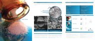
Optimal Differential Profilometry<br />
Calcium Phosphate - “Beyond Nano Size”<br />
Thick Oxide Layer<br />
Hydrophilic Surface Increases Wettability<br />
Site-Specific Surface Modification<br />
Predictable stability during the early dental<br />
implant treatment phase was a significant factor<br />
in the development of OSSEAN ® 1 . The surface<br />
is designed to activate the body’s natural<br />
healing abilities and to induce primary stability<br />
during the critical first two weeks after<br />
placement.<br />
Robotically Micro Blasted<br />
Each <strong>Intra</strong>-<strong>Lock</strong> ® implant is Robotically Micro-<br />
Blasted in a clean room environment for<br />
precise control. Robotic sequencing and<br />
differential treatment is utilized to preserve the<br />
cutting groove geometry (sharpness) and permit<br />
each region of the implant to obtain optimal<br />
topography and surface roughness 2 .<br />
Multi-Phase Cleaning and<br />
Surface Treatment<br />
Additional procedures derived from the semiconductor<br />
industry and refined for bio-medical<br />
applications ensure a clean surface. OSSEAN ®<br />
Surface is exceptionally free from contaminants<br />
as confirmed by XPS-ESCA (Electron<br />
Spectroscopy for Chemical Analysis).<br />
Robotic<br />
Micro-Blasting Preserves<br />
Cutting Edge Geometry<br />
Cellular Level -<br />
Enhanced Osteoblast<br />
Attachment<br />
Thick Oxide Layer<br />
Calcium Phosphate in<br />
Molecular Fusion<br />
Multi-Process Cleaning<br />
Keeps Surface Free from<br />
Contaminants<br />
Molecular Level -<br />
Improved Fibrin<br />
Attachment<br />
Standard Ossean ®<br />
Surface<br />
Hydrophilic Surface
20,000,000X<br />
Molecular Structure<br />
Calcium Phosphate<br />
200,000X<br />
Nano Structure<br />
5,000X<br />
Micro Structure<br />
100X<br />
Macro Structure
Micro-Nano Fractal Topography<br />
OSSEAN ® Surface structure is engineered to increase host-to-implant biocompatibility and<br />
biomechanical response 3 . It is characterized by having a surface topography that is<br />
similar at all levels of magnification, from the surface to the nanoscale level.<br />
The repetitive nature of this surface is defined as one that is “fractal” in<br />
nature. As with a set of Russian dolls, the structure when viewed at<br />
different levels of magnification has the same basic characteristics.<br />
The surface beyond the nanometric level displays an ideal surface for fibrin attachment. At the micrometric<br />
level, the pattern is appropriate for platelet deposition 4 . Under lower magnification, the pattern shows<br />
receptor sites that encourage the growth of osteoblasts 5 .<br />
AFM: Atomic Force Microscopy<br />
OSSEAN ® Surface profilometry is well characterized with Atomic Force Microscopy, showing explicit<br />
details of its complex surface at various levels of observations 6 .<br />
This image shows a surface of 20µm x 20µm.
Calcium Phosphate – Molecular Impregnation<br />
When viewed at a maximum SEM resolution (200,000X), OSSEAN ® Surface is free of any particles. However,<br />
when examined under Auger X-Ray Spectroscopy, calcium phosphate molecules (more than a thousand times<br />
smaller than nano particles) are revealed. They are present in the Titanium oxide layer as molecules<br />
(Molecular Impregnation), well beyond the nanometric level. These molecules have a greater binding energy<br />
than larger particles of calcium phosphate. In addition to extreme stability, the calcium phosphate retains its<br />
bioactive properties 7 .<br />
AUGER Spectroscopy:<br />
An electron beam of 15nanometers diameter is shot at various locations on the Ossean ® Surface. The edge, bottom<br />
and an intermediate location of the surface all show peaks of Ca and P. They are combined in a Calcium Phosphate<br />
chemical state as confirmed by the XPS-ESCA.<br />
References:<br />
1- Berglundh T, Abrahamsson I, Lang NP, Lindhe J. De novo alveolar bone formation adjacent to endosseous implants. A model study in the dog.<br />
Clin Oral Impl Res, 14, 2003, 251–262.<br />
2- Coelho & Al. Early Bone Healing Around Different Implant Bulk Designs and Surgical Techniques. A Study in Dogs. Clinical Implant Dentistry and<br />
Related Research. In Press.<br />
3- Piatelli & Al. Histomorphometric Evaluation of Bioceramic Molecular Impregnated and Dual Acid Etched Implant Surfaces in the Human Posterior Maxilla.<br />
Clinical Implant Dentistry and Related Research. In Press.<br />
4- Lena Kikuchi & Al. Platelet interactions with calcium-phosphate-coated surfaces. Biomaterials, Volume 26, Issue 26, Pages 5267-5426 (September 2005).<br />
5- Vetrone F. & Al. Nanoscale Oxidative Patterning of Metallic Surfaces to Modulate Cell Activity and Fate, Nan Lett., 9 (2), pp 659-665.<br />
6- Coelho PG. Personal communication. Manuscript in preparation. 2008.<br />
7- Marin C. & Al. Removal Torque and Histomorphometric Evaluation of Bioceramic Grit-Blasted/Acid-Etched and Dual Acid-Etched Implant Surfaces:<br />
An Experimental Study in Dogs. J of Perio, Volume 79 • Number 10, Oct 2008. (1942-1949). doi: 10.1902/jop.2008.080106.
GLOBAL HEADQUARTERS<br />
6560 West Rogers Circle, Bldg. 24<br />
Boca Raton, Florida 33487 USA<br />
Tel: 877-330-0338<br />
www.intra-lock.com • info@intra-lock.com<br />
<strong>Intra</strong>-<strong>Lock</strong> ® and Ossean ® are registered trademarks of <strong>Intra</strong>-<strong>Lock</strong> International, Inc.<br />
0499 • EC Rep: <strong>Intra</strong>-<strong>Lock</strong> System Europa, Srl., I-84100 Salerno • © Copyright 2009, <strong>Intra</strong>-<strong>Lock</strong> ® CE<br />
System International • S4EN-09-2