


Create successful ePaper yourself
Turn your PDF publications into a flip-book with our unique Google optimized e-Paper software.
<strong>M267</strong> <strong>Lectures</strong> 1-5<br />
Harvey Herschman<br />
The Cell Cycle (2006)<br />
Tissues in mammals grow in two ways: (i) by increasing in cell size, as is the case for the size of the<br />
rooster comb in response to androgens (hypertrophy), or (ii) by increasing in cell number, as<br />
happens to the size of a deer's antlers in spring (hyperplasia).<br />
Within the body, we have three types of cells with respect to the cell cycle: (i) cells that are<br />
continuously dividing, such as intestinal crypt cells and erythroid stem cells, (ii) post-mitotic cells<br />
such as neurons and the nuclei of fused muscle cells that never divide and (iii) those cells that can be<br />
induced to divide (hepatocytes in response to hepatectomy, antigen-responsive lymphocytes,<br />
mammary cells during pregnancy, glial cells after wounding). Tissues have all three types of cells.<br />
The number of cell divisions per minute per individual is 20x10 6 , just for renewing body tissues (not<br />
including spermatozoa, antigen responses, etc). There are two “musts” to cell division: In order to<br />
divide cells must (i) replicate their DNA and (ii) segregate the DNA to the progeny cells in mitosis.<br />
Mitosis is the most easily observable cell cycle microscopic phenomenon. Mitotic cells appear as<br />
round, shiny balls. The first concept of a "cell cycle" was M-------M. Lots of articles appeared<br />
about this briefest of cell cycle periods. There are no distinguishing morphological features between<br />
mitoses, i.e., during “interphase”, in the light microscope. The cell cycle was thought to be a<br />
continuum of events; i.e., no qualitatively distinct events were thought to occur between mitoses.<br />
WHAT HAPPENS BETWEEN THE TWO MITOSES; i.e. DURING INTERPHASE? Is this<br />
time really occupied by a continuum or are there discrete phenomena, both qualitatively and<br />
temporally, that occur in the cycle? We will discuss (i) the sequential cyclic events between mitoses<br />
and (ii) the regulation of the process.<br />
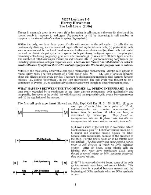
The first cell cycle experiment [Howard and Pelc, Exptl Cell Res 51: 2: 178 (1951)]. (1) grow<br />
root tips of vicia faba, do a pulse of 32 P, do<br />
INTERPHASE<br />
DNA Synthesis Gap<br />
M M<br />
Gap DNA Synthesis Gap<br />
M M<br />
Gap DNA Synthesis Gap<br />
M M<br />
S<br />
G 1<br />
G 2<br />
radioautographs, and examine incorporation of<br />
isotope into the nucleus. M takes one hour, as<br />
determined by microscopy. They found no<br />
incorporation into the M phase cells, but did see<br />
incorporation into some, but not all, interphase cells.<br />
(2) Grow a series of the root tips in colchicine, which<br />
blocks mitosis, plus 32 P. Label for various times, (2, 4,<br />
6 hours) and examine mitotic figures for label.<br />
Mitotic cells accumulate, because of the presence of<br />
the drug. For the first 6 hours or so no mitotic cells<br />
are labeled! The data imply that there exists a period<br />
prior to cell division in which no DNA synthesis<br />
occurs. After six hours, some mitotic cells are<br />
labeled; they must have synthesized DNA, gone<br />
though a period where no DNA synthesis occurred,<br />
then entered mitosis.<br />
(3) If 32 P is removed after 6-8 hours, some of the cells<br />
go into mitosis much later, and are not labeled. This<br />
implies there is a period between mitosis and the<br />
beginning of DNA synthesis when no DNA synthesis<br />
occurs.<br />
1
Let’s do a more modern experiment; pulse-label cultured cells with radioactive thymidine, a DNA<br />
precursor. Then wash, put the cells in colchicine, and do autoradiographs after this pulse label.<br />
(4) If we plot the<br />
data obtained for<br />
labeled mitoses in the<br />
presence of colchicine<br />
we get these results:<br />
:<br />
G 2<br />
M<br />
S<br />
G 1<br />
G 2<br />
S<br />
Total number of<br />
mitotic figures<br />
Total number of labeled<br />
mitotic figures<br />
M G 1 S G 2 M<br />
Putting these data together, we get a STANDARD CELL CYCLE<br />
REPRESENTATION. From other data, which I won’t describe, we<br />
can make several generalizations: (i) the S, G2 and M periods of the<br />
cycle are pretty constant. (ii) Differences in cycle time are usually<br />
due to alterations in G1. (iiii) in non-dividing tissues most cells are<br />
frozen in G1, or a distinct cell cycle phase, called "Go".<br />
AS A RESULT OF THIS EXPERIMENT, WE NOW KNOW INITIATION AND<br />
TERMINATION OF DNA SYNTHESIS ARE DISCRETE CYCLE EVENTS.<br />
DETERMINING THE CELL CYCLE PARAMETERS; G1, S, G2 and M:<br />
Number of<br />
labelled<br />
mitoses<br />
G 2 S G 2 + M + G 1<br />
Time<br />
The second procedure for determining cell cycle<br />
parameters is the use of flow micro-fluorimetry<br />
(FMF). One stains cells with a dye that causes DNA<br />
to become fluorescent. The amount of dye taken up<br />
by any cell is proportional to the DNA content of that<br />
cell. One then determines the amount of binding of<br />
dye per cell, using a laser. Individual cells go through<br />
the laser beam, and the data are analyzed by a<br />
computer.<br />
The classic method is known as labeled<br />
mitoses. One counts labeled mitoses after a<br />
pulse with a DNA label such as [ 3 H]thymidine.<br />
The generation time, GT = G1 + S<br />
+ G2 + M. M can be determined as the<br />
percent of cells that appear as mitotic figures<br />
in a randomly growing population of cells.<br />
The two major events common to all eukaryotic cell cycles are the replication of<br />
DNA (i.e, the replication of chromosomes during S phase) and chromosome<br />
segregation during mitosis (i.e, the segregation of DNA to the sibling cells).<br />
If extended, the human genome has seven feet of DNA. Faithfully replicating all this only once per<br />
cycle, and accurately packaging it for condensation and separation in individual chromosomes is a<br />
major problem. Moreover, this DNA has to be replicated and packaged during a specific portion of<br />
the cell cycle (S), and segregated exactly to two sibling cells during another cell cycle portion (M).<br />
2
A number of eukaryotic systems are used to study the cell cycle, including fly embryos, clam eggs,<br />
sea urchin eggs, yeast --where genetics can be brought into play--, and mammalian cells in culture.<br />
While I will be emphasizing mammalian somatic cells, I will draw on data from the other systems –<br />
particularly yeast. There are differences in localization, activity, and function of cell cycle<br />
components in these various systems -- not all cell cycles in all organisms are alike. However, data<br />
have shown that the cell cycle is so similar from organism to organism in eucaryotes that many<br />
basic principles can be extrapolated among all eucaryotes.<br />
Tissues in the organs of a mammal (like us!) are mixed populations of Go, randomly spaced G1, S,<br />
and G2 cells, with some cells in cycle, and others having exited the cycle. We want to study the<br />
biochemistry of the cell division process; to identify temporal points in the cell cycle. In most<br />
somatic tissues the greatest portion of cells are not dividing. We want to use populations of<br />
homogeneous, growing (i.e., dividing) cells. In the lab, to study the cell cycle of mammalian cells,<br />
we study mostly immortal cell culture cell lines derived from single cells -- BHK, CHO, 3T3, etc.<br />
We will talk about cell cycle mutants, and how these studies have contributed to our understanding<br />
of the cell cycle.<br />
SYNCHRONIZING CELLS: To study most of the major landmarks in the cell cycle we need to<br />
have homogeneous populations of cells traversing the cycle at the same time; i.e., we must have<br />
synchronous cells, whether we are studying yeast, fly, mouse or human cell cycles. What we do to<br />
study many of the events in the cycle is to (1) synchronize a randomly dividing population of cells<br />
and (2) study a given parameter as a function of cell cycle position, after release from the synchrony<br />
point. There are three major methods used to synchronize cultured cells.<br />
[1]. WE CAN SYNCHRONIZE CELLS BY BLOCKING AT SOME POINT IN THE<br />
CELL CYCLE AND ALLOWING CELLS TO ACCUMULATE AT THE BLOCK.<br />
(1) Cells can be starved for isoleucine, for PO4, or for serum. These starvation paradigms will<br />
block many cell lines in G1 [Ley and Toby, J. Cell Biol 47: 453 (1970), Xeros, Nature 194:<br />
682 (1962)], at a point referred to as the “restriction point”.<br />
(2) One can block DNA synthesis, by using drugs such as hydroxyurea, which interferes with<br />
dATP production, or amethopterin, FUdR, or aphidicolin. One of the most popular methods<br />
is the use of a high concentration of thymidine, which feedback-inhibits the production of<br />
dCTP synthesis. These drugs, in many cases, can be reversed and the cycle can be<br />
continued. One can wash out HOU; or reverse high thymidine with a wash.<br />
USING A SINGLE BLOCK TO SYNCHRONIZE CELLS:<br />
DNA synthesis<br />
inhibitor<br />
M G 1<br />
M<br />
G 1<br />
G 2 S (time greater G 2 S<br />
than G 2 + M<br />
+ G 1 )<br />
Release these cells<br />
from inhibition, and<br />
count cell number<br />
3<br />
Cell Number<br />
G 2 + M S
Note: all agents that inhibit DNA synthesis block the cells in very early S, and not at the G1/S<br />
transition, as is often claimed. By using this procedure we can determine [G2 + M] and S. One can<br />
determine G1 by subtraction, since we know the generation time [GT] = G1 + S + G2 + M.<br />
USING A DOUBLE BLOCK TO SYNCHRONIZE CELLS:<br />
Release single blocked cells for a period greater<br />
than S, but less than the sum of G2 + M + G1.<br />
Add back an inhibitor of DNA synthesis. All<br />
cells will be synchronized at "the G1/S transition".<br />
G 1 G1<br />
G 1<br />
M M M<br />
G 2 G G 2<br />
2<br />
S -DNA S +DNA S<br />
inhib<br />
inhib<br />
[2]. WE CAN SYNCHRONIZE CELLS BY MITOTIC SELECTION:<br />
Cell Cell number number<br />
Release these cells<br />
from inhibition, and<br />
count cell number<br />
S + G 2 + M<br />
Many cells grow attached to the culture dish, because the microtubules hold them in an extended<br />
shape. At mitosis, the cells round up and are only loosely attached to the dish, because the<br />
microtubules are dissociated and used to form the mitotic spindle to separate the chromosomes.<br />
One can shake off the mitotic cells, or wash them off the surface of the dish with a stream of<br />
medium. You can then replate these cells and have synchronized M/G1 cells [Robbins and Marcus,<br />
Science 144: 1152 (1964)].<br />
The advantage of mitotic selection is that there is no biochemical manipulation. The disadvantage<br />
of mitotic selection is that, since only ~5% of the cells will be in M at any given time; one gets only<br />
5% of the cells in synchrony with the wash. One can increase the yield by blocking cells at M with<br />
a drug like nocodazole (a drug which reversibly prevents microtubule polymerization). One then<br />
collects the mitotic cells, washes, and gets synchronized M/G1 cells to replate.<br />
SO-- MITOTIC SELECTION GIVES US CELLS AT M/G1. DNA INHIBITION<br />
PROCEDURES GIVE US CELLS EITHER DISTRIBUTED THROUGHOUT S, OR AT<br />
G1/S.<br />
[3]. WE CAN SYNCHRONIZE CELLS BY CENTRIFUGAL ELUTRIATION. Cells<br />
increase in size as they go through the cycle; from M to G1 to S to G2. The centrifugal elutriation<br />
rotor isolates cells that are increasingly larger in size. One can then check the position of the cells in<br />
the cycle by using flow microflourimetry (FMF) to determine DNA content [Meistrich et al, Exptl.<br />
Cell Res. 105: 169 (1979)].<br />
Cell<br />
number<br />
G 1<br />
S<br />
G 2<br />
Control early mid-E mid-L late<br />
fluorescence intensity<br />
4
3 H TdR<br />
HL LL<br />
Density<br />
Chicken cells<br />
BIOCHEMICAL CHANGES IN THE CELL CYCLE: DNA SYNTHESIS.<br />
cpm<br />
G 1<br />
RATE<br />
S<br />
G 2<br />
cpm<br />
G 1<br />
RATE<br />
AMOUNT<br />
There are a large number of questions to be addressed: What initiates DNA synthesis; i.e., is the<br />
signal positive for S, or is DNA synthesis repressed in G1 and G2? Does DNA synthesis depend on<br />
RNA synthesis? Does DNA synthesis depend on protein synthesis? How is DNA replication<br />
restricted to once per cell cycle for each gene? How does chromatin structure change during the cell<br />
cycle? How is chromatin assembled following DNA replication? Is DNA synthesized in a random<br />
or an orderly fashion -- does DNA synthesized early in one cycle always get synthesized early in a<br />
second cycle? When are specific genes replicated? When are enzymes involved in DNA replication<br />
synthesized? Does DNA damage modulate DNA synthesis?<br />
QUESTIONS CONCERNING DNA SYNTHESIS:<br />
Is DNA synthesized sequentially or randomly? [Mueller and Kajiwara, BBA 114: 108 (1966)]<br />
Cytochemical evidence suggest there exist "early replicating" and "late replicating" DNAs. Is DNA<br />
that is synthesized early in one cell cycle again synthesized early the next cell cycle?<br />
Optical<br />
density<br />
(1) Synchronize HeLa cells with a double-thymidine block at G1/S (S = 6hr).<br />
(2) Release from block, add [ 3 H]-TdR for 3 hours (ie, during the first half of S). Then<br />
switch the cells to medium containing non-radioactive, "cold" thymidine.<br />
(3) Grow the [ 3 H]-TdR labeled cells for a few generations. (They will loose synchrony).<br />
(4) Resynchronize the cells with a second double-TdR block at G1/S. Release in the<br />
presence of BudR for 3 hours.<br />
(5) Isolate DNA, shear the DNA, and centrifuge on CsCl gradient.<br />
(6). Follow radioactivity and density. If early replicating DNA is resynthesized early,<br />
then [ 3 H] will always go with the BUdR density label. If DNA synthesis is random,<br />
then [ 3 H] will be present in all fractions. RESULT: [ 3 H] AND BUdR TRAVEL<br />
TOGETHER. CONCLUSION: DNA SYNTHESIZED EARLY IN ONE<br />
REPLICATION CYCLE IS SYNTHESIZED EARLY IN THE NEXT CYCLE.<br />
CELL FUSION EXPERIMENTS SUGGEST MECHANISMS FOR<br />
CELL CYLE TRANSITIONS.<br />
Is initiation of DNA synthesis due to (1) a positive signal acting on/in S phase nuclei or (2)<br />
release from a negative signal acting on G1 or G2 cells? To put this another way, are there G1<br />
and G2 phase products which inhibit DNA synthesis, or S phase products which initiate DNA<br />
synthesis? [Rao and Johnson, Nature 225: l59 (1970)].<br />
Human cells<br />
Flu virus or<br />
polyethylene glycol<br />
Use cells that have morphologically distinct nuclei. They fused together, using<br />
inactivated sendai virus, a G1 phase cell and an S phase cell from different species,<br />
to make a "heterokaryon" – a cell with two nuclei in a common cytoplasm. Then<br />
they added [ 3 H]-TdR, and prepared autoradiographs, to identify labeled nuclei. The<br />
S phase nucleus makes DNA. In addition, the G1 nucleus is induced to make DNA.<br />
Fuse a G2 and an S cell. G2 nucleus makes no DNA; S nucleus makes DNA.<br />
Fuse a G2 phase and a G1 phase cell. No DNA synthesis in either nucleus.<br />
5<br />
S<br />
G 2<br />
M<br />
2X<br />
1X
THESE FUSION EXPERIMENTS SUGGEST [1] THAT DNA SYNTHESIS OCCURS IN<br />
RESPONSE TO A POSITIVE, NONSPECIES SPECIFIC INDUCING FACTOR PRESENT<br />
IN S PHASE CELLS AND [2] THAT G2 CELLS –WHICH HAVE DUPLICATED THEIR<br />
DNA -- HAVE INFORMATION THAT TELLS THEM TO IGNORE THE SIGNAL.<br />
Is there a "dominant factor" for chromatin condensation, or is there an inhibitor of THIS<br />
mitotic activity in interphase cells? [Johnson and Rao, Nature 226: 717 (1970)]. Fuse a mitotic<br />
cell to any interphase cell (in G1, S or G2), and the interphase cell undergoes premature<br />
chromosome condensation, or PCC. The mitotic cell is "dominant"; something present in mitotic<br />
cells can initiate chromosome condensation in interphase cells! What might this be?<br />
HISTONE SYNTHESIS<br />
C<br />
B<br />
Fraction number<br />
try ptophan<br />
arginine<br />
lysine<br />
A3<br />
A2<br />
A1<br />
Histones are synthesized in S phase. A large percent of<br />
the cell protein is histone; the histones must be<br />
duplicated, get into the nucleus, and associate with the<br />
DNA for each cell cycle. We will define histones as<br />
chromosomal (nuclear) proteins that are high in arginine<br />
and lysine, low in tryptophan and are acid extractable.<br />
The first experiment was done by Robbins and Borun<br />
[PNAS 57: 409 (1969)]. They labeled cells with [ 3 H]lys,<br />
[ 3 H]-arg or [ 3 H]-tryp, then prepared nuclei, extracted<br />
other proteins first with NaCl, then extracted histones<br />
with 0.25N HCl, ran on SDS PAGE gels, and<br />
determined radioactivity to identify histones.<br />
To determine when in the cell cycle histones are synthesized, they synchronized cells at M/G1 (how<br />
might they do this?), released, pulsed with radioactive [ 3 H]-lys/arg at various times (shown in the<br />
figure below as 1, 2 and 3), and measured histone synthesis. Histones are only synthesized in S<br />
phase. How is transcription of the histone genes restricted to S-phase??<br />
M/G 1<br />
1<br />
2<br />
3<br />
Fraction number<br />
If one adds cytosine arabinoside, an inhibitor of DNA synthesis, histone synthesis ceases!! Tight<br />
coupling exists between DNA synthesis and histone synthesis.<br />
HISTONE MODIFICATIONS DURING THE CELL CYCLE. Histones undergo a variety of<br />
modifications; including acetylation, phosphorylation, ADP ribosylation, ubiquitination, sumolation,<br />
and methylation. These modifications are primarily on the flexible, basic domains in the N and C<br />
termini of the histones.<br />
Histone phosphorylation: There is an ordered pattern of histone phosphorylation during the cell<br />
cycle. The data for histone H1 is best described. (1) H1 is not phosphorylated in resting cells. (2)<br />
Two hours prior to DNA synthesis, H1 begins to be phosphorylated. Thus "old" G1 histone (i.e,<br />
histone synthesized in previous cell cycles) --for that cell cycle-- is phosphorylated. (3) H1<br />
phosphorylation increases somewhat in S. (4) H1 phosphorylation continues to increase in G2 and<br />
in the first part of M. (5) In mitosis, H1 is rapidly and dramatically dephosphorylated.<br />
6
Histone Kinase: Zellig and Langan [BBRC 95: 1372 (1980)] showed that histone H1 kinase<br />
activity, measured in extracts of synchronized cells, increased 4-6 fold from late G2 to mid<br />
metaphase. The activity disappears at the end of M!! The H1 kinase enzyme activity had a half-life<br />
of 30 minutes. Is this due to enzyme activation? synthesis? Degradation? Remember this paper; it<br />
was "before its time".<br />
THE DISCOVERY OF CYCLIN, ANOTHER PROTEIN SYNTHESIZED CYCLICALLY<br />
DURING THE CELL CYCLE: In l983 Evans et al [Cell 33: 389 (1983)], using gel<br />
electrophoresis, observed that a protein builds up in fertilized sea urchin eggs during interphase,<br />
reaches a maximum just before/during mitosis, and is rapidly degraded in mitosis. They named this<br />
protein "cyclin". We will not talk much about embryonic systems, since I want to emphasize<br />
somatic cells. We now know that all cells contain a homologue of this sea urchin cyclin, cyclinB.<br />
This is the experiment that lead to the Nobel Prize for Tim Hunt in 2001.<br />
G 1 + S + G 2 G 1 + S + G 2 G 1 + S + G 2<br />
M M M<br />
RESCUING YEAST CDC MUTANTS WITH A HUMAN GENE: THE cdc28/cdc2 STORY.<br />
Lee Hartwell isolated temperature sensitive (ts) mutants of the yeast S. cerevisiae that – when<br />
grown at the “permissive temperature” (23 o C) – grow well. But, when shifted to the “nonpermissive”<br />
temperature (36 o C), these mutants arrest at specific points in the cell cycle –<br />
presumably the point at which the function of the gene in question is needed; what Hartwell termed<br />
the “execution point” for the function of that gene. One can easily tell where in the cycle the cdc<br />
cells are, when they arrest, by the size of the bud. These are the experiments that lead to the Nobel<br />
Prize for Lee Hartwell in 2001. Hartwell called these “cell division cycle” or cdc mutants.<br />
When shifted to the restrictive temperature, the S. cerevisiae cdc28 mutant arrests at a point in G1<br />
called START. This is the point where the cell makes its decision to initiate DNA synthesis. Paul<br />
Nurse isolated similar mutants in the fission yeast, S. pombe. cdc2 is a ts cell cycle mutant in S.<br />
pombe. S. pombe cdc2 mutants arrest predominantly at G2/M at the non-permissive temperature.<br />
What was surprising was that the S. cerevisiae cdc28 mutant and the S. pombe cdc2 mutant<br />
complement one another, even though there is great evolutionary distance between these two strains<br />
and that the former arrests at START and the latter at G2/M. However, closer analysis showed that<br />
the cdc28/cdc2 gene product actually controls both functions in both yeast strains. Because of<br />
difference in other aspects of their biology, they arrest predominantly at different points, but the<br />
cdc28/cdc2 gene is actually responsible for both functions in both strains.<br />
S. pombe can (amazingly) use a promoter from the SV40 monkey virus to express foreign genes.<br />
Lee and Nurse [Nature 327:31 (1987)] transfected a human cDNA expression library, with cDNAs<br />
driven by the SV40 promoter, into a cdc2 ts/leu - S. pombe strain. They cotransfected with a leu+<br />
plasmid and let the cells grow for a while at the permissive temperature, to first select for leu+<br />
transformants. They then plated at the restrictive temperature, and selected for growth. From one<br />
million leu+ transfectants they got 5 that could grow at the restrictive temperature. From 2 of these<br />
they could recover plasmids containing human expression cDNAs that could correct a cdc2<br />
mutation! Thus this human gene complements the yeast cell cycle mutant. The yeast cdc2 gene<br />
product is a 34 kd phosphoprotein, known at that time to have protein kinase activity in vitro. What<br />
are the properties of the human cdc28/cdc2 homologue? It is also 34 kd, is phosphorylated.<br />
Moreover the p34 cdc human homologue – like the products of the cdc2/cdc28 genes, is a protein<br />
kinase. This is the experiment that lead to the Nobel Prize for Paul Nurse.<br />
7
Draetta and Beach [Cell 54: 17 (1988)], using [35]S-labelled HeLa cells, found that the protein<br />
product of the p34 gene, the cdc2 protein kinase, complexes with a protein called p62.<br />
Percent<br />
cells in<br />
each phase<br />
G 1 S G 2<br />
2 4 6 8 10 12 Fraction<br />
p34<br />
p62<br />
p34<br />
p62<br />
casein<br />
[35]S-labelled HeLa cells were fractionated by<br />
elutriation. The p34 level [determined by western<br />
blots (row 1 of the figure)] does not change much<br />
during the cycle. However, p34 associates with a 62<br />
kd protein, p62, during G2, as shown by coimmunoprecipitation<br />
of [35]S-labeled proteins with<br />
antisera to p34 (row 2). Moreover, p34 kinase<br />
activity is greatly elevated in G2/M, using the p34IP<br />
for casein kinase activity (row 3). In addition, p62 is<br />
a substrate for p34 kinase (row 3)! The kinase activity<br />
– but not the p34 protein itself – is eliminated as a<br />
part of the M to G1 transition. The data suggest that<br />
p34 associates with p62 as a function of cell cycle<br />
position, and that p62 association is necessary for<br />
activation of p34 kinase activity on casein and on<br />
p62.<br />
The p62 protein is a cyclin. Several experiments, between 1989 and 1990, identified the molecular<br />
nature of the p62 protein.<br />
1. The yeast cdc13 gene encodes a 62 kilodalton, phosphorylated protein, p62, that complexes with<br />
yeast p34. The yeast cdc13/p62 gene was cloned [Bodner and Beach, EMBO J. 7:2321 (1988),<br />
Solomon, Cell 54: 738 (1988)] and sequenced. The product of the yeast cdc13 gene, the p62<br />
protein, is yeast cyclin!<br />
2. Is the p62 protein that associates with p34 in mammalian cells a cyclin? In experiments carried<br />
out at about the same time, Langan et al [MCB 9: 3860 (1989)] (remember that paper that "was<br />
before its time", published 8 years earlier?) showed that the mammalian histone kinase active at the<br />
G2/M time period also contains p34cdc2 and cyclin. Thus it appears that a complex of p34 kinase<br />
and cyclin (p62) is the active G2/M histone kinase; cyclin converts a relatively inactive p34 kinase<br />
to a histone kinase. A cell cycle dependent heterodimer of the p34cdc2 protein kinase and cyclin is<br />
formed to activate histone kinase activity. Loss of cyclin as the cell goes from M into G1 eliminates<br />
the M-phase specific histone kinase activity. We now call this cdc2 p34 protein kinase enzyme a<br />
cyclin-dependent kinase, or cdk.<br />
Cdc2<br />
p34<br />
Cdc2<br />
p34<br />
Inactive kinase<br />
+ cyclin + cyclin<br />
Cdc2 cyclin cyclin<br />
p34<br />
Cdc2<br />
p34<br />
cyclin cyclin<br />
active cyclin-dependent kinase<br />
HISTONE + ATP HISTONE-P + ADP<br />
8
The human cyclin B gene: Pines and Hunter [Cell 58: 833 (1989)] isolated a cDNA for a HeLa cell<br />
cyclin, cyclin B, expressed the protein and made antibodies to it. They synchronized HeLa cells at<br />
the G1/S transition by sequential thymidine and aphidicolin blocks. Following release, cyclin B<br />
RNA increased four fold from S to G2, and was relatively low in other phases of the cell cycle.<br />
[This may be the first mRNA to clearly be shown to be induced in G2.] When cells were collected<br />
in mitosis with nocodazole, then released at the M/G1 interface, the level of p62/cyclin mRNA<br />
plunged dramatically in the M to G1 transition. Northern analyses are shown below:<br />
0 2 4 6 8 10 12 14 16 18 20 22 24<br />
G 2 /M peak<br />
Hours after release from G G1/S 1/S<br />
Cyclin<br />
Histone 2A<br />
0 2 4 8 16<br />
Hours after release from mitosis<br />
The cyclin protein also was low in S, and increased in G2 (western blots).<br />
0 2 4 6 8 10 12 14 16 18 20 22 24<br />
G 2 /M peak<br />
Hours after release from G G1/S 1/S 1/S<br />
Intensity of cyclin,<br />
abitrary units<br />
10 15 20 30<br />
Hours after release from G 1 /S<br />
Cyclin protein<br />
Cyclin protein<br />
Intensity of cyclin,<br />
abitrary units<br />
0 0.5 1.0 1.5 2 4 5<br />
Hours after release from mitosis<br />
0 0.5 1.0 1.5 2 4 5<br />
Hours after release from mitosis<br />
Cyclin<br />
The cyclin of Hela cells is destroyed in the transition from M to G1, just as it is in frog eggs,<br />
clam eggs, sea urchin eggs and yeast. Pines and Hunter also showed that the Hela cell p34/cyclin<br />
complex is an H1 kinase, with highest activity in mitotic cells. To repeat: active H1 kinase is a<br />
heterodimer composed of a stable p34 subunit with latent kinase activity and cyclin -- a protein<br />
made and destroyed cyclically.<br />
MULTIPLE CYCLIN GENES ARE PRESENT IN CELLS<br />
Multiple S and G2 cyclins in mammalian cells. Pines and Hunter cloned a second cyclin, cyclin<br />
A, from Hela cells. [Nature 346: 760 (1990)]. Cyclin A is expressed slightly before before cyclin B<br />
(in S, G2, and M), and is degraded before cyclin B in mitosis. Yeast have now been shown to have<br />
six B-type cyclins; Clb1-Clb6.<br />
G1 "cyclins" in yeast. Recall that p34 cdc2/cdc28 is required at START and at G2/M in budding<br />
yeast. Three yeast "cyclins" required in G1 were isolated [Hadwiger, PNAS 86: 6255 (1989)]. They<br />
are referred to as “cln” cyclins. In addition to p34cdc28 kinase, these cln proteins are required to<br />
pass through START. Co-immunoprecipitation experiments demonstrated that the p34cdc28 protein<br />
forms complexes, active as protein kinases, with G1 cln cyclins [Wittenberg, Cell 62: 225 (1990)].<br />
9
Overlapping "G1 cyclins", "S cyclins", and "mitotic cyclins". A great deal of genetic and<br />
biochemical work has now characterized the cyclins necessary for cell cycle transitions in yeast.<br />
The Cln1-3 cyclins [which we can term “G1-cyclins”] activate p34/cdc2 kinase for G1 progression<br />
through START. The B-type cyclins Clb5 and Clb6 [which we can term “S-cyclins”] are required<br />
for S phase initiation. Clb5 and Clb6 are synthesized late in G1, overlapping Clns 1-3. Clns (G1<br />
cyclins) are first required at START, then the Clb5/6 proteins are required for S phase progression.<br />
The Clb1-4 cyclins [which we can term “M-cyclins”] are required for the completion of mitosis.<br />
Cyclins specific for various phases exist for the yeast cell cycle.<br />
The yeast p34/cdc2/cdc28 cyclin-dependent kinase can interact with different cyclins, at<br />
different phases of the yeast cell cycle, and phosphorylate alternative substrates necessary for<br />
various functions.<br />
Substrate a<br />
Substrate b<br />
Substrate c<br />
Cdc2 Cdc2 Cdc2<br />
Substrate d<br />
Substrate e<br />
Substrate f<br />
Substrate g<br />
Substrate h<br />
Substrate i<br />
G1 cyclins in mammalian cells. Using yeast mutants defective in cln1, cln2 and cln3 (the yeast G1<br />
cyclin genes), human "cyclin" genes that can complement this defect have been isolated [Lew et al,<br />
Cell 66: 1197 (1991)]-- using procedures much like the original cloning (by Lee and Nurse) of the<br />
human p34 cdc2 homologue. These genes are called cyclins C, D, and E. There are, in fact, three<br />
different D-type cyclins; D1, D2 and D3. The C, D and E cyclins are synthesized in G1 in<br />
mammalian cells! The same concept of G1-cyclins, S-cyclins and M-cyclins is also true for<br />
mammalian cells.<br />
10
DISTINCT P34CDC2-LIKE GENES AND PROTEINS IN MAMMALIAN CELLS.<br />
Pines and Hunter showed that cyclin A in human tissue culture cells can also associate with a 33kd<br />
protein that "is related to but distinct from p34cdc2." This protein complex also has H1 kinase<br />
activity, suggesting there may be a family of p34cdc2-like proteins. This protein became known as<br />
cdk2.<br />
A family of mammalian cyclin-dependent protein kinases, or CDKs, was subsequently cloned.<br />
Cdk2 protein associates with cyclin A [Tsai et al, Nature 353: 174 (1991)]. Subsequently a family<br />
of at least ten proteins related to p34cdc2 was cloned [Meyerson et al, EMBO J. 11: 2809 (1992)].<br />
Thus, although in yeastcdc2/cdc28 appears to control both G1/S and G2/M transitions, in<br />
mammalian cells a family of related genes are responsible for these (and possibly other) transitions.<br />
Mammalian cells have distinct cyclin dependent kinases and cyclins that catalyze the<br />
phosphorylation of alternative substrates required for completion of various cell cycle<br />
functions.<br />
WHAT ARE THE ROLES OF THE VARIOUS CYCLIN DEPENDENT KINASES, OR<br />
CDKs, AND THE VARIOUS CYCLINS IN MAMMALIAN CELLS?<br />
HOW DO THESE CATALYTIC KINASE SUBUNITS AND CYCLICALLY PRODUCED<br />
REGULATORY SUBUNITS REGULATE CELL CYCLE PROGRESSION AND<br />
TRANSITIONS?<br />
WHAT ARE THE SUBSTRATES PHOSPORYLATED BY THE CDKS?<br />
WHAT’S A CYCLIN? Anything with a “cyclin box” – a site for interacting with a CDK, and<br />
demonstration of an interaction with a CDK to increase its kinase activity. Here is an illustration<br />
of the value of having a whole genome sequence: by these molecular definitions, in budding<br />
yeast there are 22 cyclins and 5 CDKs. Nine cyclins can activate CDC28. CDC28 is the only<br />
CDK known to have an essential role in cell cycle progression in yeast [Andrews and<br />
Measday, Trends in Genetics 14: 66 (1998)].<br />
11
A summary of major cyclin and Cdk<br />
interactions in the mammalian cell cycle. We<br />
now know that, in mammalian cells, cyclin B<br />
and cyclin A, along with p34cdc2, are involved<br />
in the G2/M transition. Cyclin A:Cdk2 also<br />
seems to be involved in control of DNA<br />
synthesis. We also know that cyclins A and E,<br />
and cdk2, are involved in initiation of<br />
replication of DNA.<br />
The three D-type cyclins vary between<br />
cell types; they may carry out different<br />
functions when complexed with various cyclindependent<br />
kinases. It appears that complexes<br />
of cyclin E and/or cyclin D with Cdk2 and<br />
Cdk4 are involved in entry into G1.<br />
cdc2<br />
M<br />
cdc2 CDK? Cyclin<br />
cdc2 CDK? cdc2 CDK? ???<br />
Cyclin<br />
???<br />
Cyclin<br />
A&B<br />
G 2<br />
Cyclin<br />
B<br />
p cdc2<br />
p<br />
G 1<br />
CDK<br />
2,4& 5<br />
Cyclin<br />
D1,D2<br />
D3<br />
PCNA<br />
CDK2<br />
Cyclin<br />
E p107 &<br />
E2F<br />
S CDK2<br />
Cyclin<br />
A p107 &<br />
Cyclin<br />
E2F<br />
B<br />
Since the experiment of Lee and Nurse (page 7 of the lecture notes) in 1987, we have gone from<br />
descriptive biology to mechanistic studies of the cell cycle. This is why I referred to the Lee and<br />
Nurse experiment as a “landmark experiment,” leading to the Nobel Prize.<br />
.<br />
12<br />
cdc2
WHAT ARE THE SUBSTRATES OF THE CYCLIN-DEPENDENT KINASES?<br />
A CDK reaction is required for human cells to traverse the G1/S boundary. In traversing the<br />
cell cycle, human cells decide at the "restriction point" (similar to the S. cerevisiae START<br />
decision) whether to exit the cell cycle and enter Go, or to commit to another cell cycle. If cells<br />
commit to progression through the cell cycle, they activate a transcription factor, E2F, necessary<br />
to transcribe genes whose products are needed for DNA synthesis. In early G1, E2F is bound to<br />
another protein --we will call it the restriction boundary protein, or RB protein for now-- and<br />
E2F is not active as a transcription factor. RB protein is not phosphorylated in non-dividing, Goarrested<br />
cells, or in cells emerging into G1 from M. Cell cycle-dependent phosphorylation of RB<br />
releases E2F from the RB/E2F complex, and permits E2F to activate transcription of genes<br />
necessary to enter S. The CDK4/cyclin D1 complex, which forms as cells synthesize cyclin D1 in<br />
G1, phosphorylates RB protein, leading to release of active E2F transcription factor.<br />
RB must subsequently be dephosphorylated before cells complete mitosis, so that it can<br />
again monitor the decision to remain in the cell cycle or to exit the cell cycle and enter the Go<br />
phase, the next time the cell progresses to the restriction point.<br />
RB RB RB<br />
Cyclin<br />
c<br />
D1<br />
c Cyclin<br />
D1<br />
CDK<br />
4<br />
E2F E2F<br />
G o , Early G 1<br />
Cell cycle progression<br />
Late G 1 , Early S<br />
Among the genes transcriptionally regulated by E2F are c-myc, cyclin E (necessary to initiate DNA<br />
synthesis), B-myb, p107, pRB, E2F-1, E2F-2, cyclin A, p34cdk2, DNA polymerase alpha,<br />
HsORC1, dihydrofolate reductase, thmidine kinase, thymidylate sythase, PCNA, RRM2 and<br />
Histone 2A. [Lavia and Jansen-Durr, Bioessays 23: 221 (1999)].<br />
Recall that histones are transcribed only during the S phase of the cell cycle. How is this<br />
controlled? Each mammalian histone had a distinct promoter, with different cis-acting<br />
regulatory sequences that bind different transcription factors. Cyclin E expression is induced in<br />
response to E2F activation. Cyclin E forms a heterodimer with Cdk2. Zhao et al [Genes & Dev.<br />
12, 456-461(1998] identified NPAT (nuclear protein mapped to the AT locus) as a protein that<br />
interacts with, and is phosphorylated by, Cdk2/cyclin E. Using antibodies to NPAT, Zhao et al<br />
[Genes & Dev.. 14, 2283-2297 (2000)] found that NPAT localizes to histone gene complexes<br />
(you should read how this is done). Cyclin E also co-localizes to these sites [Ma et al, Genes &<br />
Dev.. 12, 2298-2313 (2000)]. Localization occurs in S phase, when histone transcription occurs.<br />
The idea is that cyclin E is expressed at G1/S, interacts with Cdk2, phosphorylates NPAT at the<br />
histone gene locus and that phosphorylated NPAT co-ordinates the transcription of the histone<br />
gene family, either by acting as a chromatin-rearranging machine, a co-activator, etc.<br />
NPAT NPAT NPAT NPAT<br />
pp<br />
pp<br />
NPAT NPAT NPAT NPAT<br />
NPAT NPAT NPAT NPAT X X X<br />
H1 H2A H2B<br />
Fold Induction Induction<br />
H4 H4-1 Control<br />
Cyclin E Cdk2<br />
NPAT NPAT NPAT NPAT<br />
pp<br />
pp<br />
pp<br />
pp<br />
NPAT NPAT NPAT NPAT NPAT NPAT<br />
pp<br />
pp<br />
pp<br />
pp<br />
pp<br />
pp<br />
RB RB<br />
RB<br />
NPAT NPAT NPAT NPAT NPAT NPAT<br />
pp<br />
pp<br />
pp<br />
pp<br />
pp<br />
pp<br />
p<br />
H1 H2A H2B<br />
If one transfects cells with a [histone<br />
promoter][luciferase] reporter gene, cotransfection<br />
with an NPAT expression vector<br />
increases transcription 10 fold. NPAT must be<br />
phosphorylated at a Cdk2/cyclinE site for this<br />
increase in transcription to occur.<br />
13
w.t. hpm CyclinAHA<br />
cdk2<br />
Histone H1<br />
p107<br />
E2F<br />
How do cyclin-dependent kinases choose their substrates? Mammalian<br />
The crystal structure of cyclin A-cdk2 has been solved (Jeffery et al,<br />
Nature 376: 313, 1998). A hydrophobic patch on the surface of cyclin A,<br />
in which the sequence is conserved in many cyclins, is apparent.<br />
Schulman et al [PNAS 95: 10453, (1998)] thought this patch might direct<br />
substrate recognition. Many of the substrates of cyclin A-cdk2 have the<br />
sequence RXLFG. Mutations were made in the cyclin A hydrophobic<br />
patch (mutant htm), complexes were purified by immunoprecipitation,<br />
and the ability of the cyclin A-cdk2 complexes to interact with<br />
and phosphorylate substrates was examined. The wild-type and mutated<br />
complexes could both phosphorylate H1. However, the mutant complex could not phosphorylate<br />
RB as well as the complex containing the wild type protein, and has no activity on several other<br />
[cyclin A][cdk2] substrates. These data suggest that, as predicted from the structural data, the<br />
substrate is chosen and presented to the kinase by interaction with the specific cyclin of the<br />
cdk/cyclin heterodimer.<br />
The big questions are:<br />
1. Which cyclins interact with which Cdk catalytic subunits?<br />
2. How are the activities of the various cyclin-dependent kinases regulated?<br />
3. What are the substrates for the various cyclin-dependent kinases?<br />
4. What cell functions are regulated by these CDK phosphorylations?<br />
5. What phosphatases reverse the cyclin-dependent kinase-mediated events (e.g., Rb,<br />
H1)?<br />
6. How are cyclins degraded?<br />
7. By what other mechanisms are the various cyclin-dependent kinase complexes<br />
activated and inactivated?<br />
THE CYCLINS AND THE CYCLIN-DEPENDENT CATALYTIC KINASE<br />
SUBUNITS ARE ALSO SUBJECT TO CYCLIC PHOSPHORYLATION.<br />
CyclinB (a G2/M-cyclin) accumulates in late S and in G2, and complexes with the p34cdc2 protein.<br />
Why, then, do we not see H1 kinase activity in G2, when cyclin B and p34cdc2 are complexed?<br />
Instead, we find the kinase activity shooting up in mitosis. (1) During early G2 --when the<br />
p34cdc2/cyclin complex exists, but its H1 kinase activity is not yet high-- the p34cdc2 protein is<br />
phosphorylated, on both tyrosine and threonine [Draetta and Beach, CELL 54: 17 (1988)]. The<br />
tyrosine/threonine phosphorylation occurs in the ATP binding site of p34, and inactivates the kinase<br />
--even though it is complexed with cyclin B. (2) When cells enter mitosis, the threonine and tyrosine<br />
of the p34 kinase in the complex is dephosphorylated, and kinase activity increases dramatically. We<br />
conclude that Tyr/thr phosphorylation negatively regulates p34/cyclin B M-phase kinase activity.<br />
Other studies suggest a distinct threonine (thr161) site on the p34cdc2 protein must be<br />
phosphorylated to activate M-phase kinase [Gould et al EMBO J. 10: 3297 (1991)].<br />
The amounts of cyclin B and p34cdc2 do not go up dramatically during the later part of G2<br />
and entry into M, but the kinase activity of the complex goes up dramatically at G2/M, as a<br />
result of dephosphorylation of the catalytic p34cdc2 subunit.<br />
14
HERE IS A SCHEMATIC OF THE PHOSPHORYLATIONS OF THE M-PHASE KINASE.<br />
WHAT ENZYMES ARE RESPONSIBLE FOR THE PHOSPHORYLATION AND<br />
DEPHOSPHORYLATION OF THE p34cdc2 PROTEIN IN THE PROGRESSION OF THE<br />
CELL CYCLE?<br />
Cycin B<br />
is<br />
degraded<br />
Active<br />
CDK!!<br />
T14Y15<br />
cdc2 T14Y15<br />
cdc2 T14Y15<br />
cdc2 T14Y15<br />
cdc2 T14Y15<br />
cdc2 T14Y15<br />
cdc2 T14Y15<br />
cdc2<br />
T161<br />
T14Y15<br />
cdc2 T14Y15<br />
cdc2 T14Y15<br />
cdc2<br />
Cyclin<br />
B<br />
T161<br />
T14Y15<br />
cdc2 T14Y15<br />
cdc2 T14Y15<br />
cdc2 T14Y15<br />
cdc2 T14Y15<br />
cdc2 T14Y15<br />
cdc2 T14Y15<br />
cdc2<br />
T161<br />
inactive p<br />
ATP<br />
p<br />
G1<br />
G1<br />
M G2<br />
PPase<br />
S<br />
T14 dephosphorylation<br />
Y15 dephosphorylation<br />
Cyclin<br />
B<br />
T14Y15<br />
cdc2 T14Y15<br />
cdc2 T14Y15<br />
cdc2 T14Y15<br />
cdc2 T14Y15<br />
cdc2 T14Y15<br />
cdc2 T14Y15<br />
cdc2<br />
p p<br />
Cyclin T161<br />
B p<br />
T14Y15<br />
cdc2 T14Y15<br />
cdc2 T14Y15<br />
cdc2 T14Y15<br />
cdc2<br />
p p<br />
Cyclin T161<br />
B<br />
1. What kinase phosphorylates p34cdc2 on threonine and tyrosine (T14Y15) in the active<br />
site to suppress the activity of this enzyme?<br />
2. What kinase phosphorylates p34cdc2 on threonine 161?<br />
3. What phosphatases dephosphorylate p34cdc2 on these residues as the cells traverse<br />
the cell cycle?<br />
15<br />
p<br />
(inactive)<br />
(still inactive)
Genetic studies in yeast demonstrate that the products of other genes influence the mitotic<br />
p34/cyclin complex activity.<br />
Mik 1<br />
wee1<br />
Cdc2<br />
p34<br />
cdc 25<br />
cdc 13<br />
(Cyclin)<br />
Interphase mitosis<br />
G 2<br />
M<br />
Three genes – wee1, mik1, and cdc25 – appear to regulate Cdc2<br />
kinase activity at G2/M. A schematic from the known genetics is<br />
shown. Gene products are shown as stimulating (by the arrows) or<br />
inhibiting (by the blunt bars) the activity of other gene products. The<br />
cdc2 gene product is the p34cdc2 kinase, the cdc13 gene product is<br />
cyclin. These two proteins, p34cdc2 kinase and cyclin, are shown<br />
acting in concert to induce mitosis. In the absence of both wee1 and<br />
mik1 cells can enter mitosis prematurely, without completing DNA<br />
synthesis, because they have too much CDK activity.<br />
Protein kinases that regulate M-phase [p34/cyclin B] kinase. The product of the yeast wee1<br />
gene is a both a tyrosine and a threonine/serine kinase. Using recombinant, bacculovirus-produced<br />
proteins, Parker et al [PNAS 89: 2917 (1992)] demonstrated that p34cdc2 by itself is not a substrate,<br />
but that the p34cdc2 component of the p34cdc2/cyclin B heterodimer is a substrate. Phosphorylation<br />
of p34cdc2/cyclin B blocked H1 kinase activity of an in vitro assembled H1 kinase (p34cdc2 and<br />
cyclin B complex).<br />
The human wee1 gene was cloned by rescuing a yeast wee1 mutant with a human cDNA expression<br />
library [Igarashi et al, Nature 353: 80 (1991)] (the Lee and Nurse experiment all over again).<br />
Overexpression of human wee1 in HeLa cells prevents cell cycle progression [McGowan and<br />
Russell EMBO J 12:75 (1993)]. [How would you do this experiment? What would the results look<br />
like, in a graph or table?] These data suggest that Wee1 must be inactivated for a cell to make a<br />
transition into M. Watanabe et al [EMBO J 14:1878 (1995)] demonstrated hyperphosphorylation<br />
and subsequent degradation of Wee1 in HeLa cells.<br />
wee1 cooperates with another gene, mik1 [Lundgren et al, Cell 64:1111 (1991)]. Mik1 is also a<br />
protein tyrosine kinase! Both genes are necessary for p34cdc2 tyr phosphorylation in vivo. wee1 and<br />
mik1 are essential for p34cdc2 phosphorylation.<br />
PROTEIN PHOSPHATASES THAT REGULATE THE M-PHASE KINASE.<br />
The yeast cdc25 gene product is a protein tyrosine and threonine phosphatase [Strausfeld et al,<br />
Nature 351: 242 (1991); Gautier et al, Cell 67; 197 (1991)]. The enzyme shows remarkable<br />
substrate specificity for tyrosine-phosphorylated p34cdc2. The enzyme can also dephosphorylate the<br />
adjacent threonine residue; it a both a ser/thr and a tyrosine phosphatase of remarkable specificity.<br />
Dephosphorylation of p34cdc2 protein by cdc25 protein phosphatase activates the kinase activity of<br />
the p34/cyclin complex.<br />
.<br />
Cdc 25<br />
p<br />
p<br />
p34 cyclin<br />
Wee 1<br />
p34 cyclin<br />
Histone Histone p<br />
Histone Histone p<br />
16
% G 2 + M<br />
The cdc25 gene product, a cdc2 phosphatase, appears to be expressed primarily in the G2 phase of<br />
the cell cycle, both in S. pombe [Moreno, Nature 344: 549 (1990)] and in Hela cells [Sadhu, PNAS<br />
87: 5139 (1990)].<br />
Hela Cells<br />
cdc 25 mRNA<br />
cdc 25 mRNA<br />
cdc 25 mRNA<br />
Hela cells Yeast cells<br />
% Septated<br />
% Septated<br />
M<br />
M<br />
Time (20 min intervals)<br />
Yeast cells<br />
Several mammalian homologues of Cdc25 have now been cloned. Cdc25C can dephosphorylate<br />
p34cdc2, the G2 Cdk. What phosphatases act on Cdk2, Cdk4, Cdk6, etc?<br />
cdc2<br />
Cyclin<br />
B<br />
PPase<br />
p<br />
p<br />
cdc2<br />
Wee 1 Cyclin<br />
B<br />
Cdc 25<br />
Cdc 25<br />
Histone<br />
p<br />
+ ATP<br />
p<br />
cdc2<br />
Cyclin<br />
B<br />
mitosis<br />
Histone p Histone p<br />
Cdc 25 message<br />
Phosphorylation activates the<br />
phosphatase activity of the Cdc25<br />
protein. The Cdc2 protein component<br />
of the Cdc2/cyclin B complex (the<br />
“histone kinase”) must be<br />
dephosphorylated at residues 14 and<br />
15. Active Cdc2/cyclin B histone<br />
kinase can also phosphorylate the<br />
Cdc25 phosphatase; moreover,<br />
phosphorylation of Cdc25 activates<br />
this protein phosphatase. Thus a "selfamplification"<br />
is set up, in which a<br />
small amount of active cdc2/cyclinB<br />
can activate the cdc25 phosphatase,<br />
which then activates more kinase.<br />
[Hoffmann et al, EMBO J. 12:53<br />
(1993)]. This sets up a cycle in which<br />
very rapid activation of p34cdc2/<br />
cyclin B histone kinase can occur.<br />
A Cyclin-dependent kinase phosphorylates and regulates cyclin-dependent kinases; cyclindependent<br />
kinase cascades. In order to be activated, p34 cdc2/28 must be phosphorylated on<br />
Thr161. The enzyme that carries out this phosphorylation is called Cdc2-activating Kinase, or CAK.<br />
Fisher and Morgan [Cell 78: 713 (1994)] demonstrated that CAK is composed of a cyclindependent<br />
protein kinase Cdk catalytic sub-unit called MO15 and a new G1 cyclin, called cyclin H.<br />
CAK can activate both Cdc2 (a G2/M Cdk) and Cdk2 (a G1 Cdk).<br />
17
NEGATIVE REGULATION OF THE CELL CYCLE, CYCLINS AND CDKS<br />
Eukaryotic cell progression through the cell cycle is regulated by the sequential formation,<br />
activation and subsequent deactivation of a series of cyclin-dependent kinases. Are there ways<br />
in which external agents can interfere with the sequential cyclin/cdk activations, and regulate<br />
progression through the cycle?<br />
Cdc28/Clb2 Cdc28/Cln2<br />
pET pETSIC1 pET pETSIC1<br />
Purified Sic1 inhibits the Clb2-associated<br />
kinase, but not the Cln2 kinase in in vitro.<br />
Hist H1<br />
HisHASic1 (pETSIC1) was added to yeast extracts from<br />
CLB2HA3 orGAL-CLN2HA3. After 30 min. incubaton at 4o HisHASic1 (pETSIC1) was added to yeast extracts from<br />
CLB2HA3 orGAL-CLN2HA3. After 30 min. incubaton at 4 C,<br />
Clb2 or Cln2, Sic1 and their associated protiens were<br />
immunprecipitated with 12CA5 Antibody, and H1 kinase<br />
activity was measured. In control lanes (pET), E. coli extracts<br />
from cells containing the empty vector were added.<br />
oC, Clb2 or Cln2, Sic1 and their associated protiens were<br />
immunprecipitated with 12CA5 Antibody, and H1 kinase<br />
activity was measured. In control lanes (pET), E. coli extracts<br />
from cells containing the empty vector were added.<br />
cdc28<br />
SIC 1<br />
p p<br />
Clb2<br />
cdc28<br />
Clb2<br />
SIC 1<br />
Histone Histone p<br />
p40/Sic1, regulates the G1/S transition in yeast.<br />
p40/Sic1 was originally identified as a protein that<br />
associates with cdc28/cyclinB complexes. It was<br />
purified and shown to inhibit the activity of Cdc28-<br />
Clb kinase in vitro [Mendenhall, Science 259:216<br />
(1993)]. In yeast, cdc28 p34 protein can complex<br />
with Clb2 (an S phase cyclin). When the p34<br />
cdc28/Clb2 complex is associated with p40/Sic1<br />
protein, this cdk/cyclin complex is inactive as a<br />
kinase. p40/Sic1 inhibition is specific, inhibiting p34<br />
cdc28 complexed to Clb5 and Clb2 (S/G2 cyclins),<br />
but not Cln2 (G1 cyclin). [Schwob et al, Cell 79: 233<br />
(1994)]. To enter S, p40/Sic1 protein is<br />
phosphorylated and then degraded, releasing the<br />
inhibition of the CDK activity. Think about the<br />
following question: how does Sic1 get degraded?<br />
Cyclin dependent kinase [CDK] activities are<br />
regulated by a group of molecules known as cyclindependent<br />
kinase inhibitors, or CKIs. Sic1 is now<br />
termed a cyclin-dependent kinase inhibitor, or CKI. The<br />
CKIs of yeast inactivate cyclin-dependent kinases even if<br />
tyr15 and thr14 are dephosphorylated and thr161 is<br />
phosphorylated.<br />
Recall that we wanted to ask the question “Are there ways in which external agents can<br />
interfere with the sequential cyclin/cdk activations, and regulate progression through the cycle?”<br />
The answer is “yes”, both in yeast and in mammalian cells.<br />
Regulation of a CKI joins the cell cycle and response to external stimuli that inhibit cell<br />
growth, both in yeast and mammalian cells. Yeast have two mating types, a and α. In response to<br />
α-factor, which is released by α-mating type yeast, a-mating type yeast exit<br />
the cell cycle in G1, and prepare to mate. What makes proliferating, haploid<br />
cells withdraw from the cell cycle at G1 in response to mating factors? The<br />
far1 gene is required for mating-factor induced cell arrest. Expression of the<br />
far1 gene is induced by binding of α-factor to the α-factor receptor<br />
expressed on type a cells. Far1 protein associates with p34/cdc28<br />
complexes to the G1 Cln1 and Cln2 proteins, and inactivates their<br />
kinase activity, but does not inhibit the activity of p34Cdc28/ClnB<br />
complex. Far1 is an inducible G1 cyclin-specific CKI [Peter and Herskowitz,<br />
Science 265: 1228 (1994)].<br />
18
We have seen that there are at least two yeast CKI molecules in yeast. p40/Sic1, a clb-specific<br />
cyclin, is a part of the intrinsic cell cycle machinery, regulating the G1/S transition. Far1 is a cln<br />
specific cyclin. Far 1 mutants have normal cell cycle control in proliferating cells; Far1, an "add<br />
on" to the cell cycle machinery, controls progression through the cell cycle in response to external<br />
stimuli.<br />
TGF-β is a “growth factor” that stops proliferation of human keratinocytes.<br />
p15 protein is also a CKI. TGF-β induces expression of p15 message and<br />
protein in human keratinocytes, producing an inhibitor of G1 Cdk/cyclin<br />
kinases [Hannon and Beach, Nature 371: 267 (1994)].<br />
Morgan [Nature 374:131 (1995)] summarized the principles of CDK regulation:<br />
19
PROTEOLYSIS RULES: MECHANISM OF MITOTIC CYCLIN DEGRADATION<br />
There are two excellent earlier reviews on this topic; King et al, Science 274: 1652 (1996), and<br />
Hoyt, Cell 91: 149 (1997).<br />
Before beginning the discussion of proteolysis and the cell cycle, we need to review the phases<br />
of mitosis, the transition from G2 to G1. Sister chromatid separation occurs in the transition<br />
from metaphase to anaphase. The degradation of cyclin B is required for the exit from<br />
telophase into the subsequent interphase.<br />
cdc2/28<br />
Cohesins<br />
cyclin deg.<br />
G 2 Prophase Metaphase Anaphase Telophase G 1<br />
-p34cdc2 +p34cdc2 -p34cdc2 +p34cdc2 0 15 30 50 0 15 30 50<br />
min min<br />
interphase mitotic<br />
Active cyclinB/p34cdc2 gets one into mitosis. How do we<br />
get out? To get out of the mitotic state we must inactivate<br />
the p34cdc2 kinaseWe also know that cyclin B must be<br />
degraded for the cell to exit the mitotic state. What is the<br />
signal for cyclin B degradation? How is cyclin B<br />
degraded? If [35S]radio-labeled cyclin is prepared<br />
and added to an interphase extract from frog embryo<br />
cells it is stable. If active p34cdc2 kinase (i.e.,<br />
[p34cdc2][cyclinB]) is also added, the [ 35 S] cyclin B is<br />
rapidly degraded [Felix, Nature 346: 379 (1990)]. What triggers the degradation of the<br />
radioactive cyclin in these extracts?<br />
Cyclin B degradation uses the ubiquitin system. Glotzer et al [Nature 349: 132 (1991)] used a<br />
system in which they generated a “stable mitotic extract” (you should read this paper and the<br />
relevant papers, to see how this was done). They also made a hybrid protein of the N-terminal of<br />
cyclin (containing what is now termed the "cyclin destruction box", or “D-box”; RXXLXXXXN)<br />
fused to protein A (for ease of purity of the radiolabeled, recombinant protein). Protein A binds to<br />
immunoglobulins, so the fusion protein can be purified on an immunoglobulin-coated bead. They<br />
showed this cyclin-protein A fusion protein, labeled with radioactive 125 I, could be degraded by<br />
mitotic extracts, as Felix et al showed for native cyclin (vida supra). Long exposure of the gels<br />
suggested a "ladder" of higher molecular weight forms. By using affinity binding of the 125 I-cyclinprotein<br />
A protein to IgG (used for both illustrations below) they showed convincingly that “stable<br />
mitotic extracts”, which degrade cyclin, form ubiquitin intermediates of the cyclin-protein A<br />
complex. Interphase extracts do not form the ubiquitin complexes. I suggest you read this paper.<br />
buffer<br />
buffer<br />
M extract<br />
M extract<br />
I extract<br />
Time 0 6 12 20 25 30 35 40 60<br />
20
Does p34cdc2/cyclin B activate a cyclin-specific enzyme involved in ubiquitination of cyclin in<br />
M? Does p34cdc2/cyclin kinase make cyclin a better substrate in M phase for a constitutive<br />
ubiquitination system?<br />
The protein complex that degrades ubiquitinated proteins in cells is called the “26S<br />
proteasome.” It appears to always be present in cells. The major question to answer is “What<br />
targets proteins destined for degradation to the proteasome in a cell-cycle specific manner<br />
(e.g., cyclin B in this case)?”<br />
cyclin B<br />
cdc2 p<br />
cdc2 p<br />
p p<br />
cyclin B<br />
cyclin B sythesis<br />
cdc25<br />
p p<br />
cdc2 p<br />
cdc2 p<br />
p p<br />
cyclin B<br />
cdc2 p<br />
cdc2 p<br />
cyclin B<br />
+<br />
cdc2<br />
protein X<br />
protein X<br />
p<br />
p<br />
protease<br />
Felix et al suggest that activated M-phase<br />
kinase (p34cdc2/cyclinB) phosphorylates a<br />
substrate, X, that leads to the activation of a<br />
protease that will then degrade cyclinB. We<br />
now know this degradation pathway includes<br />
phosphorylation and ubiquitination of<br />
cyclinB. New synthesis of cyclinB allows<br />
reactivation of the M-phase kinase in the next<br />
G2 period. A corollary of this hypothesis is<br />
that the protease that degrades G2 cyclins in<br />
M must be inactivated in the next cell cycle;<br />
otherwise cyclinB could not be degraded, and<br />
the next cycle could not occur.<br />
The components in clam extracts that ligate ubiquitin to cyclin B have been identified.<br />
[Hershko et al, JBC 269:4940 (1994)]. One component is active only from M-phase extracts.<br />
Moreover, this component can be activated from interphase extracts by p34cdc2/cyclin B, as<br />
suggested by Felix. The cyclin ubiquitinylation activity is part of a large protein multimer. This<br />
result suggests a mechanism by which the destruction of the cyclin of G2/M Cdk activity can be<br />
initiated and cells can make the transition to interphase. The enzyme complex that ubiquitinates<br />
cyclin B in M phase is called the “cyclosome” or the Anaphase Promoting Complex or APC<br />
(because it is necessary for the transition from G2 into anaphase). The cyclosome/APC marks<br />
cyclin B for degradation by ubiquitination, preparing cyclin B for degradation by the proteasome.<br />
Clearly, the cyclin B degrading system must be turned off prior to G2 of the following cell<br />
cycle, so that cells can proceed through a subsequent cell cycle; cells must be able to make<br />
“stable” cyclin B at a later time, for the next cell cycle.<br />
Cyclin<br />
B<br />
p<br />
Cyclin<br />
B<br />
proteasome<br />
APC<br />
Ub<br />
Ub<br />
p<br />
Cyclin<br />
B<br />
Ub<br />
Ub<br />
Ub Ub Ub Ub<br />
Ub UbUb<br />
What turns off the cyclin B<br />
degradation system – i.e., what<br />
turns off the cyclosome/APC – so<br />
cyclin B can be synthesized, and<br />
form the p34cdc2/cyclin B<br />
complex required for mitosis in<br />
the next cell division?<br />
Cyclin B proteolysis continues in G1 in yeast, until p34/cdc28 is reactivated by Cln G1 cyclins.<br />
Once the cyclin B degradation system --the APC-to-proteasome complex-- is triggered, how long<br />
does it stay active? Is cyclin B all degraded within the period of M, or can it keep being degraded as<br />
cells enter interphase?<br />
21
cyclin B RNA cyclin B protein H1 kinase activity<br />
Cyclin B2 (ClnB2) protein cannot accumulate in cells deprived of G1 cyclins. Amon et al [Cell<br />
77: 1037 (1994)] constructed a yeast cell mutated in the three Cln G1 cyclins, and carrying a<br />
methionine-suppressible G1 Cln2 gene. In the presence of met, these cells arrest in G1, because they<br />
are not expressing G1 cyclins. If these same cells carry a ClnB2 gene driven by the Gal promoter,<br />
one can regulate the expression of the cyclin B gene. If these cells are given methionine, they have<br />
no clns; they arrest in G1. When stimulated with galactose, they accumulate ClnB2 mRNA, but no<br />
ClnB2 protein or, therefore, H1 kinase activity. Cyclin B protein is degraded in G1. Expression of<br />
G1 cyclins is necessary to shut off cyclin B degradation.<br />
If the procedure described above is repeated with cells washed free of met, and given galactose,<br />
CLN2 expression will occur. Within 15 minutes, cyclin B protein and H1 kinase activity appear;<br />
degradation of the cyclin B protein is abrogated. Accumulation of G1 cyclins leads to inactivation of<br />
CLB proteolysis.<br />
Cyclin B is degraded in G1 if the G1 CLNs are not synthesized; i.e., until the G1 CLNs are<br />
synthesized. The APC remains active until it is (somehow) shut off by the G1-Cdk activity that<br />
requires G1 cyclins. The APC machinery, which marks cyclin B for degradation, is turned off<br />
when the G1 cyclins are synthesized.<br />
A summary of CLN production, CLB production and cyclin B destruction machinery in yeast.<br />
CLNs CLNs<br />
CLBs<br />
G1 S M G1 S M<br />
Start Finish Start<br />
Cyclin Oscillations in yeast<br />
CLBs<br />
APC mediated destruction<br />
machinery for cyclin B<br />
CLN1, CLN2 and CLN3 associated kinases are absent in early G1, reach peak levels<br />
at the G1-S transition, and decline as cells enter G2. These kinases not only promote<br />
budding and DNA synthesis, but also inactivate CLB cyclin proteolysis. Activation of<br />
the mitotic kinase during G2 causes repression of CLN1, CLN2 and CLN3 synthesis<br />
and onset of M phase and ultimately leads to the reactivation of CLB proteolysis.<br />
Cyclin B proteolysis then persists during G1 until the reactivation of CLN cyclins<br />
22
How is the APC/cyclosome activated to target cyclin B for degradation? Identification of the<br />
cell-cycle regulated component of the complex that targets cyclin B destruction by the<br />
proteasome. Identifying the genes that encode the protein components of the APC/cyclosome.<br />
WT<br />
Cdc16-123<br />
Cdc23-304<br />
Cse1-22<br />
pGAL-CLB2<br />
-GAL<br />
+GAL<br />
Irniger et al [Cell 81: 269 (1995)] made a ClnB2-β-gal fusion, and<br />
expressed this gene in the Amon et al [cln1-, cln2-, cln3-] mutant.<br />
When the cells are arrested in G1 (because G1 CLNs are not<br />
synthesized), colonies transcribe clnB2-βgal mRNA. Parental colonies<br />
remain white, because the fusion protein is degraded by the<br />
cyclosome/APC complex. If a cell is mutant in the cyclosome/APC<br />
cyclin B degrading system, colonies will be blue. They obtained three<br />
mutants -- all previously known genes; CDC16, CDC23 and CSE1.<br />
This question has also been approached biochemically. When one fractionates frog egg extracts for<br />
the components necessary to ubiquitinate cyclin B, the cell cycle-determining factor is a complex<br />
that contains the CDC27 and CDC16 proteins. [King et al, Cell 81:279 (1995)]. Immunodepletion<br />
of these proteins prevents cyclin B ubiquitination. This is the mitotic E3 component of the cyclin B<br />
ubiquitination complex suggested by Hersko (1994).<br />
Antibody staining demonstrated that human CDC27 and CDC16 proteins co-localize to<br />
centrosomes and mitotic spindles. It appears a complex of Cdc27, Cdc16 and other proteins is a<br />
machine – the cyclosome/APC – regulated in M, that mediates cyclin B destruction.<br />
NON-CYCLIN PROTEINS ALSO MUST BE TARGETED FOR<br />
DEGRADATION BY THE CYCLOSOME/APC COMPLEX FOR CELLS TO<br />
TRAVERSE MITOSIS.<br />
If a non-degradable cyclin B is present, cells do not arrest in anaphase. Sister chromatid separation<br />
still occurs, and the cells arrest in telophase. Therefore, cyclin B inactivation cannot be the trigger<br />
for anaphase sister chromatid separation. However, if one blocks APC activity, either genetically or<br />
by injecting antibodies to APC components, sister chromatid/chromosome separation is blocked;<br />
anaphase is not completed. These findings create a paradox: entry into anaphase does not require<br />
degradation of mitotic cyclins (e.g., cyclin B), yet anaphase remains dependent upon proteolysis<br />
initiated by the action of the APC. APC/cyclosome directed degradation of some other proteins –<br />
before cyclin is degraded – must be necessary for this sister chromatid separation -- i.e.,<br />
metaphase to anaphase -- to occur.<br />
CUT2 is a protein found in S. pombe that increases in anaphase, and accumulates in G1 if<br />
cells are mutant in APC components. The data suggest that CUT2 must be degraded by the APC for<br />
cell cycle progression to continue in M. Expression of a non-degradable form of CUT2 (without a<br />
D-box) blocks cells in anaphase, i.e., before they enter telophase. [Funabiki et al, Nature 381:438<br />
(1996)]. CUT2 degradation must occur before cyclin B degradation.. A similar story holds for a<br />
protein known as PDS1 in S. cerevisiae. (1) PDS1 is degraded during mitosis, and its degradation<br />
requires an active cyclosome/APC [Cohen-Fix et al, Genes and Development 10:3081 (1996)]. (2)<br />
Over-expression of non-degradable PDS1 (PDS1 without a D box) arrests cells in metaphase (i.e.,<br />
the cells don’t enter anaphase). (3) Degradation of PDS1 requires an intact D box and functional<br />
APC. PDS1 is the budding yeast equivalent of CUT2 in the fission yeast.<br />
CUT2 and PDS1 are also known as securins. Securin binds to a molecule called separase. Separase<br />
(the Esp1 protein in budding yeast, and the CUT1 in fission yeast) is a protease that is inhibited by<br />
securin. When CUT2/PGS1/securin is degraded by the APC/proteasome prior to anaphase, separase<br />
can be activated by proteolytic activity. Separase then cleaves the family of molecules known as<br />
cohesions, which hold the sister chromatids in anaphase together, allowing the sister chromatids to<br />
separate and anaphase to proceed. [Reviewed in Peters, Mol. Cell. 9:931 (2002); Yanagida, Genes to<br />
Cells 5:1 (2000)]].<br />
23
So – the APC/proteasome-directed destruction of proteins such as CUT2/PDS1/securin is<br />
necessary for cells to move from metaphase to anaphase. But mitotic cyclins (and some other<br />
proteins that are involved in spindle formation) are not destroyed until later in the mitotic<br />
sequence, after telophase, in order for cells to exit mitosis and enter G1.<br />
securin<br />
Proteolysis at metaphase, anaphase, telophase, G1 transitions [King et al, Science 274: 1652 (1996)]<br />
How is the APC activity regulated so that distinct proteins (e.g., CUT2/PDS1/securin versus<br />
cyclinB) are ubiquitinated by the APC, and delivered to the proteasome for degradation, at<br />
different times – at different stages – during the G2 to G1 transition through M?<br />
Schwab et al [Cell 90:683 (1997)] demonstrated that<br />
yeast cells deficient for a protein called HCT1/CDH1<br />
are impaired in their ability to degrade mitotic cyclin<br />
B2. In the experiment shown at the right, G1 phase<br />
wild-type yeast cells and G1 phase hct1 mutant cells<br />
were isolated by elutriation. At various times after<br />
isolation of G1 phase cells and continued growth,<br />
populations of cells were analyzed by western<br />
blotting for Clb2 protein. Hct1 mutant cells cannot<br />
degrade Clb2.<br />
These authors also demonstrated that HCT1 does not direct<br />
APC/proteasome activity to the PDS1/securin protein. In this<br />
experiment HCT1 is expressed from an inducible gal promoter.<br />
Cells were treated with galactose to induce HCT1 expression, and<br />
the ability of the cells to degrade both Clb2 and PDS1 was examined.<br />
Clb2 is rapidly degraded, PDS1/securin is not degraded.<br />
24
The data of Schwab et al suggest that HCT1 directs the activity of the APC/cyclosome<br />
ubiquitination system to mitotic cyclins, but not to proteins such as PDS1/CUT2/securin – a protein<br />
that must be degraded to initiate anaphase. In a similar experiment, Visintin et al [Science 278: 460-<br />
463 (1997)] demonstrated that the CDC20 protein directs the APC/cyclosome activity to<br />
PDS1/securin, but not to mitotic cyclins. Hoyt [Cell 91:149 (1997)] proposes the following model:<br />
Cdc20 and Htc1 have been cloned. When recombinant Htc1 is added to interphase<br />
cyclosome/APC preparations, the cyclin B ubiquitination ability of the complex is greatly<br />
stimulated (Fang et al, Mol. Cell 2: 163, 1998); i.e., the APC/cyclosome activity is stimulated.<br />
The APC cdc20 complex peaks in activity in M, and is responsible for Pds1p/securin<br />
proteolysis and anaphase onset. APC Cdh1 cyclosome activity peaks in Anaphase/G1, and is<br />
responsible for promoting Clb2p proteolysis and consequent G1 entry from M. [Nasmyth,<br />
TIBS 24:98 (1999)].<br />
1 2 3 4 5 6<br />
Time (min) 0 15 30<br />
0 15 30 60<br />
What determines whether a target protein (e.g. cyclin B or<br />
CUT2/securin) is directed to the proteasome by the E3 ligase<br />
APC CDH1 or by the E3 ligase APC CDC20 ? Remember the cyclin B<br />
“destruction box” [Glotzer et al, Nature 349: 132 (1991)]? Many<br />
APC CDH1 substrates contain this sequence, RXXLXXXXN. Cdc20<br />
is, itself, a target of APC CDH1 ; Cdc20 is rapidly degraded at the end<br />
of mitosis. Reconstituted mitotic extracts containing CDH1<br />
degrade Cdc20; without recombinant CDH1 the Cdc20 protein is<br />
stable [Pfleger and Kirschner, Genes & Devel. 14:655 (2000)]. By<br />
mutating regions of the Cdc20 protein and adding them to<br />
APC CDH1 , they identified a KENXXXN sequence required for<br />
APC CDH1 -dependent ubiquitination. Lanes 1, 3 and 5 are w.t.<br />
Cdc20, lanes 2,4 and 6 are the mutant. The Cdc20 protein is<br />
destroyed in an APC CDH1 extract, the mutant is not. APC CDH1<br />
substrates may contain a KEN box.<br />
Pfleger et al [Genes & Devel. 15:2396 (2001)] subsequently showed the CDH1 and Cdc20<br />
proteins can bind directly, through their N termini, to the substrates that they direct to the<br />
proteasome.<br />
APC APC<br />
cdc20 cdh1<br />
proteasome proteasome<br />
Substrate Substrate 11<br />
25<br />
Substrate Substrate 22
G1 CYCLINS (AND OTHER PROTEINS) ARE PREPARED FOR<br />
PROTEASOME DEGRADATION BY A DISTINCT UBIQUITINATION<br />
MECHANISM – THE SCF COMPLEX.<br />
The cdc34 gene, which causes arrest at the G1/S transition at the non-permissive temperature,<br />
encodes a ubiquitination enzyme. Mutants in the cdc34 gene arrest at the G1/S transition. Cloning<br />
and sequencing the cdc34 gene showed that it has homology to ubiquitin-conjugating enzymes.<br />
Recombinant cdc34 protein has ubiquitin-conjugating activity with histone 2A (how would you<br />
demonstrate this experimentally?) [Goebl et al Science 241:1331 (1988)].<br />
Cdc34p ubiquitinates Cln2 and leads to Cln2 destruction. Cln2, a G1 cyclin, is the regulatory<br />
subunit of the G1 CDK, cdc28p34/cln2, that binds to Cdc28/p34. The Cln2 in the complex is<br />
phosphorylated. In these in vitro reactions, an additional modified form of Cln2-P is observed. This<br />
Cln2-P modified form can be precipitated with anti-ubiquitin. The ubiquitination occurs in extracts<br />
of wt. cdc34 cells, but not extracts of cdc34 t.s. cells. Moreover, ubiquitination did not occur in<br />
cdc28 t.s. extracts; phosphorylation of cln2 is required for ubiquitination of Cln2 by the cdc34<br />
enzyme. [Deshaies et al EMBO J. 14:303 (1995)].<br />
Cln3-beta gal remaining<br />
Cdc34<br />
w.t.<br />
15 30 45 60<br />
Chase time (min)<br />
Cln3 is also phosphorylated by Cdc28/p34, and is then ubiquitinated by<br />
a Cdc34-dependent step, leading to Cln3 degradation. Cln3, another G1<br />
cyclin, is also ubiquitinated. Cln3 is also not degraded in the cdc28 t.s.<br />
mutant. Finally, Cln3 is not degraded in a cdc34 t.s. mutant [Yaglom et al,<br />
Mol. Cell. Biol. 15:731 (1995)]. In this experiment, a Cln3/β-gal fusion<br />
protein was expressed in wt cdc34 cells or in a cdc34 mutant. The proteins<br />
were labeled for five minutes with radioactive amino acids, then the cells<br />
were chased with cold amino acids. At the times shown, Cln3-βgal was<br />
immunoprecipitated and subjected to electrophoresis and autoradiography.<br />
CYCLIN-DEPENDENT KINASE INHBITORS, OR CKIs, ARE ALSO<br />
DEGRADED BY A CDC34-DEPENDENT UBIQUITINATION SYSTEM.<br />
Sic1 degradation in yeast requires Cdc34 enzyme activity. Recall that Sic1 – a CKI inhibitor of<br />
the CDK activity required for the G1/S transition; i.e., an inhibitor of CDKs that contain S-phase<br />
cyclins – regulates the G1/S transition. Sic1 must be phosphorylated by a cdc28/-G1 cyclindependent<br />
step, and Sic1 is then degraded. An essential role for the yeast G1 CDKs is to promote<br />
the transition to S phase by the phosphorylation of Sic1, leading to its degradation. How is Sic1<br />
degraded and removed, to allow S phase to begin? Sic1 degradation in late G1 depends on Cdc34!<br />
[Schwob et al Cell 79:233 (1994)]. (How would you do the experiment?)<br />
The transition from G1 to S should be a “step function” –<br />
i.e, it should be a rapid initiation of DNA synthesis – but<br />
it should occur only after a series of events in G1 has<br />
taken place. Moreover, one doesn’t want this transition to<br />
be dependent on small errors in the level of<br />
phosphorylation of Sic1. Nature has solved this by<br />
requiring that six out of nine phosphorylation sites on<br />
Sic1 be phosphorylated before ubiquitination/degradation<br />
becomes effective. This results in at least a finite minimal<br />
G1 phase period. [Nash et al, Nature 414:514-521 (2001).<br />
This results in a delayed, but sharp initiation of Sic1<br />
degradation, activation of cdc2/S cyclin protein kinase<br />
activity, and the G1/S transition.<br />
26
There are several other genes whose products are required for the G1/S transition; genes<br />
which – when mutated – produce phenotypes like cdc34 (i.e., arrest at G1/S). Mutations in the<br />
skp1, cdc53 and cdc4 genes all produce phenotypes similar to that of a cdc34 mutation.<br />
S-phase<br />
cyclin<br />
cdc28<br />
Sic 1<br />
Skp1<br />
S-phase<br />
cyclin<br />
cdc28<br />
cdc4<br />
Cdc53<br />
Cyclin<br />
cdc28<br />
ub<br />
cdc34<br />
S-phase<br />
cyclin<br />
cdc28<br />
Sic 1<br />
Skp1<br />
p p<br />
cdc4<br />
Cdc53<br />
S-phase<br />
cyclin<br />
cdc28<br />
Sic Sic 1<br />
p p<br />
ub<br />
cdc34<br />
ub ubububub<br />
ub ub ubub<br />
ubub<br />
ubub<br />
ub<br />
A macromolecular complex recognizes<br />
phosphorylated Sic1 protein and catalyzes its<br />
ubiquitination. Skowyra et al [Cell 91:209<br />
(1997)] expressed recombinant forms of all these<br />
proteins in an insect cell system and used coimmunoprecipitation<br />
procedures to demonstrate<br />
that these four proteins – Cdc34, Cdc4, Cdc53,<br />
and Skp1 – could form a macromolecular<br />
complex. Feldman et al [Cell 91: 221 (1997)]<br />
demonstrated that this complex, in the presence<br />
of ubiquitin and the E1 and E2 enzymes, can<br />
ubiquitinate phosphorylated Sic1. The cdc4<br />
protein recognizes the multi-phosphorylated Sic1<br />
protein, and causes its binding to the<br />
ubiquitinating complex.<br />
Ccd4 associates with Skp1 through a motif on Cdc4 that is called an F-box. The complex that<br />
ubiquitinates Sic1 is called an SCF complex (for Skp1, Cdc53, F-box protein).<br />
Bai et al [Cell 86:263 (1996)] identified a number of proteins that contain F-box domains. They<br />
suggested that different F box domain proteins might interact with the SCF complex (i.e., with<br />
Skp1, Cdc53 and Cdc34) and – by interacting with alternative target proteins – lead to the<br />
destruction of such target proteins by ubiqitination and proteasome degradation.<br />
Skowra et al [Cell 91:209 (1997)] then demonstrated that this is the case: Another F-box protein,<br />
Grr1, associates with Skp1, Cdc53, and Cdc34. This complex does not bind to phospho-Sic1, but<br />
does bind to phospho-Cln2 and phospho-Cln3 – two G1 cyclins that are degraded as cells make the<br />
G1 to S transition. It appears that the Skp1, Cdc53, Cdc34 complex – the SCF complex – can<br />
interact with a variety of F-box proteins, via the Skp1 recognition site. The F-box containing<br />
subunits target the SCF ubiquitination activity to different substrates.<br />
This should all sound familiar; just as the G2/M APC/cyclosome ubiquitinating machine<br />
can be targeted by to distinct substrates -- Cdc20 and Hct1/Cdh1-- , the G1/S SCF complex<br />
ubiquitinating machine can be targeted to distinct substrates by distinct F-box proteins.<br />
27
Wee1 is also degraded by an SCF Cdc34 mechanism. Recall that wee1 (a kinase that<br />
phosphorylates p34cdc2) in S. pombe must be inactivated for cells to move from G2 into M, and<br />
that the wee1 protein is phosphorylated and degraded. How is this degradation accomplished?<br />
Using an oocyte extract system, Michael and Newport [(Science 282: 1886, 1998)] demonstrated<br />
that a dominant-negative cdc34 mutant can block wee1 degradation. Thus, in addition to<br />
ubiquitinating G1 phase cyclins and CKIs, an SCF complex regulates entry into mitosis by<br />
initiating wee1 protein degradation. What F-box protein targets wee1 kinase for ubiquitination?<br />
Tome-1, a protein connecing the APC- and SCF-directed protease cell cycle mechanisms.<br />
Percent wee1<br />
H1 H1 kinase kinase activity activity<br />
100<br />
50<br />
0 50 100 150<br />
Time (min)<br />
0 50 100<br />
Time (min)<br />
1<br />
2<br />
Ayad et al [Cell 113:101-113 (2003)] used a biochemical search to find<br />
new substrates for APC CDH1 . They in vitro translated pools of cDNAs,<br />
mixed them with [mitotic extracts + CDH1] vs [mitotic extracts without<br />
CDH1], and looked for proteins degraded in the presence of CDH1. They<br />
identified several substrates. One they called Tome-1. As expected for a<br />
APC CDH1 substrate, Tome-1 is degraded in a cell cycle dependent manner<br />
similar to cyclin B. Western blots are shown for G1/S synchronized cells.<br />
The Tome-1 sequence contains what looks like an F-box. Tome-1 co-IPs<br />
with Skp-1 and Cul-1. Is it an F-box protein that directs SCF-dependent<br />
ubiquitination and degradation of some substrate? If Tome-1 is reduced in<br />
cells by using a small interfering RNA (2), wee1 degradation is inhibited.<br />
A control oligo (1) does not block wee1 degradation. The experiment was<br />
done by pulse labeling cells with [ 35 S]cysteine after incubation with the<br />
oligos, then doing immunoprecipitates at various times after labeling.<br />
Non-mitotic extracts can be stimulated to enter mitosis, and activate CDK<br />
activity on histones. If Tome-1 is depleted from extracts with anti-Tome-1,<br />
and the extracts are then stimulated “to enter mitosis”; i.e, to activate<br />
histone kinase, Tome-1 depleted extracts enter mitosis much later than<br />
IgG depleted extracts. Tome-1 is named for trigger of mitotic entry.<br />
Tome-1 helps to regulate the transition from G2 into M by acting as an F-box protein leading to<br />
the degradation of wee1, shifting the wee1/cdc25 balance in the direction of cdc25. This causes<br />
dephosphorylation of cdc2 and activation of the cdc2/cyclin B kinase activity required for<br />
mitosis. However, Tome-1 is degraded by an APC-dependent process, so that wee1 can be built<br />
back up in the next cycle, preventing premature mitosis. Tome-1 connects the APC and SCF<br />
protein degradation cycles.<br />
From the yeast data base, there are over 20 “F-box” containing proteins that are candidates for<br />
SCF-regulatory molecules that control cell cycle transitions of various types, by promoting<br />
ubiquitination and degradation Koepp et al [Cell 97:431 (1999)].<br />
.<br />
Through a combination of genetics and biochemistry many substrates for the APC/cyclosome<br />
complex (for the G2/M/G1 transition) and many substrates for the SCF complex for<br />
interphase transitions have now been identified. This list should grow now rapidly<br />
.<br />
28
SCF<br />
APC<br />
APC<br />
CDH1<br />
CDH1<br />
CDH1<br />
APC<br />
Grr1<br />
Cyclin Cyclin BB<br />
Tome-1 Tome-1<br />
Cln1 Cln1 Cln1<br />
Sic1 Sic1<br />
G 1<br />
M<br />
Cdc 20 separase separase<br />
securin securin<br />
separase<br />
29<br />
SCF<br />
oo oo<br />
o o<br />
o<br />
S<br />
G 2<br />
cdc4<br />
wee1<br />
A SUMMARY OF PROTEOLYSIS IN THE CELL CYCLE<br />
SUBSTRATE (SCF) FUNCTION F BOX PROTEIN<br />
Cln1 a G1 cyclin Grr1<br />
Cln2 a G1 cyclin Grr1<br />
Cln3 a G1 cyclin ?<br />
Clb5 S phase cyclin ?<br />
Sic1 a CDK inhibitor Cdc4<br />
Far1 a CDK inhibitor Cdc4<br />
Rum1 b CDK inhibitor Pop1 + Pop2<br />
Swe1 a /Wee1 c Mitotic inhibitory Met30/?<br />
kinase<br />
Cdc6 a /Cdc18 b DNA replication factor<br />
Cdc4/?<br />
Gcn4 a Transcription factor for<br />
Cdc4<br />
amino acid regulation<br />
Armadillo d /β-catenin e Wnt/Wg-activated Slimb/β-TRCP<br />
transcription factor<br />
Cubitus interruptus (Ci) d Hedgehog-activated Slimb<br />
transcription factor<br />
E2F-1 e Cell cycle transcription<br />
Skp2<br />
factor<br />
Gic2 e Polarized bud growth Grr1<br />
regulator<br />
IKB e NF-KB inhibitor β-TRCP<br />
CD4 e (via HIV Vpu) T H cell HIV receptor β-TRCP<br />
Putative Substrates<br />
Cyclin D1 e G1 cyclin<br />
Cyclin E e G1 cyclin<br />
p21 e CDK inhibitor<br />
p27 e CDK inhibitor<br />
p57 e CDK inhibitor<br />
Superscripts refer to organism: a , S. cerevisiae; b , S. pombe; c , X. laevis; d ,<br />
D. melanogaster; e , H. sapiens; f , S. solidissima; and g , A. nidulans.<br />
SCF Tome-1<br />
SUBSTRATE (APC) FUNCTION<br />
Clb2 a Mitotic cyclin<br />
Cyclin A e,f Mitotic cyclin<br />
Cyclin B c,e,f Mitotic cyclin<br />
Cdc20 a /p55Cdc e APC regulator<br />
Cdc5 a Mitotic kinase and APC regulator<br />
Pds1 a /Cut2 b Anaphase inhibitor<br />
Geminin c DNA replication inhibitor<br />
Ase1 a Spindle protein<br />
NIMA g Mitotic kinase<br />
Tome-1 wee 1<br />
Superscripts refer to organism: a , S. cerevisiae; b , S. pombe;<br />
c , X. laevis; d , D. melanogaster; e , H. sapiens; f , S. solidissima;<br />
and g , A. nidulans.
CKIs in mammalian cells are also degraded by a ubiquitin-dependent mechanism. p27(Kip1),<br />
a CKI, is present in quiescent cells. In quiescent cells, p27 degrading activity is low, and the protein<br />
is stable. Following mitogenic stimulation, p27 is ubiquitinated, p27 is more rapidly degraded, and<br />
cells move into the cycle. If we remove growth factors, the ubiquitination reaction is suppressed,<br />
p27 is elevated, and cells withdraw from cycle. p27 levels go down as the cells go into cycle,<br />
without any change in mRNA level or change in rate of protein synthesis [Pagano et al Science<br />
1995]. Spruck and Strohmaier [Cell Cycle 1: 250 (2002)] review mammalian SCF ubiquitin ligases.<br />
Steve Reed [Nature reviews in Molecular Cell Biology 4:855 (2003)] reviews the relationship<br />
between cyclic phosphorylation reactions, cyclic degradation reactions and the cell cycle.<br />
DNA REPLICATION LICENSING. ONCE AND ONLY ONCE FOR DNA SYNTHESIS.<br />
Recall (1) that replication of the DNA must occur once, and only once, for each gene in a cell<br />
cycle, and (2) that – when G2 phase cells are fused to S phase cells – the G2 phase cell DNA<br />
does not replicate, despite the presence of a factor that can cause G1 phase cell DNA to replicate.<br />
Is there a factor that “licenses ” DNA replication, that is removed after replication in S phase? If<br />
so, the DNA must be re-licensed before the next S phase.<br />
DNA replication occurs at origins of<br />
replication. There is a complex, called the<br />
origin replication complex, or ORC, that<br />
binds to replication origins. The ORC is<br />
associated with origins of replication<br />
throughout the life cycle (how might you<br />
demonstrate this?) Licensing occurs as the<br />
cells complete mitosis: Cdc18p, Cdt1p, and<br />
the MCM proteins (a multi-subunit oligomer<br />
called the mini-chromosome maintenance<br />
oligomer) are loaded onto the ORC,<br />
licensing it for replication.<br />
The licensed replication complex is activated for DNA replication at the G1/S transition. In<br />
yeast, this activation requires Cdc28/clb5 or Cdc28/clb6 –“the S phase cylcins”. In mammalian<br />
cells, this activation requires Cdk2-cyclinE. The activation of the licensed replication complex<br />
by the CDK results in additional proteins binding to the origin. The ORC is left at the origin,<br />
while the assembled, activated replication machinery proceeds down the DNA. The functional<br />
target(s) of the CDK required to activate a licensed origin are not yet known.<br />
When the MCM replication complex leaves the origin after initiation of DNA synthesis, the<br />
origin is converted to the unlicensed state. How does a cell prevent re-replication?<br />
Genetic and biochemical studies have shown that the cdc18p is degraded by a F-box/SCF<br />
ubiquitination mechanism. The cdc6/18 protein has to be resynthesized in G1, in order to license<br />
the DNA for subsequent replication.<br />
Without knowing about cell cycle mutants and proteolytic cycles in eukaryotic cells, we could not<br />
have begun to understand this “must” of cell cycle biology. Nishitani and Lygerou [Genes to Cells<br />
7: 523 (2002)] have reviewed DNA licensing recently.<br />
30
HIGH THROUGHPUT SCREENING, GENOMICS, BIO INFORMATICS AND<br />
PROTEOMICS COME TO CELL CYCLE ANALYSIS.<br />
The analysis of events in the cell cycle – particularly in yeast – have been the target of high<br />
throughput screens, genomic and proteomic analysis.<br />
Genomics, proteomics bioinformatics and cell cycle.<br />
High throughput screens: do the temperature sensitive screens<br />
of Hartwell and Nurse identify all cell cycle genes? It is<br />
possible, and in fact likely, that the temperature sensitive screens<br />
of Hartwell and Nurse would not identify all genes involved in cell<br />
cycle control. (You might want to think about reasons why this is<br />
the case). Stevenson et al [PNAS 98: 3946 (2001)] used overexpression<br />
of yeast genomic fragments and cDNA libraries, in<br />
which the genes are over-expressed from the GAL1 promoter, to<br />
look for genes that would cause cell cycle arrest in S. cerevisiae<br />
when the cells are shifted to galactose. Using high-throughput<br />
screening, 150,000 colonies were screened and 179 genes/ORFs<br />
were identified that modulate the cell cycle. Cells were analyzed<br />
by flow cytometry after 6-8 hours in the presence of galactose.<br />
Strains in the left column are shifted toward a 1C amount of DNA<br />
relative to cells containing a control plasmid, cells in the right<br />
column are shifted toward a 2C content of DNA. 43% of the<br />
known genes cloned had previously been associated with cell<br />
cycle. 19 new ORFs were identified that modulated cell cycle.<br />
Seven of these ORFs cause accumulation in M phase, when<br />
examined microscopically.<br />
Spellman et al. [Mol Biol Cell. 9: 3273 (1998)] synchronized yeast cells three ways (with mating<br />
factor, using the cdc15 ts mutant, and using an elutriation rotor). They extracted RNA at various<br />
times after release from the cell cycle blocks, and then performed microarray analyses of mRNA<br />
levels for the whole yeast genome, versus RNA from exponentially growing cells. They first<br />
used “phase analysis,” in which they plotted the peak time of expression for these genes. They<br />
found 800 cell cycle regulated genes; 300 in G1, 71 in S, 121 in G2, 195 in M and 113 “M/G1”<br />
genes. They compared the upstream promoter sequences of the genes, and found increased<br />
presence of appropriate transcription factor response elements.<br />
They then used “cluster analysis”, which “sorts through all the data to find the pairs of genes that<br />
behave most similarly in each experiment, and then progressively adds other genes to the initial<br />
pairs to form clusters of apparently co-regulated genes.” Nine clusters were identified (3 in G1,<br />
2 in S, 1 in M, and 3 in M/G1). About half the 800 cell cycle regulated genes are in these nine<br />
clusters. Analysis of the 5’ regions of the genes in a cluster shows that such genes share common<br />
promoter elements. They suggest these data “provide a foundation for understanding the<br />
transcriptional mechanisms of cell cycle regulation”. For example, within the G1 group of genes<br />
is the “CLB2 cluster” of 76 genes . 58% have at least one copy of ACGCGT (the target of MBF;<br />
a heterodimer of Mbp1p and Swi6p; see below), vs 6% of control genes. 52% have a<br />
CRCGAAA (the target of SBF; a heterodimer of Swi1p and Swi6p, see below), vs 13% of<br />
control genes. There are several other “clusters” that peak in G1.<br />
Can we discover anything new with this analysis? The “MET cluster” of 20 genes “was<br />
completely unexpected”. Ten genes are known to be involved in methionine biosynthesis,<br />
sequences of the others suggest roles in this pathway. 15 have AACTGTGG sequences.<br />
31
All the data from Spellman et al, 400,000 data points, are freely available on the web for<br />
investigation and manipulation by any informatics aficionados.<br />
However, all is not necessarily well….. Shedden and Cooper [Nuc. Acids. Res. 30:2920 (2002)]<br />
have reviewed this paper and conclude (1) “…when the degree of cyclicity for genes in different<br />
experiments are compared, a large degree of non-reproducibility is found.” (2) “Specific genes<br />
can show a wide range of cyclical behavior between different experiments; a gene with high<br />
cyclicity in one experiment can show essentially no cyclicity in another experiment.”<br />
Cell cycle gene profiling has also been extended to mammalian cells.<br />
Cho et al [Nature Genetics 27: 48 (2001) synchronized fibroblasts by a double thymidine block<br />
and analyzed mRNA profiles every two hours by microarray, using Affymetrix arrays, for<br />
cyclically expressed genes. A duplicate experiment was performed. Twelve “patterns” were<br />
identified, which included 731 transcripts. An attempt to classify these patterns into groups of<br />
genes involved in related functions (e.g, amino acid metabolism, cell adhesion, etc). Again, the<br />
entire data set is available on the web for analysis.<br />
Whitfield et al [Mol. Biol. Cell 13: 1977 (2002)] synchronized Hela cells by (1) double<br />
thymidine block, (2) high thymidine followed by nocodazole or (3) mitotic collection and<br />
pooling on ice. Cells were released, RNA was isolated at various times, and microarray analysis<br />
was performed. RNAs were prepared and assayed using spotted cDNA arrays. They identified<br />
~850 cell cycle regulated genes. Extensive temporal correlations and functional classifications<br />
are provided. Like the previously cited studies, the entire data set is available for analysis.<br />
Whitfield et al compared the ~700 genes from the Cho et al paper with 595 genes that mapped,<br />
in their own study, to unique gene clusters. Only 96 genes were identified as cell cycle<br />
regulated in both studies!! They comment “We have no ready explanation of this difference in<br />
results, except to note that there are differences in the cell lineage, the microarray technology,<br />
and in the analysis methods…. We suspect that the most significant differences may well be in<br />
the degree of synchrony achieved…”<br />
Again, all is not well….. Shedden and Cooper [PNAS 99: 4379 (2002)] have also reviewed the<br />
data from the Cho et al article. They performed a statistical analysis of the data and conclude that<br />
the cyclic variations observed by Cho et al could be explained by chance fluctuations, and<br />
conclude “We propose that the microarray results do not support the proposal that there are<br />
numerous cell-cycle-specifically expressed genes in human cells.” An unpublished response to<br />
the Shedden and Cooper article can be read at<br />
http://www.salk.edu/docs/labs/chipdata/response.html. Dr. Cooper’s comment on the response of<br />
Cho and Lockhart can be found at http://www-personal.umich.edu/~cooper/Replytocho.pdf.<br />
Dr. Cooper argues that the chemical methods of synchrony result in “cyclic” gene expression<br />
patterns that reflect responses to particular chemical treatments, and not functions of the cell<br />
cycle. He argues that essentially all “synchrony” experiments (not just those involved in<br />
microarray analysis) are flawed in their premises, design and interpretation. For those of you<br />
interested in his alternative point of view regarding the cell cycle, you can consult his web site,<br />
http://www-personal.umich.edu/%7Ecooper/, where his own personal views are presented, as<br />
well as links to many of his papers.<br />
32
A total genomic analysis of transcription factors and cis-acting genomic sequences<br />
regulating cell-cycle specific gene expression:<br />
A variety of transcription factors have been shown to bind to<br />
the upstream regions of members of the various “clusters” of<br />
cell-cycle regulated genes in S. cerevisiae. Many of the<br />
genes expressed in the G1 phase of the yeast cell cycle bind<br />
either SBF, a heterodimer of Swi4p and Swi6p, or MBF, a<br />
heterodimer of Mbp1p and Swi6p. Swi4p and Mbp1p are<br />
DNA binding proteins; Swi6p may have a regulatory<br />
function. Iyer et al [Nature 409: 533 (2001)] used chromatin<br />
immunoprecipitation assays to identify genes that bind SBF<br />
and MBF targets. They used an array of intergenic<br />
sequences, as well as coding sequences, from the yeast<br />
genome to identify sequences that are coimmunoprecipitated<br />
with Swi6, Swi4p or Mbp1. Using data<br />
for enrichment of sequences in the ChIP assay and<br />
expression data from Spellman et al, they identified about<br />
200 candidate target genes. 49% of the Swi4 ChIP targets<br />
contain CRCGAAA, the SBF target; only 10% of all<br />
intergenic regions have this sequence. 55% of the MBF<br />
putative targets have the ACGCGN sequence; only 20% of<br />
all intergenic regions have this sequence.<br />
Of particular interest: “Not every promoter bound by SBF or MBF in vivo contained a<br />
recognizable consensus binding site. Moreover, most of the coding sequences and many of the<br />
promoters that contain the consensus sequences show no evidence of binding to these factors in<br />
vivo.” Using the data of Spellman et al, they found that 66% of the genes identified by ChIP<br />
with Swi4p,Swi6 or Mbp1 are cell cycle regulated, as opposed to 13% of all yeast genes.<br />
CHECKPOINTS AND THE CELL CYCLE<br />
If a cell were to begin to divide DNA up into daughter cells before the DNA has been completely<br />
replicated, the result would be catastrophic for the cell.<br />
If there are errors in the DNA replication, then it would be disadvantageous to the cell to segregate<br />
incorrectly replicated DNA.<br />
Reinitiation of replication before duplicated genomes are segregated to daughter cells by mitosis<br />
would be harmful to the cell.<br />
To control for these processes cells have developed "checkpoints". For example, cells monitor the<br />
extent of DNA synthesis, and do not allow mitosis to occur unless all the DNA has been replicated.<br />
Failure of this sort of checkpoint results in premature chromosome condensation. Similarly, if DNA<br />
is damaged the cell will not permit mitosis to occur until the damage has been corrected. Finally,<br />
cells will not initiate a new round of synthesis of DNA until the 4c DNA has been reduced to a<br />
diploid complement by mitosis. Each of these checkpoint controls is genetically determined;<br />
mutations in specific genes in yeast can cause (i) PCC, (ii) replication of damaged DNA, and (iii)<br />
reduplication of DNA prior to mitosis. If cells have damaged DNA they cannot enter mitosis, unless<br />
the RAD9 gene is not functioning; this is a "checkpoint". Similarly, if cells have not completed<br />
DNA synthesis, they cannot enter mitosis (recall the cell fusion experiments with M phase cells, in<br />
which PCC occurs and kills the cells). These data are the experimental evidence for the existence of<br />
"checkpoints" in the cell cycle, where completion of prior events is monitored before subsequent<br />
events are allowed to occur.<br />
33
Fold Induction Fold Induction<br />
Fold Induction<br />
H4 H4-1 Control<br />
H4 H4-1 Control<br />
H4 H4-1 Control<br />
34<br />
Fold Fold Induction<br />
H4 H4-1 Control


