The Small B-Cell Lymphomas
The Small B-Cell Lymphomas
The Small B-Cell Lymphomas

Create successful ePaper yourself
Turn your PDF publications into a flip-book with our unique Google optimized e-Paper software.
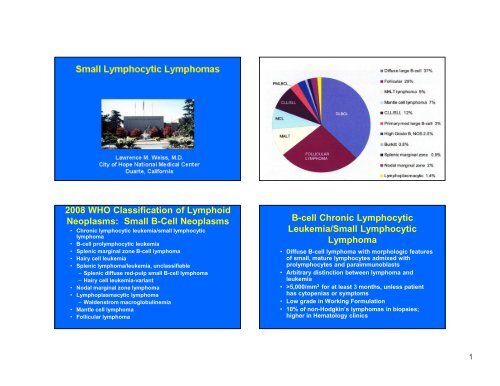
2008 WHO Classification of Lymphoid<br />
Neoplasms: <strong>Small</strong> B-<strong>Cell</strong> Neoplasms<br />
• Chronic lymphocytic leukemia/small lymphocytic<br />
lymphoma<br />
• B-cell prolymphocytic leukemia<br />
• Splenic marginal zone B-cell lymphoma<br />
• Hairy cell leukemia<br />
• Splenic lymphoma/leukemia, unclassifiable<br />
– Splenic diffuse red-pulp small B-cell lymphoma<br />
– Hairy cell leukemia-variant<br />
• Nodal marginal zone lymphoma<br />
• Lymphoplasmacytic lymphoma<br />
– Waldenstrom macroglobulinemia<br />
• Mantle cell lymphoma<br />
• Follicular lymphoma<br />
B-cell Chronic Lymphocytic<br />
Leukemia/<strong>Small</strong> Lymphocytic<br />
Lymphoma<br />
• Diffuse B-cell lymphoma with morphologic features<br />
of small, mature lymphocytes admixed with<br />
prolymphocytes and paraimmunoblasts<br />
• Arbitrary distinction between lymphoma and<br />
leukemia<br />
• >5,000/mm 3 for at least 3 months, unless patient<br />
has cytopenias or symptoms<br />
• Low grade in Working Formulation<br />
• 10% of non-Hodgkin’s lymphomas in biopsies;<br />
higher in Hematology clinics<br />
1
Monoclonal B-<strong>Cell</strong> Lymphocytosis<br />
• Defined as < 5 x 10 9 cells/L, with no tissue<br />
involvement<br />
• Same phenotype as CLL<br />
• 3-5% in general population over 50 years<br />
• 5% progress to CLL overall; about 1% per year<br />
• Possesses 13q deletion in 50% of cases, similar to<br />
CLL<br />
• Has mutated Ig gene repertoire that differs from<br />
CLL<br />
CLL/SLL: Architecture<br />
• Effacement of architecture or, more rarely,<br />
interfollicular<br />
• Pseudofollicular pattern (proliferation<br />
centers)<br />
2
CLL/SLL: Cytology<br />
• <strong>Small</strong> lymphocytes:<br />
– May be same size or slightly larger than normal<br />
lymphocytes<br />
– Round nuclei, mature clumped chromatin, and<br />
inconspicuous nucleoli<br />
– Can show some atypia or plasmacytoid features<br />
• Prolymphocytes:<br />
– Slightly larger but look similar<br />
• Paraimmunoblasts:<br />
– Larger cells with dispersed chromatin, and central<br />
eosinophilic nucleoli and more abundant cytoplasm<br />
• Reed-Sternberg-like cells (very rare)<br />
3
CLL/SLL: Immunophenotype and<br />
Gene Rearrangement Studies<br />
• CD20 and other B lineage markers<br />
• CD43, CD5, and CD23+ in 95%<br />
• CD10, FMC-7, CD79b-<br />
• ZAP-70, CD38+ in a subset (correlates with<br />
non-mutator)<br />
• Clonal lg gene rearrangements<br />
• 40-50% of cases non-mutated (poorer<br />
prognosis and 50-60% with somatic<br />
hypermutation)<br />
CD20 CLL<br />
CD43 CLL<br />
CD20<br />
CD20<br />
CD5<br />
4
CD20 CD23<br />
MUM1<br />
CLL/SLL: Molecular Cytogenetics<br />
• 80% show clonal aberrations<br />
– Deletions at 13q (50%) (good prognosis)<br />
– Deletions at 11q22-23 (20%) (aggressive)<br />
– Trisomy 12 (20%) (aggressive)<br />
– Deletions at 17p13 (10%) (aggressive)<br />
– Deletions at 6q21 (5%) (aggressive)<br />
• 20% show normal cytogenetics<br />
CLL/SLL: Progression<br />
• General increase in paraimmunoblasts over time—not<br />
progression<br />
• Prolymphocytoid progression occurs in about 15% (CD5 +)<br />
• Progression to DLBCL occurs in about 5%<br />
– Architectural effacement seen<br />
– Usually CD5 -<br />
– Usually clonal related (unmutated)<br />
– May be clonally unrelated, usually occurring in<br />
mutated cases<br />
– Poor survival (median 1 year)<br />
• Progression to classical HL occurs in
B-<strong>Cell</strong> PLL: Morphology<br />
• Blood: Medium-sized cell (2x normal lymphocyte),<br />
round nucleus, with moderately condensed chromatin,<br />
prominent central nucleolus, and small amount of<br />
faintly basophilic cytoplasm<br />
• Bone marrow: Interstitial or nodular intertrabecular<br />
infiltrate<br />
• Spleen: Expanded white and red pulp<br />
• Lymph node: Diffuse or vaguely nodular infiltrates<br />
without pseudofollicles<br />
B-<strong>Cell</strong> Prolymphocytic Leukemia<br />
• Neoplasm of B prolymphocytes affecting blood, bone<br />
marrow, spleen, and occasionally other tissues<br />
• Prolymphocytes must exceed 55% of lymphoid cells in<br />
the peripheral blood<br />
• Aggressive neoplasm<br />
• Cases of transformed CLL excluded<br />
• Cases with t(11;14) excluded<br />
• Extremely rare<br />
B-cell prolymphocytic leukemia<br />
6
B-<strong>Cell</strong> PLL: Molecular<br />
• Clonal Ig rearrangements<br />
• Unmutated in 50%<br />
• Complex karyotypes<br />
• Deletion of 17p in 50% (associated with<br />
p53 gene mutations)<br />
• 13q deletions in 25%<br />
• By definition, no t(11;14)<br />
B-<strong>Cell</strong> PLL: Phenotype<br />
• CD20 and other pan-B cell antigens<br />
• CD43 + in majority<br />
• CD5 + in 25%<br />
• CD23 + in 10-20%<br />
• ZAP-70 + in 50%<br />
Splenic B-<strong>Cell</strong> MZL: Histology<br />
• Surrounds and replaces the splenic white pulp<br />
germinal centers, effaces the follicle mantle and<br />
merges with a peripheral zone of larger cells<br />
• Infiltrates the red pulp<br />
• <strong>Small</strong> cells dominate centrally, merging with a<br />
peripheral zone of small to medium-sized cells<br />
with more dispersed chromatin and abundant<br />
pale cytoplasm, which resemble marginal zone<br />
cells and interspersed transformed cells<br />
• Plasmacytic differentiation may be occasionally<br />
seen<br />
7
CD20<br />
Splenic B-<strong>Cell</strong> MZL: Immunophenotype<br />
and Molecular<br />
• CD20 and pan-B-cell antigen +<br />
• IgM and often IgD +<br />
• CD5, CD10, CD23, CD43, bcl-6, cyclin D1 –<br />
• CD103 usually negative<br />
• Clonal Ig gene rearrangements, with somatic<br />
hypermutations seen in 50%<br />
• Loss of 7q31-32 in 40%<br />
• Trisomy 3q in some cases<br />
Hairy <strong>Cell</strong> Leukemia<br />
• Neoplasm of small B lymphoid cells with oval nuclei<br />
and abundant cytoplasm with “hairy” projections in<br />
bone marrow and peripheral blood, diffusely infiltrating<br />
bone marrow and splenic red pulp<br />
• Patients are usually middle-aged to elderly adults with<br />
median age of 55 years and 5:1 M:F ratio<br />
• Tumor infiltrates may also occur in liver, lymph nodes,<br />
and skin<br />
• Tumor cells are TRAP positive<br />
• Express CD20 and other pan-B cell markers, CD103,<br />
CD25, CD123, DBA.44, annexin A1, and CD11c<br />
• Cyclin D1 stains a subset of the cells<br />
9
Hairy <strong>Cell</strong> Leukemia-Variant (HCL-v)<br />
• B-chronic lymphoproliferative disorder that<br />
architecturally resembles classic HCL but exhibits<br />
variant hematologic and/or cytologic features<br />
• Variant hematologic features include leukocytosis<br />
and monocytosis<br />
• Variant cytologic features include prominent<br />
nucleoli, blastic or convoluted nuclei, or lack of hairy<br />
projections<br />
• TRAP -, CD25 -, annexin A1 -<br />
• Poor prognosis to HCL therapy<br />
DBA.44 Cyclin D1<br />
11
Splenic Diffuse Red Pulp<br />
<strong>Small</strong> B-<strong>Cell</strong> Lymphoma<br />
• Uncommon lymphoma with a diffuse pattern of<br />
involvement of the splenic red pulp (cords and<br />
sinusoids) by small monomorphous B-lymphocytes<br />
• Also involves bone marrow sinusoids, commonly<br />
with a villous morphology<br />
• DBA.44 +, IgG +, IgD –<br />
• Must exclude all other known lymphomas involving<br />
the spleen<br />
• May be overlap with hairy cell leukemia-variant<br />
12
Nodal MZBL: Histology<br />
• Infiltration around reactive follicles and extending<br />
into interfollicular areas<br />
• May see follicular colonization or remnants of<br />
follicles<br />
• Varying numbers of centrocyte/monocytoid-like<br />
lymphoid cells, plasma cells, and scattered<br />
transformed cells<br />
• Plasma cells may dominate<br />
• May be many transformed cells, but not<br />
patternless sheets, which indicates transformation<br />
to diffuse large B-cell lymphoma<br />
CD20<br />
Nodal Marginal Zone B-cell Lymphoma<br />
• Primary nodal B-cell neoplasm that morphologically<br />
resembles lymph nodes involved by marginal zone<br />
lymphoma of extranodal or splenic type, but without<br />
evidence of extranodal or splenic disease<br />
• Cases with involvement of extranodal sites (1/3),<br />
Hashimoto thyroiditis or Sjögren syndrome should be<br />
considered to have nodal involvement by MALT<br />
lymphoma<br />
• Involves peripheral lymph nodes, and occasionally<br />
bone marrow and peripheral blood<br />
• About 2% of lymphomas<br />
• May be associated with hepatitis C<br />
13
Nodal MZBL: Phenotype and Molecular<br />
• CD20 and other pan-B antigens +<br />
• CD43+ in 50%; bcl-2 in 80%<br />
• CD5, CD10, CD23, bcl-6, cyclin D1 -<br />
• Clonal Ig rearrangements<br />
• Not associated with translocations found in<br />
MALTomas but may have trisomy 3, 7, 18<br />
14
cl-2 MBCH bcl-2 MBCL<br />
Lymphoplasmacytic Lymphoma<br />
• Neoplasm of small B lymphocytes, plasmacytoid<br />
lymphocytes, and plasma cells<br />
• Usually involves bone marrow, and sometimes lymph<br />
nodes and spleen<br />
• Usually associated with Waldenstrom<br />
macroglobulinemia (IgM monoclonal gammopathy with<br />
bone marrow involvement by LPL)<br />
• Many cases may be associated with hepatitis C<br />
infection (particularly in Italy); some cases familial<br />
• Rare; must be differentiated from plasmacytoid<br />
variants of extranodal or nodal marginal zone<br />
lymphoma and chronic lymphocytic leukemia<br />
• Rare; 20:1 M:F ratio<br />
Pediatric MZBCL<br />
• Usually confined to head and neck lymph nodes<br />
• Often show PGTC<br />
• BCL-2 in only 40%; CD43 coexpression in 70%<br />
• Needs to be distinguished from atypical marginal<br />
zone hyperplasia found in tonsil or appendix<br />
(monotypic lambda but polyclonal)<br />
Lymphoplasmacytic Lymphoma:<br />
Histology<br />
• Diffuse or interfollicular proliferation<br />
• No proliferation centers<br />
• Population of small lymphocytes, plasmacytoid<br />
lymphocytes, and plasma cells<br />
• No monocytoid cells (think marginal zone lymphoma)<br />
• May see Dutcher bodies<br />
• May see scattered large transformed cells<br />
• May see scattered mast cells, epithelioid histiocytes<br />
• May see PAS + material, amyloid, or crystal-storing<br />
histiocytes<br />
15
Lymphoplasmacytic Lymphoma:<br />
Immunophenotype and Molecular<br />
• CD20 and other pan-B antigen +, but may be - in a<br />
subset<br />
• Cytoplasmic Ig; usually IgM, sometimes IgG, rarely IgA<br />
• CD10, bcl-6, CD23 –; CD5 -/+<br />
• CD138, CD38+/-, but may be in a subset<br />
• t(9;14) rarely found (despite initial studies)<br />
• No distinctive molecular markers<br />
16
CD20 CD79a<br />
CD138<br />
LPL / к<br />
LPL / λ<br />
<strong>Lymphomas</strong> with Plasmacytic Features<br />
• Lymphoplasmacytic<br />
• CLL/SLL<br />
• MALT lymphoma<br />
• Nodal marginal zone lymphoma<br />
• Follicular lymphoma<br />
• DLBCL<br />
• Plasmablastic lymphoma<br />
17
Mantle <strong>Cell</strong> Lymphoma<br />
• B-cell neoplasm composed of monomorphic small to<br />
medium-sized lymphoid cells with irregular nuclear<br />
contours<br />
• Well-defined clinicopathological and genotypic entity<br />
• Usually involves lymph nodes, but may be seen in<br />
spleen, bone marrow, blood, gastrointestinal tract<br />
(e.g., lymphomatous polyposis), and Waldeyer ring<br />
• Clinical: Median survival of 3-5 years; not curable in<br />
most cases<br />
• About 5% of non-Hodgkin lymphomas<br />
• Indolent variant: Patients present with high WBC,<br />
splenic involvement, and less LN involvement<br />
Mantle <strong>Cell</strong> Lymphoma: Architecture<br />
• Mantle cell: May be restricted to inner mantle zone,<br />
narrow mantles, or, more commonly, expanded<br />
mantles, with retained germinal centers<br />
• Follicular: Proliferation extends to obliterate central<br />
reactive germinal center<br />
• Vaguely nodular: Hard to see pre-existent germinal<br />
centers<br />
• Diffuse: Most common<br />
18
Mantle <strong>Cell</strong> Lymphoma: Cytology<br />
• Monomorphous population of small to medium-sized<br />
lymphoid cells<br />
• Slightly irregular nuclear contours<br />
• Relatively mature chromatin<br />
• Inconspicuous nucleoli<br />
• No admixed larger lymphoid cells<br />
• May see scattered epithelioid histiocytes<br />
Mantle <strong>Cell</strong> Lymphoma:<br />
Cytologic Variants<br />
• Other variants<br />
– Mimicking small lymphocytic<br />
lymphoma<br />
– Mimicking marginal zone B-cell<br />
lymphoma<br />
• Blastic<br />
– Classic: Resembles lymphoblastic<br />
lymphoma; 2-3 mitoses/HPF<br />
– Pleomorphic: Resembles large cell<br />
lymphoma<br />
19
Mantle <strong>Cell</strong> Lymphoma:<br />
Immunophenotype<br />
• CD20 and pan-B antigen +<br />
• Cyclin D1+ in 95%, SOX-11+ in 100%<br />
• CD5+ in 90%; CD43+ in 80%; FMC-7 +/-<br />
• CD10 -, Bcl-6 -, CD23 -/+<br />
• IgM + IgD, with 1:1 K:L ratio<br />
• Cases negative for cyclin D1 are usually positive for<br />
cyclin D2 or cyclin D3<br />
• Ki-67 and p53 may be useful in identifying blastic<br />
variants<br />
Cyclin D1 cyclinD1 TCRBCL<br />
20
Mantle <strong>Cell</strong> Lymphoma: Molecular<br />
Genetics<br />
• t(11;14) found in >95% of cases, translocating cyclin<br />
D1 to the immunoglobulin heavy chain gene<br />
• Rare cases lacking t(11;14) may have translocations<br />
involving cyclin D2<br />
• Other non-random secondary gross cytogenetic<br />
changes and/or specific gene mutations are<br />
frequently seen<br />
• ATM gene<br />
• p53 gene<br />
Differential Diagnosis of <strong>Small</strong> Lymphocytic<br />
Lymphoproliferations<br />
CLL/<br />
SLL<br />
Lymphoplasmacytic<br />
Marg Follic Mantle<br />
Ig 98% 98% 98% 90% 98%<br />
Heavy IgM +<br />
IgD<br />
IgM IgM, D,<br />
A<br />
IgM, G IgM +<br />
IgD<br />
K:L 2:1 2:1 2:1 2:1 1:1<br />
CD20 99% 98% 98% 100% 99%<br />
CD43 98% 60% 50% 2% 80%<br />
CD5 96% 25% 10% 1% 90%<br />
CD10 1% 1% 1% 95% 1%<br />
CD23 90% 10% 5% 15% 5%<br />
Cyclin D1 5% 5% 0% 0% 95%<br />
21