Immunofixation electrophoresis (IFE)
Immunofixation electrophoresis (IFE)
Immunofixation electrophoresis (IFE)

Create successful ePaper yourself
Turn your PDF publications into a flip-book with our unique Google optimized e-Paper software.
TEST<br />
<strong>Immunofixation</strong> <strong>electrophoresis</strong> (<strong>IFE</strong>)<br />
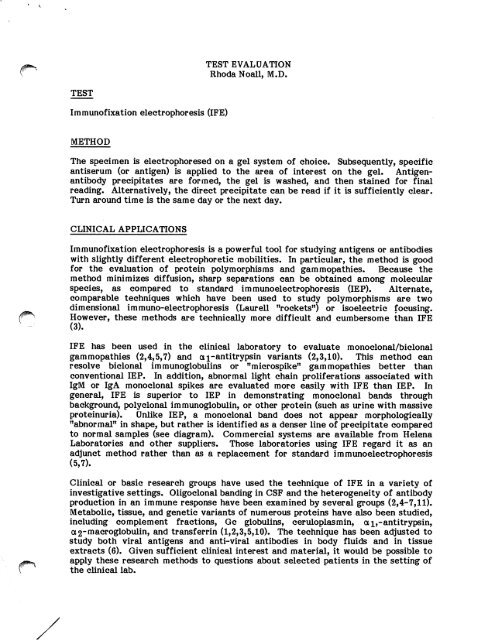
METHOD<br />
TEST EVALUATION<br />
Rhoda Noall, M.D.<br />
The specimen is electrophoresed on a gel system of choice. Subsequently, specific<br />
antiserum (or antigen) is applied to the area of interest on the gel. Antigenantibody<br />
precipitates are formed, the gel is washed, and then stained for final<br />
reading. Alternatively, the direct precipitate can be read if it is sufficiently clear.<br />
Turn around time is the same day or the next day.<br />
CLINICAL APPLICATIONS<br />
<strong>Immunofixation</strong> <strong>electrophoresis</strong> is a powerful tool for studying antigens or antibodies<br />
with slightly different electrophoretic mobilities. In particular, the method is good<br />
for the evaluation of protein polymorphisms and gammopathies. Because the<br />
method minimizes diffusion, sharp separations can be obtained among molecular<br />
species, as compared to standard Immuno<strong>electrophoresis</strong> (IEP). Alternate,<br />
comparable techniques which have been used to study polymorphisms are two<br />
dimensional immuno-<strong>electrophoresis</strong> (Laurell "rockets") or isoelectric focusing.<br />
(^ However, these methods are technically more difficult and cumbersome than <strong>IFE</strong><br />
(3).<br />
<strong>IFE</strong> has been used in the clinical laboratory to evaluate monoclonal/biclonal<br />
gammopathies (2,4,5,7) and ai-antitrypsin variants (2,3,10). This method can<br />
resolve biclonal immunoglobulins or "microspike" gammopathies better than<br />
conventional IEP. In addition, abnormal light chain proliferations associated with<br />
IgM or IgA monoclonal spikes are evaluated more easily with <strong>IFE</strong> than IEP. In<br />
general, <strong>IFE</strong> is superior to IEP in demonstrating monoclonal bands through<br />
background, polyclonal immunoglobulin, or other protein (such as urine with massive<br />
proteinuria). Unlike IEP, a monoclonal band does not appear morphologically<br />
"abnormal" in shape, but rather is identified as a denser line of precipitate compared<br />
to normal samples (see diagram). Commercial systems are available from Helena<br />
Laboratories and other suppliers. Those laboratories using <strong>IFE</strong> regard it as an<br />
adjunct method rather than as a replacement for standard immuno<strong>electrophoresis</strong><br />
(5,7).<br />
Clinical or basic research groups have used the technique of <strong>IFE</strong> in a variety of<br />
investigative settings. Oligoclonal banding in CSF and the heterogeneity of antibody<br />
production in an immune response have been examined by several groups (2,4-7,11).<br />
Metabolic, tissue, and genetic variants of numerous proteins have also been studied,<br />
including complement fractions, Gc globulins, ceruloplasmin, ai,-antitrypsin,<br />
a2-macroglobulin, and transferrin (1,2,3,5,10). The technique has been adjusted to<br />
study both viral antigens and anti-viral antibodies in body fluids and in tissue<br />
extracts (6). Given sufficient clinical interest and material, it would be possible to<br />
apply these research methods to questions about selected patients in the setting of<br />
the clinical lab.
j/^P^v<br />
TECHNICAL AND CLINICAL LIMITATIONS<br />
Page 2<br />
<strong>Immunofixation</strong> <strong>electrophoresis</strong> is technically uncomplicated and rapid, especially<br />
when compared to alternate methodology such as isoelectric focusing. The<br />
preparations are dependent on the electrophoretic method, the quality of the<br />
antisera, and the staining method. Specimens may be sera, other body fluids, or<br />
tissue extracts. The gels can be read directly for qualitative evaluation, or if<br />
<strong>electrophoresis</strong> and staining methodology are sufficiently clean, densitometry can be<br />
used (3). Gel systems have included agarose, cellulose-acetate, and starch gel<br />
<strong>electrophoresis</strong>. After the antigen-antibody precipitates are formed, any stain or<br />
visualization method can be used. Published variations include Coomasie blue,<br />
nigrosin, amido black, FITC-conjugated antibodies, horse radish peroxidaseconjugated<br />
antibodies, and autoradiography for maximum sensitivity. Direct or<br />
indirect antibody techniques have been employed for different sensitivities (see<br />
chart). Although the range of optimum antibody-antigen concentration is narrower<br />
in <strong>IFE</strong> than IEP, prozone phenomena are easily detected as a zone of central<br />
clearing (7). <strong>IFE</strong> is superior to other techniques such as isoelectric focusing or<br />
rocket <strong>electrophoresis</strong> in technical ease, turn-around time, amount of sample<br />
required, and numbers of samples which can be run with ease. For example, using<br />
<strong>IFE</strong>, a larger number of samples can be screened for a j-antitrypsin polymorphisms<br />
in less time and using less experienced personnel than using rocket <strong>electrophoresis</strong><br />
(3). However, in some worker's laboratories, immunofixation <strong>electrophoresis</strong> is less<br />
reproducible and requires more skill than IEP. Finally, although this technique has<br />
not found the widespread use of IEP, in the correct setting of sufficient appropriate<br />
material <strong>IFE</strong> can be a useful adjunct method in protein evaluation.<br />
MOLECULE<br />
monoclonal lg<br />
monoclonal lg<br />
albumin<br />
albumin<br />
albumin<br />
STAIN<br />
direct Ab-conjugated to HPO<br />
indirect Ab-conjugated to FITC<br />
0.1% nigrosine (direct stain,<br />
no antibody applied to gel)<br />
0.1% nigrosine after rabbit<br />
anti-human antibody<br />
indirect Ab-conjugated to HPO<br />
or alkaline phosphatase<br />
DETECTED<br />
CONCENTRATION<br />
2.6 ng/yl<br />
>2.6 ng/yl<br />
500 ng/yl<br />
125 ng/wl<br />
31 ng/ul<br />
REF<br />
5<br />
5<br />
9
yi#8|!tesy<br />
r^<br />
Diagram (from Ref. 7)<br />
Biclonal gammopathy<br />
SPEP $» to<br />
anti-X<br />
anti-K<br />
a n t i - M fi t f t<br />
%<br />
electrophoreses are all in register<br />
REFERENCES<br />
I<br />
Polyclonal gammopathy<br />
SPEP m a •» Jg»^<br />
a n t i - X W<br />
a n t i - K *<br />
a n t i - M f c<br />
anti-A *»<br />
anti-G $<br />
all electrophoreses in register, anti<br />
Page 3<br />
bodies applied to "boxed" area of SPEP<br />
1. Alper CA, Johnson AM: <strong>Immunofixation</strong> <strong>electrophoresis</strong>: A technique for the<br />
study of protein polymorphism. Vox Sang 17:445-452, 1969.<br />
2. Ritchie RF, Smith R: <strong>Immunofixation</strong>. I. General principles and application<br />
to agarose gel <strong>electrophoresis</strong>. Clin Chem 22(4):497-499, 1976.<br />
3. Ritchie RF, Smith R: <strong>Immunofixation</strong>. n. Application to typing of<br />
a;rantitrypsin at acid pH. Clin Chem 22(10):1735-1737, 1976.<br />
4. Ritchie RF, Smith R: <strong>Immunofixation</strong>. HI. Application to the study of<br />
monoclonal proteins. Clin Chem 22(10):1982-1985, 1976.<br />
5. Cawley LP, Minard BJ, Tourtellotte WW, Ma BI, Chelle C: <strong>Immunofixation</strong><br />
electrophoretic techniques applied to identification of proteins in serum and<br />
cerebrospinal fluid. Clin Chem 22(8):1262-1268, 1976.<br />
6. Nordal HJ, Vandvik B, Norrby E: Demonstration of electrophoretically<br />
restricted virus-specific antibodies in serum and cerebrospinal fluid by imprint<br />
electroimmunofixation. Scand J Immunol 7:381-388, 1978.<br />
7. Sun T, Lien YL, Degnan T: Study of gammopathies with immunofixation<br />
<strong>electrophoresis</strong>. Am J Clin Pathol 72:5-11, 1979.<br />
8. Voller A, Bartlett A, Bidwell D (ed). Immunoassays for the 80[s. University<br />
Park Press, Baltimore, 1981, pp. 55-56.
V<br />
^^PSSPSSy<br />
Page 4<br />
9. Chang C-H, Inglis NR: Convenient immunofixation <strong>electrophoresis</strong> on<br />
cellulose acetate membrane. Clin Chim Acta 65:91-97, 1975.<br />
10. Johnson AM: Genetic typing of a j- antitrypsin by immunofixation<br />
<strong>electrophoresis</strong>. Identification of subtypes of PiM. J Lab Clin Med 87(1):152-<br />
163, 1976.<br />
11. George PM, Lorier M, Donaldson I: An evaluation of cerebrospinal fluid<br />
oligoclonal banding confirmed by immunofixation on agarose gel. J Neurol,<br />
Neurosurg, Psychiat 46(6):500-504, 1983.