full-text pdf - Departement für Chemie und Biochemie
full-text pdf - Departement für Chemie und Biochemie
full-text pdf - Departement für Chemie und Biochemie

You also want an ePaper? Increase the reach of your titles
YUMPU automatically turns print PDFs into web optimized ePapers that Google loves.
THE DEPARTMENT OF CHEMISTRY AND BIOCHEMISTRY OF THE UNIVERSITY OF BERN<br />
Chimia 55 (2001) 1009–1013<br />
© Schweizerische Chemische Gesellschaft<br />
ISSN 0009–4293<br />
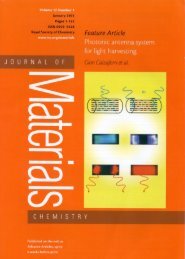
Organic–Inorganic Composites as<br />
Photonic Antenna<br />
Gion Calzaferri*<br />
1009<br />
CHIMIA 2001, 55, No. 12<br />
Abstract: A photonic antenna system is an organized multi-component arrangement in which several<br />
chromophoric molecular species absorb the incident light and channel the excitation energy to a common<br />
acceptor component. Photonic antenna materials, based on dye–zeolite L composites, collect excitation<br />
energy and transport it over large distances. Fine tuning of dye-loaded zeolite L photonic antenna materials<br />
in the size range of 30–3000 nm was realized. Stationary and time-resolved luminescence experiments on an<br />
ensemble and space and time resolved luminescence measurements on single crystal antenna materials have<br />
been carried out. Coupling of the photonic antenna information to an external device is in progress.<br />
Keywords: Energy migration · Energy transfer · Light harvesting · Supramolecular · Zeolite L<br />
Microporous structures containing atoms,<br />
clusters, molecules or complexes<br />
provide a source of new materials with<br />
exciting properties [1–6]. Zeolites are especially<br />
appealing crystalline inorganic<br />
microporous materials. Some of them occur<br />
in nature as a component of the soil.<br />
Natural and synthetic zeolites possess a<br />
large variety of well-defined internal<br />
structures such as uniform cages, cavities<br />
or channels [7–10]. A useful feature of<br />
zeolites is their ability to host molecular<br />
guests [3] and quantum-sized particles<br />
[11] within the intravoid space. Chromophore-loaded<br />
zeolites have been investigated<br />
for different purposes such as interfacial<br />
electron transfer, microlasers, second<br />
harmonic generation, frequency doubling<br />
and optical bistabilities giving rise<br />
to persistent spectral hole burning [12–<br />
21]. The role of the zeolite framework is<br />
*Correspondence: Prof. Dr. G. Calzaferri<br />
Department of Chemistry and Biochemistry<br />
University of Bern<br />
Freiestrasse 3<br />
CH–3012 Bern<br />
Tel.: +41 31 631 4236<br />
Fax: +41 31 631 3994<br />
E-Mail: Gion.Calzaferri@iac.unibe.ch<br />
http://dcbwww.unibe.ch/groups/calzaferri<br />
to act as a host for realizing the desired<br />
geometrical properties and for stabilizing<br />
the incorporated molecules. Incorporation<br />
of chromophores into the cavities of<br />
zeolites can be achieved in different<br />
ways, depending on the substances used<br />
and on the desired properties: from the<br />
gas phase [22–24], by ion exchange if<br />
cations are involved [3][25–28] by crystallization<br />
inclusion [29] or by performing<br />
an in situ synthesis inside the zeolite<br />
cages[11][30][31].<br />
Plants are masters of the efficient<br />
transformation of sunlight into chemical<br />
energy. In this process, every plant leaf<br />
acts as a light-harvesting device, in which<br />
photonic energy is absorbed in the form<br />
of sunlight and transported by chlorophyll<br />
molecules for the purpose of energy<br />
transformation. The light is absorbed by<br />
an arrangement consisting of a few h<strong>und</strong>red<br />
chlorophyll molecules in a protein<br />
environment. This photonic antenna allows<br />
fast energy transfer from an electronically<br />
excited molecule to unexcited<br />
neighbor molecules in such a way that the<br />
excitation energy reaches the reaction<br />
center with high probability. Trapping<br />
occurs there [32–34]. In this natural device<br />
the formation of aggregates is prevented<br />
by fencing the chlorophyll molecules<br />
in polypeptide cages. A similar approach<br />
is possible by enclosing dyes inside<br />
a microporous material and by<br />
choosing conditions such that the volume<br />
of the cages and channels is able to uptake<br />
only monomers but not aggregates.<br />
An artificial photonic antenna system<br />
is an organized multi-component arrangement<br />
in which several chromophoric<br />
molecular species absorb the incident<br />
light and transport the excitation energy<br />
to a common acceptor component. Imaginative<br />
attempts to build an artificial antenna<br />
different from ours have been presented<br />
in the literature [35]. Multinuclear<br />
luminescent metal complexes [36–38],<br />
multichromophore cyclodextrins [39],<br />
Langmuir Blodgett films [40–43], dyes<br />
in polymer matrices [44–46], and dendrimers<br />
[47] have been investigated.<br />
Some sensitization processes in silver<br />
halide photographic materials [48] and<br />
also the spectral sensitization of polycrystalline<br />
titanium dioxide films bear, in<br />
some cases, aspects of artificial antenna<br />
systems [49–51]. The systems developed<br />
by us so far are of bi-directional type,<br />
based on zeolite L as a host material,<br />
and able to collect and transport excitation<br />
over relatively large distances<br />
[3][22][26–28][52–55] Light transport<br />
is made possible by specifically organized<br />
dye molecules which mimic the natural<br />
function of chlorophyll. The zeolite<br />
L crystal structure consists of a continuous<br />
one-dimensional tube system. We<br />
have filled each individual tube with successive<br />
chains of different joint but noninteracting<br />
dye molecules. Light shining<br />
on the cylinder is first absorbed and the<br />
electronic excitation energy is then transported<br />
by the dye molecules inside the<br />
tubes to the cylinder ends.
THE DEPARTMENT OF CHEMISTRY AND BIOCHEMISTRY OF THE UNIVERSITY OF BERN 1010<br />
A schematic view of the photonic antenna<br />
is illustrated in Fig. 1. The monomeric<br />
dye molecules are represented by<br />
colored rectangles. The dye molecule,<br />
which has been excited by absorbing an<br />
incident photon, transfers its electronic<br />
excitation energy to another one. After a<br />
series of such steps the electronic excitation<br />
energy reaches a trap, pictured as red<br />
rectangles. The energy migration is in<br />
competition with spontaneous emission,<br />
radiationless decay and photochemically<br />
induced degradation. Very fast energy<br />
migration is therefore crucial if a trap<br />
should be reached before other processes<br />
can take place. These conditions impose<br />
not only spectroscopic but also decisive<br />
geometrical constraints on the material.<br />
Favorable conditions for realizing a<br />
photonic antenna are a high concentration<br />
of monomeric dye molecules with<br />
high luminescence quantum yield, ideal<br />
geometrical arrangement of the chromophores<br />
and an optimal size of the device.<br />
Dyes at high concentration have the tendency<br />
to form aggregates which in general<br />
show very fast radiationless decay<br />
[56][57]. The formation of aggregates<br />
can be prevented by fencing dyes inside a<br />
microporous material and by choosing<br />
conditions such that the volume of the<br />
cages and channels is only able to uptake<br />
monomers but not aggregates. Linear<br />
channels running through crystals allow<br />
the formation of highly anisotropic dye<br />
assemblies. Our investigations have been<br />
concentrated on zeolite L as a host. The<br />
reason for this is that neutral dyes as well<br />
as cationic dyes can be inserted into the<br />
channels of zeolite L and that synthesis<br />
procedures for controlling the morphology<br />
of zeolite L crystals in the size regime<br />
from 30 nm to about 3000 nm are available<br />
[53][58–60]. Many results obtained<br />
on zeolite L are valid for other nanoporous<br />
materials as well. In Fig. 2 we show<br />
a scanning electron microscopy picture<br />
of a zeolite L material with nice morphology.<br />
The hexagonal shape of the crystals<br />
can easily be recognized. For simplicity<br />
we describe them as crystals of cylinder<br />
morphology. A space-filling top view<br />
and a side view of the zeolite L framework<br />
is illustrated in Fig. 3. The primitive<br />
vector c corresponds to the channel axis<br />
while the primitive vectors a and b are<br />
perpendicular to it, enclosing an angle of<br />
60°.<br />
We distinguish between three types of<br />
dye molecules.<br />
i) Molecules small enough to fit into a<br />
single unit cell. Some examples are biphenyl,<br />
hydroxy-TEMPO, fluorenone<br />
and methylviologen (MV 2+ ). Structural<br />
CHIMIA 2001, 55, No. 12<br />
Fig. 1. Representation of a cylindrical nanocrystal consisting of organized dye molecules acting<br />
as donors (blue) and acceptors (red) acting as trap. Left: The donors are in the middle part of the<br />
crystal and the acceptors at the front and the back of each channel. Right: The donors are at the<br />
front and the back of each channel and the acceptors are in the middle part.<br />
Fig. 2. SEM picture of a zeolite L sample.<br />
Fig. 3. Framework of zeolite L. Upper: top view, perpendicular to the c-axis, displayed as stick-<br />
(left) and as Van der Waals- (right) representation with a dye molecule entering the zeolite channel.<br />
Lower: Side view of a channel along the c-axis, without bridging oxygen atoms (left). Schematic<br />
view of some channels in a hexagonal zeolite crystal with cylinder morphology.
THE DEPARTMENT OF CHEMISTRY AND BIOCHEMISTRY OF THE UNIVERSITY OF BERN<br />
details of the latter are known based on<br />
vibrational spectroscopy, Rietveld refinement<br />
of X-ray data and molecular<br />
modeling [25].<br />
ii) Molecules with a size which makes it<br />
hard to guess if they align along the caxis<br />
or if they find a way to fit into a single<br />
unit cell. Oxonine, pyronine, and<br />
thionine are molecules of this type [54].<br />
iii) Molecules which are so large that<br />
they have no other choice but to align<br />
along the c-axis. Many examples fit into<br />
this category. It is important to know if<br />
molecules can occupy at least part of the<br />
same unit cell, so that they can interact<br />
via their π-system or if they can ‘only<br />
touch each other’ so that their electronic<br />
coupling is negligible.<br />
While for molecules of type (i) not<br />
only translational but also large amplitude<br />
modes can be activated, the latter are<br />
severely or even <strong>full</strong>y restricted for molecules<br />
of type (ii) and (iii). This has consequences<br />
on their stability and also on<br />
Table. Dye molecules and abbreviations.<br />
their luminescence quantum yield which<br />
in general increases. An example we<br />
have investigated is the very light-sensitive<br />
DPH which is dramatically stabilized<br />
when inserted into zeolite L [22]. In other<br />
cases a dramatic increase of stability is<br />
observed because reactive molecules<br />
which are too large or anions such as<br />
hypochlorite have no access because they<br />
cannot enter the negatively charged channels<br />
[3]. Representative dyes that we<br />
have inserted in zeolite L are listed in the<br />
Table. Many of them lead to strongly<br />
luminescent materials. Some exceptions<br />
are fluorenone, MV 2+ , ResH, and hydroxy-TEMPO.<br />
We now show an experiment which<br />
illustrates the photonic antenna principle<br />
of Fig. 1; see [53]. Py + and Ox + were<br />
fo<strong>und</strong> to be very convenient dyes for this<br />
purpose. The channels of zeolite L are<br />
first loaded with Py + and then modified at<br />
both sides with Ox + . The latter acts as a<br />
luminescent trap which gets excited via<br />
1011<br />
CHIMIA 2001, 55, No. 12<br />
radiationless energy transfer from an excited<br />
Py + . If radiationless relaxation is<br />
not considered, Ox + can lose its energy<br />
only by fluorescence, as it cannot transfer<br />
it back to Py + because of its lower excitation<br />
energy. Fluorescence of excited Py + ,<br />
internal conversion, and intersystem<br />
crossing compete with the energy migration<br />
and energy transfer.<br />
We expect that the trapping efficiency<br />
decreases with increasing crystal length<br />
for otherwise constant parameters, specifically<br />
constant Py + loading, because<br />
the excitation energy has to migrate over<br />
an increasingly large distance to reach a<br />
trap. This can be tested if materials with<br />
different average crystal lengths are<br />
available. Since we have been able to<br />
prepare these materials, experiments<br />
with crystals of the following average<br />
length were carried out: 1, 300 nm; 2, 500<br />
nm; 3, 850 nm; 4, 1400 nm; and 5, 2400<br />
nm. These crystals where loaded with<br />
Py + so that the occupation probability
THE DEPARTMENT OF CHEMISTRY AND BIOCHEMISTRY OF THE UNIVERSITY OF BERN 1012<br />
was always the same, namely 0.11. They<br />
were then modified with two Ox + molecules<br />
on average at both ends of the channels.<br />
The fluorescence of a thin layer on<br />
quartz was measured at room temperature<br />
after specific excitation of Py + at<br />
460 nm. The fluorescence reported in<br />
Fig. 4 is scaled to the same height at the<br />
Py + emission maximum, as before. It<br />
shows a very strong increase of the Ox +<br />
emission with decreasing crystal length.<br />
One can calculate that the front–back<br />
trapping efficiency increases from 0.33<br />
up to 0.91. This means that in the 300 nm<br />
crystals, 90% of the emitted light is due<br />
to energy migration along the Py + and<br />
transfer to the luminescent traps Ox + . Interestingly,<br />
in these experiments carried<br />
out at constant Py + loading, there is also a<br />
small shift of the Py + maximum, from<br />
525 nm for the smallest crystals to 530<br />
nm for the largest ones. The maximum of<br />
the Ox + emission remains at 605 nm.<br />
This wavelength shift is most probably<br />
due to self-absorption and re-emission<br />
because the absorption depth increases<br />
with increasing crystal size despite of the<br />
constant Py + loading [53]. The measurements<br />
were made on thin dye-loaded zeolite<br />
L layers on quartz plates. The pronounced<br />
increasing trapping efficiency<br />
with decreasing length of the zeolite L<br />
<strong>und</strong>erlines the interpretation that the an-<br />
tenna behavior is mainly governed by radiationless<br />
energy migration [61–65],<br />
supported by some self-absorption and<br />
re-emission. The latter causes a shift in<br />
the maximum of the Py + fluorescence<br />
spectrum with increasing loading, but<br />
also with increasing crystal size at constant<br />
loading [3]. Some internal reflection<br />
also occurs, especially in the larger<br />
crystals [65].<br />
We conclude that zeolite L is a very<br />
suitable framework for organizing a wide<br />
variety of chromophores. Its structure is<br />
such that the formation of non-fluorescent<br />
dimers inside the channels can be<br />
prohibited and chromophores can be<br />
aligned in a certain direction. We have<br />
shown that this host/guest system can be<br />
used to make very efficient nanoscale bidirectional<br />
photonic antenna materials. A<br />
broad spectral absorption range can be<br />
achieved by using several different cationic<br />
and neutral dyes. We were interested<br />
in reversing the scheme in Fig. 1 (left)<br />
and to have an acceptor dye in the center,<br />
and the donors at both ends, Fig. 1 (right),<br />
because such systems could be useful for<br />
analytical purposes or to develop a new<br />
generation of LEDs. It turned out that it is<br />
indeed possible to prepare such materials<br />
and to perform stationary energy migration<br />
experiments on an ensemble and<br />
space and time resolved measurements<br />
CHIMIA 2001, 55, No. 12<br />
on single crystals [63–65]. It is a challenge<br />
to couple the antenna material to a<br />
device, e.g. a semiconductor. Preparation<br />
of organized zeolite monolayers on<br />
flat surfaces has been reported [66][67].<br />
The interface between the chromophoreloaded<br />
zeolite and the semiconductor<br />
becomes very important for the coupling<br />
purpose. We expect that ‘stopcock molecules’<br />
could function as a bridge between<br />
the chromophores in the zeolite L channels<br />
and the device surface (Fig. 5) [65].<br />
This opens a whole new exciting research<br />
area.<br />
Acknowledgments<br />
This work was supported by the Swiss National<br />
Science Fo<strong>und</strong>ation project NFP 47<br />
(4047-057481) and by the B<strong>und</strong>esamt <strong>für</strong> Energie,<br />
Project 10441.<br />
Received: October 17, 2001<br />
[1] R.F. Cracknell, K.E. Gubbins, M. Maddox,<br />
D. Nicholson, Acc. Chem. Res. 1995,<br />
28, 281.<br />
[2] J.M. Thomas, Angew. Chem. 1999, 111,<br />
3800.<br />
[3] G. Calzaferri, D. Brühwiler, S. Megelski,<br />
M. Pfenniger, M. Pauchard, B. Hennessy,<br />
H. Maas, A. Devaux, U. Graf, Solid State<br />
Sciences 2000, 2, 421; G. Calzaferri,<br />
Chimia 1998, 52, 525.<br />
[4] J.P. Langley, J. Hulliger Chem. Soc. Revs.<br />
1999, 28, 279.<br />
Fig. 4. Upper: SEM pictures of the investigated<br />
zeolite L samples with different crystal length:<br />
1, 300 nm; 2, 500 nm; 3, 850 nm; 4, 1400 nm;<br />
5, 2400 nm. Lower: Fluorescence intensity,<br />
after specific excitation of only Py + at 460 nm<br />
(scaled to the same height at the maximum of<br />
the Py + emission) of Py + loaded and Ox + modified<br />
zeolite L crystals with constant Py + loading,<br />
for different crystal lengths. The Ox + modification<br />
was two molecules at both ends of the<br />
channel, on average.
THE DEPARTMENT OF CHEMISTRY AND BIOCHEMISTRY OF THE UNIVERSITY OF BERN<br />
Fig. 5. Principle of the stopcock approach. Some channels of a zeolite filled with dye molecules<br />
(green rectangles) which are closed with a stopcock molecule, consisting of a head, a spacer, and<br />
a label.<br />
[5] A. Douhal, T. Fiebig, M. Chachisvilis,<br />
AH. Zewail, J. Phys. Chem. A 1998, 102,<br />
1657.<br />
[6] Y.S. Park, S.Y. Um, K.B. Yoon, J. Am.<br />
Chem. Soc. 1999, 121, 3193.<br />
[7] D.W. Breck, Zeolite Molecular Sieves,<br />
John Wiley & Sons, New York, 1974.<br />
[8] W.M. Meier, D.H. Olson, C. Baerlocher,<br />
‘Atlas of Zeolite Structure Types’, Elsevier,<br />
London, 1996.<br />
[9] S.L. Suib, Chem. Rev. 1993, 93, 803.<br />
[10] F. Schüth, <strong>Chemie</strong> in unserer Zeit 1995,<br />
29, 42.<br />
[11] G. Calzaferri, D. Brühwiler, S. Glaus, D.<br />
Schürch, A. Currao, C. Leiggener, J. Imaging<br />
Sci. Technol. 2001, 45, 331.<br />
[12] G. Calzaferri, M. Lanz, J. Li, J. Chem.<br />
Soc., Chem. Commun. 1995, 1313.<br />
[13] K.B. Yoon, Y.S. Park, J.K. Kochi, J. Am.<br />
Chem. Soc. 1996, 118, 12710.<br />
[14] U. Vietze, O. Krauss, F. Laeri, G. Ihlein,<br />
F. Schüth, B. Limburg, M. Abraham,<br />
Phys. Rev. Lett. 1998, 81, 4628.<br />
[15] S.D. Cox, T.E. Gier, G.D. Stucky, J. Bierlein,<br />
J. Am. Chem. Soc. 1988, 110, 2986.<br />
[16] G. Schulz-Ekloff, in ‘Advanced Zeolite<br />
Science and Applications, Studies in Surface<br />
Science and Catalysis’, Eds. J.C.<br />
Jansen, M. Stöcker, H.G. Karge, J.<br />
Weitkamp, Elsevier; Amsterdam, 1994,<br />
Vol. 85, p. 145.<br />
[17] M. Ehrl, F.W. Deeg, C. Bräuchle, O.<br />
Franke, A. Sobbi, G. Schulz-Ekloff, D.<br />
Wöhrle, J. Phys. Chem. 1994, 98, 47.<br />
[18] R. Hoppe, G. Schulz-Ekloff, D. Wöhrle,<br />
C. Kirschhock, H. Fuess, in ‘Zeolites and<br />
Related Microporous Materials: State of<br />
the Art 1994. Studies in Surface Science<br />
and Catalysis’, Eds. J. Weitkamp, H.G.<br />
Karge, H. Pfeifer, W. Hölderich, Elsevier,<br />
Amsterdam, 1994, Vol. 84, pp. 821.<br />
[19] J. Caro, F. Marlow, M. Wübbenhorst,<br />
Adv. Mater. 1994, 6, 413.<br />
[20] D. Wöhrle, G. Schulz-Ekloff, Adv. Mater.<br />
1994, 6, 875.<br />
[21] V. Ramamurthy, D.R. Sanderson, D.F.<br />
Eaton, J. Am. Chem. Soc. 1993, 115,<br />
10438.<br />
[22] M. Pauchard, A. Devaux, G. Calzaferri,<br />
Chem. Eur. J. 2000, 6, 3556.<br />
[23] D. Brühwiler, N. Gfeller, G. Calzaferri, J.<br />
Phys. Chem. B 1998, 102, 2923.<br />
[24] B. Müller, G. Calzaferri, J. Chem. Soc.,<br />
Faraday Trans. 1996, 92, 1633.<br />
[25] B. Hennessy, S. Megelski, C. Marcolli, V.<br />
Shklover, C. Bärlocher, G. Calzaferri, J.<br />
Phys. Chem. B 1999, 103, 3340.<br />
[26] G. Calzaferri, N. Gfeller, J. Phys. Chem.<br />
1992, 96, 3428.<br />
[27] F. Binder, G. Calzaferri, N. Gfeller, Solar<br />
Energy Mat. and Sol. Cells 1995, 38, 175.<br />
[28] F. Binder, G. Calzaferri, N. Gfeller, N.<br />
Proc. Ind. Acad. Sci. 1995, 107, 753.<br />
[29] M. Bockstette, D. Wöhrle, I. Braun, G.<br />
Schulz-Ekloff, Microp. Mesop. Mater.<br />
1998, 23, 83.<br />
[30] P. Lainé, M. Lanz, G. Calzaferri, Inorg.<br />
Chem. 1996, 35, 3514.<br />
[31] D. Brühwiler, R. Seifert, G. Calzaferri, J.<br />
Phys. Chem B 1999, 103, 6397.<br />
[32] Z.G. Fetisova, A.M. Freiberg, K.E.<br />
Timpmann, Nature 1988, 334, 633.<br />
[33] X. Hu, K. Schulten, Phys. Tod. 1997, 50,<br />
28.<br />
[34] W. Kühlbrandt, D.N. Wang, Nature 1991,<br />
350, 130.<br />
[35] H.R. Kerp, H. Donker, R.B.M. Koehorst,<br />
T.J. Schaafsma, E.E. van Faassen, Chem.<br />
Phys. Lett. 1998, 298, 302.<br />
[36] V. Balzani, S. Campagna, G. Denti, A. Juris,<br />
S. Serroni, M. Venturi, Sol. Ener. Mat.<br />
Sol. Cells 1995, 38, 159.<br />
[37] V. Balzani, S. Campagna, G. Denti, A. Juris,<br />
S. Serroni, M. Venturi, Coord. Chem.<br />
Rev. 1994, 132, 1.<br />
[38] C.V. Kumar, A. Chaudhary, Microp.<br />
Mesop. Mater. 1999, 32, 75.<br />
[39] L. Jullien, J. Canceill, B. Valeur, E. Bardez,<br />
J.-M. Lehn, Angew. Chem. 1994, 106,<br />
2582.<br />
1013<br />
CHIMIA 2001, 55, No. 12<br />
[40] H. Bücher, K.H. Drexhage, M. Fleck, H.<br />
Kuhn, D. Möbius, F.P. Schäfer, J. Sondermann,<br />
W. Sperling, P. Tillmann, J. Wiegand,<br />
Mol. Crystals 1967, 2, 199.<br />
[41] F. Willig, in ‘Photochemical Conversion<br />
and Storage of Solar Energy’, Eds. E.<br />
Pelizzetti, M. Schiavello, Kluwer Academic<br />
Publishers, Dordrecht, 1991, pp.<br />
235-249.<br />
[42] N. Tamai, T. Yamazaki, I. Yamazaki, J.<br />
Phys. Chem. 1987, 91, 841.<br />
[43] J.A. Pescatore, Jr., I. Yamazaki, J. Phys.<br />
Chem., 1996, 100, 13333.<br />
[44] B.A. Gregg, U. Resch, J. Photochem. Photobiol.,<br />
A: Chem., 1995, 87, 157.<br />
[45] S.E. Webber, Chem. Rev. 1990, 90, 1469.<br />
[46] D.M. Watkins, M.A. Fox, J. Am. Chem.<br />
Soc. 1996, 118, 4344.<br />
[47] V. Balzani, P. Ceroni, S. Gestermann, M.<br />
Gorka, C. Kauffmann, M. Maestri, F.<br />
Vögtle, ChemPhysChem 2000, 4, 224.<br />
[48] D.M. Sturmer, D.W. Heseltine, in ‘The<br />
Theory of the Photographic Process’, Ed.<br />
T.H. James, Macmillan Publishing Co.,<br />
New York, 1977, pp. 194-234.<br />
[49] C.A. Bignozzi, R. Argazzi, J.R. Schoonover,<br />
G.J. Meyer, F. Scandola, Solar Energy<br />
Mater. Sol. Cells 1995, 38, 187.<br />
[50] H. Tributsch, ‘Proc. IPS-10’, Eds. Z.W.<br />
Tian, Y. Cao, Intern. Academic Publishers,<br />
Beijing, 1993, pp. 235-247.<br />
[51] R. Memming, H. Tributsch, J. Phys.<br />
Chem. 1971, 75, 562.<br />
[52] N. Gfeller, S. Megelski, G. Calzaferri, J.<br />
Phys. Chem. B 1999, 103, 1250.<br />
[53] S. Megelski, G. Calzaferri, Adv. Function.<br />
Mater. 2001, 11, 277.<br />
[54] S. Megelski, A. Lieb, M. Pauchard, A.<br />
Drechsler, S. Glaus, C. Debus, A. J.<br />
Meixner, G. Calzaferri, J. Phys. Chem. B<br />
2001, 105, 25.<br />
[55] N. Gfeller, S. Megelski, G. Calzaferri, J.<br />
Phys. Chem. B 1998, 102, 2433.<br />
[56] M. Kasha, H.R. Rawls, M. Ashraf El-Bayoumi,<br />
Pure Appl. Chem. 1965, 11, 371.<br />
[57] E.G. McRae, M. Kasha, J. Chem. Phys.<br />
1958, 28, 721.<br />
[58] M. Tsapatsis, T. Okubo, M. Lovallo, M.E.<br />
Davis, Mat. Res. Soc. Symp. Proc. 1995,<br />
371, 21.<br />
[59] S. Ernst, J. Weitkamp, Catal. Today 1994,<br />
19, 27.<br />
[60] P.A. Anderson, A.R. Armstrong, A.<br />
Porch, P.P. Edwards, L.J. Woodall, J.<br />
Phys. Chem. B 1997, 101, 9892.<br />
[61] N. Gfeller, G. Calzaferri, J. Phys. Chem. B<br />
1997, 101, 1396.<br />
[62] M. Pfenniger, G. Calzaferri, ChemPhys<br />
Chem 2000, 1, 211.<br />
[63] M. Pauchard, S. Huber, R. Méallet-Renault,<br />
H. Maas, R. Pansu, G. Calzaferri,<br />
Angew. Chem. Int. Ed. 2001, 40, 2839.<br />
[64] G. Calzaferri, H. Maas, M. Pauchard, M.<br />
Pfenniger, A. Dévaux, Advances in Photochemistry,<br />
2002, Vol 27, in press.<br />
[65] G. Calzaferri, M. Pauchard, H. Maas, S.<br />
Huber, A. Khatyr, T. Schaafsma, J. Mater.<br />
Chem. 2002, in press.<br />
[66] P. Lainé, R. Seifert, R. Giovanoli, G. Calzaferri,<br />
New J. Chem. 1997, 21, 453.<br />
[67] K. Ha, Y.-J. Lee, D.-Y. Jung, H.J. Lee,<br />
K.B. Yoon, Adv. Mater. 2000, 12, 1114.