Arterial Blood Gases - Surgery
Arterial Blood Gases - Surgery
Arterial Blood Gases - Surgery

Create successful ePaper yourself
Turn your PDF publications into a flip-book with our unique Google optimized e-Paper software.
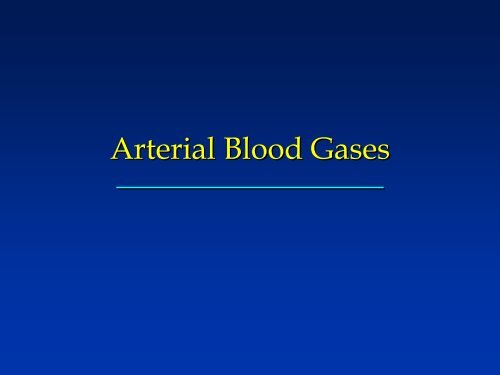
<strong>Arterial</strong> <strong>Blood</strong> <strong>Gases</strong>
PO 2<br />
<strong>Arterial</strong> <strong>Blood</strong> <strong>Gases</strong><br />
SO 2<br />
measured<br />
calculated<br />
pulse oximetry<br />
PCO 2<br />
pH<br />
BE/BD<br />
HCO 3 -<br />
venous CO 2<br />
ventilation<br />
oxygenation<br />
Acid-base
<strong>Arterial</strong> PO P (Pa 2 (PaO2) Normal: 80 – 100 mm Hg breathing room air<br />
at sea level in healthy young adults (103- (103 0.5<br />
x age)<br />
PaO 2 affected by<br />
– FIO2 PEEP Lung function<br />
– Age Ventilation Altitude<br />
PAO2 PAO = FIO2(P FIO (PB-PH PH20) 0) – PaCO2 PaCO x 1.2<br />
PAO2 PAO = FIO2(700) FIO (700) - PaCO2 PaCO x 1.2<br />
Always interpret PaO 2 in relation to FIO 2
Oxygen saturation (%)<br />
Oxyhemoglobin Dissociation<br />
Curve<br />
100<br />
90<br />
80<br />
70<br />
60<br />
50<br />
40<br />
30<br />
20<br />
10<br />
0<br />
0 10 20 30 40 50 60 70 80 90 100<br />
PO 2 (mm Hg)
PaO 2/SaO /SaO2 Shifting of the Oxyhemoglobin Dissociation curve<br />
-Temperature<br />
-pH<br />
-2,3, DPG (stored blood loses 2,3, DPG)<br />
-Dyshemoglobins (carboxy, fetal, methhgb)<br />
Shift to left facilitates Oxygen loading<br />
Shift to right facilitates Oxygen unloading
PaO 2/SaO /SaO2 30 mm Hg = 60% saturation<br />
60 mm Hg = 90% saturation<br />
40 mmHg = 75% saturation<br />
Oxygen delivery = Oxygen content x cardiac output<br />
Oxygen content = PaO2 (0.003) + Hgb(1.34)%sat<br />
Once PaO2 exceeds 70 mmHg further increases do<br />
not increase oxygen delivery
<strong>Arterial</strong> PCO P<br />
(PaCO<br />
CO 2 (Pa<br />
Normal: 35 to 45 mm Hg<br />
CO 2)<br />
↑ PaCO 2 = hypoventilation<br />
– Respiratory center depression<br />
– Neuromuscular disease<br />
– Pulmonary disease<br />
↓ PaCO 2 = hyperventilation<br />
– Central<br />
– Pain<br />
– Anxiety<br />
– Iatrogenic
Acid-Base Acid Base Balance<br />
pH = 6.1 + log HCO 3 -<br />
pH ~ HCO 3 -<br />
PCO 2<br />
0.03 × PCO 2<br />
metabolic component<br />
respiratory component<br />
When HCO 3 - is 24 mmol/L and PaCO2 is 40 mm Hg, the pH is 7.40
pH 7.35 – 7.45<br />
Normal Values<br />
PaCO2 35-45 35 45 mmHg<br />
HCO3- HCO3 22-26 22 26 meq/L<br />
BE/BD –2 2 to +2<br />
Base Excess or Base Deficit reflects the non-<br />
respiratory portion of acid-base acid base balance<br />
Includes RBC buffering
Acid-Base Acid Base Disorders<br />
Primary disturbance<br />
– Acidosis: pH < 7.35<br />
• Respiratory: ↑ Pa<br />
• Metabolic: ↓ HCO<br />
• BE: normal<br />
– Alkalosis: pH > 7.45<br />
• Respiratory: ↓ Pa<br />
• Metabolic: ↑ HCO<br />
• BE: normal<br />
PaCO CO2 HCO -<br />
3<br />
PaCO CO2 HCO -<br />
3
Rules<br />
Acid-Base Acid Base Disorders<br />
For a 0.08 change in pH – PaCO 2 changes 10<br />
mmHg<br />
7.40 40 7.32 50 7.48 30<br />
Respiratory compensation is rapid<br />
Metabolic compensation is slow
Acid-Base Acid Base Disorders<br />
Compensation<br />
– Change in PaCO PaCO2<br />
to correct pH with<br />
metabolic acid-base acid base imbalance<br />
• e.g., hyperventilation occurs with<br />
metabolic acidosis<br />
– Change in HCO -<br />
3 to correct pH with<br />
respiratory acid-base acid base imbalance<br />
• e.g., HCO -<br />
3 increases with respiratory<br />
acidosis<br />
Compensation<br />
↔<br />
↓<br />
pH ~ HCO 3 -<br />
PCO 2<br />
↓↑<br />
↓↑
Respiratory Acidosis<br />
Uncompensated: ↓ pH, ↑ PaCO PaCO2,<br />
, nl BE,<br />
HCO -<br />
3<br />
Uncompensated:<br />
Compensated: nl pH, ↑ Pa<br />
PaCO CO2, , ↑ BE, HCO -<br />
3<br />
Causes: respiratory center depression,<br />
neuromuscular disease, lung disease<br />
Treatment: treat cause, mechanical<br />
ventilation, buffers
Respiratory Alkalosis<br />
Uncompensated: ↑ pH, ↓ PaCO PaCO2,<br />
, nl BE,<br />
HCO -<br />
3<br />
Uncompensated:<br />
Compensated: nl pH, ↓ Pa<br />
PaCO CO2, , ↓ΒΕ, ΒΕ, HCO<br />
Causes: respiratory center stimulation,<br />
iatrogenic<br />
Treatment: treat cause<br />
HCO 3 -
Metabolic Alkalosis<br />
Uncompensated: ↑ pH, ↑ HCO -<br />
3 , nl PaCO Pa<br />
Compensated: nl pH, ↑ HCO -<br />
3 , ↑ Pa<br />
PaCO<br />
CO 2<br />
CO 2<br />
Causes: hypokalemia, nasogastric suctioning<br />
or vomiting, contraction alkalosis, bicarbonate<br />
administration, steroid therapy<br />
Treatment: treat cause, KCl, volume, diamox,<br />
NH 4Cl, Cl, arginine monohydrochloride, HCl
Metabolic Acidosis<br />
Uncompensated: ↓ pH, ↓ HCO -<br />
3 , nl PaCO Pa<br />
Compensated: nl pH, ↓ HCO -<br />
3 , ↓ Pa<br />
PaCO<br />
CO 2<br />
CO 2<br />
Causes: hypoxia (lactic acidosis), diabetes<br />
(ketoacidosis), renal failure (uremic acidosis),<br />
GI loss of HCO -<br />
3 (diarrhea), renal loss of<br />
HCO -<br />
3 (renal tubular acidosis, diamox),<br />
poisons (aspirin, methanol, ethylene glycol)<br />
Treatment: treat cause, buffer
Acid-Base Acid Base Interpretation<br />
Classify the disturbance: acidosis, alkalosis,<br />
metabolic, respiratory<br />
Determine the degree of compensation:<br />
uncompensated, partially compensated, fully<br />
compensated<br />
Identify the cause of the disturbance<br />
Develop a treatment plan
Acid-Base Acid Base Interpretation<br />
Disorder pH PaCO 2 HCO 3 -<br />
Respiratory acidosis<br />
Uncompensated ↓↓ ↑↑ N<br />
Partially compensated ↓ ↑↑ ↑<br />
Fully compensated N ↑↑ ↑↑<br />
Respiratory alkalosis<br />
Uncompensated ↑↑ ↓↓ N<br />
Partially compensated ↑ ↓↓ ↓<br />
Fully compensated N ↓↓ ↓↓<br />
Metabolic acidosis<br />
Uncompensated ↓↓ N ↓↓<br />
Partially compensated ↓ ↓ ↓↓<br />
Fully compensated N ↓↓ ↓↓<br />
Metabolic alkalosis<br />
Uncompensated ↑↑ N ↑↑<br />
Partially compensated ↑ ↑ ↑↑<br />
Fully compensated N ↑↑ ↑↑
↓<br />
Test Your Skills<br />
pH = 7.25<br />
PaCO2 = 57<br />
HCO 3 - = 24<br />
pH ~ HCO 3 -<br />
PCO 2<br />
↔<br />
↑
Test Your Skills<br />
pH = 7.25<br />
PaCO 2 = 40<br />
HCO 3 - = 17<br />
pH ~ HCO 3 -<br />
↓ ↔ ↓<br />
PCO 2
↔<br />
Test Your Skills<br />
pH = 7.38<br />
PaCO 2 = 60<br />
HCO 3 - = 34<br />
pH ~ HCO 3 -<br />
PCO 2<br />
↑<br />
↑
↓<br />
Test Your Skills<br />
pH = 7.28<br />
PaCO 2 = 28<br />
HCO 3 - = 13<br />
pH ~ HCO 3 -<br />
PCO 2<br />
↓<br />
↓
Mechanical Ventilation<br />
Variables<br />
Mode<br />
FIO2 and PEEP<br />
Tidal Volume and frequency<br />
I:E ratio, inspiratory time
Modes<br />
CMV or assist control – every breath is the<br />
same volume or pressure, time<br />
IMV – spontaneous breaths are allowed<br />
between mandatory breaths<br />
IMV<br />
Pressure support – a set pressure is delivered<br />
with each breath the patient takes (a boost)<br />
CPAP/PEEP – elevated end expiratory<br />
pressure<br />
CPAP/PEEP
Tidal Volume & Frequency<br />
Control minute ventilation & PaCO2 PaCO<br />
VE = f x VT V<br />
PaCO2 PaCO = VCO2/V VCO /VA<br />
VA = VT V – Vds<br />
Postop – 8-12 12 mL/kg<br />
Restrictive – 4-8 8 mL/kg<br />
Obstructive – 8-10 10 mL/kg
Tidal Volume – Weight & Height<br />
The major determinant of lung volume is height<br />
not weight<br />
Women – 45.5 + 2.3 (Ht in inches -60) 60)<br />
Men - 50 + 2.3 (Ht in inches – 60)<br />
Modify tidal volume to maintain airway plateau<br />
pressure < 30 cm H2O
Control oxygenation<br />
PEEP and FIO2<br />
FIO2 FIO start at 100% and move down using SpO2 SpO<br />
PEEP – 5 cm H20 H 0 minimum<br />
ARDS – 10 – 20 cm H2O<br />
COPD – 5-10 10 cm H2O<br />
PEEP is titrated to oxygenation, lung mechanics,<br />
oxygen delivery or other clinician determined<br />
endpoints
Writing Ventilator Orders<br />
Mode (A/C, IMV, PSV)<br />
Pressure or tidal volume<br />
Frequency<br />
FIO2<br />
PEEP<br />
Goals of support<br />
Better to write adjust FIO2 to maintain SpO2 > 92%<br />
then to write six orders to reduce FIO2
Terminology<br />
Weaning implies the gradual withdrawal of<br />
support<br />
Liberation from mechanical ventilation is more<br />
appropriate<br />
Liberation may not require weaning<br />
Extubation is removal of the ET tube<br />
Decannulation is removal of the tracheostomy<br />
tube
Minute Volume<br />
pain, anxiety<br />
sepsis, DS, VCO2<br />
Resistive<br />
Airway, secretions<br />
bronchospasm<br />
Elastic<br />
Lung compliance<br />
chest wall compliance<br />
PEEPi<br />
Weaning Failure<br />
Load<br />
Capacity<br />
Ventilatory Drive<br />
sedation, brain injury<br />
Neuromuscular<br />
Spinal injury,<br />
polyneuropathy<br />
Hyperinflation<br />
malnutrition<br />
electrolytes<br />
Chest Wall<br />
flail chest, pain
WHEANS NOT<br />
Wheezes heezes<br />
Heart eart disease<br />
Electrolytes<br />
Anxiety, nxiety, airway problems, alkalosis<br />
Neuromuscular euromuscular disease<br />
Sepsis, epsis, sedation<br />
Nutrition utrition (over and underfeeding)<br />
Opiates, piates, obesity<br />
Thyroid hyroid disease<br />
Ely EW, RCCNA 2000;6:303
Weaning Readiness<br />
Daily Screen – 5 Criteria<br />
Patient coughs when suctioned<br />
No continuous vasopressor or sedative<br />
infusions<br />
PaO2//FIO PaO //FIO2 > 200<br />
PEEP < 8 cm H2O H<br />
f/V /VT < 105 for one minute<br />
Ely NEJM 1996;335:1864
Spontaneous Breathing Trials<br />
All pts who pass the daily screen – SBT 30 mins<br />
Termination of the SBT<br />
Resp rate > 35 for > 5 mins<br />
SpO2 < 90% for > 30 secs<br />
20% increase or decrease in heart rate for > 5 mins<br />
SBP > 180 or < 90 for 60 secs consecutively<br />
Agitation, anxiety, diaphoresis > baseline for > 5<br />
minutes<br />
Ely NEJM 1996;335:1864