Micrognathozoa - Ecology & Evolutionary Biology
Micrognathozoa - Ecology & Evolutionary Biology
Micrognathozoa - Ecology & Evolutionary Biology

Create successful ePaper yourself
Turn your PDF publications into a flip-book with our unique Google optimized e-Paper software.
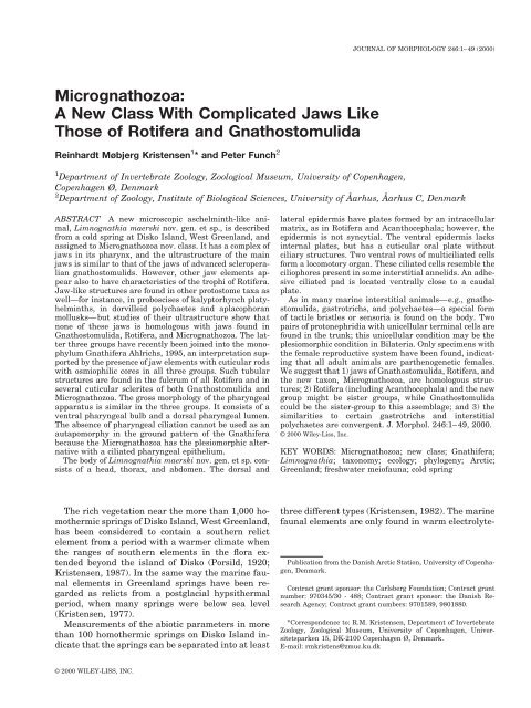
<strong>Micrognathozoa</strong>:<br />
A New Class With Complicated Jaws Like<br />
Those of Rotifera and Gnathostomulida<br />
Reinhardt Møbjerg Kristensen 1 * and Peter Funch 2<br />
1<br />
Department of Invertebrate Zoology, Zoological Museum, University of Copenhagen,<br />
Copenhagen Ø, Denmark<br />
2<br />
Department of Zoology, Institute of Biological Sciences, University of Åarhus, Åarhus C, Denmark<br />
ABSTRACT A new microscopic aschelminth-like animal,<br />
Limnognathia maerski nov. gen. et sp., is described<br />
from a cold spring at Disko Island, West Greenland, and<br />
assigned to <strong>Micrognathozoa</strong> nov. class. It has a complex of<br />
jaws in its pharynx, and the ultrastructure of the main<br />
jaws is similar to that of the jaws of advanced scleroperalian<br />
gnathostomulids. However, other jaw elements appear<br />
also to have characteristics of the trophi of Rotifera.<br />
Jaw-like structures are found in other protostome taxa as<br />
well—for instance, in proboscises of kalyptorhynch platyhelminths,<br />
in dorvilleid polychaetes and aplacophoran<br />
mollusks—but studies of their ultrastructure show that<br />
none of these jaws is homologous with jaws found in<br />
Gnathostomulida, Rotifera, and <strong>Micrognathozoa</strong>. The latter<br />
three groups have recently been joined into the monophylum<br />
Gnathifera Ahlrichs, 1995, an interpretation supported<br />
by the presence of jaw elements with cuticular rods<br />
with osmiophilic cores in all three groups. Such tubular<br />
structures are found in the fulcrum of all Rotifera and in<br />
several cuticular sclerites of both Gnathostomulida and<br />
<strong>Micrognathozoa</strong>. The gross morphology of the pharyngeal<br />
apparatus is similar in the three groups. It consists of a<br />
ventral pharyngeal bulb and a dorsal pharyngeal lumen.<br />
The absence of pharyngeal ciliation cannot be used as an<br />
autapomorphy in the ground pattern of the Gnathifera<br />
because the <strong>Micrognathozoa</strong> has the plesiomorphic alternative<br />
with a ciliated pharyngeal epithelium.<br />
The body of Limnognathia maerski nov. gen. et sp. consists<br />
of a head, thorax, and abdomen. The dorsal and<br />
The rich vegetation near the more than 1,000 homothermic<br />
springs of Disko Island, West Greenland,<br />
has been considered to contain a southern relict<br />
element from a period with a warmer climate when<br />
the ranges of southern elements in the flora extended<br />
beyond the island of Disko (Porsild, 1920;<br />
Kristensen, 1987). In the same way the marine faunal<br />
elements in Greenland springs have been regarded<br />
as relicts from a postglacial hypsithermal<br />
period, when many springs were below sea level<br />
(Kristensen, 1977).<br />
Measurements of the abiotic parameters in more<br />
than 100 homothermic springs on Disko Island indicate<br />
that the springs can be separated into at least<br />
© 2000 WILEY-LISS, INC.<br />
JOURNAL OF MORPHOLOGY 246:1–49 (2000)<br />
lateral epidermis have plates formed by an intracellular<br />
matrix, as in Rotifera and Acanthocephala; however, the<br />
epidermis is not syncytial. The ventral epidermis lacks<br />
internal plates, but has a cuticular oral plate without<br />
ciliary structures. Two ventral rows of multiciliated cells<br />
form a locomotory organ. These ciliated cells resemble the<br />
ciliophores present in some interstitial annelids. An adhesive<br />
ciliated pad is located ventrally close to a caudal<br />
plate.<br />
As in many marine interstitial animals—e.g., gnathostomulids,<br />
gastrotrichs, and polychaetes—a special form<br />
of tactile bristles or sensoria is found on the body. Two<br />
pairs of protonephridia with unicellular terminal cells are<br />
found in the trunk; this unicellular condition may be the<br />
plesiomorphic condition in Bilateria. Only specimens with<br />
the female reproductive system have been found, indicating<br />
that all adult animals are parthenogenetic females.<br />
We suggest that 1) jaws of Gnathostomulida, Rotifera, and<br />
the new taxon, <strong>Micrognathozoa</strong>, are homologous structures;<br />
2) Rotifera (including Acanthocephala) and the new<br />
group might be sister groups, while Gnathostomulida<br />
could be the sister-group to this assemblage; and 3) the<br />
similarities to certain gastrotrichs and interstitial<br />
polychaetes are convergent. J. Morphol. 246:1–49, 2000.<br />
© 2000 Wiley-Liss, Inc.<br />
KEY WORDS: <strong>Micrognathozoa</strong>; new class; Gnathifera;<br />
Limnognathia; taxonomy; ecology; phylogeny; Arctic;<br />
Greenland; freshwater meiofauna; cold spring<br />
three different types (Kristensen, 1982). The marine<br />
faunal elements are only found in warm electrolyte-<br />
Publication from the Danish Arctic Station, University of Copenhagen,<br />
Denmark.<br />
Contract grant sponsor: the Carlsberg Foundation; Contract grant<br />
number: 970345/30 - 488; Contract grant sponsor: the Danish Research<br />
Agency; Contract grant numbers: 9701589, 9801880.<br />
*Correspondence to: R.M. Kristensen, Department of Invertebrate<br />
Zoology, Zoological Museum, University of Copenhagen, Universitetsparken<br />
15, DK-2100 Copenhagen Ø, Denmark.<br />
E-mail: rmkristens@zmuc.ku.dk
2 R. M. KRISTENSEN AND P. FUNCH<br />
rich, radioactive springs (“salt springs”). The dominant<br />
type of spring on Disko Island is an electrolytepoor<br />
spring with only “normal freshwater” species.<br />
The “southern” vegetation around the springs is a<br />
result of the so-called “greenhouse effect” brought<br />
about by the snow and ice cover in winter.<br />
Compared with the “warm” springs of Disko Island,<br />
very few investigations had been carried out in<br />
cold springs, which may be frozen up to 8 months of<br />
the year. The freshwater fauna of the cold (heterothermic)<br />
springs differ greatly from those of the<br />
homothermic springs. The flora and fauna of a cold<br />
spring (Isunngua/Mudderbugten) was compared<br />
with that of the relatively cold homothermic springs<br />
(Sullorsuaq/Kvandalen) during a field course in Arctic<br />
<strong>Biology</strong>, 1994, at Disko Island. To our surprise,<br />
we found a new type of animal in the cold spring<br />
(Kristensen, 1995; Kristensen and Funch, 1995) epiphytic<br />
on water mosses. Here we fully describe the<br />
new taxon, discuss its phylogenetic position, and<br />
correct some misconceptions about this unique animal<br />
(see Ahlrichs, 1997; Herlyn and Ehlers, 1997).<br />
The first specimens of the new species were collected<br />
in 1979 at Disko Island. These three specimens<br />
were then labeled Rotifera because they were<br />
not observed alive and were strongly contracted in<br />
the formaldehyde/glycerol preparation. Later, we<br />
recognized the complicated jaw apparatus. The new<br />
animal has several superficial similarities to monogonont<br />
rotifers, especially in the pharyngeal apparatus.<br />
Therefore, we compared the extremely complex<br />
jaw apparatus with the more simple mastax of<br />
several species of Monogononta by scanning electron<br />
microscopy (SEM). Recently, excellent SEM analysis<br />
of the sclerite system of the rotifer mastax has been<br />
published (Markevich, 1989; De Smet, 1996, 1997).<br />
Surprisingly the jaw apparatus of the new animal<br />
clearly has elements similar to the scleroperalian<br />
gnathostomulids (Kristensen and Nørrevang, 1977,<br />
1978; Herlyn and Ehlers, 1997), especially in the<br />
main jaws (articularium and dentarium). We compare<br />
the lamellarization of the dentarium and the<br />
fibularization of the apophysis in the family Gnathostomulidae<br />
(Riedl and Rieger, 1972) with similar<br />
structures in the main jaws of the new animal.<br />
Gnathostomulida was described by Ax (1956) as<br />
an order of Turbellaria (Platyhelminthes). Later,<br />
Riedl (1969) established a new phylum for the<br />
gnathostomulids and Sterrer (1972) confirmed the<br />
new status of the group, but he also mentioned that<br />
gnathostomulids share characteristics with both<br />
Platyhelminthes and Aschelminthes. In the excellent<br />
review article of Lammert (1991) the Gnathostomulida<br />
was also included in the Aschelminthes.<br />
Zoology textbooks, such as Ruppert and Barnes<br />
(1994), follow the idea of Sterrer et al. (1985) and<br />
Lammert (1991) and include the Gnathostomulida<br />
in the Aschelminthes. Nielsen (1995) included them<br />
in Annelida. Littlewood et al. (1998) were the first to<br />
sequence 18S ribosomal DNA from a species of<br />
Gnathostomulida. In comparing this sequence to<br />
that of other phyla they came to the conclusion that<br />
the Gnathostomulida is a member of a Nematoda <br />
Chaetognatha clade. Zrzavy´ et al. (1998) came to<br />
another conclusion. They used the 18S ribosomal<br />
DNA data of Gnathostomulida from Littlewood et al.<br />
(1998) and combined these with morphological characters<br />
in a total-evidence approach. Analysis of this<br />
huge dataset indicated a monophylum Neotrichozoa<br />
consisting of Gnathostomulida and Gastrotricha. So<br />
far, the only agreement is that gnathostomulids are<br />
protostomian worms.<br />
Recently, Rieger and Tyler (1995) and Ahlrichs<br />
(1995) cited ultrastructural evidence for a sistergroup<br />
relationship of Gnathostomulida with<br />
Rotifera-Acanthocephala. Ahlrichs (1995) established<br />
a new monophylum Gnathifera for these<br />
groups. Later, Ahlrichs (1997) included our new<br />
taxon as “New group A” in his phylogenetic diagram<br />
of Gnathifera. He cited our unpublished Danish report<br />
from the Arctic Field Course (Kristensen and<br />
Funch, 1995). Unfortunately, the same new taxon<br />
was also mentioned as “New group 1” (Kristensen,<br />
1995). “New group A” for our unnamed taxon and its<br />
inclusion in Gnathifera were mentioned again in the<br />
article of Herlyn and Ehlers (1997). They argued<br />
that the trophi of rotifers and the cuticular jaws of<br />
gnathostomulids are homologous. Their conclusion<br />
was based on a transmission electron microscopy<br />
(TEM) investigation of the pharynx of the scleroperalian<br />
gnathostomulid, Gnathostomula paradoxa.<br />
Recently, Sørensen (2000) made the first SEMinvestigation<br />
of the two main type jaws (“compact”<br />
and “basket” type) within the Gnathostomulida. His<br />
study gives further support for a closer relationship<br />
with rotifers by virtue of similarities with the jaws<br />
and trophi of advanced monogonont rotifers. Sørensen<br />
shows that the pseudofulcrum of Rastrognathia<br />
macrostoma resembles the fulcrum in the<br />
rotiferan family Dicranophoridae. The cuticular elements,<br />
the sclerofibrillae, are almost identical in<br />
the two groups. Our new taxon from Greenland belongs<br />
to Gnathifera and supports the monophyly of<br />
this taxon comprising the Gnathostomulida, Rotifera<br />
(including Seison), and Acanthocephala. Consequently,<br />
Gnathostomulida is not as closely related to<br />
the Turbellaria (Platyhelminthes) as suggested by<br />
Ax (1956, 1985, 1989, 1996).<br />
Finally, we considered other possibilities for a<br />
phylogenetic relationship for the “New Group 1,”<br />
because we doubt that Aschelminthes is monophyletic,<br />
a hypothesis that has gained support from<br />
recent phylogenetic analyses using morphological<br />
data (Nielsen et al., 1996; Sørensen et al., 2000),<br />
molecular data and combined data (Giribet et al.,<br />
2000). There are similarities in outer morphology—<br />
e.g., the ventral trunk ciliation and sensoria—<br />
between certain gastrotrichs (Hyman, 1951; Ruppert,<br />
1991b) and the new animal. The phylogenetic<br />
significance of these structures is briefly discussed.
The free-swimming chordoid larva of Cycliophora<br />
(Funch and Kristensen, 1995; Funch, 1996) has a<br />
gastrotroch of compound cilia as in the ventral ciliation<br />
of the new animal. This is probably an adaptation<br />
to crawling and not a homology. We discuss<br />
the hypothesis of polyphyletic origin of Aschelminthes<br />
(Winnepenninckx et al., 1995) in the context<br />
of groups, such as the interstitial polychaete<br />
families Diurodrilidae and Dorvilleidae (see Kristensen<br />
and Niilonen, 1982; Eibye-Jacobsen and<br />
Kristensen, 1994) having ventral ciliophores both on<br />
the head and abdomen, as does our new animal.<br />
Furthermore, the family Dorvilleidae has a cuticular<br />
jaw in the pharyngeal apparatus. The recently described<br />
species of dorvilleid, Neotenotrocha sterreri,<br />
has a swimming behavior like that of a rotifer and a<br />
jaw apparatus similar to the trophi of rotifers. In<br />
light of the similarities between Neotenotrocha,<br />
“New Group 1,” and Rotifera the old theory of Semper<br />
(1872) that the rotifers are simply neotenic annelids<br />
is discussed.<br />
MATERIALS AND METHODS<br />
Substrate and Abiotic Factors<br />
in the Isunngua Spring<br />
The sources of the Isunngua spring are located<br />
1,030–1,120 m from the coast at 50 m above sea<br />
level in a cold moor vegetation. The outflow runs<br />
through two more moors at 315–345 m and 200–296<br />
m from the coast. Between the last two moor areas<br />
and the sea the outflow runs in a 30–50 cm-deep<br />
water channel in well-sorted sand from the Cretaceous.<br />
The outlet from the second moor is the type<br />
locality at a position: 69°43.799’N and 51°56.549’W.<br />
The spring temperature on 5 August 1994 was 5°C,<br />
the conductivity 62 mho, and the pH in the field<br />
was 6.4. The radioactivity was only slightly higher<br />
than the background.<br />
The new animal was found only in the last two<br />
moors and the outlets from the moors. More than<br />
100 specimens were collected epiphytic in the<br />
mosses on 22 July 1994, 25 July 1994, 5 August<br />
1994, and 20 May 1995. About 5 kg moss and soil<br />
were collected 5 August 1994 and carried to Copenhagen<br />
and kept in culture in a 4°C refrigerator with<br />
constant light and airing. The animals reproduced<br />
in the moss culture until 28 February 1997, when an<br />
unknown fungus overgrew the culture. The moss<br />
species on which the new animal lives epiphytically<br />
comprise the following species: Aulocommium<br />
palustre, Calliergon sarmenfosum (the dominant<br />
species), Drepanocladus intermedius, Paludella<br />
squarrosa, and Tomenthypnum nitens (soil species).<br />
Living Materials<br />
Mosses with the rhizoids kept at 4°C were<br />
squeezed in spring water for detritus and meiofauna<br />
and the rinsings decanted through a 32-m mesh<br />
MICROGNATHOZOA: A NEW CLASS<br />
net. The epiphytic meiofauna was placed in spring<br />
water or distilled water in Petri dishes at room<br />
temperature and inspected under a stereomicroscope<br />
using magnifications of 40–100. After<br />
about a half hour the new animal could be observed<br />
free-swimming in the water column. The animals<br />
adhere strongly to Pasteur pipettes, so Irwin loops<br />
were use to transfer specimens to the microslides,<br />
where they were mounted in spring water and photographed<br />
in the differential interference contrast<br />
microscope (DIC, Nomarski technique) or videotaped<br />
with a camera mounted on a Zeiss phase contrast<br />
microscope.<br />
The holotype (Fig. 1) and one paratype (Fig. 2)<br />
were drawn using camera lucida (magnification<br />
2,000). These habitus drawings were made at 4°C,<br />
when the animals were still alive but had been<br />
slightly squeezed. The measurements of total length<br />
and width of living specimens are about 20% larger<br />
than the same animal later fixed with osmiumtetroxide<br />
vapor. The drawings and measurements of<br />
all sensoria, ciliophores, adhesive ciliated pad, and<br />
the dorsal plates could only be made on living but<br />
slightly squeezed animals (Figs. 3, 4). Some animals<br />
from the culture were prepared for SEM (Figs. 5, 6)<br />
and a prominent oral plate that was not included in<br />
the drawings was observed.<br />
Some animals laid eggs in small salt cellars and<br />
some of these eggs were prepared for SEM (Fig. 7).<br />
Wholemount Preparations<br />
Twenty-eight specimens were fixed with a drop of<br />
4% formaldehyde (buffered with borax) in the spring<br />
water or with 1% osmium-tetroxide vapor directed<br />
onto the hanging drop of water on the microslides.<br />
Thereafter the specimens were mounted with a coverslip<br />
in the fixative and the fixative was replaced by<br />
2% glycerol in distilled water that evaporated to<br />
glycerol over several days; the mounts were finally<br />
sealed with Glyceel.<br />
Seven living mounted specimens were treated<br />
with 2% sodium hypochlorite, using the technique<br />
described by Riedl and Rieger (1972) for isolating<br />
the jaws of Gnathostomulida. The specimen was<br />
mounted under a coverslip in spring water in a<br />
squeezed preparation. Thereafter, sodium hypochlorite<br />
was sucked in with bibulous paper. The jaws<br />
and the basal plate (Figs. 8, 9) were strongly resistant<br />
to oxidation; however, the outer oral plate, the<br />
different lamellae, ligaments, and symphyses became<br />
transparent and disappeared under the<br />
bleaching process.<br />
The photomicrographs were taken during the<br />
bleaching process of the jaw apparatus (Figs. 10–<br />
15). After bleaching, the tissue debris was removed<br />
with distilled water, glycerol was added, and the<br />
coverslip was sealed with Glyceel. All wholemounts<br />
were examined with phase contrast and DIC optics<br />
and drawn using a camera lucida. For drawings of<br />
3
4 R. M. KRISTENSEN AND P. FUNCH<br />
the pharyngeal apparatus and the complex jaw system<br />
(Figs. 8, 9), a Wild drawing tube mounted on a<br />
Wild M20 microscope was used, allowing a drawing<br />
magnification of 10,000.<br />
Electron Microscopical Techniques<br />
Adult specimens and eggs used for SEM were<br />
fixed in 4% buffered formaldehyde or osmiumtetroxide<br />
vapor. Specimens were transferred<br />
through an acetone dehydration series and were<br />
critical-point dried using carbon dioxide; thereafter,<br />
they were mounted on aluminum SEM stubs with<br />
double-sided tape.<br />
A new technique was developed for SEM observations<br />
of the jaw apparatus. For this, several living<br />
animals were placed in a small drop of distilled<br />
water directly on the aluminum SEM stub. When<br />
the distilled water evaporated, the dried animals<br />
adhered strongly to the SEM stub. The soft tissue<br />
was then removed by 2% sodium hypochlorite and<br />
the cleaned jaw apparatus was rinsed several times<br />
with distilled water. All SEM preparations were<br />
sputter-coated with gold and examined in a JEOL<br />
JSM-840 scanning electron microscope (Figs. 16,<br />
17).<br />
For TEM, six adult animals were fixed in a mixture<br />
of three aldehydes, a so-called trialdehyde fixation<br />
(Lake, 1973), in 0.1 M sodium cacodylate<br />
buffer (see Kalt and Tandler, 1971). All specimens<br />
were postfixed in 1% osmium-tetroxide with 0.1 M<br />
sodium cacodylate buffer for 1hat20°C. After fixation,<br />
the animals were dehydrated in an ethanol<br />
series, transferred to propylene oxide, and finally<br />
embedded in epoxy resin type TAAB 812. The ultrathin<br />
serial sections were stained with uranyl acetate<br />
and lead citrate (Reynolds, 1963). TEM examinations<br />
(Figs. 18–33) were performed with a JEOL<br />
JEM-100SX transmission electron microscope.<br />
DESCRIPTION<br />
Phylum: Gnathifera Ahlrichs, 1995<br />
<strong>Micrognathozoa</strong>, new class<br />
Diagnosis. Acoelomate metazoans with bilateral<br />
symmetry and epidermal dorsal and lateral intracellular<br />
plates. Epidermis cellular, not syncytial.<br />
Body divided into a head with the pharyngeal apparatus,<br />
an accordion-like thorax, and an abdomen<br />
with a dorsal anus, which may be functional only<br />
periodically. Ventral epidermis with thick glycocalyx<br />
and two rows of multiciliated ciliophores. Ventral<br />
mouth opening surrounded by a cuticular oral<br />
plate. Epidermis of the mouth cavity and the pharyngeal<br />
apparatus also with a cuticle. Jaw apparatus<br />
with one unpaired and nine paired major sclerites.<br />
Females with one pair of ovaries. One egg per<br />
clutch. Two pairs of protonephridia with monociliated<br />
terminal cells. Direct development.<br />
Etymology. Micro, gnathos and zoa are Greek for<br />
“small,” “jaws,” and “animal,” referring to the small<br />
animal with complex jaws.<br />
Limnognathida, new order<br />
Diagnosis. Same as the class and with a life-cycle<br />
with free-living individuals in freshwaters.<br />
Limnognathiidae, new family<br />
Diagnosis. Same as the class.<br />
Limnognathia gen. nov.<br />
Diagnosis. Same as the class.<br />
Type species. Limnognathia maerski, new species<br />
by designation (Figs. 1, 2).<br />
Etymology. Limnos and gnathos are Greek for<br />
“freshwater” and “jaws,” referring to the habitat being<br />
freshwater; feminine gender.<br />
Limnognathia maerski sp. nov.<br />
Diagnosis. Mature females 105–152 m long, juveniles<br />
85–107 m long, ovoid sculptured egg 40 <br />
30 m. Stiff sensoria consisting of: 1) one pair of<br />
apicalia, 2) one pair of frontalia, 3) five pairs of<br />
lateralia, 4) three pairs of dorsalia, and 5) two pairs<br />
of caudalia, each with a ring-shaped epidermal<br />
socket. Second dorsalia (Fig. 1, do 2) on the thorax<br />
are double and may lack cilia.<br />
Etymology. To honor Maersk McKinney Møller,<br />
who sponsored the new research vessel Porsild for<br />
the Arctic Station. The new animal was discovered<br />
during the maiden trip with the new research vessel<br />
in 1994.<br />
Type material. The holotype (MIC 0001, ZMUC)<br />
is an adult female with two unsculptured oocytes.<br />
This wholemount slide, together with 19 paratypes<br />
(MIC 0002-MIC 0020, ZMUC), is deposited in the<br />
Zoological Museum of Copenhagen, Denmark. Seven<br />
sodium hypochlorite-treated jaws are also deposited<br />
on microslides (MIC 0021-MIC 0023, ZMUC). Three<br />
paratypes and two jaw apparatuses are located on<br />
SEM stubs. Six paratypes are ultrasectioned and<br />
located on 105 grids. The type material is placed in<br />
ZMUC and a single paratype is in the National<br />
3<br />
Fig. 1. Holotype of Limnognathia maerski nov. gen. et nov. sp.<br />
Dorsal view of the slightly squeezed living female from the spring<br />
at Isunngua, Disko Island, W. Greenland. A few details from<br />
study after the animal was fixed are included. ac, apicalia; an,<br />
anus; ap, apical plate; at, apical cilia tuft; ca, cauda of the main<br />
jaws (ja); cd 1-cd 2, caudalia; do 1-do 3, dorsalia; dp, dorsal plate; fg,<br />
flagellar head structure; fr, frontalia; la 1-la 5, lateralia; lp, lateral<br />
plate; mg, midgut; oo 1-oo 2, oocytes; ph, pharyngeal apparatus; sg,<br />
salivary gland; ta, tail (pygidium).
MICROGNATHOZOA: A NEW CLASS<br />
Figure 1<br />
5
6 R. M. KRISTENSEN AND P. FUNCH<br />
Museum of Natural History, Smithsonian Institution<br />
(USNM), Washington DC, USA.<br />
Type locality. The type material was extracted<br />
from living water mosses from the cold Isunngua<br />
spring (69°43.799’N, 51°56.549’W), located at the<br />
eastern corner of Disko Island, West Greenland<br />
(Figs. 34–39). The living material was collected by<br />
the authors on 22 July 1994, 25 July 1994, 5 August<br />
1994, and 20 May 1995. The holotype was from the<br />
5 August 1994 collection.<br />
Additional material. Three strongly retracted<br />
animals had been mounted on microslides in 1979.<br />
This material is from a cold homothermic spring<br />
close to Lymnaea lake (69°42.297N, 52°11.435W)<br />
in the valley of Sullorsuaq/Kvandalen (Fig. 36). The<br />
specimens were collected during a survey for tardigrades<br />
in July 1979 (Kristensen, 1982).<br />
Description of the Holotype (Adult Female)<br />
The holotype was observed alive and after fixation<br />
with 1% osmium-tetroxide and preparation of a glycerol<br />
wholemount. The total length of the living animal<br />
was 142 m and the maximum width of the<br />
abdomen was 55 m. The drawing (Fig. 1) is in<br />
dorsal view. The body seems segmented or divided<br />
into a two-parted head, accordion-like thorax, and<br />
ovoid abdomen with a small retractile pygidium (tail).<br />
External anatomy. The whole dorsal part (dorsum)<br />
of the animal is covered with plates. The animal<br />
bears sensoria on all parts of the body. As in<br />
many marine interstitial animals, e.g., gnathostomulids,<br />
gastrotrichs, and polychaetes, these tactile<br />
bristles (stiff, adjoined cilia) consist of more than one<br />
cilium. In each sensorium the cilia seem to emerge<br />
from one cell. However, this observation is not yet<br />
confirmed with TEM. In the holotype, two to three<br />
adjoined stiff cilia arise from a circular reinforcement,<br />
the socket. Still, when the bristle disappears<br />
under fixation the socket can be recognized as a ring<br />
with a pore in the middle. The tactile sensoria are<br />
always found in pairs, serially arranged on the body.<br />
The arrangement is consistent in all investigated<br />
specimens of Limnognathia maerski, including the<br />
holotype. On the anterior part of the head are four<br />
pairs of bristles. A pair of long frontalia (fr, 34 m<br />
long) and a pair of shorter apicalia (ac, 23 m long)<br />
are located on the frontal margin of the head. These<br />
sensoria are directed forward. Two pairs of laterally<br />
oriented sensoria (la1 and la2,22m long) are located<br />
between the sutures of the first two lateral<br />
headplates. The posterior part of the head lacks<br />
bristles. The thorax has two pairs of lateralia (la3 and la4,25m long) and two pairs of dorsalia (do1, 18 m long and do2, without ciliary structure). They<br />
are located on middorsal plates. The posterior pair<br />
(do2) lacks the external ciliary structures in the holotype,<br />
and the socket for the sensory structure is a<br />
double structure (8-shaped) and has a pore in the<br />
anterior part of the structure.<br />
The abdomen has a single pair of lateralia (la 5,24<br />
m long) located in a constriction that divides the<br />
abdomen into two parts. Furthermore, a shorter pair<br />
of dorsalia (do 3, 15 m long), which is oriented<br />
dorso-caudally, is located on two thin dorso-lateral<br />
plates close to the triangular anal plate. Dorsocaudally,<br />
a pair of caudalia (cd 1,19m long) and<br />
ventro-caudally another pair of caudalia (cd 2,22m<br />
long) are located. Both pairs are oriented caudally<br />
and the sockets of the ventro-caudal bristles are<br />
located on the two caudal plates, which can be seen<br />
only in the ventral view (Fig. 2), and therefore are<br />
not seen in the holotype. All sensoria break off easily<br />
and in the wholemount preparation of the holotype<br />
only the sockets of the sensoria can be observed.<br />
Three other ciliary structures could be observed<br />
on the head of the holotype when it was alive: 1) One<br />
small apical cilia tuft (at) was observed between the<br />
two apicalia. It consists of four short, stiff cilia,<br />
although eight cilia were seen in some paratypes.<br />
The cilia are about 8 m long and are not adjoined,<br />
but it seems that they arise from the same epidermal<br />
cell. 2) Close to the base of each frontalium, a<br />
long single flagellum-like structure (30 m long) is<br />
present (fg). This structure has the typical stroke of<br />
a flagellum. 3) Between the frontalia and the first<br />
pair of lateralia, a broom of short cilia is present.<br />
These cilia are perhaps a part of the ventral preoral<br />
cilia field (see Fig. 2, pc). These cilia persist in the<br />
wholemount preparation.<br />
The characteristic dorsal plates can best be investigated<br />
in the head. Here it is clearly seen that each<br />
plate comprises 3–4 epidermal cells. The borders<br />
between cells are shown as broken lines in Figure 1.<br />
An exception seems to be the thick apical plate (ap),<br />
which strongly reflects light in DIC microscopy and<br />
seems to be formed by a single giant epidermal cell.<br />
The forehead is separated from the posterior part by<br />
a constriction. The forehead consists of eight plates<br />
and the posterior part consists of five plates.<br />
The thorax has thick dorsal plates (dp) and more<br />
flexible lateral plates (lp). The lateral plates work<br />
like an accordion and the thorax can change shape<br />
rapidly from a broad and solid structure to a thinner,<br />
longer, and flexible structure. Between the dorsal<br />
and lateral plates, up to five smaller rod-like to<br />
rhomboid plates are located.<br />
The abdomen is subdivided dorsally with a sulcus<br />
(incomplete transverse furrow). The plates are large<br />
and there are no distinctions between dorsal and<br />
3<br />
Fig. 2. Limnognathia maerski nov. gen. et nov. sp. Ventral<br />
view of living paratype (slightly squeezed) from the spring at<br />
Isunngua, Disko Island. ab, abdomen; ad, adhesive ciliated pad;<br />
cd 1-cd 2, caudalia; cp, caudal plate; rb 1-rb 2, refractive bodies; ey,<br />
eye structure ?; fg, flagellar head structure; hc, head ciliophore;<br />
he, head; me, mid-ventral sensorium; mo, mouth opening, oo 1-oo 2,<br />
oocytes; pc, preoral cilia field; ph, pharyngeal apparatus; pr 1-pr 2,<br />
protonephridia; tc, trunk ciliophore; th, thorax (neck).
Figure 2
8 R. M. KRISTENSEN AND P. FUNCH<br />
lateral plates. The dorsal triangular anal plate is<br />
thick and seems to lack an anal opening.<br />
Internal structures. The thick dorsal plates obscure<br />
many of the internal structures in the holotype.<br />
Some of these structures could be seen in the<br />
allotype from the ventral view (Fig. 2).<br />
The most conspicuous structure in the holotype<br />
and all the paratypes is the large pharyngeal apparatus<br />
(25 23 m) located in the head. From the<br />
dorsal view all three pairs of jaw-like structures (ja)<br />
can be seen, but the subunits in these complex structures<br />
can only be distinguished in sodiumhypochlorite-treated<br />
animals. The cross-striated<br />
muscles are not drawn in Figure 1 to avoid complicating<br />
the figure. The muscles are best analyzed<br />
with TEM. The ovoid mouth opening is surrounded<br />
by a true cuticle and therefore it can be seen from<br />
the dorsal view through the transparent tissue. The<br />
pharyngeal apparatus will be described in detail<br />
below. The large salivary glands (sg) seem to open<br />
inside the midgut.<br />
The digestive system continues with a short<br />
esophagus, which leads to a large midgut, consisting<br />
of large transparent endoderm cells. The midgut<br />
totally lacks ciliary structures. The anus has been<br />
difficult to locate. It seems that Limnognathia<br />
maerski has only a temporary opening. In the holotype,<br />
the anal plate covers the rectum and anus.<br />
The ovary is paired and two large oocytes dominate<br />
the abdomen. A third right oocyte is seen close<br />
to the large oocyte (oo 1). A gonopore was not observed.<br />
Description of Allotype<br />
The animal was observed for several hours at 4°C<br />
by DIC optics in a cooling room at Arctic Station.<br />
Later, observations were continued on the wholemount<br />
glycerol preparation fixed by 1% osmiumtetroxide<br />
vapor. The drawing was made with camera<br />
lucida in Greenland of the ventral view (Fig. 2).<br />
Details of the pharyngeal apparatus were added<br />
later. The total length of the living animal was 127<br />
m; maximum width of the abdomen was 47 m.<br />
External gross anatomy. The entire ventral<br />
part (ventrum) of the animal lacks intracellular<br />
plates, except for the two caudal plates (cp), which in<br />
the living animal can be seen ventrally; the pair of<br />
caudo-ventral caudalia (cd2) is located on these<br />
plates. Furthermore, a large cuticular oral plate is<br />
present, but it was only observed after the animal<br />
was fixed.<br />
It is clearly seen from a ventral view that the head<br />
is divided into two parts. A sulcus separates the<br />
forehead from the posterior part. The accordionshaped<br />
thorax is divided by five annulations (transverse<br />
furrows), which are very flexible and therefore<br />
disappear after fixation. The abdomen has a small<br />
sulcus, but it does not continue as a transverse furrow<br />
to the midventral part side. In a few paratypes,<br />
a very thin furrow was seen in the anterior part of<br />
the abdomen. This furrow was not as distinct as the<br />
annulations on the thorax.<br />
The oval mouth opening (Fig. 2, mo) is located<br />
midventrally on the anterior edge of a spade-shaped<br />
oral plate. The large oral plate is not drawn in Figure<br />
2, but is seen clearly on the SEM micrograph of<br />
a paratype (Fig. 6, op). The width of the oral plate in<br />
the allotype is 21 m, and the length is 24 m. The<br />
oral plate consists of true cuticle and lacks ciliation<br />
(Figs. 25, 27, op). In retracted animals the oral plate<br />
reaches far posterior to the edge of the ventral thorax.<br />
In the allotype preparation the shape of the<br />
mouth is a small oval opening, as in all wholemount<br />
preparations of Limnognathia maerski. Therefore it<br />
was very interesting to see the behavior of the living<br />
allotype. In a relaxed swimming position, the mouth<br />
opening has the typical oval form, but when the<br />
animal feeds the two ventral jaw elements (ja 1) can<br />
be protruded out through the mouth as two small<br />
arms to grasp the substrate. Furthermore, if the<br />
animal swallows some unwanted items the large<br />
cross-striated muscles in the head retract the dorsal<br />
part of the forehead, which is lifted upward and<br />
backward. Consequently, most of the pharyngeal<br />
apparatus with the whole jaw apparatus could be<br />
seen to stick out of the mouth. Several fast movements<br />
of all the jaws elements often accompany this<br />
behavior. The movements consist both of a snapping<br />
reaction and of turning the cauda of the main jaws<br />
(Fig. 1, ca) forward and backward. We called this<br />
action of the jaws a “vomit” behavior. One of the<br />
paratypes sectioned for TEM is fixed with the forehead<br />
lifted upward and the jaws protruded.<br />
Ciliophores and ventral ciliation. The ventral<br />
ciliation is well developed and all body parts have a<br />
complex ciliation, unlike ciliation in taxa such as<br />
Gastrotricha, Rotifera, and Gnathostomulida. The<br />
forehead is covered ventrally by rows of single cilia.<br />
The rows of cilia are formed as arcs, leading particles<br />
directly to the mouth opening. From the mouth<br />
opening itself several stiff sensory cilia stick out like<br />
a broom.<br />
Lateral to the oral plate a quite different ciliation<br />
exists. Four pairs of specialized ciliated areas were<br />
seen in the allotype. One pair is located at the forehead<br />
and three pairs are located at the posterior<br />
part of the head. The cilia in each area are stiff and<br />
they arise from a single rectangular cell. The cilia<br />
are not bounded by a common membrane, as in the<br />
gastrotrichs, but they move in unison nevertheless.<br />
Similar multiciliated cells in other invertebrates are<br />
called ciliophores (Kristensen and Niilonen, 1982;<br />
Eibye-Jacobsen and Kristensen, 1994). The ciliophores<br />
are large epidermal cells with numerous cilia<br />
whose basal bodies are ordered in regular rows. The<br />
head ciliophores in Limnognathia maerski are located<br />
in the same position as the metastomial ciliophores<br />
in the two interstitial polychaetes Diurodrilus<br />
westheidei and Neotenotrocha sterreri.
The dominating ventral ciliation in Limnognathia<br />
maerski is located on the thorax and the abdomen.<br />
Two rows of trunk ciliophores exist. The length of<br />
each ciliophore is about 5 m and the width is about<br />
15 m. The cilia are ordered in four rows. There are<br />
about 20 cilia in each row. In the allotype there exist<br />
18 pairs of trunk ciliophores, ten pairs on the thorax<br />
and eight on the abdomen. There is no difference<br />
between the ciliophores of the thorax and those of<br />
the abdomen.<br />
Just behind the last abdominal ciliophores is a<br />
large adhesive ciliated pad (Fig. 2, ad). The pad<br />
MICROGNATHOZOA: A NEW CLASS<br />
Figs. 3, 4. Limnognathia maerski nov. sp. et nov. sp. Micrographs (DIC) of live specimens. Fig. 3: Habitus photo of the whole animal<br />
divided into head (he), thorax (th), and abdomen (ab). Note the different size of the oocytes (oo 1 and oo 2). br, brain; gc, midgut cells;<br />
ja 1, ventral jaw; ph, pharyngeal apparatus. Fig. 4: Ventral view of strongly squeezed caudal part. ad, adhesive ciliated pad consisting<br />
of ten ciliated cells (ce); bb, basal bodies in rows; ep, unciliated epidermal cells; gs, glue secretion; oo 1, oocyte; tc; trunk ciliophore.<br />
consists of two paired groups of five cells. The cilia in<br />
the pad are also stiff, but they are longer than the<br />
cilia in the ciliophores and they do not beat in unison.<br />
In the strongly squeezed paratype (Fig. 4, gs), a<br />
secretion is squeezed out from a pore midventral<br />
between the two clusters of cells. We assume the<br />
secretion is adhesive. This assumption was supported<br />
by live observations of the allotype. The animal<br />
stuck to the substrate with the posterior ventral<br />
part when we tried to remove it with a Pasteur pipette.<br />
The action of the trunk ciliophores was also observed<br />
in the living allotype. The trunk ciliophores<br />
9
10 R. M. KRISTENSEN AND P. FUNCH<br />
Figs. 5, 6. Limnognathia maerski nov. gen et nov. sp. Females. SEM. From culture, Copenhagen. Fig. 5: Lateral view of a slightly<br />
shrunken specimen showing the dorsal (dp) and lateral plates (lp). The locomotory cilia are ventral ciliation with trunk ciliophores (tc)<br />
and an adhesive ciliated pad (ad) posteriorly. Note the position of one of each of some of the paired sensoria: lateralia (la 2-la 4) and<br />
dorsalia (do 2). Fig. 6: Ventral view of animal with anterior preoral cilia field (pc), head (hc) and trunk ciliophores (tc), and a posterior<br />
adhesive ciliated pad (ad). The mouth (mo) is partly covered by food (fo) or detritus and situated anterior to the oral plate (op). Note<br />
the position of some of the paired lateralia (la 3-la 5) and caudalia (cd 1-cd 2). cp, caudal plate.<br />
are locomotory organs and they beat when the animal<br />
crawled on the substrate (mosses) and when the<br />
animal was swimming. It could be that the preoral<br />
ciliary field is used together with the head ciliophores<br />
as a broom to collect small detritus particles.<br />
This theory was not supported by live observations,<br />
however. It seems that the animals feed by grasping<br />
food particles directly by the ventral jaws. The anterior<br />
ciliation instead could be involved in swimming.<br />
Internal structures. The few internal structures<br />
that could be seen in the living allotype included two<br />
pairs of protonephridia (Fig. 2, pr 1-pr 2), one pair in<br />
the thorax and another pair in the anterior part of<br />
the abdomen. The flame cells could be observed beating<br />
inside the lateral protonephridium, but the<br />
nephridiopore could not be located (see later, “Excretory<br />
Structures”).<br />
The oocytes in the two ovaria are similar to the<br />
oocytes in the holotype, but the right oocytes are<br />
further developed and larger (diameter 28 m). In<br />
the left side of the abdomen of the allotype two<br />
characteristic globular bodies are located. We call<br />
them refractive bodies (Fig. 2, rb 1-rb 2). Their function<br />
is unknown, but they are only seen in mature<br />
females with large oocytes or sculptured eggs.<br />
Other Paratypes<br />
More than 100 specimens were observed alive<br />
from the cold spring of Isunngua. Many of them
were used for video recordings, or squeeze or jaw<br />
preparations. This material does not exist any<br />
longer, but has been documented with video recordings,<br />
drawings, and micrographs (Figs. 3, 4). Three<br />
paratypes were used for SEM (Figs. 5, 6) and six for<br />
TEM. The TEM material will be treated under “Ultrastructural<br />
TEM Morphology.” The holotype, allotype,<br />
and an additional 27 specimens were drawn<br />
using a camera lucida, measured, and then kept as<br />
wholemount preparations on glass microslides.<br />
The length of the adult animals ranges from 105–<br />
152 m, with an average length of 123.3 m (n<br />
23). We defined adult females as those where both<br />
ovaries with oocytes are present. The length of the<br />
juveniles, being those without any signs of gonads,<br />
ranges from 85–107 m, with an average length of<br />
93.0 m (n 7). When we compared juveniles with<br />
adults no differences in the jaws, sensoria, or dorsal<br />
plates were observed. The number of rows of the<br />
double ciliophores varied, however. A minimum<br />
number of ten rows was observed in the small juveniles,<br />
while a maximum of 18 rows was seen in the<br />
allotype. It is therefore likely that development is<br />
direct, although we never observed a juvenile hatching<br />
from an egg. The ciliophores can be observed<br />
easily using DIC optics (Fig. 4). In the SEM preparations<br />
we made (Fig. 6) the trunk ciliophores (tc)<br />
and the adhesive ciliated pad (ad) were covered with<br />
a thin layer of mucus or glue, and all the unique<br />
characters could not be distinguished. The head ciliophores<br />
(hc) are organized as compound cilia. The<br />
sensoria are often lost in SEM preparations (Figs. 5,<br />
6) and only the sockets are seen (cd 1). If the sensorium<br />
MICROGNATHOZOA: A NEW CLASS<br />
Fig. 7. Limnognathia maerski nov. gen. et nov. sp. SEM. A newly laid sculptured egg from a<br />
female. From Isunngua, 25 July 1994.<br />
is present, it could be observed that the structure consists<br />
of more than one stiff cilium (Fig. 6, la 3).<br />
Development<br />
The smallest female (105 m) had two small oocytes,<br />
one in each ovary. Usually there is only one<br />
large oocyte and a smaller oocyte in the opposite<br />
ovary (Fig. 3). A large animal (152 m) had developed<br />
two large oocytes (right oocytes, d 45 m; left<br />
oocytes, d 39 m). In several animals the female<br />
gamete had developed a chorion or eggshell. We call<br />
this gamete an egg. The egg can be enormous relative<br />
to the small size of the animal, and the egg may<br />
occupy most of the abdomen. Only one egg develops<br />
per clutch. The smallest egg with a chorion had a<br />
diameter of 31 m and was unsculptured. The largest<br />
egg in the ovary had a diameter of 48 m and<br />
was sculptured, with osmiophilic dots at the surface.<br />
We could provoke the females to lay the egg. When<br />
we kept the animals at room temperature (20°C) the<br />
animals left the detritus or the moss leaves and<br />
swam around in the water column. When we transferred<br />
the specimens to small watch glasses the<br />
animals would lay eggs at the bottom. The egglaying<br />
behavior was difficult to observe, but it<br />
seemed that the animal bent ventrally, and that the<br />
egg burst out from the ventral side close to the<br />
adhesive pad. All eggs were of the same size and<br />
were quite sticky. We recognized two types of eggs.<br />
One type was unsculptured and burst easily, and the<br />
second type was sculptured (Fig. 7) and could be<br />
removed more easily from the watch glass. The free<br />
11
12 R. M. KRISTENSEN AND P. FUNCH<br />
egg is oval (30 40 m) and other observed eggs<br />
ranged in size from 40–60 m at the longest axis.<br />
The unsculptured eggs could be abortive, while the<br />
sculptured eggs were capable of further development.<br />
The justification for this assumption is that<br />
the whole population of adult females would deliver<br />
the oocytes and eggs when they were kept at high<br />
temperature. We did not observe any cleavage in the<br />
eggs. Furthermore, we never found eggs with jaws of<br />
juveniles inside.<br />
We looked very intensively for smaller males both<br />
in Greenland and in the culture in Copenhagen.<br />
Unfortunately, we never observed any, and therefore,<br />
we expect that the species reproduces by parthenogenesis,<br />
at least during the summer. We cannot<br />
rule out that the animal is hermaphroditic. The<br />
two refractive bodies (Fig. 2, rb) may be a part of the<br />
male reproductive system. The smallest juvenile (85<br />
m) was observed alive. The head was relatively<br />
large (35 m) and the thorax weakly developed. As<br />
mentioned above, it had only ten rows of ciliophores<br />
and it may be the ciliophores of the thorax that are<br />
lacking. It moved exactly like the adults and it already<br />
had a full set of jaws in the pharyngeal apparatus.<br />
The animals were kept in a moss culture in a<br />
4°C refrigerator for more than 2 years. During this<br />
period only seven juveniles were observed and measured.<br />
Fine Structure of the Jaw Apparatus<br />
The fine structure of the “true” cuticularized parts<br />
of the pharyngeal apparatus is important for a discussion<br />
of the phylogenetic position of Limnognathia.<br />
We therefore describe the isolated cuticularized<br />
parts (sclerites) of the pharyngeal apparatus in<br />
this section.<br />
A complete investigation of the ultrastructure of<br />
the whole pharyngeal apparatus, e.g., cuticular elements,<br />
epidermal cells, and the mesodermal crossstriated<br />
muscles, is needed. The only way to do that<br />
is to combine SEM and TEM techniques (Fiege,<br />
1990). A less sophisticated technique is to serialsection<br />
the entire pharyngeal apparatus and make<br />
three-dimensional reconstructions of the location of<br />
all cuticular elements, the nuclei of the epidermal<br />
cells, and the cross-striated muscles. This technique<br />
has been used with great success in the eutardigrades<br />
(Eibye-Jacobsen, 1997). Unfortunately, this<br />
technique is time-consuming and requires singlehole<br />
grids for serial sectioning. We sectioned six<br />
pharyngeal apparatuses, but we used the more secure<br />
200-mesh grids. The additional information obtained<br />
from the TEM study was crucial to understanding<br />
the general organization of the pharyngeal<br />
apparatus; for example, that the cuticularized parts<br />
are extracellular and that the jaw apparatuses are<br />
built in the same way as the jaws of gnathostomulids.<br />
The results from our TEM study of the pharyn-<br />
geal apparatus will be treated under the section on<br />
ultrastructure.<br />
In this section, we use the technique described by<br />
Riedl and Rieger (1972) for gnathostomulids and<br />
thereafter combine light microscopical observations<br />
with SEM micrographs. We first observed living,<br />
squeezed animals by DIC optics (Figs. 10,11). A few<br />
animals were treated with OsO 4-vapor and were<br />
strongly squeezed (Fig. 12). This allowed us to see<br />
the dentarium and articularium of the main jaws<br />
(ja 2). Soon it became clear that some parts of the<br />
cuticular jaw apparatus were hidden in the epidermal<br />
tissues and the muscles. In order to reveal the<br />
hidden parts, whole animals were treated with 2%<br />
sodium hypochlorite (Figs. 13–15). This bleaching<br />
process allowed us to see the main cuticular parts,<br />
the basal plates, the three pairs of jaws, and the<br />
complex structure of the fibularium with its many<br />
fenestrae and fibulae. These structures are strongly<br />
resistant to the bleaching process. Unfortunately,<br />
all ligaments, the oral plate, and the lamellae orales<br />
dissolved immediately. Consequently, the ventral<br />
jaws (ja 1) and the accessory sclerites (as 1,as 2) were<br />
lost in all sodium hypochlorite preparations. It soon<br />
became obvious that the fine structure of the cuticularized<br />
pieces was too small and complex to be fully<br />
resolved with light microscopy. A modified SEM<br />
technique of Koehler and Hayes (1969) was therefore<br />
introduced (Figs. 16, 17). In this technique the<br />
soft tissue was also removed with sodium hypochlorite.<br />
The three-dimensional configurations were<br />
added to the two schematic drawings (Figs. 8, 9),<br />
which were made with the camera lucida technique.<br />
When the DIC micrographs (Figs. 10–15) were compared<br />
with the SEM micrographs (Figs. 16, 17), it<br />
was obvious that both techniques have their advantages.<br />
When we used DIC it was possible to observe<br />
the living animals first and later to treat the animals<br />
with sodium hypochlorite. This affords the opportunity<br />
to observe the cuticular elements in situ<br />
attached to epidermal cells, ligaments, and muscles.<br />
We never observed all cuticular parts together using<br />
SEM and the thin lamellae of the fibularium were<br />
twisted, so it gave the impression that the structure<br />
is much more flattened compared to similar structures<br />
in living specimens.<br />
On the other hand, many details were seen only<br />
with SEM and these structures are still not well<br />
understood, e.g., the free teeth between the main<br />
jaws (ja 2) and the dorsal jaws (ja 3), the pseudodentes<br />
(p.de), the trochanter (tr), and the spinula (sp) of the<br />
dorsal jaws (see Figs. 16, 17). We have to admit that<br />
our interpretation is preliminary and we may have<br />
misinterpreted several structures, but otherwise we<br />
hope the readers will understand that the complexity<br />
of the jaw apparatus of Limnognathia maerski<br />
is far beyond what is seen in other invertebrates<br />
such as gnathostomulids, rotifers, and dorvilleid<br />
polychaetes. For a comparison of the fine structure
of their jaw apparatuses and that of Limnognathia,<br />
see below.<br />
We used a new terminology for the more important<br />
cuticular parts of the jaw apparatus. We employed<br />
the Latin nomenclature in many general<br />
structures, such as apophysis, fenestra, trochanter,<br />
etc. Unfortunately, this nomenclature has been used<br />
in gnathostomulids, and to some extent in rotifers as<br />
well. We are still not sure if these structures are<br />
homologous in the three groups, but the ultrastructure<br />
of some of the cuticular elements (Rieger and<br />
Tyler, 1995) indicates that this is the case. The<br />
following description of the main elements 1) the<br />
basal plates, 2) the lamellae orales, 3) the ventral<br />
jaws, 4) the main jaws, 5) the two fibularia, and 6)<br />
the dorsal jaws is given from the ventral perspective<br />
to the dorsal view (Figs. 8–17).<br />
Basal Plates (ba)<br />
A pair of molar-like structures (Figs. 9, 16, ba) is<br />
located on the ventro-caudal part of the mouth opening.<br />
These two structures can be extruded out from<br />
the lower lips when the animal forages. The molar<br />
structure is triangular in shape, with five cusps of<br />
heavily sclerotized material. The two molars are the<br />
first elements to be seen in optical sections with DIC<br />
techniques (Figs. 11, 13) in ventral view. Each molar<br />
is 2 m wide and 1.5 m high. The outer cuticular<br />
part of the basal plates is fixed to thin jointed lamella<br />
plates, which continue inside the lower part of<br />
the mouth cavity. The thin plates were not observed<br />
with DIC optics, but were clearly seen in specimens<br />
treated with sodium hypochlorite (Figs. 8, 9, 16, 17).<br />
The thin lamellar parts are 4 m long and are fused<br />
rostrally with a 1.5 m-long suture. Two large crossstriated<br />
muscles seem to attach close to the molar<br />
portion of the basal part. In connection with the<br />
basal plates, five additional dentes oralis (de.o) are<br />
seen in the SEM micrographs (Figs. 16, 17). These<br />
structures were not observed by DIC optics and were<br />
consequently omitted from Figure 9. They could<br />
have been hidden by the outer cuticular rim of the<br />
mouth opening itself.<br />
Lamellae Orales<br />
Lamellae orales were observed only in living animals<br />
and in the TEM micrographs. They consist of<br />
two halfcone-shaped structures on the rostral part of<br />
the mouth cavity. In ventral optical sections the<br />
lamellae orales are seen as two arched upper lips<br />
(Fig. 11). These structures may constitute a continuous,<br />
folded cuticular membrane covering the upper<br />
part of the mouth cavity. By DIC techniques (Fig.<br />
11) the lamellae orales are seen as two arcs with<br />
delicate striation. At least 13 folds are observed in<br />
each arc. The lamellae orales support the dorsal<br />
mouth cavity and prevent cavity collapse during<br />
feeding. The lamellae orales quickly disappear in<br />
MICROGNATHOZOA: A NEW CLASS<br />
the sodium hypochlorite bleaching process and the<br />
collapsed lamellae can be seen as an extra,<br />
membrane-like structure outside the cleaned jaw<br />
apparatus (Figs. 13–15).<br />
Ventral Jaws (Pseudophalangia)<br />
The ventral jaws consist of two pairs of strongly<br />
cuticularized elements, the pseudophalangia (ja1) and the accessory sclerites (as1). These two elements<br />
are joined with a ligament and a ball-and-socket<br />
joint. Furthermore, a ligament connects the pseudophalangium<br />
to the fibula ventralis of the fibularium.<br />
Both ligaments and the large cross-striated<br />
muscle disappeared when sodium hypochlorite was<br />
added, and the pseudophalangium was found independent<br />
from the rest of the jaw apparatus. The<br />
accessory sclerite, on the other hand, stays attached<br />
to the lateral part of the fibularium (Figs. 13, 14, 16,<br />
17), even after the pseudophalangium disappears.<br />
The link between the pseudophalangium, the fibularium,<br />
and the accessory sclerite is much more complicated<br />
than we have drawn in the squeezed preparation<br />
of the jaw apparatus (Figs. 8, 9).<br />
The pseudophalangium is a large sclerite and it<br />
can be quite moveable, facilitated by the large crossstriated<br />
muscles. The muscle attachment is formed<br />
as a fenestra pseudophalangialis (fe.p) at the swollen<br />
base of the sclerite. Both pseudophalangia can be<br />
protruded through the mouth opening under foraging.<br />
In relaxed swimming behavior the two pseudophalangia<br />
would be located latero-rostral of the<br />
mouth opening (Figs. 8, 9). The tip of each pseudophalangium<br />
consists of four large digits; in a few<br />
specimens a fifth smaller digit also was observed as<br />
a dorsal thumb. The length of the pseudophalangium<br />
is 12 m and the length of the accessory sclerite<br />
is 5 m. Both structures are a hollow tube-like<br />
structure in cross section (see below).<br />
Main Jaws<br />
The main jaws are pincer-like. The pincer consists<br />
of the paired dentarium and the unpaired articularium<br />
with the symphysis. Dorso-caudally the main<br />
structure again is split into a symmetrical cauda<br />
(Figs. 8, 9, 12, 15–17, ca), which is the apodeme for<br />
the posterior pharyngeal muscle sac. Ventrocaudally<br />
the symphysis (Figs. 9, 17, sy) continues as<br />
an unpaired element. The fibularium, the largest<br />
element in the jaw apparatus, attaches to the main<br />
jaws laterally. The fibularium could be regarded as a<br />
part of the main jaws, but will be treated separately<br />
because of its complexity.<br />
The dentarium is the teeth-bearing distal part<br />
of the pincer. Each branch of the pincer has an<br />
arc-shaped arrangement of teeth which can be<br />
observed from a lateral view (Fig. 12). The ventral<br />
arc (arcus ventralis) is prominent and possesses<br />
five large teeth, the dentes ventrales (Fig. 9, d.ve).<br />
13
14 R. M. KRISTENSEN AND P. FUNCH<br />
Figs. 8, 9. Limnognathia maerski nov. gen. et nov. sp. Semi-schematic drawings of the two pharyngeal apparatuses based on DIC<br />
and SEM techniques. The drawings are constructed from two different animals. Fig. 8: Dorsal view. Fig. 9: Ventral view. as 1, accessory<br />
sclerite of ventral jaw (ja 1); as 2, accessory sclerite of dorsal jaw (ja 3); ba, basal plate; ca, cauda; d.do, dentes dorsales; df, dorsal<br />
fibularium; d.me, dentes mediales; d.se, dorsal serratum; d.te, dentes terminalis; d.ve, dentes ventrales; fd 1-fd 3, three fenestrae of<br />
dorsal fibularium; fe.d, fenestra dentarialis; fe.p, fenestra pseudophalangialis, fe.s, fenestra symphysis; fi.d, fibula dorsalis; fi.v, fibula<br />
ventralis; fl 1-fl 4, four fenestrae of lateral fibularium; fv 1-fv 2, two fenestrae of ventral fibularium; ja 1, ventral jaw (pseudophalangium);<br />
ja 2, main jaw consisting of dentarium and articularium; ja 3, dorsal jaw; lf, lateral fibularium; l.or, lamellae oralis; mo, mouth opening;<br />
o.la, outer lamella of fibularium; pd 1, pseudodigit of ventral jaw; pd 2, pseudodigit of dorsal jaw; p.de, pseudodentes on the dorsal jaw;<br />
sp, spinula of dorsal jaw; sy, symphysis of main jaw; tr, trochanter of dorsal jaw, vf, ventral fibularium.
The dentation of the median part of the arc (Fig. 9,<br />
d.me) remains beyond the resolution of the light<br />
microscope (Fig. 12, d.me). With SEM, however, it<br />
seems that three small dentes mediales are<br />
present, and in TEM preparations even more teeth<br />
are seen in cross section. The structure of the<br />
dorsal arc is much more difficult to interpret.<br />
MICROGNATHOZOA: A NEW CLASS<br />
Figure 9<br />
When the dorsal arc (arcus dorsalis) is observed in<br />
the light microscope with DIC optics (Fig. 12,<br />
d.do), it seems to consist of a large tooth, the<br />
dentes terminalis, followed by five tread-like<br />
structures. When cleaned jaws are observed with<br />
SEM, it is seen that the dentes terminalis are not<br />
connected to the dorsal arc, but lie free at the<br />
15
16 R. M. KRISTENSEN AND P. FUNCH<br />
Figures 10–15
surface (Fig. 16, d.te). We still believe it is the<br />
same structure as seen by DIC optics, because the<br />
dentes dorsalis (d.do) are also located superficially<br />
as thin tread-like teeth on the dorsal arc.<br />
Caudal to the arc-structure, the dorsal edge of the<br />
dentarium has a serrated appearance. This dorsal<br />
serratum (Figs. 8, 16, d.se) consists of 20 small saw<br />
teeth on each side of the pincer-arms. Just before the<br />
symphysis the dentarium has a pair of small holes<br />
(Figs. 8, 16, fe.d). The symphysis continues ventrally<br />
as an unpaired caudal part (Figs. 9, 14, 17, sy). The<br />
dorsal part of the articularium is more complicated.<br />
Behind the articulation of the two arms in the dentarium,<br />
is a 4 m-long single piece of the articularium.<br />
This lamella structure is penetrated by a<br />
large hole, the fenestra symphysis (Figs. 8, 12, 15,<br />
16, fe.s). The articularium has two symmetrical<br />
cauda (ca). We believe that this structure is the<br />
apodeme for the large caudal muscle-sac.<br />
The Two Fibularia<br />
The fibularium consists of cuticularized fibulae<br />
and fenestrae. Each fenestra is a window with cellular<br />
tissue surrounded by cuticle. The cellular part<br />
is involved in forming the extracellular fibulae. The<br />
unique fibularium is perhaps only a part of the apophysis<br />
of the main jaws as seen in some gnathostomulids<br />
and rotifers, but in Limnognathia the<br />
structure has developed to an extreme degree. The<br />
fibularium in Limnognathia is a three-dimensional<br />
structure that totally surrounds the main jaws as<br />
three compartments: the ventral, lateral, and dorsal<br />
fibularia.<br />
Figs. 10–15. Limnognathia maerski nov. gen. et nov. sp. DIC<br />
micrographs of jaws. Figs. 13–15. The jaw apparatus of the same<br />
animal as Figure 11 treated with sodium hypochlorite. The dissolved<br />
lamellae oralis (l.or) is seen at the upper left corner in all<br />
three photos. Fig. 10: Living animal, middorsal optical section,<br />
strongly squeezed preparation. The dorsal fibularium (df) is attached<br />
to the main jaws (ja 2). Note the symphysis (sy) of the<br />
articularium in the middle and the accessory sclerite (as 1)ofthe<br />
ventral jaw to the right. Fig. 11: Living animal, ventral optical<br />
section focused on ventral jaws (ja 1) with the accessory sclerite<br />
(as 1), lamellae oralis (l.or), and basal plates (ba). The fenestrae of<br />
both the ventral (vf) and lateral fibularium (lf) contain several<br />
nuclei (nu). Fig. 12: Strongly squeezed, osmium-fixed animal. To<br />
the left a large cross-striated muscle (mu) seems to attach to the<br />
dorsal jaw (ja 3). The main jaws (ja 2) are twisted. The arcs of the<br />
dentarium with teeth (d.do, d.me, d.ve) are seen to the right. The<br />
articularium with a large fenestra symphysis (fe.s) and the two<br />
symmetrical arms of the cauda (ca) are recognized. Fig. 13: Ventral<br />
optical section focused on the basal plates (ba), ventral jaw<br />
(ja 1), accessory sclerite (as 2), and the strong fibula ventralis (fi.v)<br />
in the ventral fibularium (vf). Fig. 14: Midventral optical section<br />
focused on the ventral arc of dentarium with teeth (d.ve), the<br />
symphysis (sy) of the articularium, the lateral (lf) and the dorsal<br />
fibularium (df) with the strong cutilarized fibula (fi.d). Fig. 15:<br />
Dorsal optical section focused on the dorsal jaw (ja 3), the outer<br />
lamella (o.la) of the fibularium and the symmetrical arms of the<br />
cauda (ca). fe.s, fenestra symphysis.<br />
MICROGNATHOZOA: A NEW CLASS<br />
The ventral fibularium is characterized by a<br />
strong fibula ventralis (Figs. 9, 13, fi.v). This fibula<br />
runs across the ventral fibularium as a straight bar<br />
and it connects the basal parts of the pseudophalangium.<br />
The epidermal cells in the ventral fibularium<br />
have large nuclei and abundant cytoplasm (Figs. 10,<br />
11). There are at least five ventral fenestrae with<br />
large cells. The lateral fibularium has four fenestrae<br />
whose cells have large nuclei. Each nucleus nearly<br />
fills the lateral fenestra (Fig. 11). The fibulae of the<br />
lateral fibularium are thin. We could only recognize<br />
three fibulae of the dorsal fibularium. The fibula<br />
dorsalis (Figs. 8, 14, 16, 17, fi.d) is thick and strongly<br />
sclerotized.<br />
The outer limit of the fibularium consists of a<br />
thick lamella that can be seen as a nearly circular<br />
dorsal structure (o.la) by DIC optics (Fig. 15) or as a<br />
robust structure by SEM (Figs. 16, 17). The fibularium<br />
is attached to the dentarium rostrally but is<br />
embedded in the cellular tissue as an apophysial<br />
structure in its caudal part.<br />
Dorsal Jaw System<br />
The pharyngeal channel passes through the two<br />
arcs of the main jaws and continues dorso-caudally<br />
to a short esophagus. Before entering the esophagus<br />
the food particles must pass through the third jaw<br />
system (ja3). The dorsal jaws are in many ways<br />
similar to the pseudophalangia. These dorsal cuticular<br />
structures consist of a pair of arm-shaped rods<br />
(Figs. 8, 15–17, ja3). Rostrally, each arm has four<br />
large digits, a smaller dorsal thumb-like digit, and<br />
several small teeth, which could not be seen by light<br />
microscopy. The caudal part of the dorsal jaw also<br />
extends a ligament to an accessory sclerite (Fig. 8,<br />
as2). The ligament disappears with sodium hypochlorite<br />
treatment and the pair of accessory sclerites<br />
is therefore missing in the SEM preparation (Figs.<br />
16, 17).<br />
The dorsal jaws are surely serially homologous<br />
with the ventral jaws, but have a more complicated<br />
structure. The jaws have a dorsal trochanter (tr) for<br />
the attachment of a large striated muscle and a<br />
midventral spinula (sp) attaching the fibula dorsalis<br />
(fi.d). Furthermore, the caudal part is attached to<br />
the outer lamina of the fibularium (Fig. 16, o.la). The<br />
paired dorsal jaw system is also connected to the<br />
fibularium in the sodium-hypochlorite-treated animals<br />
(Figs. 15–17).<br />
Ultrastructural TEM Morphology<br />
The ultrastructural observations by TEM are<br />
based on trialdehyde-fixed materials (Figs. 18–33).<br />
A few artifacts were clearly seen by TEM. The spacious<br />
body cavity between the digestive system and<br />
the epidermal cells (see Figs. 18, 22) is an artifact of<br />
inappropriate fixation. The same osmotic problem<br />
may be seen in the dorsal epidermal cells with the<br />
17
18 R. M. KRISTENSEN AND P. FUNCH<br />
Figs. 16, 17. Limnognathia maerski nov. gen. et nov. sp. SEM. Jaw apparatus treated with sodium hypochlorite. The dorsal<br />
fibularium (df) is twisted ventrally compared to the view in Figure 14. Note also: The ventral jaws (pseudophalangia), the accessory<br />
sclerite, and lamellae oralis are lacking. Fig. 16: Dorsal view. Fig. 17: Lateral view. ar, articularium of main jaws (ja 2); as 1, accessory<br />
sclerite to ventral jaw (which is lacking); ba, basal plates; ca, cauda; de, dentarium of main jaws (ja 2); de.o, dentes oralis; d.se, dorsal<br />
serratum; d.te, dentes terminales; fd 1-fd 3, fenestrae of dorsal fibularium; fe.d, fenestra dentarialis; fe.s, fenestra symphysis; fi.d, fibula<br />
dorsalis; ja 2, main jaws consisting of articularium (ar) and dentarium (de); ja 3, dorsal jaw with pseudodentes (p.de), trochanter (tr) and<br />
spinula (sp); o.la, outer lamella; sy, symphysis.
plates and the nearly mature egg (Fig. 22). The thick<br />
glycocalyx may also become separated from the ventral<br />
ciliated cells, but otherwise the trialdehyde fixation<br />
was excellent, especially for low magnifications<br />
of different cell structures.<br />
Integumentary Structures<br />
The main integumentary structures in all adult<br />
females can be divided into the nonciliated dorsal<br />
epidermis (dorsal and lateral plates) with an intracellular<br />
matrix layer and into the ventral<br />
locomotory/feeding organ with multiciliated epidermal<br />
cells covered with a glycocalyx. The glycocalyx<br />
may be thin and simple or, as on the adhesive ciliated<br />
pad, thick and complex. Furthermore, the epidermal<br />
sensory cells (sensoria) consist of monociliary<br />
to multiciliary cells without a cuticle. The only<br />
external cuticular structure is the ventral oral plate,<br />
which is formed by nonciliated epidermal cells.<br />
Other epidermal cells in the pharyngeal apparatus<br />
produce hard cuticular parts (Fig. 23), but the ultrastructure<br />
of these is treated in the section “Digestive<br />
System,” below. All epidermal structures are nonsyncytial.<br />
Syncytia were not observed at all in Limnognathia<br />
maerski.<br />
Dorsal and Lateral Intracellular Plates<br />
The dorsal epidermis in Limnognathia maerski is<br />
cellular, unciliated, and generally 2–5 m thick (Fig.<br />
18, ep). In the sutures between the plates the epidermis<br />
is much thinner, being less than 0.2 m<br />
thick. A very thin glycocalyx (120 nm) covers the cell<br />
membranes of the epidermis (Fig. 21). Cell constancy<br />
(eutely) might be present in the epidermis.<br />
Two to four cells form each dorsal plate. The pronounced<br />
apical plate seems to be formed by a single<br />
large epidermal cell. The cell borders can be seen by<br />
DIC optics through the plate structure as dotted<br />
lines (Fig. 1). The dotted structure can be interpreted<br />
by TEM observations. It consists of two junctions<br />
(Fig. 26, dj) with a distance of about 0.5 m,<br />
where the two cells form some interdigitations. The<br />
cell processes from one cell extend deeply into the<br />
apposing cell. Two other junctional complexes are<br />
present between neighboring epidermal cells. Between<br />
the two middorsal epidermal cells, which<br />
form the first dorsal plate in the thoracic region (Fig.<br />
18, dp), a very characteristic cell junction complex is<br />
present (Fig. 21). Distally, the two cells form a large<br />
intercellular space (ic) distal to a gap junction,<br />
which can be up to 0.5 m long. A unique type of<br />
septate junction is present between several dorsolateral<br />
epidermal cells (Fig. 19, zj) on the borders<br />
between plates (see review, Green and Bergquist,<br />
1982). The junction consists of three to seven<br />
bridges, which open up like a zipper when the animal<br />
is treated with sodium hypochlorite, and the<br />
plates are separating. We named this new type of<br />
MICROGNATHOZOA: A NEW CLASS<br />
junction the zipper junction. The nucleus of the dorsal<br />
epidermal cells is round to oval and has heterochromatin<br />
close to the nuclear membrane. The cytoplasm<br />
in these epidermal cells is osmiophobic, with<br />
few vesicles and mitochondria (Figs. 18, 19).<br />
The dorsal and lateral plates are situated inside<br />
the epidermal cells. A plate consists of a conspicuous<br />
intracellular matrix layer (Figs. 19, 21, im). The<br />
matrix layer can be differentiated into a strongly<br />
osmiophilic outer layer and a less osmiophilic inner<br />
layer. The intracellular-matrix layer may range in<br />
thickness from 0.1–0.3 m. It has been very difficult<br />
to observe the outer cell membrane of the epidermal<br />
cell (Figs. 19, 26), but with high magnification the<br />
cell membrane is seen as a typical unit membrane<br />
consisting of two osmiophilic layers with an osmiophobic<br />
layer in between (Fig. 21, cm). The plate<br />
structure is clearly located beneath the cell membrane,<br />
and is therefore intracellular.<br />
The lateral plates are formed like the dorsal<br />
plates (Figs. 18, 20, lp) but they seem to be more<br />
flexible than the thick dorsal plates. The lateral<br />
plate continues to the ventral side, where the border<br />
between the plate and the ciliated ventrum is easily<br />
observed (Fig. 20). Usually, the lateral epidermis<br />
forms one to two folds in the junction between the<br />
plate structure and the stiff, flat ventrum.<br />
Ventral Ciliated Epidermis<br />
The ventrum has four quite different ciliated<br />
structures: the preoral cilia field, the head ciliophores,<br />
the trunk ciliophores, and the adhesive ciliated<br />
pad (Figs. 2, 5, 6, 18, 22). The preoral ciliary<br />
field consists of five rows of single cilia on both sides<br />
of the head and two rows of fronto-lateral rows of<br />
shorter cilia. The ciliary roots are relatively short.<br />
These cilia are associated with a labyrinth of extracellular<br />
membranes. Similar membrane structures<br />
are seen in the pharyngeal apparatus associated<br />
with the pharyngeal cilia. The cilia may work as a<br />
broom during foraging, but they are clearly also<br />
involved in swimming behavior. The four pairs of<br />
head ciliophores are located lateral to the oral plate.<br />
These ciliophores consist of rectangular cells with<br />
stiff compound cilia. The ciliary roots are relatively<br />
long.<br />
The pairs of 18 trunk ciliophores are a unique<br />
character for Limnognathia maerski. They are the<br />
locomotory organ when the animal is creeping on the<br />
substrate. Each cell has four rows of cilia, each with<br />
about 20 stiff compound cilia. The beat of one row of<br />
compound cilia is synchronized. Each cilium is surrounded<br />
at its base by an extracellular ring of cuticle.<br />
The same feature is seen in the adhesive ciliated<br />
pad (Fig. 32, cu). Often, four ciliophores are seen in<br />
cross section. There exist only two rows of cells, but<br />
they overlap slightly, so the distal part of the anterior<br />
row is also cut. The ciliophores are all covered<br />
with a thick glycocalyx (Fig. 18, gx).<br />
19
20 R. M. KRISTENSEN AND P. FUNCH<br />
Figures 18–21
The adhesive ciliated pad consists of ten cells (Fig.<br />
4). The cilia are ordered in register like those of the<br />
trunk ciliophores. The cross-striated ciliary root<br />
passes through the entire cell. The ciliary root may<br />
attach to the nucleus, which lies proximally in the<br />
cell. The glycocalyx is thick and is ordered in a<br />
characteristic network of osmiophilic granules with<br />
thin fibers (Fig. 32, gx).<br />
Oral Plate<br />
The oral plate was not visible by DIC optics in the<br />
living animals observed in Greenland. The jaw<br />
structure and the pharyngeal bulb completely overshadowed<br />
the outer cuticular plate. Later, the thin<br />
but large oral plate was discovered using SEM (Fig.<br />
6, op) and TEM (Fig. 25, op). The oral plate is very<br />
flexible and can be transformed from a flat plate into<br />
a cone-shaped structure when the ventral jaws are<br />
protruded. The internal part of the oral plate is<br />
osmium-negative, as are the main elements in the<br />
jaws. The tubular subunits are seen between two<br />
unit membranes similar to plasma membrane (Fig.<br />
27, tu).<br />
Sensory Structures<br />
The tactile bristles consist of stiff cilia arising<br />
from a ring-shaped socket (Fig. 5, do2, la3). This<br />
reinforcement consists of a thickening in the intracellular<br />
matrix layer. The pore in the middle where<br />
the cilia arise is naked, covered only with the cell<br />
membrane. The cilium may be surrounded by very<br />
short microvilli. It seems likely that all stiff cilia<br />
lack the central doublet of microtubules. There may<br />
be only one stiff cilium or up to three long, stiff, and<br />
adjoined cilia, as in the lateralia. Other sensory<br />
structures comprising cilia are located around the<br />
mouth opening. The ciliary structures in the phar-<br />
Figs. 18–21. Limnognathia maerski nov. gen. et nov. sp.<br />
TEM. Fig. 18: Cross section of the animal just behind the pharyngeal<br />
apparatus. Note the large vesicle (ve) and the Golgi<br />
apparatus (go) inside the midgut cell (gc). The midgut (mg) lacks<br />
cilia. The dorsum is covered with epidermal cells (ep) forming the<br />
dorsal plates (dp) and lateral plates (lp), while the ventrum consists<br />
of trunk ciliophores (tc) that are only covered with glycocalyx<br />
(gx). The two ventral nerve cords (vn) are located close to the<br />
lateral protonephridia (pr). Fig. 19: High magnification of a new<br />
type of septate junction, the zipper junction (zj), between two<br />
dorsolateral epidermal cells. The plate, or intracellular matrix<br />
layer (im), is formed inside the epidermal cell (ep). ic, intercellular<br />
space. Fig. 20: A close-up of Figure 18 showing a midgut cell<br />
(gc), the right protonephridium (pr), and the right ventral nerve<br />
cord (vn). The arrow indicates the junction between the lateral<br />
plate (lp) and the ciliated ventrum that is only covered with<br />
glycocalyx (gx). The muscle (mu) is obliquely cross-striated. Fig.<br />
21: High magnification of junction between two middorsal epidermal<br />
cells showing the intracellular matrix layer (im) inside the<br />
epidermal cell (ep). The two epidermal cells form a gap junction<br />
(gj) after a distal intercellular space (ic). The cell membrane (cm)<br />
is only covered with a very thin glycocalyx (gx).<br />
MICROGNATHOZOA: A NEW CLASS<br />
ynx (Figs. 22, 23, ci) may also have a sensory function.<br />
A pair of round vesicular structures in front of the<br />
head was observed both in the allotype (Fig. 2) and<br />
in several paratypes (Fig. 40). They are osmiophilic<br />
and may be lipoid eye granules. The granules disappear<br />
in wholemount preparation, but an eye structure<br />
similar to a phaosome was observed with TEM<br />
just in front of the pharyngeal structure.<br />
Glands and Secretion<br />
Limnognathia maerski is poor in epidermal<br />
glands. The only epidermal gland complex is associated<br />
with the adhesive ciliated pad. It is located in<br />
the tail (Fig. 29, ag) and consists of two cells filled<br />
with large vesicles. These cells are large, up to 10<br />
m. The substructure in the glands does not resemble<br />
the duo-gland systems in other meiofaunal animals;<br />
it looks more like mucous glands as found in<br />
kinorhynchs. The glands seem to open just behind<br />
the adhesive ciliated pad. In a strongly compressed<br />
animal the glandular material is extruded (Fig. 4,<br />
gs). The glands related to the digestive system will<br />
be described in that section.<br />
Nervous System<br />
Brain. The large cerebral ganglion occupies most<br />
of the head in front of the pharyngeal apparatus<br />
(Fig. 3, br). It is slightly bilobate. The neuronal cell<br />
bodies form the outer rim of the brain and surround<br />
the central neuropile (Fig. 29, br). The basal laminae<br />
are poorly developed in the head, so it is unclear<br />
whether the brain is intra- or subepithelial in position.<br />
The perikarya of the outer rim consist of about<br />
175 round cell bodies, each with a large nucleus. The<br />
heterochromatin forms several clusters inside the<br />
nucleus. The commissural connections between the<br />
two brain lobes are prominent. The commissure consists<br />
of neuropile fibers, which contain vesicles ranging<br />
in diameter from 40–100 nm. A ventral longitudinal<br />
nerve cord extends from each brain lobe. Other<br />
nerve cords were not observed, but they may be<br />
present because nerves were observed in the pharyngeal<br />
apparatus.<br />
Ventral nerve cords. A pair of ventral longitudinal<br />
nerve cords extends lateroventrally to the pharyngeal<br />
apparatus. Each consists of about 15–17<br />
nerve fibers filled with neurovesicles (Fig. 20). The<br />
ventral nerve cords are deeply submerged and they<br />
seem to be subepidermal, but again the basal lamina<br />
is hard to locate. It is therefore very difficult to state<br />
the position of the whole nervous system. The two<br />
nerve cords may be associated with a double ganglion<br />
in the thorax and a caudal ganglion close to the<br />
tail (pygidium).<br />
21
Figs. 22–24. Limnognathia maerski nov. gen. et nov. sp. TEM. Fig. 22: Latero-longitudinal section of the whole animal. Note the<br />
pharyngeal apparatus (ph), the salivary gland (sg), the midgut (mg), and nearly mature oocyte of the right side (oo 1). The buccal gland<br />
(bg) is located dorsal of the pharyngeal lumen. Only a few trunk ciliophores (tc) are cut. Fig. 23: Close-up of the pharyngeal apparatus<br />
seen in Figure 22. Note the true cross-striated pharyngeal muscles (pm), the negative staining of the main jaws (ja 2), and the cilia (ci)<br />
in the pharynx. The epithelial cells (ec) lie in clusters surrounded by the fibularium (fi). Fig. 24: The lumen of the midgut with<br />
microvilli (mv) from the endodermal cells.
Musculature<br />
The musculature consists of fiber-form muscle<br />
cells. Myosyncytia are not present. The somatic<br />
muscles consist of several longitudinal fiber cells<br />
attaching to various parts of the trunk and fibers<br />
running through the entire trunk, attaching in the<br />
caudal end close to the tail. The dorsoventral muscles<br />
attach through the epidermal cells on the edge<br />
of lateral plates. Circular muscles were not observed.<br />
Minute muscle fibers attaching one epidermal<br />
plate to another contract the thorax. Such contractions<br />
were observed in many living animals. The<br />
muscles of the pharyngeal apparatus are classically<br />
cross-striated (Fig. 23, pm), while many of the somatic<br />
muscles in the trunk are obliquely striated<br />
(Fig. 20, mu). The number of sarcomeres varies from<br />
one to five in the pharyngeal muscles. The length of<br />
each sarcomere is about 2 m. A continuous Z-line is<br />
absent. Instead, there are five to seven Z-discs at<br />
sites where the Z-components would be expected.<br />
All the pharyngeal muscles observed attach to<br />
epidermal cells (Fig. 25, mu). We never observed<br />
muscles attaching directly to the cuticle. The epidermal<br />
cells may have both microtubules and threadlike<br />
fibers in the muscle attachment (Fig. 28, mt).<br />
Furthermore, the somatic muscles never attach directly<br />
on the dorsal and lateral plates. The connection<br />
is always through an epidermal cell (Fig. 26,<br />
ep), which contains the intracellular plate structure.<br />
The somatic muscles (Fig. 26, mu) may either lack a<br />
basal lamina or the lamina is very diffuse and thin.<br />
Since embryological data are missing, ideas on the<br />
origin of the musculature are based solely on the<br />
location of the musculature, proximally to the epidermal<br />
cells and the basement membrane (two basal<br />
laminae). When the basal lamina is lacking or is<br />
diffuse, as in Limnognathia maerski, the origin of<br />
the musculature remains uncertain. Myoepithelial<br />
muscle cells were not observed either in the body<br />
wall or associated with the sclerites of the pharyngeal<br />
apparatus. We therefore suggest that all muscles<br />
of L. maerski are of mesodermal origin.<br />
Digestive System<br />
Mouth opening. The oval mouth opening (Figs.<br />
2, 9, mo) is located ventrally on the frontal edge of<br />
the oral plate (Fig. 6, mo). We observed living animals<br />
with a slightly protruded mouth cone, which<br />
suddenly protruded most of the pharyngeal apparatus<br />
out of the mouth (see Discussion). To be capable<br />
of that the whole rim of the mouth must be flexible.<br />
The flexible part is the upper lip, which can be<br />
expanded enormously. The lower lip is attached to<br />
the basal plate complex and cannot be everted. A<br />
multicellular, ciliary organ surrounds the anterior<br />
margin of the mouth and may, in fact, constitute<br />
part of the buccal cavity (Figs. 25, 27, 29, bc). These<br />
cilia stick out of the mouth when the animal is<br />
MICROGNATHOZOA: A NEW CLASS<br />
foraging. They do not beat like locomotory cilia, but<br />
only move rigidly. They may be chemoreceptors.<br />
Pharyngeal apparatus. The fine structure of<br />
the hard part of the jaw apparatus has been described<br />
under “Description.” Here we will deal with<br />
the cellular parts and the ultrastructure of the solid<br />
parts of the pharyngeal apparatus.<br />
The pharyngeal apparatus of Limnognathia<br />
maerski consists of the following structures: epithelial<br />
cells forming the cuticular structures, gland cells<br />
covering the buccal cavity, cuticular jaw elements<br />
(sclerites), the buccal nervous system, sensory or<br />
ciliated cells, and the pharynx musculature.<br />
The epithelial cells in the pharyngeal bulb secrete<br />
a true cuticle that covers most of the mouth cavity<br />
and the pharyngeal lumen itself; furthermore, these<br />
cells also form the cuticular sclerites (the jaw system).<br />
Some epithelial cells lie inside the sclerites<br />
(Fig. 27, ja 1) and connect to other epithelial cells or<br />
muscle cells through fenestrae. Other epithelial cells<br />
lie in clusters surrounded with thin cuticle, as in the<br />
fibularium (Fig. 23, fi). In adult specimens, the epithelial<br />
cells lack any indication of secretory activity,<br />
suggesting that generation of the cuticle only happened<br />
once (already in the egg?) and that the cuticle<br />
is not molted. The shape of cuticle-generating cells<br />
ranges from round in the fibularium to elongated<br />
inside the jaw elements. There are only a few organelles<br />
and their cytoplasm is osmiophobic. The<br />
nucleus shows greater areas of condensed chromatin<br />
than in cells with a secretory activity. All features<br />
suggest that these cells are resting cells.<br />
Unicellular buccal glands seem to be indistinct,<br />
but several glandular cells are scattered around the<br />
anterior part of the mouth cavity. One large dorsal<br />
gland of about 20 cells covers the upper lumen of the<br />
pharynx (Fig. 22, bg). These cells have enlarged<br />
perinuclear and cytoplasmic cisternae, several mitochondria,<br />
and numerous secretory vesicles—<br />
features indicating secretory activity.<br />
The buccal nervous system is not well investigated<br />
but two large buccal nerves (each with 17<br />
nerve fibers) run laterally between the two halves of<br />
the fibularium and the main jaws. A buccal ganglion<br />
may be present but was not observed.<br />
Limnognathia maerski has overwhelming numbers<br />
of pharyngeal solid parts, the sclerites. The<br />
main parts consist of two ventral basal plates fused<br />
posteriorly, two dorsal lamellae orales, two fibularia,<br />
and three sets of jaws, where the main jaws are<br />
fused and consist of a dentarium and an articularium.<br />
Furthermore, most of the lumen of the pharynx<br />
is covered with a thin cuticle or glycocalyx.<br />
By DIC optics we observed that each half of the<br />
thin lamellae orales (Figs. 9, 11, l.or) consists of<br />
vertical rows of 12 longitudinal cuticular rods or<br />
tubes. Therefore, it was not surprising to discover by<br />
TEM that all other sclerites have the same features,<br />
e.g., the fused part of the basal plate (Fig. 27, tu).<br />
The tubes consist of lucent, osmiophobic material<br />
23
24 R. M. KRISTENSEN AND P. FUNCH<br />
Figures 25–28
surrounding a dense osmiophilic core. The tubular<br />
substructures are embedded in true cuticle, so in<br />
cross section the sclerite appears porous and in longitudinal<br />
section the sclerite is striated due to the<br />
many vertical rows of cuticular tubes. How these<br />
structures are generated is still uncertain, but in<br />
one sclerite we observed microvilli in the tubes. It<br />
seems that the epithelial cell microvilli secrete the<br />
tubes. Later, when the microvilli are retracted they<br />
leave a dense core in the middle. In fact, we observed<br />
the osmiophilic microfibers in the middle of these<br />
microvilli.<br />
The tubular substructure may be hidden in the<br />
sclerotized part of the sclerite, e.g., the pseudodigits<br />
of the ventral jaws (pseudophalangia) and in molar<br />
parts of the basal plates. The “arm” of the pseudophalangium<br />
is hollow in the middle. This lumen<br />
still contains the cells that generated the sclerite.<br />
The tubular substructure can be observed as a striation<br />
in the lamellae orales and the symphysis by<br />
DIC optics. While the diameter of the tubes in these<br />
two sclerites is up to 0.4 m, the diameter of the<br />
tubes in all other cuticular elements is about 0.2 m.<br />
Ciliated epithelial cells (Fig. 23, ci) without cuticle<br />
are located dorsal of the two jaw halves of the dentarium<br />
(Fig. 23, ja 2). Microvilli are rarely observed<br />
in the pharyngeal lumen. Only one type of microvillus<br />
is present, short with an electron-dense core,<br />
being similar to the microvilli of the gut epithelium<br />
(Fig. 24, mv).<br />
The muscular part of the pharyngeal system is<br />
observed as three-lobed by light microscopy. One<br />
median, caudal lobe attaches to the two arms of the<br />
cauda and two lateral lobes attach to the three sets<br />
of jaws (Fig. 12, mu). By TEM we observed many<br />
smaller muscle bundles attaching to the accessory<br />
sclerites of both the ventral and the dorsal jaws. The<br />
fine structure of all these muscles is beyond the<br />
scope of this article, and serial sections of the whole<br />
pharyngeal bulb are needed to determine the true<br />
location of each muscle bundle. A capsular muscle<br />
Figs. 25–28. Limnognathia maerski nov. gen. et nov. sp. TEM.<br />
Pharyngeal apparatus and epidermal cells. Fig. 25: Cross section<br />
through the cuticular oral plate (op) and the anterior of the<br />
pharyngeal apparatus with basal plates (ba), the ventral jaw (ja 1)<br />
and the main jaws (ja 2); bc, buccal cilia; ci, cilia in the pharynx;<br />
ep; epidermal cell; hc, head ciliophores; mu, muscle. Fig. 26: The<br />
interdigitated junction (dj) between two latero-dorsal epidermal<br />
cells. Note that the matrix layer is intracellular in the epidermal<br />
cell (ep). The muscle (mu) is surrounded by a diffuse basal lamina<br />
(bl). Fig. 27: Close-up of the pharyngeal apparatus seen in Figure<br />
25. The jaw structures (ba, basal plate) and (pd 1, pseudodigit) are<br />
extracellular, as is the oral plate (op) surrounding the mouth<br />
opening. The tubular elements (tu) with the dense core are seen<br />
both in the basal plate and the oral plate. The left ventral jaw<br />
(ja 1) has an epidermal cell inside the cuticular structure. Fig. 28:<br />
Muscle-epidermal junction attached to the jaw elements (ja).<br />
Note that the epidermal cells (ep) have both microtubuli (mt) and<br />
thread-like (tf) fibers (tonofilaments) in the muscle-attachment.<br />
The muscle (mu) is cross-striated.<br />
MICROGNATHOZOA: A NEW CLASS<br />
observed in several gnathostomulids seems not to be<br />
present in Limnognathia maerski. The whole pharyngeal<br />
bulb is surrounded only by a weakly developed<br />
basement membrane.<br />
Esophagus. The esophagus is dorsal of the pharyngeal<br />
apparatus. It is very short and lacks cilia or<br />
microvilli. The esophageal lumen is usually closed,<br />
but a sphincter muscle was not observed. The esophagus<br />
is surrounded laterally by a pair of large salivary<br />
glands. It seems that the salivary glands do not<br />
open into the pharyngeal apparatus or into the<br />
esophagus. They open into the lumen of the midgut.<br />
Further investigations are needed before it can be<br />
decided if the salivary glands are homologous with<br />
stomach glands in other Gnathifera.<br />
Salivary glands. The so-called salivary glands<br />
are located latero-posteriorly of the pharyngeal apparatus<br />
(Fig. 1, sg) and they interdigitate with the<br />
midgut cells. The cytoplasm of the salivary glands is<br />
osmiophilic and numerous granules and vesicles are<br />
present (Fig. 22, sg). The large nuclei are lobed, with<br />
a centrally positioned nucleolus. The salivary gland<br />
cells have the ultrastructure of highly active cells<br />
that export enzymes to the digestive system.<br />
Midgut. The midgut fills up most of the thoracic<br />
region and the anterior part of the abdomen (Figs. 1,<br />
3, 18). The midgut consists of cells with large vesicles<br />
and a Golgi apparatus (Fig. 18, ve, go) that<br />
surround the lumen of the midgut and peripheral<br />
cells with smaller vesicles and giant nuclei. These<br />
nuclei can reach a diameter of 4 m and are the<br />
second-largest nuclei in the entire animal. Only the<br />
nucleus of the oocyte may be larger, at up to 10 m<br />
in diameter. In well-fed animals the lumen of the<br />
midgut is a labyrinth of crevices (Fig. 18, mg), but in<br />
other animals an oval lumen is present in cross<br />
section. All midgut cells have a brush border of<br />
small microvilli, only 0.2 m long (Fig. 24, mv). Each<br />
microvillus has an osmiophilic core of two to three<br />
fibers, and from the surface of the microvillus numerous<br />
fine osmiophilic fibers extend out into the<br />
lumen of the midgut.<br />
Rectum and anal system. Limnognathia maerski<br />
lacks a true cuticle-lined hindgut, as seen in<br />
many other invertebrates with a cuticle, except for<br />
Gnathostomulida and some Gastrotricha that also<br />
lack the permanent anus. Instead, the gut cells decrease<br />
in size posteriorly and end on the edge of the<br />
triangular anal plate (Fig. 1, an). The six most caudal<br />
cells, the rectal cells (Fig. 33, rc), differ from the<br />
midgut cells. They lack the microvilli and only<br />
slightly interdigitate with the epidermal cell. The<br />
lumen in the gut disappears and only a lacunar<br />
system exists between the cells. Two small muscles<br />
terminate at the edge of the dorsal anal plate, and<br />
may be involved in opening what we interpret as a<br />
periodically functioning anus. There is no basement<br />
membrane between the rectal cells, the two muscles,<br />
and the epidermal cells. Because basal laminae are<br />
poorly developed in any case in Limnognathia<br />
25
26 R. M. KRISTENSEN AND P. FUNCH<br />
Figs. 29–31. Limnognathia maerski nov. gen. et nov. sp. TEM. Brain, pharyngeal apparatus, ventral trunk ciliophores, and the<br />
excretory system. Fig. 29: Latero-longitudinal section of the whole animal. ag, adhesive gland; bc, buccal cilia; br, brain; mo, mouth;<br />
oo 1, oocyte; ph, pharyngeal apparatus; pr 1-pr 2, protonephridia; tc, trunk ciliophores. Fig. 30: Close-up of the anterior protonephridium<br />
seen in Figure 29. Note the terminal cell (tl) with flagellum (ft), ciliary root (cr) and microvilli (mv). The canal cell (cc) forms the outer<br />
rods (or) of the weir. The nucleus (nu) of the terminal cell is located laterally on the canal. mi, mitochondria. Fig. 31: Canal cell (cc)<br />
with nucleus (nu), autodesmosome (au), and two flagella (ft), each surrounded by 9–10 microvilli (mv).
maerski, it is unclear whether these rectal cells are<br />
ectodermal or mesodermal in origin, but they differ<br />
from the epidermal cells in having many vesicles<br />
and mitochondria. In this way they look more like<br />
the midgut cells. Defecation was never observed.<br />
Body Cavity<br />
There is no coelom in the context of an extracellular<br />
fluid-filled space (pseudocoel) or of mesodermal<br />
lining cells (eucoelom). In nearly all cross and longitudinal<br />
sections the animal is totally compact<br />
(Figs. 29, 33), but in a few cross and longitudinal<br />
sections a cavity is observed between the epidermis<br />
and the midgut (Figs. 18, 22). This may be an arti-<br />
MICROGNATHOZOA: A NEW CLASS<br />
Fig. 32. Limnognathia maerski nov. gen. et nov. sp. TEM. The adhesive ciliated pad, where two<br />
(ce 1 and ce 2) of the ten adhesive cells are shown. The nucleus (nu) is located proximally in the cell.<br />
The cells are multiciliated; each cilium (ci) has a long ciliary root (cr). The cell surface is covered<br />
by a thick glycocalyx (gx). Each cilium is surrounded proximally with a tube-shaped cuticle (cu).<br />
fact of fixation and the shrinking it causes. The<br />
extracellular matrix between the epidermis and mesodermal<br />
muscles consists of a very thin to diffuse<br />
basal lamina (Fig. 26). A distinct basement membrane<br />
was never observed between mesodermal cells<br />
and the endodermal part of the digestive system or<br />
the ectodermal ventral nerve cord (Fig. 20, vn). All<br />
mesodermal cells are fixed and amebocytes are not<br />
present. We therefore believe that the micrognathozoans<br />
are fundamentally acoelomate.<br />
Excretory Structures<br />
The excretory system of <strong>Micrognathozoa</strong> consists<br />
of two pairs of protonephridia lying ventrolateral to<br />
27
28 R. M. KRISTENSEN AND P. FUNCH<br />
Fig. 33. Limnognathia maerski nov. gen. et nov. sp. TEM. Cross section of the animal in the<br />
anal region. an, temporary anus; D, dorsal; mu, muscle; nu, nucleus; oo 1, mature oocyte; oo 2,<br />
secondary oocyte; rc, rectal cells; tc, trunk ciliophores; V, ventral.<br />
the gut, one pair in the thoracic region and the other<br />
anterior in the abdominal region (Figs. 2, 29,<br />
pr 1-pr 2). In Limnognathia maerski, the protonephridium<br />
consists of four terminal cells, two canal<br />
cells, and a single nephridiopore cell. The nephridiopore<br />
cell opens laterally between two lateral<br />
plates. All cells in the protonephridium are monociliated,<br />
so in a cross section in the distal part of the<br />
protonephridium, seven cilia are seen—four flagellalike<br />
cilia from the four terminal cells, two short cilia<br />
from the two canal cells, and one cilium close to the<br />
wall of the nephridiopore cell (Fig. 20).<br />
The terminal cell forms a cylindrical weir apparatus<br />
or ciliary pit (Fig. 30), which consists of nine to<br />
ten stiff microvilli (inner rods of the weir) and the<br />
single flagellum in the middle. Between the microvilli<br />
the weir is filled with an extracellular striated<br />
material (Fig. 31). This material is formed inside<br />
large, coated vesicles in the cytoplasm of the<br />
terminal cell and is released by the vesicles into the<br />
weir apparatus. The striated material may be homologous<br />
with the fenestration lamina in other invertebrates.<br />
The terminal cell contains a few mitochondria<br />
and an abortive ciliary root (Fig. 30, cr).<br />
The cell body and nucleus of the terminal cell are<br />
located laterally on the weir, as is the nucleus of the<br />
canal cell, a cell that engulfs the weirs from two<br />
different terminal cells. The weirs lie in pairs in the<br />
proximal part of the canal cell (Fig. 31). The canal<br />
cell also has its own flagellar structure, which enters<br />
the lumen just after the weir. The canal cell surrounds<br />
the two weirs and closes itself with an autodesmosome<br />
(Fig. 31, au). In a few cases the canal<br />
cell also is involved in formation of the weir. The<br />
canal forms the outer rods (Fig. 30, or) that surround<br />
two flagella and the two sets of inner rods<br />
(Figs. 30, 31, mv). This interpretation could be<br />
wrong. The terminal cell may have two flagella, as in<br />
some Turbellaria and all known Kinorhyncha, but in<br />
the six animals investigated there were always four<br />
terminal cells and only two canal cells. The nephridiopore<br />
cell is not well investigated (Fig. 20). It looks<br />
very similar to the canal cell and it also forms a<br />
canal by closing on itself with an autodesmosome.<br />
The two weirs do not reach to the nephridiopore cell,<br />
but four of the six cilia are the flagella from the four<br />
terminal cells; two are shorter cilia from the canal<br />
cell and one is the cilium from the nephridiopore<br />
cell. In the wall of the nephridiopore these seven<br />
ciliary structures often may be seen (Fig. 20, pr). If<br />
our interpretation is correct, all cells in the protonephridium<br />
are monociliated.<br />
Reproductive Components<br />
After five years of investigation, we found only<br />
mature females of Limnognathia maerski and we
assume that the species reproduces by parthenogenesis,<br />
as do many other freshwater meiofaunal animals,<br />
such as bdelloid rotifers and echiniscid heterotardigrades.<br />
There is a small chance the animal<br />
could be a hermaphrodite (see below, “refractive<br />
bodies”) or we may have overlooked dwarf males in<br />
our culture.<br />
The investigated part of the reproductive system<br />
is anatomically simple. There always exist paired,<br />
compact ovaries in mature animals (Figs. 1–3). The<br />
ovary seems to consist only of oocytes; nongerminal<br />
sheath cells and oviducal cells were not observed.<br />
Usually only one egg of the two oocytes matures per<br />
clutch; just before oviposition this egg may fill onethird<br />
of the entire abdomen. The mature ovum is<br />
located ventro-posteriorly (Fig. 22). In the final<br />
stage of vitellogenesis, when a thin eggshell is developed,<br />
it lies in direct contact with the rectal cells<br />
(Fig. 33). Oviposition has not been thoroughly examined.<br />
Video documentation failed just when the eggs<br />
were laid, but we suggest that oviposition may occur<br />
through a permanent ventral pore just behind the<br />
adhesive pad, and not through the temporary dorsal<br />
anus as we first thought.<br />
Vitellogenesis may be autosynthetic, i.e., yolk is<br />
synthesized within the oocyte from low-weight molecules<br />
that diffuse across the thin oolemma directly<br />
from the midgut. Evidence for that is seen at earlier<br />
stages of vitellogenesis before the eggshell is formed<br />
(Fig. 22). Here the oocyte is in direct contact with the<br />
midgut. The midgut contains large vesicles in the<br />
region where it contacts the oocyte. The oocyte has<br />
an enlarged nucleus with a nucleolus, many mitochondria,<br />
and prominent endoplasmic reticulum.<br />
This clearly indicates that the oocyte itself is very<br />
active in the formation of the osmiophilic yolk granules.<br />
The mature egg itself also forms the eggshell. Two<br />
types of eggshell surfaces were observed on newly<br />
laid eggs. One type of eggshell is smooth and sticky;<br />
the other is strongly sculptured (Fig. 7). We first<br />
thought that the unsculptured, sticky egg was an<br />
abortive egg, considering that we had introduced<br />
them into the depression slides at too high temperatures.<br />
Later, both types of eggs were found in the<br />
ovary of different specimens. It seems that smaller<br />
females lay sticky eggs, while larger females lay<br />
sculptured eggs. Thus, there is a possibility that<br />
there exist two types of eggs, as in gastrotrichs and<br />
rotifers. The sticky eggs may be quick-developing<br />
summer eggs (tachyblastic) and the sculptured eggs<br />
may be resting winter eggs (opsiblastic).<br />
Two refractive bodies found in close contact with<br />
the mature egg (Fig. 2, rb 1-rb 2) may be accessory<br />
organs. The function of these two structures is not<br />
understood, and more TEM investigations are<br />
needed. We believe that it is unlikely that these are<br />
part of a male reproductive system, as found recently<br />
in freshwater chaetonotid gastrotrichs. There<br />
MICROGNATHOZOA: A NEW CLASS<br />
is no evidence for modified spermatozoa inside these<br />
two bodies.<br />
Live Observations<br />
More than 100 living specimens of Limnognathia<br />
maerski have been observed, both in Greenland and<br />
in the moss culture in Copenhagen. Usually the<br />
animals were collected by an Irwin loop when they<br />
were swimming. If we tried to collect animals sitting<br />
on the substrate, they adhered very strongly to the<br />
substrate with their adhesive pad. Color photos<br />
(Figs. 40, 41) were taken immediately after the animals<br />
were removed from the culture in Copenhagen.<br />
These photos showed that the body shape was<br />
flexible; the thorax especially could contract like an<br />
accordion, but it could also constrict to a narrow,<br />
neck-like shape, and then the animal looked nearly<br />
like a figure eight.<br />
Video technique was used to observe swimming<br />
and crawling behaviors. The swimming behavior<br />
was very characteristic, because the animal moves<br />
in a spiral. It is a slow movement, different from<br />
that of monogonont rotifers, which are common in<br />
the cold spring from Isunngua. It seems that the<br />
trunk ciliophores are used both in swimming and in<br />
crawling on the substrate. The preoral ciliary field is<br />
not the main locomotory organ, as we first expected.<br />
Specimens were never observed moving backwards,<br />
as nearly all gnathostomulids do, when they reverse<br />
their long cilia. Kept cool at 4°C and mounted on a<br />
depression microslide with a cover slip, the animals<br />
could be observed alive for about 5 h. When the<br />
animal crawls on the substrate, e.g., pieces of moss<br />
leaf or detritus, it moves by slow gliding, using the<br />
ciliophores in unison. The gliding is caused by the<br />
trunk ciliophores in exactly the same way as the<br />
ciliophores in Diurodrilus (Kristensen and Niilonen,<br />
1982), or as the two rows of ventral ciliation in some<br />
of the chaetonotid gastrotrichs (Ruppert and<br />
Barnes, 1994). The gastrotrich genus Chaetonotus<br />
was also represented in the same moss samples as<br />
Limnognathia, but these animals were easily distinguished<br />
because Chaetonotus has scales and lacks<br />
jaws in the pharyngeal apparatus.<br />
The foraging behavior of Limnognathia maerski<br />
was also observed several times. It consists of a<br />
rhythmic oscillation of the head from side to side. It<br />
seems like food items, which could be blue-green<br />
bacteria or diatoms, are collected with the preoral<br />
ciliary field (Fig. 2, pc) and picked up by the pseudophalangia.<br />
The pseudophalangia may be extruded<br />
out of the mouth opening, and it was observed that<br />
they could pick up a single diatom, which later was<br />
macerated by the main jaws. Whole diatoms were<br />
never observed in the midgut in any of the type<br />
specimens. The vomit behavior, which was described<br />
under “the allotype,” is very similar to the same<br />
behavior in one species of gnathostomulids, Problognathia<br />
minima (see Sterrer and Farris, 1975). Most<br />
29
30 R. M. KRISTENSEN AND P. FUNCH<br />
of the pharyngeal apparatus is protruded out<br />
through the mouth, followed by a fast snapping reaction<br />
of the main jaws. Defecation was never observed<br />
in any of the animals, and it was first thought<br />
that the anus was located ventrally. Later, when we<br />
had located the dorsal anal tissue by TEM investigations,<br />
we could also see by DIC optics that the<br />
midgut continues dorsally, directly into a closed rectum,<br />
which ends on the edge of the most posterior<br />
triangular dorsal plate. There is no permanent functional<br />
anal pore. Such a pore would only open to the<br />
exterior, when defecation is happening. A closed<br />
anal pore is found in several other meiofaunal<br />
groups. The anus may be secondarily closed, as in<br />
some loriciferans and in all Heterotardigrada. All<br />
Eutardigrada have a permanent open cloaca and<br />
defecation behavior is very easy to observe. The<br />
chance of observing defecation behavior in living<br />
Limnognathia is small, and we did not succeed in<br />
making that observation, although we videoed the<br />
animals for hours.<br />
DISCUSSION<br />
Comparison Between <strong>Micrognathozoa</strong><br />
and Gastrotricha<br />
The phylum Gastrotricha consists of only about<br />
500 species of microscopical acoelomate worms. The<br />
phylum is divided into two orders, the marine Macrodasyida<br />
and the marine and freshwater<br />
Chaetonotida. The phylum is one of the best TEMinvestigated<br />
groups in the world (see the excellent<br />
review of Ruppert, 1991b). The so-called primitive<br />
groups of gastrotrichs may have monociliated epidermal<br />
cells, a plesiomorphic character shared with<br />
all gnathostomulids (Rieger, 1976). Superficially,<br />
<strong>Micrognathozoa</strong> look like chaetonotid gastrotrichs.<br />
Like all gastrotrichs, the chaetonotids have only<br />
ventral locomotory cilia, with which they glide<br />
smoothly over the substrate. In some chaetonotids<br />
the locomotory cilia are arranged in two longitudinal<br />
bands, as in Limnognathia maerski. Furthermore,<br />
the ciliary bands of chaetonotids are grouped in cirri<br />
like the ciliophores of Limnognathia. The cilia (including<br />
the locomotory cilia) of gastrotrichs are always<br />
surrounded by the exocuticle (Ruppert, 1991b)<br />
and this has been considered a good autapomorphy<br />
for the phylum. The cilia of the adhesive pad of<br />
Limnognathia clearly have a cuticular membrane<br />
most proximally, but this membrane does not continue<br />
all the way out on the cilia (Fig. 32). The<br />
similarities between the <strong>Micrognathozoa</strong> and Gastrotricha<br />
seem to be only superficial, and are not<br />
supported by ultrastructural evidence. The differences<br />
in the pharyngeal apparatus and the integumental<br />
structures are so large that the other similarities<br />
must be considered convergence. All<br />
gastrotrichs have a myoepithelial pharyngeal bulb<br />
with a triangular lumen that is lined by a layer of<br />
cuticle. This type of pharyngeal system with a pha-<br />
ryngeal channel running through the center of the<br />
bulb is very different from the jaw system and the<br />
ventrally placed pharyngeal bulb seen in both Gnathostomulida<br />
and <strong>Micrognathozoa</strong>. Furthermore, in<br />
the two last-mentioned groups the bulb consists of<br />
an epithelial and a muscle layer. The epidermis of<br />
gastrotrichs is covered with a true cuticle, which in<br />
some gastrotrichs may be specialized to form scales<br />
or spines. There is no evidence for an intracellular<br />
plate structure, as in <strong>Micrognathozoa</strong> and Rotifera.<br />
We therefore consider gastrotrichs to be members of<br />
the Cycloneuralia clade (Nielsen, 1995) and Gastrotricha<br />
is not the sister-group to Gnathostomulida<br />
(Wallace et al., 1996; Cavalier-Smith, 1998; Zrzavy´<br />
et al., 1998), but to the Introverta clade, which includes<br />
Nematoda, Nematomorpha, Priapulida, Kinorhyncha,<br />
and Loricifera (Nielsen et al., 1996; Sørensen<br />
et al., 2000).<br />
Comparison Between <strong>Micrognathozoa</strong><br />
and Gnathostomulida<br />
The phylum Gnathostomulida consists of microscopical,<br />
acoelomate worms that live exclusively in<br />
marine sediments, especially those with a high content<br />
of hydrogen sulfide and a lack of oxygen. The<br />
approximately 80 described species may be found in<br />
cryptic habitats such as stromatolites, cold seeps, or<br />
deep in the sediments, avoiding oxygen (for review,<br />
see Sterrer, 1998). The monociliated epidermis and<br />
protonephridia, the blind tubular gut system and<br />
acoelomate body condition (Lammert, 1991) may<br />
support the theory of an ancient origin of Gnathostomulida<br />
and they may have survived from the<br />
Precambrian, when oxygen was nearly lacking<br />
(Fenchel and Riedl, 1970; Boaden 1975, 1989). However,<br />
the complex cuticular jaws and extremely complex<br />
hermaphroditic gonads do not support the theory<br />
that gnathostomulids should be the evolutionary<br />
bridge between a cnidarian ancestor and the Platyhelminthes<br />
(Ruppert and Barnes, 1994). The phylogenetic<br />
position of Gnathostomulida has been very<br />
turbulent. Haszprunar (1996a) regarded them to be<br />
the “most primitive” of all spiralian Metazoa, while<br />
Nielsen (1995) considered them to be specialized<br />
polychaetes. Before presenting a comparison between<br />
the new group, <strong>Micrognathozoa</strong>, and Gnathostomulida,<br />
we therefore outline a short historical<br />
review of the “enigmatic” Gnathostomulida (see<br />
Sterrer et al., 1985).<br />
Gnathostomulida was described by Ax (1956) as<br />
an order of Turbellaria, but the group was known<br />
since the 1920s by Remane (unpublished). Riedl<br />
(1969) established a new phylum for the gnathostomulids<br />
and Sterrer (1972) supported this, but he<br />
also mentioned that gnathostomulids share characteristics<br />
with both Platyhelminthes and Aschelminthes.<br />
Ax (1985, 1989, 1996) could not give up his<br />
first impression and placed the Platyhelminthes<br />
(flatworms) and Gnathostomulida as sister taxa in
the clade Plathelminthomorpha. This theory was<br />
supported by four synapomorphies: 1) hermaphroditism,<br />
2) direct transfer of sperm and internal fertilization,<br />
3) thread-like sperm, and 4) lack of mitosis<br />
in somatic cells. The three first-mentioned<br />
autapomorphies are very weak characters because<br />
they are observed in many other lines of Metazoa,<br />
and the fourth character may be a plesiomorphic<br />
character. The monociliated epidermal cells (Rieger,<br />
1976; Rieger and Mainitz, 1977) may also be a plesiomorphic<br />
character present in all gnathostomulids<br />
and some gastrotrichs, e.g., Chordodasys and Xenodasys<br />
(Rieger, 1976; Ruppert, 1991b).<br />
Sterrer et al. (1985) questioned the link between<br />
the Platyhelminthes and Gnathostomulida on the<br />
basis of the ventral pharyngeal bulb, the true crossstriated<br />
muscles, and the presence of a periodically<br />
functioning anus (Knauss, 1979). Kristensen and<br />
Nørrevang (1977, 1978) indicated similarities between<br />
polychaetes (Dorveillidae) and gnathostomulids<br />
in jaw formation and structure. They also stated<br />
that the similarities between the mastax of Rotifera<br />
(Koehler and Hayes, 1969) and Gnathostomulida<br />
seem rather more inconclusive (see below, “Comparison<br />
Between <strong>Micrognathozoa</strong> and Annelida”). The<br />
molecular data (18S ribosomal DNA) are not really<br />
helping to clarify the phylogenetic position of<br />
Gnathostomulida. Littlewood et al. (1998) indicate a<br />
relationship with the Gnathostomulida and a Nematoda<br />
Chaetognatha clade. Both Zrzavy´ et al.<br />
(1998) and Giribet et al. (2000) offer phylogenetic<br />
hypotheses based on combined morphological and<br />
molecular data. Although the datasets in the two<br />
analyses are quite similar, their analyses differ and<br />
Zrzavy´ et al. (1998) suggest a close relationship between<br />
the Gnathostomulida and Gastrotricha<br />
(Neotrichozoa), while Giribet et al. (2000) suggest<br />
that Gnathostomulida is a sister-group to Cycliophora<br />
Syndermata. Recently, Rieger and Tyler<br />
(1995) and Ahlrichs (1995, 1997) published ultrastructural<br />
evidence indicating a sister-group relationship<br />
of Gnathostomulida with Rotifera-<br />
Acanthocephala. Ahlrichs (1995) established a new<br />
monophylum Gnathifera for the three mentioned<br />
groups. Sørensen (2000) used a new SEM technique<br />
to clean both the rotifer trophi and the jaws of gnathostomulids.<br />
His observations support a close relationship<br />
between Gnathostomulida and Rotifera.<br />
The TEM investigation of pharyngeal epithelium,<br />
pharyngeal musculature, and the pharyngeal jaws<br />
has already shown the same evidence (Herlyn and<br />
Ehlers, 1997). In the same study, Herlyn and Ehlers<br />
(1997) suggest that the absence of pharyngeal ciliation<br />
is an autapomorphy for the Gnathifera. We<br />
disagree with that interpretation. Pharyngeal cilia<br />
in Rotifera are clearly present (see fig. 26, Clément<br />
and Wurdak, 1991), and cilia are present in the<br />
pharyngeal epithelium of <strong>Micrognathozoa</strong> as well<br />
(Fig. 23). Gnathostomulida, however, seems to lack<br />
the ciliation in the pharyngeal epithelium; instead,<br />
MICROGNATHOZOA: A NEW CLASS<br />
they have thick microvilli (fig. 13, Kristensen and<br />
Nørrevang, 1977). It is therefore interesting that<br />
Limnognathia maerski has both cilia and microvilli<br />
(Figs. 23, 29) in the pharyngeal epithelium.<br />
In this research, we used the same SEM technique<br />
as described by Sørensen (2000) for Gnathostomulida<br />
and also used in Rotifera (De Smet, 1996, 1997)<br />
to clean the cuticular elements of Limnognathiapharyngeal<br />
apparatus. It should therefore be easy to<br />
compare the solid parts of the pharyngeal system in<br />
the three groups. Unfortunately the comparison is<br />
not easy. The largest dissimilarity is between the<br />
“compact” type of gnathostomulid jaws found in the<br />
order Filospermoidea. Several similarities are<br />
present between the jaws of <strong>Micrognathozoa</strong> and the<br />
“basket type” of gnathostomulid jaws found in the<br />
order Bursovaginoidea. Especially, basket jaws of<br />
the suborder Scleroperalia are very similar to the<br />
main jaws of Limnognathia. The basket type is also<br />
more similar to the trophi (jaws) of Eurotatoria than<br />
the compact jaws. These findings are very confusing<br />
because the compact type of jaws has always been<br />
considered as the plesiomorphic condition within the<br />
Gnathostomulida (Sterrer, 1972). If the compact<br />
type is the plesiomorphic condition, several elements<br />
of rotiferan trophi and micrognathozoan jaws<br />
may have been formed by convergence, so that they<br />
look similar to the basket jaws, or the ground pattern<br />
for both rotiferan trophi and micrognathozoan<br />
jaws was also present in the stem group of Gnathostomulida.<br />
Then the compact jaws in filospermoid<br />
gnathostomulids are a secondary reduction. Within<br />
the Rotifera we have the same problem. The trophi<br />
of the marine genus Seison (Order Seisonidea) are<br />
very aberrant and are not easy to compare with the<br />
trophi of the Eurotatoria (Ahlrichs, 1995; Segers and<br />
Melone, 1998).<br />
The similarities between the isolated jaws of the<br />
genus Gnathostomula and the main jaws of Limnognathia<br />
are really astonishing (Riedl and Rieger, 1972).<br />
There are similarities, which are so convincing that<br />
they may be homologies. Similarities are seen, not<br />
only in the dentarium, where three arcs of teeth are<br />
present in both genera, but also in the articularium.<br />
The articularium consists of two symphysis lamellae<br />
that are joined by a symphysis. The cuticularized<br />
part of the main jaws in Limnognathia ends in a<br />
paired cauda system, as in Gnathostomula. InLimnognathia<br />
the caudae are symmetrical, as in, e.g.,<br />
Gnathostomula microstyla (see Riedl and Rieger,<br />
1972). The fibularium of Limnognathia may be better<br />
understood if we follow the theory (Riedl and<br />
Rieger, 1972) of fibularization of the involucrum in<br />
the family Gnathostomulidae. The fibularium may<br />
be homologous to the apophysis and the involucrum<br />
system of Gnathostomulida. If so, then the fibularium<br />
is developed to an extreme degree in Limnognathia.<br />
As in gnathostomulids, each fenestra contains<br />
a single cell, but instead of three fenestrae (in<br />
each jaw) in Gnathostomulida there exist at least<br />
31
32 R. M. KRISTENSEN AND P. FUNCH<br />
eight fenestrae in Limnognathia on both sides of<br />
each main jaw element (Figs. 8, 9). It is very difficult<br />
to find counterparts in the gnathostomulid jaws to<br />
the other sclerites in Limnognathia jaws. The basal<br />
plates of Limnognathia are paired but partly fused.<br />
The basal plate of gnathostomulids consists of a<br />
single plate, but may be secondarily fused of several<br />
elements. The two lamellae orales in Limnognathia<br />
have the same substructure as the lamellae addentalis<br />
in Valvognathia pogonostoma. They may in fact<br />
constitute a continuous, folded cuticle, covering a<br />
large part of the pharyngeal lumen. The ventral<br />
jaws and dorsal jaw elements are not observed in<br />
any Gnathostomulida. The complexity of the jaw of<br />
Limnognathia is greater than that of jaws of any<br />
invertebrate group and far beyond the complexity of<br />
Gnathostomulida. Unfortunately, they are also<br />
among the smallest jaws, so subunits in the sclerites<br />
can only be observed by high-resolution SEM.<br />
The new group <strong>Micrognathozoa</strong> supports the theory<br />
that the jaws of gnathostomulids are homologous<br />
with rotiferan trophi. The jaws of Limnognathia<br />
have several elements that are similar to<br />
those of gnathostomulids; other elements may be<br />
closer to some of the sclerites of rotiferan trophi.<br />
A closer relationship (sister-group relationship)<br />
between <strong>Micrognathozoa</strong> and Gnathostomulida was<br />
supposed when we discovered Limnognathia in 1994<br />
(Kristensen and Funch, 1995). This was based on<br />
the SEM investigations of the main jaws of Limnognathia<br />
and the jaws of scleropleralian gnathostomulids<br />
and also on behavioral observations of Problognathia<br />
minima and Limnognathia maerski.<br />
Problognathia minima was described from Castle<br />
Harbour of Bermuda (Sterrer and Farris, 1975). It is<br />
a very small, plump gnathostomulid that lacks the<br />
ability to swim backwards. Furthermore, the manner<br />
in which Problognathia protrudes the pharyngeal<br />
apparatus (Sterrer and Farris, 1975) closely<br />
resembles the “vomit” behavior we observed in Limnognathia.<br />
The two refractive bodies of the allotype<br />
(Fig. 2, rb) were thought to be bursae or prebursae,<br />
like the round bursa of Problognathia. Unfortunately,<br />
we still do not have any TEM photographs of<br />
refractive bodies, but after investigations of more<br />
than 50 living specimens of Limnognathia it seems<br />
that the bodies are glandular structures and there is<br />
no evidence that L. maerski should be a hermaphrodite.<br />
The new SEM investigations of the trophi<br />
of planktonic Rotifera (Sørensen and Kristensen,<br />
2000) and the TEM investigations of L. maerski may<br />
indicate that the Rotifera is the sister-group to <strong>Micrognathozoa</strong>,<br />
and not the Gnathostomulida.<br />
Comparison Between <strong>Micrognathozoa</strong><br />
and Rotifera<br />
The rotifers were discovered just after the invention<br />
of the microscope, but Leeuwenhoek could not<br />
distinguish them from Protozoa and they were in-<br />
cluded in the group Infusoria (for historical review,<br />
see Hyman, 1951). Today more than 1,500 species<br />
have been described, the majority from freshwater<br />
environments, e.g., zooplankton, algae, and mosses<br />
(Nogrady et al., 1993). Several species of the class<br />
Bdelloidea may enter into cryptobiosis and can then<br />
be found in extremely dry habitats such as the<br />
Namib desert, where they form anhydrobiotic tuns<br />
under quartz stone (unpublished observations), or<br />
on the surface of glaciers, where they may have<br />
survived for many years in a cryobiotic stage in the<br />
cryoconite holes (De Smet and Van Rompu, 1994).<br />
Still, the active stage always needs water and all<br />
rotifers are aquatic animals. The marine species of<br />
rotifers have generally been neglected. In many<br />
textbooks rotifers are only treated as freshwater<br />
animals, although planktonic rotifers (e.g., the genus<br />
Synchaeta) are very important in nutrient recycling<br />
in all marine systems. Recently, marine rotifers<br />
of the class Seisonida have been placed centrally<br />
in phylogenetic discussions of the origin of the<br />
Rotifera (Ahlrichs, 1993a, 1997; Ricci et al., 1993;<br />
Segers and Melone, 1998). The monophyly of the<br />
Rotifera has been questioned and the class Seisonida<br />
(only one genus Seison) has been placed as a<br />
sister-group to the exclusively parasitic phylum Acanthocephala<br />
(Ahlrichs, 1995, 1997; Herlyn and<br />
Ehlers, 1997). The new theory to include the Acanthocephala<br />
as an ingroup inside the Rotifera is<br />
based primarily on ultrastructural observations of<br />
the integumentary structures. Storch and Welsch<br />
(1969) first discovered several ultrastructural similarities<br />
between the rotiferan and acanthocephalan<br />
integuments. Later, the monophylum Syndermata<br />
was proposed for Rotifera and Acanthocephala (see<br />
Ahlrichs, 1995), based on several other ultrastructural<br />
characters. Molecular data strongly support<br />
such a monophyletic group (Winnepenninckx et al.,<br />
1995, 1998) but the interrelationship inside the<br />
clade remains unclear (Garey et al., 1998; Welsch,<br />
2000). Unfortunately, we cannot compare important<br />
morphological characters present in <strong>Micrognathozoa</strong><br />
with potential homolog characters in Acanthocephala<br />
because Acanthocephala totally lacks an alimentary<br />
tract, including the pharyngeal apparatus<br />
(Dunagan and Miller, 1991). We can only state that<br />
<strong>Micrognathozoa</strong> lacks a syncytial epidermis, one of<br />
the proposed autapomorphies for the Syndermata<br />
clade.<br />
One of the autapomorphies for all Rotifera is the<br />
complicated jaw apparatus (the mastax) that always<br />
contains cuticular parts elements, the trophi. The<br />
plesiomorphic condition for the trophi is the condition<br />
in which the trophi (singular: trophus) comprise<br />
four different kinds of cuticular elements called sclerites<br />
(Markevich, 1989). Before we compare the trophi<br />
of Rotifera and the jaw apparatus of <strong>Micrognathozoa</strong>,<br />
we should briefly define the four primary<br />
cuticular elements of the rotiferan trophi.
The central parts of the trophi consist of two<br />
paired sclerites, the rami. The rami are held together<br />
posteriorly by a flexible cuticular ligament, so<br />
the two sclerites can be moved independently by<br />
muscles. Each ramus is formed by three epithelial<br />
cells (Markevich, 1989). The epithelial cells form<br />
chambers inside the sclerites, and each of these<br />
opens to the surface of the sclerite by a small window,<br />
or fenestra. At least two windows, basifenestra<br />
and sub-basifenestra, are used today in systematic<br />
studies of monogonont rotifers (Markevich, 1989; De<br />
Smet, 1997). According to Markevich (1989), a third<br />
window, the fenestrulum, might be present apically<br />
on the ramus. Each ramus may have a strong dentition<br />
going all the way down to the symphysis, but<br />
often the distal part of the ramus is smooth.<br />
The unpaired fulcrum is positioned caudally to the<br />
symphysis of the rami. By light microscopy and SEM<br />
(Sørensen, 2000) the fulcrum appears striated,<br />
which is caused by longitudinally arranged tubes.<br />
This striation may be present in other sclerites, but<br />
often the microtubes are embedded in a homogeneous<br />
matrix layer and the subunits cannot be observed<br />
by SEM. The fulcrum serves as the cuticular<br />
muscle attachment for the large abductor muscles,<br />
which are responsible for moving the rami like forceps.<br />
The paired unci are positioned apically or ventroapically<br />
to the paired rami. Each uncus may be a<br />
single large tooth or a large plate with several teeth.<br />
The uncus seems to lack windows or fenestra. The<br />
unci and the rami are responsible for handling the<br />
food and may crush food particles in different ways.<br />
The last pair of primary sclerites is the manubria.<br />
They are located lateral to the rami. Each manubrium<br />
is attached to the uncus by a thin ligament.<br />
An extensor muscle attaches to the posterior part of<br />
the uncus and the caudal part of the manubrium is<br />
responsible for the movement of these two sclerites.<br />
The manubrium is formed by three epithelial cells,<br />
each lying in a chamber or on the surface of the<br />
manubrium. If all three chambers with fenestra are<br />
present the openings are referred to as the anterior,<br />
the median, and the posterior fenestra.<br />
Secondary sclerites may be present in some monogonont<br />
trophi as well in the Seison trophi. The secondary<br />
sclerites form the epipharynx (De Smet,<br />
1997; Segers and Melone, 1998) or pseudoepipharynx<br />
(Seison; Ahlrichs, 1995). At least in the genus<br />
Dicranophorus, the paired epipharynx consists of<br />
striated plates. The location of the epipharynx may<br />
vary from one species to another (De Smet, 1997),<br />
but often it is found anterior to the unci. Two pairs<br />
of accessory sclerites may be present between the<br />
uncus and the manubrium. These sclerites are the<br />
supramanubrium and the intramalleus.<br />
In principle, all cuticular elements of Rotifera are<br />
formed by tubular rods with a dense osmiophilic core<br />
(fig. 27, Clément and Wurdak, 1991; fig. 1, Rieger<br />
and Tyler, 1995), as in Gnathostomulida and Mi-<br />
MICROGNATHOZOA: A NEW CLASS<br />
crognathozoa, but in different species the sclerites<br />
may be reduced or highly modified, so the tubular<br />
subunit can only be observed in the unpaired fulcrum<br />
(Segers and Melone, 1998; Sørensen and Kristensen,<br />
2000). When the fulcrum is reduced, as in<br />
the class Bdelloidea, the tubular subunits are difficult<br />
to recognize, but we still believe that the tubular<br />
subunits were present in all sclerites in the stem<br />
species of Rotifera, and they are an autapomorphy<br />
for Gnathostomulida, Rotifera, and <strong>Micrognathozoa</strong>.<br />
There is no evidence that the cuticular sclerites may<br />
molt or that the juveniles of rotifers have fewer<br />
sclerites than the adults. The embryo inside the<br />
rotiferan egg already has all elements. In the same<br />
way, the cuticular elements of the gnathostomulid<br />
jaw may degenerate (Sterrer, 1968), but molting of<br />
the cuticular elements is not observed in gnathostomulids<br />
or in Limnognathia.<br />
It is not easy to homologize the jaw elements of<br />
Limnognathia with the sclerites of rotiferan trophi.<br />
The complexity of the Limnognathia jaw is much<br />
greater than that of any known rotiferan trophi. At<br />
least seven paired elements and one unpaired cuticular<br />
element are present in Limnognathia jaws. The<br />
greatest similarity is found in the trophi of the family<br />
Lindiidae, which have the cardate type of trophi<br />
(Nogrady et al., 1993). Cardate trophi are similar to<br />
the virgate trophi that are common in marine monogonont<br />
rotifers (Synchaeta; Sørensen and Kristensen,<br />
2000). The virgate type may be the plesiomorphic<br />
type of trophi with Eurotatoria. Like the<br />
trophi of Synchaeta, where the manubria, unci, and<br />
fulcrum curve (Koste, 1978), the jaw apparatus of<br />
<strong>Micrognathozoa</strong> has a globular form.<br />
Using the genus Lindia trophi as a model may be<br />
dangerous if the cardate trophi are an advanced<br />
type within the Rotifera. We had the same problem<br />
with Gnathostomula jaws within the Gnathostomulida.<br />
The comparison between the trophi of Lindia<br />
and Limnognathia is as follows: 1) The unci and the<br />
ventral jaws (pseudophalangia) are homologous; 2)<br />
The manubria and the accessory sclerites attached<br />
to the ventral jaws are homologous; 3) The rami and<br />
the dentarium of the main jaws are homologous; 4)<br />
The fulcrum and the articularium of the main jaws<br />
are homologous; 5) The epipharynx and the lamellae<br />
orales are homologous; 6) The fibularia are elements<br />
of rami, which have developed to an extreme degree<br />
in Limnognathia; 7) The dorsal jaws with accessory<br />
sclerites are an autapomorphy for <strong>Micrognathozoa</strong>;<br />
and 8) The basal plates are absent in Rotifera<br />
(present in Gnathostomulida).<br />
There are several problems with these eight statements.<br />
The major problem is the main jaws. They<br />
look much more like the jaws of gnathostomulids.<br />
The fulcrum of the rotiferan trophi is never split into<br />
two symmetrical arms, like the cauda of Gnathostomula<br />
and Limnognathia. Furthermore, the dentes<br />
terminales (Fig. 8, d.te) may be homologous with the<br />
uncinal teeth, and then the fibularium may be the<br />
33
34 R. M. KRISTENSEN AND P. FUNCH<br />
manubrium (Ahlrichs, pers.com.). More extensive<br />
SEM investigations of the jaws using higher magnifications<br />
may clarify these questions. The synapomophies<br />
for Rotifera, Gnathostomulida, and <strong>Micrognathozoa</strong><br />
are very limited, but a possible unique<br />
character is that the epithelial cells lie inside the<br />
sclerites and form openings to the outside via the<br />
windows or fenestra. Recently Sørensen (2000) has<br />
shown that the fenestra dorsalis in Rastrognathia<br />
macrostoma also contains a cell. All fenestrae of<br />
Limnognathia maerski have the same feature (Figs.<br />
10, 11).<br />
Except for the possible homology of the cuticular<br />
jaws of Limnognathia and the trophi of rotifers,<br />
there are very few similarities in the ultrastructure<br />
of the two groups. In fact, the micrognathozoans lack<br />
structures that could be interpreted as homologous<br />
to the corona, the germovitellarium, the retrocerebral<br />
complex, and the syncytial origin of many tissues,<br />
as is seen in rotifers. The intracellular skeletal<br />
lamina found in Syndermata and in <strong>Micrognathozoa</strong><br />
is probably homologous, in spite of the lack of this<br />
structure on the ciliated ventrum of Limnognathia.<br />
The integument of Limnognathia exhibits a distinctive<br />
intracellular dense matrix layer (Fig. 21,<br />
im) that resembles the intracytoplasmic skeletal<br />
lamina of rotifers (Clément and Wurdak, 1991). The<br />
skeletal lamina in rotifers has been considered homologous<br />
to a similar structure in the integument of<br />
Acanthocephala, and this shared feature has been<br />
used to suggest a sister-group relationship between<br />
Rotifera and Acanthocephala (Storch and Welsch,<br />
1969). But in Rotifera and Acanthocephala the intracellular<br />
lamina are in a syncytial epidermis,<br />
while in <strong>Micrognathozoa</strong> the dense matrix is within<br />
a cellular epidermis. The cellular epidermis of the<br />
<strong>Micrognathozoa</strong> is likely to represent the plesiomorphic<br />
condition as it is found in most invertebrates.<br />
Otherwise, the apical position of an electrondense,<br />
intracytoplasmic lamina in the epidermis,<br />
just beneath the cell membrane, is shared in all<br />
three groups. The lamina could be considered a specialized<br />
terminal web, with a skeletal function for<br />
somatic muscle attachment in the absence of a true<br />
extracellular cuticle. Such a molting cuticle is missing<br />
in all three groups. Other integumental structures<br />
in <strong>Micrognathozoa</strong> are not similar to those of<br />
Rotifera. <strong>Micrognathozoa</strong> has multiciliated epidermal<br />
cells, as in Rotifera, and, typically, rotifers possess<br />
a corona consisting of a circumapical band and<br />
a buccal field. <strong>Micrognathozoa</strong> has no structure that<br />
could be considered homologous to the rotiferan corona.<br />
The locomotory cilia in <strong>Micrognathozoa</strong> are<br />
without exception ventral, and they extend to the<br />
posterior trunk. A ciliated band that encircles the<br />
anterior is not present. Furthermore, the cilia of the<br />
corona in Rotifera are restricted to the anterior end,<br />
although some rotifers, such as species belonging to<br />
the family Dicranophoridae, have a ventral anterior<br />
ciliated field of some extent.<br />
An organ similar to the characteristic germovitellarium<br />
of rotifers is not present in <strong>Micrognathozoa</strong>.<br />
Both Rotifera and <strong>Micrognathozoa</strong> have protonephridia,<br />
but their ultrastructure differs. The rotiferan<br />
terminal cell is multiciliated (Ahlrichs<br />
1993a,b), while the micrognathozoan terminal organ<br />
is monociliated. The stomach of Eurotatoria is typically<br />
ciliated; in some species it is syncytial. The<br />
micrognathozoan gut is neither; it consists of an<br />
epithelium with a brush border of microvilli extending<br />
into the gut lumen. The stomach of Seisonida<br />
also exhibits a brush border of microvilli (Ricci et al.,<br />
1993; Ahlrichs, 1995). Most rotifers have a dorsal<br />
cloaca, although members of the genus Asplanchna<br />
have no anus. The condition with a temporary anus<br />
in <strong>Micrognathozoa</strong> is not known in rotifers.<br />
Comparison Between Gnathifera Ahlrichs,<br />
1995 and Annelida<br />
The gross anatomy of the body of Limnognathia<br />
maerski appears similar to that of some interstitial<br />
polychaetes (cf. Westheide, 1990). As in many marine<br />
interstitial animals, e.g., turbellarians, gnathostomulids,<br />
gastrotrichs, and interstitial polychaetes,<br />
tactile bristles or sensoria are present on the body of<br />
<strong>Micrognathozoa</strong>. It is well known that these sensory<br />
structures have developed by convergence in the<br />
different phyla as an adaptation to living in the<br />
interstitium between sand grains (Swedmark, 1964;<br />
Ax, 1966). The problem with L. maerski is that it is<br />
neither marine nor interstitial. It has been overlooked<br />
that many freshwater and epiphytic meiofaunal<br />
animals also have tactile bristles, e.g.,<br />
chaetonotid gastrotrichs and aelosomatid annelids.<br />
The tactile bristles always consist of compound cilia,<br />
but they may have a different origin. The tactile<br />
bristles of gnathostomulids are all formed from several<br />
monociliary cells. In annelids, however, the<br />
bristle may be formed by either a single multiciliary<br />
cell or by several monociliary cells. The long stiff<br />
bristles (apicalia, frontalia, dorsalia, lateralia, and<br />
caudalia) of L. maerski consist of one to three long<br />
cilia. The lateralia, dorsalia, and caudalia have a<br />
ring-shaped to oval opening, the aperture, through<br />
the epidermal cell. Such an aperture often is found<br />
in the dorsal antenna of loricated rotifers (Notholca<br />
ikaitophila; Sørensen and Kristensen, 2000) but until<br />
now has not been observed in annelids. However,<br />
the repetition of the tactile bristles on the body is not<br />
found in rotifers, but is present in several interstitial<br />
dorvilleid polychaetes (Neotenotrocha sterreri;<br />
Eibye-Jacobsen and Kristensen, 1994) and in the<br />
exclusively interstitial annelid order Diurodrilida<br />
(see Diurodrilus westheideri; Kristensen and Niilonen,<br />
1982), and in L. maerski. Furthermore, the<br />
lateral tactile bristles (lateralia) are similar in pattern<br />
in both annelids and L. maerski. Three trunk<br />
dorsalia are present serially in L. maerski, and they<br />
are always paired. In contrast, the dorsalia in Diu-
odrilus are located only on the protostomium (head)<br />
and the dorsalia may be paired (two pairs) or unpaired<br />
(two single middorsal structures). We believe<br />
that tactile bristles have developed by convergence<br />
in several meiofaunal groups, including the Annelida<br />
and <strong>Micrognathozoa</strong>, but the similarities are<br />
surprising.<br />
More surprising is the presence of ciliophores in<br />
both Diurodrilus and Limnognathia. Ciliophores are<br />
large, ventral epidermal cells with numerous cilia to<br />
which basal bodies are attached in regular rows. The<br />
beat of the cilia in one row is synchronized and the<br />
ciliary roots are very long in both Diurodrilus (fig.<br />
14, Kristensen and Niilonen, 1982) and Limnognathia<br />
(Fig. 20). The cilia are not bounded by a common<br />
membrane, as in gastrotrichs (Ruppert, 1991b).<br />
The prostomial ciliophores of Diurodrilus have a<br />
very specific pattern and have been used in species<br />
determination. These ciliophores may be used as a<br />
broom to collect small detritus particles. In Diurodrilus,<br />
three rows of prostomial ciliophores exist: an<br />
anterior, a central, and two lateral groups. The anterior<br />
rows of prostomial ciliophores have rectangular<br />
bases, while the large central and the small<br />
lateral groups vary considerably. In addition, three<br />
to six pairs of metastomial ciliophores usually flank<br />
the pharyngeal apparatus. Kristensen and Niilonen<br />
(1982) stated that these prostomial and metastomial<br />
( peristomial) ciliophores may be an autapomorphy<br />
for the genus Diurodrilus. However, head ciliophores<br />
have also been found recently in dorvilleid<br />
polychaetes (Eibye-Jacobsen and Kristensen, 1994)<br />
and now in Limnognathia. Both protostomial and<br />
peristomial ciliophores are present in the small dorvilleid,<br />
Neotenotrocha sterreri. Here the bases of the<br />
ciliophores are rectangular. The rows of both the<br />
protostomial and peristomial ciliophores are not<br />
only located on the ventral side, as in Diurodrilus<br />
and Limnognathia, but the two rows of ciliophores<br />
in N. sterreri continue onto the dorsal side as well. It<br />
is too early to state whether the head ciliophores are<br />
homologous in the two annelid species.<br />
The preoral ciliary field (Fig. 2, pc) in Limnognathia<br />
superficially looks like ciliophores, but the cilia are<br />
not placed in regular rows or groups. The most surprising<br />
findings in Limnognathia are the lateral ciliophores<br />
that flank the pharyngeal apparatus (Fig.<br />
2, hc), exactly in the same way as in Diurodrilus (fig.<br />
2 in Kristensen and Niilonen, 1982). The head ciliophores<br />
of both Limnognathia and Neotenotrocha<br />
have a rectangular base, while only the anterior<br />
group of ciliophores of Diurodrilus has this very<br />
characteristic configuration. This presumed homology<br />
between the head ciliophores of <strong>Micrognathozoa</strong><br />
and Annelida probably represents a plesiomorphic<br />
condition, because it is very difficult to believe that<br />
the head ciliophores of <strong>Micrognathozoa</strong> and Annelida<br />
could be convergent structures. Consequently,<br />
head ciliophores have been secondarily reduced sev-<br />
MICROGNATHOZOA: A NEW CLASS<br />
eral times independently in various taxa, and that is<br />
not a very parsimonious hypothesis.<br />
Trunk ciliophores are only found in Diurodrilus<br />
and Limnognathia. The 18 trunk ciliophores of Limnognathia<br />
consist of double cells (Figs. 2, 4). All<br />
ciliophores are very similar, with four rows of cilia in<br />
each cell. The first rows of trunk ciliophores are<br />
located just behind the oral plate (Fig. 6). In living<br />
and slightly contracted animals (Fig. 2), the first row<br />
may overlap the oral plate, but the trunk ciliophores<br />
are always located on the thorax and the abdomen,<br />
and not on the head. Just behind the last row of<br />
trunk ciliophores is located the adhesive ciliated pad<br />
(Figs. 2, 4, 32). The cilia of the pad may be modified<br />
trunk ciliophores, which have changed from a locomotory<br />
function to being adhesive. The adhesive<br />
glands seem to open in a midventral position between<br />
the two clusters of five large ciliated cells. In<br />
Diurodrilus the number of trunk ciliophores is constant<br />
within the species, but shows marked differences<br />
between the species. The plesiomorphic condition<br />
seems to be 15 trunk ciliophores, which means<br />
three ciliophores on each trunk segment. Each ciliophore<br />
has ten rows of cilia ordered in a very strict<br />
way. In Diurodrilus westheidei there exist an anterior<br />
postpharyngeal (ppc) and a posterior preanal<br />
ciliary field (see fig. 2, Kristensen and Niilonen,<br />
1982). The preanal ciliary field closely resembles the<br />
adhesive ciliated pad in Limnognathia, but in Diurodrilus<br />
the ciliary pad is not adhesive. The adhesive<br />
glands (duo glands) are located in the so-called<br />
toes. By DIC optics it looks as though the trunk<br />
ciliophores of Diurodrilus consist of only one element<br />
(not double, as in Limnognathia). However, by<br />
TEM technique it is seen that there may be up to<br />
four ciliophores forming the specialized ciliated areas,<br />
which form the locomotory organs (“Membranellen,”<br />
Ax, 1967). In an as-yet undescribed Diurodrilus<br />
species from Australia with dorsal plates,<br />
the first two rows of trunk ciliophores are still not<br />
fused. These ciliophores consist of four ciliary fields.<br />
The three to nine rows of ciliophores comprise two<br />
cells, and the last (posterior) row nine is reduced to<br />
a single ciliophore. The rows of trunk ciliophores in<br />
the genus Diurodrilus may vary considerably, and<br />
ciliophore pattern is used to determine the different<br />
species (Mock, 1981; Villora-Moreno, 1996). The<br />
number of rows of trunk ciliophores may be related<br />
to the dorsal reinforcements or to the so-called dorsal<br />
cuticular plates. These structures were described<br />
for the first time by Ax (1967) in D. ankeli. They are<br />
absent in D. westheidei and in D. minimus, and are<br />
present as very delicate structures (Mock, 1981) in<br />
D. subterraneus. Species with strong dorsal reinforcements<br />
seem to have fewer rows of ciliophores.<br />
The rows of ciliophores may be reduced to nine and<br />
it is always the posterior rows that are reduced. In<br />
the new species of Diurodrilus from Queensland<br />
(Australia), the dorsal reinforcements as well as the<br />
fine structure of the trunk ciliophores can only be<br />
35
36 R. M. KRISTENSEN AND P. FUNCH<br />
observed when the animal is alive. The dorsal plates<br />
are not only present on the prostomium and metastomium<br />
( peristomium) as described in D. ankeli,<br />
but are also present on all five trunk segments. In<br />
wholemount preparations, the so-called cuticular<br />
plates disappear, as in Limnognathia. New TEM<br />
investigations are greatly needed to determine<br />
whether the dorsal reinforcements in Diurodrilus<br />
may also be intracellular matrix plates, as in Limnognathia.<br />
In the classic work of Ax (1956), in which he described<br />
the gnathostomulids, he stated that “Archiannelida”<br />
and Gnathostomulida could not be related<br />
because the “Archiannelida” lack jaws (p. 553,<br />
Ax, 1956). Many new forms with jaws in the socalled<br />
“Archiannelida” have been found since 1956<br />
(for review, see Westheide, 1982; Westheide and von<br />
Nordheim, 1985). Today the group “Archiannelida”<br />
has been abandoned by nearly all scientists and the<br />
different families are included in the class<br />
Polychaeta. The “Archiannelida” with jaws are all<br />
included in the polychaete family Dorvilleidae<br />
(Eibye-Jacobsen and Kristensen, 1994).<br />
Ax (1956) cited Remane (unpubl.) as the first person<br />
to have stated that gnathostomulids were related<br />
to annelids (“Archiannelida”). Later, Kristensen<br />
and Nørrevang (1977, 1978) came to the<br />
same conclusion when they described the pharyngeal<br />
apparatus of the gnathostomulids Rastrognathia<br />
macrostoma and Valvognathia pogonostoma.<br />
The muscular part of the pharyngeal<br />
apparatus is similar to that found in various annelids,<br />
being ventral, with the pharyngeal tract passing<br />
dorsally to the muscular bulb. In Turbellaria, to<br />
which gnathostomulids were first assigned (Ax,<br />
1956), the pharyngeal muscles surround the pharynx<br />
that passes through the middle of the muscular<br />
pharyngeal bulb. As a consequence, Kristensen and<br />
Nørrevang (1977, 1978) followed the idea of Riedl<br />
(1969) and regarded Gnathostomulida as a separate<br />
phylum, clearly unrelated to Platyhelminthes,<br />
thinking that gnathostomulids were closely related<br />
to annelids. They based their hypothesis on the formation<br />
and structure of the jaw apparatus of the<br />
dorvilleid polychaete, Ophryotrocha sp. These TEM<br />
investigations were never published because the fixation<br />
was of poor quality.<br />
Later, Kristensen and Niilonen (1982) found similarities<br />
between the Diurodrilidae and Gnathostomulida.<br />
In some gnathostomulids the pharyngeal<br />
area is provided with glands, which, with respect to<br />
number and location, may be compared to the glands<br />
of the Diurodrilidae. Some aberrant spermatozoa of<br />
gnathostomulids (suborder Scleroperalia) have microvillar<br />
feet that superficially look like the<br />
mushroom-shaped bodies from the mature spermatozoa<br />
of Diurodrilus subterraneus. The similarity,<br />
however, is due to convergence. When Kristensen<br />
and Eibye-Jacobsen (1995) made a thorough TEM<br />
investigation of spermiogenesis of D. subterraneus,<br />
they also noted important differences at the functional<br />
level. The spermatozoa of D. subterraneus are<br />
flagellar and the mushroom-shaped bodies are not<br />
involved in their movement. Those spermatozoa of<br />
Gnathostomula that have been investigated (Graebner,<br />
1969) are aflagellar, and movement is performed<br />
with the aid of microvilli, which look like the<br />
mushroom-shaped bodies. The theory regarding the<br />
Gnathostomulida–Annelida relationship proposed<br />
by Kristensen and Nørrevang (1977) was totally neglected<br />
by other scientists, except for Nielsen (1995),<br />
who denied gnathostomulids phylum status and incorporated<br />
them in Annelida as “a highly specialized<br />
polychaete group.”<br />
A few years ago the finding of Remane’s hypothetical<br />
“archiannelids with jaws” (Neotenotrocha sterreri)<br />
would have supported Nielsen’s theory about<br />
the gnathostomulids as polychaetes. Today we know<br />
that the jaws of dorvilleids (Purschke, 1987) are<br />
built quite differently from those of the gnathostomulids<br />
(Herlyn and Ehlers, 1997). When Eibye-<br />
Jacobsen and Kristensen (1994) described N. sterreri,<br />
they abandoned the sister-group relationship<br />
between gnathostomulids and annelids. Instead,<br />
they included the new taxon in the polychaete family<br />
Dorvilleidae. They followed the new concepts (see<br />
Westheide, 1987) that neoteny and progenesis may<br />
have a major role in the adaptive radiation of interstitial<br />
annelids and the characteristics of the interstitial<br />
dorvilleids may be included, although Neotenotrocha<br />
did not “fit” into any of the hypothetical<br />
reduction series for interstitial polychaetes (Westheide<br />
and Riser, 1983; Westheide, 1984, 1987). It<br />
has been thought that the loss of setae and parapodia<br />
happened after the loss of jaws and other elements<br />
of the pharyngeal apparatus. Suddenly, Neotenotrocha<br />
turned up with a full set of jaw elements,<br />
but with reduced parapodia and without polychaete<br />
setae. It appears that the archiannelid problem will<br />
never be resolved by morphological data. That all<br />
so-called archiannelids are only very advanced<br />
polychaetes is very easy to show in a hypothetical<br />
reduction series, but the “true” evolutionary events<br />
of archiannelids may be more complex.<br />
Unfortunately, it is possible that some “archiannelids”<br />
may be primarily adapted to interstitial life<br />
very early in animal evolution, e.g., Diurodrilidae<br />
and Lobatocerebridae (Rieger, 1980), whereas Protodrilidae,<br />
Saccocirridae, Protodriloidae, Nerillidae,<br />
and interstitial Dorvilleidae are secondarily modified<br />
polychaetes. It is not possible for morphological<br />
data alone to show the difference between primary<br />
adaptation to interstitial life or secondary adaptation<br />
happening several times in several phyla without<br />
any close relationship. In fact, a modern cladistic<br />
analysis would have difficulties helping us with a<br />
new animal such as Limnognathia maerski. In gross<br />
anatomy it clearly exhibits characters of an interstitial<br />
annelid, e.g., tactile bristles, ciliophores, and the<br />
body plan.
The hypothesis that <strong>Micrognathozoa</strong> could be an<br />
interstitial polychaete is not supported by our TEM<br />
and SEM observations, but this theory should be<br />
examined in the light of the old theory of Semper<br />
(1872), who proposed that Rotifera are only simple<br />
neotenic annelids. When Semper discovered the aberrant<br />
rotifer Trochosphaera in 1859, he stated that<br />
this rotifer was of the primitive ancestral type, and<br />
all rotifers developed by neoteny from a trochophore<br />
larva of an annelid. This theory was abandoned after<br />
it was clearly shown that Trochosphaera is a<br />
very specialized floscularicean rotifer (Hyman,<br />
1951). Still, the Annelida–Rotifera relationship<br />
should not be ruled out in any discussion. Today,<br />
most scientists believe that rotifers evolved from a<br />
small, ciliated, creeping ancestor (Ruppert and<br />
Barnes, 1994) and not from a trochophore-like ancestor.<br />
The well-developed buccal field seen in some<br />
Dicranophorus species may represent a vestige of<br />
the ancestral ciliation, the neurotroch. Many vermiform<br />
phyla have ventral ciliation, but the similarities<br />
between the neotenous polychaetes, e.g., Neotenotrocha,<br />
are very convincing. The behavior of N.<br />
sterreri is rotifer-like, especially swimming and foraging<br />
behavior. The jaw apparatus consists superficially<br />
of a trophi-like structure, or perhaps it is more<br />
similar to the jaws of Limnognathia maerski. The<br />
pincer-like mandibles of Neotenotrocha superficially<br />
resemble the main jaws (ja 2)ofLimnognathia, the<br />
free denticles of the maxillary apparatus resemble<br />
the pseudophalangia (ja 1), and the basal plates resemble<br />
the molar structure (also called the basal<br />
plates) in Limnognathia. Without our TEM knowledge<br />
of dorvilleid-jaws (Purschke, 1987), the rotiferan trophi<br />
(Clément and Wurdak, 1991; Rieger and Tyler,<br />
1995; Kleinow and Wratil, 1996) and gnathostomulid<br />
jaws (Herlyn and Ehlers, 1997), we could not<br />
resolve the type of Limnognathia jaws. The tubular<br />
substructures of the Limnognathia jaw are seen<br />
clearly in Figure 27, and such substructures are<br />
present in the fulcrum of all Rotifera and in the<br />
articularium (lamellae symphyses) of Gnathostomulida,<br />
but never in dorvilleid Polychaeta. The explanation<br />
for the similarity in gross morphology of the<br />
jaws of dorvilleid polychaetes (Neotenotrocha) and<br />
micrognathozoans (Limnognathia) is convergence.<br />
Furthermore, Neotenotrocha is an advanced neotenic<br />
polychaete, and its jaws can very easily be<br />
placed into a reduction series from complex dorvilleid<br />
jaws (Westheide, 1982) to the situation in which<br />
jaws are totally lost, as in dinophilids (Eibye-<br />
Jacobsen and Kristensen, 1994).<br />
When Ahlrichs (1995) established the monophylum<br />
Gnathifera (Gnathostomulida Rotifera Seison<br />
Acanthocephala), he indicated Platyhelminthes<br />
as the sister-group. The monophyly of<br />
Platyhelminthes and Gnathifera may be seen in a<br />
new light after the discovery of the <strong>Micrognathozoa</strong>.<br />
Limnognathia maerski has several characters in<br />
common with both Gnathostomulida/Rotifera and<br />
MICROGNATHOZOA: A NEW CLASS<br />
interstitial Annelida. Except for the tactile bristles<br />
(compound cilia), Limnognathia shows no similarities<br />
with the Platyhelminthes. The tactile bristles<br />
are seen in many meiofaunal groups, and these sensory<br />
structures may be an adaptation to small size<br />
or to an interstitial way of life (Ax, 1966). Platyhelminthes<br />
lack all the characters found in the pharyngeal<br />
apparatus of Gnathifera, and today several authors<br />
(Nielsen, 1995; Sørensen et al., 2000) place<br />
Platyhelminthes as an ingroup in Euspiralia under<br />
the monophylum Parenchymia (Platyhelminthes <br />
Nemertini). To remove the Platyhelminthes as the<br />
sister-group to Gnathifera and place them as an<br />
ingroup in Euspiralia causes a new problem: What is<br />
the sister-group to the monophylum Gnathifera?<br />
There are two possibilities: 1) the Euspiralia is the<br />
sister-group to Gnathifera or 2) the Aschelminthes<br />
is a monophylum and Cycloneuralia (Gastrotricha <br />
Nematoda Nematomorpha Priapulida Kinorhyncha<br />
Loricifera) is the sister-group to Gnathifera.<br />
Limnognathia shares some characters in common<br />
with Euspiralia, especially Annelida, which are not<br />
seen in the other members of Gnathifera. These<br />
characters are the ciliation of the pharyngeal epithelium<br />
and ventral ciliation (ciliophores). It is too early<br />
to state that these characters are plesiomorphic.<br />
Monophyletic, Paraphyletic, or Polyphyletic<br />
Origin of Aschelminthes<br />
The Aschelminthes currently consist of two<br />
clades: Cycloneuralia (Gastrotricha, Nematoda;<br />
Nematomorpha, Priapulida, Kinorhyncha, and<br />
Loricifera) and Gnathifera (Gnathostomulida, Eurotatoria,<br />
Seisonida, Acanthocephala, and the new<br />
group, <strong>Micrognathozoa</strong>). Other groups have been<br />
suggested to belong to Aschelminthes, such as Tardigrada<br />
(Ruppert and Barnes, 1994), Entoprocta<br />
(Hyman, 1951; Bartolomaeus, 1993), and Chaetognatha<br />
(Nielsen, 1995). Although we find these ideas<br />
interesting, we will not discuss them further in this<br />
section.<br />
The phylum Aschelminthes was proposed by<br />
Grobben in 1910 (see Hyman, 1951) to cover an<br />
assemblage of animal groups with the so-called<br />
pseudocoel or pseudocoelom. The five groups were<br />
Rotifera, Gastrotricha, Kinorhyncha, Nematoda,<br />
and Nematomorpha. Later, Hyman (1951) included<br />
the Acanthocephala and Entoprocta as “the<br />
pseudocoelomate Bilateria.” Still, in many textbooks<br />
the name “Pseudocoelomata” is used for Aschelminthes,<br />
inspired by Hyman’s coelom theory: “primitive”<br />
Acoelomata without coelom, Pseudocoelomata<br />
with a persisting blastocoel, and “advanced” Eucoelomata<br />
with “true coelom.” Unfortunately, the coelom<br />
characters alone have no value in the interpretation<br />
of metazoan phylogeny, at least not at the<br />
higher levels such as classes or phyla (Haszprunar,<br />
1996b). Bartolomaeus (1993) has excellently discussed<br />
the coelom concept on the basis of his new<br />
37
38 R. M. KRISTENSEN AND P. FUNCH<br />
ultrastructural studies. Rotifera, Acanthocephala,<br />
large Nematoda, and one family of Loricifera (Pliciloricidae)<br />
are pseudocoelomate. In Priapulida, all<br />
three body cavity conditions exist: acoelomate,<br />
pseudocoelomate, and a body cavity with an incomplete<br />
epithelial lining (eucoelomate) in Meiopriapulus<br />
fijiensis (see Storch et al., 1989). Other Aschelminthes<br />
are acoelomate, e.g., Gnathostomulida,<br />
Gastrotricha, many small Nematoda, and some<br />
Loricifera and Kinorhyncha (Kristensen, 1995).<br />
Several authors have been aware of the pitfall of<br />
recognizing the Pseudocoelomata (Aschelminthes)<br />
as a monophylum because they require special consideration<br />
in terms of convergent evolution (Ruppert,<br />
1991a). The so-called pseudocoelom is the embryonic<br />
blastocoel that persists in the adult<br />
organism, regardless of the time at which it first<br />
appeared during ontogeny. Rieger (1976) regarded<br />
the pseudocoelom or pseudocoel as a neotenic condition<br />
secondarily derived from coelomate ancestors<br />
rather than a plesiomorphic character in metazoan<br />
evolution (see Brusca and Brusca, 1990; Haszprunar,<br />
1996b; Valentine, 1997). Others still believe<br />
that Aschelminthes constitute a monophyletic group<br />
(Nielsen, 1995), but they now avoid the term<br />
Pseudocoelomata. When the phylum Loricifera was<br />
described (Kristensen, 1983), it was assigned to Aschelminthes<br />
on the basis of at least one synapomorphic<br />
character shared with each other phylum of<br />
Aschelminthes, except for the Gnathostomulida.<br />
Some of these characters have later been recognized<br />
as not synapomorphic but clearly convergent, e.g.,<br />
the similarities of the lorica of rotifers with dorsal<br />
antennae and the lorica of Nanaloricidae with<br />
flosculae are not homologous but convergent. The<br />
Loricifera clearly belongs to the Cycloneuralia-clade<br />
and the Rotifera to the Gnathifera-clade.<br />
Likewise, it can be difficult to answer the question<br />
about the phylogenetic position of <strong>Micrognathozoa</strong>:<br />
is the new animal Limnognathia maerski an aschelminth?<br />
The ultrastructure of the jaw clearly indicates<br />
affinities with the Gnathifera. It lacks the<br />
syncytia in the epidermis and therefore cannot be<br />
included in Syndermata (Rotifera and Acanthocephala),<br />
but it has an intracellular matrix in the dorsal<br />
plates, as in many Rotifera. The ventrum of<br />
L. maerski has multiciliated cells and cannot be<br />
included in Gnathostomulida (epidermal cells are<br />
always monociliated), but L. maerski has a temporary<br />
dorsal anus like Gnathostomula. The monociliation<br />
of the terminal cell in the protonephridia and<br />
the ciliated pharynx may be plesiomorphic characters<br />
shared with other Euspiralia.<br />
The question about monophyly, paraphyly, or<br />
polyphyly of Aschelminthes is very difficult to answer<br />
(Winnepenninckx et al., 1995; Wallace et al.,<br />
1996) and there exists considerable disagreement<br />
between morphological and molecular data. When<br />
we described Cycliophora (Funch and Kristensen,<br />
1995, 1997), we placed the new group as a sister-<br />
group to the Entoprocta/Ectoprocta clade in Euspiralia<br />
(Protostomia). We argued that the chordoid<br />
larva of Symbion pandora is a modified trochophore<br />
(Funch, 1996) in our conclusion. Later, Winnepenninckx<br />
et al. (1998) used the 18S ribosomal DNA<br />
dataset to include Cycliophora in the Rotifera/<br />
Acanthocephala clade. In a new cladistic approach<br />
built only on morphological data, Sørensen et al.<br />
(2000) placed Entoprocta as a sister-group to Cycliophora<br />
in the Euspiralia. Ectoprocta is placed in the<br />
Deuterostomia by these authors.<br />
While nearly all morphologists discussed the<br />
monophyletic or paraphyletic nature of Aschelminthes<br />
(Lorenzen, 1985; Ehlers et al., 1996;<br />
Nielsen et al., 1996; Neuhaus et al., 1996), new<br />
molecular data may indicate a polyphyletic origin of<br />
Aschelminthes (Aguinaldo et al., 1997). The new<br />
phylogenetic analysis of 18S ribosomal DNA sequences<br />
indicates a close relationship between Panarthropoda<br />
(Tardigrada, Onychophora, Arthropoda)<br />
and molting Aschelminthes (Nematoda, Nematomorpha,<br />
Priapulida, Kinorhyncha [and Loricifera?]).<br />
Ecdysis (the molting cycle) has only arisen once in<br />
the Animal Kingdom. The new clade is called Ecdysozoa<br />
and contains all molting animals (Garey<br />
and Schmidt-Rhaesa, 1998). The Aschelminthes<br />
should then be a polyphyletic group. This new theory<br />
could explain why Tardigrada may have characters<br />
from both Aschelminthes and Euarthropoda.<br />
The theory is also supported by fossil evidence from<br />
the Cambrian (Conway Morris, 1989; Kristensen,<br />
1991). Some segmental worms, e.g., Opabinia, could<br />
be aschelminths with a segmental cuticle, e.g., as<br />
the kinorhynchs. Unfortunately, the clade Ecdysozoa<br />
is not supported by many morphological data,<br />
e.g., the Gastrotricha, belonging to the Cycloneuralia<br />
(Nielsen et al., 1996), do not molt. Schmidt-<br />
Rhaesa et al. (1998) suggest Gastrotricha as a sistergroup<br />
to Ecdysozoa based on the synapomorphic<br />
characters: triradiate muscular sucking pharynx,<br />
terminal mouth opening, and bilayered cuticle with<br />
a trilaminate epicuticle. Most morphologists have<br />
placed Gastrotricha as a sister-group to the<br />
Nematoda/Nematomorpha clade. The new animal<br />
Limnognathia maerski does not resolve the problem<br />
about the Aschelminthes phylogenetic relationship,<br />
which still remains unclear.<br />
Additional Meiofauna of the<br />
Isunngua Spring<br />
The usually characteristic fauna of warm homothermic<br />
springs (Kristensen, 1987) was not found in<br />
the cold spring of Isunngua (Figs. 34–36). Instead, a<br />
cold water fauna seen in some mud volcanoes and<br />
cold homothermic springs was found. There is a rich<br />
fauna of turbellarians in the spring, including the<br />
kalyptorhynch genus Geoplana. The gastrotrich<br />
fauna is relatively poor; only one species, Chaetonotus<br />
sp., was found. The rotifer fauna in the sediment
Figs. 34–36. Maps of the type locality for Limnognathia maerski. Fig. 34: Map of Greenland indicating Disko Island. Fig. 35: Map<br />
of Disko Island showing Kvandalen. Fig. 36: Close-up of Mudderbugten (The Bay of Mud) and Kvandalen (The Valley of Angelika)<br />
showing the type locality, the cold spring at Isunngua, the Lymnaea lake where Limnognathia maerski was collected in 1979, and all<br />
the known homothermic springs in Kvandalen.
40 R. M. KRISTENSEN AND P. FUNCH<br />
Figs. 37–41. The type locality and living Limnognathia maerski nov. gen. et sp. Fig. 37: The first sledge expedition from Arctic<br />
Station to Isunngua (5 April 1978). Fig. 38: The type locality for Limnognathia maerski. Note the moss cushions where more than 100<br />
specimens were collected 25 July 1994. Fig. 39: The base camp for the Arctic Field Course in 1994. The small cold Isunngua spring was<br />
used for drinking water and the spring can be seen just behind the camp (22 July 1994). Fig. 40: Limnognathia maerski nov. gen. et<br />
nov. sp. Ventral view of living Limnognathia maerski. The so-called eye spots can be seen as round vesicles just in front of the<br />
pharyngeal bulb. The animal is 115 m long. Fig. 41: Limnognathia maerski nov. gen. et nov. sp. Dorsal view of a slightly squeezed<br />
animal. The sensoria located on the head and the nucleus in the large oocyte are clearly seen. The animal is 110 m long.
Figs. 42–46. Search for Limnognathia maerski year-round. Fig. 42: Isunngua spring in autumn (27 August 1996). Fig. 43: Digging<br />
down to the type locality (5 May 1998). Fig. 44: The spring was totally frozen beneath 1.5 m snow cover (5 May 1998). Note the research<br />
vessel Porsild in the Vaigat Strait and Arveprinsens Ejland more than 50 km away. Fig. 45: The third moor and Isunngua spring early<br />
in the spring (20 May 1995). Only one juvenile was collected. Fig. 46: Breaking the sea ice to reach Isunngua (20 May 1995). Note the<br />
Cretaceous sand on the beach of Isunngua and the mountain Pingu in the background.
42 R. M. KRISTENSEN AND P. FUNCH<br />
consists of Nothololca squamula f. lapponica, Lepadella<br />
patella, and Encentrum unicinatum. The rotifer<br />
fauna in the water mosses in the spring consists<br />
of Colurella obtusa, Lepadella acuminata,<br />
Lecane lunaris, Trichocerca weberi, T. longiseta, and<br />
T. rattus rattus.<br />
The tardigrade fauna is very interesting because<br />
two of the species were originally described from a<br />
mud volcano (Eohypsibius nadjae) and a cold homothermic<br />
spring near Isunngua (Microhypsibius<br />
minimus). The other members of the tardigrade<br />
fauna are the following species: Amphibolus nebulosus,<br />
A. weglarskae, Dactobiotus ambiguus, Dactobiotus<br />
nov. sp., Diphascon scoticum, Hypsibius arcticus,<br />
H. dujardini, Isohypsibius elegans, Macrobiotus<br />
echinogenitus, M. richtersi, and Murryon pullari. All<br />
the mentioned species are very common in freshwater<br />
(lakes, ponds, or rivers) or in wet soil at Disko<br />
Island. The copepod Bryocampus arcticus was rare,<br />
as well as other members of the meiofaunal crustaceans.<br />
The marine fauna element found in the radioactive<br />
salt springs at Disko Island (Kristensen,<br />
1977, 1982, 1987) was not present in the Isunngua<br />
spring.<br />
Ecological Remarks<br />
The senior author visited the Isunngua area the<br />
first time by a sledge expedition from Arctic Station<br />
in the winter of 1978 (Fig. 37). The temperature at<br />
noon was measured as 29°C on 5 April 1978. The<br />
spring of Isunngua has abiotic parameters similar to<br />
other small springs dominated by melt water at<br />
Disko Island: the temperature varies throughout the<br />
year and the springs have a very low conductivity<br />
(43–63 mho). The pH (6.4) is low because the<br />
spring is running through three moor systems. After<br />
the last moor, the spring runs in a deep channel of<br />
well-sorted Cretaceous sand (Figs. 38, 39, 42). There<br />
exists a rich interstitial meiofauna (e.g., the rotifer<br />
Notholca squamula) in this former tropical sand, but<br />
the taxon Limnognathia maerski was only found<br />
associated with the spring mosses. The radioactivity<br />
in the spring is very low (about 40–60 counts per<br />
second). In the salt springs at Disko Island the radioactivity<br />
may be up to 1,500 cps (Kristensen,<br />
1987). The cold spring of Isunngua is totally frozen<br />
during the long Arctic winter. At Disko Island this<br />
means that all springs, which are not homothermal,<br />
are frozen from about early November to early May.<br />
The type locality was visited twice in May (Figs.<br />
43–46). Both in 1995 and 1998 spring came early to<br />
Disko Island and we could reach the remote place by<br />
the research vessel Porsild without any problem<br />
with sea ice. On 21 May 1995, the spring was not<br />
covered with snow and the water temperature was<br />
already 4°C, while the air temperature was 1.5°C.<br />
The meiofauna in the spring was already activated.<br />
Seven species of monogonont rotifers and five species<br />
of tardigrades were found in the spring mosses,<br />
but only a single specimen of Limnognathia maerski<br />
was found. The length of the specimen measured<br />
only 84 m. It is absolutely the smallest specimen<br />
we have ever found. The animal was transparent<br />
and it could be a newly hatched juvenile. It could<br />
indicate that Limnognathia survives the Arctic winter<br />
as eggs, a different life strategy than some of the<br />
other meiofaunal animals in the spring, which<br />
clearly overwinter in all different stages of the life<br />
cycle, e.g., the large tardigrade Amphibolus nebulosus,<br />
which was present in the spring as eggs, juveniles,<br />
and adults.<br />
The spring was revisited on 5 May 1998. The air<br />
temperature was 2.2°C. The spring was covered<br />
with nearly two meters of snow (Figs. 43, 44). The<br />
crew of Porsild dug down to the Isunngua spring;<br />
the spring was totally frozen and the ice temperature<br />
in the spring was 0.1°C. Frozen moss samples<br />
were carried to the Arctic Station at Qeqertarsuaq.<br />
Several species of rotifers, including two species of<br />
bdelloids and four species of monogononts, were observed.<br />
The very rare tardigrade Eohypsibius nadjae<br />
(see Bertolani and Kristensen, 1987) and a species of<br />
the gastrotrich genus Chaetonotus were also present<br />
in the frozen samples. Not a single specimen of Limnognathia<br />
was found. This finding supports the theory<br />
that with Limnognathia overwintering as eggs,<br />
the eggs hatch early in the summer and the maximum<br />
of the population will be in July–August. Late<br />
in August the population begins to decline. The two<br />
different eggs (unsculptured and sculptured) found<br />
in August 1994 may be summer eggs and overwintering<br />
eggs (Fig. 7). The so-called unsculptured,<br />
abortive eggs laid by several females may not be<br />
induced by high temperature in the culture, but<br />
could instead be true summer eggs. The sculptured<br />
eggs could then be winter eggs.<br />
Temperature data recorders placed in the submerged<br />
mosses at the type locality in the winter of<br />
1996/97 from 26 October to 1 May showed that Limnognathia<br />
maerski endures minimum temperatures<br />
(2.7°C) at the end of October. Typical winter temperatures<br />
in the frozen mosses are remarkably stable<br />
and range between 1.0 to 0.5°C beneath the<br />
protective snow cover, with an average for the whole<br />
period of 0.8°C. Summer temperatures were measured<br />
between 2 July and 16 August 1997, each for<br />
4.5 h. The average temperature in this period was<br />
8.7°C. The highest temperatures are in the afternoon<br />
and evening (maximum temperature: 14.2°C<br />
was measured on 6 July 1997, 6 PM). Low summer<br />
temperatures are typically in the morning (minimum<br />
temperature: 4.6°C was measured on 16 August<br />
1997, 6 AM). We attempted to record temperatures<br />
for an entire year, but the recorder was lost.<br />
Probably the powerful melting water flushed the<br />
recorder out into the sea. Although Limnognathia<br />
maerski lives in Arctic springs, the temperatures<br />
encountered are moderate. Adaptations to cope with<br />
low temperatures do not seem to be important for
the adults. However, adaptation to attach firmly to<br />
the substrate is important. Limnognathia probably<br />
endures powerful currents caused by melting water<br />
in the spring. Eggs are probably the stages that<br />
disperse over longer distances and they might survive<br />
low temperatures.<br />
Zoogeography<br />
If rather similar vermiform phyla had diverged<br />
already in the deep Precambrian (Wray et al., 1996;<br />
Sterrer, 1998), as some molecular data indicate, the<br />
zoogeographic data cannot help us in the phylogenetic<br />
discussion, e.g., the cosmopolitan distribution<br />
of some low-water species of Gnathostomulida and<br />
Tardigrada without obvious mechanisms for longrange<br />
dispersal only tell us that they are groups that<br />
go back to the supercontinent Pangaea in early Mesozoic<br />
time. The earlier supercontinent Rodinia (see<br />
McMenamin, 1998) in the Precambrian would not<br />
have any influence on worldwide distribution of recent<br />
species. Recently, what seems to be a member<br />
of <strong>Micrognathozoa</strong> was found in Antarctica (De<br />
Smet, pers. com.). The bipolar distribution of <strong>Micrognathozoa</strong><br />
may indicate that the group is very<br />
ancient. The problem is that the spring at Isunngua<br />
is young compared with other springs throughout<br />
the world. In the last glaciation the Isunngua spring<br />
was, as were many of the homothermic springs at<br />
Disko Island, about 100 meters below sea level.<br />
When glaciation ended in the area about 6,900 years<br />
ago, the land masses rose and subsequently the<br />
spring was left above sea level. This uplifting of the<br />
land masses happened so slowly that the marine<br />
forms of animals could adapt to freshwater in some<br />
of the homothermic springs (Kristensen, 1977). The<br />
new taxon, Limnognathia maerski, is found in a cold<br />
spring that is frozen for nearly half of the year. It<br />
cannot go into anhydrobiosis, as do many of the<br />
other members of the meiofauna in the spring. Thus,<br />
it has a very low dispersal range. The animal may be<br />
a marine relict in the spring, but there are no other<br />
marine animals in the Isunngua spring. More than<br />
20 years research for marine interstitial meiofauna<br />
just outside the spring in Disko Bay (Kristensen and<br />
Niilonen, 1982; Kristensen and Nørrevang, 1982)<br />
did not produce any <strong>Micrognathozoa</strong>. The discovery<br />
of three specimens of Limnognathia from 1979 in<br />
the valley of Sullorsuaq/Kvandalen may indicate<br />
that the new taxon belongs to an ancient true freshwater<br />
fauna. The source of the cold homothermic<br />
spring at Sullorsuaq (Fig. 36) is about 100 meters<br />
above sea level, and there is no evidence to assume<br />
that the springs were under sea level during the last<br />
glaciation. The finding of a “missing link” between<br />
the marine gnathostomulids and the marine/<br />
freshwater rotifers in a remote Arctic spring is very<br />
enigmatic, and we have no explanation as to why the<br />
animal was not discovered before.<br />
MICROGNATHOZOA: A NEW CLASS<br />
Phylogenetic Position of <strong>Micrognathozoa</strong><br />
The discovery of the new taxon, Limnognathia<br />
maerski, gave the impetus to the proposal of the<br />
homology of gnathostomulid and rotifer jaws (Rieger<br />
and Tyler, 1995) and the phylogenetic discussion of<br />
the validity of the monophylum Gnathifera (Ahlrichs,<br />
1997; Herlyn and Ehlers, 1997). When we<br />
observed the first living specimens of L. maerski in<br />
1994, it became clear to us that it was a unique<br />
animal (Kristensen, 1995; Kristensen and Funch,<br />
1995). We have therefore discussed these new findings<br />
of morphological characters with several of our<br />
colleagues and a new computer-assisted cladistic<br />
analysis of all Metazoa will soon be published (Sørensen<br />
et al., 2000). In this cladistic analysis both<br />
the phylogenetic position of Cycliophora and <strong>Micrognathozoa</strong><br />
will be treated. Here, we discuss only the<br />
phylogenetic position of <strong>Micrognathozoa</strong> within the<br />
monophylum Gnathifera (see Fig. 47).<br />
We consider the possession of cuticular jaws with<br />
tubes (rods) as a synapomorphy for all Gnathifera<br />
(Fig. 47, apomorphy 1). Jaw-like structures are<br />
found in other taxa as well, for instance, in the<br />
probosces of kalyptorhynch turbellarians, in dorvilleid<br />
polychaetes, and aplacophoran mollusks, but<br />
studies of their ultrastructure show that none of<br />
these jaws are homologous with the jaws in Gnathostomulida,<br />
<strong>Micrognathozoa</strong>, and Rotifera (Rieger<br />
and Tyler, 1995; this article). We are uncertain of<br />
the origin of the basal plates in Gnathifera. A single<br />
cuticular basal plate could belong to the ground pattern<br />
of Gnathifera; consequently, basal plates were<br />
lost in the stem lineage of Syndermata and became<br />
paired and modified in the stem lineage of <strong>Micrognathozoa</strong>.<br />
A second alternative is that paired basal<br />
plates were present in the ancestor of Gnathifera,<br />
and were completely lost in Syndermata. A third<br />
alternative is that the basal plates in Gnathostomulida<br />
and <strong>Micrognathozoa</strong> are not homologous. We<br />
consider the loss of the digestive system, including<br />
the pharyngeal apparatus, as an autapomorphic<br />
character for Acanthocephala.<br />
The presence of a monociliated cellular epidermis<br />
has been postulated to be present in the stem species<br />
of the Bilateria (Ehlers, 1985; Ax, 1985), but Sørensen<br />
et al. (2000) regard that a multiciliated epidermis<br />
evolved in the ancestor of Bilateria. Monociliation<br />
in Gnathifera is only found on the overall<br />
surface of the body in Gnathostomulida and in the<br />
protonephridia of both Gnathostomulida and <strong>Micrognathozoa</strong>.<br />
Rieger (1976) interpreted complete<br />
monociliation with microvilli surrounding the cilium<br />
as a plesiomorphic character. However, the long<br />
flagellar-like ciliation present in all gnathostomulids<br />
may be interpreted as an autapomorphic character<br />
for gnathostomulids. Absence of motile cilia on<br />
the dorsal and lateral sides and ventral multiciliated<br />
epidermal cells may be interpreted as a synapomorphy<br />
of <strong>Micrognathozoa</strong> and Syndermata (Ro-<br />
43
44 R. M. KRISTENSEN AND P. FUNCH<br />
Figure 47
tifera Acanthocephala). We consider the reduction<br />
of ventral ciliation in Eurotatoria Seisonida and<br />
the total absence of epidermal ciliation in Acanthocephala<br />
(Dunagan and Miller, 1991) as apomorphic<br />
characters. Ventral ciliation may be a plesiomorphic<br />
character for Limnognathia, while the presence of<br />
ventral ciliophores is an autapomorphy for <strong>Micrognathozoa</strong>.<br />
The thick dorsal and lateral epidermal<br />
plates in <strong>Micrognathozoa</strong> are not cuticular but are<br />
formed by an intracellular matrix layer, as in Syndermata.<br />
We consider this character as a synapomorphy<br />
for <strong>Micrognathozoa</strong> and Syndermata (Fig.<br />
47, apomorphy 3). The syncytial epidermis is an<br />
autapomorphy for the clade Syndermata, which includes<br />
the Eurotatoria, Acanthocephala, and Seisonida.<br />
The new taxon, <strong>Micrognathozoa</strong>, lacks syncytia.<br />
We also consider the presence of separate sexes as<br />
a plesiomorphic character in Gnathifera, and the<br />
presence of hermaphroditism and a complex reproductive<br />
system in Gnathostomulida as autapomorphic<br />
characters. Furthermore, we follow the idea of<br />
Sterrer (1972) that the presence of a filiform spermatozoon<br />
with one flagellum and an acrosome is a<br />
plesiomorphic character, and that all other types of<br />
spermatozoa are derived from this type.<br />
Figure 47 shows our phylogeny of the main groups<br />
of Gnathifera. It differs somewhat from Ahlrichs<br />
(1997) and Herlyn and Ehlers (1997). The two question<br />
marks indicate two other possibilities for relationships<br />
in Gnathifera. The ultrastructure of the<br />
main jaws of Limnognathia maerski is similar to the<br />
jaws of advanced scleroperalian gnathostomulids,<br />
especially in the jaw apophyses and the paired<br />
cauda. We have therefore previously suggested that<br />
Gnathostomulida and <strong>Micrognathozoa</strong> were closely<br />
related (Kristensen and Funch, 1995). New SEM<br />
data on the jaws of Haplognathia rosea and Rastrognathia<br />
macrostoma (see Sørensen, 2000) made us<br />
Fig. 47. Phylogeny of the Gnathifera. The cladogram is modified<br />
from Ahlrichs (1997), Herlyn and Ehlers (1997), and Sørensen et<br />
al. (2000). Our suggestion is shown with solid lines. The broken<br />
line with a question mark denotes the suggestion of Ahlrichs<br />
(1997) and the dotted line with a question mark is our alternative<br />
suggestion (Kristensen and Funch, 1995). Ventral views of the<br />
animals and their jaws. From left to right: Haplognathia rosea<br />
(Filospermoidea, Gnathostomulida); Gnathostomulida microstyla<br />
(Bursovaginoidea, Gnathostomulida); Limnognathia maerski<br />
nov. gen et nov. sp. (<strong>Micrognathozoa</strong>); Synchaeta vorax (Monogononta,<br />
Eurotatoria); Echinorhynchus gadi (Palaeacanthocephala,<br />
Acanthocephala); Seison annulatus (Seisonida, Rotifera). The<br />
black squares 1–11 mark syn- and autapomorphies. 1. Gnathifera.<br />
Cuticular jaws with tubes composed of lucent material surrounding<br />
an electron-dense core. 2. Gnathostomulida. Hermaphrodites<br />
with complex reproductive system. Pharyngeal apparatus with<br />
muscle capsule. 3. <strong>Micrognathozoa</strong> Syndermata. Epidermis<br />
with intracellular matrix, motile cilia absent on the surface of<br />
dorsal and lateral trunk regions. 4. Filospermoidea. Elongation of<br />
the body with a long and slender rostrum. Compact jaws with<br />
8–12 longitudinally arranged rods in lamellae symphyses. 5.<br />
MICROGNATHOZOA: A NEW CLASS<br />
consider that some of these jaw characters could<br />
belong to the ground pattern of Gnathifera, e.g., the<br />
longitudinally arranged rods in the lamellae symphysis<br />
of gnathostomulids might be homologous<br />
with the striation present in the fulcrum of Eurotatoria<br />
(for review, see Markevich, 1989) and Seisonida<br />
(Segers and Melone, 1998). Other characters,<br />
such as the presence of a paired cauda, might be<br />
convergently developed in some species of Gnathostomula<br />
and Limnognathia. But it is too early to<br />
conclude that the paired fibularia of Limnognathia<br />
and the fibularization of the jaw apophyses of<br />
Gnathostomula belong to the ground pattern of<br />
Gnathifera. A consequence of fibularization belonging<br />
to the ground pattern of Gnathifera is that the<br />
so-called solid type (arc type) of jaws in some<br />
gnathostomulids represents a derived type. However,<br />
this contradicts the most accepted classification<br />
of Gnathostomulida (Sterrer, 1972), where the<br />
compact arc type is considered to have the plesiomorphic<br />
characters and the row type with fibularization<br />
has the apomorphic characters. Sterrer’s<br />
classification of the jaws is strongly supported by other<br />
characters, e.g., the sperm types and the fine structure<br />
of the reproductive organs. We therefore suggest that<br />
<strong>Micrognathozoa</strong> is the sister-group to the taxon Syndermata,<br />
and not Gnathostomulida. An important synapomorphy<br />
for the clade <strong>Micrognathozoa</strong> Syndermata<br />
is the presence of an intracellular matrix within<br />
the epidermis. The micrognathozoans do not belong to<br />
Syndermata (Eurotatoria Acanthocephala Seisonida),<br />
because Limnognathia totally lacks the syncytia<br />
in the epidermis (Fig. 47, apomorphy 7).<br />
Lorenzen (1985) argued that the characteristic<br />
sacs, lemnisci in acanthocephalans, were in fact<br />
present in bdelloid rotifers but had been overlooked.<br />
Based on this and other morphological similarities<br />
Lorenzen suggested that acanthocephalans were<br />
merely aberrant bdelloid rotifers. The first phyloge-<br />
Bursovaginoidea. Fibularization of the jaw apophyses. Myosyncytia<br />
in the pharyngeal apparatus. Complex female organs with a<br />
bursa and often a vagina. Male opening with an injectory penis<br />
often consisting of 8–12 intracellular rod-like structures. Spermatozoon<br />
strongly modified, lacking flagellar or centriolar structures.<br />
6. <strong>Micrognathozoa</strong>. Ventral locomotory organ consists of<br />
two rows of multiciliated cells (trunk ciliophores). Ventral epidermis<br />
covered only with a thin glycocalyx, except for the large<br />
cuticular oral plate. Dorsal and lateral intracellulary plates<br />
present. The solid parts of the jaws are very complex, with paired<br />
basal plates, paired lamellae orales, paired fibularia, and three<br />
sets of paired jaws. 7. Syndermata. Syncytial epidermis; epidermal<br />
cells with apical crypts. Eutelic epidermis. Spermatozoon<br />
with anteriorly inserted flagellum. 8. Eurotatoria Acanthocephala.<br />
Spermatozoon without acrosome. 9. Epizoic. The head<br />
ciliation consists of a few tufts of stiff bristles. The trophi form an<br />
anterior cuticular sheath. Presence of spermatophore. 10. Eurotatoria.<br />
Corona, unpaired retrocerebral glands, female reproductive<br />
organs with vitellarium. 11. Acanthocephala. Endoparasitic<br />
with a two-host life cycle. Epidermal lacunar system. Proboscis<br />
with intraepidermal hooks. Females with a uterine bell.<br />
45
46 R. M. KRISTENSEN AND P. FUNCH<br />
netic analyses of the relationship of acanthocephalans<br />
to rotifers, using nuclear 18S ribosomal DNA<br />
and mitochondrial 16 ribosomal RNA genes, gave<br />
strong support for a sister-group relationship between<br />
Acanthocephala and Bdelloidea (Garey et al.,<br />
1998). Ahlrichs (1997) argued that ultrastructural<br />
features such as the presence of dense bodies in the<br />
spermatozoa and an epidermis with similar bundles<br />
of filaments in both Acanthocephala and Seisonida<br />
gave support for a sister-group relationship (Fig. 47,<br />
dotted line), a point of view supported by Herlyn and<br />
Ehlers (1997). We are in favor of a monophyletic<br />
taxon Eurotatoria consisting of the two classes<br />
Monogononta and Bdelloidea based on a number of<br />
morphological characters (Fig. 47). This point of<br />
view is supported by a computer-generated cladistic<br />
analysis of Animalia (Sørensen et al., 2000), where<br />
the sister-group to Eurotatoria is Acanthocephala.<br />
The loss of the acrosome in the spermatozoon is a<br />
synapomorphy supporting this relationship (Fig. 47,<br />
apomorphy 8). Thus, acanthocephalans are in fact<br />
large parasitic rotifers. A recent phylogenetic study<br />
by Welch (2000) using a nuclear protein-coding gene<br />
and comprising sequences for several species of rotifers<br />
actually supports our interpretation, and the<br />
same conclusion was made by García-Varela et al.<br />
(2000) in their molecular study of Acanthocephala<br />
and Eurotatoria.<br />
In the classical taxonomy of rotifers the order<br />
Seisonidea is placed in the class Digononta, also<br />
containing Bdelloidea (Beauchamp, 1965; Nogrady<br />
et al., 1993). Segers and Melone (1998), on the other<br />
hand, treat Seisonidea as an independent class of<br />
rotifers. We consider the presence of paired gonads<br />
as a plesiomorphic character for Gnathifera and suggest<br />
that Digononta is paraphyletic. Seisonidea is<br />
instead the sister-group to Eurotatoria Acanthocephala.<br />
The two marine species of Seison are very<br />
aberrant rotifers, especially in trophi. In fact, different<br />
studies disagree in labeling the sclerites consistently<br />
(see Segers and Melone, 1998).<br />
CONCLUSION<br />
The jaws of Gnathostomulida, Rotifera, and the<br />
new group <strong>Micrognathozoa</strong> are homologous structures.<br />
The ultrastructure of cuticular elements of<br />
the jaws in all three groups has a tubular substructure.<br />
The tube-like rods consist of an osmiophilic<br />
core surrounded by an osmiophobic layer of cuticle.<br />
The rods may be present only in special areas or the<br />
whole jaw apparatus is built of parallel-layered rods.<br />
The three mentioned groups Acanthocephala have<br />
been included in the monophylum group Gnathifera.<br />
The absence of pharyngeal ciliation cannot be used as<br />
an autapomorphy of the Gnathifera, as suggested by<br />
Herlyn and Ehlers (1997). The <strong>Micrognathozoa</strong> has a<br />
ciliated pharyngeal epithelium, as do several interstitial<br />
polychaetes. Furthermore, the pharyngeal epithelium<br />
is covered with a thin cuticle, as in Rotifera.<br />
The new animal, Limnognathia maerski nov. gen.<br />
et sp., is probably closely related to both Rotifera<br />
and Gnathostomulida. Rotifera Acanthocephala<br />
and <strong>Micrognathozoa</strong> might be sister-groups, while<br />
Gnathostomulida may be the sister-group to this<br />
assemblage. The monophyly of Plathelminthomorpha<br />
(Platyhelminthes and Gnathostomulida) and<br />
Platyhelminthes (see Ax, 1996; Haszprunar, 1996a)<br />
is doubtful. The discovery of Limnognathia maerski<br />
supports the sister-group relationship of Gnathostomulida<br />
and Syndermata (Rotifera-Acanthocephala<br />
clade), as first stated by Rieger and Tyler (1995) and<br />
later supported by Ahlrichs (1995, 1997) when he<br />
established the monophylum Gnathifera. The new<br />
molecular data of Gnathostomula based on 18S ribosomal<br />
DNA are interesting, but also confusing.<br />
The data of Littlewood et al. (1998) supported a<br />
relationship with Gnathostomulida and the<br />
Nematoda-Chaetognatha clade, while the data of<br />
Zrzavy´ et al. (1998) supported an old theory about a<br />
sister-group relationship between Gastrotricha and<br />
Gnathostomulida (Neotrichozoa). The old theory<br />
was based on morphological evidence, such as the<br />
presence of monociliated epidermal cells in all Gnathostomulida<br />
and some Gastrotricha (Rieger, 1976),<br />
but we consider the monociliation in the epidermal<br />
cells and the terminal cells of the protonephridium<br />
as a plesiomorphic character in all Euspiralia, as did<br />
Rieger (1976), and so not sufficient to support the<br />
monophyly of Gnathostomulida Gastrotricha. Before<br />
we can abandon the clade Neotrichozoa, however,<br />
we need to have molecular data on <strong>Micrognathozoa</strong>.<br />
The ultrastructural similarities of the<br />
new animal to certain gastrotrichs and interstitial<br />
polychaetes may be convergent, or may show the<br />
plesiomorphic condition in Euspiralia.<br />
Autapomorphies for <strong>Micrognathozoa</strong> are the following:<br />
on the dorsum, the presence of dorsal plates<br />
sclerotized by an intracellular matrix, serially homologous<br />
tactile bristles consisting of one to three<br />
cilia, and zipper-junctions between all dorsal epidermal<br />
cells; on the ventrum are 18 trunk ciliophores<br />
and paired ventral head ciliophores flanking the<br />
pharyngeal apparatus, a posterior adhesive ciliated<br />
pad, and sclerotization only in an oral plate; all<br />
other ventral cells lack cuticle and sclerotization.<br />
The complexity of the cuticular pharyngeal elements<br />
is greater than in other members of Gnathifera and<br />
its special features constitute good autapomorphies.<br />
Limnognathia maerski cannot be included in any<br />
existing group without difficulty. Therefore, we have<br />
established a new class, <strong>Micrognathozoa</strong>, for the<br />
new freshwater animal, indicating that it has the<br />
same rank as Gnathostomulida and Syndermata in<br />
the Gnathifera. We suppose a closer relationship<br />
(sister-group relationship) to the Syndermata than<br />
to Gnathostomulida, and we support the theory of<br />
monophyly of Gnathifera.
ACKNOWLEDGMENTS<br />
We gratefully acknowledge the collaboration with<br />
the other two teachers, Dr. Jacob Larsen (Botanical<br />
Institute, University of Copenhagen) and Dr. Helge<br />
A. Thomsen (Danish Institute for Fisheries Research),<br />
and all our students at the Arctic Field<br />
Course, Godhavn, 1994. We thank the crews of both<br />
the old and the new research vessel Porsild, and we<br />
thank Ms. Stine Elle, Cand. Scient. Aslak Jørgensen,<br />
Mr. Geert Brovad, and Mrs. Birgitte Rubæk,<br />
who made all the final drawings and illustrations;<br />
Ms. Eva von Linstow Roloff for excellent technical<br />
support in ultrasectioning. The moss species were<br />
kindly determined by Dr. Gert Steen Mogensen (Botanical<br />
Museum, University of Copenhagen). We<br />
also thank Cand. Scient. Martin V. Sørensen and<br />
Dr. Wilko H. Ahlrichs for interesting discussions on<br />
a number of matters relevant to this article. We<br />
especially thank Professor Dr. Frederick W. Harrison,<br />
who forced us after a 4-year delay to publish our<br />
new findings, and the Dean of Science Faculty of<br />
University of Copenhagen, Dr. Henrik Jeppesen,<br />
who always supported our Arctic Research. Financial<br />
assistance came from the Carlsberg Foundation<br />
(970345/30 - 488) and the Danish Research Agency<br />
(9701589) to R.M. Kristensen, and a grant to P.<br />
Funch from the Danish Research Agency (9801880).<br />
LITERATURE CITED<br />
Aguinaldo AMA, Turbeville JM, Lindford LS, Rivera MC, Garey<br />
JR, Raff RA. 1997. Evidence for a clade of nematodes, arthropods<br />
and other moulting animals. Nature 387:489–493.<br />
Ahlrichs WH. 1993a. Ultrastructure of the protonephridia of Seison<br />
annulatus (Rotifera). Zoomorphology 113:245–251.<br />
Ahlrichs WH. 1993b. On the protonephridial system of the<br />
brackish-water rotifer Proales reinhardti (Rotifera, Monogononta).<br />
Microfauna Mar 8:39–53.<br />
Ahlrichs WH. 1995. Ultrastruktur und Phylogenie von Seison<br />
nebaliae (Grube 1859) und Seison annulatus (Claus 1876). Göttingen:<br />
Cuvillier Verlag. p 310.<br />
Ahlrichs WH. 1997. Epidermal ultrastructure of Seison nebaliae<br />
and Seison annulatus, and a comparison of epidermal structures<br />
within the Gnathifera. Zoomorphology 117:41–48.<br />
Ax P. 1956. Die Gnathostomulida, eine rätselhafte Wurmgruppe<br />
aus dem Meeressand. Abh math-naturw Kl Akad Wiss Mainz<br />
8:1–32.<br />
Ax P. 1966. Die Bedeutung der interstiellen Sandfauna für allgemeine<br />
Probleme der Systematik, Ökologie und Biologie. Veröffentl.<br />
d. Inst. f. Meeresforschung Bremerhaven, 2. Sonderband:<br />
15–66.<br />
Ax P. 1967. Diurodrilus ankeli nov. spec. (Archiannelida) von der<br />
nordamerikanischen Pazifikküste. Ein Beidrag zur Morphologie,<br />
Systematik und Verbreitung der Gattung Diurodrilus. Z<br />
Morphol Oeko Tiere 60:5–16.<br />
Ax P. 1985. The position of the Gnathostomulida and Plathelminthes<br />
in the phylogenetic system of the Bilateria. In: Conway<br />
Morris S, George JD, Gibson R, Platt HM, editors. The origins<br />
and relationships of lower invertebrates. Oxford: Clarendon<br />
Press. p 168–180.<br />
Ax P. 1989. Basic phylogenetic systematization of the Metazoa.<br />
In: Fernholm B, Bremer K, Jörnvall H, editors. The hierarchy of<br />
life. Amsterdam: Elsevier Science Publishers. p 229–245.<br />
Ax P. 1996. Multicellular animals: a new approach to the phylogenetic<br />
order in nature, vol. 1. Berlin: Springer-Verlag. p 225.<br />
MICROGNATHOZOA: A NEW CLASS<br />
Bartolomaeus T. 1993. Die Leibeshöhlenverhältnisse und<br />
Nephridialorgane der Bilateria-Ultrastruktur, Entwicklung<br />
und Evolution. Habilitationsschrift. Göttingen: Universität<br />
Göttingen. p 293.<br />
Bertolani R, Kristensen RM. 1987. New records of Eohypsibius<br />
nadjae Kristensen, 1982, and revision of the taxonomic position of<br />
two genera of Eutardigrada (Tardigrada). In: Bertolani R, editor.<br />
<strong>Biology</strong> of tardigrades. Modena: U.Z.I. 1, Mucci. p 359–372.<br />
Boaden PJS. 1975. Anaerobiosis, meiofauna and early metazoan<br />
evolution. Cah Biol Mar 15:367–378.<br />
Boaden PJS. 1989. Meiofauna and the origins of the Metazoa.<br />
Zool J Linn Soc 96:217–227.<br />
Brusca RC, Brusca GJ. 1990. Invertebrates. Sunderland, MA:<br />
Sinauer Associates. p 992.<br />
Cavalier-Smith T. 1998. A revised six-kingdom system of life. Biol<br />
Rev Camb Philos Soc 73:203–266.<br />
Clément P, Wurdak E. 1991. Rotifera. In: Harrison FW, Ruppert<br />
EE, editors. Microscopic anatomy of invertebrates, vol. 4. Aschelminthes.<br />
New York: Wiley-Liss. p 219–297.<br />
Conway Morris S. 1989. Burgess Shale faunas and the Cambrian<br />
explosion. Science 246:339–346.<br />
De Smet WH. 1996. Rotifera. The Proalidae (Monogononta), vol.<br />
4. In: Dumont HJF, editor. Guides to identification of microinvertebrates<br />
of the continental waters of the world, vol. 9. Amsterdam:<br />
SPB Academic. p 102.<br />
De Smet WH. 1997. Rotifera. The Dicranophoridae (Monogononta),<br />
vol. 5. In: Dumont HJF, editor. Guides to identification<br />
of microinvertebrates of the continental waters of the world,<br />
vol. 12. Amsterdam: SPB Academic Publishing. p 325.<br />
De Smet WH, Van Rompu EA. 1994. Rotifera and Tardigrada<br />
from some cryoconite holes on a Spitsbergen (Svalbard) glacier.<br />
Belg J Zool 124:27–37.<br />
Dunagan TT, Miller DM. 1991. Acanthocephala. In: Harrison FW,<br />
Ruppert EE, editors. Microscopic anatomy of invertebrates, vol. 4.<br />
Aschelminthes. New York: Wiley-Liss. p 299–332.<br />
Ehlers U. 1985. Das phylogenetische System der Plathelminthes.<br />
Stuttgart: Gustav Fischer.<br />
Ehlers U, Ahlrichs W, Lemburg C, Schmidt-Rhaesa A. 1996.<br />
Phylogenetic systematization of the Nemathelminthes (Aschelminthes).<br />
Verh Dtsch Zool Ges 89:8.<br />
Eibye-Jacobsen J. 1997. Development, ultrastructure and function<br />
of the pharynx of Halobiotus crispae Kristensen, 1982<br />
(Eutardigrada). Acta Zool (Stockholm) 78:329–347.<br />
Eibye-Jacobsen D, Kristensen RM. 1994. A new genus and species<br />
of Dorvilleidae (Annelida, Polychaeta) from Bermuda, with<br />
a phylogenetic analysis of Dorvilleidae, Iphitimidae and Dinophilidae.<br />
Zool Scr 23:107–131.<br />
Fenchel T, Riedl RJ. 1970. The sulfide system: a new biotic<br />
community underneath the oxidized layer of marine sandy bottoms.<br />
Mar Biol 7:225–268.<br />
Fiege D. 1990. Morphology of the wings of euthecosomatous<br />
pteropods. Am Malac Bull 8:27–36.<br />
Funch P. 1996. The chordoid larva of Symbion pandora (Cycliophora)<br />
is a modified trochophore. J Morphol 230:231–263.<br />
Funch P, Kristensen RM. 1995. Cycliophora is a new phylum with<br />
affinities to Entoprocta and Ectoprocta. Nature 378:711–714.<br />
Funch P, Kristensen RM. 1997. Cycliophora. In: Harrison FW,<br />
Woollacott RM, editors. Microscopic anatomy of invertebrates,<br />
vol. 13. Lophophorates, Entoprocta, and Cycliophora. New<br />
York: Wiley-Liss. p 392–462.<br />
García-Varela M, Pérez-Ponce de León G, de la Torre P, Cummings<br />
MP, Sarma SSS, Laclette JP. 2000. Phylogenetic relationships<br />
of Acanthocephala based on analysis of 18S ribosomal<br />
RNA gene sequences. J Mol Evol 50:532–540.<br />
Garey JR, Schmidt-Rhaesa A. 1998. The essential role of “minor”<br />
phyla in molecular studies of animal evolution. Am Zool 38:<br />
907–917.<br />
Garey JR, Schmidt-Rhaesa A, Near TJ, Nadler SA. 1998. The<br />
evolutionary relationships of rotifers and acanthocephalans.<br />
Hydrobiologia 387/388:83–91.<br />
Giribet G, Distel DL, Polz M, Sterrer W, Wheeler WC. Triploblastic<br />
relationships with emphasis on the acoelomates, and the<br />
position of Gnathostomulida, Cycliophora, Plathelminthes and<br />
47
48 R. M. KRISTENSEN AND P. FUNCH<br />
Chaetognatha; a combined approach of 18S rDNA sequences<br />
and morphology. Syst Biol (in press).<br />
Graebner I. 1969. Vergleichende elektronenmikroskopische Untersuchung<br />
der Spermienmorphologie und Spermiogenese einiger<br />
Gnathostomula-Arten: Gnathostomula paradoxa (Ax 1956),<br />
Gnathostomula axi (Kirsteuer 1964), Gnathostomula jenneri<br />
(Riedl 1969). Microskopie 24:131–160.<br />
Green CR, Bergquist PR. 1982. Phylogenetic relationships within<br />
the Invertebrata in relation to the structure of septate junctions<br />
and the development of “occluding” junctional types. J Cell Sci<br />
53:279–305.<br />
Haszprunar G. 1996a. Plathelminthes and Plathelminthomorpha<br />
— paraphyletic taxa. J Zoo Syst Evol Res 34:41–48.<br />
Haszprunar G. 1996b The Mollusca: coelomate turbellarians or<br />
mesenchymate annelids? In: Taylor JD, editor. Origin and evolutionary<br />
radiation of the Mollusca. Oxford: Oxford University<br />
Press. p 1–28.<br />
Herlyn H, Ehlers U. 1997. Ultrastructure and function of the<br />
pharynx of Gnathostomula paradoxa (Gnathostomulida).<br />
Zoomorphology 117:135–145.<br />
Hyman LH. 1951. The invertebrates: Acanthocephala, Aschelminthes,<br />
and Entoprocta. The pseudocoelomate Bilateria. New<br />
York: McGraw-Hill. p 572.<br />
Kalt MR, Tandler B. 1971. A study of fixation of early amphibian<br />
embryos for electron microscopy. J Ultrastruct Res 36:633–645.<br />
Kleinow W, Wratil H. 1996. On the structure and function of the<br />
mastax of Brachionus plicatilis (Rotifera), a scanning electron<br />
microscope analysis. Zoomorphology 116:169–177.<br />
Knauss EB. 1979. Indication of an anal pore in Gnathostomulida.<br />
Zool Scr 8:181–186.<br />
Koehler JK, Hayes TL. 1969. The rotifer jaw: a scanning and<br />
transmission electron microscope study. I. The trophi of Philodina<br />
acuticornis odiosa. J Ultrastruct Res 27:402–418.<br />
Koste W. 1978. Rotatoria, Die Rädertiere Mitteleuropas. Textband<br />
Tafelband. Berlin, Stuttgart: Gebrüder Borntraeger. p<br />
673.<br />
Kristensen RM. 1977. On the marine genus Styraconyx (Tardigrada,<br />
Heterotardigrada, Halechiniscidae) with description of a<br />
new species from a warm spring on Disko Island. Astarte 10:<br />
87–91.<br />
Kristensen RM. 1982. New aberrant eutardigrades from homothermic<br />
springs on Disko Island, West Greenland. In: Nelson<br />
DR, editor. Proceedings of the Third International Symposium<br />
on the Tardigrada. Johnson City, TN: East Tennessee State<br />
University Press. p 203–220.<br />
Kristensen RM. 1983. Loricifera, a new phylum with Aschelminthes<br />
characters from meiobenthos. Z Zool Syst Evolut Forsch<br />
21:163–180.<br />
Kristensen RM. 1987. The “southern” flora and the “marine”<br />
fauna elements in the homothermic springs on Disko Island,<br />
West Greenland. In: Petersen GH, editor. Grönland Exkursion,<br />
2.-25. August 1987. Kiel: Institut für Polarökologie. p 202–226.<br />
Kristensen RM. 1991. 6. New higher taxa — presented by their<br />
discoverers. Loricifera — a general biological and phylogenetic<br />
overview. Verh Dtsch Ges 84:231–246.<br />
Kristensen RM. 1995. Are Aschelminthes pseudocoelomate or<br />
acoelomate? In: Lacivelli G, Valvassori R, Candia Carnevali<br />
MD, editors. Body cavities function and phylogeny. Selected<br />
symposia and monographs. U.Z.I. 8. Modena: Mucci. p 41–43.<br />
Kristensen RM, Eibye-Jacobsen D. 1995. Ultrastructure of spermiogenesis<br />
and spermatozoa in Diurodrilus subterraneus<br />
(Polychaeta, Diurodrilidae). Zoomorphology 115:117–132.<br />
Kristensen RM, Funch P. 1995. En ny aschelminth med<br />
gnathostomulid-lignende kæber fra en kold kilde ved Isunngua.<br />
In: Ehrhardt C, editor. Arktisk Biologisk Feltkursus,<br />
Qeqertarsuaq/ Godhavn 1994. Copenhagen: University of<br />
Copenhagen. p 73–82.<br />
Kristensen RM, Niilonen T. 1982. Structural studies on Diurodrilus<br />
Remane (Diurodrilidae fam.n.), with description of Diurodrilus<br />
westheidei sp.n. from the Arctic interstitial meiobenthos,<br />
W. Greenland. Zool Scr 11:1–12.<br />
Kristensen RM, Nørrevang A. 1977. On the fine structure of<br />
Rastrognathia macrostoma gen. et sp. n. placed in Rastrognathiidae<br />
fam. n. (Gnathostomulida). Zool Scr 6:27–41.<br />
Kristensen RM, Nørrevang A. 1978. On the fine structure of<br />
Valvognathia pogonostoma gen. et sp. n. (Gnathostomulida,<br />
Onychognathiidae) with special reference to the jaw apparatus.<br />
Zool Scr 7:179–186.<br />
Kristensen RM, Nørrevang A. 1982. Description of Psammodrilus<br />
aeificator sp. n. (Polychaeta), with notes on the Arctic interstitial<br />
fauna of Disko Island, W. Greenland. Zool Scr 11:265–279.<br />
Lake PS. 1973. Trialdehyde fixation of crustacean tissue for electron<br />
microscopy. Crustaceana (Leiden) 24:244–246.<br />
Lammert V. 1991. Gnathostomulida. In: Harrison FW, Ruppert<br />
EE, editors. Microscopic anatomy of invertebrates, vol. 4. Aschelminthes.<br />
New York: Wiley-Liss. p 19–39.<br />
Littlewood DTJ, Telford MJ, Clough KA, Rohde K. 1998. Gnathostomulida<br />
— an enigmatic metazoan phylum from both morphological<br />
and molecular perspectives. Mol Phylogenet Evol<br />
9:72–79.<br />
Lorenzen S. 1985. Phylogenetic aspects of pseudocoelomate evolution.<br />
In: Conway Morris S, George JD, Gibson R, Platt HM,<br />
editors. The origins and relationships of lower invertebrates.<br />
Oxford: Clarendon Press. p 210–223.<br />
Markevich GI. 1989. Morphology and principal organization of<br />
the sclerite system of the rotifer mastax. In: Biologiya, Sistematika<br />
i Funksionalnaya Morfologiya Presnovadnyk Zhivotnyk.<br />
Trudy Inst. Biol. Vnutrenney Vod. Acad. Nauk S.S.S.R:27-82<br />
(in Russian).<br />
McMenamin MS. 1998. The garden of Ediacara. Discovery of the<br />
first complex life. New York: Columbia University Press. p 295.<br />
Mock H. 1981. Zur Kenntnis von Diurodrilus subterraneus<br />
(Polychaeta, Dinophilidae) aus dem Sandhang der Nordseeinsel<br />
Sylt. Helgol Wiss Meeresunters 34:329–335.<br />
Neuhaus B, Kristensen RM, Lemburg C. 1996. Ultrastructure of<br />
the cuticle of Nemathelminthes and electron microscopical localization<br />
of chitin. Verh Dtsch Zool Ges 89:221.<br />
Nielsen C. 1995. Animal evolution: interrelationships of the living<br />
phyla. Oxford: Oxford University Press. p 467.<br />
Nielsen C, Scharff N, Eibye-Jacobsen D. 1996. Cladistic analyses<br />
of the animal kingdom. Biol J Linn Soc Lond 57:385–410.<br />
Nogrady T, Wallace RL, Snell TW. 1993. Rotifera. In: Dumont<br />
HJF, editor. Guides to the identification of the microinvertebrates<br />
of the continental waters of the world, vol. 4. Rotifera.<br />
The Hague: SPB Academic Publishing. p 142.<br />
Porsild MP. 1920. The flora of Disko Island and the adjacent coast<br />
of West Greenland from 66°-71° N Lat. with remarks on phytogeography,<br />
ecology, flowering, fructation and hibernation.<br />
Meddr Grønland 58:1–156.<br />
Purschke G. 1987. Anatomy and ultrastructure of ventral pharyngeal<br />
organs and their phylogenetic importance in<br />
Polychaeta (Annelida). IV. The pharynx and jaws of the Dorvilleidae.<br />
Acta Zool (Stockholm) 68:83–105.<br />
Reynolds ES. 1963. The use of lead citrate at high pH as an<br />
electronopaque stain in electron microscopy. J Cell Biol 17:208–<br />
212.<br />
Ricci C, Melone G, Sotgia C. 1993. Old and new data on Seisonidea<br />
(Rotifera). Hydrobiologia 255/256:495–511.<br />
Riedl RJ. 1969. Gnathostomulida from America. Science 163:445–<br />
452.<br />
Riedl RJ, Rieger RM. 1972. New characters observed on isolated<br />
jaws and basal plates of the family Gnathostomulidae (Gnathostomulida).<br />
Z Morph Tiere 72:131–172.<br />
Rieger RM. 1976. Monociliated epidermal cells in Gastrotricha:<br />
significance for concepts of early metazoan evolution. Z Zool<br />
Syst Evolut Forsch 14:198–226.<br />
Rieger RM. 1980. A new group of interstitial annelids (Lobatocerebridae,<br />
nov. fam.) and its significance for metazoan phylogeny.<br />
Zoomorphology 95:41–84.<br />
Rieger RM, Mainitz M. 1977. Comparative fine structure study of<br />
the body wall in Gnathostomulida and their phylogenetic position<br />
between Platyhelminthes and Aschelminthes. Z Zool Syst<br />
Evolut Forsch 15:9–35.
Rieger RM, Tyler S. 1995. Sister-group relationship of Gnathostomulida<br />
and Rotifera–Acanthocephala. Invert Biol 114:186-<br />
188.<br />
Ruppert EE. 1991a. Introduction to the aschelminth phyla: a<br />
consideration of mesoderm, body cavities and cuticle. In:<br />
Harrison FW, Ruppert EE, editors. Microscopic anatomy of<br />
invertebrates, vol. 4. Aschelminthes. New York: Wiley-Liss. p<br />
1–17.<br />
Ruppert EE. 1991b. Gastrotricha. In: Harrison FW, Ruppert EE,<br />
editors. Microscopic anatomy of invertebrates, vol. 4. Aschelminthes.<br />
New York: Wiley-Liss. p 41–109.<br />
Ruppert EE, Barnes RD. 1994. Invertebrate zoology, 6th ed.<br />
Philadelphia: Saunders College Publishing. p 1056.<br />
Schmidt-Rhaesa A, Bartolomaeus T, Lemburg C, Ehlers U, Garey<br />
JR. 1998. The position of the Arthropoda in the phylogenetic<br />
system. J Morphol 238:263–285.<br />
Segers H, Melone G. 1998. A comparative study of trophi morphology<br />
in Seisonidea (Rotifera). J Zool Lond 244:201–207.<br />
Semper C. 1872. Zoologische Aphorismen, III. Trochosphaera. Z<br />
Wiss Zool 22:310–322.<br />
Sterrer W. 1968. Beiträge zur Kenntniss der Gnathostomulida. I.<br />
Anatomie und Morphologie des Genus Pterognathia Sterrer.<br />
Ark F Zool 22:1–125.<br />
Sterrer W. 1972. Systematics and evolution within the Gnathostomulida.<br />
Syst Zool 21:151–173.<br />
Sterrer W. 1998. Gnathostomulida from the (sub)tropical northwestern<br />
Atlantic. In: van der Steen LJ, editor. Studies on the<br />
natural history of the Caribbean region, vol. 74. Amsterdam:<br />
Publications Foundation for Scientific Research in the Caribbean<br />
Region: 142. p 1–186.<br />
Sterrer W, Farris RA. 1975. Problognathia minima n. g., n. sp.,<br />
representative of a new family of Gnathostomulida, Problognathiidae<br />
n. fam. from Bermuda. Trans Am Microsc Soc 94:<br />
357–367.<br />
Sterrer W, Mainitz M, Rieger RM. 1985. Gnathostomulida: enigmatic<br />
as ever. In: Conway Morris S, George JD, Gibson R, Platt<br />
HM, editors. The origins and relationships of lower invertebrates.<br />
Oxford: Clarendon Press. p 181–199.<br />
Storch V, Welsch U. 1969. Über den Aufbau des Rotatorienintegumentes.<br />
Z Zellforsch 95:405–414.<br />
Storch V, Higgins RP, Morse MP. 1989. Internal anatomy of<br />
Meiopriapulus fijiensis (Priapulida). Trans Am Microsc Soc 108:<br />
245–261.<br />
Swedmark B. 1964. The interstitial fauna of marine sand. Biol<br />
Rev 39:1–42.<br />
Sørensen MV. 2000. An SEM study of the jaws of Haplognathia<br />
rosea and Rastrognathia macrostoma (Gnathostomulida), with<br />
a preliminary comparison with the rotiferan trophi. Acta Zool<br />
(Stockholm) 81:9–16.<br />
MICROGNATHOZOA: A NEW CLASS<br />
Sørensen MV, Kristensen RM. 2000. Marine Rotifera from Ikka<br />
Fjord, SW Greenland, with the description of a new species<br />
from the rare mineral ikaite. Meddr Grønland (Bioscience) 51:<br />
1–46.<br />
Sørensen MV, Funch P, Hansen AJ, Willerslev E, Olesen J. 2000.<br />
On the phylogeny of Metazoa in light of Cycliophora and <strong>Micrognathozoa</strong>.<br />
Zool Anz (in press).<br />
Valentine JW. 1997. Cleavage patterns and the topology of the<br />
metazoan tree of life. Proc Natl Acad Sci 94:8001–8007.<br />
Villora-Moreno S. 1996. <strong>Ecology</strong> and distribution of the Diurodrilidae<br />
(Polychaeta), with redescription of Diurodrilus benazzi.<br />
Cah Biol Mar 37:99–108.<br />
Wallace RL, Ricci C, Melone G. 1996. A cladistic analysis of<br />
pseudocoelomate (aschelminth) morphology. Invert Biol 115:<br />
104–112.<br />
Welch DBM. 2000. Evidence from a protein-coding gene that<br />
acanthocephalans are rotifers. Invert Biol 119:17–26.<br />
Westheide W. 1982. Ikosipodus carolensis gen. et sp. n. an interstitial<br />
neotenic polychaete from North Carolina, U.S.A., and its phylogenetic<br />
relations within Dorvilleidae. Zool Scr 11:117–126.<br />
Westheide W. 1984. The concept of reproduction in polychaetes<br />
with small body size: adaptation in interstitial species. In:<br />
Fischer A, Pfannenstiel H-D, editors. Polychaete Reproduction.<br />
Fortschr Zool 29:265–287.<br />
Westheide W. 1987. Progenesis as a principle of meiofauna evolution.<br />
J Nat Hist 21:843–854.<br />
Westheide W. 1990. Polychaetes: interstitial families. In: Kermack<br />
DM, Barnes RSK, editors. Synopsis of the British Fauna<br />
(new series). Oestgeest: Universal Book Services/Dr. W. Bachhuys.<br />
p 152.<br />
Westheide W, Riser NW. 1983. Morphology and phylogenetic<br />
relationships of the neotenic interstitial polychaete Apodotrocha<br />
progenerans n. gen., n. spec. (Annelida). Zoomorphology<br />
103:67–87.<br />
Westheide W, von Nordheim H. 1985. Interstitial Dorvilleidae<br />
(Annelida, Polychaeta) from Europe, Australia and New Zealand.<br />
Zool Scr 14:183–199.<br />
Winnepenninckx B, Backeljau T, Mackey LY, Brooks JM, De<br />
Wachter R, Kumar S, Garey JR. 1995. 18S rRNA data indicate<br />
that Aschelminthes are polyphyletic in origin and consist of at<br />
least three distinct clades. Mol Biol Evol 12:1132–1137.<br />
Winnepenninckx B, Backeljau T, Kristensen RM. 1998. Relations<br />
of the new phylum Cycliophora. Nature 393:636–638.<br />
Wray GA, Levinton JS, Shapiro LH. 1996. Molecular evidence for<br />
deep Precambrian divergences among metazoan phyla. Science<br />
274:568–573.<br />
Zrzavy´ J, Stanislav S, Kepka P, Bezdék A. 1998. Phylogeny of the<br />
Metazoa based on morphological and 18S ribosomal DNA evidence.<br />
Cladistics 14:249–285.<br />
49