Enzyme Structure and Interaction with Inhibitors
Enzyme Structure and Interaction with Inhibitors
Enzyme Structure and Interaction with Inhibitors

Create successful ePaper yourself
Turn your PDF publications into a flip-book with our unique Google optimized e-Paper software.
RELATED WORK<br />
<strong>Enzyme</strong> <strong>Structure</strong> <strong>and</strong><br />
<strong>Interaction</strong> <strong>with</strong> <strong>Inhibitors</strong><br />
T<br />
he metabolic studies described in the<br />
previous article represent a relatively<br />
straightforward application of NMR<br />
spectroscopy. The positions of the various<br />
resonances <strong>and</strong> their heights allow one to<br />
determine the presence <strong>and</strong> amount of metabolic<br />
intermediates <strong>and</strong> products <strong>and</strong>, in<br />
turn, the specific metabolic pathways involved.<br />
These tracer studies <strong>with</strong> stableisotope<br />
labels are thus directly analogous to<br />
studies <strong>with</strong> radiolabels such as carbon- 14.<br />
But NMR spectra contain a wealth of<br />
additional information, Changes in resonance<br />
position <strong>and</strong> shape under various<br />
conditions can reveal important structural<br />
<strong>and</strong> dynamic features of complex biological<br />
macromolecules. Here we will review some<br />
results of an extensive series of studies on the<br />
‘ 3<br />
C-labeled enzyme dihydrofolate reductase,<br />
or DHFR. The idea was to explore how<br />
much we could learn about structure <strong>and</strong><br />
dynamics using NMR techniques in combination<br />
<strong>with</strong> isotopic labeling.<br />
<strong>Enzyme</strong>s are particularly interesting to<br />
study from this point of view since both their<br />
structure <strong>and</strong> dynamics may be important to<br />
their function of catalyzing biochemical reactions,<br />
In the familiar “lock <strong>and</strong> key” model<br />
of enzyme catalysis. a structurally rigid enzyme<br />
“lock” can bind only the structurally<br />
complementary substrate “keys.” Extensive<br />
crystallographic data have also fostered this<br />
picture of enzymes as rigid structures. But<br />
recent evidence suggests that enzyme dynamics<br />
is also at work in recognition <strong>and</strong><br />
catalysis.<br />
We chose to study the enzyme DHFR<br />
because of its clinical relevance. Its function<br />
is to “activate” the vitamin folic acid by<br />
catalyzing the oxidation-reduction reaction<br />
where dihydrofolate is an inactive form of<br />
folic acid, <strong>and</strong> tetrahydrofolate is its ac-<br />
by Robert E. London<br />
Dihydrofolate<br />
Methotrexate<br />
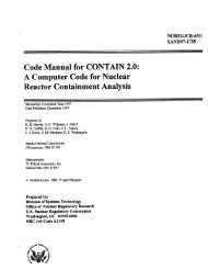
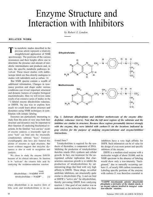
Fig. 1. Substrate dihydrofolate <strong>and</strong> inhibitor methotrexate of the enzyme dihydrefolate<br />
reductase (DHFR). Note that the left most regions of the substrate <strong>and</strong> the<br />
inhibitor are similar in structure. Because these regions presumably interact strongly<br />
<strong>with</strong> the enzyme, they were labeled <strong>with</strong> carbon-13 (at the locations indicated by<br />
gray circles) for the purpose of studying enzyme/substrate <strong>and</strong> enzyme/inhibitor<br />
interactions.<br />
tivated form.* inhibitors have a very high affinity for<br />
Tetrahydrofolate is required for the syn- DHFR. Such information can be of value for<br />
thesis of thymidine, a component of DNA. the design of even more potent <strong>and</strong> specific<br />
Inhibiting the production of tetrahydrofolate inhibitors of the enzyme.<br />
therefore retards DNA synthesis <strong>and</strong> cellular DHFR is a medium-sized enzyme <strong>with</strong> a<br />
growth. In fact, one treatment for the un- molecular weight of about 20,000, <strong>and</strong> its<br />
regulated cellular replication that char- NMR spectrum in the absence of labeling<br />
acterizes cancerous growth is to inhibit the would show only a tow-intensity “backproduction<br />
of tetrahydrofolate by ad- ground,” due to naturally occurring carministering<br />
drugs that bind <strong>with</strong> very high bon-13, of nearly 800 carbon resonances.<br />
affinity to DHFR. These drugs, so-called Labeling specific portions of the enzyme<br />
antifolate inhibitors, are structurally quite<br />
similar to dihydrofolate (Fig, 1) <strong>and</strong> can bind<br />
at DHFR’s “active site” far dihydrofolate,<br />
<strong>with</strong> carbon-13 was therefore essential to<br />
thereby preventing DHFR from catalyzing<br />
reaction 1. One goal of our studies was to<br />
underst<strong>and</strong>, at the molecular level, why these<br />
*NADPH (nicotinamide adenine dinucleotide<br />
phosphate], a derivative of the vitamin niacin, is<br />
an enzyme cofactor involved in biological oxida-<br />
tion-reductim reactions.<br />
68 Summer 1983 LOS ALAMOS SCIENCE<br />
co 2
Metabolism as it happens<br />
— METHOTREXATE<br />
() TRYPTOPHAN RESIDUES<br />
N / /<br />
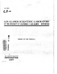
Fig. 2. Backbone ribbon drawing, showing the locations of the enzyme cofactor<br />
NADPH <strong>and</strong> of the inhibitor methotrexate, of the enzyme DHFR (a linked chain of<br />
167 amino acid residues) derived from the microorganism Lactobacillus casei.<br />
[Adapted from J. T. Bolin, D. J. Filman, D.A. Matthews, R. C. Hamlin, <strong>and</strong> J.Kraut,<br />
Journal of Biological Chemistry 257, 13650 {1982).] By occupying the enzyme’s<br />
that converts this inactive form of the vitamin folic acid to its active form. NMR<br />
spectra for DHFR derived from the microorganism Streptococcus faecium <strong>and</strong><br />
containing the 13<br />
C-labeled residue of the amino acid tryptophan provided information<br />
about the dynamic of the enzyme <strong>and</strong> the interaction between enzyme <strong>and</strong><br />
inhibitor. The approximate positions of the four tryptophan residues in DHFR derived<br />
from S. faecium are those indicated in the drawing, if it is assumed that DHFR<br />
derived from S. faecium <strong>and</strong> L. casei are homologous.<br />
LOS ALAMOS SCIENCE Summer 1983<br />
RELATED WORK<br />
enhance selected peaks <strong>and</strong> thereby reduce<br />
the complexity <strong>and</strong> increase the sensitivity of<br />
these studies.<br />
X-ray crystallographic studies have<br />
shown that enzymes are long strings of<br />
peptide-linked amino acids (that is, the<br />
amino group of one acid residue binds to the<br />
carboxylic acid group of the next). These<br />
long strings of amino acids fold in a complex<br />
way to form a globular structure (Fig. 2). To<br />
study the sensitivity of NMR spectra to<br />
structure, we labeled selected amino acid<br />
residues of DHFR <strong>with</strong> carbon-13 <strong>and</strong><br />
measured the spectra of the labeled enzyme<br />
in its globular form <strong>and</strong> again after its<br />
structure had been changed into a r<strong>and</strong>om<br />
coil by the addition of urea.<br />
The carbon- 13 labeling was accomplished<br />
by first labeling the amino acids methionine,<br />
arginine, <strong>and</strong> tryptophan <strong>and</strong> then growing<br />
the microorganism Streptococcus faecium in<br />
media containing one of these labeled amino<br />
acids. S. faecium, which is a good source of<br />
the enzyme DHFR, incorporates the labeled<br />
amino acids into the DHFR molecules. The<br />
labeled DHFR was then isolated from the<br />
microorganism, <strong>and</strong> its NM R spectra were<br />
obtained for the globular <strong>and</strong> r<strong>and</strong>om coil<br />
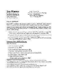
configurations. Figure 3 shows the results for<br />
DHFR labeled <strong>with</strong> [3- 13<br />
C ]tryptophan. The<br />
spectrum for the globular form of DHFR<br />
shows several carbon- 13 resonances corresponding<br />
to different positions along the<br />
polypeptide backbone <strong>and</strong> therefore to different<br />
chemical environments of the individual<br />
tryptophan residues <strong>with</strong>in the enzyme.<br />
Note that most of these so-called chemical<br />
shifts disappear when the enzyme structure<br />
is disrupted into a r<strong>and</strong>om coil. Thus NMR<br />
spectra are sensitive to structure.<br />
Looking more closely at the spectrum for<br />
the globular structure, we note that it has five<br />
resolved resonances although there are only<br />
four tryptophan residues in each enzyme<br />
molecule. Evidently a single tryptophan residue<br />
is responsible for the two adjacent peaks<br />
near 110 ppm. This splitting probably indicates<br />
that the enzyme takes on two dif-<br />
69
RELATED WORK<br />
ferent configurations in the region of that<br />
particular residue.<br />
The resonance at 106 ppm is also noteworthy<br />
because it is much broader than the<br />
others, a fact suggesting that the residue<br />
responsible for the resonance is located in a<br />
portion of the enzyme that undergoes confirmational<br />
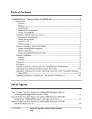
changes <strong>with</strong> time. Spectra taken<br />
at different temperatures (Fig. 4) confirmed<br />
this suggestion. The resonance at 106 ppm<br />
(resonance 4)exhibits a strongly temperaturedependent<br />
linewidth believed to be associated<br />
<strong>with</strong> “breathing” of the enzyme. That is, the<br />
enzyme adopts an ensemble of molecular<br />
conformations leading to temperature-dependent<br />
dynamic behavior. Figure 4 also shows<br />
that this “breathing’” phenomenon disappears<br />
when the enzyme is complexed <strong>with</strong> the<br />
inhibitor 3',5'-dichloromethotrexate, (The<br />
structure of this inhibitor is like that of<br />
methotrexate in Fig. 1 except that chlorine<br />
replaces hydrogen at positions 3 <strong>and</strong> 5 on the<br />
benzene ring.) The resonance then becomes<br />
sharp, indicating that a part of the binding<br />
energy between enzyme <strong>and</strong> inhibitor<br />
stabilizes a particular subset of enzyme conformations.<br />
For DHFR derived from S.<br />
faecium we found that the substrate<br />
dihydrofolate <strong>and</strong> each of the inhibitors<br />
studied lead to sharpening of resonance 4. In<br />
contrast, NADPH, the enzyme cofactor in<br />
reaction 1, does not significantly sharpen the<br />
resonance. Presumably, the NADPH binds to<br />
a portion of the enzyme molecule more remote<br />
from the particular tryptophan residue<br />
responsible for resonance 4.<br />
The addition of substrate or inhibitor<br />
molecules can also lead to changes in resonance<br />
position, or chemical shifts. For example.<br />
the spectra at 15 degrees Celsius in Fig.<br />
4 show that dichloromethotrexate causes a<br />
slight shift of resonance 1 to the right. Such<br />
chemical shifts reflect interactions between<br />
the enzyme <strong>and</strong> the bound molecules. In<br />
general, our results demonstrate the sensitivity<br />
of NMR spectra to the precise folded<br />
enzyme structure. <strong>Interaction</strong>s among specific<br />
pairs of residues are important in produc-<br />
70<br />
ing chemical shifts, <strong>and</strong> these shifts are<br />
altered when the enzyme binds other<br />
molecules. Dynamic behavior such as<br />
“breathing” is also observable, but whether<br />
this behavior observed in DHFR is significant<br />
for catalysis (for example, whether it<br />
helps the enzyme to “recognize” the<br />
substrate dihydrofolate) is still an open question.<br />
A second set of studies <strong>with</strong> DHFR was<br />
designed to investigate the basis for the high<br />
affinity between the inhibitor methotrexate<br />
<strong>and</strong> DHFR. Rather than labeling the<br />
enzyme, we chose to label the inhibitor. The<br />
label was placed in that portion of methotrexate<br />
thought to interact strongly <strong>with</strong><br />
DHFR (see Fig. I, where the position of the<br />
carbon- 13 label is marked by a gray circle).<br />
We found that the NMR spectrum of a<br />
solution containing labeled methotrexate <strong>and</strong><br />
DHFR exhibits two carbon-13 resonances,<br />
one corresponding to inhibitor molecules<br />
that are free in solution <strong>and</strong> one corresponding<br />
to inhibitor molecules that are tightly<br />
associated <strong>with</strong> the enzyme. The fact that the<br />
single carbon- 13 label exhibits two resonances<br />
is a reflection of the very high<br />
affinity of methotrexate for the enzyme.<br />
We then varied the pH of the solution<br />
containing the enzyme <strong>and</strong> the labeled inhibitor<br />
<strong>and</strong> found that the two resonances<br />
behave very differently (Fig. 5). The resonance<br />
ascribed to the uncompleted inhibitor<br />
undergoes a large shift in position at a pH of<br />
about 5.7, This so-called titration behavior<br />
indicates that the uncormplexed inhibitor accepts<br />
a proton (H+) in the following reversible<br />
reaction:<br />
The pH at which the curve’s large shift is<br />
centered (5.7) is called the pK of this proc-<br />
H<br />
Globular<br />
<strong>Structure</strong><br />
112 110 108 106 104<br />
ppm
Metabolism as it happens<br />
15°c<br />
L<br />
25° C<br />
L<br />
15°c<br />
in presence<br />
of inhibitor<br />
112 110 108 106 104<br />
ppm<br />
LOS ALAMOS SCIENCE Summer 1983<br />
Fig, 4. Carbon-13 resonances observed<br />
for [3- 13<br />
C]tryptophan-labeled DHFR at<br />
various temperatures <strong>and</strong> at 15 degrees<br />
Celsius in the presence of the inhibitor<br />
3',5'-dichloromethotrexate. Resonance 4<br />
exhibits a temperature-dependent linewidth<br />
that becomes a sharp resonance in<br />
the presence of the inhibitor. The tryptophan<br />
residue thought to be responsible<br />
2 4 6 8 10<br />
pH<br />
RELATED WORK<br />
ess. It represents the pH value at which half<br />
of the molecules are protonated <strong>and</strong> half<br />
unprotonated. Thus the one resonance observed<br />
for the uncompleted inhibitor is actually<br />
an average produced by its protonated<br />
<strong>and</strong> unprotonated forms. In contrast, the<br />
resonance ascribed to the enzyme-complexed<br />
methotrexate remains fixed near the resonance<br />
of the protonated form of the uncompleted<br />
inhibitor. These data indicate that<br />
the protonated form of methotrexate is the<br />
potent enzyme inhibitor: in other words, a<br />
strong interaction between protonated methotrexate<br />
<strong>and</strong> the enzyme must be the critical<br />
factor in making methotrexate an effective<br />
inhibitor. This conclusion is supported by the<br />
x-ray crystallographic data of Matthews <strong>and</strong><br />
coworkers at the University of California,<br />
San Diego. They found that in the crystalline<br />
state the enzyme-complexed methotrexate is<br />
protonated <strong>and</strong> hydrogen-bonded to a negatively<br />
charged amino acid (aspartate-26) of<br />
the enzyme.<br />
Figure 5 shows that the pK for protonation<br />
of the enzyme-complexed inhibitor is<br />
well above 10, the highest pH value used in<br />
the study. Such a large difference between<br />
the pK of the enzyme-complexed inhibitor<br />
<strong>and</strong> that of the free inhibitor is a measure of<br />
the binding energy between the inhibitor <strong>and</strong><br />
the enzyme. The strength of binding was not<br />
accurately known prior to these studies <strong>and</strong><br />
had in fact been incorrectly determined using<br />
conventional ultraviolet spectroscopic techniques.<br />
It is an impressive achievement of the<br />
isotopic labeling/NMR method that of all the<br />
interactions among amino acid residues<br />
<strong>and</strong> between amino acids <strong>and</strong> solvent<br />
molecules that stabilize the enzyme structure,<br />
we can probe a single one <strong>and</strong> quantify<br />
its strength. ■