An unusual stress fracture of the proximal phalanx of the great toe: A ...
An unusual stress fracture of the proximal phalanx of the great toe: A ...
An unusual stress fracture of the proximal phalanx of the great toe: A ...

Create successful ePaper yourself
Turn your PDF publications into a flip-book with our unique Google optimized e-Paper software.
Introduction<br />
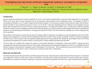
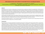
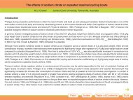
<strong>An</strong> <strong>unusual</strong> <strong>stress</strong> <strong>fracture</strong> <strong>of</strong> <strong>the</strong> <strong>proximal</strong> <strong>phalanx</strong> <strong>of</strong> <strong>the</strong> <strong>great</strong> <strong>toe</strong>: A case report<br />
G.C. Pitsis* & J.P. Best Eastern Suburbs Sports Medicine Centre, Sydney, Australia<br />
Stress <strong>fracture</strong> <strong>of</strong> <strong>the</strong> <strong>proximal</strong> <strong>phalanx</strong> <strong>of</strong> <strong>the</strong> <strong>great</strong> <strong>toe</strong> is rare. A medline search was conducted using <strong>the</strong> key words “<strong>stress</strong> <strong>fracture</strong>” and”“<strong>great</strong><br />
<strong>toe</strong>” resulting in three specific references. Yokoe and Mannoji in 1986 published a case series <strong>of</strong> a sprinter, a Kendo player (Japanese fencer),<br />
and a rugby player with this injury (1). Similarly, Shiraishi and colleagues in 1993 reported 3 cases <strong>of</strong> this injury in a long distance runner, a<br />
volleyball player, and a soccer player (2). Matsusue and colleagues in 1992 reported this injury in a shot-putter (3). Interestingly, <strong>the</strong>se <strong>stress</strong><br />
<strong>fracture</strong>s have only been reported among adolescent athletes. Most <strong>stress</strong> <strong>fracture</strong>s were associated with halux valgus deformity.<br />
The pathophysiology involves running and jumping activities inducing vertical ground reaction forces several times that <strong>of</strong> body weight. This<br />
results in large repetitive truncation forces on <strong>the</strong> base <strong>of</strong> <strong>the</strong> <strong>proximal</strong> <strong>phalanx</strong> from <strong>the</strong> windlass action <strong>of</strong> <strong>the</strong> plantar aponeurosis and muscle<br />
action, in addition to axial compressive forces (4).<br />
Hallux valgus is a postulated associated biomechanical predisposing factor to this injury due to <strong>the</strong> bowstring effect <strong>of</strong> extensor hallucis longus<br />
and adductor hallucis acting on <strong>the</strong> <strong>great</strong> <strong>toe</strong>. This may increase abduction forces on <strong>the</strong> first metatarsophalangeal joint. The resulting tensile<br />
strain on <strong>the</strong> medial collateral ligament and <strong>the</strong> medial head <strong>of</strong> <strong>the</strong> flexor hallucis brevis may predispose to an avulsion type <strong>stress</strong> <strong>fracture</strong> <strong>of</strong><br />
<strong>the</strong> medial base <strong>of</strong> <strong>the</strong> <strong>proximal</strong> <strong>phalanx</strong> <strong>of</strong> <strong>the</strong> <strong>great</strong> <strong>toe</strong> (1,2,3,5,6).<br />
It is expected this type <strong>of</strong> <strong>stress</strong> <strong>fracture</strong> would heal with conservative treatment <strong>of</strong> 4 – 6 weeks non weight-bearing rest. A graduated return to<br />
activity over a fur<strong>the</strong>r 4 – 6 weeks is usually required. Biomechanical factors need to be addressed appropriately.<br />
Case Report<br />
The patient is a 41-year-old nulliparous non-pr<strong>of</strong>essional triathlete who was clinically diagnosed with a <strong>stress</strong> <strong>fracture</strong> <strong>of</strong> <strong>the</strong> medial base <strong>of</strong> <strong>the</strong><br />
<strong>proximal</strong> <strong>phalanx</strong> <strong>of</strong> <strong>the</strong> left <strong>great</strong> <strong>toe</strong> confirmed by x-ray (Figure 1). Radiologically <strong>the</strong> <strong>stress</strong> <strong>fracture</strong> was undisplaced and involved approximately<br />
one third <strong>of</strong> <strong>the</strong> articular surface.<br />
Pain in <strong>the</strong> left <strong>great</strong> <strong>toe</strong> was experienced after progressive moderate increased training load over a 12-week period leading into a triathlon.<br />
This included 20 kilometers <strong>of</strong> running per week. A mild ache was felt 2 weeks before competition. After completing <strong>the</strong> 65-kilometer cycle and<br />
500-meter swim <strong>of</strong> <strong>the</strong> triathlon uneventfully, moderately severe pain was experienced during <strong>the</strong> first 500 meters <strong>of</strong> <strong>the</strong> 4.5-kilometer run. On<br />
completion <strong>of</strong> <strong>the</strong> race <strong>the</strong> patient could not fully weight-bear due to severe pain.<br />
Print<br />
Index<br />
Table <strong>of</strong> Contents<br />
Quit
<strong>An</strong> <strong>unusual</strong> <strong>stress</strong> <strong>fracture</strong> <strong>of</strong> <strong>the</strong> <strong>proximal</strong> <strong>phalanx</strong> <strong>of</strong> <strong>the</strong> <strong>great</strong> <strong>toe</strong>: A case report<br />
G.C. Pitsis* & J.P. Best Eastern Suburbs Sports Medicine Centre, Sydney, Australia<br />
The patient has a past history <strong>of</strong> multiple <strong>stress</strong> <strong>fracture</strong>s over <strong>the</strong> last 4<br />
years including <strong>the</strong> left fibula in June 1997, <strong>the</strong> left calcaneus in October<br />
1998, and <strong>the</strong> right fibula in April 2000, suggesting a low threshold for<br />
<strong>stress</strong> <strong>fracture</strong>s.<br />
A Dual Energy X-ray Absorptiometry (DEXA) scan in November 1998<br />
revealed osteopenia. At <strong>the</strong> time <strong>of</strong> scanning <strong>the</strong> patient had 14 months<br />
<strong>of</strong> amenorrhoea. Following treatment with Alendronate (Fosamax),<br />
hormone replacement <strong>the</strong>rapy (Ogen 1.25 mg and Provera 2.5mg), and<br />
calcium, follow-up DEXA scans in July 2000 and May 2001 were normal<br />
(Table 1). The patient continues to use hormone replacement <strong>the</strong>rapy<br />
and reports normal menstruation.<br />
Figure 1. Radiograph at diagnosis<br />
Print<br />
Index<br />
Table <strong>of</strong> Contents<br />
Quit
<strong>An</strong> <strong>unusual</strong> <strong>stress</strong> <strong>fracture</strong> <strong>of</strong> <strong>the</strong> <strong>proximal</strong> <strong>phalanx</strong> <strong>of</strong> <strong>the</strong> <strong>great</strong> <strong>toe</strong>: A case report<br />
Table 1. DEXA scan results<br />
Table 1: DEXA scan results<br />
G.C. Pitsis* & J.P. Best Eastern Suburbs Sports Medicine Centre, Sydney, Australia<br />
Nov 1998 July 2000 May 2001<br />
Lumbar age-matched (Z) score: -1.4 -1.1 -0.6<br />
Lumbar young-adult (T) score: -1.8 -1.2 -1.1<br />
Proximal femur age-matched (Z) score: -1.0 -0.8 -0.4<br />
Proximal femur young-adult (T) score: -1.4 -1.0 -0.9<br />
The patient has asthma well controlled on regular Pulmicort and Brycanyl as required. She has needed a short coarse <strong>of</strong> oral prednisone on 2<br />
occasions for severe exacerbation <strong>of</strong> her asthma at a younger age. She has a family history <strong>of</strong> ischaemic heart disease and hypertension.<br />
Initial clinical examination revealed point tenderness over <strong>the</strong> medial base <strong>of</strong> <strong>the</strong> <strong>proximal</strong> <strong>phalanx</strong> <strong>of</strong> <strong>the</strong> left <strong>great</strong> <strong>toe</strong>. There was no associated<br />
hallux valgus. She has been wearing orthotics for <strong>the</strong> last 15 years to correct hyper-pronation. They were last renewed 2 years ago and are in<br />
satisfactory condition.<br />
Excellent clinical and radiological union was achieved with 6 weeks non weight-bearing rest with crutches. A graduated return to activity was<br />
achieved over a fur<strong>the</strong>r 6 weeks. A 2 nd – 5 th metatarsal bar was used to <strong>of</strong>f-load <strong>stress</strong> from <strong>the</strong> <strong>great</strong> <strong>toe</strong>.<br />
Print<br />
Index<br />
Table <strong>of</strong> Contents<br />
Quit
<strong>An</strong> <strong>unusual</strong> <strong>stress</strong> <strong>fracture</strong> <strong>of</strong> <strong>the</strong> <strong>proximal</strong> <strong>phalanx</strong> <strong>of</strong> <strong>the</strong> <strong>great</strong> <strong>toe</strong>: A case report<br />
G.C. Pitsis* & J.P. Best Eastern Suburbs Sports Medicine Centre, Sydney, Australia<br />
Figure 2. Radiograph 4 weeks<br />
post-diagnosis<br />
Print<br />
Index<br />
Table <strong>of</strong> Contents<br />
Quit
Discussion<br />
<strong>An</strong> <strong>unusual</strong> <strong>stress</strong> <strong>fracture</strong> <strong>of</strong> <strong>the</strong> <strong>proximal</strong> <strong>phalanx</strong> <strong>of</strong> <strong>the</strong> <strong>great</strong> <strong>toe</strong>: A case report<br />
G.C. Pitsis* & J.P. Best Eastern Suburbs Sports Medicine Centre, Sydney, Australia<br />
The patient’s <strong>stress</strong> <strong>fracture</strong> was <strong>unusual</strong> as it occurred simply with relatively low levels <strong>of</strong> <strong>stress</strong> and normal bone mineral density. In addition,<br />
<strong>the</strong>re was no associated hallux valgus, and was not consistent with <strong>the</strong> previously documented age group <strong>of</strong> adolescence.<br />
Stress <strong>fracture</strong>s are more common in female athletes with menstrual disturbances (7). Alendronate (Fosamax) is used for treatment in<br />
osteoporosis. Hormone replacement <strong>the</strong>rapy with Ogen 1.25 mg and Provera 2.5mg may be effective in reducing <strong>the</strong> negative effects <strong>of</strong><br />
menstrual disturbances on bone mineral density (8). The addition <strong>of</strong> calcium in <strong>the</strong> exercising individual may also promote increase bone<br />
mineralisation (9). She is currently being investigated from an endocrinological viewpoint.<br />
This case report supports <strong>the</strong> opinion that <strong>stress</strong> <strong>fracture</strong>s <strong>of</strong> <strong>the</strong> <strong>proximal</strong> <strong>phalanx</strong> <strong>of</strong> <strong>the</strong> <strong>great</strong> <strong>toe</strong> respond well to non weight-bearing rest with<br />
graduated return to activity. This has been recorded previously (1,2). In addition, Shiraishi and colleagues (1) found one <strong>of</strong> <strong>the</strong> patients<br />
proceeded to non-union as a result <strong>of</strong> continued training. Biomechanical factors need to be addressed appropriately (10).<br />
Evidence <strong>of</strong> clinical union occurred before radiological union at 4 weeks post-injury. This supports current understanding <strong>of</strong> <strong>the</strong> temporal<br />
relationship <strong>of</strong> clinical and radiological union. Thus clinical status <strong>of</strong> <strong>the</strong> <strong>fracture</strong> is <strong>of</strong> paramount importance when assessing progress <strong>of</strong> union<br />
<strong>of</strong> <strong>the</strong> <strong>fracture</strong>. Radiological investigations will confirm <strong>fracture</strong> position, and document evidence <strong>of</strong> union after 4 weeks.<br />
Print<br />
Index<br />
Table <strong>of</strong> Contents<br />
Quit
<strong>An</strong> <strong>unusual</strong> <strong>stress</strong> <strong>fracture</strong> <strong>of</strong> <strong>the</strong> <strong>proximal</strong> <strong>phalanx</strong> <strong>of</strong> <strong>the</strong> <strong>great</strong> <strong>toe</strong>: A case report<br />
G.C. Pitsis* & J.P. Best Eastern Suburbs Sports Medicine Centre, Sydney, Australia<br />
Figure 3. Radiograph 8 weeks<br />
post-diagnosis<br />
Print<br />
Index<br />
Table <strong>of</strong> Contents<br />
Quit
References<br />
<strong>An</strong> <strong>unusual</strong> <strong>stress</strong> <strong>fracture</strong> <strong>of</strong> <strong>the</strong> <strong>proximal</strong> <strong>phalanx</strong> <strong>of</strong> <strong>the</strong> <strong>great</strong> <strong>toe</strong>: A case report<br />
G.C. Pitsis* & J.P. Best Eastern Suburbs Sports Medicine Centre, Sydney, Australia<br />
Yokoe K. and Mannoji T. Stress <strong>fracture</strong> <strong>of</strong> <strong>the</strong> <strong>proximal</strong> <strong>phalanx</strong> <strong>of</strong> <strong>the</strong> <strong>great</strong> <strong>toe</strong>: a report <strong>of</strong> three cases. American Journal <strong>of</strong> Sports Medicine<br />
1986; 14 (3): 240-242.<br />
Shiraishi M, Mizuta H, Kubota K, et al. Stress <strong>fracture</strong> <strong>of</strong> <strong>the</strong> <strong>proximal</strong> <strong>phalanx</strong> <strong>of</strong> <strong>the</strong> <strong>great</strong> <strong>toe</strong>. Foot and <strong>An</strong>kle 1993; 14 (1): 28-34.<br />
Matsusue Y, Yamamuro T, Nakagawa Y, et al. Stress <strong>fracture</strong> <strong>of</strong> <strong>the</strong> <strong>proximal</strong> <strong>phalanx</strong> <strong>of</strong> <strong>the</strong> <strong>great</strong> <strong>toe</strong>: <strong>the</strong> case report <strong>of</strong> a shot-putter. Clinical<br />
Journal <strong>of</strong> Sports Medicine 1992; 2(4): 279-282<br />
Brukner P, Bennell K and Ma<strong>the</strong>son G. Stress <strong>fracture</strong>s. Blackwell Science, 2000. pp 183-184.<br />
Eisele S and Sammarco G. Fatigue <strong>fracture</strong>s <strong>of</strong> <strong>the</strong> foot and ankle in athletes. Journal <strong>of</strong> Bone and Joint Surgery – American volume 1993 Feb.<br />
75 (2): 290-298.<br />
Hershman E and Mailly T. Stress <strong>fracture</strong>s. Clinics in Sports Medicine 1990 Jan. 9 (1): 183-214<br />
Brukner P and Bennell K. Stress <strong>fracture</strong>s in female athletes. Diagnosis, management and rehabilitation. Sports Medicine 1997 Dec. 24<br />
(6):419-429<br />
Fields C and Fricker P. Medical problems in athletes. Blackwell Science, 1997.<br />
Brukner P and Khan K. Clinical sports medicine, second edition. McGraw Hill 2000.<br />
Knapp T and Garrett W. Stress <strong>fracture</strong>s: general concepts. Clinics in Sports Medicine 1997 Apr. 16(2): 339-356.<br />
Print<br />
Index<br />
Table <strong>of</strong> Contents<br />
Quit
<strong>An</strong> <strong>unusual</strong> <strong>stress</strong> <strong>fracture</strong> <strong>of</strong> <strong>the</strong> <strong>proximal</strong> <strong>phalanx</strong> <strong>of</strong> <strong>the</strong> <strong>great</strong> <strong>toe</strong>: A case<br />
report<br />
G.C. Pitsis* & J.P. Best<br />
Eastern Suburbs Sports Medicine Centre, Sydney, Australia<br />
INTRODUCTION: Stress <strong>fracture</strong> <strong>of</strong> <strong>the</strong> <strong>proximal</strong> <strong>phalanx</strong> <strong>of</strong> <strong>the</strong> <strong>great</strong> <strong>toe</strong> is rare. A medline search was<br />
conducted using <strong>the</strong> key words “<strong>stress</strong> <strong>fracture</strong>” and “<strong>great</strong> <strong>toe</strong>” resulting in three specific references. Yokoe and<br />
Mannoji in 1986 published a case series <strong>of</strong> a sprinter, a Kendo player (Japanese fencer), and a rugby player with<br />
this injury (1). Similarly, Shiraishi and colleagues in 1993 reported 3 cases <strong>of</strong> this injury in a long distance runner,<br />
a volleyball player, and a soccer player (2). Matsusue and colleagues in 1992 reported this injury in a shot-putter<br />
(3). Interestingly, <strong>the</strong>se <strong>stress</strong> <strong>fracture</strong>s have only been reported among adolescent athletes. Most <strong>stress</strong> <strong>fracture</strong>s<br />
were associated with halux valgus deformity.<br />
The pathophysiology involves running and jumping activities inducing vertical ground reaction forces several<br />
times that <strong>of</strong> body weight. This results in large repetitive truncation forces on <strong>the</strong> base <strong>of</strong> <strong>the</strong> <strong>proximal</strong> <strong>phalanx</strong><br />
from <strong>the</strong> windlass action <strong>of</strong> <strong>the</strong> plantar aponeurosis and muscle action, in addition to axial compressive forces (4).<br />
Hallux valgus is a postulated associated biomechanical predisposing factor to this injury due to <strong>the</strong> bowstring<br />
effect <strong>of</strong> extensor hallucis longus and adductor hallucis acting on <strong>the</strong> <strong>great</strong> <strong>toe</strong>. This may increase abduction<br />
forces on <strong>the</strong> first metatarsophalangeal joint. The resulting tensile strain on <strong>the</strong> medial collateral ligament and <strong>the</strong><br />
medial head <strong>of</strong> <strong>the</strong> flexor hallucis brevis may predispose to an avulsion type <strong>stress</strong> <strong>fracture</strong> <strong>of</strong> <strong>the</strong> medial base <strong>of</strong><br />
<strong>the</strong> <strong>proximal</strong> <strong>phalanx</strong> <strong>of</strong> <strong>the</strong> <strong>great</strong> <strong>toe</strong> (1,2,3,5,6).<br />
It is expected this type <strong>of</strong> <strong>stress</strong> <strong>fracture</strong> would heal with conservative treatment <strong>of</strong> 4 – 6 weeks non weightbearing<br />
rest. A graduated return to activity over a fur<strong>the</strong>r 4 – 6 weeks is usually required. Biomechanical factors<br />
need to be addressed appropriately.<br />
CASE REPORT: The patient is a 41-year-old nulliparous non-pr<strong>of</strong>essional triathlete who was clinically<br />
diagnosed with a <strong>stress</strong> <strong>fracture</strong> <strong>of</strong> <strong>the</strong> medial base <strong>of</strong> <strong>the</strong> <strong>proximal</strong> <strong>phalanx</strong> <strong>of</strong> <strong>the</strong> left <strong>great</strong> <strong>toe</strong> confirmed by x-ray<br />
(Figure 1). Radiologically <strong>the</strong> <strong>stress</strong> <strong>fracture</strong> was undisplaced and involved approximately one third <strong>of</strong> <strong>the</strong> articular<br />
surface.<br />
Pain in <strong>the</strong> left <strong>great</strong> <strong>toe</strong> was experienced after progressive moderate increased training load over a 12-week<br />
period leading into a triathlon. This included 20 kilometers <strong>of</strong> running per week. A mild ache was felt 2 weeks<br />
before competition. After completing <strong>the</strong> 65-kilometer cycle and 500-meter swim <strong>of</strong> <strong>the</strong> triathlon uneventfully,<br />
moderately severe pain was experienced during <strong>the</strong> first 500 meters <strong>of</strong> <strong>the</strong> 4.5-kilometer run. On completion <strong>of</strong> <strong>the</strong><br />
race <strong>the</strong> patient could not fully weight-bear due to severe pain.<br />
The patient has a past history <strong>of</strong> multiple <strong>stress</strong> <strong>fracture</strong>s over <strong>the</strong> last 4 years including <strong>the</strong> left fibula in June<br />
1997, <strong>the</strong> left calcaneus in October 1998, and <strong>the</strong> right fibula in April 2000, suggesting a low threshold for <strong>stress</strong><br />
<strong>fracture</strong>s.<br />
A Dual Energy X-ray Absorptiometry (DEXA) scan in November 1998 revealed osteopenia. At <strong>the</strong> time <strong>of</strong><br />
scanning <strong>the</strong> patient had 14 months <strong>of</strong> amenorrhoea. Following treatment with Alendronate (Fosamax), hormone<br />
replacement <strong>the</strong>rapy (Ogen 1.25 mg and Provera 2.5mg), and calcium, follow-up DEXA scans in July 2000 and<br />
May 2001 were normal (Table 1). The patient continues to use hormone replacement <strong>the</strong>rapy and reports normal<br />
menstruation.
Figure 1. Radiograph at diagnosis<br />
Table 1: DEXA scan results<br />
Nov 1998 July 2000 May 2001<br />
Lumbar age-matched (Z) score: -1.4 -1.1 -0.6<br />
Lumbar young-adult (T) score: -1.8 -1.2 -1.1<br />
Proximal femur age-matched (Z) score: -1.0 -0.8 -0.4<br />
Proximal femur young-adult (T) score: -1.4 -1.0 -0.9<br />
The patient has asthma well controlled on regular Pulmicort and Brycanyl as required. She has needed a short<br />
coarse <strong>of</strong> oral prednisone on 2 occasions for severe exacerbation <strong>of</strong> her asthma at a younger age. She has a<br />
family history <strong>of</strong> ischaemic heart disease and hypertension.<br />
Initial clinical examination revealed point tenderness over <strong>the</strong> medial base <strong>of</strong> <strong>the</strong> <strong>proximal</strong> <strong>phalanx</strong> <strong>of</strong> <strong>the</strong> left<br />
<strong>great</strong> <strong>toe</strong>. There was no associated hallux valgus. She has been wearing orthotics for <strong>the</strong> last 15 years to correct<br />
hyper-pronation. They were last renewed 2 years ago and are in satisfactory condition.<br />
Excellent clinical and radiological union was achieved with 6 weeks non weight-bearing rest with crutches. A<br />
graduated return to activity was achieved over a fur<strong>the</strong>r 6 weeks. A 2 nd – 5 th metatarsal bar was used to <strong>of</strong>f-load<br />
<strong>stress</strong> from <strong>the</strong> <strong>great</strong> <strong>toe</strong>.
Figure 2. Radiograph 4 weeks post-diagnosis<br />
DISCUSSION: The patient’s <strong>stress</strong> <strong>fracture</strong> was <strong>unusual</strong> as it occurred simply with relatively low levels <strong>of</strong><br />
<strong>stress</strong> and normal bone mineral density. In addition, <strong>the</strong>re was no associated hallux valgus, and was not<br />
consistent with <strong>the</strong> previously documented age group <strong>of</strong> adolescence.<br />
Stress <strong>fracture</strong>s are more common in female athletes with menstrual disturbances (7). Alendronate (Fosamax)<br />
is used for treatment in osteoporosis. Hormone replacement <strong>the</strong>rapy with Ogen 1.25 mg and Provera 2.5mg may<br />
be effective in reducing <strong>the</strong> negative effects <strong>of</strong> menstrual disturbances on bone mineral density (8). The addition <strong>of</strong><br />
calcium in <strong>the</strong> exercising individual may also promote increase bone mineralisation (9). She is currently being<br />
investigated from an endocrinological viewpoint.<br />
This case report supports <strong>the</strong> opinion that <strong>stress</strong> <strong>fracture</strong>s <strong>of</strong> <strong>the</strong> <strong>proximal</strong> <strong>phalanx</strong> <strong>of</strong> <strong>the</strong> <strong>great</strong> <strong>toe</strong> respond<br />
well to non weight-bearing rest with graduated return to activity. This has been recorded previously (1,2). In<br />
addition, Shiraishi and colleagues (1) found one <strong>of</strong> <strong>the</strong> patients proceeded to non-union as a result <strong>of</strong> continued<br />
training. Biomechanical factors need to be addressed appropriately (10).<br />
Evidence <strong>of</strong> clinical union occurred before radiological union at 4 weeks post-injury. This supports current<br />
understanding <strong>of</strong> <strong>the</strong> temporal relationship <strong>of</strong> clinical and radiological union. Thus clinical status <strong>of</strong> <strong>the</strong> <strong>fracture</strong> is<br />
<strong>of</strong> paramount importance when assessing progress <strong>of</strong> union <strong>of</strong> <strong>the</strong> <strong>fracture</strong>. Radiological investigations will confirm<br />
<strong>fracture</strong> position, and document evidence <strong>of</strong> union after 4 weeks.
Figure 3. Radiograph 8 weeks post-diagnosis<br />
REFERENCES:<br />
1. Yokoe K. and Mannoji T. Stress <strong>fracture</strong> <strong>of</strong> <strong>the</strong> <strong>proximal</strong> <strong>phalanx</strong> <strong>of</strong> <strong>the</strong> <strong>great</strong> <strong>toe</strong>: a report <strong>of</strong> three cases.<br />
American Journal <strong>of</strong> Sports Medicine 1986; 14 (3): 240-242.<br />
2. Shiraishi M, Mizuta H, Kubota K, et al. Stress <strong>fracture</strong> <strong>of</strong> <strong>the</strong> <strong>proximal</strong> <strong>phalanx</strong> <strong>of</strong> <strong>the</strong> <strong>great</strong> <strong>toe</strong>. Foot and<br />
<strong>An</strong>kle 1993; 14 (1): 28-34.<br />
3. Matsusue Y, Yamamuro T, Nakagawa Y, et al. Stress <strong>fracture</strong> <strong>of</strong> <strong>the</strong> <strong>proximal</strong> <strong>phalanx</strong> <strong>of</strong> <strong>the</strong> <strong>great</strong> <strong>toe</strong>:<br />
<strong>the</strong> case report <strong>of</strong> a shot-putter. Clinical Journal <strong>of</strong> Sports Medicine 1992; 2(4): 279-282<br />
4. Brukner P, Bennell K and Ma<strong>the</strong>son G. Stress <strong>fracture</strong>s. Blackwell Science, 2000. pp 183-184.<br />
5. Eisele S and Sammarco G. Fatigue <strong>fracture</strong>s <strong>of</strong> <strong>the</strong> foot and ankle in athletes. Journal <strong>of</strong> Bone and Joint<br />
Surgery – American volume 1993 Feb. 75 (2): 290-298.<br />
6. Hershman E and Mailly T. Stress <strong>fracture</strong>s. Clinics in Sports Medicine 1990 Jan. 9 (1): 183-214<br />
7. Brukner P and Bennell K. Stress <strong>fracture</strong>s in female athletes. Diagnosis, management and rehabilitation.<br />
Sports Medicine 1997 Dec. 24 (6):419-429<br />
8. Fields C and Fricker P. Medical problems in athletes. Blackwell Science, 1997.<br />
9. Brukner P and Khan K. Clinical sports medicine, second edition. McGraw Hill 2000.<br />
10. Knapp T and Garrett W. Stress <strong>fracture</strong>s: general concepts. Clinics in Sports Medicine 1997 Apr. 16(2):<br />
339-356.