Effect of juvenile hormone analogue (Admiral) on embryogenesis of ...
Effect of juvenile hormone analogue (Admiral) on embryogenesis of ...
Effect of juvenile hormone analogue (Admiral) on embryogenesis of ...

You also want an ePaper? Increase the reach of your titles
YUMPU automatically turns print PDFs into web optimized ePapers that Google loves.
Egypt. Acad. J. biolog. Sci., 2 (1): 165 - 176 (2009)<br />
E.mail.egyptianacademic@yahoo.com ISSN: 1687–8809<br />
Received: 11/2/2009 www.eajbs.eg.net<br />
<str<strong>on</strong>g>Effect</str<strong>on</strong>g> <str<strong>on</strong>g>of</str<strong>on</strong>g> <str<strong>on</strong>g>juvenile</str<strong>on</strong>g> <str<strong>on</strong>g>horm<strong>on</strong>e</str<strong>on</strong>g> <str<strong>on</strong>g>analogue</str<strong>on</strong>g> (<str<strong>on</strong>g>Admiral</str<strong>on</strong>g>) <strong>on</strong> <strong>embryogenesis</strong> <str<strong>on</strong>g>of</str<strong>on</strong>g> the s<str<strong>on</strong>g>of</str<strong>on</strong>g>t tick<br />
Argas persicus (Oken)<br />
Nadia H. Ahmed; Wafaa A. Radwan; Noha A. Guneidy and<br />
Shimaa S. Mohammed<br />
Department <str<strong>on</strong>g>of</str<strong>on</strong>g> Entomology, Faculty <str<strong>on</strong>g>of</str<strong>on</strong>g> Science, Ain-Shams University<br />
ABSTRACT<br />
Embry<strong>on</strong>ic development <str<strong>on</strong>g>of</str<strong>on</strong>g> the fowl tick A. persicus was investigated during<br />
cleavage stage, blastoderm formati<strong>on</strong>, gastrulati<strong>on</strong> and organogenesis. Cleavage<br />
started 1 h post-ovipositi<strong>on</strong> (POP) and is indicated by presence <str<strong>on</strong>g>of</str<strong>on</strong>g> vitellophages <str<strong>on</strong>g>of</str<strong>on</strong>g><br />
different sizes. Blastoderm is formed at 48 h POP. Sec<strong>on</strong>dary vitellophages are<br />
observed by 24 h POP. By 72 h POP, the germ band is formed. At 96 h POP, while<br />
the embry<strong>on</strong>ic envelops are formed, gastrulati<strong>on</strong> <str<strong>on</strong>g>of</str<strong>on</strong>g> the germ band takes place.<br />
Segmentati<strong>on</strong> and differentiati<strong>on</strong> <str<strong>on</strong>g>of</str<strong>on</strong>g> germ band, as well as, blastokinesis occur at 120<br />
h POP. The stomodaeal and proctodaeal invaginati<strong>on</strong> started to differentiate at 120 h<br />
POP. The anterior and posterior midgut rudiments can be observed at 144 h POP. By<br />
168 h POP, the nervous system, as well as, rectal sac and malpighian tubules are<br />
formed.<br />
Histological study revealed that applicati<strong>on</strong> <str<strong>on</strong>g>of</str<strong>on</strong>g> JHA (<str<strong>on</strong>g>Admiral</str<strong>on</strong>g>) to newly laid<br />
eggs <str<strong>on</strong>g>of</str<strong>on</strong>g> A. persicus blocked the embry<strong>on</strong>ic development <str<strong>on</strong>g>of</str<strong>on</strong>g> affected eggs at cleavage<br />
stage and before blastoderm and pole cells formati<strong>on</strong>. In 24 h-old treated eggs, few<br />
cleavage nuclei appeared with absence <str<strong>on</strong>g>of</str<strong>on</strong>g> sec<strong>on</strong>dary vitellophages and pole cells. In<br />
48 h-old treated eggs, the blastoderm was rudimentary and irregularly arranged. In 72<br />
h-old treated eggs, disintegrati<strong>on</strong> <str<strong>on</strong>g>of</str<strong>on</strong>g> the rudimentary blastoderm and cracking <str<strong>on</strong>g>of</str<strong>on</strong>g> the<br />
cytoplasm could be seen. Starting from 96-old eggs until hatching at 168 h, complete<br />
destructi<strong>on</strong> <str<strong>on</strong>g>of</str<strong>on</strong>g> the eggs was clear.<br />
Key words: Argas persicus, Juvenile <str<strong>on</strong>g>horm<strong>on</strong>e</str<strong>on</strong>g> <str<strong>on</strong>g>analogue</str<strong>on</strong>g>, <strong>embryogenesis</strong><br />
INTRODUCTION<br />
Several histological studies <str<strong>on</strong>g>of</str<strong>on</strong>g> the <strong>embryogenesis</strong> <str<strong>on</strong>g>of</str<strong>on</strong>g> insects especially those<br />
bel<strong>on</strong>ging to order Diptera (West et al., 1968; Sandescu ahd Tacu, 1970; Raminani<br />
and Cupp, 1978 and Radwan et al., 1993). Hymenoptera (Tawfic, 1975) and<br />
Hemiptera (Shaarawi et al., 1982 and Ajidagba et al., 1983) has been reported.<br />
However, little informati<strong>on</strong> <strong>on</strong> the <strong>embryogenesis</strong> <str<strong>on</strong>g>of</str<strong>on</strong>g> ticks, especially the s<str<strong>on</strong>g>of</str<strong>on</strong>g>t ticks<br />
was available. Preliminary studies have been indicated for the s<str<strong>on</strong>g>of</str<strong>on</strong>g>t tick Aragas<br />
persicus (El-Rammah, 1981). Most investigati<strong>on</strong>s <str<strong>on</strong>g>of</str<strong>on</strong>g> tick <strong>embryogenesis</strong> were<br />
focused <strong>on</strong> hard ticks such as the camel tick Hyalomma dromedarii (El-Kammah et<br />
al., 1982); the cattle tick Boophilus annulatus (El-Kammah et al., 1987).<br />
Embry<strong>on</strong>ic development is subject to inhibiti<strong>on</strong> by the same horm<strong>on</strong>ally active<br />
materials which are effective in blocking the metamorphosis <str<strong>on</strong>g>of</str<strong>on</strong>g> postembry<strong>on</strong>ic insects<br />
(Slama and Williams, 1966). The embry<strong>on</strong>ic development can be blocked as early as<br />
the blastoderm stage by exposing unfertilized eggs to juvenoids (Riddiford and<br />
Williams, 1967). Applicati<strong>on</strong> <str<strong>on</strong>g>of</str<strong>on</strong>g> JHA <strong>on</strong> newly emerged females, as well as, newly<br />
deposited eggs may have the same effect <strong>on</strong> embry<strong>on</strong>ic development (Matolin, 1970).<br />
Blocking <str<strong>on</strong>g>of</str<strong>on</strong>g> embry<strong>on</strong>ic development during or after blastokinesis was observed in the
Nadia Helmy et al.<br />
bed bug Cimex lectularius by applicati<strong>on</strong> <str<strong>on</strong>g>of</str<strong>on</strong>g> JHA to either freshly laid eggs or parent<br />
females (Shaarawy et al., 1982).<br />
Bassal (1974) reported that acyclic terpene (ACT) completely blocked<br />
embry<strong>on</strong>ic development <str<strong>on</strong>g>of</str<strong>on</strong>g> eggs that had not reached the vitellogenesis stage or were<br />
in this stage at the time <str<strong>on</strong>g>of</str<strong>on</strong>g> ACT applicati<strong>on</strong> to female. However, embryological<br />
development <str<strong>on</strong>g>of</str<strong>on</strong>g> the northern deer tick Ixodes dammini is not disrupted by different<br />
c<strong>on</strong>centrati<strong>on</strong>s <str<strong>on</strong>g>of</str<strong>on</strong>g> insect growth regulator “fenoxycarb” (Slusser and S<strong>on</strong>enshine,<br />
1992).<br />
Inhibiti<strong>on</strong> <str<strong>on</strong>g>of</str<strong>on</strong>g> embry<strong>on</strong>ic development at different stages was observed in the<br />
tick Boophilus annulatus (El-Kammah et al., 1987) after treatment with different<br />
pesticides.<br />
In a previous work (Radwan et al., 2009) the JHA (<str<strong>on</strong>g>Admiral</str<strong>on</strong>g>) proved to be<br />
effective in reducing egg viability <str<strong>on</strong>g>of</str<strong>on</strong>g> the Fowl tick Argas persicus both by topical<br />
applicati<strong>on</strong> and immersi<strong>on</strong> <str<strong>on</strong>g>of</str<strong>on</strong>g> eggs in soluti<strong>on</strong> <str<strong>on</strong>g>of</str<strong>on</strong>g> different c<strong>on</strong>centrati<strong>on</strong>s.<br />
The present study aimed at detailed histological study <str<strong>on</strong>g>of</str<strong>on</strong>g> <strong>embryogenesis</strong> <str<strong>on</strong>g>of</str<strong>on</strong>g><br />
normal and JHA-treated eggs <str<strong>on</strong>g>of</str<strong>on</strong>g> the fowl tick Argas persicus using the light<br />
microscope.<br />
MATERIALS AND METHODS<br />
Ticks:<br />
The s<str<strong>on</strong>g>of</str<strong>on</strong>g>t tick, Argas persicus (Oken) was collected from a domestic chicken<br />
house at Banisweif Governorate, Egypt. The ticks were col<strong>on</strong>ized into the laboratory<br />
at 27 o C+1 & 75% R.H. and 16 hrs. daylight (as described by Kaiser, 1966).<br />
Treatment:<br />
The <str<strong>on</strong>g>juvenile</str<strong>on</strong>g> <str<strong>on</strong>g>horm<strong>on</strong>e</str<strong>on</strong>g> <str<strong>on</strong>g>analogue</str<strong>on</strong>g> (<str<strong>on</strong>g>Admiral</str<strong>on</strong>g>, from Sumitomo Company) was<br />
provided by Pr<str<strong>on</strong>g>of</str<strong>on</strong>g>. Reda Fadeel, Ain-Shams University, Faculty <str<strong>on</strong>g>of</str<strong>on</strong>g> Science.<br />
Newly laid eggs (0-1) h postovipositi<strong>on</strong> (POP) were treated topically by 1.5,<br />
15, 150 and 1500 µg <str<strong>on</strong>g>of</str<strong>on</strong>g> the horm<strong>on</strong>al material in 15 µl acetome / 30 eggs respectively.<br />
The horm<strong>on</strong>al material was applied topically by micropipette directly <strong>on</strong> eggs.<br />
In dipping technique, newly laid eggs (0-1) h postovipositi<strong>on</strong> were immersed<br />
in 100-200 µl acet<strong>on</strong>e soluti<strong>on</strong> c<strong>on</strong>taining <strong>on</strong>e <str<strong>on</strong>g>of</str<strong>on</strong>g> four different doses (100, 150, 170,<br />
200 µg / 30 eggs) <str<strong>on</strong>g>of</str<strong>on</strong>g> <str<strong>on</strong>g>Admiral</str<strong>on</strong>g>. The experiment was repeated 5 times (30 X 5) and<br />
eggs were dipped for 1 minute in each soluti<strong>on</strong>. Eggs used as c<strong>on</strong>trol were treated<br />
with appropriate amount <str<strong>on</strong>g>of</str<strong>on</strong>g> pure acet<strong>on</strong>e. Selected stages from normal (0-1 h, 48 h<br />
and 96 h after ovipositi<strong>on</strong>) and treated eggs (0-1 h, 48 h and 96 h after treatment) were<br />
processed for histological study.<br />
Histological Study:<br />
The F1 egg batches <str<strong>on</strong>g>of</str<strong>on</strong>g> ten females were selected to study the embry<strong>on</strong>ic<br />
development. Some eggs from each batch were allowed to hatch to assure that batch<br />
was viable. To determine the stages <str<strong>on</strong>g>of</str<strong>on</strong>g> embry<strong>on</strong>ic development, samples were taken<br />
at intervals <str<strong>on</strong>g>of</str<strong>on</strong>g> 24 h for 168 h (the hatching period). Eggs were prepared for<br />
microscopic examinati<strong>on</strong> as follows:<br />
Eggs were dechori<strong>on</strong>ated in 6% sodium hypochlorite for 5 min and then<br />
washed 3 times for 20 min with distilled water. Eggs were then placed in aqueous<br />
bouin’s soluti<strong>on</strong> for 24-48 hrs depending <strong>on</strong> the stage <str<strong>on</strong>g>of</str<strong>on</strong>g> development and dehydrated<br />
in c<strong>on</strong>secutive baths <str<strong>on</strong>g>of</str<strong>on</strong>g> 30, 50, 70, 85, 95 and 100% ethanol. These steps were<br />
followed by clearing in xylene, embedding in paraffin and secti<strong>on</strong>ing 5-6 µm. Thick<br />
secti<strong>on</strong>s using a rotator microtome were placed <strong>on</strong> slides and stained using modified<br />
method <str<strong>on</strong>g>of</str<strong>on</strong>g> Harris, hematoxylin stain.
<str<strong>on</strong>g>Effect</str<strong>on</strong>g> <str<strong>on</strong>g>of</str<strong>on</strong>g> <str<strong>on</strong>g>juvenile</str<strong>on</strong>g> <str<strong>on</strong>g>horm<strong>on</strong>e</str<strong>on</strong>g> <str<strong>on</strong>g>analogue</str<strong>on</strong>g> (<str<strong>on</strong>g>Admiral</str<strong>on</strong>g>) <strong>on</strong> <strong>embryogenesis</strong> <str<strong>on</strong>g>of</str<strong>on</strong>g> Argas persicus <br />
RESULTS AND DISCUSSION<br />
Histological study <str<strong>on</strong>g>of</str<strong>on</strong>g> normal <strong>embryogenesis</strong>:<br />
Saggital secti<strong>on</strong> <str<strong>on</strong>g>of</str<strong>on</strong>g> s<str<strong>on</strong>g>of</str<strong>on</strong>g>tened eggs were examined as early as (0-1 h) “newly<br />
laid eggs” (Fig. 1) and 12 h (POP) to describe structure and cleavage <str<strong>on</strong>g>of</str<strong>on</strong>g> the zygote.<br />
The successive stages were examined at intervals <str<strong>on</strong>g>of</str<strong>on</strong>g> 24 hours for 168 hours (hatching<br />
period).<br />
At the present study, no mitotic and meiotic stages can be observed, even as<br />
early as 0 h postovipositi<strong>on</strong>. This agrees with finding <str<strong>on</strong>g>of</str<strong>on</strong>g> Ajidagba et al. (1983), where<br />
no meiotic figures were observed in Stomoxys calcitrans at 15 min after depositi<strong>on</strong>.<br />
This is also the case in Apanteles glomeratus (Tawfik, 1975) and in Musca domestica<br />
(Radwan et al., 1993).<br />
Cleavage and blastoderm formati<strong>on</strong>:<br />
The <strong>on</strong>set <str<strong>on</strong>g>of</str<strong>on</strong>g> cleavage in Argas persicus is 1 hour post-ovipositi<strong>on</strong> (POP)<br />
(about 1% development), 3 h POP (about 1.4% development) in each <str<strong>on</strong>g>of</str<strong>on</strong>g><br />
Ornithodorous moubata (Aeschliman, 1958) and the hard tick Hyalomma dromedarri<br />
(El-Kammah et al. 1982). The <strong>on</strong>set <str<strong>on</strong>g>of</str<strong>on</strong>g> cleavage in Boophilus annulatus is 1-2 h POP<br />
(about 3% development).<br />
In A. persicus cleavage nuclei appeared during the 12 h POP. The protoplasm<br />
<str<strong>on</strong>g>of</str<strong>on</strong>g> the egg differentiated into a densely staining peripheral layer, the periplasm, and an<br />
inner cytoplasmic network c<strong>on</strong>taining the yolk granules (Fig. 2).<br />
At the posterior end <str<strong>on</strong>g>of</str<strong>on</strong>g> the egg, just beneath the vitelline envelope, lies the<br />
germ line determinant as a distinct irregular line <str<strong>on</strong>g>of</str<strong>on</strong>g> darkly stained granules. After 24<br />
h (about 14% development), cleavage nuclei have increased in number and migrated<br />
to the periphery <str<strong>on</strong>g>of</str<strong>on</strong>g> the egg; but some migrate back into the yolk as sec<strong>on</strong>dary<br />
vitellophages (Fig. 3). At the same time, dark granules begin to appear in the<br />
periplasm at the posterior pole <str<strong>on</strong>g>of</str<strong>on</strong>g> the egg (pole cells) (Fig. 3). Primary vitellophages<br />
observed in the centre and delimiting cell furrows are clear.<br />
Formati<strong>on</strong> <str<strong>on</strong>g>of</str<strong>on</strong>g> sec<strong>on</strong>dary vitellophages is also described in Stomoxys calcitrans<br />
(Ajidagba et al., 1983) and Phlebotomus papastasi (Abbassy et al., 1995a) . The<br />
vitellophages have a variety <str<strong>on</strong>g>of</str<strong>on</strong>g> functi<strong>on</strong>s. They are c<strong>on</strong>cerned with breakdown <str<strong>on</strong>g>of</str<strong>on</strong>g><br />
yolk, and later when the yolk is enclosed in the mid gut, they may form part <str<strong>on</strong>g>of</str<strong>on</strong>g> the<br />
mid gut epithelium. They are also involved in the formati<strong>on</strong> <str<strong>on</strong>g>of</str<strong>on</strong>g> new cytoplasm and<br />
are resp<strong>on</strong>sible for c<strong>on</strong>tracti<strong>on</strong> <str<strong>on</strong>g>of</str<strong>on</strong>g> the yolk (Giorgi and Nordin, 1994).<br />
Pole cells can be seen arranged at posterior pole <str<strong>on</strong>g>of</str<strong>on</strong>g> the egg. In A. persicus<br />
they have no definite number. This is also true in Musca domestica (Radwan et al.,<br />
1993), while in sand fly (Abbassy et al., 1995a) there is a variable number <str<strong>on</strong>g>of</str<strong>on</strong>g> pole<br />
cells (6-10). Mitotic activity during this period <str<strong>on</strong>g>of</str<strong>on</strong>g> development in very high.<br />
The blastoderm formati<strong>on</strong> in A. persicus eggs occur 2 days POP (about 28%<br />
development), while in Hyalomma dromedarri (El-Kammah et al., 1982) 8 days POP<br />
(about 40% development), 5-8 days POP (about 42% development) in Boophilus<br />
annulatus (El-Kammah et al., 1987). This variati<strong>on</strong> is probably due to difference in<br />
prehatching periods.<br />
Gastrulati<strong>on</strong>:<br />
In the present study, at 72 h POP (about 43% development), the blastoderm<br />
thickens al<strong>on</strong>g the midventral line, forming the germ band (Fig. 4). The cells <strong>on</strong> the<br />
dorsal and lateral sides <str<strong>on</strong>g>of</str<strong>on</strong>g> the blastoderm become flattened to form the serosa. This is<br />
also the case in Musca domestica (Radwan, et al., 1993), Phlebotomus papatasi
Nadia Helmy et al.<br />
(Abbassy et al., 1995a). In Manduca sexta, serosa is formed 12 h POP (about 10%<br />
development) (Lamber and Dorn, 2001).<br />
On the dorsal side a small group <str<strong>on</strong>g>of</str<strong>on</strong>g> cells invaginate slightly to c<strong>on</strong>stitute the<br />
dorsal organ (Fig. 5). By 120 h POP (about 71% development) the dorsal organ is<br />
absorbed in the yolk.<br />
While the embry<strong>on</strong>ic envelopes are formed at 96 POP (57% development)<br />
gastrulati<strong>on</strong> <str<strong>on</strong>g>of</str<strong>on</strong>g> the germ band in A. periscus takes place where it first appears in the<br />
middle regi<strong>on</strong> <str<strong>on</strong>g>of</str<strong>on</strong>g> the egg and then spread anteriorly and posteriorly. This also occurs<br />
in Boophilus annulatus (El-Kammah et al., 1987). However, in Drosophila<br />
melanogaster (Turner and Mahowald, 1977). The invaginati<strong>on</strong> <str<strong>on</strong>g>of</str<strong>on</strong>g> the germ band first<br />
appeared at anterior pole <str<strong>on</strong>g>of</str<strong>on</strong>g> the egg and then spread ventrolaterlly as paired<br />
mesodermal bands.<br />
The germ band is formed <str<strong>on</strong>g>of</str<strong>on</strong>g> multilayer strips <str<strong>on</strong>g>of</str<strong>on</strong>g> cells that are differentiated<br />
into two layers <str<strong>on</strong>g>of</str<strong>on</strong>g> ectoderm and mesoderm (Fig.5).<br />
Segmentati<strong>on</strong>:<br />
The germ band extends the anterior length <str<strong>on</strong>g>of</str<strong>on</strong>g> the egg <strong>on</strong> the ventral side and is<br />
differentiated by 120 h POP. The ectoderm cells proliferated in the anteroventral area<br />
to form a U-shape symmetrical band (Fig. 6). The opithosome and four ambulatory<br />
segments were formed <strong>on</strong> <strong>on</strong>e side <str<strong>on</strong>g>of</str<strong>on</strong>g> the germ band c<strong>on</strong>sisting <str<strong>on</strong>g>of</str<strong>on</strong>g> 3-4 layers <str<strong>on</strong>g>of</str<strong>on</strong>g> cells.<br />
The precheliceral and pedipalp lobes are formed <strong>on</strong> the other side c<strong>on</strong>sisting <str<strong>on</strong>g>of</str<strong>on</strong>g> 2-3<br />
layers <str<strong>on</strong>g>of</str<strong>on</strong>g> cells. The porti<strong>on</strong> <str<strong>on</strong>g>of</str<strong>on</strong>g> the germ band extending <strong>on</strong> the dorsal side <str<strong>on</strong>g>of</str<strong>on</strong>g> the egg<br />
become c<strong>on</strong>tracted and shorten (Fig. 6). As a result <str<strong>on</strong>g>of</str<strong>on</strong>g> this c<strong>on</strong>tracti<strong>on</strong>, the mouth<br />
parts and the bases <str<strong>on</strong>g>of</str<strong>on</strong>g> the ambulatory segments are shifted in positi<strong>on</strong> simultaneously<br />
to a more ventral positi<strong>on</strong> (Fig. 7).<br />
In the present study, the germ band differentiati<strong>on</strong> and segmentati<strong>on</strong> <str<strong>on</strong>g>of</str<strong>on</strong>g> the<br />
external features are observed within 24 h <str<strong>on</strong>g>of</str<strong>on</strong>g> the germ band formati<strong>on</strong>. This is also<br />
true in O. moubata (Aeschliman, 1958) and in Hyalomma dromedarii (El-Kammah et<br />
al., 1982).<br />
Blastokinesis:<br />
Initially (0-96 h) the ventral side <str<strong>on</strong>g>of</str<strong>on</strong>g> the embryo faces the ventral side <str<strong>on</strong>g>of</str<strong>on</strong>g> the<br />
egg (Fig. 7). At 120 h POP the embryo undergoes and opposite l<strong>on</strong>gitudinal rotati<strong>on</strong><br />
(180 o ), causing the ventral side <str<strong>on</strong>g>of</str<strong>on</strong>g> the embryo to face the dorsal side <str<strong>on</strong>g>of</str<strong>on</strong>g> the egg (Fig.<br />
8). This phenomen<strong>on</strong> has been observed also in Aedes aegypti (Raminani and Cupp.,<br />
1978), Manduca sexta (Dorn et al., 1987a), Phlebotomus papatasi (Abbassy et al.,<br />
1955b) and Antheraea yamami (Baba et al., 1997).<br />
Organogenesis:<br />
The mouth parts development:<br />
At 144 h POP (about 85% development), the palps, hypostome and basis <str<strong>on</strong>g>of</str<strong>on</strong>g><br />
the chelicera are beginning to differentiate. The mouth parts opened to the<br />
anteroventral line <str<strong>on</strong>g>of</str<strong>on</strong>g> the embryo (Fig. 8). The buccal apparatus was visible. By 168 h<br />
POP, the ambulatory segments 1, 2, 3 increased in length while the fourth segment<br />
remained undeveloped. The mouth parts also enlarged in size. The basis <str<strong>on</strong>g>of</str<strong>on</strong>g><br />
chelicerae came together in fr<strong>on</strong>t <str<strong>on</strong>g>of</str<strong>on</strong>g> the mouth while the median lobes <str<strong>on</strong>g>of</str<strong>on</strong>g> the<br />
pedipalps unite together to develop the hypostome. The remainder <str<strong>on</strong>g>of</str<strong>on</strong>g> each pedipalp<br />
form a functi<strong>on</strong>al palp (Fig. 9). This also occurs in Hyalomma dromedarri (El-<br />
Kammah et al., 1982) and in Phlebotomus papatasi (Abbassy et al., 1995 b) with<br />
respect to different in number <str<strong>on</strong>g>of</str<strong>on</strong>g> embry<strong>on</strong>ic unit.<br />
The alimentary canal:<br />
At 120 h POP (about 71% development) a pouch is first formed from<br />
el<strong>on</strong>gati<strong>on</strong> and invaginati<strong>on</strong> <str<strong>on</strong>g>of</str<strong>on</strong>g> the ectodermal cells anterior and posterior to
<str<strong>on</strong>g>Effect</str<strong>on</strong>g> <str<strong>on</strong>g>of</str<strong>on</strong>g> <str<strong>on</strong>g>juvenile</str<strong>on</strong>g> <str<strong>on</strong>g>horm<strong>on</strong>e</str<strong>on</strong>g> <str<strong>on</strong>g>analogue</str<strong>on</strong>g> (<str<strong>on</strong>g>Admiral</str<strong>on</strong>g>) <strong>on</strong> <strong>embryogenesis</strong> <str<strong>on</strong>g>of</str<strong>on</strong>g> Argas persicus <br />
ambulatory segments (Fig. 8). At 144 h POP, the stomodaeal and proctodaeal<br />
invaginati<strong>on</strong> are well developed (Fig. 10). It is difficult to distinguish between the<br />
differentiated mesodermal band and the anterior and posterior mid gut rudiment. This<br />
is also true in Phlebotomus papatasi (Abbassy et al., 1995 b). In the present study,<br />
the stomodaeum did not invaginate before the presumptive anterior mid gut rudiment<br />
cells, but they invaginate nearly at the same time. The invaginating posterior mid gut<br />
rudiment and proctodaeal cells are not histologically distinct as are those <str<strong>on</strong>g>of</str<strong>on</strong>g> the<br />
anterior mid gut rudiment and stomodaeum. This is also true in Culex fatigans<br />
(Davis, 1966).<br />
In A. persicus, the rectal sac and malpighian tubules (Fig. 11) are formed at<br />
168 h POP (about 100% development). In Hyalomma dromedarri (El-Kammah et al.,<br />
1982) the proctodaeum is formed <strong>on</strong> 11 days POP (about 52% development) and the<br />
rectal sac, anus and malpighian tubules is formed 17-18 days POP (about 80%<br />
development) and in Anthreaea yamami (Baba et al., 1997) the protodaeum formati<strong>on</strong><br />
occurs at 36 h POP (about 15% development).<br />
The nervous system:<br />
At 120 h POP, the germ band became short and broad by the multiplicati<strong>on</strong> <str<strong>on</strong>g>of</str<strong>on</strong>g><br />
the ventral regi<strong>on</strong> <str<strong>on</strong>g>of</str<strong>on</strong>g> the ectoderm cells and the movement <str<strong>on</strong>g>of</str<strong>on</strong>g> these cells towards the<br />
ventral invaginati<strong>on</strong> forming gangli<strong>on</strong>ic tissue (Fig. 12). At 168 h POP, the brain<br />
appeared as two gangli<strong>on</strong>ic mass dorsal to the prechelicerae lobe. These two<br />
gangli<strong>on</strong>ic mass are separated by the oesophagus. The dorsal gangli<strong>on</strong> is the supraoesophageal<br />
gangli<strong>on</strong>, and the ventral <strong>on</strong>e is the suboesophageal gangli<strong>on</strong>, which is<br />
formed from neural cell aggregates <str<strong>on</strong>g>of</str<strong>on</strong>g> the gnathal regi<strong>on</strong> (Fig. 12). Generally, the<br />
nervous system in A. persicus appeared 144 h POP (about 85% development)<br />
developing from the ectoderm. The basic development <str<strong>on</strong>g>of</str<strong>on</strong>g> the nervous system shows<br />
that it is alike throughout the Argasidae and Ixodids (Eichenberger, 1970 and<br />
Anders<strong>on</strong>, 1973). The outline <str<strong>on</strong>g>of</str<strong>on</strong>g> nervous system appeared later in H. dromedarri (El-<br />
Kammah et al., 1982) 15 days POP (about 80% development), while in the O.<br />
moubata (Aeschliman, 1958) 72 h POP (about 30% development). In Phlebotomus<br />
papatasi (Abbassy et al., 1995c) brain formati<strong>on</strong> started at 108 h POP (about 50%<br />
development) from ectodermal cells. By 156 h POP (about 72% development), the<br />
brain is completely developed.<br />
The embryo <str<strong>on</strong>g>of</str<strong>on</strong>g> A. persicus became enclosed by a cuticular membrane (Fig.<br />
11). The yolk mass remained dorsally in a yolk sac surrounded by a membrane.<br />
Hatching started 7 days POP in A. persicus, 9-10 and 20-21 days POP in the O.<br />
moubata (Aeschliman, 1958), H. dromedarri (El-Kammah et al., 1982) respectively,<br />
and in Thermobia domestica (Rost et al., 2004) 14 days POP. Until prehatching stage<br />
the alimentary canal was not clearly divided into branches. This may explain the fact<br />
that the larval stages <str<strong>on</strong>g>of</str<strong>on</strong>g> A. persicus and H. dromedarri cannot feed before four days<br />
(El-Kammah and Abdel Wahab, 1979).<br />
Histological study <str<strong>on</strong>g>of</str<strong>on</strong>g> treated eggs:<br />
The development <str<strong>on</strong>g>of</str<strong>on</strong>g> eggs treated with <str<strong>on</strong>g>Admiral</str<strong>on</strong>g> appeared to be blocked at<br />
cleavage divisi<strong>on</strong> and before blastoderm and pole cells formati<strong>on</strong>. In 0 h-old treated<br />
eggs, vacuolati<strong>on</strong> <str<strong>on</strong>g>of</str<strong>on</strong>g> cytoplasm could be seen (Fig. 13). During the following 24 h <str<strong>on</strong>g>of</str<strong>on</strong>g><br />
treatment, <strong>on</strong>ly few cleavage nuclei appeared with absence <str<strong>on</strong>g>of</str<strong>on</strong>g> the pole cells and<br />
sec<strong>on</strong>dary vitellophages (Fig. 14). In some affected eggs <strong>on</strong>ly few cleavage nuclei<br />
migrated towards the periphery and some <str<strong>on</strong>g>of</str<strong>on</strong>g> them entered the periplasm. Generally<br />
the yolk is irregularly deposited, and the destructi<strong>on</strong> <str<strong>on</strong>g>of</str<strong>on</strong>g> eggs c<strong>on</strong>tinues by<br />
disintegrati<strong>on</strong> <str<strong>on</strong>g>of</str<strong>on</strong>g> the yolk mass into tiny droplets (Fig. 15). At 48 h-old eggs treated
Nadia Helmy et al.<br />
with <str<strong>on</strong>g>Admiral</str<strong>on</strong>g> the blastoderm was <str<strong>on</strong>g>of</str<strong>on</strong>g>ten rudimentary and irregularly arranged (Fig.<br />
16). At 72 h-old eggs treated with <str<strong>on</strong>g>Admiral</str<strong>on</strong>g>, we can observe the disintegrati<strong>on</strong> <str<strong>on</strong>g>of</str<strong>on</strong>g> the<br />
rudimentary blastoderm, the cracking <str<strong>on</strong>g>of</str<strong>on</strong>g> the cytoplasm (Fig. 17).Treated eggs at 96 h<br />
until hatching at 168 h, show complete destructi<strong>on</strong> <str<strong>on</strong>g>of</str<strong>on</strong>g> eggs (Fig. 18). Blockage <str<strong>on</strong>g>of</str<strong>on</strong>g><br />
<strong>embryogenesis</strong> during cleavage by applicati<strong>on</strong> <str<strong>on</strong>g>of</str<strong>on</strong>g> different <str<strong>on</strong>g>juvenile</str<strong>on</strong>g> <str<strong>on</strong>g>horm<strong>on</strong>e</str<strong>on</strong>g><br />
<str<strong>on</strong>g>analogue</str<strong>on</strong>g>s was also reported in Musca domestica (Shanbaky et al. 1993) and in cat<br />
flea Ctenocephalides felis (Meola et al., 1993b & Palma et al. 1993). While in Cimex<br />
lectularius (Shaarawi et al., 1982) blockage <str<strong>on</strong>g>of</str<strong>on</strong>g> <strong>embryogenesis</strong> by <str<strong>on</strong>g>juvenile</str<strong>on</strong>g> <str<strong>on</strong>g>horm<strong>on</strong>e</str<strong>on</strong>g><br />
<str<strong>on</strong>g>analogue</str<strong>on</strong>g>s occur during or after blastokinesis. Ultrastructure studies <str<strong>on</strong>g>of</str<strong>on</strong>g> A. persicus<br />
eggs treated with JHA (<str<strong>on</strong>g>Admiral</str<strong>on</strong>g>) (Rad wan et al., 2008) dem<strong>on</strong>strated that ovicidal<br />
activity <str<strong>on</strong>g>of</str<strong>on</strong>g> <str<strong>on</strong>g>Admiral</str<strong>on</strong>g> resulted from an inhibiti<strong>on</strong> <str<strong>on</strong>g>of</str<strong>on</strong>g> the embry<strong>on</strong>ic development at<br />
cleavage stage and before blastoderm and pole cell formati<strong>on</strong>. Gadallah et al. (1989)<br />
showed that treatment <str<strong>on</strong>g>of</str<strong>on</strong>g> A. arbaeus with <str<strong>on</strong>g>juvenile</str<strong>on</strong>g> <str<strong>on</strong>g>horm<strong>on</strong>e</str<strong>on</strong>g> III lowers the total protein<br />
<strong>on</strong>ly during the first two days <str<strong>on</strong>g>of</str<strong>on</strong>g> <strong>embryogenesis</strong>.<br />
REFERENCES<br />
Abassy, M.M.; Helmy, N.; Osman, M.; Cope, S.E. and Presley, S.M. ( 1995a).<br />
Embryogenesis <str<strong>on</strong>g>of</str<strong>on</strong>g> the sand fly Phlebotomus papatasi (Diptera:<br />
Psychodidae): cell cleavage, blastoderm formati<strong>on</strong>, and gastrulati<strong>on</strong>. Ann.<br />
Entomol. Am., 88(6): 809-814.<br />
Abassy, M.M.; Helmy, N.; Osman, M.; Cope, S.E. and Presley, S.M. ( 1995b).<br />
Embryogenesis <str<strong>on</strong>g>of</str<strong>on</strong>g> the sand fly Phlebotomus papatasi (Diptera:<br />
Psychodidae): organogenesis including segmentati<strong>on</strong>, blastokinesis,<br />
mouthparts, and alimentary canal. Ann. Entomol. Am., 88 (6): 815-820.<br />
Abassy, M.M.; Helmy, N.; Osman, M.; Cope, S.E. and Presley, S.M. ( 1995c).<br />
Embryogenesis <str<strong>on</strong>g>of</str<strong>on</strong>g> the sand fly Phlebotomus papatasi (Diptera:<br />
Psychodidae): organogenesis <str<strong>on</strong>g>of</str<strong>on</strong>g> the nervous system, tracheal system,<br />
muscular system, heart, and g<strong>on</strong>ad rudiment. Ann. Entomol. Am.,<br />
88(6): 821-826.<br />
Aeschlimann, A. ( 1958). Developpment embry<strong>on</strong>naire d’ornithodorus moubata<br />
(Murray) et transmissi<strong>on</strong> transovarienne, de Borrielia dutt<strong>on</strong>i. Acta Trop.,<br />
15: 15-64.<br />
Ajidagba, P.; Pitts, C.W. and Bay, D.E. (1983). Early <strong>embryogenesis</strong> in the stable fly<br />
(Diptera: Muscidae). Ann. Entomol. Am., 76: 616-623.<br />
Anders<strong>on</strong>, D.T. ( 1973). Embryology and phylogeny in annelids and Arthropods.<br />
Internati<strong>on</strong>al sr. Pure and Applied Biol., 50: 499-505.<br />
Baba, H.; Kuwabara, N. and Iwashita, Y. ( 1997). Scanning electr<strong>on</strong> microscopic<br />
observati<strong>on</strong> <strong>on</strong> the <strong>embryogenesis</strong> in the wild silkmoth Antheraea<br />
yamamai. J. Sericul. Sci. Japan, 66(2): 99-106.<br />
Bassal, T.T.M. ( 1974). Biochemical and physiological studies <str<strong>on</strong>g>of</str<strong>on</strong>g> certain ticks<br />
(Ixodoidae): Activity <str<strong>on</strong>g>of</str<strong>on</strong>g> <str<strong>on</strong>g>juvenile</str<strong>on</strong>g> <str<strong>on</strong>g>horm<strong>on</strong>e</str<strong>on</strong>g> <str<strong>on</strong>g>analogue</str<strong>on</strong>g>s during <strong>embryogenesis</strong><br />
in Hyalomma dromedarii Kock (Ixodidae). Z. Parasitenk., 45: 85-89.<br />
Davis, C.W.C. (1966). A comparative study <str<strong>on</strong>g>of</str<strong>on</strong>g> larval <strong>embryogenesis</strong> in the mosquito<br />
Culex fatigans wiedermann (Diptera: Culicidae) and the sheep fly Lucilia<br />
sericata Meigen (Diptera: Calliphoridae). I. Descripti<strong>on</strong> <str<strong>on</strong>g>of</str<strong>on</strong>g> embry<strong>on</strong>ic<br />
development. Aus. J. Zool., 15: 547-579.<br />
Dorn, A.; Bish<str<strong>on</strong>g>of</str<strong>on</strong>g>f, S.T. and Gilbert, L.I. ( 1987a). An incremental analysis <str<strong>on</strong>g>of</str<strong>on</strong>g> the<br />
embry<strong>on</strong>ic development <str<strong>on</strong>g>of</str<strong>on</strong>g> the tobacco horn worm, Manduca sexta. Int. J.<br />
Invert. Reprod. Dev., 11: 137-158.
<str<strong>on</strong>g>Effect</str<strong>on</strong>g> <str<strong>on</strong>g>of</str<strong>on</strong>g> <str<strong>on</strong>g>juvenile</str<strong>on</strong>g> <str<strong>on</strong>g>horm<strong>on</strong>e</str<strong>on</strong>g> <str<strong>on</strong>g>analogue</str<strong>on</strong>g> (<str<strong>on</strong>g>Admiral</str<strong>on</strong>g>) <strong>on</strong> <strong>embryogenesis</strong> <str<strong>on</strong>g>of</str<strong>on</strong>g> Argas persicus <br />
Eichenberger, G. ( 1970). Das zenterlanerven system v<strong>on</strong> Ornithodorus moubata<br />
(Marray), (Ixodidea: Argasidae) und seine postembry<strong>on</strong>al. Entwi-chung.<br />
Acta Trop., 27(1): 15- 53.<br />
El-Kammah, K.M. ( 1981). Embry<strong>on</strong>ic development <str<strong>on</strong>g>of</str<strong>on</strong>g> the fowl tick Argas<br />
(persicargas) persicus. Egypt J. Histol., 4(2): 269-274.<br />
El-Kammah, K.M.; Adham, F.K.; Tadross, N.R. and Osman, M. (1982). Embry<strong>on</strong>ic<br />
development <str<strong>on</strong>g>of</str<strong>on</strong>g> the camel tick Hyalomma dromedarii (Ixodoidea:<br />
Ixodidae). J. Acarol., 8(1): 47-54.<br />
El-Kammah, K.M.; Goma, E.S.A. and Oyoun, L.I. (1987). Embry<strong>on</strong>ic development<br />
<str<strong>on</strong>g>of</str<strong>on</strong>g> the cattle tick Boophilus annulatus (Say). (Ixodoidea: Ixodidae). Egypt.<br />
J. Histol., 10(1): 77-78.<br />
El-Kammah, K.M. and Abdel Wahab, K.S. (1979). Argas (Persicarges) persicus life<br />
cycle under c<strong>on</strong>trolled and outdoor c<strong>on</strong>diti<strong>on</strong>. Acarologia., 21: 163-172.<br />
Giorgi, F. and Nordin, J.H. (1994). Structure <str<strong>on</strong>g>of</str<strong>on</strong>g> yolk granules in oocytes and eggs <str<strong>on</strong>g>of</str<strong>on</strong>g><br />
Blatella germanica and their interacti<strong>on</strong> with vitellophages and<br />
endosymbiotic bacteria during<br />
40: 1077-1092.<br />
granule degradati<strong>on</strong>. J. Insect Phsiol.,<br />
Kaiser, M.N. (1966). The subgenus persicargas (Ixodoidea: Argasidae: Argas). 3. The<br />
life cycle <str<strong>on</strong>g>of</str<strong>on</strong>g> A. (P.) arboreus and standardized rearing method for<br />
argasid ticks. Ann. Ent. Soc. Amer., 59: 496-502.<br />
Lamber, A. and Dorn, A. ( 2001). The serosa <str<strong>on</strong>g>of</str<strong>on</strong>g> Manduca sexta (Insecta:<br />
Lepidoptera): Ontogeny, secretory activity, structural changes, and functi<strong>on</strong>al<br />
c<strong>on</strong>siderati<strong>on</strong>s. Tissue cell., 33(6): 580-595.<br />
Matolin, S. ( 1970). <str<strong>on</strong>g>Effect</str<strong>on</strong>g>s <str<strong>on</strong>g>of</str<strong>on</strong>g> a <str<strong>on</strong>g>juvenile</str<strong>on</strong>g> <str<strong>on</strong>g>horm<strong>on</strong>e</str<strong>on</strong>g> <str<strong>on</strong>g>analogue</str<strong>on</strong>g> <strong>on</strong> <strong>embryogenesis</strong> in<br />
Pyrrhocoris apterus L. Acta Entomol. Bohemoslov., 67: 9-12.<br />
Meola, S.; Palma, R. and Meola, R.W. (1993b). Flea eggs: target <str<strong>on</strong>g>of</str<strong>on</strong>g> the new IGR <strong>on</strong><br />
animal treatments, pp. 207-213 Ink. B. Wildey and W.H. Robins<strong>on</strong> (eds. J,<br />
Proceedings, 1 st<br />
Internati<strong>on</strong>al c<strong>on</strong>ference <strong>on</strong> Insect Pests in the Urban<br />
Envir<strong>on</strong>ment, St. John’s college,<br />
Exeler, UK.<br />
Cambridge, England. BPcc Wheat<strong>on</strong>s,<br />
Palma, K.G.; Meola, S.N. and Meola, R.W. (1993). Mode <str<strong>on</strong>g>of</str<strong>on</strong>g> acti<strong>on</strong> <str<strong>on</strong>g>of</str<strong>on</strong>g> pyriproxyfen<br />
and methoprene <strong>on</strong> eggs <str<strong>on</strong>g>of</str<strong>on</strong>g> Ctenocephalides felis (Siph<strong>on</strong>optera:<br />
pulicidae). J. Med. Entomol., 30: 421-426.<br />
Radwan, W.A.M.; Enan, R.A.; Shanbaky, N.M. and Hassaneen, H.E. ( 1993).<br />
Embry<strong>on</strong>ic development <str<strong>on</strong>g>of</str<strong>on</strong>g> the house fly Musca domestica (L.). 1.<br />
Histological studies <str<strong>on</strong>g>of</str<strong>on</strong>g> blastoderm formati<strong>on</strong> and gastrulati<strong>on</strong>. Ain<br />
Shams Science bulletin., 31: 101-118.<br />
Radwan, W.A.; Helmy, N.; Guneidy, N.A. and Mohammed, S.S. ( 2008).<br />
Ultrastructure <str<strong>on</strong>g>of</str<strong>on</strong>g> normal and JHA-treated eggs <str<strong>on</strong>g>of</str<strong>on</strong>g> the s<str<strong>on</strong>g>of</str<strong>on</strong>g>t tick Argas<br />
persicus (Oken). J. Egypt. Soc. Parasitol., 38(3): 823-832.<br />
Radwan, W.A.; Helmy, N.; Guneidy, N.A. and Mohammed, S.S. (2009). <str<strong>on</strong>g>Effect</str<strong>on</strong>g> <str<strong>on</strong>g>of</str<strong>on</strong>g> the<br />
<str<strong>on</strong>g>juvenile</str<strong>on</strong>g> <str<strong>on</strong>g>horm<strong>on</strong>e</str<strong>on</strong>g> <str<strong>on</strong>g>analogue</str<strong>on</strong>g> (<str<strong>on</strong>g>Admiral</str<strong>on</strong>g>) <strong>on</strong> viability <str<strong>on</strong>g>of</str<strong>on</strong>g> eggs and<br />
postembry<strong>on</strong>ic development <str<strong>on</strong>g>of</str<strong>on</strong>g> the<br />
Acad. J. biolog. Sci., 2(1): 37-45.<br />
s<str<strong>on</strong>g>of</str<strong>on</strong>g>t tick Argas persicus (Oken). Egypt.<br />
Raminani, L.N. and Cupp, E.W. ( 1978). Embryology <str<strong>on</strong>g>of</str<strong>on</strong>g> Aedes agypti L. (Diptera:<br />
Culicidae) organogenesis. J. Insect. Morphol. Embryol., 7(3): 273-296.<br />
Riddiford, L.M. and Williams, C.M. ( 1967). The effect <str<strong>on</strong>g>of</str<strong>on</strong>g> Juvenile <str<strong>on</strong>g>horm<strong>on</strong>e</str<strong>on</strong>g><br />
<str<strong>on</strong>g>analogue</str<strong>on</strong>g>s <strong>on</strong> the embry<strong>on</strong>ic development <str<strong>on</strong>g>of</str<strong>on</strong>g> the silk worms. Proc. Nat.<br />
Acad. Sci. U.S.A., 57: 595-601.
Nadia Helmy et al.<br />
Rost, M.M.; Poprawa, I. and Klag, J. (2004). Ultrastructure <str<strong>on</strong>g>of</str<strong>on</strong>g> the pleuropodium in 8d-old<br />
embryos <str<strong>on</strong>g>of</str<strong>on</strong>g> Thermobia domestica (Packard) (Insecta: Zygentoma).<br />
Ann. Entomol. Soc. Am., 97(3): 541-547.<br />
Sandescu, I. and Tacu, V. (1970). Embry<strong>on</strong>ic development in Anopheles labranchiae<br />
atroparvus (Diptera: Culicidae). Arch. Roum. Pathol. Exp. Et Microbiol.,<br />
29: 421-436.<br />
Shaarawi, F.A.I.; Radwan, W.A.M. and Soliman, S.A. (1982). <str<strong>on</strong>g>Effect</str<strong>on</strong>g>s <str<strong>on</strong>g>of</str<strong>on</strong>g> juvenoids<br />
<strong>on</strong> some biological aspects <str<strong>on</strong>g>of</str<strong>on</strong>g> the bedbug Cimex lectularius L. II. <str<strong>on</strong>g>Effect</str<strong>on</strong>g> <strong>on</strong><br />
egg viability and <strong>embryogenesis</strong>. Bull. Fac. Sci., K.A.U., Jeddah., 6: 35-47.<br />
Shanbakey, N.M.; Enan, R.A.; Radwan, W.A. and Hasaneen, H.E. (1993). <str<strong>on</strong>g>Effect</str<strong>on</strong>g> <str<strong>on</strong>g>of</str<strong>on</strong>g><br />
<str<strong>on</strong>g>juvenile</str<strong>on</strong>g> <str<strong>on</strong>g>horm<strong>on</strong>e</str<strong>on</strong>g> <str<strong>on</strong>g>analogue</str<strong>on</strong>g> <strong>on</strong> embry<strong>on</strong>ic and post embry<strong>on</strong>ic development <str<strong>on</strong>g>of</str<strong>on</strong>g><br />
Musca domestica (L.). 18 th internati<strong>on</strong>al c<strong>on</strong>ference for statistics, computer<br />
science, scientific and social applicati<strong>on</strong>s, Cairo, Egypt, pp: 169-190.<br />
Slama, K. and Williams, C.M. (1966). The <str<strong>on</strong>g>juvenile</str<strong>on</strong>g> <str<strong>on</strong>g>horm<strong>on</strong>e</str<strong>on</strong>g>, the sensitivity <str<strong>on</strong>g>of</str<strong>on</strong>g> the<br />
bug, Pyrrhocoris apterus to a horm<strong>on</strong>ally active factor in american paper<br />
pulp. Biol. Bull., 130: 235-240.<br />
Slusser, J.H. and S<strong>on</strong>enshine, D.E. (1992). Absence <str<strong>on</strong>g>of</str<strong>on</strong>g> ovicidal effects <str<strong>on</strong>g>of</str<strong>on</strong>g> fenoxycarb<br />
in the tick Ixodes dammini as observed by light, scanning and transmissi<strong>on</strong><br />
electr<strong>on</strong> microscopy. J. Med. Entomol., 29(1): 115-117.<br />
Tawfik, M.F.S. ( 1975). The embry<strong>on</strong>ic development <str<strong>on</strong>g>of</str<strong>on</strong>g> Apanteles glomeratus L.<br />
(Hymenoptera: Brac<strong>on</strong>idae). Bull. Soc. Ent. Egypt, LIX (301).<br />
Turner, F.R. and Mahowald, A.P. ( 1977). Scanning electr<strong>on</strong> microscopy <str<strong>on</strong>g>of</str<strong>on</strong>g><br />
Drosophila melanogaster <strong>embryogenesis</strong>. II. Gastrulati<strong>on</strong> and segmentati<strong>on</strong><br />
(Diptera: Drosophilidae). Dev. Biol., 57(2): 403-416.<br />
West, J.A.; Cantwell, G.F. and Shortino, J.J. (1968). Embryology <str<strong>on</strong>g>of</str<strong>on</strong>g> the house fly,<br />
Musca domestica (Diptera: Muscidae) to blastoderm stage. Ann. Ent. Soc.<br />
Am., 61: 13-17.
<str<strong>on</strong>g>Effect</str<strong>on</strong>g> <str<strong>on</strong>g>of</str<strong>on</strong>g> <str<strong>on</strong>g>juvenile</str<strong>on</strong>g> <str<strong>on</strong>g>horm<strong>on</strong>e</str<strong>on</strong>g> <str<strong>on</strong>g>analogue</str<strong>on</strong>g> (<str<strong>on</strong>g>Admiral</str<strong>on</strong>g>) <strong>on</strong> <strong>embryogenesis</strong> <str<strong>on</strong>g>of</str<strong>on</strong>g> Argas persicus <br />
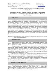
Plate I: Embryogenesis <str<strong>on</strong>g>of</str<strong>on</strong>g> s<str<strong>on</strong>g>of</str<strong>on</strong>g>t tick, Argas persicus (Oken), cleavage, blatoderm and germ band<br />
formati<strong>on</strong>:<br />
Fig. 1: Sagittal secti<strong>on</strong> through 0 h old egg, showing the chori<strong>on</strong> (ch), vitelline membrane (V),<br />
periplasm (P), yolk (Y), cytoplasm (CY), and germ line determinant (GLD) in the posterior<br />
pole <str<strong>on</strong>g>of</str<strong>on</strong>g> the egg. (X = 400).<br />
Fig. 2: Sagittal secti<strong>on</strong> through 12 h old egg, showing periplasm (P), cleavage nuclei (CN), cytoplasm<br />
(CY) and yolk (Y). (X = 400).<br />
Fig. 3: Sagittal secti<strong>on</strong> through 24 h old egg, showing arrangement <str<strong>on</strong>g>of</str<strong>on</strong>g> cleavage nuclei at the periphery,<br />
pole cells (PC), sec<strong>on</strong>dary vitellophages (SV), cleavage nuclei (CN), and delimiting cell<br />
furrows (CF). (X = 1000).<br />
Fig. 4: Sagittal secti<strong>on</strong> through 72 h old egg, showing blastoderm thickening to form germ band (GB),<br />
formati<strong>on</strong> <str<strong>on</strong>g>of</str<strong>on</strong>g> the serosa (Ser). (X = 1000).<br />
Fig. 5: Sagittal secti<strong>on</strong> through 96 h old egg, showing the dorsal organ (DO), ectoderm (Ect),<br />
mesoderm (Mes). (X = 1000).<br />
Fig. 6: Saggital secti<strong>on</strong> through 96 h old egg, showing germ band lobes (GBL), amni<strong>on</strong> (Am), germ<br />
band (GB), serosa (Ser). (X = 1000).
ab1<br />
P<br />
ab2<br />
ab3<br />
ab4 op<br />
Pch<br />
Proc Stom<br />
bc<br />
Fig. (7)<br />
d<br />
Fig. (8)<br />
f<br />
i<br />
Fig. (9)<br />
Nadia Helmy et al.<br />
Plate II<br />
Plate II: Embryogenesis <str<strong>on</strong>g>of</str<strong>on</strong>g> s<str<strong>on</strong>g>of</str<strong>on</strong>g>t tick, Argas periscus (Oken), segmentati<strong>on</strong>, blastokinesis, and<br />
organogenesis:<br />
Fig. 7: Sagittal secti<strong>on</strong> through 120 h old egg, showing movement <str<strong>on</strong>g>of</str<strong>on</strong>g> the ambulatory segments (ab1–<br />
ab4) and mouth parts (opsithosome (OP), precheliceral lobe (Pch), pedipalp (P) and dorsal<br />
surface (d). (X = 600).<br />
Fig. 8: Sagittal secti<strong>on</strong> through 120 h old egg, showing blastokinesis, proctodaeum (Proc), stomodaeum<br />
(Stom), buccal cavity (bc), and gangli<strong>on</strong>ic mass (gm). (X = 400).<br />
Fig. 9: Sagittal secti<strong>on</strong> through 144 h old egg, showing the buccal cavity (bc), ambulatory segment (ab1<br />
– ab4), hypostome (H), basis <str<strong>on</strong>g>of</str<strong>on</strong>g> chelicerae (bch), gangli<strong>on</strong>ic mass (gm), pedipalp (P), and yolk<br />
sac (Ys). (X = 600).<br />
Fig. 10: Sagittal secti<strong>on</strong> through 144 h old egg, showing the stomodeael invaginati<strong>on</strong> (Stom),<br />
proctodeael invaginatin (Proc), an terior mid gut rudiment (AMR), and posterior mid gut<br />
rudiment (PMR). (X = 400).<br />
Fig.11:Sagittal secti<strong>on</strong> through fully formed embryo, showing rectal sac (rs), cuticular membrane (ctl)<br />
and yolk sac (Ys). (X = 600).<br />
Fig.12:Sagittal secti<strong>on</strong> through fully formed embryo, showing oesophagus (Oe), supraoesophageal<br />
gangli<strong>on</strong> (Soe), suboesephageal gangli<strong>on</strong> (Suboe) and yolk sac (Ys). (X = 600).<br />
Ctl<br />
ab4<br />
ab3 ab2<br />
YS<br />
Fig (10)<br />
Fig. (11)<br />
rs<br />
soe<br />
oe<br />
ab1<br />
suboe<br />
bch<br />
P<br />
Fig. (12)
<str<strong>on</strong>g>Effect</str<strong>on</strong>g> <str<strong>on</strong>g>of</str<strong>on</strong>g> <str<strong>on</strong>g>juvenile</str<strong>on</strong>g> <str<strong>on</strong>g>horm<strong>on</strong>e</str<strong>on</strong>g> <str<strong>on</strong>g>analogue</str<strong>on</strong>g> (<str<strong>on</strong>g>Admiral</str<strong>on</strong>g>) <strong>on</strong> <strong>embryogenesis</strong> <str<strong>on</strong>g>of</str<strong>on</strong>g> Argas persicus <br />
Fig. (14)<br />
Fig. (13)<br />
Fig. (15)<br />
CN<br />
CN<br />
Rch<br />
Plate III<br />
Fig. (16)<br />
Fig. (17)<br />
Fig. (18)<br />
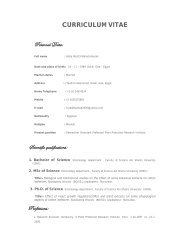
Plate III: Embryogenesis <str<strong>on</strong>g>of</str<strong>on</strong>g> A. persicus eggs treated with JHA (<str<strong>on</strong>g>Admiral</str<strong>on</strong>g>):<br />
Fig. 13: Sagittal secti<strong>on</strong> <str<strong>on</strong>g>of</str<strong>on</strong>g> 0 h old treated egg, showing vacuolati<strong>on</strong> <str<strong>on</strong>g>of</str<strong>on</strong>g> the cytoplasm (V). (X = 400).<br />
Fig. 14: Sagittal secti<strong>on</strong> through 12 h- old treated egg, showing decrease in number <str<strong>on</strong>g>of</str<strong>on</strong>g> cleavage nuclei<br />
(CN) at periphery and absence <str<strong>on</strong>g>of</str<strong>on</strong>g> the pole cells. (X = 600).<br />
Fig. 15: Sagittal secti<strong>on</strong> through 24 h-old treated egg, showing few cleavage nuclei migrating to the<br />
periphery, and ruptured chori<strong>on</strong> (Rch). (X = 1000).<br />
Fig. 16: Sagittal secti<strong>on</strong> through 48 h-old treated egg, showing rudimentary and irregularly arranged<br />
cells <str<strong>on</strong>g>of</str<strong>on</strong>g> blastoderm (RBL). (X = 1000).<br />
Fig. 17: Sagittal secti<strong>on</strong> through 72 h-old treated egg, showing disintegrati<strong>on</strong> <str<strong>on</strong>g>of</str<strong>on</strong>g> the rudimentary<br />
blastoderm, and cracking <str<strong>on</strong>g>of</str<strong>on</strong>g> the cytoplasm (CC). (X = 1000).<br />
Fig. 18: Sagittal secti<strong>on</strong> through 96 h-old treated egg, showing complete lysis and degerati<strong>on</strong> <str<strong>on</strong>g>of</str<strong>on</strong>g> the<br />
egg c<strong>on</strong>tent. (X = 1000).<br />
CC<br />
RBL
Nadia Helmy et al.<br />
ARABIC SUMMARY<br />
( ﻦﻛوأ)<br />
<br />
<br />
( ) <br />
دﻮﻤﺤﻣ <br />
– <br />
– ناﻮﺿر ﺪﻤﺣأ ءﺎﻓو – ﺪﻤﺣأ <br />
<br />
– <br />
– تاﺮﺸﺤﻟا ﻢﻠﻋ ﻢﺴﻗ<br />
ﺔﻠﺣﺮﻣ لﻼﺧ <br />
،<br />
<br />
<br />
( ﺞﻠﻔﺘﻟا)<br />
<br />
. <br />
24<br />
. <br />
<br />
<br />
. ﺔﻋﺎﺳ 72<br />
. <br />
<br />
<br />
120<br />
. ﺔﻋﺎﺳ 96 ﺮﻤﻋ ﺪﻨﻋ<br />
<br />
144 ﺪﻨﻋ . <br />
. ﺔﻋﺎﺳ 168<br />
. ﻂﺳﻮﺘﻤﻟا<br />
<br />
( لا<br />
) <br />
ﺮﻤﻋ ﺪﻨﻋو . <br />
( ﺞﻠﻔﺘﻟا)<br />
<br />
48 ﺪﻌﺑو . <br />
24<br />
ﺔﻘﺒﻃ ءﺎﻔﺘﺧا ﻆﺣﻮﻟ . <br />
ﻦﻣ ﺔﻋﺎﺳ 96 ﻦﻣ ءاﺪﺘﺑا . ﺔﻠﻣﺎﻌﻤﻟا ﻦﻣ ﺔﻋﺎﺳ 72<br />
<br />
.<br />
<br />
168 ﺪﻨﻋ ﺲﻘﻔﻟا ﻰﺘﺣ ﺔﻠﻣﺎﻌﻤﻟا