
You also want an ePaper? Increase the reach of your titles
YUMPU automatically turns print PDFs into web optimized ePapers that Google loves.
360<br />
PHYSICAL SIGNS<br />
<strong>The</strong> <strong>Babinski</strong> <strong>sign</strong><br />
J W Lance<br />
.............................................................................................................................<br />
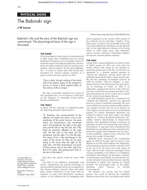
<strong>Babinski</strong>’s life and the story of the <strong>Babinski</strong> <strong>sign</strong> are<br />
summarised. <strong>The</strong> physiological basis of the <strong>sign</strong> is<br />
discussed.<br />
..........................................................................<br />
.......................<br />
Correspondence to:<br />
Professor J W Lance,<br />
Wales Medical Centre,<br />
66 High Street, Randwick,<br />
NSW 2031, Australia;<br />
jimlance@bigpond.com<br />
Received 27 February<br />
2002<br />
Accepted 3 May 2002<br />
.......................<br />
www.jnnp.com<br />
Downloaded from<br />
jnnp.bmj.com on March 31, 2012 - Published by group.bmj.com<br />
THE NAME<br />
Of all neurologists whose name is commemorated<br />
in daily usage, that of <strong>Babinski</strong> may not exceed<br />
Romberg in frequency but overshadows all in its<br />
dramatic impact and clinical implication. Wartenberg<br />
was said to evoke his name in rejecting compromise<br />
with an emotive “By the great <strong>Babinski</strong>,<br />
no!”. 1 It seems to matter little that Remak first<br />
described the extensor plantar response in a<br />
patient with transverse myelitis in 1893 2 :<br />
“One is able, through stroking of the distal<br />
half of the plantar aspect of the metatarsus<br />
primus, to evoke a fairly isolated reflex of<br />
the extensor hallucis longus.”<br />
<strong>The</strong> <strong>sign</strong> is generally attributed to a lesion of<br />
the pyramidal tract. It is of interest to look back<br />
on the diagnosis of pyramidal lesions before<br />
Remak and <strong>Babinski</strong>.<br />
THE TRACT<br />
In about 150 AD, Aretaeus of Cappadocia made<br />
the following perceptive observations 3 :<br />
“If, therefore, the commencement of the<br />
affection be below the head, such as the<br />
membrane of the spinal marrow, the parts<br />
which are homonymous and connected<br />
with it are paralysed; the right on the right<br />
side and the left on the left side. But if the<br />
head be primarily affected on the right<br />
side, the left side of the body will be<br />
paralysed; and the right, if on the left side.<br />
<strong>The</strong> cause of this is the interchange in the<br />
origin of the nerves, for they do not pass<br />
along on the same side, the right on the<br />
right side, until their terminations; but each<br />
of them passes over to the other side from<br />
that of its origin, decussating each other in<br />
the form of the letter X.”<br />
And there the matter rested for some 1700<br />
years.<br />
van Gijn, in his definitive monograph 4 and<br />
article, 5 summarised the current knowledge of<br />
the pyramidal syndrome before the recognition of<br />
the extensor plantar response. Knee and ankle<br />
jerks, clonus, withdrawal of the lower limb in<br />
response to pain and diminished cutaneous<br />
reflexes on the affected side in hemiplegia had<br />
J Neurol Neurosurg Psychiatry 2002;73:360–362<br />
been recognised. In the second (1893) edition of<br />
his textbook, Gowers 6 describes “rigidity” of the<br />
limbs after a lesion of the pyramidal tracts with<br />
increased tendon jerks and clonus on the affected<br />
side. In the third edition of Diseases of the Nervous<br />
System in 1899 (three years after <strong>Babinski</strong>’s<br />
report), Gowers 7 mentions “clasp-knife rigidity,”<br />
but not the extensor plantar response.<br />
THE MAN<br />
Joseph Felix Francois <strong>Babinski</strong> was born in Paris<br />
of Polish parents in 1857, two years after his<br />
brother Henri, with whom he was destined to<br />
spend the greater part of his life. 4 In 1879 he was<br />
appointed to a general medical position as<br />
“interne des hôpitaux,” during which time he<br />
published anatomical studies on the muscle spindle<br />
and the pathology of multiple sclerosis. In<br />
1885 he became “chef de clinique” to Jean-<br />
Martin Charcot who had become the first professor<br />
of neurology in France in 1882 at La<br />
Salpetrière, a gunpowder factory in the 17th century<br />
that evolved into an asylum and then a hospital,<br />
becoming one of the world’s great centres<br />
for the study of neurological disease.<br />
In 1890 <strong>Babinski</strong> passed the examination for<br />
“Médecin des Hôpitaux” and the way appeared<br />
clear for a career in academic neurology. <strong>The</strong> next<br />
step would be an associate professorship (professeur<br />
agrégé) but this was not to be because of<br />
what appears to have been an act of professional<br />
jealousy by Charles Bouchard. Bouchard was<br />
trained by Charcot and their names are linked<br />
together as “Charcot-Bouchard aneurysms” preceding<br />
cerebral haemorrhage in hypertensive<br />
patients. After Bouchard became a professor of<br />
pathology in 1879, his relationship with Charcot<br />
deteriorated. 8 It may have been coincidental that<br />
Bouchard was presiding over the examination for<br />
professeur agrégé when <strong>Babinski</strong> was an unsuccessful<br />
candidate, whereas three of the five who<br />
passed were pupils of Bouchard. 8<br />
<strong>Babinski</strong> never attempted the examination<br />
again. In 1895 he became chief of service at the<br />
Hôpital de la Pitié which adjoins the Salpetrière,<br />
and remained in that post until he retired in 1922<br />
at the age of 65. He wrote on a wide variety of<br />
topics and his fame attracted neurologists from<br />
overseas including S A K Wilson, C G Chaddock,<br />
and Robert Wartenberg. 8 He provided a stimulus<br />
to neurosurgery, particularly in reporting the successful<br />
removal of intracerebral tumours and the<br />
localisation of spinal cord tumours on clinical<br />
grounds. He encouraged some of his pupils,<br />
including Clovis Vincent, to become<br />
neurosurgeons. 9<br />
His manner was austere and his clinical<br />
practice was weird by present day standards.<br />
Patients entered into his consulting room naked
Downloaded from<br />
jnnp.bmj.com on March 31, 2012 - Published by group.bmj.com<br />
<strong>The</strong> <strong>Babinski</strong> <strong>sign</strong> 361<br />
and, after a skimpy history, were subjected to physical examination.<br />
van Gijn 4 recounts the story that a male patient, when<br />
he was dismissed after his head had been examined with the<br />
aid of a galvanometer, pointed at his penis and asked<br />
plaintively “you don’t have anything to make it work again?”.<br />
<strong>Babinski</strong> lived with his brother Henri, a mining engineer<br />
and an inspired chef whose Gastronomie Pratique ran to nine<br />
editions and whose skill doubtless contributed to the impressive<br />
bulk of both men. <strong>Babinski</strong> continued to attend the hospital<br />
for consultations after his retirement.<br />
He died in 1932, a year after Henri. 4<br />
THE SIGN<br />
In 1896 <strong>Babinski</strong> presented a brief paper to the Biological<br />
Society of Paris, translated as “On the cutaneous plantar reflex<br />
in certain organic disorders of the nervous system”. 4 He had<br />
observed that pricking of the sole on the healthy side of a<br />
patient with hemiplegia or lower limb monoplegia caused<br />
withdrawal of the lower limb with flexion of the toes on the<br />
metatarsal bones. In contrast, the same stimulus applied to<br />
the sole on the affected side caused extension of the toes at the<br />
metatarso-phalangeal joints, even in patients who were<br />
unable to move their toes voluntarily. He later referred to<br />
“stroking” of the sole rather than pricking as the adequate<br />
stimulus, a point that should not be lost on today’s registrars<br />
or residents, many of whom warn their patients that they are<br />
about to scrape their feet or use some other potentially intimidating<br />
term.<br />
<strong>Babinski</strong>’s definitive description appeared in 1898. 10 An<br />
English translation is included in van Gijn’s monograph. 4 He<br />
stated that the “phenomenon of the toes” is most easily elicited<br />
from the lateral aspect of the sole and that the reaction of<br />
the anatomical extensor is most conspicuous in the first or the<br />
first two toes. He demonstrated the extensor response in<br />
hemiplegic and paraparetic patients and stated that he had<br />
observed it in Friedreich’s ataxia. He attributed the <strong>sign</strong> to<br />
dysfunction of the pyramidal tract and pointed out that it was<br />
usually associated with exaggeration of tendon reflexes and<br />
clonic movements but that “this relation is far from indissoluble.”<br />
He concluded by drawing attention to the presence of the<br />
<strong>sign</strong> in the newborn, an association that had not escaped the<br />
Figure 1 Botticelli’s Virgin and Angels. (From<br />
Lance JW, McLeod JG. A physiological approach<br />
to clinical neurology, 3rd ed. London: Butterworths,<br />
1981:143.)<br />
attention of a renaissance artist (fig 1). <strong>The</strong> reversion of the<br />
plantar response to flexor occurs at variable times from the age<br />
of seven months to a year or more. <strong>The</strong> results of many publications<br />
have been tabulated by van Gijn, 4 who concluded that<br />
the relation of this change to the onset of walking was probably<br />
indirect. Confusion may arise in infants because the grasp<br />
reflex, which is most easily elicited by stimulation of the ball<br />
of the foot, involves flexion of the toes. 11 It usually disappears<br />
between six and 12 months of age and appears to be related to<br />
the age of standing. 11<br />
<strong>Babinski</strong> had noted that the <strong>sign</strong> appeared transiently on<br />
the affected side in a man during a Jacksonian fit and bilaterally<br />
in another patient suffering from strychnine poisoning. I<br />
had the opportunity to observe such a brief alteration in a<br />
child of mine subject to night terrors. As I was attempting to<br />
comfort her I ran my thumb lightly against the lateral aspect<br />
of her sole as one does and observed a definite <strong>Babinski</strong><br />
response. As soon as the paroxysm was over the response<br />
reverted to flexor.<br />
In 1903, <strong>Babinski</strong> remarked during a case presentation that<br />
abduction of the toes might accompany extension of the toes<br />
in a pyramidal lesion but was by no means constant. 12 This was<br />
later known as “le <strong>sign</strong>e de l’éventail” (the fan <strong>sign</strong>).<br />
THE CAUSE<br />
<strong>The</strong> normal plantar response to cutaneous stimuli of the sole<br />
can be considered a superficial reflex like the abdominal and<br />
cremasteric reflexes that are abolished by an upper motor<br />
neurone lesion. 4 It is then replaced by the <strong>Babinski</strong> response.<br />
<strong>The</strong> upgoing toe is regarded anatomically as extension of the<br />
great toe but physiologically it is part of a flexor reflex, apparently<br />
disinhibited by loss of upper motor neurone control, and<br />
its receptive field may extend in some instances to the leg or<br />
thigh. 13 14 This led to the description of many “reflexes” such as<br />
Chaddock’s and Oppenheim’s <strong>sign</strong>s which were simply different<br />
ways of eliciting the <strong>Babinski</strong> <strong>sign</strong>. 15<br />
Although the <strong>sign</strong> usually accompanies spasticity, and has<br />
been described as being caused by infarction apparently<br />
limited to one medullary pyramid in three cases cited by van<br />
Gijn, 4 its causation by lesions of the pyramidal tract has been<br />
questioned. Nathan and Smith 16 studied patients before and<br />
www.jnnp.com
362 Lance<br />
after operations on the spinal cord (anterolateral cordotomy),<br />
correlating clinical findings with the extent of the surgical<br />
lesion. <strong>The</strong>y found that destruction of the anterior half of the<br />
spinal cord may be associated with a <strong>Babinski</strong> response,<br />
whereas the <strong>sign</strong> could be absent with histologically verified<br />
lesions of the lateral corticospinal (pyramidal) tract. Later,<br />
Nathan 17 reported 44 patients subjected to cordotomy for relief<br />
of pain from cancer. <strong>The</strong> <strong>Babinski</strong> response was found in general<br />
to be present after lesions of the corticospinal tract and<br />
not with lesions elsewhere in the cord. Nevertheless, a<br />
transient <strong>Babinski</strong> response could be observed after anterior<br />
lesions and some patients with lesions of the tract retained<br />
normal plantar responses.<br />
Landau and Clare 18 considered that the patients with<br />
pyramidal lesions that they studied who did not develop<br />
extensor responses had peripheral nerve damage or were<br />
recovering from shock. <strong>The</strong>y felt that the correlation between<br />
the <strong>sign</strong> of <strong>Babinski</strong> and pyramidal tract dysfunction was <strong>sign</strong>ificant<br />
but added that it would be “absurd to deny that<br />
lesions of non-pyramidal internuncial pathways may facilitate<br />
release phenomena at the spinal level.” What evidence is there<br />
for this proposition?<br />
<strong>The</strong>re is an important difference in comparison with the<br />
decerebrate spinal cat that throws some light on the matter. In<br />
the decerebrate cat the stretch reflex of the quadriceps muscle<br />
becomes more active as the degree of stretch (that is, muscle<br />
length) is increased. 19 In contrast, in those chronic spinal animal<br />
preparations with increased muscle tone, the reflex<br />
response of the quadriceps becomes progressively less as the<br />
degree of stretch is increased, analogous to the clasp-knife<br />
response in human spasticity. As the reverse applies to flexor<br />
muscles (that is, increasing muscle stretch enhances the reflex<br />
response) it appears that receptors sensitive to stretch, such as<br />
group II afferent fibres, inhibit the stretch reflex of hind limb<br />
extensors and facilitate that of the flexors as long as the<br />
stretch is maintained. <strong>The</strong>se flexor reflex afferents (FRA) are<br />
normally suppressed by the dorsal reticulospinal system<br />
which arises from the pontomedullary reticular formation and<br />
descends in the dorsolateral funiculus of the spinal cord. 20<br />
Burke et al made discrete lesions in the reticular formation and<br />
upper quadrantic sections in the spinal cord of the decerebrate<br />
cat, 19 which transformed the length dependent facilitation of<br />
decerebrate rigidity into the length dependent inhibition of<br />
spinal spasticity. <strong>The</strong>se changes observed experimentally can<br />
readily be applied to the human situation.<br />
Using the H reflex as an indicator of motor neurone<br />
excitability in spastic patients, it was shown that stretch of the<br />
calf muscles diminished the amplitude of the H reflex recorded<br />
from the calf while stretch of the pretibial flexor muscles augmented<br />
the H reflex recorded from those muscles 21 —that is, the<br />
flexor reflex afferents have been released in spastic patients, as<br />
in the chronic spinal cat. This explains the clasp-knife<br />
phenomenon in human spasticity, in which the tonic stretch<br />
reflex in quadriceps is inhibited by increasing muscle length<br />
beyond the mid-point of knee flexion. It also explains the<br />
enhancement of the flexor protective response, including the<br />
<strong>Babinski</strong> <strong>sign</strong>. In cats the inhibitory reticulospinal pathway is<br />
directed from the motor cortex by parapyramidal fibres that<br />
descend in the medial part of the internal capsule and medial<br />
area of the midbrain dorsal to the cerebral peduncle. 22 If one<br />
can extrapolate to humans from these studies in the cat, any<br />
disturbance in function of this cortico-reticulospinal tract,<br />
which is closely applied to the pyramidal tract throughout its<br />
course from cortex to spinal segmental levels, would release<br />
flexor reflexes, including the <strong>Babinski</strong> <strong>sign</strong>. 23<br />
www.jnnp.com<br />
Downloaded from<br />
jnnp.bmj.com on March 31, 2012 - Published by group.bmj.com<br />
THE LEGACY<br />
It thus appears that the <strong>Babinski</strong> <strong>sign</strong> is an indication of withdrawal<br />
of supraspinal control of flexor reflexes in the lower<br />
limbs. Clinically it can be equated with inactivation, transient<br />
or permanent, of the upper motor neurone, which term implicates<br />
corticoreticulospinal fibres as well as the pyramidal tract<br />
anywhere in its path from cerebral cortex to termination in<br />
the cord.<br />
Flexor reflexes are prominent in the newborn and young<br />
infant. In order for the baby to stand, flexor reflexes in the<br />
lower limbs must be brought under control by the dorsal<br />
reticulospinal tract. For the baby to walk the flexor synergy<br />
must then be harnessed to the motor cortex as part of the<br />
walking pattern. With the maturation of upper motor neurone<br />
pathways the <strong>Babinski</strong> <strong>sign</strong> disappears and the great toe goes<br />
down on stimulation of the sole. If upper motor neurone control<br />
is temporarily suspended, as in an epileptic fit, or is abolished<br />
by disease the <strong>Babinski</strong> <strong>sign</strong> reappears.<br />
<strong>The</strong>re is a tendency to abolish eponymous names for <strong>sign</strong>s<br />
or clinical syndromes but they do serve to remind us of the<br />
debt we owe our forebears. Should we abandon the term <strong>Babinski</strong><br />
<strong>sign</strong> in favour of “the upgoing toe” or “extensor plantar<br />
response”? I would side with Wartenberg in saying: “By the<br />
great <strong>Babinski</strong>, no!”.<br />
REFERENCES<br />
1 Aird RB. Obituary: Robert Wartenberg. Neurology 1957;7:146–7.<br />
2 Pearce JMS. <strong>Babinski</strong> or Remak? J R Coll Physicians Lond 1996;30:190.<br />
3 Adams F. <strong>The</strong> extant works of Aretaeus the Cappadocian. London:<br />
Sydenham Society, 1856:306. (Reprint, Boston: Milford House, 1972.)<br />
4 van Gijn J. <strong>The</strong> <strong>Babinski</strong> <strong>sign</strong> – a century. Utrecht: Universiteit Utrecht,<br />
1996.<br />
5 van Gijn J. <strong>The</strong> <strong>Babinski</strong> <strong>sign</strong>: the first hundred years. J Neurol<br />
1996;243:675–83.<br />
6 Gowers CR. A manual of diseases of the nervous system, 2nd ed.<br />
Philadelphia: Blakiston, Son and Co, 1893:83.<br />
7 Gowers WR. A manual of diseases of the nervous system, 3rd ed, vol 1.<br />
London: Churchill, 1899:255–65.<br />
8 Iragui VJ. <strong>The</strong> Charcot–Bouchard controversy. Arch Neurol<br />
1986;43:290–5.<br />
9 Lanzino G, di Pierro CG, Laws ER. One century after the description of<br />
the “<strong>sign</strong>”: Joseph <strong>Babinski</strong> and his contribution to neurosurgery.<br />
Neurosurgery 1997;40:822–8.<br />
10 <strong>Babinski</strong> J. Du phénomène des orteils et de sa valeur semiologique.<br />
Semaine Médicale 1898;18:321–2.<br />
11 Dietrich HF. A longitudinal study of the <strong>Babinski</strong> and plantar grasp<br />
reflexes in infancy. Arch Neurol Psychiatry 1957;94:265–71.<br />
12 <strong>Babinski</strong> J. De L’abduction des orteils. Rev Neurol 1903;11:728–9.<br />
13 Walshe F. <strong>The</strong> <strong>Babinski</strong> plantar response, its forms and its physiological<br />
and pathological <strong>sign</strong>ificance. Brain 1956;79:529–56.<br />
14 Estanol R. Temporal course of the threshold and size of the receptive<br />
field of the <strong>Babinski</strong> <strong>sign</strong>. J Neurol Neurosurg Psychiatry<br />
1983;46:1055–7.<br />
15 Wartenberg R. <strong>The</strong> <strong>Babinski</strong> reflex after fifty years. JAMA<br />
1947;135:763–7.<br />
16 Nathan PW, Smith MC. <strong>The</strong> <strong>Babinski</strong> response: a review and new<br />
observations. J Neurol Neurosurg Psychiatry 1955;18:250–9.<br />
17 Nathan PW. Effects on movement of surgical incisions into the human<br />
spinal cord. Brain 1994;117:337–46.<br />
18 Landau WM, Clare MH. <strong>The</strong> plantar reflex in man, with special<br />
reference to some conditions where the extensor response is<br />
unexpectedly absent. Brain 1959;82:321–55.<br />
19 Burke D, Knowles L, Andrews C, et al. Spasticity, decerebrate rigidity,<br />
and the clasp-knife phenomenon: an experimental study in the cat. Brain<br />
1972;95:31–48.<br />
20 Lundberg A. Control of spinal mechanisms from the brain. In: Tower DB,<br />
ed. <strong>The</strong> nervous system, vol 1. New York: Raven Press, 1975:253–65.<br />
21 Burke D, Lance JW. Studies of the reflex effects of primary and<br />
secondary spindle endings in spasticity. In: Desmedt JE, ed. New<br />
developments in electromyography and clinical neurophysiology, vol 3.<br />
Basel: Karger, 1973:475–95.<br />
22 Ashby P, Andrews C, Knowles L, et al. Pyramidal and extrapyramidal<br />
control of tonic mechanisms in the cat. Brain 1972;95:21–30.<br />
23 Lance JW. <strong>The</strong> control of muscle tone, reflexes and movement: Robert<br />
Wartenberg Lecture. Neurology 1980;30:1303–13.
References<br />
Email alerting<br />
service<br />
Notes<br />
<strong>The</strong> <strong>Babinski</strong> <strong>sign</strong><br />
J W Lance<br />
J Neurol Neurosurg Psychiatry 2002 73: 360-362<br />
doi: 10.1136/jnnp.73.4.360<br />
Updated information and services can be found at:<br />
http://jnnp.bmj.com/content/73/4/360.full.html<br />
<strong>The</strong>se include:<br />
To request permissions go to:<br />
http://group.bmj.com/group/rights-licensing/permissions<br />
To order reprints go to:<br />
http://journals.bmj.com/cgi/reprintform<br />
To subscribe to BMJ go to:<br />
http://group.bmj.com/subscribe/<br />
Downloaded from<br />
jnnp.bmj.com on March 31, 2012 - Published by group.bmj.com<br />
This article cites 14 articles, 9 of which can be accessed free at:<br />
http://jnnp.bmj.com/content/73/4/360.full.html#ref-list-1<br />
Article cited in:<br />
http://jnnp.bmj.com/content/73/4/360.full.html#related-urls<br />
Receive free email alerts when new articles cite this article. Sign up in the<br />
box at the top right corner of the online article.