View - ResearchGate
View - ResearchGate
View - ResearchGate

You also want an ePaper? Increase the reach of your titles
YUMPU automatically turns print PDFs into web optimized ePapers that Google loves.
EDITOR<br />
MZ Khan, SENRA Academic Publishers<br />
Burnaby, British Columbia, Canada<br />
ASSOCIATE EDITORS<br />
Errol Hassan, University of Queensland<br />
Gatton, Australia<br />
Paul CH Li, Simon Fraser University<br />
Burnaby, British Columbia, Canada<br />
EDITORIAL STAFF<br />
Jasen Nelson<br />
Walter Leung<br />
Sara<br />
Hao-Feng (howie) Lai<br />
Ben Shieh<br />
MANAGING DIRECTOR<br />
Mak, SENRA Academic Publishers<br />
Burnaby, British Columbia, Canada<br />
The Canadian Journal of Pure and Applied<br />
Sciences (CJPAS-ISSN 1715-9997) is a<br />
peer reviewed multi-disciplinary specialist<br />
journal aimed at promoting research<br />
worldwide in Agricultural Sciences,<br />
Biological Sciences, Chemical Sciences,<br />
Computer and Mathematical Sciences,<br />
Engineering, Environmental Sciences,<br />
Medicine and Physics (all subjects).<br />
Every effort is made by the editors, board<br />
of editorial advisors and publishers to see<br />
that no inaccurate or misleading data,<br />
opinions, or statements appear in this<br />
journal, they wish to make clear that data<br />
and opinions appearing in the articles are<br />
the sole responsibility of the contributor<br />
concerned. The CJPAS accept no<br />
responsibility for the misleading data,<br />
opinion or statements.<br />
Editorial Office<br />
E-mail: editor@cjpas.ca<br />
: editor@cjps.net<br />
SENRA Academic Publishers<br />
7845 15th Street Burnaby<br />
British Columbia V3N 3A3 Canada<br />
www.cjpas.net<br />
E-mail: senra@cjpas.ca<br />
Print ISSN 1715-9997<br />
Online ISSN 1920-3853<br />
Board of Editorial Advisors<br />
Volume 3, Number 3<br />
Oct 2009<br />
CANADIAN JOURNAL OF<br />
PURE AND APPLIED SCIENCES<br />
Richard Callaghan<br />
Gordon McGregor Reid<br />
University of Calgary, AB, Canada<br />
North of England Zoological Society, UK<br />
David T Cramb<br />
Pratim K Chattaraj<br />
University of Calgary, AB, Canada<br />
Indian Institute of Technology, Kharagpur, India<br />
Matthew Cooper<br />
Andrew Alek Tuen<br />
Grand Valley State University, AWRI, Muskegon, MI, USA Institute of Biodiversity, Universiti Malaysia Sarawak, Malaysia<br />
Anatoly S Borisov<br />
Dale Wrubleski<br />
Kazan State University, Tatarstan, Russia<br />
Institute for Wetland and Waterfowl Research, Stonewall, MB, Canada<br />
Ron Coley<br />
Dietrich Schmidt-Vogt<br />
Coley Water Resource & Environment Consultants, MB, Canada Asian Institute of Technology, Thailand<br />
Chia-Chu Chiang<br />
Diganta Goswami<br />
University of Arkansas at Little Rock, Arkansas, USA Indian Institute of Technology Guwahati, Assam, India<br />
Michael J Dreslik<br />
M Iqbal Choudhary<br />
Illinois Natural History, Champaign, IL, USA<br />
HEJ Research Institute of Chemistry, Karachi, Pakistan<br />
David Feder<br />
Daniel Z Sui<br />
University of Calgary, AB, Canada<br />
Texas A&M University, TX, USA<br />
David M Gardiner<br />
SS Alam<br />
University of California, Irvine, CA, USA<br />
Indian Institute of Technology Kharagpur, India<br />
Geoffrey J Hay<br />
Biagio Ricceri<br />
University of Calgary, AB, Canada<br />
University of Catania, Italy<br />
Chen Haoan<br />
Zhang Heming<br />
Guangdong Institute for drug control, Guangzhou, China<br />
Chemistry & Environment College, Normal University, China<br />
Hiroyoshi Ariga<br />
C Visvanathan<br />
Hokkaido University, Japan<br />
Asian Institute of Technology, Thailand<br />
Gongzhu Hu<br />
Indraneil Das<br />
Central Michigan University, Mount Pleasant, MI, USA<br />
Universiti Malaysia, Sarawak, Malaysia<br />
Moshe Inbar<br />
Gopal Das<br />
University of Haifa at Qranim, Tivon, Israel<br />
Indian Institute of Technology , Guwahati, India<br />
SA Isiorho<br />
Melanie LJ Stiassny<br />
Indiana University - Purdue University, (IPFW), IN, USA<br />
American Museum of Natural History, New York, NY, USA<br />
Bor-Luh Lin<br />
Kumlesh K Dev<br />
University of Iowa, IA, USA<br />
Bio-Sciences Research Institute, University College Cork, Ireland.<br />
Jinfei Li<br />
Shakeel A Khan<br />
Guangdong Coastal Institute for Drug Control, Guangzhou, China<br />
University of Karachi, Karachi, Pakistan<br />
Collen Kelly<br />
Victoria University of Wellington, New Zealand<br />
Xiaobin Shen<br />
Hamid M.K.AL-Naimiy<br />
University of Melbourne, Australia<br />
University of Sharjah, UAE<br />
Maria V Kalevitch<br />
Eric L Peters<br />
Robert Morris University, PA, USA<br />
Chicago State University, Chicago, IL, USA<br />
Xing Jin<br />
Roustam Latypov<br />
Hong Kong University of Science & Tech.<br />
Kazan State University, Kazan, Russia<br />
Leszek Czuchajowski<br />
Frances CP Law<br />
University of Idaho, ID, USA<br />
Simon Fraser University, Burnaby, BC, Canada<br />
Basem S Attili<br />
Guangchun Lei<br />
UAE University, UAE<br />
Ramsar Convention Secretariat, Switzerland<br />
David K Chiu<br />
Atif M Memon<br />
University of Guelph, Ontario, Canada<br />
University of Maryland, MD, USA<br />
Gustavo Davico<br />
SR Nasyrov<br />
University of Idaho, ID, USA<br />
Kazan State University,Kazan, Russia<br />
Andrew V Sills<br />
Russell A Nicholson<br />
Georgia Southern University Statesboro, GA, USA<br />
Simon Fraser University, Burnaby, BC, Canada<br />
Charles S. Wong<br />
Borislava Gutarts<br />
University of Alberta, Canada<br />
California State University, CA, USA<br />
Greg Gaston<br />
Sally Power<br />
University of North Alabama, USA<br />
Imperial College London, UK
LIFE SCIENCES<br />
CONTENTS<br />
Volume 3, Number 3<br />
October 2009<br />
Marta Leonor Marulanda and Ana María López<br />
Characterization of Thornless Rubus glaucus in Colombia .................................................................................................................. 875<br />
Rajbir Bhatti, S Kaur, J Singh and MPS Ishar<br />
Ameliorative Effect of Volatile Oil from Cinnamomum zeylanicum on Hyperalgesia in Alloxan Diabetic Rats ................................. 887<br />
SM Imamul Huq, Kanta Parvin, Sylvia Rahman and JC Joardar<br />
Response of Cowpea (Vigna sinensis L.) to Arsenic ............................................................................................................................ 897<br />
FF Hammad and AH Salama<br />
The Influence of Mono-Dispersed Tin – Doped Indium Oxide Nanopowders on its Dielectric Properties .......................................... 903<br />
JR Williams, F Al-Nabhani and A Al-Hamdi<br />
The Microwave-Assisted Solvent Extraction of Propranolol from Tablets........................................................................................... 911<br />
Saber A Sakr, Sobhy E Hassab-Elnabi and Dalia A El-Ghonaimy<br />
Effect of Green Tea on Cytogenetic Changes Induced by Gibberellin A 3 in Human Lymphocyte Culture .......................................... 917<br />
Abiodun Falodun and Osahon Obasuyi<br />
Phytochemical Screening and Evaluation of Stem Bark Extract of Khaya senegalensis (Meliaceae) on Methicillin Resistant<br />
Staphyloccocus areus ............................................................................................................................................................................ 925<br />
Nasim Karim and Syed Sanowar Ali<br />
Jugular Vein Cannulation in Rats – A Mini Review ............................................................................................................................. 929<br />
Short Communication<br />
Ghazala Yasmeen, Zaheer M Khan and Adil Akbar<br />
A Study on the Induced Effect of Β-Cypermethrin on Skin of Euphlyctis cyanophlyctis ..................................................................... 937<br />
PHYSICAL SCIENCES<br />
Falayi EO and Beloff N<br />
Global Study of Geomagnetic Induced Current using Time Derivatives of Geomagnetic Fields ......................................................... 943<br />
Thomas Mathew, AD Vyas and Deepti Tripathi<br />
Dielectric Properties of Some Edible and Medicinal Oils at Microwave Frequency ............................................................................ 953<br />
Godwin NO Asemota<br />
On a Class of Computable Convex Functions ...................................................................................................................................... 959<br />
TR Sundararaman, V Ramamurthi and N Partha<br />
Effectiveness of Lignite Coagulant for Removal of Textile Dyes from Aqueous Solutions and Textile Waste Water......................... 967<br />
Ajay Kumar Singh<br />
A Theoretical Study of the Threshold Voltage Sensitivity to Process Variation in Symmetric<br />
Double Gate Mos Devices .................................................................................................................................................................... 975<br />
Olayinka O Ajani and Obinna C Nwinyi<br />
Synthesis and Evaluation of Antimicrobial Activity of Phenyl and Furan-2-yl[1,2,4] triazolo[4,3-a]<br />
quinoxalin-4(5h)-one and their hydrazone Precursors .......................................................................................................................... 983<br />
A Hussain, A Seidel-Morgenstern and E Tsotsas<br />
Effect of Top Layer’s Material and Flow Direction on Mass Transfer Through Multi-Layer Ceramic Membranes ............................ 993<br />
S Askari<br />
A New Proof for the Euler Theorem in the Complex Numbers Theory .............................................................................................. 1001
SENRA Academic Publishers, Burnaby, British Columbia<br />
Vol. 3, No. 3, pp. 875-885, 2009<br />
ISSN: 1715-9997<br />
CHARACTERIZATION OF THORNLESS RUBUS GLAUCUS IN COLOMBIA<br />
*Marta Leonor Marulanda and Ana María López<br />
Biodiversity and Biotechnology Group, Universidad Tecnológica de Pereira<br />
La Julita, Pereira, Colombia<br />
ABSTRACT<br />
High phenotypical plasticity has been identified within the species Rubus glaucus Benth, commonly known as ‘mora de<br />
castilla’ or Castilla blackberry, in Colombia’s coffee-growing area using AFLP molecular markers as well as<br />
morphological characters. Thornless plants have been observed among the blackberry materials grown. These plants<br />
present the same characteristics of productivity as thorny varieties are widely distributed. A first approximation to the<br />
genetic relationship between thorny and thornless materials indicated that thornless blackberry could possibly originate<br />
from the departments of Risaralda or Quindío. This work focuses on identifying the genetic and morpho-agronomic<br />
differences in thornless R. glaucus materials found in the coffee-growing region, especially in the department of<br />
Risaralda. Materials were collected from five different locations: two in the department of Risaralda, one in the<br />
department of Caldas, and two in the department of Quindío. For the morpho-agronomic characterization, 40 farmers<br />
were selected in the municipalities of Santa Rosa de Cabal and Guática, Risaralda, each farmer planting 50 plants from<br />
each of the five different collection sites, which had been multiplied in vitro, as well as 50 plants of thornless blackberry<br />
propagated by farmers, totaling 12,000 plants. Eight microsatellite (SSR) sequences were used to study 23 regional<br />
accessions of Rubus, including thorny and thornless R. glaucus, both cultivated and wild. Genetic and molecular<br />
differences were observed between thornless blackberry materials of different origins.<br />
Keywords: Rubus glaucus, thornless blackberry, Castillo blackberry, genetic characterization, Colombia.<br />
INTRODUCTION<br />
American Rubus species are perennial shrubs that show a<br />
broad range of growth habits: from erect to semi-erect to<br />
creeping (Daubeny, 1996). In addition, their<br />
morphological and phenotypical characteristics are<br />
probably among the most well known. Rubus species of<br />
economic importance present perennial roots, foliage that<br />
produces biannual productive canes or branches with<br />
flower structures, and new branches, known as primary<br />
branches, which present vegetative growth and form<br />
tillers (Moore and Skirvin, 1990).<br />
In temperate regions, the prolonged exposure of<br />
blackberries to the cold induces both vegetative and<br />
primary branches to develop flower buds or productive<br />
branches, also known as female branches, which die after<br />
producing fruit. Some species produce flowers without<br />
being exposed to the cold in addition to another very<br />
desirable characteristica productive primary branch<br />
capable of producing flower branches during the early<br />
growth cycle (López-Medina and Moore, 1999; Lopez-<br />
Medina et al., 2000). However, in the case of Rubus<br />
glaucus, the formation of female branches does not occur<br />
with the same mechanisms. According to popular belief,<br />
this formation is not a physiological phenomenon strongly<br />
controlled by environmental conditions, as described<br />
*Corresponding author email: ubioteve@utp.edu.co<br />
927<br />
elsewhere in the world, but is determined rather by<br />
genetic factors.<br />
Another interesting characteristic of most Rubus species<br />
is the absence of thorns or brambles. For example,<br />
commercial varieties ‘Chester Thornless’, ‘Thornfree’,<br />
and ‘Thornless Evergreen’ are thornless genotypes from<br />
the United States. This monogenic characteristic is<br />
controlled by a recessive gene that has been widely<br />
studied in Europe and the United States (Jennings and<br />
Ingram, 1983; Hall, 1990; Skirvin et al., 2005; Strik et al.,<br />
2006). In Colombia, farmers of the department of Quindío<br />
discovered a thornless blackberry biotype, which has been<br />
multiplied by farmers throughout the country, without<br />
really understanding its agronomic and genetic<br />
characteristics. This thornless blackberry offers great<br />
potential for blackberry cultivation not only in the<br />
Quindío region but throughout the country.<br />
Previous work carried out by Aguilar (Aguilar, SB. 2006)<br />
and Marulanda et al. (2007) revealed high phenotypical<br />
and molecular plasticity in R. glaucus in Colombia’s<br />
coffee-growing region. Other wild species of Rubus found<br />
in the area and nearby lots planted to the Castilla<br />
blackberry were also submitted to morphological and<br />
molecular characterization, using amplified fragment<br />
length polymorphism (AFLP) and simple sequence repeat<br />
(SSR) molecular markers.
876<br />
Some of the cultivated thornless blackberry genotypes<br />
present the same productivity and same fruit size as<br />
thorny plants. Because of their interesting phenotypical<br />
characteristics and reduced production costs, these<br />
materials, commonly known as thornless blackberry, have<br />
been massively disseminated by farmers in the region<br />
using vegetative methods. Marulanda et al. (2007) found<br />
two possible sites of origin of thornless R. glaucus<br />
materials: one is located in the department of Risaralda<br />
and the other in the department of Quindío. There could<br />
be other sources of thornless R. glaucus, but further study<br />
is necessary.<br />
Many studies using molecular markers, such random<br />
amplified polymorphic DNA (RAPD), AFLP, and SSR,<br />
have been conducted in Rubus (Graham and MacNicol,<br />
1995; Graham et al., 2002, and 2004; Marulanda and<br />
Márquez, 2001; Marulanda et al., 2007; Amsellem et al.,<br />
2000 and 2001). However, the studies conducted with<br />
SSR markers are of particular interest because these<br />
markers are co-dominant, highly replicable, frequent in<br />
most eukaryotes, and reveal a high allelic diversity<br />
(Mohan et al., 1997).<br />
The SSR of related species has been used in diversity and<br />
genetic variability studies (Stafne et al., 2005) and several<br />
have demonstrated the success of this strategy, which is<br />
PUEBLO RICO<br />
SANTUARIO<br />
LA CELIA<br />
MISTRATO<br />
APIA<br />
BALBOA<br />
BELEN DE UMBRIA<br />
Canadian Journal of Pure and Applied Sciences<br />
LA VIRGINIA<br />
GUATICA<br />
San José<br />
Montenegro<br />
La Tebaida<br />
Quimbaya<br />
Riosucio<br />
Aserma<br />
MARSELLA<br />
QUINCHIA<br />
PEREIRA<br />
S ANTA ROSA DE<br />
CABAL<br />
DOSQUEBRADAS<br />
Armenia<br />
Buenavista<br />
Pijao<br />
Génova<br />
Supia<br />
Viterbo Palestina<br />
Belalcazar<br />
Risaralda<br />
Chinchiná<br />
Filandia<br />
Circasia<br />
Calarca<br />
Marmato<br />
Filadelfia<br />
Córdoba<br />
La Merced<br />
Pacora<br />
Neira<br />
Salento<br />
Aguadas<br />
Salamina<br />
Aranzazu<br />
Manizales<br />
Villamaría<br />
based on the transferability of SSR primers between<br />
species and between genera of the Rosacea family<br />
(Ashley et al., 2003; Cipriani et al., 1999; Decroocq et al.,<br />
2003; Dirlewanger et al., 2002; Graham et al., 2002;<br />
Lewers et al., 2005; Marulanda et al., 2007).<br />
For example, Stafne et al. (2005) used SSR developed<br />
from Glen Moy red raspberries (R. idaeus ssp. idaeus)<br />
reported by Graham et al. (2002, 2004). On the other<br />
hand, Amsellem et al. (2001) used SSR from wild<br />
blackberry (R. alceifolius) from Asia; Lewers et al. (2005)<br />
used SSR from strawberry (Fragaria × ananassa), Ashley<br />
et al. (2003) used SSR from Fragaria virginiana; and<br />
James et al. (2003) SSR from Fragaria vesca to evaluate<br />
parental genotypes of North American raspberry and<br />
blackberry and determine the level of polymorphism<br />
present in parental lines. Stafne et al. (2005) found 60<br />
SSRs available for evaluating American blackberries and<br />
raspberries.<br />
This study therefore aims to (1) identify the genetic and<br />
morpho-agronomic differences of thornless R. glaucus<br />
materials grown in the department of Risaralda and (2)<br />
characterize cultivated and wild Rubus materials found in<br />
Colombia’s coffee-growing region both genetically and<br />
morphologically, using SSR molecular markers.<br />
Marulanda<br />
Pensilvania<br />
Manzanares<br />
Samana<br />
Marquetalia<br />
Conventions<br />
Victoria<br />
Sampling sites<br />
Department of Caldas<br />
Dorada<br />
Department of Risaralda<br />
Department of Quindío<br />
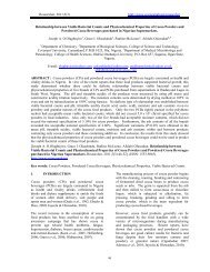
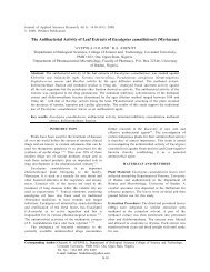
Fig. 1. Collection sites of cultivated and wild blackberries in Colombia’s coffee-growing region.
MATERIALS AND METHODS<br />
Collection of Study Materials<br />
From 2005 to 2008, plants of cultivated and wild<br />
blackberry species were collected in several<br />
municipalities of Colombia’s coffee-growing region<br />
where blackberry is grown (Fig. 1).<br />
Morpho-agronomic Characterization<br />
Thornless R. glaucus accessions were collected from five<br />
different collection sites in three different departments of<br />
Colombia (Risaralda, Caldas, and Quindío). Each<br />
accession was assigned a code beginning with the letter P<br />
and numbered 1 to 5. Table 1 lists the different sampling<br />
sites and the identification code assigned to each material.<br />
Blackberry plants were multiplied in vitro and, after<br />
hardening, a total of 50 plants from each of the five<br />
collection sites (P1, P2, P3, P4, P5) were delivered to 20<br />
farmers in each of the two municipalities of Risaralda<br />
(Santa Rosa de Cabal and Guática). Thornless plants from<br />
Santa Rosa de Cabal propagated by the farmers (P6) were<br />
used as control. Overall, each farmer established 300<br />
plants, totaling 6,000 plants per municipality and 12,000<br />
plants for the entire experiment. Fifteen morphoagronomic<br />
variables were evaluated as follows: number<br />
of female and male branches, number of runners, length<br />
of internodes of female and male branches, width and<br />
length of folioles on female and male branches, stem<br />
diameter on female and male branches, plant height at<br />
weeks 15 and 32 after planting, number of flower buds,<br />
and days to flowering.<br />
Semi-partial R 2<br />
Marulanda and López<br />
STATISTICAL ANALYSIS<br />
Treatments* Treatments*<br />
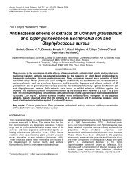
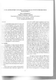
Fig. Fig. 2. 2. Dendogram and and cluster analysis based on on morpho-agronomic characterization.<br />
Treatments* see table 2<br />
Treatments* see table 2<br />
877<br />
A multivariate analysis was performed and groups were<br />
identified by cluster analysis, using the SAS program.<br />
Information on origins and sites was compared and 12<br />
combinations were identified for the cluster analysis<br />
(Table 2). Data were supported by the results of the<br />
multiple correspondence analysis.<br />
Evaluating Thorny and Thornless Accessions using<br />
SSR Markers<br />
Twenty-three microsatellite sequences developed by<br />
Amsellem (2001) and Graham et al. (2002 and 2004)<br />
were evaluated and, of these, eight SSRs were selected for<br />
their polymorphism and amplification quality. Twentythree<br />
regional accessions of Rubus, including thorny and<br />
thornless R. glaucus, both cultivated and wild, were<br />
analyzed as follows.<br />
9 wild and cultivated accessions of thornless R.<br />
glaucus<br />
4 wild accessions of thorny R. glaucus<br />
2 cultivated accessions of thornless R. glaucus<br />
1 accession of Rubus rosifolius<br />
6 accessions of Rubus urticifolius<br />
1 accession of Rubus robustus<br />
To determine groups of genetic diversity, the genetic<br />
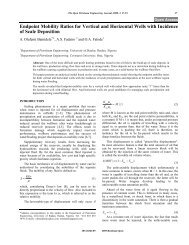
similarity (GSij) was calculated based on the formulas of
878<br />
Dice (1945) and Nei and Li (1979). Genotypes were<br />
grouped based on dissimilitude values (1-GSij) between<br />
all genotype pairs (Sneath and Sokal, 1973), using the<br />
unweighted pair-group method with arithmetic average<br />
(UPGMA) with the statistical package NTSys PC version<br />
2.02i (Rolhf, 1998).<br />
RESULTS AND DISCUSSION<br />
Morpho-agronomic Characterization<br />
Table 3 presents the cluster grouping of the 12<br />
combinations described, while table 4 presents the<br />
average, minimum, and maximum values, standard error,<br />
and coefficient of variation for each morpho-agronomic<br />
variable, regardless of the site and of the material. Data on<br />
plant height at weeks 15 and 32 after planting present<br />
coefficients of variation of 85.9% and 58.2%,<br />
respectively. This variation can be attributed to the<br />
difference in vegetative growth presented by plants,<br />
which is strongly influenced by agricultural tasks. The<br />
variable “number of runners” presents a coefficient of<br />
variation of 82.6%, with a maximum value of 3, a<br />
minimum value of 0, and an average value of 1 runner,<br />
which can be attributed to the difficulty in identifying<br />
these branches because they are easily confused with thin<br />
branches.<br />
Group 1<br />
Group 2<br />
Canadian Journal of Pure and Applied Sciences<br />
33% 42% 67% 90%<br />
Dice similarity coefficient<br />
According to principal component analysis, the variables<br />
described are classified into six components, in such a<br />
way that those contributing the greatest variation with<br />
regard to total variation are located in the first component<br />
and those contributing the smallest variation in the sixth<br />
component (Table 5).<br />
Variables presenting the greatest variation were, in<br />
descending order: length of foliole on the male branch,<br />
width of foliole on the male branch, stem diameter on the<br />
male branch, number of runners, and number of male<br />
branches. The number of male branches indirectly<br />
determines the productive capacity of a material because,<br />
after pruning, male branches can become female<br />
branches.<br />
The contribution of variables related to the productive<br />
capacity, such as number of female branches and number<br />
of flower buds, to total variation was smaller, respectively<br />
ranking fifth and sixth, which indicates that these<br />
variables alone have a smaller capacity to differentiate<br />
materials. The variable “number of flower buds” is<br />
directly related to fruit production and presents the<br />
smallest contribution to total variation, meaning that it is<br />
highly unlikely that this variable can be used to<br />
differentiate materials.<br />
0. 00 0.25 0.50 0.75 1. 0 0<br />
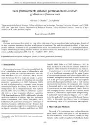
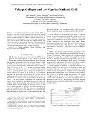
Fig. 3. Dendrogram of blackberries estimated by Dice similarity index (1945), based on SSR markers.<br />
50 R. glaucus thornless.<br />
90 R. glaucus thornless.<br />
99 R. glaucus thornless.<br />
46 R. glaucus thornless<br />
95 R. glaucus thorny<br />
54 R. glaucus thorny<br />
97 R. glaucus thorny<br />
86 R. glaucus thorny<br />
102 R. glaucus thorny<br />
P1 R. glaucus thornless<br />
P2 R. glaucus thornless<br />
P3 R. glaucus thornless<br />
P5 R. glaucus thornless<br />
22 R. glaucus thorny<br />
P4 R. glaucus thornless<br />
32 R. rosifolius<br />
33 R. robustus<br />
44 R. urticifolius<br />
82 R. urticifolius<br />
37 R. urticifolius<br />
64 R. urticifolius<br />
107 R. urticifolius<br />
106 R. urticifolius
The dendrogram in figure 2 shows the grouping of<br />
materials depending on the combination of site of origin<br />
and planting site. Two groupings are possible: the first<br />
separates the materials originating from Villamaría<br />
(Caldas) and Génova (Quindío) and planted in Santa Rosa<br />
de Cabal and Guática from the others. The second<br />
conserves the distance from these combinations, but<br />
separates materials originating from SENA-Manizales<br />
(Caldas) and Pereira (Risaralda) and planted in Guática.<br />
Overall, the dendrogram shows that the materials<br />
originating from Villamaría (Caldas) and Génova<br />
Marulanda and López<br />
879<br />
(Quindío) are promising and different, regardless of the<br />
site where they are planted.<br />
Based on the dendrogram, the three groups were<br />
submitted to cluster analysis (Table 3), resulting in the<br />
following combination of materials and planting sites:<br />
Cluster 1: Material originating from Santa Rosa de Cabal<br />
and planted in Santa Rosa de Cabal; material from Santa<br />
Rosa de Cabal propagated by farmers and planted in<br />
Santa Rosa de Cabal (control); material originating from<br />
Table 1. Collection sites of blackberry materials to be evaluated in the field and assigned code.<br />
Collection site Department Assigned ID code<br />
Santa Rosa de Cabal Risaralda P1<br />
SENA-Manizales Caldas P2<br />
Alto Manzano, Pereira Risaralda P3<br />
Cumaral, Génova Quindío P4<br />
Papayal, Villamaría Caldas P5<br />
Material from Santa Rosa de Cabal propagated by farmers (control) Risaralda P6<br />
Table 2. Combinations used in the cluster analysis, origin of materials and evaluation site.<br />
Combination Origin Evaluation site<br />
1 Santa Rosa de Cabal Santa Rosa de Cabal<br />
2 Santa Rosa de Cabal (control) Santa Rosa de Cabal<br />
3 Santa Rosa de Cabal Guática<br />
4 Santa Rosa de Cabal (control) Guática<br />
5 SENA-Manizales Santa Rosa de Cabal<br />
6 Pereira Santa Rosa de Cabal<br />
7 SENA-Manizales Guática<br />
8 Pereira Guática<br />
9 Génova Santa Rosa de Cabal<br />
10 Villamaría Santa Rosa de Cabal<br />
11 Génova Guática<br />
12 Villamaría Guática<br />
Table 3. Materials evaluated at two sites and group identification by cluster analysis.<br />
Combination Origin Site Cluster 1 Cluster 2<br />
1 Santa Rosa de Cabal Santa Rosa de Cabal 1 1<br />
2 Santa Rosa de Cabal (control) Santa Rosa de Cabal 1 1<br />
3 Santa Rosa de Cabal Guática 1 1<br />
4 Santa Rosa de Cabal (control) Guática 1 1<br />
5 SENA-Manizales Santa Rosa de Cabal 1 1<br />
6 Pereira Santa Rosa de Cabal 1 1<br />
7 SENA-Manizales Guática 3 1<br />
8 Pereira Guática 3 1<br />
9 Génova Santa Rosa de Cabal 2 2<br />
10 Villamaría Santa Rosa de Cabal 2 2<br />
11 Génova Guática 2 2<br />
12 Villamaría Guática 2 2
880<br />
Santa Rosa de Cabal and planted in Guática; material<br />
originating in SENA-Manizales and planted in Santa Rosa<br />
de Cabal; material originating in Pereira and planted in<br />
Santa Rosa de Cabal.<br />
Cluster 2: Material originating in Génova and planted in<br />
Santa Rosa de Cabal; material originating in Villamaría<br />
and planted in Santa Rosa de Cabal; material originating<br />
in Génova and planted in Guática, and material<br />
originating in Villamaría and planted in Guática.<br />
Cluster 3: Material originating in SENA-Manizales and<br />
planted in Guática; material originating in Pereira and<br />
planted in Guática.<br />
Canadian Journal of Pure and Applied Sciences<br />
Table 4. Values obtained for morpho-agronomic variables.<br />
This grouping corroborates the data presented in the<br />
dendrogram—that materials originating from Villamaría<br />
and Génova can be differentiated, regardless of the<br />
planting site (Cluster 2). The cluster analysis for the three<br />
groups and Duncan’s multiple comparison test at 5%,<br />
indicate that the only variable that differentiates the three<br />
clusters is “plant height at week 32”. Whereas cluster<br />
analysis for any two groups and the results of the<br />
minimum difference test, at 5%, indicate that the only<br />
variable that differentiated any two groups was the<br />
number of male branches.<br />
Based on the results obtained, materials originating from<br />
Santa Rosa de Cabal, SENA-Manizales, and Pereira<br />
Variable Average Minimum Maximum<br />
Standard<br />
error<br />
C.V. (%)<br />
Number of female branches 10.6 4 28 0.51 43.9<br />
Number of male branches 2.4 0 4 0.09 35.3<br />
Number of runners 0.98 0 3 0.09 82.6<br />
Length of internodes of female branches 6.62 4.5 8.5 0.09 13.0<br />
Length of internodes of male branches 7.28 3.0 10.0 0.12 14.6<br />
Width of folioles on female branches 21.04 15.0 29.5 0.30 12.6<br />
Length of folioles on female branches 16.35 12.0 21.0 0.21 11.5<br />
Width of folioles on male branches 21.29 11.0 26.0 0.30 12.6<br />
Length of folioles on male branches 16.60 8.5 19 0.22 12.2<br />
Stem diameter on female branches 4.30 2.0 8.0 0.14 28.5<br />
Stem diameter on male branches 4.80 2.5 8.0 0.11 21.5<br />
Plant height at week 15 23.81 3.0 99.0 2.28 85.9<br />
Plant height at week 32 145.30 16.0 354 9.40 58.2<br />
Number of flower buds 32.31 11 95 1.37 37.1<br />
Days to flowering 168.9 143 190 1.80 9.24<br />
Table 5. Variables identified in each component.<br />
Groups of factors Variable<br />
First factor Width of folioles on male branches<br />
Length of folioles on male branches<br />
Number of runners<br />
Stem diameter of male branches<br />
Number of male branches<br />
Second factor Width of folioles on female branches<br />
Length of folioles on female branches<br />
Third factor Stem diameter on female branches<br />
Plant height at 15 weeks after planting<br />
Plant height at 32 weeks after planting<br />
Days to flowering<br />
Fourth factor Length of internodes on female branches (cm)<br />
Fifth factor Number of females branches<br />
Length of internodes on male branches (cm)<br />
Sixth factor Number of flower buds
differ, in general, from materials originating from Génova<br />
and Villamaría for all study variables and individually<br />
(univariate analysis) for the variables “number of male<br />
branches” and “plant height at week 32”.<br />
“Days to flowering” was another variable presenting a<br />
low coefficient of variation because flowering is closely<br />
linked to a unified physiological condition of blackberry,<br />
regardless of origin of the material or planting site.<br />
Foliole length and width on both male and female<br />
branches are variables with unique characteristics for<br />
thornless blackberry and are also important in<br />
differentiating materials. Variables presenting the highest<br />
coefficient of variation were “plant height at 15 and 32<br />
weeks after planting”, for which plants presented<br />
differences in their vegetative growth associated with<br />
fertilization and weed control, as well as the variable<br />
“number of runners” because some farmers found it<br />
difficult to identify these branches in the field, confusing<br />
them with thin branches or they want to attribute the lack<br />
of crop vigor to the presence of these branches.<br />
Evaluating Thornless Blackberry Accessions using<br />
SSR Markers<br />
A total of 57 alleles with eight loci were obtained as a<br />
result of the evaluation of 23 genotypes belonging to four<br />
Rubus species: R. glaucus, R. urticifolius, R. robustus, and<br />
R. rosifolus. The genotypes of R. glaucus included in the<br />
study belonged to two groups: thorny and thornless. The<br />
Marulanda and López<br />
Table 6. Description of the SSRs used for genetic characterization of Rubus species.<br />
Locus 1 SSR<br />
mRaCIRRIV2A8 1 (CA) 12<br />
(CT)11<br />
Average<br />
PIC 2<br />
value<br />
0.08678<br />
881<br />
number of alleles per SSR marker ranged from 3 to 11<br />
(Table 6). Polymorphic bands were obtained in thornless<br />
R. glaucus genotypes, evidencing that genetic variability<br />
does exist among these materials, which were considered<br />
to be very uniform because of their limited genetic origin<br />
and their massive multiplication by cuttings and<br />
widespread distribution by blackberry farmers in the<br />
region over the last five years.<br />
The dendrogram in figure 3 shows 67% similarity for<br />
Group 1, which includes all R. glaucus accessions. The<br />
material differing most from the others was the P4<br />
thornless blackberry from Génova, Quindío—an outlier<br />
from the rest of the group due to molecular differences.<br />
At 90% similarity, two groups are formed that separate<br />
the remaining R. glaucus accessions. One group gathers<br />
thornless blackberry materials P1, P2, P3, and P5; P1 and<br />
P2, which are 100% similar with the SSR markers used.<br />
Both P4, originating from Génova (Quindio), and P5,<br />
originating from Villamaría (Caldas) show large<br />
differences at the molecular level. The dendrogram also<br />
shows other thornless blackberry genotypes that do not<br />
differ significantly from cultivated genotypes such as 50,<br />
90, 99, and 46.<br />
Analyses using SSR Molecular Markers<br />
The SSRs used differentiated the studied genotypes by<br />
specie, producing exclusive bands for each and separating<br />
Rubus species 4 Expected<br />
Thornless Thorny R. R. R.<br />
R. band size<br />
R. glaucus glaucus rosifolius urticifolius robustus (pb)<br />
1-3* 1-2* 1 1 1 191-237<br />
mRaCIRRIV2F4 1 mRaCIRRI1G3<br />
(CT) 8<br />
(CA) 17<br />
(CT)11<br />
0.38143 6-11* 8-11* 2 3 3 180-242<br />
1 Rubus 105b<br />
(GA) 28 0.43543 5-6* 2-6* 1 1-2* 2 195-265<br />
2 Rubus 98d<br />
(AG) 8 0.4444 1-3 1-3 1-4 1-2* 1-2 165/173/181<br />
2 (GAA) 5<br />
(GA)6<br />
NA 3 1-2 1-2 1 1 1 173<br />
Rubus 76b 2 (CT) 5<br />
(CT) 4<br />
0.30215 1-3* 1-2 1 1 1-2 190-210<br />
Rubus 16a 2 (AT) 8<br />
(GT) 11<br />
Rubus 116a 2 (CT) 12<br />
(T)10<br />
0.31519 4 4-6* 4 2-4* NAmp 5 169<br />
0.79717 2-5* 2 4 3-5* 5 299<br />
1 1 = Derived from Rubus alceifolius (Amsellem et al., 2001); 2 = Derived from Rubus (red raspberry) hybrid species (Graham et<br />
al., 2002, 2004).<br />
2 PIC = Polymorphism index content.<br />
3 NA = non-available.<br />
4 Presence of polymorphism.<br />
5 NAmp = No amplification.
882<br />
R. glaucus from other Rubus species, thus proving to be<br />
powerful markers for inter- and intra- specific studies.<br />
Polymorphic bands were obtained in thornless R. glaucus<br />
genotypes, evidencing genetic variability within these<br />
materials traditionally considered highly homogenous<br />
because of their restricted genetic origin, their massive<br />
multiplication by stakes, and their broad distribution by<br />
farmers in the region over the past five years. The<br />
morpho-agronomic data obtained are highly consistent<br />
with those obtained with molecular data.<br />
Badjakov (2007) characterized 48 raspberry accessions<br />
from Bulgaria, using nine SSR markers developed by the<br />
Scottish Crop Research Institute by Graham et al. (2002<br />
and 2004). He obtained 59 alleles. The number of alleles<br />
per locus varied from 4 to 10, with an average of 6.5 per<br />
locus. The SSRs allowed him to calculate the genetic<br />
distance between the materials under study as well as<br />
analyze their heterozygosity, thanks to the co-dominant<br />
character of the SSRs. Of the nine SSRs used by<br />
Badjakov (2007), two of themRubus 76b and Rubus<br />
98dwere also used in this study to evaluate different<br />
Rubus species as well as thorny and thornless accessions<br />
of R. glaucus collected in Colombia (Tables 6 and 7).<br />
The species R. urticifolius was characterized by the<br />
presence of eight exclusive bands; R. rosifolius by the<br />
presence of four exclusive bands; R. robustus by the<br />
presence of two exclusive bands; and R. glaucus by the<br />
presence of nine exclusive bands. When using SSR Rubus<br />
116a, thornless blackberry genotypes P4 and P5 were<br />
characterized by the presence of 1 exclusive band or allele<br />
and, when SSR Rubus 116a was used, thornless<br />
blackberry genotypes P1, P2, P3, P4, and P5 were<br />
characterized by the presence of 2 exclusive bands or<br />
Canadian Journal of Pure and Applied Sciences<br />
Table 7. Exclusiveness of markers in Rubus materials analyzed with SSRs.<br />
alleles. All thornless R. glaucus genotypes were<br />
differentiated by the SSRs mRaCIRRIV2F4 and<br />
mRaCIRRIV2A8 (Table 7).<br />
The highest number of single alleles was observed in R.<br />
glaucus, followed by R. urticifolius. The SSRs used<br />
allowed the Rubus species under study to be differentiated<br />
by their band patterns and the presence of bands exclusive<br />
to each species (Table 7). The presence of single bands<br />
for thornless R. glaucus genotypes (P1, P2, P3, P4, and<br />
P5) allowed the molecular characterization and<br />
differentiation of thornless blackberry genotypes. DNA<br />
profiles made it possible to clearly differentiate these<br />
materials from other R. glaucus genotypes (Table 7).<br />
Of the 60 microsatellite markers described and used by<br />
Stafne et al. (2005) to evaluate the diversity of North<br />
American Rubus species, four were selected for this<br />
study: Rubus 98d, Rubus 105b, Rubus 116a, and Rubus<br />
16a (Graham et al., 2002, 2004). The molecular marker<br />
mRaCIRRIV2A8 of Rubus alceifolius (Amsellem et al.,<br />
2001) was used also to evaluate Colombian Rubus species<br />
and genotypes (Tables 6 and 7).<br />
When the results of this study were compared with those<br />
obtained by Stafne et al. (2005), who found Rubus 98d to<br />
be monomorphic and Rubus 105b, Rubus 116a, and<br />
mRaCIRRIV2A8 to be polymorphic, our results differed<br />
regarding the number of alleles identified per each<br />
primer. When used to analyze Colombian species and<br />
genotypes, the SSRs produced the following results:<br />
Rubus 98d (3 alleles), Rubus 105b (7 alleles), Rubus 116a<br />
(11 alleles), and Rubus 16a (7 alleles) (Tables 6 and 7).<br />
The highest number of alleles per locus was obtained with<br />
the SSR markers Rubus 116a and mRaCIRRIV2F4, with<br />
Marker<br />
Alleles specific to<br />
species/genotype (no.)<br />
Rubus species/genotype<br />
Rubus 105b 3 R. urticifolius<br />
Rubus 105b 1 R. rosifolius and R. robustus<br />
mRaCIRRIV2A8 2 R. glaucus (thornless)<br />
mRaCIRRIV2A8 2 R. glaucus<br />
mRaCIRRIV2F4 1 R. glaucus (thornless)<br />
mRaCIRRIV2F4 6 R. glaucus<br />
Rubus 98d 1 R. urticifolius<br />
Rubus 98d 1 R. glaucus<br />
Rubus 76b 4 R. glaucus (thornless)<br />
mRaCIRRI1G3 3 R. glaucus<br />
Rubus 16a 2 R. urticifolius<br />
Rubus 116a 1 R. glaucus (P4 and P5)*<br />
Rubus 116a 2 R. glaucus (P1, P2, P3, P4, P5)*<br />
Rubus 116a 2 R. rosifolius<br />
Rubus 116a 1 R. urticifolius
11 alleles each (Table 7). Markers mRaCIRRIV2F4 and<br />
Rubus 76b presented the highest number of polymorphic<br />
bands in thornless R. glaucus genotypes. Other markers<br />
that produced a high number of alleles per locus were<br />
Rubus 16a (9 alleles), mRACIRRI1G3 (6 alleles), Rubus<br />
105b (7 alleles), mRaCIRRIV2A8, (5 alleles), and Rubus<br />
76b (5 alleles).<br />
When Colombian Rubus species and accessions were<br />
evaluated using with eight SSRs, a total of 57 alleles were<br />
obtained. The SSRs produced Rubus 76b (6 alleles) and<br />
Rubus 98d (3 alleles). Of the eight SSRs used, five<br />
belonged to the microsatellite series obtained in Rubus<br />
idaeus by Graham et al. (2002 and 2004) and three SSRs<br />
were obtained in Rubus alceifolus by Amsellem et al.<br />
(2001).<br />
These results corroborate the efficient transferability and<br />
usefulness of the R. alceifolius markers described by<br />
Amsellem et al. (2001), as well as the markers described<br />
by Graham et al. (2002 and 2004) developed in red<br />
raspberries. The eight SSRs were highly polymorphic in<br />
Colombian Rubus species and accessions.<br />
Furthermore, according to the results of Stafne et al.<br />
(2005), who evaluated eight SSR markers developed by<br />
Amsellem et al. (2001) in R. alceifolius, amplification<br />
was only obtained with mRaCIRRI1D3 and<br />
mRaCIRRIV2A8, which turned out to be polymorphic. In<br />
the current study, 22 alleles of a total of 57 were obtained<br />
with the SSR of R. alceifolius. The Colombian Rubus<br />
species evaluated shared 38.59% of the alleles of R.<br />
alceifolus; of a total of 57 alleles, 42 were obtained with<br />
markers obtained from red raspberry. Colombian Rubus<br />
species shared 62.68% of the alleles of R. idaeus.<br />
In other studies carried out by Marulanda et al. (2007)<br />
that evaluate other Colombian Rubus genotypes and<br />
species, amplification was obtained in six of the eight<br />
SSR markers used by Amsellem (2001). These results<br />
contrast with those obtained by Stafne et al. (2005), who<br />
only obtained amplification with two of these markers,<br />
perhaps because of the divergent evolution of the study<br />
species and because R. alceifolius is an Asian type of the<br />
subgenus Rubus, probably more related phylogenetically<br />
with R. glaucus, which means that R glaucus shares 75%<br />
of the SSRs of R alceifolius (Stafne et al., 2005;<br />
Marulanda et al., 2007).<br />
In the evaluation of 96 Rubus accessions of the Oregon<br />
germplasm bank, Castillo (Castillo, NRF. 2006) obtained<br />
12 pairs of SSR primer derived from Rubus and several<br />
from R. idaeus that generated between 3 and 16 alleles<br />
per locus, for a total of 98 alleles and an average number<br />
of eight alleles per primer. These results are very similar<br />
to those obtained in the evaluation of Colombian Rubus<br />
species and accessions in which eight SSRs generated<br />
Marulanda and López<br />
883<br />
between 3 and 11 alleles per primer, for a total of 57<br />
alleles and an average of 7.25 alleles per locus.<br />
CONCLUSIONS<br />
The analysis allows the differentiation of materials<br />
originating from Villamaría (P5) and Génova (P4) and<br />
planted in both Santa Rosa and Guática and those<br />
originating from SENA-Manizales (P2) and Pereira (P3)<br />
and planted in Guática. The most promising materials are<br />
those originating from Villamaría (P5) and Génova (P4),<br />
regardless of the planting site.<br />
The variables with the lowest coefficient of variation were<br />
days to flowering and length and width of foliole. These<br />
variables are valuable because of their characteristics<br />
unique to thornless blackberry materials and their<br />
important contribution in the differentiation of materials.<br />
The variables with the highest coefficient of variation<br />
were plant height at week 15 after planting, plant height at<br />
week 32 after planting, and number of runners.<br />
The variables that contribute most to the differentiation of<br />
materials were length of folioles on the male branch,<br />
width of folioles on the male branch, stem diameter on the<br />
male branch, number of runners, and number of male<br />
branches.<br />
The variables that contribute less to the differentiation of<br />
materials were those related to productive capacity, such<br />
as number of female branches and number of flower buds,<br />
indicating that these variables, when used independently,<br />
are less capable of differentiating materials.<br />
The number of flower buds—a variable directly related to<br />
fruit production—contributes the least to total variation.<br />
In other words, it is highly unlikely that this variable<br />
serves to differentiate materials.<br />
Polymorphic bands and bands exclusive to certain<br />
genotypes were obtained from thornless R. glaucus,<br />
indicating that genetic variability does exist within these<br />
materials.<br />
The SSRs grouped the Rubus species studied, and<br />
exclusive bands were identified for genotyping each<br />
species.<br />
The SSR markers of R. alceifolius and R. idaeus (red<br />
raspberry) were highly polymorphic in Colombian Rubus<br />
species and accessions.<br />
ACKNOWLEDGEMENTS<br />
Our special thanks to the blackberry farmers of the<br />
department of Risaralda. Our sincere appreciation to the<br />
Governor’s Office of the department of Risaralda and<br />
COLCIENCIAS for co-financing this project; to Juan
884<br />
Manuel Vásquez, undergraduate agronomy student for<br />
collecting field data; to Dr. César Sierra of the Colombian<br />
Agriculture and Livestock Institute (ICA) for monitoring<br />
the health of evaluation plots and accompanying the field<br />
evaluations; to Dr. Esther Cecilia Montoya for her support<br />
in experiment design and statistical analyses; to Juliana<br />
Arias Villegas, environment administrator, for planning<br />
and managing the present project. Our sincere thanks also<br />
go to the laboratory and nursery staff of the Biodiversity<br />
and Biotechnology Group, Universidad Tecnológica de<br />
Pereira.<br />
REFERENCES<br />
Amsellem, L., Dutech, C. and Billotte, N. 2001. Isolation<br />
and characterization of polymorphic microsatellite loci in<br />
Rubus alceifolius Poir (Rosaceae), an invasive weed in La<br />
Reunion island. Molecular Ecology Notes. 1:33-35.<br />
Amsellem, L., Noyer JL., Le Bourgeois, T. and Hossaert-<br />
McKey, M. 2000. Comparison of genetic diversity of the<br />
invasive weed Rubus alceifolius Poir. (Rosaceae) in its<br />
native range and in areas of introduction, using amplified<br />
fragment length polymorphism (AFLP) markers.<br />
Molecular Ecology. 9:443-455.<br />
Ashley, MV., Wilk, JA., Styan, SMN., Craft, KJ., Jones,<br />
KL., Feldheim, KA., Lewers, KS. and Ashman, TL. 2003.<br />
High variability and disomic segregation of<br />
microsatellites in the octoploid Fragaria virginiana Mill<br />
(Rosaceae). Theoretical and Applied Genetics. 107:1201-<br />
1207.<br />
Badjakov, IK. 2007. Program on Euro-berry Research:<br />
from Genomics to sustainable production, Quality and<br />
Health. STSM–REPORT.<br />
Cipriani, G., Lot, G., Huang, W-G., Marrazzo, MT.,<br />
Peterlunger, E. and Testolin, R. 1999. AC/GT and AG/CT<br />
microsatellite repeats in peach [Prunus persica (L.)<br />
Batsch]: Isolation, characterization, and cross species<br />
amplification in Prunus. Theoretical and Applied<br />
Genetics. 99:65-72.<br />
Daubeny, HA. 1996. Brambles. In: Fruit Breeding: Vine<br />
and Small Fruit Crops, Volume II. Eds. Janick, J. and<br />
Moore, JN. p. 109-190. John Wiley and Sons Inc, New<br />
York, N.Y.<br />
Decroocq, V., Fave, MG., Hagen, L., Bordenave, L. and<br />
Decroocq, S. 2003. Development and transferability of<br />
apricot and grape EST microsatellite markers across taxa.<br />
Theoretical and Applied Genetics. 106:912-922.<br />
Dice, LR. 1945. Measures of the amount of ecological<br />
association between species. Ecology. 26:297-302.<br />
Dirlewanger, E., Crosson, P., Tavaud, M., Aranzana, MJ.,<br />
Poizat, C., Zanetto, A., Arus, P. and Laigret, F. 2002.<br />
Development of microsatellite markers in peach (Prunus<br />
persica) (L. Batsch) and their use in genetic diversity<br />
Canadian Journal of Pure and Applied Sciences<br />
analysis in peach and sweet cherry (Prunus avium L).<br />
Theoretical and Applied Genetics. 105:127-138.<br />
Graham, J. and MacNicol, RJ. 1995. An examination of<br />
the ability of RAPD markers to determine the<br />
relationships within and between Rubus species. Theor.<br />
Appl Genet. 90: 1128-1132.<br />
Graham, J., Smith, K., MacKenzie, K., Jorgenson, L.,<br />
Hackett, C. and Powell, W. 2004. The construction of a<br />
genetic linkage map of red raspberry (Rubus idaeus<br />
subsp. idaeus) based on AFLPs, genomic-SSR and EST-<br />
SSR markers. Theoretical and Applied Genetics. 109:740-<br />
749.<br />
Graham, J., Smith, K., Woodhead, M. and Russell, J.<br />
2002. Development and use of simple sequence repeat<br />
SSR markers in Rubus species. Molecular Ecology Notes.<br />
2:250-252.<br />
Hall, HK. 1990. Blackberry breeding, Plant Breeding<br />
Reviews. 8:249-312.<br />
James, CM., Wilson, F., Hadonou, AM. and Tobutt, KR.<br />
2003. Isolation and characterization of polyploid<br />
microsatellites in diploid strawberry (Fragaria vesca L.)<br />
for mapping, diversity studies and clone identification.<br />
Molecular Ecology Notes. 3:171-173.<br />
Jennings, DL. and Ingram, R. 1983. Hybrids of Rubus<br />
parviflorus (Nutt.) with raspberry and blackberry, and the<br />
inheritance of spinelessness derived from this species.<br />
Crop Research (Horticultural Research.). 23:95-101.<br />
Lewers, KS., Styan, SM., Hokanson, SC. and Bassil, NV.<br />
2005. Strawberry GenBank-derived and genomic simple<br />
sequence repeat (SSR) markers and their utility with<br />
strawberry, blackberry, and red and black raspberry.<br />
Journal of the American Society for Horticultural Science.<br />
130:102-115.<br />
Lopez-Medina, J. and Moore, JN. 1999. Chilling<br />
enhances cane elongation and flowering in primocanefruiting<br />
blackberries. HortScience 34/44:638-640.<br />
Lopez-Medina, J., Moore, JN. and McNew, RW. 2000. A<br />
proposed model for inheritance of primocane fruiting in<br />
tetraploid erect blackberry. Journal of the American<br />
Society for Horticultural Science. 125:217-221.<br />
Marulanda, ML., López, AM. and Aguilar, SB. 2007.<br />
Genetic diversity of wild and cultivated Rubus species in<br />
Colombia using AFLP and SSR markers. Crop Breeding<br />
and Applied Biotechnology. 7:243-253.<br />
Marulanda, MP. and Márquez, MP. 2001. Caracterización<br />
de la diversidad genética de Rubus glaucus Benth<br />
mediante el empleo de marcadores moleculares (RAPD).<br />
Actualidades Biológicas. 23(74):57-63.<br />
Mohan, M., Nair, S., Bhagwat, A., Krishna, TG., Yano,<br />
M., Bhatia, CR. and Sasaki, T. 1997. Genome mapping,
molecular markers and marker assisted selection in crop<br />
plants. Molecular Breeding. 3:87-103.<br />
Moore, JN. and Skirvin, RM. 1990. Blackberry<br />
management. In: Small Fruit Crop Management. Eds.<br />
Galletta, GJ. and Himelrick, DG. Prentice Hall,<br />
Englewood Cliffs, NJ, USA. 214-244.<br />
Nei, M. and Li, WH. 1979. Mathematical model for<br />
studying genetic variation in terms of restriction<br />
endonuclease. Proceedings of the National Academy of<br />
Science of the USA.79:5267-5273.<br />
Rohlf, FJ. 1998. NTSYS-pc numerical taxonomy and<br />
multivariate analysis system, Version 2.02. Exeter<br />
Publications, New York, USA.<br />
Skirvin, RM., Motoike, S., Coyner, M. and Norton, M.<br />
2005. Rubus spp. cane fruit. In: Biotechnology of Fruit<br />
and Nut Crops. Ed. Litz, RE. CABi Publishing,<br />
Cambridge, MA, USA. 566-580.<br />
Sneath, PHA. and Sokal, RR. 1973. Numerical taxonomy.<br />
Freeman, San Francisco. pp. 573.<br />
Stafne, E., Clark, J., Weber, C., Graham, J. and Lewers,<br />
K. 2005. Simple sequence repeat (SSR) markers for<br />
genetic mapping of raspberry and blackberry. Journal of<br />
the American Society for Horticultural<br />
Science. 130(5):722-728.<br />
Strik, B., Finn, C., Clark, J., and Bañados, M. 2006.<br />
Worldwide production of blackberries. Department of<br />
Horticulture, Oregon State University, October 31st.<br />
Blackberry, Berry Crops, About Infonet.<br />
Received: Nov 6, 2008, Revised: Aug 27, 2009; Accepted: Aug 29, 2009<br />
Marulanda and López<br />
885
SENRA Academic Publishers, Burnaby, British Columbia<br />
Vol. 3, No. 3, pp. 887-895, 2009<br />
ISSN: 1715-9997<br />
AMELIORATIVE EFFECT OF VOLATILE OIL FROM CINNAMOMUM<br />
ZEYLANICUM ON HYPERALGESIA IN ALLOXAN DIABETIC RATS<br />
*Rajbir Bhatti 1 , S Kaur 1 , J Singh 2 and MPS Ishar 1<br />
1 Department of Pharmaceutical Sciences, Guru Nanak Dev University, Amritsar, Punjab<br />
2 Department of Pharmacology, Government Medical College, Amritsar, Punjab, India<br />
ABSTRACT<br />
Diabetic neuropathy is generally considered to be one of the most common complications of diabetes. Neuropathic pain<br />
is the most troublesome and early symptom of diabetic neuropathy, and has been recognized as one of the most difficult<br />
types of pain to treat due to its multifactorial pathogenesis. The aim of the present study was to evaluate the<br />
antinociceptive effect of volatile oil from Cinnamomum zeylanicum on hyperalgesia due to alloxan induced diabetes in<br />
rats. Diabetes was induced with single intra peritoneal injection of alloxan monohydrate (150mg/kg b.w.). Animals were<br />
divided into different groups. Treatment groups received cinnamon oil from 3 rd day onward upto 14 th day at different<br />
doses (5, 10 and 20mg/kg b.w.; i.p.). Diabetic control animals received normal saline (0.9% NaCl; 1ml/kg). After 2<br />
weeks, rats were tested in tail immersion and hot plate assays. Diabetic control rats exhibited significant hyperalgesia<br />
along with increased plasma glucose levels as compared with normal rats. Cinnamon oil treatment significantly<br />
decreased thermal hyperalgesia and the plasma glucose levels as compared with diabetic control rats. These results<br />
indicate the protective effect of volatile oil from the bark of Cinnamomum zeylanicum on hyperalgesia due to alloxan<br />
induced diabetes in rats.<br />
Keywords: Cinnamomum zeylanicum, diabetic neuropathy, hyperalgesia.<br />
INTRODUCTION<br />
Diabetic neuropathy (DN) is generally considered to be<br />
one of the most common complications of diabetes,<br />
affecting both types of diabetes equally (Vinik et al.,<br />
1992). DN affects up to 50% of patients with diabetes<br />
(Dyck et al., 1993). It is generally related to the duration<br />
and severity of hyperglycaemia. However, it may also<br />
occur acutely even with hypoglycaemia (Harati, 1996).<br />
Pain is the most troublesome and early symptom of<br />
diabetic neuropathy (Vinik, 2004). Neuropathic pain is<br />
mostly characterized by pain which can occur<br />
spontaneously as a result of exposure to normally mildly<br />
painful stimuli, i.e. hyperalgesia (Brown and Asbury,<br />
1984). Although hyperglycaemia (Green et al., 1992),<br />
neuronal loss (Dyck et al., 1985) have been reported to be<br />
responsible for the change in pain perception, the exact<br />
aetiological factors involved are still under investigation<br />
(Anjaneyulu and Chopra, 2006). Experimentally induced<br />
diabetic rats have been used as a model of chronic pain<br />
with signs of hyperalgesia and allodynia due to diabetic<br />
neuropathy that may reflect symptoms observed in<br />
humans (Anjaneyulu and Chopra, 2006). Alloxan at a<br />
dose of 150mg/kg bw i.p., has been reported to induce<br />
reproducible and persistant hyperglycaemia in rats (Ryle<br />
et al., 1984; Diniz et al., 2008). Alloxan-injected diabetic<br />
rats have been reported to exhibit thermal allodynia and<br />
hyperalgesia, tested on hot-plate and tail-immersion<br />
*Corresponding author email: rajbirkb@yahoo.com<br />
assays (Morani and Bodhanker, 2007). Both hyperalgesia<br />
and allodynia have been reported to be established after<br />
14 days of alloxan treatment (Aubel et al., 2004).<br />
Currently, there is growing interest in herbal remedies due<br />
to a number of side effects associated with hypoglycaemic<br />
agents used in allopathic medicine (Kim et al., 2006). The<br />
effects of these plants may delay the development of<br />
diabetic complications and correct the metabolic<br />
abnormalities. It is believed that herbal medicines have<br />
lesser side effects and also have slow onset of these side<br />
effects (Habib et al., 2005).<br />
Cinnamon is one of the most common traditional folk<br />
herbs used in Korea, China and Russia for diabetes<br />
mellitus (Bailey and Day, 1989). Cinnamon bark is<br />
widely used as a spice and flavouring agent. Common<br />
cinnamon correctly refers to “true cinnamon”, or its<br />
synonym Ceylon cinnamon (Cinnamomum verum, C.<br />
zeylanicum) (Jellin, 2006). Common and cassia cinnamon<br />
have been shown to be generally safe when ingested and<br />
to have many pharmacological properties, such as<br />
antioxidant activity (Singh et al., 2007), antimicrobial<br />
activity (Tabak et al., 1999; Ooi et al., 2006) anti<br />
inflammatory activity (Tung et al., 2008) and antifungal<br />
activity (Cheng et al., 2008). Common and Cassia<br />
cinnamon are well known for their pharmacological<br />
properties in the treatment of diabetes (Khan et al., 2003;<br />
1
888<br />
Verspohl et al., 2005). The analgesic and antiinflammatory<br />
effects of Cinnamomum zeylanicum have<br />
been described in experimental studies (Atta and<br />
Alkofahi, 1998). Moreover, the major component found<br />
in cinnamon leaf and bark volatile oil, cinnamaldehyde<br />
(Singh et al., 2007) has been studied for its inhibitory<br />
effects on pro-inflammatory mechanisms (Chao et al.,<br />
2008), NF-κB activation (Liao et al., 2008), nitric oxide<br />
production (Lee et al., 2005) and cyclooxygenase-2<br />
inhibitory activity (Guo et al., 2006). However, the role of<br />
cinnamon in diabetic neuropathy has not been<br />
investigated so far. Therefore, the present study was<br />
designed to investigate the effect of cinnamon oil on<br />
diabetic neuropathy.<br />
MATERIALS AND METHODS<br />
Plant Material<br />
C. zeylanicum (Lauraceae) was purchased from National<br />
Medicos, Amritsar, India. The species was identified and<br />
authenticated by Dr. Amarjit Singh Soodan, Guru Nanak<br />
Dev University, Amritsar. [voucher specimen<br />
(SR./Bot.Sci./0345) is deposited in the herbarium of the<br />
Department of Botanical and Environmental Sciences,<br />
Guru Nanak Dev University, Amritsar, India].<br />
Extraction of volatile oil<br />
Volatile oil was extracted according to the method<br />
reported by Chang and Cheng (2002) with little<br />
modification. Briefly, 500g of the dried bark was taken,<br />
broken into small pieces and hydrodistilled with a<br />
Clavenger apparatus for 6 h. The light yellow coloured<br />
essential oil was collected and dried over anhydrous<br />
sodium sulphate and, after filtration, stored in screw tight<br />
bottle at – 10°C.<br />
Experimental Animals<br />
Wistar albino rats of either sex (150-200g) were housed in<br />
3 per cage, with food and water ad libitum for several<br />
days before the beginning of the experiment. The animals<br />
were kept on straw bedding in cages with a natural light:<br />
dark cycle and had free access to standard rodent food<br />
pellets and water. Animals were acclimatized to the<br />
laboratory conditions (room temperature 25±5°C) for one<br />
week before the start of experiment. All the experiments<br />
were conducted between 09.00 and 17.00 hrs.<br />
Preparation of Drugs<br />
Preparation of Alloxan solution (15mg/ml)<br />
150mg of alloxan was accurately weighed and then<br />
dissolved into 10 ml of distilled water. Freshly prepared<br />
solution was used in each experiment.<br />
Preparation of Cinnamon Oil (5, 10 & 20mg/ml)<br />
50, 100 & 200µL of Cinnamon Oil was taken out and<br />
dissolved in 10ml of dimethyl sulfoxide (DMSO; 0.05%)<br />
to produce 5, 10 & 20 mg/ml of the cinnamon oil.<br />
Canadian Journal of Pure and Applied Sciences<br />
Preparation of Glipizide (10mg/ml)<br />
50mg of Glipizide was accurately weighed and dissolved<br />
in DMSO (0.05%) and volume made upto 5ml.<br />
Preparation of Fluoxetine (2mg/ml)<br />
20mg of fluoxetine was accurately weighed and dissolved<br />
in 10ml of distilled water.<br />
Experimental Protocols<br />
In the present study, total of 42 rats were used. The rats<br />
were divided into 7 groups of 6 rats each.<br />
Group I- Non-Diabetic group (n=6). The animals were<br />
injected with normal saline (0.9%, NaCl; 1 ml/kg) instead<br />
of the corresponding treatments.<br />
In the rest of the groups the animals were fasted overnight<br />
prior to alloxan treatment and diabetes was induced by a<br />
single intraperitoneal injection of freshly prepared alloxan<br />
(150mg/kg bw) and the treatment with the cinnamon oil<br />
and glipizide was started on day 3. The blood glucose<br />
levels were estimated on 3 rd day of alloxan treatment and<br />
the animals with fasting blood glucose levels more than<br />
150mg/dl were considered diabetic and included in the<br />
study. The treatment with different drugs was as follows:<br />
Group II- Diabetic control group (n = 6). The animals<br />
were administered normal saline (1 ml/kg) from 3 rd day<br />
onward upto 14 days.<br />
Group III- Diabetic vehicle group (n = 6). The diabetic<br />
animals were administered a daily dose of vehicle<br />
dimethyl sulfoxide (DMSO; 0.05%; 1ml/kg) from 3 rd day<br />
onward upto 14 days.<br />
Group IV- Glipizide treated group (n = 6). The animals<br />
were treated with glipizide at a dose of 10mg/kg; ip., from<br />
3 rd day onward upto 14 days.<br />
Group V- Fluoxetine treated group (n = 6). The animals<br />
were treated with fluoxetine at a dose of 20mg/kg; i.p.<br />
dissolved in distilled water from 3 rd day onward to 14<br />
days. Fluoxetine was used as the standard for assessment<br />
of thermal hyperalgesia.<br />
Group VI - Cinnamon oil (5mg/kg) group (n = 6). A<br />
daily dose of 5mg/kg of cinnamon oil was administered<br />
from 3 rd day onward to 14 days.<br />
Group VII - Cinnamon oil (10mg/kg) group (n = 6). A<br />
daily dose of 10 mg/kg was administered from 3 rd day<br />
onward to 14 days.<br />
Group VIII - Cinnamon oil (20mg/ kg) group (n = 6). A<br />
daily dose of 20mg/kg was administered from 3 rd day<br />
onward upto 14 days.
Estimation of Fasting Blood Glucose (FBG)<br />
For the estimation of blood glucose, blood samples were<br />
taken from retro orbital plexus. Blood samples were kept<br />
at room temperature for 5-10 minutes and were allowed to<br />
clot. After clotting the samples were centrifuged at 2000<br />
rpm for 10 minutes to separate plasma. Separated plasma<br />
was used for the estimation of fasting blood glucose by<br />
GOD/POD method using commercial kit (Span<br />
Diagnostics Ltd, Surat).<br />
Assessment of thermal hyperalgesia<br />
Thermal hyperalgesia was assessed with tail-immersion<br />
and hot plate tests. Preliminary threshold to tailimmersion<br />
and hot-plate responses, by taking the means<br />
of two consecutive stable values which did not differ by<br />
more than 10% were determind (Chopra and Anjaneyulu,<br />
2006). Nociceptive latency was measured at 15, 30, 60,<br />
120 and 180 min. after the administration of drug. The<br />
nociceptive latency was expressed as mean ± S.E.M.<br />
Tail-immersion (warm water) test<br />
The tail was immersed in a warm water bath (52.55 0 C)<br />
until tail withdrawl (flicking response) or signs of struggle<br />
were observed, the cut-off time was 12s. The hyperalgesic<br />
response in the tail withdrawl test is generally attributed<br />
to central mechanisms (Chopra and Anjaneyulu, 2006).<br />
Hot-Plate test<br />
The hyperalgesic response on the hot plate is considered<br />
to result from a combination of central and peripheral<br />
mechanisms (Chopra and Anjaneyulu, 2006). In this test,<br />
animals were individually placed on a hot-plate (Eddy’s<br />
Hot-Plate) with the temperature adjusted to 551 0 C. The<br />
latency to the first sign of paw licking or jump response to<br />
avoid the heat was taken as on index of the pain<br />
threshold; the cut-off time was 10 s in order to avoid<br />
damage to the paw.<br />
STATISTICAL ANALYSIS<br />
Results were expressed as Mean±SEM. The intergroup<br />
variation was measured by one way analysis of variance<br />
(ANOVA) followed by Tukey’s test. Statistical<br />
significance was considered at p < 0.05. The statistical<br />
analysis was done using the Sigma Stat Statistical<br />
Software version 2.3.<br />
RESULTS<br />
Yield of Volatile Oil from Bark of C. zeylanicum<br />
6.5ml of volatile oil was obtained from hydrodistillation<br />
of 500g of cinnamon bark. The percentage yield of the<br />
cinnamon oil was 1.3%. The density of the oil was found<br />
to be 1.059g/ml. The crude volatile oil so obtained was<br />
more than 98% pure cinnamaldehyde as evidenced the 1 H<br />
& 13 C NMR and IR spectra of the oil. In the 1 H NMR<br />
Bhatti et al. 889<br />
(400 M Hz, CDCl3 solvent) the aldehydic proton showed<br />
up at 9.67 (1 H, doublet, J = 7.22 Hz), aromatic protons<br />
along with C-olefinic H formed a 6H multiplet at 7.76<br />
– 7.28 and the C-olefinic H appeared as a double<br />
doublet at 6.89 (1H, J = 16.00 and7.72 Hz); there was no<br />
other significant resonance signal in the proton NMR of<br />
the oil. Presence of only cinnamaldehyde is also<br />
corroborated by 13 C NMR through resonances at 192.91<br />
(CHO), 152.23 (olefinic C), 133.85 (quaternary<br />
aromatic carbon) and other resonances at δ 1309.974<br />
(olefinic C ), 128.873 (m-C of aromatic ring), 128.395<br />
(o-C of aromatic ring), 128.263 (p-C of aromatic ring).<br />
The aldehyde band in the IR spectrum appeared at 1681<br />
cm -1 . The determined density of the oil 1.059g/ml is also<br />
close to the value reported for pure cinnamaldehyde<br />
(1.048).<br />
Effect of alloxan injection on Fasting Blood Glucose<br />
(FBG) in rats<br />
The levels of fasting blood glucose were found to be<br />
significantly higher in diabetic rats than those of normal<br />
(non-diabetic) rats after 1 and 2 weeks of intraperitoneal<br />
administration of alloxan (Fig.1).<br />
Effect of various therapeutic interventions on Fasting<br />
Blood Glucose in alloxan induced diabetic rats<br />
The fasting blood glucose levels were estimated in<br />
different groups on 7 th and 14 th day of the alloxan<br />
treatment. Glipizide treatment was found to produce a<br />
significant decrease in fasting blood glucose in treated<br />
rats as compared with that of untreated control rats. The<br />
treatment with the vehicle did not alter the increase in<br />
FBG of alloxan treated animals. The treatment with<br />
cinnamon oil at 5, 10 and 20mg/kg dose was found to<br />
decrease the fasting blood glucose significantly in dose<br />
dependent manner on 7 th and 14 th day as compared to<br />
diabetic control group. The maximum decrease in fasting<br />
blood glucose was achieved in 20mg/kg treatment group<br />
and was similar to that of glipizide treatment (Fig. 2).<br />
Effect of alloxan injection on tail immersion<br />
nociceptive threshold in rats<br />
The tail immersion nociceptive threshold was observed to<br />
be significantly decreased in diabetic rats as compared<br />
with that of normal rats. Hyperalgesia was evident in tail<br />
immersion test after 1 week, the maximum decrease in<br />
pain threshold was observed at 2 weeks after alloxan<br />
injection in rats (Fig. 3).<br />
Effect of various therapeutic interventions on tail<br />
immersion nociceptive threshold in alloxan – inducted<br />
diabetic rats<br />
A significant increase in nociceptive threshold in tail<br />
immersion test was observed in diabetic rats treated with<br />
fluoxetine (20mg/kg) i.p. as compared with that of<br />
untreated diabetic control rats. Treatment with vehicle did
890<br />
not produce any change in the nociceptive threshold in<br />
rats. Treatment with cinnamon oil at doses 5, 10 and 20<br />
mg/kg was found to increase the nociceptive threshold in<br />
tail immersion test significantly in the treated rats as<br />
comared to that of untreated diabetic control rats. The<br />
data is expressed as mean ± S.E.M. The maximum<br />
protective effect against thermal hyperalgesia was<br />
observed at 30 min. after administration of volatile oil<br />
from C. zeylanicum. The protective effect produced by<br />
cinnamon oil on thermal hyperalgesia was found to be<br />
more than that of standard dug fluoxetine in tailimmersion<br />
assay. The maximum dose-dependent decrease<br />
in nociceptive threshold in tail-immersion assay was<br />
observed at the dose of 20mg/kg in alloxan-induced<br />
diabetic rats (Fig. 4).<br />
Canadian Journal of Pure and Applied Sciences<br />
Fig. 1. Effect of alloxan-injection on Fasting Blood Glucose in Rats after 1 and 2 weeks.<br />
p < 0.05; a, as compared to normal.<br />
Fig. 2. Effect of various pharmacological interventions on Fasting Blood Glucose Diabetic Rats on Day 14.<br />
p < 0.05; a, as compared to normal; b, as compared to Diabetic Control.<br />
Effect of alloxan injection on hot plate nociceptive<br />
threshold in rats<br />
The nociceptive threshold was observed to be<br />
significantly decreased in diabetic rats as compared with<br />
that of normal rats in hot plate assay. Hyperalgesia was<br />
evident in hot plate test after 1 week, the maximum<br />
decrease in pain threshold was observed at 2 weeks after<br />
alloxan injection in rats (Fig. 5).<br />
Effect of various therapeutic interventions on hot plate<br />
nociceptive threshold in alloxan – inducted diabetic<br />
rats<br />
A significant increase in nociceptive threshold in hot plate<br />
test was observed in diabetic rats treated with fluoxetine<br />
(20mg/kg) i.p. as compared with that of untreated diabetic
control rats. Treatment with vehicle did not produce any<br />
change in the nociceptive threshold in rats. Treatment<br />
with cinnamon oil at doses 5, 10 20mg/kg was found to<br />
increase the nociceptive threshold in hot plate test<br />
significantly in the treated rats as compared to that of<br />
untreated diabetic control rats. The data is expressed as<br />
mean ± S.E.M. The maximum protective effect against<br />
thermal hyperalgesia was observed at 30min. after<br />
administration of volatile oil from C. zeylanicum. The<br />
protective effect produced by cinnamon oil on thermal<br />
hyperalgesia was found to be more than that of standard<br />
dug fluoxetine in hot plate assay. The maximum dosedependent<br />
decrease in nociceptive threshold in tailimmersion<br />
assay was observed at the dose of 20mg/kg in<br />
alloxan-induced diabetic rats (Fig. 6).<br />
Bhatti et al. 891<br />
Fig. 3. Effect of alloxan-injection on tail-withdrawl latency in tail-immersion test in rats, as observed after 1 and 2<br />
weeks.<br />
a=p
892<br />
the diabetic control rats as compared to that of the normal<br />
(non-diabetic rats) indicating that alloxan-induced<br />
diabetic rats exhibit thermal hyperalgesia. It has been<br />
reported that alloxan-injected diabetic rats exhibit thermal<br />
allodynia and hyperalgesia, tested on hot-plate and tailimmersion<br />
assays (Morani and Bodhanker, 2007). Both<br />
hyperalgesia and allodynia have been reported to be<br />
established after 14 days of alloxan treatment (Aubel et<br />
al., 2004), which was observed behaviorally. The<br />
hyperalgesic response in tail-withdrawal test is generally<br />
attributed to central mechanisms whereas the hyperalgesic<br />
response on hot plate is attributed to the combination of<br />
Canadian Journal of Pure and Applied Sciences<br />
Fig. 5. Effect of alloxan-injection on paw-licking latency in hot-plate test in rats, as observed after 1 and 2 weeks.<br />
a=p
multifactorial, include the polyol pathway (Finegold et<br />
al., 1983), glycation (Wada and Yagihashi, 2005) and<br />
oxidative stress (Chung et al., 2003; Vincent et al., 2004).<br />
Oxidative stress could damage the nerves by direct toxic<br />
effects or with different biochemical changes that lead to<br />
endothelial dysfunction (Goycheva et al., 2006). The<br />
volatile oil from C. zeylanicum has been reported to<br />
possess antioxidant activity (Shobana and Naidu, 2000;<br />
Singh et al., 2007). Cinnamaldehyde which is the chief<br />
component of the cinnamon oil may be responsible for the<br />
antioxidant activity. Cinnamon has been reported to have<br />
a very high concentration of antioxidants chiefly<br />
cinnamaldehyde which make it an excellent candidate for<br />
disorders arising from oxidant stress (Su et al., 2007; Jang<br />
et al., 2007). Also cinnamon is documented to inhibit<br />
aldose reductase (Aida et al., 1987), the latter plays a vital<br />
role in conversion of glucose to sorbitol. This sorbitol is<br />
further converted to fructose by sorbitol dehydrogenase<br />
which leads to the depletion of organic osmolites such as<br />
taurine, myoinositol etc. (Zhu et al., 1999). Myoinositol<br />
depletion results in inactivation of Na + /K + ATPase, Na +<br />
retention and cellular edema (Brownlee, 2001). Also the<br />
conversion of glucose to sorbitol depletes the cellular<br />
stores of NADPH which are required for the conversion<br />
of oxidized glutathione to reduced glutathione (Brownlee,<br />
2001). All these factors play vital role in various<br />
complications of diabetes (Brownlee, 2001). From the<br />
above discussion it may tentatively be suggested that the<br />
protective effect of cinnamon oil may be attributed to its<br />
insulin enhancing and antioxidant effects.<br />
Further, studies have demonstrated that even with<br />
stringent blood glucose control, the prevention of<br />
neuropathy is not successful which suggest that there may<br />
be a release of early mediators between hyperglycaemiainduced<br />
metabolic and enzymatic changes and the nerve<br />
damage (Sharma et al., 2006). Once these mediators are<br />
released, it is possible that they modulate neuronal<br />
homeostasis independly of the initial metabolic stimulus<br />
(Sharma et al., 2006). Previous studies have shown that<br />
chronic hyperglycaemia accelerates the production of<br />
endogenous tumor necrosis factor- (TNF-) in<br />
microvascular and neuronal tissues (Kuhad et al., 2008).<br />
Cinnamaldehyde has been reported to have inhibitory<br />
effects on TNF- (Liao et al., 2008). Cinnamaldehyde has<br />
been demonstrated to inhibit TNF--induced signaling<br />
pathways by inhibiting the expression of cell adhesion<br />
molecules in endothelial cells by suppressing nuclear<br />
factor-kappa B (NF-κB) activation (Liao et al., 2008). It<br />
has been shown that clinical application of the agents that<br />
suppress the production and/or activity of TNF- may<br />
inhibit the development and exacerbation of chronic<br />
diabetic complications (Satoh et al., 2003).<br />
Superoxide and nitric oxide have been reported as the<br />
other key mediators which combine to form peroxynitrite,<br />
Bhatti et al. 893<br />
which rapidly causes protein nitration or nitrosylation,<br />
lipid peroxidation, DNA damage and cell death and to<br />
have direct toxic effects on the nerve tissue leading to<br />
neuropathic pain (Kuhad et al., 2008). Cinnamon has<br />
been evaluated for its NO-suppressing activity via<br />
different pathways such as the blocking of inducible nitric<br />
oxide synthase (iNOS) expression, the inactivation of<br />
iNOS catalytic function and the scavenging of NO (Tsai<br />
et al., 2007).<br />
Cinnamaldehyde has also been reported to reduce IL-1βinduced<br />
cyclooxygenase-2 (COX-2) activity in rat<br />
cerebral microvascular endothelial cells significantly<br />
(Guo et al., 2006). It has been reported that COX-2 is<br />
upregulated in the peripheral nerves and dorsal root<br />
ganglia neurons in experimental diabetes and that COX-2<br />
gene inactivation or selective COX-2 inhibition provides<br />
protection against various diabetic peripheral neuropathy<br />
defects (Kellogg et al., 2008). COX-2 upregulation leads<br />
to an altered prostaglandin profile with an increased<br />
production of vasoconstricting PGH2, thromboxane-A2<br />
(TXA2) and PGF 2α and reduction of vasodilatory<br />
prostacyclin (PGI2), thereby favouring vasoconstriction<br />
and ischemia (Kuhad et al., 2008). It has also been<br />
demonstrared that COX-2 gene deficient experimental<br />
diabetic rats showed a differential protection against<br />
biochemical and functional markers of experimental<br />
neuropathy and that the extent of this protection appeared<br />
to be dependent on the degree of COX-2 gene deficiency<br />
(Kellogg and Pop-Busui, 2005).<br />
CONCLUSION<br />
On the basis of present results and above discussed<br />
mechanisms, the antinociceptive effect of cinnamon oil in<br />
alloxan-induced diabetic rats as observed in the present<br />
study may be attributed to its hypoglycaemic, antioxidant<br />
and pro-inflammatory inhibitory effects i.e. suppressing<br />
TNF-α signaling pathway, inhibiting NF-қB activation,<br />
suppressing NO production and activity, and reducing IL-<br />
1β-induced COX-2 activity. In conclusion, it may<br />
tentatively be stated that volatile oil from Cinnamomum<br />
zeylanicum could provide a novel therapeutic approach in<br />
attenuation of hyperalgesia due to diabetes.<br />
REFERENCES<br />
Aida, K., Shindo, H., Tawata, M. and Oaya, T. 1987.<br />
Inhibition of aldose reductase activities by kampo<br />
medicines. Pl Medica. 53:131-135.<br />
Anjaneyulu, M. and Chopra, K. 2004. Quercetin<br />
attenuated thermal hyperalgesia and cold allodynia in<br />
STZ-induced diabetic rats. Ind J Expt. Bio. 42:766-769.<br />
Anjaneyulu, M. and Chopra, K. 2006. Possible<br />
involvement of cholinergic and opiod receptor<br />
mechanisms in fluoxetine mediated antinociception
894<br />
response in streptozotocin-induced diabetic mice. Eur J<br />
Pharmacol. 539:80-84.<br />
Atta, A.H. and Alkofahi, A. 1998. Anti-nociceptive and<br />
anti-inflammatory effects of some Jordanian medicinal<br />
plant extracts. J Ethnopharmacology. 60:117-124.<br />
Aubel, B., Kayser, V., Mauborgne, A., Farre, A., Hamom,<br />
M. and Bourgoin, S. 2004. Antihyperalgesic effects of<br />
cizolirtine in diabetic rats: behavioral and biochemical.<br />
B74-B77.<br />
Bailey, CJ. and Day, C. 1989. Traditional plant medicines<br />
as treatments for diabetes. Diabetes Care. 12:553-564.<br />
Brown, MJ. and Asbury, AK. 1984. Diabetic neuropathy.<br />
An. Neurol. 15(1):2-12.<br />
Brownlee, M. 2001. Biochemistry and molecular cell<br />
biology of diabetic complications. Nature. 414:813-820.<br />
Chang, ST. and Cheng, SS. 2002. Antitermitic Activity of<br />
Leaf Essential Oils and Components from Cinnamomum<br />
osmophleum. J. Agric. Food Chem. 50:1389-1392.<br />
Chao, LK., Hua, KF., Hsu, HY., Cheng, SS., Lin, IF.,<br />
Chen, CJ., Chen, ST. and Chang, ST. 2008.<br />
Cinnamaldehyde inhibits pro-inflammatory cytokines<br />
secretion from monocytes/macrophages through<br />
suppression of intracellular signaling. Food and Chem<br />
Toxicol. 46:220-231.<br />
Cheng, SS., Liu, JY. Ed-Haun, Chang, EH. and Chang,<br />
ST. 2008. Antifungal activity of cinnamaldehyde and<br />
eugenol congeners against wood-rot fungi. Bioresource<br />
Technology. 99:5145-5149.<br />
Chung, SSM., Ho, ECM., Lam, KSL. and Chung, SK.<br />
2003. Contribution of polyol pathway to diabetes induced<br />
oxidative stress. J Am Soc Nephrol. 14:S233-S236.<br />
Diniz, SF., Amorim, PLG., Cavalcante-Neto, FF., Bocca,<br />
AL., Batista, AC., Simm, GEPM. and Silva, TA. 2008.<br />
Alloxan-induced diabetes delays repair in a rat model of<br />
closed tibial fracture. Brazil J Med Bio Res. 41:373-379.<br />
Dyck, I., Hansen, S., Karnes, J., O’Brien, P., Yasuda, M.,<br />
Windebank, A. and Simmerman, B. 1985. Capillary<br />
number and percentage closed in human diabetic neuronal<br />
nerve. Nat. Acad Sci USA. 82:2513-2517.<br />
Dyck, PJ., Katz, KM., Karnes, JL., Litchy, WJ., Klein, R.,<br />
Pach, JM., Wilson, DM., Brein, PC., Melton, LJ. and<br />
Service, FJ. 1993. The prevalence by staged severity of<br />
various tupes of diabetic neuropathy,retinopathy and<br />
nephropathy in population-based cohort: the Rochester<br />
Diabetic neuropathy study. Neurology. 43:817-824.<br />
Finegold, D., Lattimer, SA., Nolle, S., Bernstein, M. and<br />
Greene, DA. 1983. Polyol pathway activity and myoinositol<br />
metabolism. A suggested relationship in the<br />
pathogenesis of diabetic neuropathy. Diabetes. 32:988-<br />
992.<br />
Canadian Journal of Pure and Applied Sciences<br />
Goycheva, P., Gadjeva, V. and Popov, B. 2006. Oxidative<br />
stress and its complications in diabetes mellitus. Trakia J<br />
Sci. 4(1):1-8.<br />
Greene, DA., Sims, AA., Stevens, MJ., Feldman, EL. and<br />
Lattimer, SA. 1992. Complications:neuropathy,<br />
pathogenetic conciderations. Diabetes Care. 15(12):1902-<br />
1925.<br />
Guo, JY., Huo, HR., Zhao, BS., Liu, HB., Li, LF., Ma,<br />
YY., Guo, SY. and Jiang, TL. 2006. Cinnamaldehyde<br />
reduces IL-1β-induced cyclooxygenase-2 activity in rat<br />
cerebral microvascular endothelial cells. Eur J Pharmacol.<br />
537:174-180.<br />
Habib, M.Y., Islam, M.S. and Khan, M.A. (2005) Herbal<br />
products: a novel approach for diabetic patients. Pak J<br />
Nutrition, 4(1):17-21.<br />
Harati, Y. 1996. Diabetes and the nervous system.<br />
Endocrinol metab Clin North Am. 25(2): 325-359.<br />
Jang, HD., Chang, KS., Huang, YS., Hsu, CL., Lee, SH.<br />
and Su, MS. 2007. Principal phenolic phytochemicals and<br />
antioxidant activities of three Chinese medicinal plants.<br />
Food Chemistry. 103:749-756.<br />
Jellin, JM. 2006. Cinnamon bark- Monograph [online].<br />
Available at www.naturaldatabase.com.<br />
Kellogg, AP., Cheng, HT. and Pop-Busui, R. 2008.<br />
Cyclooxygenase-2 pathway as apotential therapeutic<br />
target diabetic peripheral neuropathy. Curr Drug Targets.<br />
9(1):68-76.<br />
Kellogg, AP. and Pop-Busui, R. 2005. Peripheral nerve<br />
dysfunction in experimental diabetes is mediated by<br />
cyclooxygenase-2 and oxidative stress. Antioxd Redox<br />
Signal. 7:1521-1529.<br />
Khan, A., Safdar, M., Khan, MMA., Khattak, KN. and<br />
Anderson, RA. 2003. Cinnamon improves glucose and<br />
lipids of people with type II diabetes. Diabetes Care.<br />
26:3215-3218.<br />
Kim, SH., Hyun, SH. and Choung, SY. 2006. Antidiabetic<br />
effect of cinnamon extract on blood glucose I db/db mice.<br />
J Ethnopharmacology. 104:119-123.<br />
Kuhad, A., Sharma, S. and Chopra, K. 2008. Lycopene<br />
attenuates thermal hyperalgesia in a diabetic mouse model<br />
of neuropathic pain. Eur J Pain. 12:624-632.<br />
Lee, SH., Lee, SY., Son, DJ., Lee, H., Yoo, HS., Song, S.,<br />
Oh, KW., Han, DS., Kwon, BM. and Hong, JT. 2005.<br />
Inhibitory effect of 2’-hydroxycinnamaldehyde on nitric<br />
oxide production through inhibition of NF-kappa B<br />
activation in RAW 264.7 cells. Biochem Pharmacol.<br />
69(5):791-799.<br />
Liao, BC., Hsieh, CW., Liu, YC., Tzeng, TT., Sun, YW.<br />
and Wung, BS. 2008. ‘Cinnamaldehyde inhibits the tumor<br />
necrosis factor-α-induced expression of cell adhesion
molecules in endothelial cells by suppressing NF-κB<br />
activation: Effects upon IκB and Nrf2. Toxicology and<br />
Applied Pharmacology. 229:161-171.<br />
Morani, AS. and Bodhankar, SL. 2007. Neuroprotective<br />
effect of early treatment with pioglitazone and pyridoxine<br />
hydrochloride in alloxan induced diabetes in rats.<br />
Pharmacologyonline. 2:418-428.<br />
Ooi, LS., Li, Y., Kam, SL., Wang, H., Wong, EY. and<br />
Ooi, VE. 2006. Antimicrobial activities of cinnamon oil<br />
and cinnamaldehyde from the Chinese medicinal herb<br />
Cinnamomum cassia Blume. Am J Med. 34(3):511-522.<br />
Ryle, PR., Barker, J., Gaines, PA., Thomson, AD. and<br />
Chakraborty, J. 1984. Alloxan-induced diabetes in the rat<br />
- protective action of (-) epicatechin? Life Sci. 34:591-<br />
595.<br />
Satoh, J., Yagihashi, S. and Toyota, T. 2003. The possible<br />
role of tumor necrosis factor-a in diabetic polyneuropathy.<br />
Expt. Diab. Res. 4:65-71.<br />
Sharma, S., Kulkarni, SK., Agrewala. and J.N., Chopra,<br />
K. 2006. Curcumin attenuates thermal hyperalgesia in a<br />
diabetic mouse model of neuropathic pain. Eur J<br />
Pharmacol. 536:256-261.<br />
Shobana, S. and Naidu, KA. 2000. Antioxidant activity of<br />
selected Indian species. Prostaglandins, Leukotrienes and<br />
Essential Fatty Acids: 62:107-110.<br />
Singh, G., Maurya, S., Delampasona, M.P. and Catalan,<br />
CAN. 2007. A comparison of chemical, antioxidant and<br />
antimicrobial studies of cinnamon leaf and bark volatile<br />
oils, oleoresins and their constituents. Food and Chem<br />
Toxicol. 45:1650-1661.<br />
Su, L., yin, JJ., Charles, D., Zhou, K., Moore, J. and Yu,<br />
L. 2007. Total phenolic contents, chelating capacities, and<br />
radical-scavenging properties of black peppercorn,<br />
nutmeg, rosehip, cinnamon and oregano leaf. Food<br />
Bhatti et al. 895<br />
Received: April 4, 2009; Accepted: July 8, 2009<br />
Chemistry. 100:990-997.<br />
Tabak, M., Armon,R. and Neeman, I. 1999. Cinnamon<br />
extracts’ inhibitory effect on Helicobacter pylori. J<br />
Ethnopharmacology. 67:269-277.<br />
Tsai, PJ., Tsai, TH., Yu, CH. and Ho, SC. 2007.<br />
Evaluation of NO-suppressing activity of several<br />
Mediterranean culinary spices. Food Chem Toxicol.<br />
45:440-447.<br />
Tung, YT., Chua, MT., Wang, SY. and Chang, ST. 2008.<br />
Anti-inflammation activities of essential oil and its<br />
constituents from indigenous cinnamon (Cinnamomum<br />
osmophioem) twings. Biores Techn. 99:3908-3913.<br />
Verspohl, EJ., Bauer, K. and Neddermann, E. 2005.<br />
Antidiabetic Effect of Cinnamomum cassia and<br />
Cinnamomum zeylanicum In vivo and In vitro. Phytother<br />
Res. 19:203-206.<br />
Vincent, AM., Russel, JW., Low, P. and Feldman, EL.<br />
2004. Oxidative stress in the pathogenesis of diabetic<br />
neuropathy. Endocrine Rev. 25(4):612-628.<br />
Vinik, AI. 2004. Advances in diabetes for the millennium:<br />
new treatments for diabetic neuropathies. Med Gen Med.<br />
6(3):13.<br />
Vinik, AI., Holland, MT., LeBeau, JM., Liuzzi, FJ.,<br />
Stansberry, KS. and Colen, LB. 1992. Diabetic<br />
neuropathies. Diabetes Care. 15:1926-1975.<br />
Wada, R. and Yagihashi, S. 2005. Role of advanced<br />
glycation end products and their receptors in development<br />
of diabetic neuropathy. Ann N Y Acadm Sci. 1043:598-<br />
604.<br />
Zhu, XL. and Eichberg, J. 1999. A myo-inositol pool<br />
utilized for phosphotidyl inositol synthesis is depleted in<br />
sciatic nerve from streptozotocin- induced diabetic rats.<br />
Proc. Natl. Acad. Sci. USA. 87:9818-9822.
SENRA Academic Publishers, Burnaby, British Columbia<br />
Vol. 3, No. 3, pp. 897-902, 2009<br />
ISSN: 1715-9997<br />
RESPONSE OF COWPEA (VIGNA SINENSIS L.) TO ARSENIC<br />
*SM Imamul Huq, Kanta Parvin, Sylvia Rahman and JC Joardar<br />
Bangladesh-Australia Centre for Environmental Research<br />
Department of Soil, Water and Environment<br />
University of Dhaka, Dhaka-1000, Bangladesh<br />
ABSTRACT<br />
Responses of cowpea (Vigna sinensis L.) to different levels of spiked arsenic in pots with two soil series viz., Sonargaon<br />
and Dhamrai were investigated. Arsenic (As) content in plant increased with increasing As application to soil. Arsenic<br />
treated plants of cowpea were shorter in heights and at 50 mg As/L treatment, all leaves were shed after 60 days of<br />
growth. The number and size of nodules showed gradual decrease with increasing As application. The plant N decreased<br />
while plant P increased with increasing As concentration. The nodule nitrogen content showed a decreasing tendency<br />
with increasing As accumulation in plant, thus demonstrating a negative impact on Rhizobium-legume symbiotic<br />
association.<br />
Keywords: Arsenic, Cowpea, Vigna sinensis, nodulation, mineral nutrition, accumulation.<br />
INTRODUCTION<br />
Arsenic (As) contamination of ground water in<br />
Bangladesh has emerged as the largest water pollution<br />
event of all time where millions of people are exposed to<br />
As concentrations, the Bangladesh and WHO drinking<br />
water standards being far above. Another possible<br />
potential exposure pathway is through the food chain<br />
where As contaminated ground water is used for irrigation<br />
and there is report of increased concentrations of the<br />
element in soil and many food materials (Huq and Naidu,<br />
2005).<br />
Food crops such as vegetables and cereals are potential<br />
sources from which As may enter the food chain, and<br />
these can reflect the levels of As that exist in the<br />
environment in which they are grown. Leguminous plant,<br />
such as cowpea is rich in vital nutritive value protein. Any<br />
adverse effect on protein production due to use of As<br />
contaminated irrigation water may, therefore, have a<br />
negative impact on the nutritive quality of grain legume.<br />
Moreover, the presence of As at elevated levels in the<br />
growth media might adversely affect the symbiotic<br />
system, leading to a crop failure. The present study aims<br />
at understanding the risk associated with As contaminated<br />
soils for the cowpea- Rhizobium symbiotic association.<br />
MATERIALS AND METHODS<br />
The experiment was carried out with two soils viz.,<br />
Sonargaon (collected from Narayanganj) and Dhamrai<br />
(collected from Dhamrai, Dhaka) soil series being spiked<br />
with different levels of As concentrations to investigate its<br />
*Corresponding author email: imamhuq@hotmail.com<br />
1<br />
Present address: University of Development Alternative, Dhaka<br />
913<br />
transfer to and effect on the growth of cowpea. Arsenic<br />
levels of the Sonargaon and Dhamrai soils were 0.46<br />
mg/kg and 0.31 mg/kg soil, respectively. These were<br />
taken as control and As at the rates of 20, 30 and 50mg /L<br />
in water were applied as solution of sodium meta arsenite<br />
(NaAsO2). There were three replications for each<br />
treatment. The As solution was added to the treated soils<br />
from seed sowing to plant maturity. The total amount of<br />
solution added was recorded. Plastic pots of equal sizes<br />
(2.5L) were used. Two kg soil was put in each pot. The<br />
soil in each pot was mixed with triple super phosphate<br />
and muriate of potash according to the need of the soils as<br />
calculated from Fertilizer Recommendation Guide<br />
(BARC, 1997). Before sowing, seeds were mixed with<br />
Rhizobium biofertilizer procured from Bangladesh<br />
Institute of Nuclear Agriculture (BINA). Rhizobium<br />
biofertilizer is known to increase soil nitrogen by 50-300<br />
kg/ha in optimum condition (BINA, 1999). The cowpea<br />
seeds were collected from BARI, Gazipur. About 10-12<br />
cowpea seeds were sown in each of the plastic pots and<br />
then they were allowed to grow. The pots were arranged<br />
in a completely randomized way. There were a total of 24<br />
(2×4×3) pots. In Dhamrai soil at 50 mg As/L treatment,<br />
no cowpea seeds germinated. Treatment plants received<br />
watering with As solution and control plants with tap<br />
water twice a week. Plant protection measures were taken<br />
for both sets of plants. During the whole growth period,<br />
all visible symptoms were noted. In the cowpea plants on<br />
Sonargaon soil at 50mg As/L treatment, leaves shed<br />
gradually after eight weeks of germination. The plants<br />
were harvested after 90 days of seed sowing. The<br />
harvested roots were washed with deionized distilled<br />
water several times to remove ion from the ion free space
898<br />
as well as to dislodge any adhering particles on the root<br />
surface. The numbers of plants were counted and the<br />
heights of plants were measured separately. The total<br />
number of root nodules of each plant was counted and<br />
isolated with the help of forceps. In Dhamrai soil, the<br />
nodules formed were very small in size, more particularly<br />
in the treated soils. The fresh weight of roots, shoots,<br />
leaves and nodules were taken separately. The collected<br />
plant samples were air dried and then oven dried at 70 0 ±<br />
5 0 C for 48 hours and the dry weights of plant samples<br />
were noted. The dried plant samples were then ground<br />
into fine powder and preserved in small plastic vials for<br />
further analysis. After harvest, the soil samples in each<br />
pot were thoroughly air dried and homogenized and were<br />
sieved to pass through a 0.5 mm sieve for chemical<br />
analysis.<br />
For determination of As, soil was digested in aqua regia<br />
and the As in the extracts was estimated by Hydride<br />
Generated Atomic Absorption Spectrometer (HG-AAS)<br />
as described by (Portman and Riley 1964). The water<br />
soluble soil As was extracted by shaking samples of soil<br />
at a 1:5 soil-water ratios for 24 hours. Plant samples were<br />
digested with HNO3 on a digestion block. Samples were<br />
normally predigested overnight over a temperature range<br />
Table 1. Some basic properties of the selected soils.<br />
Soil<br />
Sand<br />
(%)<br />
Silt<br />
(%)<br />
Clay<br />
(%)<br />
Textural<br />
Class<br />
Sonargaon 2.33 59.58 38.08<br />
Silty Clay<br />
Loam<br />
Dhamrai 9.26 49.68 41.06 Silty<br />
Clay<br />
Canadian Journal of Pure and Applied Sciences<br />
pH<br />
of about 40-100 0 C before increasing the temperature to<br />
140 0 C for the final dissolution of the organic material.<br />
This extract was used for the determination of As and<br />
other elements of plant (Portman and Riley, 1964). The<br />
experimental data were statistically analyzed using<br />
MINITAB Package.<br />
The various physical, chemical and physiochemical<br />
properties of the soils were determined following<br />
procedures as described in the book by Huq and Alam<br />
(2005). Certified reference materials were carried through<br />
the digestion and analyzed as part of the quality<br />
assurance/quality control protocol. Reagent blanks and<br />
internal standards were used where appropriate to ensure<br />
accuracy and precision in the analysis of arsenic. Each<br />
batch of 20 samples was accompanied with reference<br />
standard samples to ensure strict QA/QC procedures.<br />
Some basic properties of the selected soils are given in<br />
table 1.<br />
RESULTS AND DISCUSSIONS<br />
Morphological change<br />
Symptoms due to As application were observed. The<br />
OM<br />
(%)<br />
Total N<br />
(%)<br />
Av. N<br />
(ppm)<br />
Av. P<br />
(ppm)<br />
As (ppm)<br />
Total Av.<br />
7.0 3.45 0.005 31.50 8.41 0.46 0.10<br />
5.9 1.30 0.020 47.25 3.02 0.31 0.15<br />
Table 2. Mean height and fresh weight (g/10 plants) of cowpea (Vigna sinensis L.) plants.<br />
Treatment (mg<br />
Sonargaon Soil Dhamrai Soil<br />
As/L)<br />
Height (m) Fresh weight (g) Height (m) Fresh weight (g)<br />
0 1.067 38.9 1.621 116.2<br />
20 1.247 22.5 1.643 86.3<br />
30 1.016 30.2 1.328 56.1<br />
50 0.889 44.6 - -<br />
LSD at 1% level, 1.075 and 0.814 for height and 4.51 and 1.83 for fresh weight in Sonargaon and Dhamrai soil, respectively.<br />
Table 3. Number, fresh and dry weights of root nodules.<br />
Treatment<br />
(mg As/L)<br />
Sonargaon Soil<br />
F.W. D.W.<br />
Number per plant<br />
(mg/plant)<br />
Dhamrai Soil<br />
F.W. D.W.<br />
Number per plants<br />
(mg/plant)<br />
0 5.5 12 5 1.2 6 2<br />
20 2.1 6 2 0.6 4 1<br />
30 1.0 4 1 0.4 2 1<br />
50 0.6 2 1 - - -<br />
LSD at 1% level, 0.38 and 0.036 for number and 0.03 and 0.04 for fresh weight of root nodule in Sonargaon and Dhamrai soil,<br />
respectively.
seeds did not germinate on Dhamrai soil at 50mg As/L<br />
treatment. The plants showed severe symptoms at higher<br />
As concentration and the symptoms became pronounced<br />
with time of exposure to arsenic stress. The symptoms<br />
were shading of leaves, reduced plant growth, and brown<br />
necrotic spots on the leaves. The brown necrotic spots<br />
were observed on old leaves at 30 and 50mg As/L<br />
Huq et al. 899<br />
Control 20mg As/L<br />
30mg As/L 50mg As/L<br />
(a)<br />
Control 20mg As/L<br />
30mg As/L<br />
(b)<br />
Fig. 1. Root nodules of cowpea: (a) Sonargaon soil; (b) Dhamrai soil.<br />
treatment. Red-brown necrotic spots on old leaves, tips<br />
and margins due to arsenic toxicity have also been<br />
reported by Aller et al. (1990) and Marin et al. (1992).<br />
Reduced chlorophyll content due to As toxicity and<br />
resulting chlorosis were reported in garden pea by<br />
Paivoke (1983). The plants grew vigorously up to<br />
approximately 45 days and then growth of treated plants
900<br />
declined and caused a reduction in plant heights over<br />
control plants. The mean values of heights (m) and fresh<br />
weights (g/10 plants) of the plants were recorded after<br />
harvest of the plants at 90 days and are presented in Table<br />
2. The plants on both soils at 20mg As/L treatment grew<br />
taller than control plants and had the maximum height<br />
(1.247 m and 1.643 m for Sonargaon and Dhamrai soil,<br />
respectively). It could be so because of the fact that the<br />
presence of As in soil might have induced the release of<br />
phosphorus and P requirement of legumes is high. The<br />
height of the plants for other treatments decreased<br />
significantly with increasing As treatment in both the soils<br />
(p = 0.000 for both the soils). Fresh weight of plants also<br />
decreased with increasing As treatment in both the soils;<br />
however, in Sonargaon soil, the trend of decrease was not<br />
uniform; a significantly higher fresh weight was observed<br />
for 30 and 50mg As/L As treatment over 20mg As/L<br />
treatment (Table 2). The effect of As on fresh weight was<br />
statistically significant (p = 0.000 for both the soils). Sizes<br />
(Fig. 1) and numbers and weights (Table 3) of the root<br />
nodules were reduced with increasing As treatment (p =<br />
0.000 for Sonargaon soil and p = 0.005 and 0.025 for<br />
number and weight respectively for Dhamrai soil).<br />
Arsenic accumulation in plants<br />
The mean values of arsenic concentration (mg/kg) in root,<br />
shoot, and leaf of Sonargaon and Dhamrai soils are<br />
presented in figure 2. Corresponding soil As<br />
concentrations (mg/kg) after harvest are also presented in<br />
figure 2. It is important to note that in control plants As<br />
concentrations increased slightly through the uptake of<br />
water extractable As present (Table 1) in both the soils.<br />
Arsenic concentrations in root, shoot and leaf increased<br />
with increasing arsenic treatment more in the Sonargaon<br />
than in the Dhamrai soil. The reasons could be attributed<br />
to the differences in clay contents of the two soils and soil<br />
reaction. Dhamrai soil contained higher clay and the pH<br />
was lower than the Sonargaon soil. Higher clay content in<br />
As concentration (mg/kg)<br />
25<br />
20<br />
15<br />
10<br />
5<br />
0<br />
Root Shoot Leaf Soil after harvest<br />
-5<br />
0 10 20 30 40 50<br />
Arsenic treatment (mg/L)<br />
Canadian Journal of Pure and Applied Sciences<br />
soil is known to retain As more tenaciously and make it<br />
less phytoavailable (Joardar et al., 2005) while higher pH<br />
make the As more phytoavailable (Huq and Naidu, 2003).<br />
Arsenic treatment in soil had a significant (p = 0.000 in all<br />
the cases and in both the soils) increase in As<br />
accumulation in all parts of cowpea. Arsenic<br />
accumulation was found to be higher in root followed by<br />
shoot and leaf.<br />
The consequence might be due to the fact that the upward<br />
transport of As from roots was inhibited by its high<br />
toxicity to the membranes of radicle (Wauchope, 1983;<br />
Barrachina et al., 1995). Higher accumulations of arsenic<br />
in roots in many plants have also been reported by many<br />
investigators e.g. in arum (Parvin et al., 2006), in<br />
marigold and ornamental arum (Huq et al., 2005), in rice<br />
(Huq and Naidu, 2005; Odanka et al., 1985) in lodgepole<br />
pines (Frans et al., 1988; Yamare, 1989), in alfalfa<br />
(Maclauchlan et al., 1988), and in lettuce and wheat<br />
(Kapustka et al., 1995). Accumulation of arsenic in some<br />
aquatic plants like duckweed (Spirodela polyrhiza Dh<br />
116) and water fern (Azolla pinnata var. pinnata Dh 113)<br />
is also in report (Aziz, 2002). After harvest, the soil As<br />
was also found higher in Dhamrai soil than in Sonargaon<br />
soil and it might be due to the fact that the high clay<br />
content in Dhamrai soil retained high amount of As (Huq<br />
et al., 2006).<br />
A mass balance of arsenic was calculated between the<br />
amount of As added to soil and the amount of As in plant<br />
and in soil. A substantial fraction of the total As applied<br />
remained unaccounted for. This fraction, however, varied<br />
between the two soils. The unaccounted As was higher<br />
except at 50 mg As/L treatment in Sonargoan soil. The<br />
uncounted fraction was 39%, 27% and 25% in Sonargaon<br />
soil for 20, 30 and 50mg As/L treatment respectively and<br />
it was 36.4% and 28.3% in Dhamrai soil at 20 and 30mg<br />
As/L treatment respectively. It needs to be mentioned<br />
again that in Dhamrai soil the seeds treated with 50mg<br />
As concentration (mg/kg)<br />
15<br />
10<br />
5<br />
Root Shoot Leaf Soil after harvest<br />
0<br />
0 5 10 15 20 25 30<br />
-5<br />
Arsenic treatment (mg/L)<br />
Fig. 2. Arsenic concentrations in different parts of cowpea: (a) Sonargaon soil (b) Dhamrai soil.
Table 4. Nitrogen and Phosphorus contents in plant parts and in soils after harvest.<br />
Soil<br />
Sonargaon<br />
Dhamrai<br />
As/L did cause no germination. The possible reason for<br />
the unaccounted fraction might be that the method used<br />
for extraction was not able to bring most of the As into<br />
solution from soil, or there was a loss of arsenic through<br />
volatilization by biomethylation process active in soil.<br />
Mineral nutrition as affected by arsenic toxicity<br />
Increasing levels of As had a pronounced effect on the<br />
nutrient uptake as well as its transport in plants. To<br />
evaluate the effect of As on N and P concentrations in<br />
root, shoot and leaf both N and P were analyzed.<br />
Nitrogen content<br />
Mean values of the nitrogen content in soil after harvest<br />
and in different parts of cowpea are presented in table 4.<br />
Nitrogen content significantly decreased in plant roots<br />
with increased As concentration (p = 0.000 for both the<br />
soils) whereas there has been an increase in the shoot N<br />
content of cowpea grown on Sonargaon soil (p = 0.043).<br />
There was no significant reduction of leaf nitrogen.<br />
Decreased nitrogen content in different plant parts might<br />
be due to reduced growth and reduced number of root<br />
nodules.<br />
It needs to be mentioned here that the nodules formed on<br />
Dhamrai soil were not sufficient enough to be analyzed<br />
separately. These were analyzed with the roots. The poor<br />
growth of nodules, even in the control plants on Dhmarai<br />
soil could be due to the fact that the Dhamrai soil does not<br />
contain indigenous cowpea Rhizobium; the nodulation<br />
that occurred could have been due to inoculation with<br />
biofertilizer. In Sonargaon soil, however, the nodule<br />
nitrogen concentrations were 6.1%, 3.6%, 3.1% and 2.8%,<br />
in 0, 20, 30 and 50 mg As/L treated plants respectively.<br />
The corresponding nodule As concentrations were 0.5,<br />
5.6, 7.9, and 10.3 mg/kg respectively. This shows that<br />
with increasing As accumulation in nodules, the nodule<br />
nitrogen also decreased. From this observation, it is<br />
realized that Rhizobium-legume symbiotic association is<br />
affected by high level of As in the growth medium. It has<br />
been observed that high concentration of As has a<br />
depressing effect on the in vitro growth of Rhizobium<br />
(data not shown in this paper) Similar effects might have<br />
occurred in vivo too.<br />
Huq et al. 901<br />
Treatment Root (%) Shoot (%) Leaf (%) Soil (%)<br />
(mg As/L) N P N P N P N P (total)<br />
0 7.7 0.1 1.7 0.2 3.9 0.2 0.08 0.06<br />
20 5.0 0.2 2.1 0.2 4.8 0.4 0.11 0.08<br />
30 4.5 0.5 2.0 0.2 3.5 0.4 0.07 0.08<br />
50 4.3 0.4 2.2 0.2 3.7 0.4 0.05 0.09<br />
0 7.9 0.3 2.8 0.2 3.4 0.2 0.10 0.08<br />
20 4.7 0.3 2.7 0.2 3.4 0.4 0.12 0.08<br />
30 4.9 0.4 2.7 0.2 3.5 0.4 0.10 0.07<br />
Root nodules of control plants in Sonargaon soil<br />
contained the highest amount of nitrogen (6.1%) among<br />
all other plant parts. Nodule nitrogen concentrations<br />
significantly differed among control and all treated plants<br />
and decreased with increased As concentrations. Paivoke<br />
(1983) reported that the nodulation and nitrogen fixation<br />
were depressed and plant nitrogen content decreased at As<br />
treatments in garden pea. The antagonistic effects<br />
between As and nitrogen have also been reported in rice<br />
(Yamare, 1989) and in silver beet (Merry et al., 1986).<br />
Phosphorus content<br />
Phosphorus content in different plant parts are shown in<br />
Table 4. Phosphorus content increased with As treatment<br />
compared to the control in roots but there was no<br />
difference among the treatments. Leaves contained<br />
comparatively higher amount of phosphorus than root and<br />
shoot. But the increased phosphorous content in root and<br />
leaf was highly significant (p = 0.000 for root and leaf in<br />
both the soils) whereas in shoot it was insignificant in<br />
both the soils. Increased uptake of P in presence of As<br />
might be due to the fact that arsenate and phosphate are<br />
taken up by the same plasma membrane transport system<br />
(Meharg and Macnair, 1990) in plants. Increased P<br />
accumulation in presence of As in the growth medium has<br />
also been observed in hydroponically grown rice by<br />
Shaibur et al. (2006).<br />
CONCLUSION<br />
It is apparent from the present study that As in the growth<br />
medium of leguminous plants could adversely affect the<br />
symbiotic system and cause a negative effect on the<br />
nutritive quality of the legumes. Further studies with more<br />
comestible grain legumes are emphasized.<br />
REFERENCES<br />
Aller, AJ., Bernal, JL., Del Nozal, MJ. and Deban, L.<br />
1990. Effect of selected trace elements on plant growth. J.<br />
Sci. Food and Agric. 51:447-472.<br />
Aziz, A. 2002. Arsenic problems: Integrated approach for<br />
solution by floating plants. In: Bangladesh Environment.<br />
vol. 1. Eds. Ahmed, MF., Saleh, AT. and Badruzzaman,
902<br />
ABM. Bangladesh Paribesh Andolon, Dhaka,<br />
Bangladesh. pp. 186-191.<br />
BARC (Bangladesh Agricultural Research Council).<br />
1997. Fertilizer Recommendation Guide 1997. Eds.<br />
Rashid, MM., Idris, A., M., Bhuiyan, NI., Hossain,<br />
AKM., Huq, F., Talukder, AB., Panaullah, GM.,<br />
Nuruzzaman, M. and Miah, MU. BARC, Dhaka,<br />
Bangladesh. pp. 196.<br />
Barrachina, AC., Carbonell, FB. and Beneyto, JM. 1995.<br />
Arsenic uptake, distribution and accumulation in tomato<br />
plants: Effects of arsenic on plant growth and yield. J.<br />
Plant Nutr. 18(6):1237-1250.<br />
BINA (Bangladesh Institute of Nuclear Agriculture).<br />
1999. Zibanu Sar O Amader Krishi. Eds. Podder, AK.,<br />
Md. Zahurul Islam, Md. Harun-or-Rashid, Saiyera C. and<br />
Md. Niaz Uddin Pasha). Bangladesh Institute of Nuclear<br />
Agriculture, Mymensingh, Bangladesh. pp. 9.<br />
Frans, R., D. Horton and L. Burdette. 1988. Influence of<br />
MSMA on straight head, arsenic uptake and growth<br />
response in rice (Oryza sativa). Report series, Arkansas<br />
Agric. Expt. Stn. 302(12):10.<br />
Huq, SMI. and Alam, MD. 2005. A handbook on analyses<br />
of Soil, Plant and Water. BACER-DU, University of<br />
Dhaka, Bangladesh. xxii+246pp.<br />
Huq, SMI. and Naidu, R. 2003. Arsenic in groundwater of<br />
Bangladesh: Contamination in the food chain. In: Arsenic<br />
Contamination: Bangladesh Perspective. Eds. Ahmed,<br />
MF. pp. 203-226. ITN-Centre, BUET, Dhaka,<br />
Bangladesh.<br />
Huq, SMI. and Naidu, R. 2005. Arsenic in ground water<br />
and contamination of the food chain: Bangladesh<br />
Scenario. In: Natural arsenic in ground water: occurrence,<br />
remediation and management. Eds. Bundschuh, B. and<br />
Chandrasekharam. A.A. Balkema Publishers, New York.<br />
pp. 95-101.<br />
Huq, SMI., Joardar, JC. and Parvin, S. 2005. Marigold<br />
(Tagetes patula) and ornamental arum (Syngonia sp.) as<br />
phytoremediators for arsenic in pot soil. Bangladesh J.<br />
Bot. 34(2):65-70.<br />
Huq, SMI., MS., Joardar, JC. and Khan, TH. 2006.<br />
Retention of some environmental pollutants (As, Pb and<br />
Cd) in soil and subsequent uptake of these by plant<br />
(Ipomoea aquatica). Bangladesh J. Agric. & Environ.<br />
2(1):61-68.<br />
Joardar, J.C., Rashid, MH. and Huq, SMI. 2005.<br />
Adsorption of arsenic (As) in soils and in their clay<br />
fraction. Dhaka Univ. J. Biol. Sci. 14(1):51-61.<br />
Kapustka, LA., Lipton, J., Galbraith, H., Cacela, D. and<br />
Lejeune, K. 1995. Metal and arsenic impacts to soils,<br />
vegetation communities and wildlife habitat in south west<br />
Montana uplands contaminated by smelter emissions. II<br />
Canadian Journal of Pure and Applied Sciences<br />
Laboratory phytotoxicity studies. Environment toxicity<br />
and chemistry. 14(2):1905-1212.<br />
Maclauchlan, LE., Borden. JH. and Auria, JM. 1988.<br />
Distribution of As in lodgepole pines treated with<br />
MSMA. Western J. Appl. Forestry. 3(2):37-40.<br />
Marin, AR., Masscheleyn, PH. and Patrick, WH. Jr. 1992.<br />
The influence of chemical form and concentration of<br />
arsenic on rice growth and tissue arsenic concentration.<br />
Plant Soil. 139:175-183.<br />
Meharg, AA. and Macnair, MR. 1990. An altered<br />
phosphate uptake system in arsenate tolerant Holcus<br />
lanatus. New Phytol. 116:29-35.<br />
Merry, RH., Tiller, KG. and Alsten, AM. 1986. The effect<br />
of contamination of soil with copper, lead and arsenic on<br />
the growth and composition of plants II. Effect of source<br />
of contamination, varying soil pH and prior water<br />
logging. Plant Soil. 95:225-268.<br />
Odanka, Y., Tsuchi, N., Matano, O. and Goto, S. 1985.<br />
Characterization of arsenic metabolites in rice plant<br />
treated with DSMA. J. Agric. Food Chem. 33(4):757.<br />
Paivoke, A. 1983. The term effect of lead and arsenate on<br />
the growth and development, chlorophyll content and<br />
nitrogen fixation of the garden pea. Annals - Botanici -<br />
Fennici. 20(3):297-306.<br />
Parvin, S., Rashid, MH., Joardar, JC. and Huq, SMI.<br />
2006. Response of arum (Colocasia antiquorum) to<br />
different levels of arsenic (As) treatments. Dhaka Univ. J.<br />
Biol. Sci. 15(1):11-20.<br />
Portman, JE. and Riley, JP. 1964. Determination of<br />
arsenic in seawater, marine plants and silicate and<br />
carbonate sediments. Anal. Chem. Acta. 31:509-519.<br />
Shaibur, MR, N. Kitajima, N., Sugawara, R., Kondo, T.,<br />
Huq, SMI. and Kawai, S. 2006. Physiological and<br />
mineralogical properties of arsenic-induced chlorosis in<br />
rice seedlings grown hydroponically. Soil Sci. Plant Nutr.<br />
52:691-700.<br />
Wauchope, RD. 1983. Uptake, translocation and<br />
phytotoxicity of arsenic in plants. In: Industrial,<br />
biomedical, environmental perspectives. Eds. Lederer and<br />
Fensterheim. Arsenic Symp. Gaiothersburg. MD, USA.<br />
pp. 348-374<br />
Yamare, T. 1989. Mechanism and counter measures of<br />
arsenic toxicity to rice plant. Bull. Shimane Agric. Expt.<br />
Stn. 24:104.<br />
Received: May 10, 2009; Revised: July 10, 2009; Accepted: July 11,<br />
2009.
SENRA Academic Publishers, Burnaby, British Columbia<br />
Vol. 3, No. 3, pp. 903-910, 2009<br />
ISSN: 1715-9997<br />
THE INFLUENCE OF MONO-DISPERSED TIN – DOPED INDIUM OXIDE<br />
NANOPOWDERS ON ITS DIELECTRIC PROPERTIES<br />
FF Hammad 1 and *AH Salama 2<br />
1 Department of Inorganic Chemistry, National Research Center, Dokki, Giza<br />
2 Department Physical Chemistry, National Research Center, Dokki, Giza<br />
ABSTRACT<br />
Mono-dispersed tin doped indium oxide (ITO) nanopowders were prepared by wet chemical co-precipitation method at<br />
300 0 C for 3h with maintaining the In/Sn atomic ratio 90:10, 70:30 and 50: 50 wt.%. The grain size of the prepared<br />
samples was determined by TEM technique. Structural characterization of the prepared samples was studied by X-ray<br />
diffraction analysis. It is found that the samples were a mixture of cubic and rhombohedra crystals with sharp diffraction<br />
peaks except for the sample with atomic ratio 50:50 shows very poor crystalline and no peaks ascribed to the phase of<br />
the compounds are observed which confirms the amorphicity of this sample. The dielectric properties including dielectric<br />
constant, έ and dielectric loss, ε`` of the prepared samples were evaluated from the observed capacitance values in the<br />
frequency range 100 Hz to 5 MHz and in the temperature range of 25 0 C to 160 0 C. Further, from the dielectric properties<br />
studies AC- conductivity was evaluated. The activation enthalpy ΔH and the entropy change ΔS of the prepared samples<br />
were calculated. The obtained data were correlated to the structure of the prepared samples.<br />
Keywords: Indium Tin Oxide nanopowder, XRD- spectroscopy, dielectric constant έ, dielectric loss ε``, ac-conductivity.<br />
INTRODUCTION<br />
Indium tin oxide is a colorless in thin layers solid solution<br />
of In2O3 and SnO2 (typically 90% In2O3 and 10% SnO2 by<br />
weight). In2O3 is an important transparent conducting<br />
oxide (TCO) material, passing over 90% of visible light.<br />
Increased interest to In2O3, which is one of the wide band<br />
gap semi conducting materials (3.6 or ~3.7eV)<br />
(Korotcenkov, et al., 2007; Zhu et al., 2005; Rey et al.,<br />
2005) and crystallizes with C-type rare earth sesquioxide<br />
structure. Due to the wide band gap, it is transparent to<br />
the visible radiation. It is an insulator in its stoichiometric<br />
form and becomes conducting in the oxygen deficient<br />
nonstoichiometric form. Each oxygen vacancy can act<br />
like a doubly or singly ionized donor. Therefore,<br />
nonstoichiometric In2O3 exhibits high electrical<br />
conductivity similar to that of a heavily doped degenerate<br />
n-type semiconductor (Malar et al., 2005; Zhan et al.,<br />
2004). The interest in nonstoichiometric In2O3 is mainly<br />
due to the possibility of varying the electrical resistivity<br />
by more than 10 orders of magnitude by changing the<br />
oxygen content alone (Malar et al., 2005).<br />
Indium tin oxide (ITO) is an advanced ceramic material<br />
with many electronic and optical applications due to its<br />
excellent properties of high conductivity (≈10 4 Ώ -1 cm -1 )<br />
and high transparency (85-90%) to visible light, high<br />
infrared reflectivity for wavelengths higher than 1 µm<br />
(Lee and Choi, 2005).<br />
Up till now, tin-doped indium oxide (ITO) yields the<br />
lowest resistivity of about 1.10 -4 Ω cm. This, together<br />
*Corresponding author email: alia2005salama@yahoo.com<br />
885<br />
with its very good stability, are the reasons why ITO is<br />
presently used exclusively as transparent electrode<br />
material for flat panel displays, based on liquid crystals,<br />
micro plasmas or organic light emitting diodes (OLED)<br />
and sensors (Ellmer and Mientus, 2008). High<br />
conductivity of ITO is achieved by doping with Sn and<br />
thereby increasing the oxygen vacancies in the In2O3<br />
lattice. An improvement of conductivity can also be<br />
achieved by stabilizing the cubic In2O3 phase instead of<br />
the hexagonal modification (Pujilaksono et al., 2005).<br />
To increase the conductivity up to the metallic<br />
conductivity (10 3 -10 4 Ώ -1 cm -1 ), a solid solution of indiumtin<br />
oxide (ITO) with a few percent of Sn is used (Cho et<br />
al., 2006).<br />
ITO thin films of nanoparticles have been reported by<br />
Sung et al., 1996. In recent years, great attention has been<br />
paid to nanometresized materials in studies of their<br />
fundamental mechanism, such as the size effect and the<br />
quantum effect, and in applications of these materials. In<br />
particular, nanocrystalline indium-doped tin dioxide<br />
powders are widely applied by a screen-printing<br />
technique (Yang et al., 1998).<br />
The present work aims to study the effect of different<br />
additives ratio of Sn on the crystallographic structure<br />
properties and dielectric properties of In2O3 using X-ray<br />
diffraction analysis, dielectric constant ε`, dielectric loss<br />
ε`` and ac- conductivity in order to select the best<br />
composition of In / Sn having cubic structure with higher<br />
dielectric properties.
904<br />
MATERIALS AND METHODS<br />
Preparation of nano-grain ITO<br />
Indium tin oxide (ITO) nano powders of different<br />
compositions (In: Sn = 90: 10, 70: 30 and 50: 50 wt.%)<br />
were prepared from the starting materials, aqueous indium<br />
nitrate and SnCl4.5H2O. The resulting solution was stirred<br />
genetically for 2h at 65 o C and the pH of the above<br />
solution was maintained in the range 7.0-7.5. White<br />
powder solid was obtained. The samples were heated at<br />
300 o C at 3h (Jagadish et al., 2003).<br />
TEM<br />
TEM images were taken with a JEDL model 1230<br />
Transimation Electron Microscope.<br />
X-ray diffraction study<br />
A computer controlled X-ray diffraction (Diano- 8000,<br />
USA) was used with a filtered Co Kα radiation (λ=<br />
1.7903 ْ◌A). The scanning range was 20 ْ◌ -80 ْ◌ (2θ).<br />
Dielectric properties<br />
The dielectric properties (dielectric constant ε` and<br />
dielectric loss ε``) were measured as disc of 12 mm<br />
diameter, in the temperature range (298 K – 483 K) and<br />
frequency range (0.1KHz – 5 MHz) using HIOKI 3532<br />
LCR Hi – Tester.<br />
RESULTS AND DISCUSSION<br />
TEM<br />
Figure 1, shows TEM images of pure In2O3 and ITO<br />
Canadian Journal of Pure and Applied Sciences<br />
nanopowder of different compositions namely (90: 10, 70:<br />
30 and 50: 50 wt.%). It is clear that the particle was in<br />
spherical shape with size range from 10 nm to 70 nm.<br />
X-ray diffraction for ITO nanopowder<br />
Figure 2a and 2b, Shows XRD patterns of pure In2O3 and<br />
indium tin oxide (ITO) nanopowder of different<br />
composition namely (In: Sn = 90:10, 70:30 and 50:50<br />
wt.%). It is clear that there is no any diffraction line for<br />
separate In2O3 or SnO2. It is found that the samples were a<br />
mixture of cubic and rhombohedra In2O3 and SnO2 with<br />
sharp diffraction peaks. The deconvoluted X-ray peaks of<br />
(222) and (104) corresponding to cubic and rhombohedra<br />
In2O3 respectively in confirmed with the result obtained<br />
by Kim et al. (1999).<br />
Dielectric properties of ITO samples<br />
Variation of dielectric constant ε` and dielectric loss<br />
ε`` of ITO with frequency and temperatures<br />
Figure 3, shows variation of dielectric constant ε` with<br />
frequency at different temperatures for ITO samples<br />
containing different Sn 4+ ion concentrations. The<br />
measurement was carried out in the frequency range from<br />
100 Hz to 5 MHz and in the temperature range from room<br />
temperature to 160 o C. From this figure, as the frequency<br />
increases the following can be noticed:<br />
The dielectric constant (ε`) is the frequency dependent<br />
within the range used. It is clear that it decreases with the<br />
increase of frequency at lower frequency range. This<br />
decrease can be attributed to the dielectric dispersion<br />
originated from the lag of the molecules behind the<br />
alternations of the applied electric field. At the higher<br />
(1) 50 nm (2) 10nm<br />
(3) 40 nm (4) 70 nm<br />
Fig. 1. TEM image of structure ITO particles: (1) Pure In2O3, (2) (In: Sn = 90:10), (3) (In: Sn = 70:30) and (4) (In: Sn<br />
= 50:50).
frequency range almost at 1 KHz, the dielectric constant<br />
becomes frequency independent. This phenomenon can<br />
be attributed to the decrease of the apparent carrier<br />
mobility. When the ceramic was heated in air, oxygen<br />
diffuses into the specimen and this is followed by the<br />
formation of a non-conducting oxide layer at the intergranular<br />
spacing. On the oxide layer, space charges easily<br />
accumulate to form a high resistance barrier layer for the<br />
<br />
XRD Intensity (cps)<br />
1,200.0<br />
1,000.0<br />
800.0<br />
600.0<br />
400.0<br />
200.0<br />
Hammad and Salama<br />
conduction electrons (Richerson, 1992).<br />
905<br />
On the other hand, from the measured dielectric loss (ε``)<br />
shown in figure 4, increasing frequency is accompanied<br />
by a decrease in the dielectric loss with formation of a<br />
peak that shifts to higher frequency as the temperature<br />
increased. Generally, the values of the dielectric losses<br />
varied from 0.9 to 0.1. It can be noticed that, there are<br />
0.0<br />
0.0<br />
30 40 50 60<br />
Two -Theta (degree /Co<br />
70 80<br />
20 30 40 50 60<br />
2 (degree /Co <br />
70 80<br />
<br />
Fig. 2a. X-ray Diffraction pattern for pure In2O3. Fig. 2b. X-ray Different Patterns of the samples of<br />
Dielectric constant<br />
160<br />
140<br />
120<br />
100<br />
80<br />
60<br />
40<br />
20<br />
700.0<br />
600.0<br />
500.0<br />
400.0<br />
300.0<br />
200.0<br />
100.0<br />
different In: Sn ratio heated at 300 ْ◌C.<br />
140<br />
120<br />
100<br />
80<br />
60<br />
40<br />
20<br />
(In - Sn= 90 -10)<br />
(In - Sn= 70 -30)<br />
(In - Sn= 50 -50)<br />
R.T.<br />
40 0C<br />
80 0C<br />
120 0C<br />
160 0C<br />
0<br />
0 2 4 6 8<br />
(a) (b)<br />
0<br />
0 2 4 6 8<br />
R.T<br />
40 0C<br />
80 0C<br />
120 0C<br />
160 0C<br />
35<br />
30<br />
25<br />
20<br />
15<br />
10<br />
5<br />
R.T<br />
40 0C<br />
80 0C<br />
120 0C<br />
160 0C<br />
0<br />
1 3 5 7<br />
Log F<br />
(c) (d)<br />
Fig. 3. The frequency dependent of dielectric constant `a) Pure In2O3, (b) (In: Sn = 90:10) , (c) (In: Sn = 70:30) and<br />
(d) (In: Sn = 50:50).
906<br />
Dielectric Loss ε``<br />
0.9<br />
0.8<br />
0.7<br />
0.6<br />
0.5<br />
0.4<br />
0.3<br />
0.2<br />
0.1<br />
1<br />
0.9<br />
0.8<br />
0.7<br />
0.6<br />
0.5<br />
0.4<br />
0.3<br />
0.2<br />
0.1<br />
0<br />
0<br />
1 3 5 7<br />
different types of electric energy losses in the presence of<br />
alternating electric field. The d.c loss is due to the<br />
transitional motion of free charges and it’s trapping in<br />
holes. The Debye type loss is due to orientation<br />
polarization of polar groups, which are able to orient or to<br />
polarize in the electric field. The two types of losses are<br />
always present, but sometimes one of them is<br />
predominant (El-Anwar and El-Sayed, 1997). The loss<br />
absorption bands recorded in curves of ε`` vs. log F, show<br />
shift towards a higher temperature as the frequency is<br />
increased. This shift can be attributed to the increased<br />
mobility of free electrons with temperature. The position<br />
of Fm, the maxima of the loss absorption bands, for the<br />
prepared samples is listed in table 1. The apparent<br />
relaxation times for these samples are calculated from the<br />
equ.<br />
τ = 1/ 2π Fm<br />
Canadian Journal of Pure and Applied Sciences<br />
R.T.<br />
40 0C<br />
80 0C<br />
120 0C<br />
160 0C<br />
1<br />
0.9<br />
0.8<br />
0.7<br />
0.6<br />
0.5<br />
0.4<br />
0.3<br />
0.2<br />
0.1<br />
(a) (b)<br />
0.9<br />
R.T.<br />
40 0C<br />
80 0C<br />
120 0C<br />
160 0C<br />
1.6<br />
1.9<br />
2.2<br />
2.5<br />
2.8<br />
3.1<br />
3.4<br />
3.7<br />
4.0<br />
4.3<br />
4.6<br />
4.9<br />
5.6<br />
5.9<br />
6.2<br />
6.5<br />
0.8<br />
0.7<br />
0.6<br />
0.5<br />
0.4<br />
0.3<br />
0.2<br />
0.1<br />
0<br />
R.T.<br />
40 0C<br />
80 0C<br />
120 0C<br />
160 0C<br />
1 3 5 7<br />
R.T.<br />
40 0C<br />
80 oC<br />
120 0C<br />
0<br />
0 2 4 6 8<br />
Log F<br />
(c) (d)<br />
Fig. 4. The frequency dependent of dielectric loss e`` for (a) pure In2O3, (b) (In: Sn = 90:10), (c) (In: Sn = 70:30) and<br />
(d) (In: Sn = 50:50).<br />
160 0C<br />
Where, τ is the apparent relaxation time and Fm is the<br />
critical frequency corresponding to maximum energy loss<br />
ε``m. the values obtained are listed in table 1. The<br />
activation enthalpy ΔH and the entropy change ΔS of the<br />
dielectric relaxation are calculated from the usual rate<br />
equation. The calculated values of ΔH and ΔS are also<br />
listed in table 1.<br />
The observed low values of activation enthalpy ΔH of the<br />
ITO samples with different Sn 4+ ratio compared to pure<br />
In2O3, may be attributed to lower orientation polarization<br />
as a result of solid nature of the ceramics, which acquires<br />
semi-crystalline nature, polarization is partially occur in<br />
the molecule through electrons and some ions, that need<br />
low activation energy. Otherwise, the entropy change is<br />
negative indicating that the rate determining factor is the<br />
displacement of the dipoles, which possess a particular
Dielectric Constant ε`<br />
1200<br />
1000<br />
800<br />
600<br />
400<br />
200<br />
100 Hz<br />
1KHz<br />
10 KHz<br />
100 KHz<br />
1 MHz<br />
0<br />
0 50 100 150 200<br />
vibration configuration. The negative value of entropy<br />
change indicates presence of oscillation polarization and<br />
least orientation, i.e., activated molecules is in a state of<br />
low order. The relatively stable entropy change values are<br />
also indicating that the main chain and the polarization<br />
are stables. Accordingly changes that may occur are very<br />
low which make these materials good candidates to be<br />
used as substrate because of its independent on their<br />
constituents.<br />
Figure 6, shows the variation of dielectric constant ε` as a<br />
function of Sn 4+ ion concentration as a doping for In2O3.<br />
The dielectric constant ε` of pure In2O3 oxide is very high<br />
ranged from 1200 to 100 where as dielectric constant ε`<br />
recorded for the ITO samples with different ratio of is<br />
very low ranged from 123 to 10.4 for ITO (90:10), from<br />
96 to 10.3 for ITO (70:30) and finally from 33.175 to<br />
2.625 for ITO (50:50). It is clear that as the ratio of Sn 4+<br />
Hammad and Salama<br />
120<br />
100<br />
80<br />
60<br />
40<br />
20<br />
100 Hz<br />
1KHz<br />
10 KHz<br />
100 KHz<br />
1 MHz<br />
0<br />
0 50 100 150 200<br />
(a) (b)<br />
35<br />
30<br />
25<br />
20<br />
15<br />
10<br />
5<br />
100 Hz<br />
1 KHz<br />
10 KHz<br />
100 KHz<br />
1 MHz<br />
0<br />
0 50 100 150 200<br />
Temp. o C<br />
(c) (d)<br />
Fig. 5. The variation of dielectric constant as a function of temperature for (a) pure In2O3, (b) (In: Sn = 90:10), (c)<br />
(In: Sn = 70:30) and (d) (In: Sn = 50:50).<br />
907<br />
ion increased the dielectric constant decreased. This<br />
behavior may be referred to the fact that, Sn 4+ with its<br />
small ionic radius (0.62 Å) replace In 3+ ion wits high ionic<br />
radius (0.92 Å), leading to shrinkage of the unit cell of the<br />
crystal structure. In addition, the very low values of<br />
dielectric constant recorded for 50:50 samples may be<br />
referred to the amorphicity of this sample and the very<br />
grain size of it. The temperature has a strong effect on the<br />
crystal structure and polarization characteristics of ITO.<br />
Figure 5, shows variation of dielectric constant ε` of ITO<br />
samples with different concentrations of Sn 4+ ion with<br />
temperature at different frequencies (100Hz, 1 KHz, 10<br />
KHz, 100 KHz and 1 MHz). The initial increase of ε` can<br />
be attributed to the expected decrease in density, which<br />
decreases the effect of the environment. Consequently,<br />
atomic/ionic polarization tends to increase with<br />
temperature.
908<br />
Dielectric constant `<br />
Moreover, it can be assumed that, the dependence of<br />
dielectric constant on the ratio of the Sn 4+ and<br />
consequently on the grain size of the prepared samples<br />
figure 6, corresponds to the transfer of water molecules<br />
from the surface adsorbed state to the volume adsorbed<br />
state under condition of ultra – small pore volume of the<br />
nano-sized ITO samples. As ultra-fine volume applies<br />
restrictions on evaporation and diffusion.<br />
Variation of ac-conductivity of ITO with frequency<br />
To obtain conductivity (σac) due to the dielectric loss ε``<br />
taking 0.9 x 10 12 as the ratio of the farad to the<br />
Canadian Journal of Pure and Applied Sciences<br />
(a) (b)<br />
Sn 4+ Conc.<br />
Fig. 6. The variation of dielectric constant ` with different concentration of Sn +4 at (a) room temperature and (b)<br />
160 o C .<br />
Table 1. The apparent relaxation time τ (Sec.), the activation enthalpy ΔH and entropy change ΔS for ITO samples<br />
with different Sn +4 ratio.<br />
Sample τ(Sec.) ΔH ΔS<br />
R.T 40 0 C 80 0 C 120 0 C 160 0 C KJ/mole J/deg/mole<br />
Pure In2O3 2.65x10 -4 1.59x10 -4 6.36x10 -5 3.18x10 -5 1.59x10 -5 5.011x10 3 2.88x10 -2<br />
ITO (90:10) 1.99x10 -3 7.96x10 -4 3.98x10 -4 1.99x10 -4 7.96x10 -5 3.80x10 -3 -2.817x10 -2<br />
ITO (70:30) 1.99x10 -3 1.59x10 -4 1.59x10 -5 3.18x10 -6 6.37x10 -7 1.48x10 -3 1.46x10 -4<br />
ITO (50:50) 1.32x10 -3 2.65x10 -4 3.19x10 -5 1.99x10 -5 2.65x10 -6 1.94x10 -3 4.89x10 -2<br />
Table 2. The data of ac- conductivity for pure In2O3 and ITO with different ratio at room temperature and 160 0 C.<br />
Sample 100 Hz 1KHz 1 MHz<br />
Log ac (ohm -1 cm -1 ) Log ac (ohm -1 cm -1 ) Log ac (ohm -1 cm -1 )<br />
R.T 160 0 C R.T 160 0 C R.T 160 0 C<br />
Pure In2O3 -9.187 -8.999 -8.214 -7.958 -5.121 -5.040<br />
ITO (90:10) -9.839 -10.226 -9.103 -8.668 -6.014 -6.037<br />
ITO (70:30) -8.426 -8.067 -8.059 -7.037 -5.172 -3.995<br />
ITO (50:50) -7.460 -8.022 -7.002 -7.045 -4.251 -4.162<br />
electrostatic unit of capacitance and of the mho to the<br />
electrostatic unit of conductance, and using the expression<br />
for the capacitance C0 in farads of the empty condenser.<br />
A<br />
4<br />
d 0.9 10<br />
Co 12<br />
The following equation is used,<br />
εω<br />
<br />
4<br />
d 0.910<br />
12<br />
ε<br />
F<br />
<br />
1.810<br />
12<br />
ohm<br />
-1<br />
cm<br />
-1
The dielectric conductivity sums over all dissipation<br />
effects and may represent as well as an actual<br />
conductivity caused by migrating charge carriers as refer<br />
to an energy loss associated with a frequency dependence<br />
(dispersion of ε`) for example to the friction<br />
accompanying the orientation of the dipoles. The values<br />
of ac-conductivity are listed in table 2.<br />
As the measurement was carried out in air, oxygen is<br />
absorbed from the environment and since the temperature<br />
was raised from room temperature to 160 0 C, H2O<br />
molecules were desorbed from the crystal structure.<br />
Because of the large value of the specific surface area, we<br />
assumed that adsorption from the gas phase has a<br />
significant influence on the conductivity.<br />
It is known that, H2O molecules possess electron – donor<br />
properties under adsorption:<br />
H2O + S = H2O (ads) + e –<br />
Adsorbed oxygen possess electron acceptor properties<br />
½ O2 (g) + V **o + 2e = O*o<br />
We can explain the temperature dependence of the<br />
conductivity in the following way, changes in<br />
conductivity are related to the desorption and adsorption<br />
of water and oxygen molecules. Under heating, electron –<br />
donor H2O molecules desorbed and the concentration of<br />
charge carriers decreased. In addition, the electron<br />
acceptor oxygen molecules occupying the adsorption sites<br />
of desorbed H2O molecules. Molecular oxygen desorbs<br />
under heating more than H2O molecules, Since ITO is<br />
oxygen deficit compound and oxygen molecules can<br />
become embedded in the crystal lattice.<br />
CONCLUSION<br />
The crystal structures of the samples were found to be a<br />
mixture of cubic and rhombohedra structures as<br />
determined from the XRD- analysis. The dielectric<br />
constant ε` of pure In2O3 is high compared with the<br />
samples containing different Tin ion concentrations which<br />
can be attributed to the change of crystal structure from<br />
cubic to a mixture of cubic and rhombohedra. The<br />
dielectric loss ε`` was found to be frequency dependent<br />
and ranged from 0.9 to 0.1. The ac-conductivity of the<br />
dielectric loss was calculated and found to be ranged from<br />
Log σac = -9.839 (ohm -1 cm -1 ) at room temperature to Log<br />
σac = -4.162 (ohm -1 cm -1 ) at 160 0 C. The activation<br />
enthalpy and the entropy change of the prepared samples<br />
were calculated, it is found that the changes in the values<br />
are very low which make these materials good candidate<br />
to be used as substrate because of its independent on their<br />
constituents.<br />
Hammad and Salama<br />
REFERENCES<br />
909<br />
Cho, Y-S., Gi-Ra Yi, Hong, JJ., Jang, SH. and Yang, SM.<br />
2006. Colloidal Indium Tin Oxide nanoparticles for<br />
transparent and conductive films. Thin Solid Films.<br />
515(4):1864- 1871.<br />
El–Anwar, IM. and El-Sayed, AM. 1997. Some Electrical<br />
and Dielectric Properties of Titanium Substituted Li -Zn<br />
Ferrites. Egypt. J. Chem. 40 (4): 305-315.<br />
Ellmer, K. and Mientus, R. 2008. Carrier transport in<br />
polycrystalline transparent conductive oxide: A<br />
comparative study of Zinc oxide and indium oxide. Thin<br />
solid films. 516 (14):4620-4627.<br />
Jagadish, C., Ray, C., Saha, R. and Pramanik. 2003.<br />
Chemical Synethesis of Nanocrystalline Tin doped Cubic<br />
ZrO2 Powders. Mater. Letts. 57(13-14): 2140-2144.<br />
Kim, SS., Choi, SY., Park, CG. and Jin, HW. 1999.<br />
Transparent conductive ITO thin films through the sol –<br />
gel process using metal salts. Thin Solid Films. 347:155-<br />
160.<br />
Korotcenkov, G., Nazarov, M., Zamoryanskaya, MV. and<br />
Ivanov, M. 2007. Cathod-luminescence emission study of<br />
nanocrystalline indium oxide films deposited by spray<br />
pyrolysis. Thin Solid Films. 515(20-21):8065-8071.<br />
Lee, JS. and Choi, SC. 2005. Solvent effect on synthesis<br />
of indium tin oxide nanopowders by a solvothermal<br />
process. Journal of the European Ceramic Society.<br />
25(14):3307-3314.<br />
Malar, P., Mohanty, BC. and Kasiviswanathan, S. 2005.<br />
Growth and Rutherford backscattering spectrometry study<br />
of direct current sputtered indium oxide films. Thin Solid<br />
Films. 488 (1-2):26-33.<br />
Pujilaksono, B., Klement, U., Nyborg, L., Jelvestam, U.,<br />
Hill, S. and Burgard, D. 2005. X-ray Photoelectron<br />
Spectroscopy Studies of Indium Tin Oxide<br />
Nanocrystalline Powder. Materials Characterization. 54:1-<br />
7.<br />
Rey, JFQ., Plivelic, TS., Rocha, RA., Tadokoro, RA.,<br />
Torriani, I. and Muccillo, ENS. 2005. Synthesis of In2O3<br />
Nanoparticles by Thermal Decomposition of a Citrate Gel<br />
Precursor. Journal of Nanoparticle Research. 7(2-3):203.<br />
Richerson, DW. 1992. Modern Ceramic Engineering,<br />
Properties, Processing and Use in Design, Marcel Dekker,<br />
Inc. New York, Bassal, Hong Kong.<br />
Sung, XW., Huang, HC. and Kwok, HS. 1996. On the<br />
initial growth of Indium Tin Oxide on Glass. Appl. Phys.<br />
Lett. 68:2663-2665.<br />
Yang, H., Han, S., Wang, L., Kim, IlJ. and Son, YM.<br />
1998. Preparation and Characterization of Indium-doped
910<br />
tin dioxide Nanocrystalline powders. Materials Chemistry<br />
and Physics. 56:153.<br />
Zhan, Z., Song, W. and Jiang, D. 2004. Preparation and<br />
property control of nanosized indium tin oxide particle.<br />
Journal of Colloid and Interface Science. 271, 366.<br />
Canadian Journal of Pure and Applied Sciences<br />
Received: Jan 23, 2009 Revised: May 16, 2009 Accepted: June 9, 2009<br />
Zhu, H., Wang, N., Wang, L., Yao, K. and Shen, X. 2005.<br />
In Situ X-ray Diffraction Study of the Phase Transition of<br />
Nanocrystalline In(OH)3 to In2O3. Inorganic Materials. 41<br />
(6):609-612.
SENRA Academic Publishers, Burnaby, British Columbia<br />
Vol. 3, No. 3, pp. 911-915, 2009<br />
ISSN: 1715-9997<br />
THE MICROWAVE-ASSISTED SOLVENT EXTRACTION<br />
OF PROPRANOLOL FROM TABLETS<br />
*JR Williams, F Al-Nabhani and A Al-Hamdi<br />
Department of Chemistry, College of Science, Sultan Qaboos University<br />
PO Box 36, Al-Khod 123, Sultanate of Oman<br />
ABSTRACT<br />
A rapid alternative technique for the extraction of propranolol from tablets is reported. Traditionally, propranolol has<br />
been extracted using sonication, but this has proved to be significantly solvent consuming. In this study, propranolol has<br />
been extracted successfully from tablets using an optimised microwave-assisted solvent extraction (MASE) method. The<br />
optimum conditions for MASE for this application were: extraction solvent, methanol; extraction time, 45 s (5 s heat and<br />
40 s cool); and extraction solvent volume, 5.00 mL. The recovery of propranolol from tablets by the optimised MASE<br />
method was 89.8% with a RSD of 3.7%. This performance was acceptable and comparable with sonication. The<br />
determination and identification of the extracts was performed using high-performance liquid chromatography with<br />
ultraviolet detection at 290nm. A domestic microwave oven was used for the study, because an industrial MASE<br />
apparatus was uncompetitive with sonication with respect to extraction time due to the amount of time required to cool<br />
the sample following microwave heating. Safety considerations for domestic microwave ovens are discussed, including<br />
the use of a novel two-vial sample cell.<br />
Keywords: High-performance liquid chromatography, microwave-assisted solvent extraction, propranolol, sonication,<br />
tablets.<br />
INTRODUCTION<br />
One use of microwaves presently being studied is as a<br />
rapid heat source for the solvent extraction of soluble<br />
organic substances from insoluble matrices. The sample<br />
and solvent are contained together in a closed, nonmetallic<br />
extraction vessel housed inside the microwave<br />
oven. Polar solvent molecules become excited by the<br />
microwave radiation and rotate faster, generating heat<br />
(Chang, 1998). Consequently, the solvent warms up much<br />
more quickly than in conventional heating. Furthermore, a<br />
higher temperature liquid has more solvating power, so<br />
accelerating the desorption of solute molecules from the<br />
sample surface, and lower viscosity, so improving solvent<br />
penetration into matrix pores. The resultant higher<br />
pressure also facilitates solvent penetration, and keeps the<br />
solvent safely below its boiling point. Consequently, less<br />
solvent may be required to extract the same amount of<br />
material and extraction can be faster. Solute recovery can<br />
also be improved compared to conventional solvent<br />
extraction.<br />
Conventional solvent extraction methods can have major<br />
disadvantages, such as the use of significant amounts of<br />
toxic and corrosive organic solvents. Many organic<br />
solvents are expensive to buy and dispose of, as well as<br />
posing an environmental health hazard. Consequently,<br />
microwave-assisted solvent extraction (MASE) is<br />
*Corresponding author email: jrwtranmere@yahoo.co.uk<br />
927<br />
proposed as an alternative with the potential advantage of<br />
the reduced use of hazardous solvents. For such<br />
exploitation in chemical analysis, the extraction should<br />
ideally be quantitative, reproducible, efficient, and<br />
compare favourably with existing extraction methods.<br />
Medicinal drugs in tablets are routinely extracted as part<br />
of quality control programmes in the pharmaceutical<br />
industry. Pharmaceutical companies prepare tablets dosed<br />
with drug at a known concentration. Dosed drugs are then<br />
extracted to confirm the level of inclusion. In this<br />
regulated environment, a recovery of greater than 90%<br />
and a relative standard deviation (RSD) of less than 5% is<br />
required.<br />
Microwave-assisted solvent extraction has already seen<br />
some use for pharmaceutical applications. As an<br />
illustration, Labbozzetta et al. (2005) extracted naproxen<br />
from suppositories and found advantages over<br />
conventional methods. Woźniakiewicz et al. (2008)<br />
extracted tricyclic antidepressants from human serum<br />
quantitatively and reproducibly. There have only been a<br />
few reports in the chemical literature of the MASE of<br />
drugs from tablets. Eskilsson et al. (1999) reported the<br />
MASE of felodipine and its degradation product from<br />
tablets. Average recoveries were just below 100% and<br />
competitive with a validated ultrasonication method.<br />
Interestingly, the MASE method could be used<br />
successfully without the need for reducing the tablet to a<br />
powder. This was achieved by using a 5% methanol in
912<br />
acetonitrile extraction solvent. Methanol dissolved the<br />
outer covering layer and acetonitrile made the inner<br />
matrix swell, fragmenting the tablet into small pieces, and<br />
releasing the analytes. Eight years later, Hoang et al.<br />
(2007) applied MASE to the extraction of active<br />
pharmaceutical ingredients from solid dosage forms<br />
including tablets. They found that MASE gave results<br />
comparable with validated mechanical extraction<br />
procedures. In the same year, Lee et al. (2007) compared<br />
MASE, accelerated solvent extraction, and Caliper Life<br />
Sciences Tablet Processing Workstation II with a<br />
conventional manual extraction method for the extraction<br />
of a compound from spray dried dispersion tablet<br />
formulations. They found that all three modern extraction<br />
methods were faster than the traditional method. More<br />
specifically, MASE was slower than the Tablet<br />
Processing Workstation II, but gave a higher recovery<br />
than accelerated solvent extraction. A year later,<br />
Nickerson et al. (2008) also found that these same three<br />
modern extraction methods gave significantly reduced<br />
sample preparation times compared to the standard<br />
method for the extraction of active pharmaceutical<br />
ingredients from controlled release tablet formulations.<br />
This study describes a MASE method to recover the wellknown<br />
beta-blocker propranolol (as the hydrochloride)<br />
from tablets quantitatively and reproducibly. Propranolol<br />
is used to treat hypertension (Windholz, 1976).<br />
Sonication, the existing extraction method, uses<br />
significant amounts of methanol, a toxic organic solvent.<br />
It is anticipated that MASE may consume less solvent and<br />
complete the extraction faster. Quantitation was by highperformance<br />
liquid chromatography (HPLC) with<br />
ultraviolet detection at 290nm.<br />
MATERIALS AND METHODS<br />
Instrumentation, reagents and standards<br />
The industrial microwave apparatus was purchased from<br />
Milestone, Sorisole, Italy. The domestic microwave oven<br />
was manufactured by Bosch, England, UK. An ultrasonic<br />
bath was obtained from JAC, Kodo, Japan. The HPLC<br />
instrument was procured from Dionex, Germering,<br />
Germany. The propranolol hydrochloride tablets (40 mg<br />
per 200 mg tablet) were produced by AstraZeneca,<br />
Macclesfield, UK and obtained from Sultan Qaboos<br />
University Hospital Pharmacy. Pure propranolol<br />
hydrochloride (99%), methanol (HPLC grade), and<br />
ethanol (HPLC grade) were obtained from Sigma Aldrich,<br />
Steinheim, Germany. The ammonium formate<br />
(99.995%+) was procured from Sigma Aldrich, St. Louis,<br />
MO and trifluoroacetic acid (TFA) (99.9%) was<br />
purchased from Panreac, Barcelona, Spain. The deionised<br />
water used in some of the extraction solvents and HPLC<br />
mobile phase was produced in-house by passing tap water<br />
through a TKA DI 800 mixed-bed water demineraliser<br />
(Niederelbert, Germany).<br />
Canadian Journal of Pure and Applied Sciences<br />
Sample preparation<br />
Twenty pink tablets of propranolol hydrochloride were<br />
weighed and ground using a pestle and mortar to give a<br />
pinkish powder. The powdered tablets were cone and<br />
quartered until an appropriate amount was left for use as a<br />
sub-sample.<br />
Microwave-assisted solvent extraction with an<br />
industrial microwave apparatus<br />
About 100mg of sample were weighed accurately<br />
(0.1mg) into a Teflon extraction vessel, followed by<br />
15.00mL of methanol and a magnetic stir bar. A reference<br />
vessel, used for temperature control, was also assembled.<br />
The vessels were put inside the microwave irradiation<br />
chamber. The power was set to 1000W and the heating<br />
cycle was 5 min at 80 o C and 1 bar. Once the extraction<br />
programme was completed, high temperature and<br />
pressure were reached inside the vessels, so it was<br />
necessary to cool them down for 30 min before opening<br />
them. Once the vessels had cooled to room temperature,<br />
the extract was filtered through Whatman paper #1 and<br />
diluted to 25.00mL with methanol. Then, 5.00mL of that<br />
solution was diluted to 10.00mL with methanol ready for<br />
analysis by HPLC. The extractions were performed in<br />
triplicate.<br />
Microwave-assisted solvent extraction with a domestic<br />
microwave oven<br />
Sample (33.3mg) was weighed directly and accurately<br />
(0.1mg) into a tared 9.50mL hinged-lid vial (EP290<br />
Polyvial ® , LA Packaging, Yorba Linda, CA). Using a<br />
micropipette, 2.50, 3.75, or 5.00mL of extraction solvent<br />
were added to the sample. The lid was snapped shut and<br />
the vial shaken and placed inside a 50mL polypropylene<br />
centrifuge tube from Bibby Sterilin, Stone, UK. The cap<br />
was screwed on to the centrifuge tube. The two-vial<br />
system was then put inside the microwave oven and<br />
heated at 600W for 5 s. Then, it was removed from the<br />
oven, the screw cap of the outer tube removed, the tube<br />
inverted and the smaller, inner vial removed and placed<br />
upright in an ice-salt bath for 40 s. For a 90 s extraction<br />
time, this heating and cooling procedure was repeated<br />
once more. The resulting extract was filtered by pouring<br />
into a 5mL plastic syringe (Terumo, Leuven, Belgium)<br />
attached to a 13 mm diameter, 0.2-µm pore size filter unit<br />
(Schleicher and Schuell, Dassel, Germany) with a small<br />
glass sample tube underneath. The plunger of the syringe<br />
was pressed down. The filtered extracts were then ready<br />
for analysis by HPLC. The extractions were performed in<br />
triplicate.<br />
Safety considerations for the domestic microwave oven<br />
The industrial microwave apparatus has built-in safety<br />
features, such as sensors for monitoring temperature and<br />
pressure, and for the detection of solvent leaks. The<br />
domestic microwave oven, however, does not.
Consequently, a careful and considered approach needs to<br />
be adopted when using the latter apparatus for extraction<br />
purposes with flammable organic solvents.<br />
One safety measure used in this study was to place a<br />
9.50mL vial containing the tablet sample and extraction<br />
solvent inside a 50-mL tube. This step exploits Boyle’s<br />
law (Zumdahl and Zumdahl, 2007). If the pressure inside<br />
the smaller, inner vial becomes dangerously high during<br />
microwave heating, the hinged lid pops open. The larger<br />
volume of the screw-capped outer tube causes a decrease<br />
in the pressure, allowing the analyst time to switch off the<br />
oven before the pressure rises again to dangerous levels.<br />
This two-vessel arrangement also prevents vapours of<br />
flammable organic solvents entering the heating chamber.<br />
Another precaution taken in this study was the use of a<br />
weighted clear plastic safety shield in front of the oven<br />
during the extractions. The shield was obtained from<br />
Aldrich, Steinheim, Germany.<br />
Sonication<br />
The sonication extraction method was that used by Shinde<br />
et al. (1993). About 200 mg of the powdered propranolol<br />
tablets were weighed accurately (0.1mg) and transferred<br />
to a 50.00 mL volumetric flask. Then, 30.00 mL of<br />
methanol was added and the flask kept in an ultrasonic<br />
water bath at 22ºC for 5 min and finally diluted to the<br />
mark with methanol. The solution was filtered through<br />
Whatman paper #1 ready for analysis by HPLC. The<br />
extractions were performed in triplicate.<br />
High-performance liquid chromatography<br />
Quantitation was by isocratic HPLC using a method<br />
developed by Law and Appleby, (1996). A 15cm ×<br />
4.6mm i.d. strong cation-exchange column (Hichrom,<br />
Reading, UK) was employed to separate the components<br />
in the extracts. The mobile phase was 0.02 M ammonium<br />
formate in methanol-water (80:20, v/v). The pH of the<br />
mobile phase was adjusted to 2.45 by the addition of<br />
TFA. The mobile phase was pumped at a flow rate of 1<br />
mL/min by a Dionex P580 pump. In order to facilitate the<br />
introduction of sample solutions into the HPLC system, a<br />
Rheodyne 8125 injection valve (Cotati, CA) fitted with a<br />
20µL loop was needed. The loop was flushed with 100µL<br />
of sample solution immediately prior to injection. A<br />
Dionex UVD170S ultraviolet detector was used to<br />
quantify and confirm the presence of propranolol in the<br />
extracts and was set at 290nm. A DTK Pentium III<br />
computer loaded with the Dionex Chromeleon<br />
Chromatography Information Management System<br />
Software was used to observe the detector response and<br />
provide quantitative data. The peak for propranolol<br />
appeared at about 8.3 min.<br />
Williams et al.<br />
RESULTS AND DISCUSSION<br />
913<br />
An industrial MASE apparatus was considered first for<br />
the extraction of propranolol from tablets. The initial<br />
experiment gave the following promising result: 84.0 <br />
3.6% (n = 3). Its use was, however, curtailed because of<br />
the long cooling stage required (30 min) that made the<br />
process uncompetitive with sonication with respect to<br />
extraction time. Consequently, attention was switched to a<br />
domestic microwave oven, and the subsequent reduction<br />
in scale, where cooling time was much faster (40 s).<br />
In this study with the domestic microwave oven, three<br />
different variables were optimised: extraction solvent,<br />
extraction solvent volume, and extraction time.<br />
Optimisation of extraction solvent<br />
Four different extraction solvents were used: methanol,<br />
ethanol, ethanol-aqueous 1 M TFA (96.2:3.8, v/v), and<br />
0.02 M ammonium formate in ethanol-water (80:20, v/v).<br />
This final extraction solvent was adjusted to pH 2.45 by<br />
the addition of TFA. Ethanol and ethanol-based extraction<br />
solvents were tried as less hazardous alternatives to<br />
methanol and methanol-based ones. Methanol-aqueous 1<br />
M TFA (96.2:3.8, v/v) has been used to extract<br />
propranolol successfully from rodent food (Williams et<br />
al., 1996). A solution of 0.02 M ammonium formate in<br />
methanol-water (80:20, v/v) at pH 2.45 is a successful<br />
HPLC mobile phase for the separation of propranolol<br />
(Law and Appleby, 1996).<br />
The average recoveries of propranolol from tablets were<br />
very close to 90% for each of the four extraction solvents<br />
(Table 1). Analysis of variance (Morgan, 1991) showed<br />
that there was no significant difference at the 5%<br />
probability level between recoveries of propranolol using<br />
the four different extraction solvents. Methanol was<br />
chosen as the optimum extraction solvent of the four,<br />
because it gave the best precision (lowest RSD). In<br />
addition, it was the cheapest of the four extraction<br />
solvents. Furthermore, it did not require any timeconsuming<br />
preparation, unlike ethanol-aqueous 1 M TFA<br />
(96.2:3.8, v/v) and 0.02 M ammonium formate in ethanolwater<br />
(80:20, v/v).<br />
Table 1. Recoveries by MASE of propranolol from tablets<br />
using different extraction solvents. Solvent volume, 5.00<br />
mL; extraction time, 90 s; number of replicates, 3.<br />
Extraction solvent<br />
Recovery <br />
RSD (%)<br />
Methanol 88.5 1.2<br />
Ethanol 89.3 5.6<br />
Ethanol-aqueous 1 M TFA (96.2:3.8, 87.9 7.1<br />
v/v)<br />
0.02 M ammonium formate in<br />
ethanol-water (80:20, v/v) at pH 2.45<br />
89.3 4.8
914<br />
Optimisation of extraction solvent volume<br />
Three volumes of methanol were studied: 2.50mL,<br />
3.75mL, and 5.00mL. It was found that 5.00mL gave the<br />
highest average recovery of the three volumes tried (Table<br />
2). Furthermore, average recovery increased steadily with<br />
increasing extraction solvent volume. This inferred that<br />
the highest volume of the solvent gave the greatest<br />
solubility of the drug. Volumes of extraction solvent<br />
greater than 5.00mL may have given higher recoveries,<br />
but solvent volume was limited to 5.00mL by the<br />
equipment used. Larger volumes of solvent increased the<br />
frequency of the inner vial popping open.<br />
Table 2. Recoveries by MASE of propranolol from tablets<br />
using different volumes of extraction solvent. Solvent,<br />
methanol; extraction time, 90 s; number of replicates, 3.<br />
Extraction solvent volume Recovery RSD<br />
(mL)<br />
(%)<br />
2.50 74.8 1.6<br />
3.75 78.9 2.7<br />
5.00 82.7 1.8<br />
Optimisation of extraction time<br />
The last variable to be investigated was extraction time.<br />
Two times were tried: 45 and 90 s. It was found that the<br />
average recovery of propranolol was greater when<br />
extracting for 45 s (91.3 3.6%) than for 90 s (88.7 <br />
3.6%). The t-test (Morgan, 1991) at P = 0.05 showed that<br />
there was no significant difference between the recovery<br />
of propranolol at the two different times. In both cases,<br />
recoveries were about 90% and the RSDs were an<br />
acceptable 3.6%. Consequently, 45 s was chosen as the<br />
optimum extraction time.<br />
In summary, the optimum extraction conditions for<br />
MASE were extraction solvent, methanol; solvent<br />
volume, 5.00mL; and extraction time, 45 s.<br />
There was some variation of recovery by MASE from<br />
experiment to experiment given the same extraction<br />
conditions. As an illustration, the following recoveries<br />
and RSDs were obtained when methanol was the<br />
extraction solvent, extraction time was 90 s, and<br />
extraction solvent volume was 5.00mL: 88.5 1.2%<br />
(Table 1), 82.7 1.8% (Table 2), and 88.7 3.6%<br />
(Optimisation of extraction time). The 82.7% result is<br />
significantly lower than the 88.5 and 88.7% recoveries at<br />
the 5% probability level. This was presumably because an<br />
external standard was used during the study. The use of<br />
an internal standard, such as atenolol, may have given<br />
better reproducibility from experiment to experiment.<br />
Comparison of MASE with sonication<br />
The optimised MASE method was compared with the<br />
literature technique, sonication, for the extraction of<br />
Canadian Journal of Pure and Applied Sciences<br />
propranolol from tablets. It was found that sonication<br />
gave significantly higher recovery of the drug from the<br />
tablets at the 5% probability level and better precision (as<br />
measured by RSD), but recovery by MASE and its<br />
associated precision were acceptable (Table 3).<br />
Furthermore, MASE gave a faster extraction and used less<br />
extraction solvent and sample.<br />
Table 3. Comparison of MASE with sonication for the<br />
extraction of propranolol from tablets. Number of<br />
replicates, 3.<br />
Variable MASE Sonication<br />
Recovery (%) 89.8 99.5<br />
RSD (%) 3.7 0.5<br />
Solvent volume (mL) 5.00 30.0<br />
Extraction time (s) 45 300<br />
Sample size (mg) 33.3 200.0<br />
CONCLUSIONS<br />
It can be concluded that MASE is a viable alternative to<br />
the conventional extraction method for the recovery of<br />
propranolol from tablets. It has been shown that MASE<br />
recovery and precision data were acceptable. In addition,<br />
MASE was significantly faster and used less solvent than<br />
sonication. Propranolol recovery by MASE was<br />
significantly lower than by sonication. To improve<br />
recovery by MASE, it is suggested for the future that the<br />
volume of extraction solvent be increased. To overcome<br />
the problem of vial popping when using larger volumes of<br />
solvent with the present sample cell arrangement, larger<br />
volume inner vials can be purchased. Another suggestion<br />
is to increase microwave power from the present 600 W<br />
to, for example, 1000 W. The use of greater power should<br />
heat the extraction solvent faster, producing a higher<br />
temperature solvent in the sample vial for the same<br />
extraction time. Since solubility is usually greater at<br />
higher temperature, one might reasonably expect greater<br />
recovery of propranolol from the tablet. In addition to use<br />
as a sample pre-treatment step in pharmaceutical industry<br />
quality control programmes, the MASE method described<br />
in this study could also be applied successfully to student<br />
laboratory classes. The use of a novel two-vial sample cell<br />
has allowed safe use of the domestic microwave oven.<br />
Furthermore, the domestic microwave oven has been<br />
demonstrated to be advantageous compared to an<br />
industrial extractor for applications where the extraction<br />
time is relatively short. A univariate approach was taken<br />
in this study. This cautious approach was adopted,<br />
because it was unknown in advance whether the results<br />
would be reproducible since sample size was modest and<br />
the experiments were performed on a small scale. It was<br />
found that recoveries were generally reproducible despite<br />
the small scale used. Armed with this knowledge, it can<br />
be proposed that a full- or fractional-factorial approach
could also be applied to the MASE of propranolol from<br />
tablets successfully. Chemometrics is not a panacea<br />
though. Poor reproducibility with an experimental design<br />
approach can lead to anomalous results and wrong<br />
conclusions being made.<br />
ACKNOWLEDGEMENTS<br />
The authors thank Sultan Qaboos University for funding<br />
this study.<br />
REFERENCES<br />
Chang, R. 1998. Chemistry. p. 382-383. McGraw-Hill,<br />
New York, N.Y.<br />
Eskilsson, CS., Bjorklund, E., Mathiasson, L., Karlsson,<br />
L. and Torstensson, A. 1999. Microwave-assisted<br />
extraction of felodipine tablets. Journal of<br />
Chromatography A. 840:59-70.<br />
Hoang, TH., Sharma, R., Susanto, D., Di Maso, M. and<br />
Kwong, E. 2007. Microwave-assisted extraction of active<br />
pharmaceutical ingredient from solid dosage forms.<br />
Journal of Chromatography A. 1156:149-153.<br />
Labbozzetta, S., Valvo, L., Bertocchi, P. and Manna, L.<br />
2005. Focused microwave-assisted extraction and LC<br />
determination of the active ingredient in naproxen-based<br />
suppositories. Journal of Pharmaceutical and Biomedical<br />
Analysis. 39:463-468.<br />
Law, B. and Appleby, JRG. 1996. Re-evaluation of strong<br />
cation-exchange high-performance liquid chromatography<br />
for the analysis of basic drugs. Journal of<br />
Chromatography A. 725:335-341.<br />
Williams et al.<br />
Received: July 27, 2009; Revised: Aug 27, 2009; Accepted: Aug 29, 2009<br />
915<br />
Lee, C., Gallo, J., Arikpo, W. and Bobin, V. 2007.<br />
Comparison of extraction techniques for spray dried<br />
dispersion tablet formulations. Journal of Pharmaceutical<br />
and Biomedical Analysis. 45:565-571.<br />
Morgan, E. 1991. Chemometrics: Experimental Design.<br />
Wiley, Chichester, UK.<br />
Nickerson, B., Arikpo, WB., Berry, MR., Bobin, VJ.,<br />
Houck, TL., Mansour, HL. and Warzeka, J. 2008.<br />
Leveraging elevated temperature and particle size<br />
reduction to extract API from various tablet formulations.<br />
Journal of Pharmaceutical and Biomedical Analysis.<br />
47:268-278.<br />
Shinde, VM., Desai, BS. and Tendolkar, NM. 1993.<br />
Simultaneous determination of propranolol hydrochloride<br />
and hydrochlorothiazide in tablets by quantitative TLC.<br />
Indian Drugs. 31:192-196.<br />
Williams, JR., Morgan, ED. and Law, B. 1996.<br />
Comparison of supercritical, subcritical, hot, pressurized<br />
and cold solvent extraction of four drugs from rodent<br />
food. Analytical Communications. 33:15-17.<br />
Windholz, M. 1976. The Merck Index. Merck, New<br />
Jersey, USA. pp 1016.<br />
Woźniakiewicz, M., Wietecha-Posłuszny, R., Garbacik,<br />
A. and Kościelniak, P. 2008. Microwave-assisted<br />
extraction of tricyclic antidepressants from human serum<br />
followed by high performance liquid chromatography<br />
determination. Journal of Chromatography A. 1190:52-<br />
56.<br />
Zumdahl, SS. and Zumdahl, SA. 2007. Chemistry.<br />
Houghton Mifflin, New York, USA. 181-184.
SENRA Academic Publishers, Burnaby, British Columbia<br />
Vol. 3, No. 3, pp. 917-924, 2009<br />
ISSN: 1715-9997<br />
EFFECT OF GREEN TEA ON CYTOGENETIC CHANGES INDUCED<br />
BY GIBBERELLIN A3 IN HUMAN LYMPHOCYTE CULTURE<br />
*Saber A Sakr, Sobhy E Hassab-Elnabi and Dalia A El-Ghonaimy<br />
Department of Zoology, Faculty of Science, Menoufia University, Shebin El-kom, Egypt<br />
ABSTRACT<br />
Gibberllin A3 is a plant growth regulator hormone used to increase the growth and flowering of fruits and vegetables. In<br />
the present work, the cytogenetic effect of different concentrations of gibberellin A3 (0.1, 0.5, 1 and 2 mg) was studied in<br />
human lymphocyte culture. Treating cultures with gibberellin A3 induced chromosomal aberrations, sister chromatid<br />
exchanges and DNA damage. The chromosomal aberrations include gap, break, deletion and centromeric attenuation.<br />
DNA damage was detected by comet assay and by total genomic damage method using gel electrophoresis. By<br />
increasing the concentration of gibberellin A3, the number of damage cells and the damage DNA spots increase.<br />
Moreover, the gel electrophoresis method showed that there was an increase of released DNA and RNA as the<br />
concentration of gibberellin A3 increase. Green tea (Camellia sinensis) is one of the most popular bervages consumed.<br />
Green tea and its constituents were found to have a variety of therapeutic effects. The results obtained in the present<br />
investigation showed that when green tea and gibberellin A3 were simultaneously applied in the culture media, the<br />
mutagenic changes induced by gibberellin A3 were significantly reduced.<br />
Keywords: Gibberellin A3, cytogenesis, green tea, human lymphocytes.<br />
INTRODUCTION<br />
Gibberellic acids are a group of plant growth regulators<br />
that have been identified in different plants (Jones and<br />
MacMillan, 1984) and they are used in agriculture as<br />
plant regulators to stimulate both cell division and cell<br />
elongation that affect leaves as well as stems (Liu and<br />
Loy, 1976). Gibberellic acid or one of its metabolites if<br />
applied to dwarf varieties of peas, broad beans or maize,<br />
growth is greatly accelerated (Carpita et al., 1979). On the<br />
other hand, GA3 was found to have carcinogenic effect in<br />
experimental animals. Feeding toads Bufo regularis with<br />
gibberellin A3 induced hepatocellular carcinomas in 16%<br />
of the animals (El-Mofty and Sakr 1988). Moreover, El-<br />
Mofty et al. (1994) showed that gibberellin A3 induced<br />
breast and lung adenocarcinomas in mice. Gibberellic<br />
acid was found to induce chromosomal aberrations in<br />
human lymphocytes (Arutiunian and Zalinian, 1987) and<br />
mice (Bakr et al., 1999).<br />
Green tea (Camellia sinensis) is one of the most popular<br />
beverages consumed. A number of studies have shown<br />
that traditional beverages, as represented by teas, are<br />
beneficial to human health, suggesting that these<br />
beverages might warrant investigation from the viewpoint<br />
of nutrition. Green tea or its constituents were found to<br />
possess a variety of effects, including antimutation (Kada<br />
et al., 1985), antibiotic action (Toda et al., 1989),<br />
antihypercholesterolemia (Muramatsu et al., 1986),<br />
antihypertension (Hara and Tonooka, 1990),<br />
antihyperglycemia (Shimizu et al., 1988) and antiinflammatory<br />
action (Sagesaka et al., 1996). Various<br />
*Corresponding author email: Sabsak@yahoo.com<br />
931<br />
systems were used to confirm anticancer activities of<br />
green tea. These included experimental animals in which<br />
cancer was induced chemically. Green tea was found to<br />
inhibit hepatocarcinogenesis induced in rats by<br />
diethylnitrosamine (Klaunig, 1992) and suppresses the<br />
growth of urinary bladder tumor in rats induced by Nbutyl-N<br />
(4-hydroxybutyl) nitrosamine (Sato, 1999).<br />
Epidemiological studies revealed that the incidences of<br />
stomach cancers all over the world are the lowest among a<br />
population that consumes green tea on a regular basis<br />
(Fujiki et al., 1996). Gibberellic acid is used in Egypt to<br />
increase the size of some fruits and vegetables such as<br />
strawberries, grapes, tomatoes and cabbages. On the other<br />
hand, the exposure to such chemicals, increase the risk of<br />
many diseases including cancer in farmers and other<br />
agricultural uses. This stimulated us to study the side<br />
cytogenetic effect of gibberellic acid in human<br />
lymphocytes culture and an attempt to reduce its<br />
hazardous effects by green tea.<br />
MATERIALS AND METHODS<br />
Tested compounds<br />
a. Gibberellic acid (GA3) was dissolved in the tissue<br />
culture media and was applied at doses of 0.1, 0.5, 1<br />
and 2mg/ 5ml culture medium.<br />
b. Green tea aqueous extract was prepared according to<br />
Han (1997). It was applied at dose of 0.5mg/5ml<br />
culture medium.<br />
Media<br />
Human lymphocytes are easy to culture, and readily<br />
available. They do not divide and grow unless artificially
918<br />
stimulated this is usually activated by adding<br />
phytohaemagglutinin (PHA) to the cultures and results in<br />
a high mitotic yield. These lymphocyte cultures were used<br />
according to the method adopted by (Moorhead et al.,<br />
1960). The culture medium consists of RPMI 1640<br />
culture medium with L-glutamine (sigma) supplemented<br />
with 20% fetal calf serum; 1% peincillin (5000 IU/ml),<br />
streptomycin (5000mg/ml); and 0.005g%<br />
phytohaemagglutinine (PHA) (Biochrome).<br />
Applied techniques<br />
a. Chromosomal aberrations<br />
One ml heparinized whole blood from healthy nonsmoking<br />
donor was mixed with 5 ml culture medium and<br />
incubated at 37C. Gibberellic acid and green tea was<br />
added at 72 hours of incubation. Colcimed (10mg/ml;<br />
Biochrome) was used to inhibit spindle formation at a<br />
final concentration of 0.2g/ml. Two hours before<br />
harvesting and fixation, cells were arrested in metaphase<br />
by addition of colcimed. After 72 hrs, cells were<br />
centrifuged (1000rpm; 10 min) and the supernatants were<br />
discarded. The cell pellets were then resuspended in<br />
approximately 5 ml of prewarmed hypotonic solution<br />
(KCl) and incubated for 15 min. Tubes were then<br />
centrifuged at 1000 rpm for 10 min. The supernatant was<br />
removed and the pellets were thoroughly mixed with 5ml<br />
of cold fixative (1 part acetic acid to 3 parts methanol)<br />
added drop by drop; this step was repeated twice. The<br />
tubes were spanned at 900 rpm for 10 min. The cells<br />
pellets were resuspended in equal small volumes of fresh<br />
fixative to form a milky suspension. Slides were prepared<br />
by pipetting 4 drops of cells suspension on to clean,<br />
grease free slides and air dried. The slides were then<br />
stained with 10% Giemsa for 12 min; immersed in<br />
distilled water for washing; air dried for 3 days and<br />
mounted in DPX. For each treatment, at least 250 well<br />
spread metaphases were examined for chromosomal<br />
aberrations. For the mitotic index, minimum of 1000 cells<br />
per dose were counted.<br />
b. Sister chromatid exchanges (SCEs)<br />
In this experiment SCE’s technique was used, which<br />
based on the fact that after two rounds of replication in<br />
the presence of Bromodeoxyuridine (BrdU), the<br />
chromosomes contain one chromatid in which the DNA is<br />
unifilarly substituted with BrdU and one chromatid in<br />
which the DNA is bifilarly substituted. These two<br />
chromatids are stained differently with Giemsa. For this<br />
purpose, cultures were used as previously described in the<br />
detection of chromosomal aberrations.<br />
Staining technique of SCE’s<br />
Slides from BrdU-treated cultures were air-dried and kept<br />
in the dark. Three days old slides were stained using the<br />
fluorescence plus giemsa technique according to Pery and<br />
Canadian Journal of Pure and Applied Sciences<br />
Wolff (1974). With this technique a light-dark contrast<br />
between sister chromatids can be presented giving the<br />
chromosomes a “harlequin-like” appearance (unifiliarly<br />
BrdU substituted chromatid: dark: bifilarly substituted<br />
chromatid: light).<br />
Fluorescence-plus Giemsa Staining<br />
A stock solution of Hoechst 33258 (bisbenzimide) was<br />
prepared at a concentration of 0.5mg/ml in sterile H2O.<br />
This can be stored for up to 1 month in the dark at 4C.<br />
The stock Hoechst solution was diluted 1:200 with<br />
distilled water immediately prior to use. The slides were<br />
immersed in diluted Hoechst for 10 min, washed one time<br />
with distilled water (Immersing). This was followed by<br />
exposure of the slides to UV rays (254 nm) at a distance<br />
of approximately 15 cm for 60 min, covered with 2 SSC<br />
(sodium salt citrate) – solution (pH 7.0). After that the<br />
slides remained in a solution of 2 SSC (pH 7.0) at 60C<br />
for 90 min. The cells were stained with 10% Giemsa in<br />
phosphate buffer solution (pH 6.8) for 15 min, left to air<br />
dry and mounted in DPX.The SCE frequency was scored<br />
of 50 metaphases (2N = 46) in the second cycle of<br />
division (MII) per dose.<br />
c. Total damage of DNA (double strand breaks)<br />
Double strand breaks of DNA were detected according to<br />
Wlodek et al. (1991). Human lymphocytes were isolated<br />
according to Boyum (1968) from whole blood by ficoll<br />
separating solution (Sigma). The cells were washed in a<br />
medium of TGD of DNA. The isolated lymphocytes were<br />
incubated with the tested agents for 2 hr. The viability of<br />
the cells was determined by trypan blue. From the stock<br />
of lymphocytes, 0.5ml (110 6 lymphocytes) to 0.2 ml<br />
(410 5 ) were transferred to 1.5 ml ependorf tube and<br />
completed to 1 ml with medium, then treated with tested<br />
substances. Viability of treated cells was measured.<br />
Treated cells were centrifuged for 1 min by Ependorf<br />
microcentrifuge. The pellets were suspended in 15 microliter<br />
medium and loaded directly in the well of gel.<br />
Gel preparation<br />
Gels were prepared with 1.5% electrophoretic grade<br />
agarose (BRL) and 0.2% polyvinylpyrolidine (PVP;<br />
Sigma). The agarose and PVP were boiled with tris borate<br />
EDTA buffer (1 TBE buffer; 89mM Tris, 89mM boric<br />
acid, 2mM EDTA, pH 8.8). 0.5 micro-gram/ml ethidium<br />
bromide was added to gel at 40C. then gels were poured<br />
and allowed to solidify at room temperature for 1hr.<br />
before the samples were loaded. Treated human<br />
lymphocytes from 0.5 ml blood were loaded in wells. 15<br />
micro-liter of lysing buffer (50mM NaCl, 1 mM Na2<br />
EDTA, 1% SDS, pH 8.3) was added on the cells for<br />
15min. 5 l from 6X loading buffer was added on the<br />
lysis cells. Electrophoresis was performed for 2hrs at 50<br />
volt using 1X TBE buffer as running buffer. Gel was<br />
photographed using a Polaroid camera while the DNA
was visualized using a 312 nm UV light under a<br />
transilluminator.<br />
Damage scoring<br />
The intensity of intact, fragmented (released DNA) and<br />
RNA were measured as optical density by gel pro<br />
analyzer program.<br />
d. Comet assay (single strand breaks of DNA)<br />
DNA stranded breaks were measured in the alkaline<br />
comet assay using the method described by and Singh et<br />
al. (1988).The isolation and treatment of human<br />
lymphocytes with GA3 and green tea in this technique are<br />
similar to the steps of total genomic technique. Viability<br />
of the cells was determined by trypan-blue exclusion<br />
before using the cells for studies (Boyum, 1968). Scoring<br />
was performed according to Hassab El-Nabi and Sallam<br />
(2002). Examinations were done with a fluorescent<br />
microscope equipped with an excitation filter of 510 nm<br />
and barrier filter of 590 nm. The migration was evaluated<br />
by observing and measuring the nuclear DNA, where the<br />
rounded spot of DNA was considered as a normal DNA<br />
spot, while the nuclear DNA, migrating towards the<br />
anode, appeared as comet spot and considered as<br />
damaged DNA spot. Five hundred spots of DNA were<br />
examined and classified into three types: (1) normal spots;<br />
round shape, (2) damaged spots; in which the length of<br />
the migrated fragments is less than or equal to the<br />
diameter of the basal nuclear DNA, and (3) strongly<br />
damaged spots; where the length of the comet was greater<br />
than the diameter of the basal nuclear DNA.<br />
e. Statistical Analysis<br />
In the present work, the chromosomal aberrations and the<br />
number of damaged cells (single cell gel electrophoresis<br />
assay) are represented in tables as percentage (%) while,<br />
SCE's are represented as mean ± standard deviation. The<br />
significance of difference between the treated and control<br />
were calculated according to χ 2 test and Student's t-test.<br />
RESULTS<br />
1. Chromosomal aberrations<br />
Different types of chromosomal aberrations were<br />
observed in human lymphocyte cultures treated with<br />
gibberellic acid. These aberrations included chromatid<br />
gap, dicentric, chromatid break, chromatid deletion,<br />
fragment, and centromeric attenuation (Figures 1-3). An<br />
increase in the percentage of these aberrations was<br />
observed with the increase of the dose. The percentage of<br />
total chromosomal aberrations showed significant<br />
increase at doses 0.5, 1 and 2 mg of GA3 and the<br />
percentages were 5.6, 9.2 and 11.6, respectively (Table<br />
1). Conversely, cultures treated with GA3 and green tea<br />
showed a decrease in the percentage of the chromosomal<br />
aberrations when compared with those treated with GA3<br />
alone. Green tea induced no chromosomal abnormality.<br />
Sakr et al. 919<br />
2. Sister chromatide exchange<br />
Data in table 2 showed the frequencies of sister chromatid<br />
exchanges in human lymphocyte cultures treated with<br />
different doses of GA3 and green tea. The data showed<br />
that there was highly significant (p
920<br />
Fig. 3. Chromosomes with centromeric attenuation (CA)<br />
and fragment (F) in human lymphocyte cultures treated<br />
with gibberellic acid. (X 1000).<br />
Fig. 4. Photomicrograph showing single strand breaks<br />
(comet assay) of DNA of human lymphocyte cultures<br />
treated with gibberellic acid. (A) Normal DNA spots (no<br />
migration). (B) Damaged DNA spots (migration towards<br />
the anode). (C) Strong damaged DNA spots (more<br />
migration towards the anode).<br />
Electrophoretic pattern of nucleic acids of lymphocytes<br />
appeared as three main bands in gel. RNA area located at<br />
up 150 bp, nucleoprotein area located at 300-350 bp and<br />
DNA area located near the wells of gel to nucleoprotein<br />
area (Fig. 5). The DNA and RNA have orange color with<br />
ethidium bromide, while nucleoprotein has a purple color.<br />
When the gel was exposed to UV by UV transilluminator<br />
for 5 min. the color of nucleoprotein changed from purple<br />
to white. After staining the gel with commassi blue the<br />
DNA and RNA not stained while, nucleoprotein stained<br />
darkly with blue color (Hassab El-Nabi and Elhassaneen,<br />
2008). Figure 5 revealed that the intensity of DNA<br />
fragmentation increased in GA3-treated cultures. GA3<br />
Canadian Journal of Pure and Applied Sciences<br />
induced strong and obvious damage at concentrations of 1<br />
and 2mg as represented in lanes 4 and 5. On the other<br />
hand, treatment with GA3 and green tea decreased the<br />
amount of fragmented DNA. Similarly, GA3 increased the<br />
intensity of total RNA in a dose dependent manner. The<br />
optical density of released DNA is shown in figure 6.<br />
There was an increase in the intensity of released<br />
(fragmented) DNA in human lymphocyte cultures treated<br />
with GA3. The values of the optical density of released<br />
DNA were increased with values of 36.242, 50.028,<br />
65.003, and 126.44 at the dose levels of 0.1, 0.5, 1 and 2<br />
mg GA3 respectively, when compared with control.<br />
Simultaneous treatment with gibberellic acid at the<br />
previous doses, and green tea decreased the amount of<br />
fragmented DNA and the optical density values were<br />
33.28, 38.494, 45.908, and 125.03. The optical density of<br />
total RNA was increased with values of 51.992, 55.243,<br />
57.986, and 74.091 when compared with control value of<br />
45.496. Treatment with GA3 and green tea decreased the<br />
intensity of total RNA with values of 49.196, 54.42,<br />
55.337, and 58.702 when compared with that treated with<br />
GA3 (Fig. 7).<br />
Fig. 5. Gel electrophoresis showing the effect of GA3 and<br />
green tea on total DNA (section A), nucleoprotein<br />
(section B), and RNA pattern (section C) in human<br />
lymphocyte cultures; lane 1 control; lanes 2, 3, 4, 5<br />
represent human lymphocyte cultures treated with 0.1,<br />
0.5, 1, and 2 mg/ml respectively; lanes 6, 7, 8, 9 represent<br />
human lymphocyte cultures treated with the same doses<br />
of GA3 and green tea .Lane 10 : green tea .<br />
optical density (mean ± SD)<br />
160<br />
140<br />
120<br />
100<br />
80<br />
60<br />
40<br />
20<br />
0<br />
FIG(6):Optical density of released fragments of DNA<br />
control<br />
0.1mg GA<br />
0.1mg GA + GT<br />
0.5mg GA<br />
0.5mg GA+GT<br />
1mg GA<br />
1mg GA +GT<br />
2mg GA<br />
2mg GA+GT<br />
group<br />
Fig. 6. Optical density of released fragments of DNA.<br />
GT
optical density (mean ± SD)<br />
90<br />
80<br />
70<br />
60<br />
50<br />
40<br />
30<br />
20<br />
10<br />
0<br />
FIG(7) : Optical density of released RNA<br />
control<br />
0.1mg GA<br />
0.1mg GA + GT<br />
0.5mg GA<br />
0.5mg GA+GT<br />
1mg GA<br />
1mg GA +GT<br />
2mg GA<br />
2mg GA+GT<br />
group<br />
Fig. 7. Optical density of released RNA.<br />
DISCUSSION<br />
The present results showed that gibberellin A3 induced<br />
chromosomal aberrations in human lymphocyte<br />
culture.Similar result was obtained by Zalinian et al.<br />
(1990). Gibberellic acid caused highly significant increase<br />
(p
922<br />
that catechins, well-known antimutagens in tea samples,<br />
might account for the observed inhibitory effect green tea<br />
extract. Ito et al. (1989) recorded the suppression of<br />
chromosomal aberrations induced by aflatoxin-B1 in rat<br />
bone marrow cells in vivo by green tea.<br />
Table 2. Sister Chromatid exchanges (SCE’s) of human<br />
lymphocyte cultures treated with gibberellic acid and<br />
green tea.<br />
Treatment SCE’s (mean +/- SD)<br />
Control 3.4 +/- 2.12<br />
0.1 mg GA 4.84 +/- 2.49<br />
0.1 mg GA+GT 3.68 +/- 1.93<br />
0.5 mg GA 5.56 +/- 3.02**<br />
0.5 mg GA+GT 3.84 +/- 2.13<br />
1mg GA 6.28 +/- 2.6**<br />
1mg GA+GT 4.2 +/- 1.68<br />
2mg GA 6.56 +/- 2.88**<br />
2mg GA+GT 4.72 +/- 1.49<br />
** Statistically highly significant (p
known antioxidant properties, particularly the scavenging<br />
of hydroxyl free radicals. Feng et al. (2002) reported that<br />
black tea polyphenols, theaflavins, prevent cellular DNA<br />
damage using the comet assay by inhibiting oxidative<br />
stress and suppressing cytochrome P450 1A1 in cell<br />
cultures.<br />
The present results proved that green tea had a curative<br />
effect against cytogenicity of GA3. A growing body of<br />
research has demonstrated green tea polyphenols to be<br />
powerful components with anticarcinogenic and<br />
antimutagenic properties. The mechanism of these green<br />
tea components is attributed to: 1- Antioxidant and free<br />
radical scavenging activity, 2- Stimulation of<br />
detoxification systems, especially selective induction or<br />
modification of phase I and phase II metabolic enzymes<br />
with increasing the formation and excretion of detoxified<br />
metabolites of carcinogens, 3- Inhibition of biochemical<br />
markers of tumor initiation and promotion, including<br />
lowering the rate of cell replication and thus the growth<br />
and development of neoplasm, and 4- Prevention of<br />
mutagenicity and genotoxcity (Weisburger,1999). Thus in<br />
the present work, it is speculated that one or more<br />
constituents of green tea may be responsible for<br />
prevention of mutagenic effects of gibberellic acid<br />
through one or more of these mechanisms.<br />
REFERENCES<br />
Arutiunian, RM. and Zalinian, GG. 1987. Cytogenetic<br />
effect of natural mutagenesis modifiers in a human<br />
lymphocyte culture. The action of caffeine during the<br />
induction of chromosome aberrations by gibberellic acid.<br />
Tsitol. Genet. 21(2):101-105.<br />
Bakr, SM., Moussa, EM. and Khater, E. Sh. 1999.<br />
Cytogenetic evaluation of gibberellin A3 in Swiss albino<br />
mice. J Union Arab Biol. 11(A):345-351.<br />
Bewley, JD. and Black, M. 1978. Physiology and<br />
Biochemistry of Seeds in Relation to Germination, Vol. 1.<br />
Development, Germination, and Growth. Springer-<br />
Verlag, Berlin. 1:306.<br />
Boyum, A. 1968. Separation of leukocytes from blood<br />
and Bone marrow. Scand. J. Clin. Lab. Invest. 21 (Suppl.<br />
97):77-89.<br />
Carpita, NC., Nabors, MW., Ross, CW. and Petrie, NL.<br />
1979. The growth physics and water relations of red-light<br />
induced germination in lettuce seeds. III. Changes in the<br />
osmotic and pressure potential in the embryonic axes of<br />
red- and far-red-treated seeds. Planta. 144:217-224.<br />
Chen, JH., Tipoe, GL., Long, EC., SH So, H., Ka-Man<br />
Leung., Wai-Ming Tom, Fung, CW. and Nanji, AA. 2004.<br />
Green tea polyphenols prevent toxin- induced<br />
hepatotoxicity in mice by down-regulating inducible nitric<br />
Sakr et al. 923<br />
oxide-derived prooxidants. American J Clin Nutrition.<br />
80(3):742-751.<br />
Cheng, SJ. 1989. Inhibitory effect of green tea extract on<br />
promotion and related action of TPA. Zhongguo Yi Xue<br />
Ke Xue Yuan Xue Bao. 11(4):259-264.<br />
El-Mofty, MM. and Sakr, SA. 1988. Induction of<br />
neoplasm in the Egyptian toad Bufo regularis By<br />
gibberellin-A3.Oncology. 45:61-64.<br />
El-Mofty, MM., Sakr, SA., Rizk, AM. and Moussa, EA.<br />
1994. Carcinogenic effect of gibberellin A3 in Swiss<br />
albino mice. Nutr Cancer. 21:183-190.<br />
Fath, A., Bethke, PC. and Jones, RL. 1999. Barley<br />
aleurone cell death is not apoptotic: characterization of<br />
nuclease activities and DNA degradation. Plant J. 20(3):<br />
305-315.<br />
Feng, Q., Torii, Y., Uchida, K. Nakamura, Y. and Osawa,<br />
T. 2002. Black tea polyphenols, theaflavins, prevent<br />
cellular DNA damage by inhibiting oxidative stress and<br />
suppressing cytochrome P450 1A1 in cell cultures. J.<br />
Agric. Food. Chem. 50(1): 213-220.<br />
Fujie, K., Aoki, T., Ito, Y. and Maeda, S. 1993.Sisterchromatid<br />
exchanges induced by trihalomethanes in rat<br />
erythroblastic cells and their suppression by crude<br />
catechin extracted from green tea. Mutat. Res. 300(3-<br />
4):241-246.<br />
Fujiki, H., Suganuma, M., Okabe, S., Komori, A.,<br />
Sueoka, E., Sueoka, N., Kozu, T. and Sasaki, Y. 1996.<br />
Japanese green tea as a cancer preventive in humans.<br />
Nutr. Rev. 54:S67-S70.<br />
Han, C. 1997. Screening of anticarcinogenic ingredients<br />
in tea polyphenols. Cancer letters. 114:153-158.<br />
Hara, Y. and Tonooka, F.1990. Hypotensive effect of tea<br />
catechin on blood pressure of rats. Nippon Eiyo Shokuryo<br />
Gakkaishi. 43:345-348.<br />
Hassab El-Nabi, SE. and Sallam, FA. 2002. The<br />
protective effect of ellagic acid against the mutagenic<br />
potential of berelex in human lymphocyte cultures. J.<br />
Egypt. Ger. Soc. Zool. 37(C):77-98.<br />
Hassab El-Nabi, SE. and Elhassaneen, YA. 2008.<br />
Detection of DNA damage, molecular apoptosis and<br />
production of home-made ladder by using simple<br />
techniques. Biotechnology. 7(3):514-522.<br />
Ito, Y. Ohnishi, S and Fujie, K. 1989.Chromosome<br />
aberrations induced by aflatoxin B1 in rat bone marrow<br />
cells in vivo and their suppression by green tea. Mutat.<br />
Res. 222(3):253-261.<br />
Jacobsen, JV. 1977. Regulation of ribonucleic acid<br />
metabolism by plant hormones. Ann. Rev. Plant Physiol.<br />
28:537-564.
924<br />
Jacqmard, A. 1968. Early effects of gibberellic acid on<br />
mitotic activity and DNA synthesis in the apical bud of<br />
Rudbeckia bicolor. Physiol. Veg. 6: 409-416.<br />
Jones, RL. and MacMillan, M. 1984. Gibberellins. In:<br />
Advanced Plant Physiology. Ed. Wilkins, MB. Pitman,<br />
London. pp.21-52.<br />
Kada, T., Kaneo, K., Matsuzaki, S., Matsuzaki, T. and<br />
Hara, Y. 1985. Detection and chemical identification of<br />
natural bioantimutagens. Mutat. Res. 150:127-132<br />
Klaunig, JE. 1992. Chemopreventive effects of green tea<br />
components on hepatic carcinogenesis Prev. Med.<br />
21(4):510-519.<br />
Lin, YS., Tsai, YJ., Tsay, JS. and Lin, JK. 2003. Factors<br />
affecting the levels of tea polyphenols and caffeine in tea<br />
leaves. J. Agric. Food Chem. 51(7):1864-1873.<br />
Liu, PB. and Loy, JB. 1976. Action of gibberellic acid on<br />
cell proliferation in the subapical shoot meristem of<br />
watermelon seedlings. Am. J. Botany. 63:700-704.<br />
Liu, XL. 1990. Genotoxicity of fried fish extract, MelQ<br />
and inhibition by green tea antioxidant. Zhonghua Zhong<br />
Liu Za Zhi. 12(3):170-173.<br />
Moorhead, PS., Nowell, PC., Mellraan, WJ., Battips,<br />
DM. and Hungerford, DA. 1960. Chromosome<br />
preparations of leukocytes cultured from human<br />
peripheral blood. Exp. Cell Res. 20:613-616.<br />
Muramatsu, K. Fukuyo, M. and Hara, Y. 1986. Effect of<br />
green tea catechins on plasma cholesterol level in<br />
cholesterol-fed rats. J. Nutr. Sci. Vitaminol. 32:613-622.<br />
Parshad, R., Sanford, KK., Price, FM., Steele, VE.,<br />
Tarone, RE., Kelloff, GJ. and Boone, CW. 1998.<br />
Protective action of plant polyphenols on radiationinduced<br />
chromatid breaks in cultured human cells.,<br />
J.Anticancer Res. 18(5A):3263-3266.<br />
Perry, P. and Wolff, S. 1974. New Giemsa method for the<br />
differential staining of sister chromatids. Nature<br />
(London). 251:156-158.<br />
Roy, M., Chakrabarty, S., Sinha, D., Bhattacharya, RK.<br />
and Siddiqi, M. 2003. Anticlastogenic, antigenotoxic and<br />
apoptotic activity of epigallocatechin gallate: a green tea<br />
polyphenol. Mutat. Res. (523-524):33-41.<br />
Sagesaka, YM., Uemura, T., Suzuki, Y., Sugiura, T.,<br />
Yoshida, M., Yamaguchi, K. and Kyuki, K. 1996.<br />
Antimicrobial and anti-inflammatory actions of tea-leaf<br />
saponin. Yakugaku Zasshi. 116:238-243.<br />
Sasaki, YF., Yamada, H., Shimoi, K., Kator, K. and<br />
Kinae, N. 1993. The clastogen-suppressing effects of<br />
Canadian Journal of Pure and Applied Sciences<br />
green tea, Po-lei tea and Rooibos tea in CHO cells and<br />
mice. Mutat. Res. 286(2):221-232.<br />
Sato, D. 1999. Inhibition of urinary bladder tumours<br />
induced by N-butyl-N-(4-hydroxybutyl)-nitrosamine in<br />
rats by green tea. Int. J. Urol. 6:93-99.<br />
Shimizu, M., Wada, S., Hayashi, T., Arisawa, M.,<br />
Ikegaya, K., Ogaku, S., Yano, S. and Morita, N. 1988.<br />
Studies on hypoglycemic constituents of Japanese tea.<br />
Yakugaku Zasshi. 108:964-970<br />
Singh, NP., McCoy, MT., Tice, RR. and Schneider, EL.<br />
1988. A simple technique for quantitation of low levels of<br />
DNA damage in individual cells. Exp. Cell Res. 175: 184-<br />
191.<br />
Tanaka, R. 2000. Protective effects of (-)-epigallocatechin<br />
gallate and (+)-catechin on paraquat-induced genotoxicity<br />
in cultured cells. J. Toxicol. Sci. 25(3):199-204.<br />
Toda, M., Okubo, S., Ohnishi, R. and Shimamura, T.<br />
1989. Antibacterial and bactericidal activities of Japanese<br />
green tea. Jpn. J. Bacteriol. 44:669-672.<br />
Wang, ZY., Cheng, SJ., Zhou, ZC., Athar, M., Khan,<br />
WA., Bickers, DR. and Mukhtar, H. 1989. Antimutagenic<br />
activity of green tea polyphenols. Mutat. Res.<br />
223(3):273-285.<br />
Weisburger, JH.1999. Tea and health: the underlying<br />
mechanisms. Proc Soc Exp Biol Med. 220:271-275.<br />
Williams, GM. and Weisburger, JH. 1991. Chemical<br />
Carcinogenesis. In: Casarett and Doull's Toxicology: the<br />
basic science of poisons. Eds. Klaassen, CD., Amdur,<br />
MO. and Doull, J. (4th ed.). Pergamon Press, New York,<br />
pp. 127-200.<br />
Wlodek, D., Banath, J. and Olive, PL. 1991. Comparison<br />
between pulsed-field and constant-field gel<br />
electrophoresis for measurement of DNA double-strand<br />
breaks in irradiated Chinese hamster ovary cells. Int. J.<br />
Radiat. Biol. 60(5):779-790.<br />
Zalinian, GG. , Arutiumian, RM. and Sarkisian, GG.<br />
1990. The cytogenetic effect of natural mutagenesis<br />
modifiers in a human lymphocyte culture. The action of<br />
amino benzamide during the gibberellic acid induction of<br />
chromosome aberrations. Tsitol Genet. 24(3):31-34.<br />
Zhang, H., Spitz, MR., Tomlinson, GE., Schabath, MB.,<br />
Minna, JD. and Wu, X. 2002. Modification of lung cancer<br />
susceptibility by green tea extract as measured by the<br />
comet assay. Cancer. Detect. Prev. 26(6):411-418.<br />
Received: May 14, 2009; Revised: August 2, 2009;<br />
Accepted: August 5, 2009
SENRA Academic Publishers, Burnaby, British Columbia<br />
Vol. 3, No. 3, pp. 925-928, 2009<br />
ISSN: 1715-9997<br />
PHYTOCHEMICAL SCREENING AND EVALUATION OF STEM BARK<br />
EXTRACT OF KHAYA SENEGALENSIS (MELIACEAE) ON METHICILLIN<br />
RESISTANT STAPHYLOCCOCUS AREUS<br />
*Abiodun Falodun 1 and Osahon Obasuyi 2<br />
1 Department of Pharmaceutical Chemistry, 2 Department of Pharmaceutical Microbiology, Faculty of Pharmacy<br />
University of Benin, Benin City, Nigeria<br />
ABSTRACT<br />
Khaya senegalensis is an herbal medicinal plant, used locally in Nigeria and South Africa for the treatment of cough and<br />
sexually transmitted diseases. This antibacterial activity prompted the phytochemical and antibacterial investigation of<br />
this herbal plant. The stem bark of K. senegalensis was subjected to phytochemical studies using standard experimental<br />
procedures testing for secondary metabolites. The crude extract was evaluated for its antimicrobial activity using<br />
methicillin resistant strains of Staphyloccocus aureus MRSA. The result of the study revealed the presence of alkaloids,<br />
saponins, tannins and flavonoids in the plant extract. The extract exhibited significant antibacterial and MRSA activities<br />
against the tested organisms. The study therefore provides natural source for drugs used for the treatment of methicillin<br />
resistant strains (MRSA) infections.<br />
Keywords: K. senegalensis, stem bark extract, MRSA, bacterial infections.<br />
INTRODUCTI ION<br />
Natural Products are an important source of new<br />
structures leading to drugs discovery in all major disease<br />
areas. In order to find new drugs in plants, it is necessary<br />
to screen plant extracts for biological activities, with the<br />
aim of obtaining novel compounds. Once novel<br />
compounds are suspected, they are generally isolated in<br />
order to have material available for further biological and<br />
toxicological testing (Newman and Cragg, 2007). The<br />
demands of traditional medicine from the public and the<br />
growing economic importance of traditional medicine<br />
have led to the increased interest on the part of academic<br />
communities and the government. The need to study a<br />
local medicinal plant Khaya senegalensis cannot be over<br />
emphasized.<br />
Khaya senegalensis belongs to the family Meliaceae, and<br />
is commonly called African Mahogany and it is endemic<br />
in many African countries. K. senegalensis is a deciduous<br />
evergreen tree, 15-30 m high, up to 1 m in diameter, with<br />
a clean bole to 8-16 m. The plant is used in ethnomedicine<br />
for the treatment of various disease conditions such as<br />
rheumatoid arthritis, diarrhea and cough (Dalziel, 1948;<br />
Brian and Stanfield, 1966; Egwim et al., 2002). It has also<br />
been used as an anthelmintic, emetic, emmenagogue and<br />
in jaundice treatment (Gill, 1992). The effect of the<br />
extract on the rat kidney has also been reported (Joseph et<br />
al., 2003). The aqueous extract of stem bark has been<br />
reported to reduce anemia (Sanni et al., 2005), and<br />
inflammation (Lompo et al., 1998). Some limonoids have<br />
been isolated from the stems, barks, leaves and flowers of<br />
*Corresponding author email: faloabi25@yahoo.com<br />
927<br />
K. senegalensis (Nakatani et al., 2001; Adesida et al.,<br />
1971). They include phragmalin limonoids named<br />
khayanolides D and E, khayanosides, 2,<br />
6-dihydrofissinolide and two mexicanolides named<br />
khayanone and 2-hydro- xyseneganolide. Traditional<br />
herbalists in many parts of Africa (especially northern<br />
part of Nigeria) have achieved success with the use of this<br />
plant for the treatment of tuberculosis and bacterial<br />
infections.<br />
Methicillin resistant strains have assumed increasing<br />
importance both as a cause of nosocomial and community<br />
acquired infections (Jevons, 1961; Knox, 1961).<br />
Infections caused by MRSA resulted in increased lengths<br />
of hospital stay, health care cost, morbidity and mortality<br />
(Niclaes et al., 1999). The infusion of the plant extract is<br />
used by herbal healers for the treatment of cough and<br />
wounds and boils. This biological activity necessitated the<br />
scientific validation and justification of its use as herbal<br />
remedy. This study was therefore aimed at searching for<br />
natural products that could be used to combat the effect of<br />
MRSA infections among the natives since these herbal<br />
medicinal products are cheap and readily available in the<br />
tropical forest.<br />
MATERIALS AND METHODS<br />
Plant material<br />
The plant (stem bark) was collected around January 2008<br />
in Otukpo Local Government, Benue State, Nigeria.<br />
Botanical identification and authentication was done by a<br />
staff of the Department of Pharmacognosy, University of
926<br />
Benin. [Voucher specimen was deposited, no. AF<br />
2008KS].<br />
Extraction and preparation<br />
The stem bark of K. senegalensis was dried and reduced<br />
to a fine powder with the aid of a mechanical blender. The<br />
powdered sample (600g) was extracted with 700ml of<br />
distilled water by maceration for 48 hours. The residue<br />
(54.28g) was stored in a refrigerator at -4 o C until use.<br />
Phytochemical Screening<br />
The crude plant sample was subjected to phytochemical<br />
screening testing for alkaloids, saponins, tannins,<br />
flavonoid and irridoids using standard experimental<br />
procedures of Trease and Evans (2002).<br />
Biological assay<br />
MRSA organisms isolated from clinical specimens<br />
obtained from patients visiting the University of Teaching<br />
Hospital, Benin City, were used for the study.<br />
Drugs and Microbial Media<br />
The antimicrobial agents used include amoxicillin<br />
(Smithkline Becham Pharmaceuticals UK), ciprofloxacin<br />
(Ranbaxy Pharmaceuticals, India), nutrient broth and<br />
Mannitol Salt agar (MSA) Oxoid, England.<br />
Susceptibility Testing<br />
The agar diffusion method was used to determine the<br />
antimicrobial activity of the extract (NCCLS, 1999; Barry<br />
and Thornsberry, 1985; Cruickshank et al., 1975). The<br />
susceptibility of isolates to methicillin using the E-test<br />
strips (AB Biodisk) was carried out by the disk diffusion<br />
method. Each isolate was grown in the nutrient broth for<br />
18 hours. 2ml of the culture was aseptically used to seed<br />
molten mannitol salt agar which has been cooled to 45 o C,<br />
mixed gently and poured into sterile Petri dishes and<br />
allowed to solidify. The extract dissolved in dist. water<br />
was tested at 40mg/ml concentration. 2ml was delivered<br />
into wells (8mm diameter) bored into the surface of the<br />
already seeded MSA plates. Standard antibiotics<br />
concentration of ciprofloxacin (10mg/l) and amoxicillin<br />
(25mg/l) were assayed along using the agar well diffusion<br />
technique. The plates were incubated at 37 o C for 24 hours<br />
and zones of inhibition measured in mm diameter and<br />
recorded.<br />
Determination of Minimum Inhibitory Concentration<br />
(MIC)<br />
The modified agar well diffusion technique (Okeke et al.,<br />
2001) was used to determine the MIC of the extract.<br />
Two fold serial dilutions were prepared by first<br />
reconstituting in sterile distilled water, diluting to achieve<br />
a decreasing concentration range of 50 – 0.78 mg/ml.<br />
0.1ml of each dilution was introduced into MSA plates<br />
seeded with standard inoculum (approx. 10 5 cfu/ml) of the<br />
test bacterial cells. All test plates were incubated at 37 o C<br />
Canadian Journal of Pure and Applied Sciences<br />
for 24 hours. The test concentration of extract showing a<br />
clear zone of inhibition was taken as the MIC.<br />
Statistical Analysis<br />
The Student’s t-test was used for the analysis of data<br />
(Dixon and Massey, 1969).<br />
RESULTS AND DISCUSSION<br />
The phytochemical testing (Table 1) showed that the plant<br />
contained flavonoids, saponins tannins and alkaloids in<br />
agreement with the result of Gbile (1986). The results of<br />
the antibacterial activity of the extract (inhibition zone<br />
diameter in mm) at 40mg/ml concentration, ciprofloxacin<br />
10mg/ml and amoxicillin 25mg/ml against ten strains of<br />
are given in table 2. The dissolution solvent used for the<br />
extract did not show any activity at the volume used. The<br />
crude extract showed significant activity against all<br />
MRSA isolates whereas the standard antibiotics showed<br />
absence of activity against one and two isolates for<br />
ciprofloxacin and amoxicillin respectively.<br />
Table 1. Phytochemical components of aqueous extract of<br />
K. senegalensis.<br />
Phytochemical<br />
compositions<br />
Presence of components<br />
Alkaloids +<br />
Tannins +++<br />
Saponins +++<br />
Flavonoids ++++<br />
+; presence of low amount of component<br />
+++; presence of moderate amount of component<br />
++++; indicates presence of high amount of component<br />
-; indicates absence of components<br />
The result further showed that the extract exhibited<br />
similar activity (zone diameter) as amoxicillin against the<br />
bacterial strains. The MIC of the extract (Table 3) is<br />
between 1.56mg/ml to 6.25mg/ml against all isolates used<br />
in this evaluation.<br />
The observation of zones of inhibition against MRSA<br />
isolates indicates the presence of antibacterial activity<br />
which confirmed its ethnomedicinal use as anti-infective<br />
agent in bacterial infections. Further study on the<br />
prefractionation and isolation of the bioactive chemical<br />
constituents when carried out will lead to new compound<br />
which could be added to the potential list of antibacterial<br />
agents against MRSA. The work reveals for the first time<br />
marked activity of K. senegalensis against different<br />
isolates of MRSA at the concentration tested.<br />
ACKNOWLEDGEMENTS
The authors are grateful to the University of Benin<br />
Teaching Hospital (UBTH) and Faculty of Pharmacy,<br />
UNIBEN for the facilities provided in executing the<br />
research.<br />
Table 3. Minimum Inhibitory concentration of MIC<br />
aqueous extract of K.senegalensis.<br />
REFERENCES<br />
Organisms MIC (mg/mL)<br />
a 3.13<br />
b 6.25<br />
c 3.13<br />
d 3.13<br />
e 3.13<br />
f 1.56<br />
g 3.13<br />
h 3.13<br />
I 1.56<br />
J 3.13<br />
Adesida, GA., Adesogan, EK., Okorie, DA., Taylor,<br />
DAH. and Styles, BT. 1971. The limonoid chemistry of<br />
the genus Khaya (Meliaceae). Phytochemistry<br />
10:1845-1853.<br />
Barry, A. and Thornsberry, C. 1985. Susceptibility Test.<br />
Diffusion Test Procedure. J. Clin. Pathol. 19:492.<br />
Brian, H. and Stanfield, D. 1966. Field Key to the<br />
Savanna Trees of Nigeria, pp. 2-4.<br />
Cruickshank, R., Duguid, J, Marmion, P. and Swain, R.<br />
1975. Test for sensitivity of antimicrobial agent. vol. 1.<br />
Medical Microbiology. (12 th ed.). Churchill Livingstone,<br />
London. pp. 190.<br />
Dalziel, JM. 1948. The useful Plants of West Tropical<br />
Africa. Crown Agents for the colonies, London. pp. 612.<br />
Falodun and Osahon 927<br />
Dixon, W. and Massey, F. 1969. Introduction to<br />
Statistical Analysis, (3rd ed.). McGraw-Hill, New York.<br />
Egwim, E., Badru, A. and Ajiboye, K. 2002. Testing<br />
pawpaw (Carica papaya) leaves and African Mahogany<br />
Table 2. In vitro antibacterial activity of aqueous extract and standard antimicrobial agents.<br />
Micro organisms<br />
Extract<br />
(40mg/mL)<br />
CP<br />
(10 mg/mL)<br />
AMX<br />
(25 mg/mL)<br />
DW<br />
(0.2mL)<br />
a 18.5 ± 0.70 29.0 ± 0.00 - -<br />
b 15.5 ± 0.70 28.5 ± 4.9 25.5 ± 74 -<br />
c 19.5 ± 0.70 27.0 ± 0.01 22.0 ± 0.02 -<br />
d 19.0 ± 1.40 30.5 ± 0.70 16.5 ± 2.10 -<br />
e 17.0 ± 1.40 27.0 ± 1.40 22.0 ± 0.00 -<br />
f 20.5 ± 3.50 24.5 ± 0.71 19.5 ± 3.51 -<br />
g 20.0 ± 1.40 31.5 ± 0.72 17.5 ± 0.72 -<br />
h 20.0 ± 0.05 26.0 ± 0.01 14.5 ± 0.71 -<br />
I 21.5 ± 4.90 - - -<br />
J 17.5 ± 0.70 25.5 ± 0.70 20.0 ± 0.01 -<br />
Key: a – j, isolates of MRSA; CP, Ciprofloxacin; AMX, Amoxicillin; DW, distilled water; #, mean of two replicates determinations; -, absence<br />
of zone of inhibition<br />
(Khaya senegalensis) bark for antimalarial activities.<br />
Nigerian Society Experimental Biology. 2:37-39.<br />
Gbile, Z. 1986. Ethnobotany, taxonomy and conservation<br />
of medicinal plants. In: The State of Medicinal Plants<br />
Research in Nigeria. Ed. Sofowora, A. University of<br />
Ibadan Press, Ibadan, Nigeria. pp 13-29.<br />
Gill, LS. 1992. In: Ethnomedical Uses of Plants in<br />
Nigeria. Eds. Gills, LS. Uniben Press. pp. 143.<br />
Jevons, MP. 1961. Celbenin – resistant Staphylococci.<br />
British Med. J. I :124-5<br />
Knox, R. 1961. Celbenin – resistant Staphylococci.<br />
British Med. J. I :124-6<br />
Joseph, OA., Musa, TY, Evans, CE, Victor, OB. and<br />
Bernard, UE. 2003. Effect of ethanolic extract of Khaya<br />
senegalensis on some biochemical parameters of rat<br />
kidney. J. Ethnopharmacol. 88:69-72.<br />
Lompo, M., Nikiema, JB, Guissou, IP, Moes, AJ. and<br />
Fotaine, J. 1998. Topical Anti inflammatory effect of<br />
chloroform extract from Khaya senegalensis stem bark.<br />
Phytother. Res.12:448-450.<br />
Nakatani, M., Abdelgaleil., SA, Kurawaki, J., Okamura,<br />
H., Iwagawa, T. and Doe, M. 2001. Antifeedant rings B<br />
and D opened limonoids from Khaya senegalensis. J. Nat.<br />
Prod. 64:1261-1265.<br />
NCCLS, 1999. Performance Standards for Antimicrobial<br />
susceptibility Testing 9 th Information Supplement.<br />
NCCLS document M100 - S9. Wayne PA: National<br />
Committee for Clinical Laboratory Standards.
928<br />
Newman, D. and Cragg, G. 2007. Natural Products as<br />
Sources of New Drugs over the Last 25 Years Nat. Prod.<br />
70:461.<br />
Niclaes, L, Buntinx, F, Banuro, E. and Lesaffre, A. 1999.<br />
Consequences of MRSA Carriage in nursing home<br />
residents. Epidermiol Infect.122:235-239.<br />
Okeke, M., Iroegbu, C, Eze, E, Okoli, A. and Esimone, C.<br />
2001. Evaluation of extracts of the root of Landolphia<br />
Owerrience for antibacterial activity. J. Ethnopharmacol.<br />
78:119 - 127.<br />
Sanni, FS., Ibrahim, S, Esievo, KAN. and Sanni, S. 2005.<br />
Effect of Oral Administration of Aqueous extract of Kaya<br />
senegalensis stem bark on Phenyl hydrazine induced<br />
anaemia in rats. Pak. J. Bio. Sci. 8 (2):255-258.<br />
Trease, GE. and Evans, WC. 2002. Pharmacognosy. (15 th<br />
ed.) Ed. Evans Harcourt, WC. Publishers Ltd London.<br />
pp 3-48.<br />
Received: July 1, 2008; Revised: July 13, 2009; Accepted: July 14, 2009<br />
Canadian Journal of Pure and Applied Sciences
SENRA Academic Publishers, Burnaby, British Columbia<br />
Vol. 3, No. 3, pp. 929-935, 2009<br />
ISSN: 1715-9997<br />
JUGULAR VEIN CANNULATION IN RATS – A MINI REVIEW<br />
*Nasim Karim 1 and Syed Sanowar Ali 2<br />
1 Department of Pharmacology, Sindh Medical College, Dow University of Health Sciences, Karachi<br />
2 Department of Community Medicine, Jinnah Medical and Dental College, Karachi, Pakistan<br />
ABSTRACT<br />
Blood is removed from animals for a variety of scientific purposes. As suffering and distress in animals can result in<br />
physiological changes which are likely to add another variable to experimental results care should be taken to minimize<br />
stress in these laboratory animals as much as possible so that appropriate experimental results could be obtained. Various<br />
methods are in use for blood sampling in rodents like tail-clipping, retro-orbital puncture, tail puncture, jugular vein<br />
puncture, cardiac puncture, decapitation etc. Vascular cannulations are among the most widely used surgeries in research<br />
labs around the world. Cannulation for the repeated blood samples are suitable for use in all strains of rats and can be<br />
used to take blood from the femoral artery and vein, carotid artery, jugular vein, vena cava and dorsal aorta. Venous<br />
cannulation is said to have advantages which outweigh the arterial cannulation as the former is easier to implant, easier<br />
to fill, more likely to maintain patency than the latter. In this respect in addition blood sampling through the jugular<br />
catheter offers the advantage that, lost volume can easily be replaced. Thus it is possible to collect sequential samples to<br />
evaluate the time course of a response or the acquisition of a larger volume for measurement of hormones which are<br />
present at low concentrations in the circulation. Present article is a review with an attempt to provide an elaborate piece<br />
of information regarding jugular vein cannulation with precise focus in rats.<br />
Keywords: Cannulation, jugular vein, rats.<br />
INTRODUCTION<br />
Jugular vein and carotid artery cannulations are among<br />
the most widely used surgeries in research labs around the<br />
world. These cannulations are extremely important for<br />
confirmed intravenous delivery of test substances and<br />
arterial blood collections. Cannulation reduces the stress<br />
of multiple sampling as observed in association with tail<br />
vein or orbital sinus technique. Very few adverse effects<br />
including a possible rise of corticosteroids and a decrease<br />
in platelets are associated with indwelling catheters<br />
except under chronic conditions Angela (2007). A<br />
cannula (from latin word little reed) is a flexible tube<br />
which when inserted into the body is used either to<br />
withdraw fluid or insert medication Cannulation (2008).<br />
Whereas cannulation is defined as a surgical procedure<br />
involving insertion of a flexible catheter into one of the<br />
large blood vessels Cannulation (2008). Synonyms used<br />
for cannulation are canulation, cannulization, cannulisation,<br />
intubation Word Net Search (2008).<br />
Blood vessel cannulation allows repeated blood sampling<br />
from conscious, unrestrained rats while avoiding the<br />
stress due to handling, restraint and anesthesia (Yoburn et<br />
al., 1984; Suzuki et al., 1997; Reilly, 1998). Surgery may<br />
cause changes in hormonal and hemodynamic responses<br />
(Weissman, 1990). Catheter infection, while uncommon<br />
in rats is known to stimulate a stress response and is a<br />
potential consequence of long term catheterization (Popp<br />
*Corresponding author email: nsm_karim@yahoo.com<br />
927<br />
and Brennan, 1981; Bradfield et al., 1992). An adequate<br />
recovery time from surgery and aseptic surgical technique<br />
may minimize these effects but the precise time for full<br />
recovery is uncertain (Fagin, 1983). An adequate recovery<br />
time is therefore required to minimize the effects of<br />
surgery on a subsequent stressor (Garcia et al., 2000). It<br />
would be advantageous to use a cannula within days of<br />
insertion. Besides the risk of infection in a chronic<br />
catheter and the challenge of keeping the cannula patent<br />
for more than a week other physiological changes have<br />
also been observed (Terao and Shen, 1983).<br />
Blood sampling through the jugular catheter offers the<br />
advantage that lost volume can easily be replaced. Thus it<br />
is possible to collect sequential samples to evaluate the<br />
time course of a response or the acquisition of a larger<br />
volume for measurement of hormones at low<br />
concentrations in the circulation (Darlington et al., 1986;<br />
Torner et al., 2000) without critically affecting the<br />
hemodynamic variables. Chronic cannulation of the<br />
jugular vein in freely moving rats and the other larger<br />
laboratory animals is an established method for studying<br />
drug pharmacokinetics and effects (Popovic et al., 1963;<br />
Nicolaidis et al., 1974; Torres-Molina et al., 1992). It<br />
allows serial blood sampling with minimal disturbance of<br />
the animal without the need for anaesthesia. Commonly<br />
used methods for obtaining blood samples such as cardiac<br />
puncture or decapitation yield only one blood sample per<br />
animal since the animal is killed by the procedure. When
930<br />
using techniques like orbital sinus puncture and cutting of<br />
the tail serial blood sampling is possible however the<br />
sample volume that can be obtained is very small and the<br />
procedure is stressful for the animal. Drawing of blood<br />
from peripheral sites such as the tail may result in a<br />
different pharmacokinetic outcome than using blood taken<br />
from central sites (Chiou, 1989 a ; Chiou, 1989 b ).<br />
Need of Cannulation<br />
Frequent sampling increases the stress for the animal and<br />
if this is necessary the scientist should consider<br />
cannulation. Even in the short term, this technique is a<br />
favorable alternative to repeated venepuncture. Cannula<br />
(Cann.) can be implanted and used in place of multiple<br />
needle entries at any one site, or indeed in place of single<br />
sampling from several sites within a relatively short time<br />
period.<br />
Short Term and Long Term Cannulation<br />
If sampling over a few hours is required a temporary<br />
cann. (short term) such as a butterfly needle or a plastic<br />
cann. held in place with tape or some form of bandage<br />
could be used. Long term cannulation as indwelling<br />
jugular cann. is very suitable for repeated shorterm<br />
multiple sampling. Whatever the method employed,<br />
surgical skills are essential to position and fix the cann<br />
(Gellai and Valtin, 1979; Desjardins, 1986; Dennis et al.,<br />
1986; Van Dongen et al., 1990) .<br />
Maintaining Position bf Cannula<br />
In both short term (upto 1 day) and long term (2 days or<br />
more) cannulation it may be necessary to restrain the<br />
animal in some way to stop it removing the cann. but this<br />
depends very much on the species. Smaller mammals like<br />
rats are often restrained by some form of harness, swivel<br />
and tether along which the cann. runs. Cannulae can be<br />
maintained in dogs, pigs, rats and rabbits without the use<br />
of harnesses or jackets for example by using simple skin<br />
buttons and tygon tubings.<br />
Site of Cannulation<br />
Cannulation is suitable for use in all strains of rats and<br />
can be used to take blood from the femoral artery and<br />
vein, carotid artery, jugular vein, vena cava and dorsal<br />
aorta Blood Sampling Microsite Rat (2007).<br />
Patency of Cannula<br />
In small animals cannulae remain patent for at least 2 to 3<br />
days and some workers have achieved patency for 3 to 4<br />
weeks or longer. Different techniques are available to<br />
maintain the patency of the catheter Kohn et al. (1997)<br />
and Luo et al. (2000). Patency can be maintained by using<br />
suitable anticoagulant solution as heparin. It is also<br />
necessary when considering cannulation to differentiate<br />
between infusion and the administration of substances and<br />
the removal of blood. It is easier to administer compounds<br />
than to remove blood long term, as thrombi attached to<br />
Canadian Journal of Pure and Applied Sciences<br />
the end of a cann. can act as a one way value permitting<br />
infusion but restricting withdrawal.<br />
Material of Cannula<br />
Polypropylene polyethylene, nylon and rubber cannulae<br />
have been used but silicone rubber or silastic and<br />
TygonTM cannulae appears to be the most biocompatible<br />
and can be obtained in a sufficient range of sizes. Nonsilastic<br />
materials tend to cause fibrotic reactions with time<br />
whereas silastic types seem to cause little reaction, even<br />
after 18 months. The problem with silastic is that it is too<br />
flexible arid, therefore predisposed to kinking. It is also<br />
easy to obstruct the lumen by over-tightening anchoring<br />
ligatures and care should be taken to check patency at the<br />
time of operation. To avoid the problem of a small cann.<br />
kinking, a polypropylene cann. inside a larger silastic<br />
cann. can be used, or a polypropylene cann. can be coated<br />
with silastic paint. This will have the effect of combining<br />
the greater rigidity of polypropylene with the<br />
biocompatibility of silastic. This type of cann. is less<br />
easily kinked, easier to flush and will also give accurate<br />
pressure measurements than silastic alone.<br />
Placement of Jugular Cannula<br />
Two main mechanisms exist for securing a cann. First by<br />
placing it in the lumen of the vein and tying it in place to<br />
the vein itself. This is what we have done. Second the<br />
cann. can be inserted into a tributary of the target vein<br />
with the tip in the lumen of the major vein. The former<br />
method involves tying off a vein and destroying its<br />
patency but usually this has little effect on the animal.<br />
With a jugular cann. the tip may be left very close to the<br />
right atrium in the cranial or caudal vena cava. Care<br />
should be taken not to place it within the right atrium<br />
since cardiac arrhythmias may be induced and this may<br />
lead to death by atrial fibrillation.<br />
Sealing the Cannula<br />
The cann. can end in a multiple entry point for example, a<br />
silicone rubber stopper can be attached to the end of the<br />
cann. and this can be pierced several times. This has the<br />
advantage that it is self sealing and will better protect<br />
against microbial contamination of the fluid column<br />
within the cann. These can be found on a saline drip bag<br />
or the plunger seal from a 1 ml disposable syringe and can<br />
be inserted into a luer mount of a needle which in turn is<br />
inserted into the end of cann. Alternatives are filling the<br />
mount with silastic glue, plugging the end with a spigot of<br />
solid plastic or metal, heat sealing of the tube ending<br />
(Morton et al., 1993).<br />
Exit Site of Cannula<br />
Two common sites are there to make the exit of the cann.<br />
first at the back of neck and second between the shoulder<br />
blades, other exit sites are also possible but over the back<br />
is safe as animal cannot interfere with it specially rats<br />
cannot chew them in this position usually. Care should be
taken to ensure that cann. should not kink when the<br />
animal moves moreover correct length of cann. is very<br />
important because if it is too short the cann. may be<br />
pulled out or if the length of tubing is too much then it<br />
may lead to flexion or kinking. Thus when the rat grows<br />
the relative position of the catheter will be shifted which<br />
may affect its patency (Thrivikraman et al., 2002).<br />
Keeping Cannula Patent after Use<br />
When collecting blood from a cann. the anticoagulant<br />
mixture in the cann. should first be removed by drawing it<br />
into a syringe until fresh blood appears. The blood sample<br />
can then be taken. The “dead space” in the cann. then<br />
should be replaced by a fresh anticoagulant to prevent<br />
thrombosis. Appropriate anticoagulants include heparin<br />
saline (10- 1000 IU/ml) or sodium citrate (0.05% w/v) or<br />
either of these in a dense solution such as 25-50%<br />
glucose, 5-40% polyvinylpyrrolidine to maintain cann.<br />
patency, the cann. should be flushed with saline at least<br />
twice a week, if not daily.<br />
Complications with Cannulae<br />
Various complications can occur with cannulae like<br />
blockage, infection, accidental removal, gastric ulcers.<br />
Rats may develop gastric ulcers because of long term<br />
restraint and tethering (Brodie and Hanson, 1966).<br />
Appropriate use of anticoagulants & proper flushing of<br />
cann. can avoid most of these complications.<br />
Volume of Blood to be Removed<br />
Removal of around 10% of the circulating blood volume<br />
will initiate homeostatic cholinergic mechanisms. Cardiac<br />
output and blood pressure will be reduced if 15-20% of<br />
blood volume is removed. Removal of 30-40% can induce<br />
haemorrhagic shock and 40% loss can cause 50%<br />
mortality in rats (McGuill and Rowan, 1989). Circulating<br />
blood volume in rats is 50-70ml/kg assuming the animal<br />
is mature, healthy and on an adequate diet. Circulating<br />
blood volume is lower about 15% in obese and older<br />
animals. Upto 10% of the circulating blood volume can be<br />
taken on a single occasion. For repeat bleeds at shorter<br />
intervals a maximum of 1% of an animal’s circulating<br />
blood volume can be removed every 24 hours.<br />
0.01 X circulating blood volume (ml/day) roughly = 0.6<br />
ml/kg/day.<br />
Care should be taken to replace the blood by infusing<br />
equal volume of saline or equal volume of blood from a<br />
donor animal. Blood withdrawal and fluid replacement<br />
must be performed slowly over 1-2 minutes and at a<br />
steady rate. Fluid replacement should be done with sterile,<br />
warm physiologic saline equal to the volume of blood<br />
collected Mcgill University (2005).<br />
Anaesthesia for Jugular Vein Cannulation<br />
An injectable balanced anaesthetic mixture which is<br />
preferred for use is prepared by mixing 1.0ml<br />
Karim and Ali 931<br />
acepromazine maleate (10mg/ml), 4ml sterile water,<br />
2.5ml ketamine HCl and 2.5ml xylazine HCl in a sealed<br />
vial Thrivikraman et al. (2000). The older technique of<br />
using pentobarbital (nembutol) is also followed. These<br />
can be given subcutaneously or intraperitoneally with a<br />
25-G needle. For surgical anaesthesia the mixture is<br />
administered at a dose of 125-150µl/100gm body weight<br />
and for pentobarbital the dose is 30-50 mg/kg body<br />
weight Anesthesia and Analgesia of Mice and Rats<br />
(2003). Inhalation (gas) anaesthetics have been used<br />
successfully Wotjak et al. (1996) and Liebsch et al.<br />
(1998) for catheterization surgery, however these agents<br />
must be used with appropriate gas scavenging systems.<br />
Charles River Laboratories in USA are using injectable<br />
anesthetics for majority of procedures performed on<br />
rodents. Intraperitoneal route of administration is<br />
principally used to reduce potential tissue damage from<br />
intramuscular and subcutaneous injections. This is<br />
accomplished with a one inch needle ranging in size from<br />
20-23gauge. Using a larger gauge needle prevents the<br />
inadvertent introduction of anesthetics into the lumen of<br />
the abdominal viscera. Smaller gauge needles are more<br />
likely to penetrate the lumen of organs due to a high total<br />
entry force on the needle tip coupled with the small lumen<br />
size of the needle. Typically injections are administered in<br />
the lower left or lower right abdominal quadrant with the<br />
animal in the head down position. The combination of<br />
anesthetics being preferred by them for surgery is<br />
Ketamine: Xylazine:sterile water = 2:1:10, administered<br />
at the rate of 3.0ml/kg.<br />
Use of Analgesics<br />
The standard default analgesic used by North American<br />
Research Models Surgical Services is an opioid,<br />
buprenorphine hydrochloride. An alternative nonsteroidal<br />
anti-inflammatory agent (NSAID), flunixin<br />
meglumine is available for substitution. The dosages for<br />
Buprenorphine are 0.02mg/kg when under anesthesia and<br />
0.05mg/kg when awake, administered subcutaneously.<br />
The dosage for latter is 1.1mg/kg subcutaneously every<br />
12 hours if necessary Surgical Capabilities (2005). The<br />
same analgesic was dissolved in Nutella chocolate &<br />
provided to rats at a dose of 0.4mg/kg body weight every<br />
day, as a means of postoperative pain relief Flecknell<br />
(1998).<br />
Heat Loss<br />
Because the ratio of body surface area to mass is greater<br />
in small rodents than in large domestic animals, heat<br />
dissipation during surgery & post-surgical recovery is<br />
common with general anesthesia. This can cause<br />
significant variations in the metabolism of anesthetics and<br />
hence the rate of recovery. This heat loss also affects<br />
cardiovascular performance, as well as the urinary<br />
excretion of anesthetics, thereby prolonging anesthesia.<br />
For this reason during the surgical period, as well as<br />
postoperatively, supplemental heat is provided to the
932<br />
animals via heated surfaces. The temperature of the<br />
heating devices should be closely monitored to avoid<br />
harmful elevation in temperature on the skin’s surface that<br />
could result in burns. Generally the animals are removed<br />
from the heated surface when their righting reflexes are<br />
regained and they can maintain normal posture Surgical<br />
Capabilities (2005).<br />
Surgical Technique<br />
Precautions<br />
A. Surgical gloves, eye protection, long-sleeved gown,<br />
closed in shoes.<br />
B. All instruments and materials (including catheter)<br />
must be sterilized before use.<br />
C. Surgery must be performed under aseptic conditions<br />
Equipments<br />
A. Anaesthetic<br />
B. 70% Alcohol<br />
C. Heparinized saline<br />
D. Scalpel (No 22), scissors, forceps, haemostats<br />
E. Silk ligatures<br />
F. Suture material or staples<br />
G. Sterile Cannula (Cann) O.D 1.05mm x I.D 0.5mm<br />
Procedure<br />
1. Anaesthetize rat (specify dose, route and volume).<br />
2. Lay the rat on its back with the head away from the<br />
surgeon.<br />
3. Measure the distance required by the cann. (around 3-<br />
4cms or ~20 cm if exteriorizing via stainless steel<br />
spring at the back of neck).<br />
4. Shave hair on the ventral neck from midline to 1cm<br />
past jugular groove and swab the skin with alcohol.<br />
5. Make a 1.5-2.0mm incision in the neck to one side of<br />
the midline. Blunt dissect away the fat and<br />
connective tissue.<br />
6. Pass a pair of haemostats under the vein and place 2<br />
ligatures around the vein.<br />
7. Using blunt dissection, clear the area of connective<br />
tissue and fat until the bifurcation of the internal and<br />
external jugular veins is exposed.<br />
8. Loosely position ties around the internal and external<br />
branches and the common jugular vein (i.e., one tie<br />
on each of the three branches of vein).<br />
9. Raise the posterior ligatures and hemi-transect the<br />
vein between the two ligatures.<br />
10. Introduce a heparinized saline filled cann. into the<br />
vein and advance it towards the heart. Verify the<br />
patency of the catheter by withdrawing blood. Flush<br />
with heparinized saline.<br />
11. Tie the anterior ligature firmly around the catheter<br />
once inside the common jugular vein. Tie the<br />
posterior ligatures around the cann. and vein.<br />
12. Grasp the cann. and create a “stress loop”<br />
13. Secure the catheter loop with the posterior ligatures<br />
14. Shave the dorsal neck and swab with alcohol.<br />
Canadian Journal of Pure and Applied Sciences<br />
15. Make a 0.5-1.0ml incision and create a s/c tunnel<br />
using a straight pair of haemostats.<br />
16. Cut the cann. leaving 2.5-3.0cm exterior to the skin.<br />
17. Suture the skin or pass the catheter through a<br />
stainless steel spring and suture the base of the spring<br />
into the subcutaneous pocket at the back of the neck<br />
The University of Queensland (2004).<br />
In fact it would not have been possible to evaluate<br />
temporal pattern of hypothalamic pituitary adrenal (HPA)<br />
axis responses to stressors in conscious rats without the<br />
use of repeated blood sampling technique Plotsky et al.<br />
(1992) Engelmann et al. (1996), Thrivikraman et al.<br />
(1997), Thrivikraman et al. (1999), Ladd et al. (2000),<br />
Arborelius et al. (2000) and Huot et al. (2001). Vascular<br />
access techniques have been employed to collect blood<br />
samples since 1960’s Cocchetto and Bjornsson (1983)<br />
and Lestage et al. (1985) in many neuroendocrinological<br />
Pich et al. (1993), Bohus (1998) and Lightman et al.<br />
(2000) and pharmacological studies Rawlings et al.<br />
(1994), Booze et al. (1997) and Rivier et al. (1999).<br />
As most of our present knowledge in animal physiology<br />
comes from experiments performed on anesthetized<br />
animals or from in vitro studies the utility of these<br />
preparations is often curtailed when highly complex<br />
neural or hormonal regulatory mechanisms are<br />
investigated. Basal secretion rates and plasma levels of<br />
most hormones are drastically influenced by anesthesia<br />
and surgical trauma (Pettinger, 1975; Depocas and<br />
Behrens, 1977; Carruba et al., 1981) and even simple<br />
handling of animals may have profound effects. Thus<br />
from an ethical perspective, there is a moral mandate to<br />
strive to reduce stress in laboratory animals during normal<br />
husbandry as well as during and after experimental<br />
procedures. From a scientific point of view, stress is a<br />
well known and unwanted source of experimental error,<br />
because the natural response of an animal to stressors<br />
includes alterations in the normal physiology and<br />
metabolism. This may add between animal variation in<br />
responses to experimental procedures Hau et al. (2001)<br />
and Morton and Hau (2002).<br />
In conclusion stress in animals is a recognized cause of<br />
experimental errors. Use of vascular access in small<br />
laboratory animals is a delicate and very good tool for the<br />
scientists and researchers to avoid stress and carry out the<br />
time course studies. In addition it is helpful in<br />
determining the cytokine and hormonal levels using less<br />
number of animals at a time. Jugular vein cannulation<br />
being a surgical procedure, is also associated with<br />
disadvantages but these are few and minor like it requires<br />
skill and time consuming, it is not very easy in<br />
comparison to tail puncture or tail clipping and if jugular<br />
vein happens to be ruptured it can be dangerous for the<br />
animal but all these can be easily overcome by carefully<br />
practicing the surgical skill. Thus Jugular vein
cannulation is a better method of cannulation in<br />
comparison to other vascular access ports in rats as it<br />
provides the overwhelming advantage of replacing the<br />
lost volume in the animal besides giving an opportunity to<br />
get larger blood volume for measurement of hormones<br />
which are present in circulation in low concentrations.<br />
ACKNOWLEDGEMENT<br />
The authors are thankful to Professor Joshua R. Berlin,<br />
Department of Physiology & Pharmacology, University<br />
of Medicine & Dentistry, New Jersey (UMDNJ), USA<br />
with whose help the work on vascular cannulation in rats<br />
was done by the authors as a part of postdoctoral<br />
fellowship in 2006.<br />
REFERENCES<br />
Anesthesia and Analgesia of Mice and Rats. 2003.<br />
Newark Research Animal Facility Standard Operating<br />
Procedures. University of Medicine and Dentistry New<br />
Jersey, USA.<br />
Angela, Heiser. 2007. Rat jugular vein and carotid artery<br />
catheterization for acute survival studies. A Practical<br />
Guide. Springer.com.<br />
Arborelius, L., Skelton, KH., Thrivikraman, KV., Plotsky,<br />
PM., Schulz, DW. and Owens, MJ. 2000. Chronic<br />
administration of the selective cotricotropin releasing<br />
factor I receptor antagonist CP-154, 526:Behavioral,<br />
endocrine and neurochemical effects in the rat. J<br />
Pharmacol Exp Ther. 294:588-597.<br />
Blood sampling Microsite Rat: Blood vessel cannulation<br />
(surgical). 2007.<br />
http://www.nc3rs.org.uk/bloodsamplingmicrosite/page 1-<br />
2.<br />
Bohus, BGJ. 1998. Neuroendocrinology of stress,<br />
behavioral and neurobiological considerations. In:<br />
Methods in Neuroendocrinology, Ed. VandeKar, LD.<br />
CRC Press, Boca Raton FL.163-180.<br />
Booze, RM., Lehner, AF., Wallace, DR., Welch, MA. and<br />
Mactutus, CF. 1997. Dose response cocaine<br />
pharmacokinetics and metabolite profile following<br />
intravenous administration and arterial sampling in<br />
unanesthetized freely moving male rats. Neurotoxicol<br />
Teratol. 19:7-15.<br />
Bradfield, JF., Schachtman, TR., McLaughlin, RM. and<br />
Steffen, EK. 1992. Behavioral and physiologic effects of<br />
inapparent wound infection in rats. Lab Anim Sci.<br />
42:572-578.<br />
Brodie, DA. and Hanson, HM. 1966. Restraint induced<br />
gastric lesions. J Industrial Med. 12:5601-5606.<br />
Cannulation. 2008. en.Wikipedia.org/wiki/cannulation.<br />
Karim and Ali 933<br />
Cannulation. 2008. www.chop.edu/consumer/jsp/division/<br />
generic.jsp.<br />
Carruba, MO., Picotti, GB., Miodini, P., Lotz, W. and<br />
Prada, DA. 1981. Blood sampling by chronic cannulation<br />
technique for reliable measurements of catecholamines<br />
and other hormones in plasma of conscious rats. J<br />
Pharmacol Methods. 5:293-303.<br />
Chiou, WL. 1989 a . The phenomenon and rationale of<br />
marked dependence of drug concentration on blood<br />
sampling site. Implications in pharmacokinetics,<br />
pharmacodynamics, toxicology and therapeutics (Part I).<br />
Clinical Pharmacokinetics.17:75-199.<br />
Chiou, WL. 1989 b . The phenomenon and rationale of<br />
marked dependence of drug concentration on blood<br />
sampling site. Implications in pharmacokinetics,<br />
pharmacodynamics, toxicology and therapeutics (Part II).<br />
Clinical Pharmacokinetics.17:275-290.<br />
Cocchetto, DM., Bjornsson, TD. 1983. Methods for<br />
vascular access and collection of body fluids from the<br />
laboraotory rat. J Pharm Sci. 72:465-492.<br />
Darlington, DN., Shinsako, J. and Dallman, MF. 1986.<br />
Responses of ACTH, epinephrine, norepinephrine and<br />
cardiovascular system to hemorrhage. Am J Physiol.<br />
251:H612-H618.<br />
Dennis, MB Jr., Cole, JJ. and Scribner, BH. 1986.<br />
Vascular Access in large laboratory animals. In methods<br />
of Animal Experimentation. Research Surgery and Care<br />
of the Research Animal, part-A – Patient care, Vascular<br />
Access and Telemetry. Eds. Gay, WI. and JE Heavner,<br />
JE. (Vol. VII). Academic Press.143-194.<br />
Depocas, F. and Behrens, WA. 1977. Effects of handling,<br />
decapitation, anesthesia and surgery on plasma<br />
noradrenaline levels in the white rat. Can J Physiol<br />
Pharmacol. 55:212-219.<br />
Desjardins, C. 1986. Indwelling vascular cannulas. In:<br />
Methods of Animal Experimentation. Vol VII: Research<br />
Surgery and Care of the Research Animal Part-A –<br />
Patient care, Vascular Access and Telemetry. Eds. WI<br />
Gay and JE Heavner. Academic Press. 143-194.<br />
Engelmann, M., Thrivikraman, KV., Su, Y., Nemeroff,<br />
CB., Montkowski, A., Landgraf, R., Holsboer, F. and<br />
Plotsky, PM. 1996. Endocrine and behavioral effects of<br />
airpuff - startle in rats. Psychoneuro-endocrinology.<br />
21:391-400.<br />
Fagin, KD., Shinsako, J. and Dallman, MF. 1983. Effects<br />
of housing and chronic cannulation on plasma ACTH and<br />
corticosterone in the rat. Am J Physiol. 245. Endocrinol<br />
Metab 8. E515-E520.<br />
Flecknell, PA., Roughan, JV. and Stewart, R. 1998. Use<br />
of oral burprenorphine (buprenorphine jello) for
934<br />
postoperative analgesia in rats – a clinical trial Laboratory<br />
Animals. 33:169-174.<br />
Garcia, A., Marti, O., Valles, A., Dal-Zotto, S. and<br />
Armario, A. 2000. Recovery of the hypothalamic pituitary<br />
adrenal response to stress. Neuroendocrinology. 72:114-<br />
125.<br />
Gellai, M. and Valtin, H. 1979. Chronic vascular<br />
constrictions and measurements of renal function in<br />
conscious rats. Kidney International. 15:419-426.<br />
Hau, J., Anderson, E. and Carlssen, HE. 2001.<br />
Development and validation of a sensitive ELISA for<br />
quantification of secretory IgA in rat saliva and faeces.<br />
Laboratory Animals. 35:301-306.<br />
Huot, RL., Thrivikraman, KV., Meaney, MJ. and Plotsky,<br />
PM. 2001. Development of adult ethanol preference and<br />
anxiety as a consequence of neonatal maternal separation<br />
in long evan rats and reversal with antidepressant<br />
treatment. Psychopharmacology (Berl). 158:366-373.<br />
Kohn, Df., Wixson, SK., White, WJ. and Benson GJ,<br />
(Eds.) 1997. Anesthesia and analgesia in laboratory<br />
animals. Academic Press, San-Diego, CA, USA.<br />
Ladd, CO., Huot, Rl., Thrivikraman, KV., Nemeroff, CB.,<br />
Meahey, MJ. and Plotsky, PM. 2000. Long term<br />
behavioral and neuroendocrine adaptations to adverse<br />
early experience. Prog Brain Res. 122:81-103.<br />
Lestage, P., Vitte, PA., Rolinat, JP., Minot, R.,<br />
Broussolle, E. and Bobillier, P. 1985. A chronic arterial<br />
and venous cannulation method for freely moving rats. J<br />
Neurosci Methods. 13:213-222.<br />
Liebsch, G., Linthorst, ACE., Neumann, ID., Reul,<br />
JMHM., Holsboer, F. and Landgraf, R. 1998. Behavioral,<br />
physiological and neuroendocrine stress responses and<br />
differential sensitivity to diazepam in two Wistar rat lines<br />
selectively bred for high and low anxiety related behavior.<br />
Neuropsychopharmacology. 19:381-396.<br />
Lightman, SL., Windle, RJ., Julian, MD., Harbuz, MS.,<br />
Shanks, N., Wood, SA., Kershan, YM. and Ingram, CD.<br />
2000. Significance of pulsatility in the HPA axis. Novartis<br />
Found Symp. 227:244-257.<br />
Luo, YS., Luo, YL., Ashford, EB., Morin, RR., White,<br />
WJ. and Fisher, TF. 2000. Comparison of catheter lock<br />
solutions in rats. http://www.criver.com/products/surgery/<br />
resources.html.<br />
Mcgill University. 2005. University Animal Care<br />
Committee. UACC Standard Operating Procedure No. 1.<br />
Blood Collection- Rodents (Rats, mice and guinea pigs).<br />
October version.<br />
McGuill, MW. and Rowan, AN. 1989. Biological effects<br />
of blood loss: implications for sampling volumes and<br />
techniques. ILAR News. 31:5-18.<br />
Canadian Journal of Pure and Applied Sciences<br />
Morton, DB. and Hau, J. 2002. Welfare assessment and<br />
humane endpoints. In Handbook of Laboratory Animal<br />
Science. Vol 1. Eds. Hau, J. and LVan Hoosier Jr. and<br />
Boca Raton, FL. CRC Press.457-486.<br />
Morton, DB., Abbot, D., Barclay, R., Close, BS.,<br />
Ewbank, R., Gask, D., Heath, M., Mattic, S., Poole, T.,<br />
Seamer, J., Southee, J., Thompson, A., Trussell, B., West,<br />
C. and Jennings, M. 1993. Removal of blood from<br />
laboratory mammals and birds. First report of the<br />
BVA/FRAME/RSPCA/UFAW Joint Working Group on<br />
Refinement. Laboratory Animals. 27:1-22.<br />
Nicolaidis, S., Rowland, N., Meile, MJ., Marfaing-Jallat,<br />
P. and Pesez, A. 1974. A flexible technique for long term<br />
infusions in unrestrained rats. Pharmacology, Biochemistry<br />
Behaviour. 2:131-136.<br />
Pettinger, WA., Tanaka, K., Keeton, K., Campbell, WB.<br />
and Brooks, SN. 1975. Renin release, an artifact of<br />
anesthesia and its implications in rats. Proc Soc Exp Biol<br />
Med. 148:625-630.<br />
Pich, EM., Heinrichs, SC., Rivier, C., Miczek, KA.,<br />
Fisher, DA. and Koob, GF. 1993. Blockade of pituitary<br />
adrenal axis activation induced by peripheral<br />
immunoneutralization of corticotrophin releasing factor<br />
does not affect the behavioral response to social defeat<br />
stress in rats. Psychoneuroendocrinology. 18:495-507.<br />
Plotsky, PM., Thrivikraman, KV., Watts, AG. and Hauger<br />
RL. 1992. Hypothalamic pituitary adrenal axis function<br />
in the Zucker obese rat. Endocrinology. 130:1931-1941.<br />
Popovic, VP., Kent, KM. and Popovic, P. 1963.<br />
Technique of permanent cannulation of the right ventricle<br />
in rats and ground squirrels. Proceedings of the Society<br />
for Experimental Biology and Medicine. 113:599-602.<br />
Popp, MB. and Brennan, MF. 1981. Long term vascular<br />
access in the rat: Importance of asepsis. Am J Physiol.<br />
241:H606-612.<br />
Rawlings, JM., Provan, WM., Wilks, MF. and Batten, PL.<br />
1994. Comparison of two methods for determining the<br />
toxicokinetics of flazifop butyl after intravenous dosing in<br />
rats. Hum Exp Toxicol. 13:123-129.<br />
Reilly, J. 1998. Variables in animal based research part 2.<br />
Variability associated with experimental conditions and<br />
techniques. ANZCCART News. 11:1-12.<br />
Rivier, JE, Kirby, DA., Lahrichi, SL., Corrigan, A., Vale,<br />
WW. and Rivier, CL. 1999. Constrained corticotrophin<br />
releasing factor antagonists (astressin analogues with long<br />
duration of action in the rat. J Med Chem. 42:3175-3182.<br />
Surgical Capabilities. 2005. Charles River Laboratories.<br />
Reference paper (volume 13). Eds. Flecknell, PA.,<br />
Roughan, JV. and Stewart, R. Use of oral buprenorphine<br />
(buprenorphine jello) for postoperative analgesia in rats- a<br />
clinical trial. Laboratory Animals. 33: 169-174.
Suzuki, K., Koizumi, N., Hirose, H., Hokao, R.,<br />
Takemura, N. and Motoyoshi, S. 1997. Blood sampling<br />
technique for measurement of plasma arginine<br />
vasopressin concentration in conscious and unrestrained<br />
rats. Lab Anim Sci. 47:190-193.<br />
Terao, N. and Shen, DD. 1983. Alterations in serum<br />
protein binding and pharmacokinetics of 1-propranolol in<br />
the rat elicited by the presence of an indwelling venous<br />
catheter. J Pharmacol Exp Ther. 227:369-375.<br />
The University of Queensland. 2004. Standard operating<br />
procedures. Laboratory Animal Research. SOP No. AHT<br />
52.<br />
Thrivikraman, KV., Ladd, CO. and Plotsky, PM. 1999.<br />
HPA axis responses to stressors modulated by feedback<br />
sensitive and resistant neurocircuitry. Soc Neurosci Abst.<br />
25:707.<br />
Thrivikraman, KV., Nemeroff, CB. and Plotsky, PM.<br />
2000. Sensitivity to glucocorticoid mediated fast feed<br />
back regulation of the hypothalamic pituitary adrenal axis<br />
is dependent upon stressor specific neurocircuitry. Brain<br />
Res. 870:87-101.<br />
Thrivikraman, KV., Rebecca, L., Huot, P. and Plotsky, M.<br />
2002. Jugular vein catheterization for repeated blood<br />
sampling in the unrestrained conscious rat. Brain<br />
Research Protocols. 10:84-94.<br />
Thrivikraman, KV., Su, Y. and Plotsky. PM. 1997.<br />
Patterns of Fos-immunoreactivity in the CNS induced by<br />
repeated hemorrhage in conscious rats: correlations with<br />
pituitary adrenal axis activity. Stress. 2:145-158.<br />
Karim and Ali 935<br />
Torner, L., Thrivikraman, KV., Toschi, NN., Holsboer,<br />
F., Plotsky, PM. and Neumann, LD. 2000. Effect of<br />
adrenalectomy and corticosterone on hypothalamic and<br />
peripheral release of oxytocin and behavioral stress<br />
coping in male rats. Soc Neurosci Abstr. 26:422.<br />
Torres-Molina, F., Aristorena, JC., Garcia, CC., Granero<br />
L., Chesa-Jimenez, J., Pla-Delfina, J. and Peris-Ribera,<br />
JE. 1992. Influence of permanent cannulation of the<br />
jugular vein on pharmacokinetics of amoxicillin and<br />
antipyrine in the rat. Pharmaceutical Research. 9:1578-<br />
1591.<br />
Van Dongen, JJ., Remie, R., Rensema, JW. and<br />
VanWunnik, GHJ (Eds.). 1990. (Microsurgery on the<br />
laboratory rat, Part 1 Elsevier. 159-169.<br />
Weissman, C. 1990. The metabolic response to stress: an<br />
overview and update. Anesthesiology. 73:308-327.<br />
Word Net Search for: Cannulation. 2008. available at<br />
http://wordnet.princeton. edu/perl/webwns-cannulation.<br />
Wotjak, CT., Kubota, M., Liebsch, G., Montkowski, A.,<br />
Holsboer, F., Neumann, I. and Landgraf, R. 1996. Release<br />
of vasopressin within the rat paraventricular nucleus in<br />
response to emotional stress: a novel mechanism of<br />
regulating adrenocorticotropic hormone secretion? J<br />
Neurosci Methods. 16:7725-7732.<br />
Yoburn, BC., Morales, R. and Inturrisi, CE. 1984.<br />
Chronic vascular catheterization in the rat: Comparison of<br />
three techniques. Physiol Behav. 33:89-94.<br />
Received: July 3, 2008; Revised: August 5, 2009; Accepted: August 21, 2009
SENRA Academic Publishers, Burnaby, British Columbia<br />
Vol. 3, No. 3, pp. 937-941, 2009<br />
ISSN: 1715-9997<br />
Short Communication<br />
A STUDY ON THE INDUCED EFFECT OF β-CYPERMETHRIN ON SKIN OF<br />
EUPHLYCTIS CYANOPHLYCTIS<br />
INTRODUCTION<br />
The pollution of lakes and rivers with chemicals of<br />
anthropogenic origin may have adverse consequences the<br />
waters become unsuitable for drinking, irrigation, fish<br />
cultivation, household purposes, and the animal<br />
communities living in them may suffer seriously<br />
(Clickman and Lech, 1982). The amphibians are<br />
important part due to their value as indicators of<br />
environmental stress (Blaustein, 1994; Blaustein and<br />
Wake, 1995). They are in close contact with water as<br />
larvae and mostly have some contact with land as adults.<br />
Therefore, they experience both aquatic and terrestrial<br />
stressors. They have moist, permeable skin and unshelled<br />
eggs that are directly exposed to soil, water and sunlight,<br />
more sensitive to environmental toxins or to changes in<br />
patterns of temperature or rainfall than are other terrestrial<br />
vertebrate groups (Blaustein and Wake, 1990; Vitt, et al.,<br />
1990). The most obvious factors contributing to<br />
amphibian population decline are habitat destruction and<br />
alteration (Alford and Richards, 1999). A variety of<br />
pathogens affect wild amphibian populations. These<br />
include viruses, bacteria, parasites, protozoans,<br />
oomycetes, and fungi (Blaustein et al., 1994; Jancovich et<br />
al., 1997; Kiesecker and Blaustein, 1997; Longcore et al.,<br />
1999; Johnson et al., 2002). These pathogens can be the<br />
proximate causes of mortality or they can cause sublethal<br />
damage such as severe developmental and physiological<br />
deformities. Pathogens may infect amphibians at various<br />
life stages. There are some observations of pathogens<br />
causing massive die-offs of amphibians. Of particular<br />
concern to a number of investigators is whether the<br />
diseases of amphibians are novel or if they are being<br />
triggered by an environmental change. A wide array of<br />
contaminants may affect amphibian populations which<br />
include pesticides, herbicides, fungicides, fertilizers and<br />
numerous pollutants (Sparling et al., 2000). A diversity of<br />
pesticides and their residues are present in a wide variety<br />
of aquatic habitats (Harris et al., 1998; McConnell et al.,<br />
1998; Le Noir et al., 1999; Kolpin et al., 2002).<br />
Many pyrethroids have emerged as an important class of<br />
*Corresponding author email: ghazala_yasmin@live.com<br />
Ghazala Yasmeen, Zaheer M Khan and Adil Akbar<br />
Department of Zoology – Wildlife and Fisheries<br />
University of Karachi, Karachi-75270<br />
927<br />
agricultural pesticides due to their high bio-efficacy and<br />
relatively low toxicity in comparison to several<br />
organochlorine (OC) and organophosphorous (OP)<br />
pesticides (Casida et al., 1983) and are used in<br />
households, cereals, vegetable, cotton, tobacco, and other<br />
crops (Clickman and Lech, 1982; Smith and Stratton,<br />
1986). During last 50 years, the application of pesticides<br />
and chemical fertilizers has increased rapidly in Pakistan,<br />
and the volume of pesticides has been used from 665 tons<br />
in 1980 to 70,000 tons in year 2002 (Iftikhar, 2003).<br />
Several studies already have been reported that some<br />
pesticides reduced cholinesterase (ChE) in frog Rana<br />
tigrina (Khan et al., 2002 a,b and 2003 a ) and in Rana<br />
cyanophlyctis (Khan et al., 2003 b,c,d ; Khan and Yasmeen,<br />
2005; Khan et al., 2006; Khan et al., 2007 a ). There is<br />
some indication that field application of these pesticides<br />
may be deleterious to amphibians (Jolly et al., 1978;<br />
Thybaud, 1990; Berril et al., 1993; Materna et al., 1995).<br />
Pyrethroids appear to affect voltage-dependent<br />
neuromuscular sodium channels producing tremors,<br />
hyperexcitation and convulsions (Van den Bercken, 1977;<br />
Vijverberg et al., 1982; Ruigt and Van den Bercken,<br />
1986).<br />
The histology of the normal amphibian skin has been<br />
described by Patt and Patt (1969) and Green (1999). The<br />
present study was aimed to determine the induced effects<br />
of pesticide β-Cypermethrin (pyrethroid) on skin of E.<br />
cyanophlyctis.<br />
MATERIALS AND METHODS<br />
The experimental work was carried out on adults of<br />
skittering frog E. cyanophlyctis, collected from selected<br />
areas of Sindh Province. Collected frogs were brought in<br />
laboratory and kept in glass aquarium in Wildlife and<br />
Fisheries Lab, Department of Zoology, University of<br />
Karachi. Frogs were fed with prawns and insects. For the<br />
treatment of β-Cypermethrin, 0.08ml was injected in the<br />
sub-cutaneous abdominal region of frog by using insulin<br />
syringe. Weight of each frog was measured before and<br />
after treatment. The effects of pesticide were observed on
938<br />
the skin during 90 days after the treatment. Histological<br />
study of skin cells was carried out by sectioning of fixed<br />
tissue by Paraffin Section Technique. The tissue was<br />
fixed, dehydrated in graded series of alcohols, cleared in<br />
xylene and finally embedded in paraffin wax.<br />
Hematoxylin & Eosin (HE) and periodic acid-schiff stain<br />
(PAS) were used for staining. Mounted in a synthetic<br />
resin DPX (Kiernan, 1990).<br />
RESULTS AND DISCUSSION<br />
The amphibian’s skin plays important role in survival and<br />
their ability to exploit a wide range of habitats and<br />
ecological conditions (Clarke, 1997). Frog does not drink<br />
like higher animals but absorbs the required water through<br />
its skin. In the present study, after 90 days of post<br />
treatment of β-Cypermethrin, the induced effects were<br />
observed on the skin of E. cyanophlyctis, samples were<br />
examined, and it was found that the skin was covered<br />
with fibrocollagenous tissue and skeletal muscle tissue.<br />
Mild chronic inflammatory cells were also identified in<br />
the fibrocollagenous tissue with focal areas of necrosis.<br />
The microscopic studies of skin cells of treated E.<br />
cyanophlyctis showed complete necrosis of the skin, with<br />
loss of the glands and even necrosis of the underlying<br />
cutis or possibly musculature. The subcutaneous lymph<br />
sac has also disappeared (see Fig.1). The skin section<br />
shows distinctly accumulation of inflammatory material<br />
in the subcutaneous lymph sac (see Fig. 2), this most<br />
probably is a bacterial infection. The Hematoxylin and<br />
eosin (HE) stained slide (see Fig. 3) elucidates the same<br />
elements as the periodic acid-schiff (PAS) stained slide<br />
(see Fig. 4) which shows the details of figure 3 (near the<br />
upper right corner). This again explains the presence of<br />
cellular exudates in the subcutaneous lymph sac. The PAS<br />
stained slide (see Fig. 4) exhibits the normal epidermis,<br />
normal cutis and glands. The open space lined with a very<br />
delicate endothelium (slender nucleus at the epidermal<br />
side) is a lymph space. The dark violet irregular strands<br />
are normal constituent of the frog skin, and this layer is<br />
related to water management. In the right lower corner,<br />
the dense connective tissue of normal sub-cutis is visible,<br />
and no any alterations were recognized.<br />
Little is known about the diseases of wild amphibians.<br />
Many disease agents are present in healthy animals and<br />
disease occurs when immune system is compromised<br />
(Crawshaw, 1992; Alford and Richards, 1999). The<br />
observations that amphibians are dying in large numbers<br />
due to infectious disease raise the critical question<br />
concerning why amphibian immune systems are not<br />
successful in combating these pathogens? Amphibian<br />
immune defenses involve both innate and adaptive<br />
components that together lack only a few elements of<br />
mammalian immune systems (Carey et al., 1999; Du<br />
Canadian Journal of Pure and Applied Sciences<br />
Pasquier et al., 1989) some information exists on the<br />
influence of environmental factors on the effectiveness of<br />
these defenses (Carey et al., 1999), this phenomenon<br />
could be due to a pesticide as well. However, the<br />
interaction of amphibian immune systems with viruses<br />
and the defenses against fungal skin infections have not<br />
been examined in detail.<br />
The components of the innate immune system, such as<br />
macrophages, neutrophils, and antimicrobial peptides,<br />
provide the primary protection against fungal skin<br />
infections (Carey et al., 1999). Histological examinations<br />
of the epidermis of amphibians suffering from fungal skin<br />
infections reveal relatively few immune responses. Fungal<br />
penetrations into the outer layer of epidermis caused little<br />
or no inflammation of the epidermis, besides few<br />
neutrophils, lymphocytes and macrophages were observed<br />
in infected skin (Pessier et al., 1999; Taylor et al., 1999).<br />
This sort of inflammation has been described to be caused<br />
by cytokines produced by macrophages and neutrophils in<br />
response to a foreign invader (Janeway and Travers,<br />
1996). Therefore, inflammation is an important<br />
component of immune defense because it results in<br />
increased permeability of blood vessels. Since the<br />
pesticide could exert effects on blood properties,<br />
membrane permeability and other physiological<br />
parameters (Khan et al., 2002 a,b ; Khan and Yasmeen,<br />
2005 and 2008; Khan et al., 2006, 2007 a and 2008), thus<br />
it is conceivable, though it has not been proved that<br />
pesticide could affect on blood vessels permeability.<br />
The change in permeability fosters the release of soluble<br />
mediators, such as immunoglobulins and complement,<br />
and also assists in recruitment of circulating leucocytes to<br />
the site of infection. While the lack of an inflammatory<br />
response could result from a number of factors. However,<br />
the fungi could produce compounds that inhibit the<br />
inflammatory response whereas macrophages and<br />
neutrophils may not recognize these fungi as pathogens,<br />
and consequently these fungi cause insufficient tissue<br />
necrosis to stimulate the inflammatory response, or other<br />
factors. This type of relatively low involvement of<br />
macrophages in mycotic epidermal infections suggests<br />
that formation of memory T and B cells and antibodies<br />
would be limited. Few amphibians appear to survive<br />
chytrid infections (Berger et al., 1999), but if some do,<br />
they would likely face a repeat attack with little<br />
improvement in their resistance. Another study Khan et<br />
al. (2007 b ) observed the induced effect of pesticide<br />
Chlorpyrifos (organophosphate) on skin of E.<br />
cyanophlyctis, and reported that skin covered with<br />
fibrocollagenous tissue exhibiting areas of necrosis and<br />
aggregates of mild chronic inflammation.<br />
In the present study, some sections examined revealed<br />
skin is covered with fibrocollagenous tissue and skeletal
muscle tissue. Mild chronic inflammatory cells were<br />
identified in the fibrocollagenous tissue with focal area of<br />
necrosis. In a case complete necrosis of the skin, with loss<br />
of the glands and even necrosis of the underlying cutis or<br />
possibly musculature was recorded, where it seems<br />
subcutaneous lymph sac has disappeared. However, it<br />
could be that the mass of cellular exudates in the<br />
subcutaneous lymph sac. Though, in this respect<br />
inflammatory material might have appeared as a result of<br />
other factors, therefore, it could not be a sole consequence<br />
of pesticide effect, hence, it remained for confirmations.<br />
The purpose of the study was to investigate the effect of<br />
β-Cypermethrin (pyrethroid) on skittering frog E.<br />
cyanophlyctis though histopathology conclusion indicated<br />
no strong treatment-related findings. Further study is<br />
necessary to evaluate the relative susceptibility of adult<br />
frog to key pesticides in order to utilize these species as<br />
sentinels of pesticide toxicity.<br />
Fig. 1. Skin section of treated E. cyanophlyctis showing<br />
complete necrosis of the skin.<br />
Fig. 2. Skin section of treated E. cyanophlyctis showing<br />
accumulation of inflammatory material in the<br />
subcutaneous lymph sac.<br />
Yasmeen et al. 939<br />
Fig. 3. Skin section of treated E. cyanophlyctis showing<br />
the presence of cellular exudates in the subcutaneous<br />
lymph sac.<br />
Fig. 4. Skin section of treated E. cyanophlyctis showing<br />
the normal epidermis, glands and dense connective tissue<br />
of normal sub-cutis.<br />
Fig. 5. Skin section of lab standard of E. cyanophlyctis<br />
showing normal structure of skin cells.<br />
ACKNOWLEDGEMENT<br />
For diagnosis we are thankful to Professor P. Zwart,<br />
Pathologist of amphibians, reptiles and invertebrates,<br />
Utrecht University, The Netherlands.
940<br />
REFERENCES<br />
Alford, RA. and Richards, SJ. 1999. Global amphibian<br />
declines: a problem in applied ecology. Ann. Rev. Ecol.<br />
Syst. 30:133-165.<br />
Berger L, Speare, R and Hyatt, A. 1999. Chytrid fungi<br />
and amphibian declines: overview, implications and<br />
future directions. In: Declines and Disappearances of<br />
Australian Frogs. Ed. Campbell, A. Canberra<br />
Environment Australia. 21-31.<br />
Blaustein, AR. and Wake, DB. 1990. Declining<br />
amphibian populations: a global phenomenon? Trends<br />
Ecol. Evol. 5:203-4.<br />
Blaustein, AR. 1994. Chicken Little or Nero’s fiddle? A<br />
perspective on declining amphibian populations.<br />
Herpetologica. 50:85-97.<br />
Blaustein, AR. Hokit, DG., O’Hara, RK. and Holt, RA.<br />
1994. Pathogenic fungus contributes to amphibian losses<br />
in the Pacific Northwest. Biol. Conserv. 67:251-254.<br />
Blaustein, AR. and Wake, DB. 1995. The puzzle of<br />
declining amphibian populations. Sci. Am. 272:52-57.<br />
Berril, M., Bertram, S., Wilson, A., Louis, S,, Brigham,<br />
D. and Stromberg, C. 1993. Lethal and sub-lethal impacts<br />
of pyrethroid insecticides on amphibian embryos and<br />
tadpoles. Environ. Toxicol. Chem. 12:525-539.<br />
Carey, C., Cohen, N. and Rollins-Smith, L. 1999.<br />
Amphibian declines: an immunological perspective.<br />
Devel. Comp. Immunol. 23:459-472.<br />
Casida, JE., Gamnion, DW., Glickman, AH. and<br />
Lawrence, LJ. 1983. Mechanism of selective action of<br />
pyrethroid insecticides, Annu. Rev. Pharmacol. Toxicol.<br />
23:413-418.<br />
Clarke, BT. 1997. The natural history of amphibian skin<br />
secretions, their normal functioning and potential medical<br />
applications. Biol. Rev. 72:365-379.<br />
Clickman, AH. and Lech, JJ. 1982. Differential toxicity of<br />
transper-methrin in rainbow trout and mice. II. Role of<br />
target organ sensitivity. Toxicol. Appl. Pharmacol.<br />
66:162-171.<br />
Crawshaw, GJ. 1992. The role of disease in amphibian<br />
decline. In: Declines in Canadian Amphibian Populations:<br />
Designing a National Monitoring Strategy. Eds. Bishop,<br />
CA. and Pettit, KE. Can. Wildlife Serv. Occas. pap.<br />
76:67-70.<br />
Du Pasquier, L., Schwager, J. and Flajnik, MF. 1989. The<br />
immune system of Xenopus. Ann. Rev. Immunol. 7:251-<br />
275.<br />
Green, DE. 1999. Amphibian Medicine and Husbandry,<br />
Krieger publishing.<br />
Canadian Journal of Pure and Applied Sciences<br />
Harris, ML., Bishop, CA., Struger, J., Ripley, B. and<br />
Bogart, JP. 1998. The functional integrity of northern<br />
leopard frog (Rana pipiens) and green frog (Rana<br />
clamitans) populations in orchard wetlands. II. Genetics,<br />
physiology, and biochemistry of breeding adults and<br />
young of the year. Environ. Toxicol. and Chemistry.<br />
17:1338-1350.<br />
Iftikhar, A. 2003. The Integrated Pest Management<br />
Program. PARC Newsletter 23.<br />
Jancovich, JK., Davidson, EW., Morado, JF., Jacobs, BL.<br />
and Collins, JP. 1997. Isolation of a lethal virus from the<br />
endangered tiger salamander Ambystoma tigrinum<br />
stebbinsi. Dis. Aquat. Organ. 31:161-167.<br />
Janeway, CA. and Travers, P. 1996. Immunobiology.<br />
Current Biology Ltd. London.<br />
Johnson, PT., Lunde, JKB., Thurman, EM., Ritchie, EG,<br />
Wray, SN., Sutherland, DR., Kapfer, JM., Frest TJ.,<br />
Bowerman, J. and Blaustein, AR. 2002. Parasite<br />
(Ribeiroia ondatrae) infection linked to amphibian<br />
malformations in the western United States. Ecol.<br />
Monogr. 72:151-168.<br />
Jolly, AL. Jr., Avault, JW. Jr., Koonce, KL. and Graves,<br />
JB. 1978. Acute toxicity of Permethrin to several aquatic<br />
animals. Trans. Am. Fish Soc. 107: 825-827.<br />
Jr, Koonce KL. and Graves, JB. 1978. Acute toxicity of<br />
permethrin to several aquatic animals. Trans. Am. Fish<br />
Soc. 107:825-827.<br />
Khan, MZ., Fatima, F. and Ahmad, I. 2002 a . Effect of<br />
Cypermethrin on Protein Contents in Lizard Calotes<br />
versicolor in comparison to that in Frog Rana tigrina.<br />
Online Journal of Biological Sciences. 2(12):780-781.<br />
Khan, MZ., Shah, EZ., Ahmed, I. and Fatima, F. 2002 b .<br />
Effects of agricultural pesticides permethrin (pyrethroid)<br />
on protein contents in kidney and liver of lizard species<br />
Calotes versicolor in comparison to that in frog Rana<br />
tigrina. Bull. of Pure & App. Sc. 21A (2):93-97.<br />
Khan, MZ., Tabassum, R., Naqvi, SNH., Shah, EZ.,<br />
Tabassum, F., Ahmad, I., Fatima, F. and Khan, MF.<br />
2003 a . Effect of Cypermethrin and Permethrin on<br />
Cholinesterase Activity and Protein Contents in Rana<br />
tigrina (Amphibia).Turk. J. Zool. 27: 243-246.<br />
Khan, MZ., Nazia, M., Fatima, F., Rahila, T. and Gabol,<br />
K. 2003 b . Comparison of the effect of Lamda cyhalothrin<br />
with permethrin on cholinesterase activity in Rana<br />
cyanophlyctis and Rana tigrina (Ranidae: amphibian).<br />
Bull. of Pure & App. Sc. 22A (1): 43-49.<br />
Khan, MZ., Maria, Z. and Fatima, F. 2003 c . Effect of<br />
Lamda Cyhalothrin (Pyrethroid) and Monocrotophos<br />
(Organophosphate) on Cholinesterase activity in liver,<br />
kidney and brain of Rana cyanophlyctis. Korean J. Biol.<br />
Sci. 7: 165-168.
Khan, MZ., Fatima, F., Mahmood, N. and Yasmeen, G.<br />
2003 d . Comparison of Cholinesterase activity in the brain<br />
tissue of lizard Calotes versicolor with that of frog Rana<br />
cyanophlyctis under the effect of Cypermethrin, Lamda<br />
Cyhalothrin, Malathion and Monocrotophos. Bulletin of<br />
Pure and Applied Sciences. 22 A (2):105-112.<br />
Khan, MZ. and Yasmeen, G. 2005. Pesticide dependent<br />
cholinesterase activity in brain of Rana cyanophlyctis<br />
(amphibian) J. Exp. Zool. India. 8 (1):135-140.<br />
Khan, MZ., Yasmeen, G. and Hamid, S. 2006. Effect of<br />
Sundaphos (Organophosphate) and b-Cypermethrin<br />
(Synthetic Pyrethroid) on Cholinesterase activity in liver<br />
and kidney of Euphlyctis cyanophlyctis. Hamadryad.<br />
30(1&2):176-180.<br />
Khan, MZ., Rais, M. and Yasmeen, G. 2007 a . Inhibitory<br />
effects on cholinesterase activity produced by the two<br />
different pesticides on brain, liver and kidney of<br />
Euphlyctis cyanophlyctis. J. Exp. Zool. India.10(1):89-93.<br />
Khan, MZ., Yasmeen, G., Akbar, A. and Khan, MF.<br />
2007 b . Determination of induced effect of pesticide<br />
chlorpyrifos (organophosphate) on skin of Euphlyctis<br />
cyanophlyctis. J. Basic Appl. Sci. 3(2): 101-105.<br />
Khan, MZ., Yasmeen, G., Naqvi, SNH. and Perveen, A.<br />
2008. The activity of Cholinesterase and Alkaline<br />
Phosphatase in liver, kidney and brain of Euphlyctis<br />
cyanophlyctis under the effect of Chlorpyrifos and<br />
Dathrin. Canadian J. of Pure & Applied Sci. 2(2):349-<br />
356.<br />
Khan, MZ. and Yasmeen, G. 2008. Effect of Sandaphos<br />
and ß-Cypermethrin exposure on cholinesterase and<br />
alkaline phosphatase activity in liver, kidney and brain of<br />
Euphlyctis cyanophlyctis. Canadian J. of Pure & Applied<br />
Sci. 2(3):511-519.<br />
Kiernan, JA. 1990. Histological and Histochemical<br />
Methods: Theory and Practice, (2nd ed.). Pergamon Press,<br />
Oxford.<br />
Kiesecker, JM. and Blaustein, AR. 1997. Egg laying<br />
behavior influences pathogenic infection of amphibian<br />
embryos. Conserv. Biol. 11:214-220.<br />
Kolpin, DW., Furlong, ET., Meyer, MT., Thurman, EM.,<br />
Zaugg, SD., Barber, LB. and Buxton, HT. 2002.<br />
Pharmaceuticals, hormones, and other organic wastewater<br />
contaminants in US. streams, 1999-2000: a national<br />
reconnaissance. Environmental Science and Technology.<br />
36:1202-1211.<br />
LeNoir, JS., McConnell, LL., Fellers, GM., Cahill, TM.<br />
and Seiber, JN. 1999. Summertime transport of currentuse<br />
pesticides from California’s central valley to the<br />
Sierra Nevada mountain range. US Environ. Toxicol.<br />
Chem. 18:2715-2722.<br />
Yasmeen et al. 941<br />
Longcore, JE., Pessier, AP. and Nichols, DK. 1999.<br />
Batrachochytrium dendrobatidis gen. and sp. nov., a<br />
chytrid pathogenic to amphibians. Mycologia 91:219-227.<br />
Materna, EJ., Rabeni, CF. and La Point, TW. 1995.<br />
Effects of synthetic pyrethroid insecticides, esfenvalerate,<br />
on larval leopard frogs (Rana spp.) Environ. Toxicol.<br />
Chem. 14:613-622.<br />
McConnell, LL., LeNoir, JS., Datta, S. and Seiber, JN.<br />
1998. Wet deposition of current-use pesticides in the<br />
Sierra Nevada Mountain range, California, USA. Environ.<br />
Toxicol. Chem. 17:1908-1916.<br />
Patt, DI. and Patt, GR. 1969. Comparative Vertebrate<br />
Histology, Harper and Row, New York. 117-121.<br />
Pessier, AP., Nichols, DK., Longcore, JE. and Fuller, MS.<br />
1999. Cutaneous chytridiomycosis in poison dart frogs<br />
(Dendrobates spp.) and White's tree frogs (Litoria<br />
caerulea). J. Vet. Diagn. Invest. 11:194-199.<br />
Ruigt, GSF. and Van den Bercken, J. 1986. Action of<br />
pyrethroids on a nerve muscle preparation of the clawed<br />
frog Xenopus leavis. Pestic. Biochem Physiol. 25:176-<br />
187.<br />
Smith, TM. and Stratton, GW. 1986. Effect of synthetic<br />
pyrethroid insecticides on non target organisms. Residue<br />
Rev. 97:93-120.<br />
Sparling, DW., Linder, G. and Bishop, CA. 2000.<br />
Ecotoxicology of amphibians and Reptiles. SETAC.<br />
Pensacola, FL, USA.<br />
Taylor, SK., Williams, ES. and Mills, KW. 1999. Effects<br />
of malathion on disease susceptibility in Woodhouse's<br />
toads. J. Wildl. Dis. 35:436-541.<br />
Thybaud, E. 1990. Acute toxicity and bioconcentration of<br />
lindane and deltamethrin in tadpoles of Rana temporaria<br />
and the mosquito fish Gambusia affinis. Hydrobiologia.<br />
190:137-146.<br />
Van den Bercken, J. 1977. The action of allethrin on the<br />
peripheral nervous system of the frog. Pestic. Sci. 8: 692-<br />
699.<br />
Vijverberg, HPM., Zalm, JM. and Bercken, JVD. 1982.<br />
Similar mode of action of pyrethroids and DDT on<br />
sodium channel gating in myelinated nerves. Nature.<br />
259:601-603.<br />
Vitt, LJ., Caldwell, JP., Wilbur, HM. and Smith, DC.<br />
1990. Amphibians as harbingers of decay. Bio Sci.<br />
40:418-18.<br />
Received: May 4, 2009; Revised: Sept 26, 2009; Accepted: Sept 30,<br />
2009
SENRA Academic Publishers, Burnaby, British Columbia<br />
Vol. 3, No. 3, pp. 943-951, 2009<br />
ISSN: 1715-9997<br />
GLOBAL STUDY OF GEOMAGNETIC INDUCED CURRENT<br />
USING TIME DERIVATIVES OF GEOMAGNETIC FIELDS<br />
Falayi EO and Beloff N<br />
Space Science Centre, University of Sussex, Falmer, BN1 9QJ, UK<br />
ABSTRACT<br />
This report investigates variations in time derivatives of the geomagnetic field observed during great storms known to<br />
have caused disruption and to have had other adverse effects on power grids. The geomagnetic storms considered were<br />
those of 24 th November 2004, 30 th October 2003 and 28 th October 2004, which occurred during the Autumnal event, and<br />
also 7 th April 2000 and 31 st March 2001 which occurred during the Spring events. Geomagnetic field variations are<br />
associated with geoelectric field variation at the surface of the Earth which is influenced by the conductivities of<br />
different structures of the Earth’s interior. The induced electric field is proportional to the rate of change of the<br />
geomagnetic field, which explains why many researchers have used time derivatives of the geomagnetic field as a<br />
measure of GIC strength. Koen and Gaunt (2002) established that variation exceeding 30nT/min of the time derivatives<br />
of the geomagnetic field component appears to be significant, causing undesirable consequences in power grids.<br />
Keywords: Time derivatives of geomagnetic fields, GIC (Geomagnetic induced current), higher latitudes.<br />
INTRODUCTION<br />
The Sun is the primary driver of space weather and most<br />
of the events have their origin in the corona. When<br />
intense surges of solar wind reach the Earth, many<br />
changes occur in the Earth’s magnetosphere. On the<br />
dayside the solar wind dynamic pressure acts on the<br />
magnetosphere, forcing the magnetosphere current to<br />
become compressed closer to the surface of the Earth and<br />
the geomagnetic field to fluctuate. This is refers to as a<br />
geomagnetic storm. During geomagnetic storms the<br />
current in the higher latitudes changes as a result of the<br />
solar wind. These currents produce their own magnetic<br />
field and, combined with the Earth’s magnetic field, are<br />
called geomagnetically induced currents (GICs). GICs are<br />
the ground end, space weather effect which are driven in<br />
technology systems such as electrical power transmission<br />
grids, oil and gas pipelines, phone cables and railway<br />
systems by the geoelectric field induced by geomagnetic<br />
storms at the Earth’s surface.<br />
It should also be noted that space weather-related<br />
spacecraft anomalies can occur even when there is no<br />
CME (coronal mass ejection) driven storm or high-speed<br />
stream. Energy transferred from the solar wind to the<br />
magnetosphere through the merging of the interplanetary<br />
and terrestrial magnetic field builds up in the magnetotail<br />
until it is explosively released in episodic events known<br />
as magnetospheric substorms. Substorms which occur<br />
during non-storm times as well as storm times inject<br />
energetic plasma into the inner magnetosphere and can<br />
cause an electrical charge to build up on spacecraft<br />
*Corresponding author email: olukayodefalayi@yahoo.com<br />
927<br />
surfaces. The electrostatic discharge that occurs<br />
subsequently is one of the major causes of spacecraft<br />
anomalies.<br />
GIC creates problems for all technological systems<br />
leading to economic losses and social disruption. Systems<br />
in the high magnetic latitudes, such as the northern United<br />
States, Canada, Scandinavia and Russia, are at particular<br />
risk because Earth’s magnetic fields converge near the<br />
geographic poles. Oil and gas pipelines can be damaged<br />
by corrosion when buried in the ground. The voltage<br />
between a pipeline and the Earth strongly influence the<br />
electrochemical environment at the pipeline surface,<br />
which leads to corrosion occurrence (Trichtenko and<br />
Boteler, 2002; Boteler, 2000; Boteler et al., 1998;<br />
Gummow, 2002; Pirjola, 2002).<br />
GIC is a function of many parameters including time<br />
derivatives of the horizontal geomagnetic field, the<br />
electric resistance of the Earth, and the geometry and<br />
resistance of the power grid. Recently, Poppe and Jordan<br />
(2006) established that GIC also occurs at mid and low<br />
latitudes even at Africa and South Africa (Baker and<br />
Skinner, 1980; Ogunade, 1986; Osella and Favetto, 2000).<br />
In South Africa, saturation of a transformer led to serious<br />
problems followed by the collapse of the whole system<br />
during the famous Halloween storm in October-<br />
November 2003. For large storms (or increasing dB/dt<br />
levels) both observations and simulations indicate that as<br />
the intensity of the disturbance increases, the relative<br />
levels of GIC and related power system impact will also<br />
increase proportionately. In these scenarios, the scale and
944<br />
speed of the problems that could occur on exposed power<br />
grids have the potential to impact power system operators<br />
in ways they have not previously experienced. Therefore,<br />
as storm environments reach higher intensity levels, it<br />
becomes more likely that these events will precipitate<br />
widespread blackouts in exposed power grid<br />
infrastructures.<br />
GICs are driven by rapid variations in ionospheric current<br />
at high latitudes. Here the most intense GIC are<br />
experienced. These variations are thought to be related to<br />
the intensification of the electrojet during enhanced<br />
ionospheric convection conditions and to the development<br />
of the substorm current wedge during geomagnetic storms<br />
(Pulkkinen et al., 2003).<br />
The objective of this paper is to study the effect of<br />
geomagnetic storm on power grids. The geomagnetic<br />
storm considered were those of 24 th November 2004, 30 th<br />
October 2003 and 28 th October 2004, which occurred<br />
during the Autumn, and those of 7th April 2000 and 31 st<br />
March 2001 which occurred during the spring. Data<br />
obtained from www.spaceweather.gc.ca/effect_e.php<br />
have been used to determine the rate of disturbance to<br />
magnetic storms occurring in different region; auroral<br />
region (Thule, 77.4 0 ), subauroral (Iqaluit, 63.7 0 ), mid<br />
(Boulder 40.1 0 ) and lower region (Tucson, 32.2 0 ).<br />
Fig. 1. Geographic location of the observatories.<br />
OBSERVATIONS<br />
24 th November 2001<br />
The event of November 24th 2001 was a severe magnetic<br />
storm which was characterized by a pronounced SSC<br />
(sudden storm commencement) that started at 5: 50UT in<br />
the auroral region. The disturbance lasted for about 10<br />
hours with enhancement in the auroral electrojet index<br />
3242nT exceeding 3000nT with a maximum value of rate<br />
of change of disturbance of dBx/dt 150nT/min, dBy/dt<br />
150nT/min and dBz/dt 100nT/min at higher latitudes<br />
(geographic latitude Thule 77.5 o ).<br />
In the subauroral region large enhancements of the<br />
geomagnetic field were observed at 6:00UT with large<br />
Canadian Journal of Pure and Applied Sciences<br />
geomagnetic variation (dBx/dt 350nT/min, dBy/dt<br />
2000nT/min and dBz/dt 200nT/min) measured at Iqaluit<br />
(63.7 o ). The time of occurrence of the substorm at mid<br />
latitude (Boulder 40.1 o ) coincided with the onset in the<br />
subauroral region and its amplitude was not big enough<br />
for it to be considered active storm in the mid latitude<br />
region (dBx/dt 60nT/min, dBy/dt 30nT/min and dBz/dt<br />
15nT/min). It is also clear that the rate of change of<br />
disturbance from the auroral latitude reached the lower<br />
latitude at Tucson, 32.2 o (dBx/dt 60nT/min, dBy/dt<br />
20nT/min and dBz/dt 4nT/min). The large geomagnetic<br />
variation with a significant geoelectric field and high GIC<br />
magnitude took place during the geomagnetic storm in the<br />
auroral region.<br />
Leonard et al. (2000) suggested that the electric currents<br />
in the ionosphere are the major factor in geomagnetic<br />
disturbance in the auroral region and also that the<br />
geoelectric field and other parameters are essential in<br />
computing GIC in a power system. It is also known that<br />
GIC are a proxy for the rate of change of the geomagnetic<br />
field (Trivedi et al., 2007). Less geomagnetic field<br />
variation is associated with low GIC value and geoelectric<br />
fields, which implies that GIC values were less active at<br />
Tucson (geographic latitude 32.2 o ) and also that minimum<br />
values of time derivatives of the geomagnetic field were<br />
not big enough to drive the transformers at the power<br />
stations into saturation (see Fig. 2).
Fig. 2. Variation of the time derivatives on 24th<br />
November 2001 (a) auroral region at Qaanaaq. (b)<br />
subauroral region at Iqaluit. (c) mid latitude at Boulder (d)<br />
lower region at Tucson.<br />
30 th October 2003<br />
The 30th October 2003 storm, known as the Halloween<br />
storm, attracted wide attention from scientists and<br />
industrial communities. On 30th October the IMF<br />
(Interplanetary Magnetic Field) reached the Earth with a<br />
southward orientation due to the magnetic reconnection<br />
between the IMF and Earth’s magnetic field. As a result<br />
of this, the magnetic field was able to connect to the IMF<br />
directly so that energetic particles in the solar wind were<br />
free to enter the Earth’s magnetosphere along the<br />
magnetic lines. The energetic particles entered the Earth’s<br />
magnetosphere, causing the largest geomagnetic storm of<br />
2003, as measured by the low latitude Dst index, and were<br />
also associated with ionospheric intensification.<br />
The time derivatives signatures represent a characteristic<br />
scenario in the evolution of the geomagnetic field from a<br />
quiet period to a highly fluctuating one, and extended to<br />
the next day. In Figure 3, the first time derivatives<br />
signatures was seen in the early morning and another<br />
signature was observed at 16:20UT at the auroral region<br />
which was associated with increasing energy input from<br />
solar wind and dynamic reconfiguration in the<br />
magnetotail, as suggested by Pulkkinen et al. (2003).<br />
Enhancement of time derivatives of geomagnetic<br />
disturbance was also observed in the subauroral region<br />
(Iqaluit 63.7 0 ) in the early morning (1:50UT), with dBx/dt<br />
Falayi and Beloff<br />
945<br />
120nT/min, dBy/dt 220nT/min and dBz/dt 150nT/min and<br />
at 11:00UT with dBx/dt 200nT/min, dBy/dt 300nT/min<br />
and dBz/dt 100nT/min. Another signature was also<br />
noticed between the hours of 1700UT and 1800UT with<br />
dBx/dt 300nT/min, dBy/dt 300nT/min and dBz/dt<br />
50nT/min. In the mid latitudes, during the 30 th October<br />
storm the rate of disturbance was noticed pre-midnight,<br />
with dBx/dt 40nT/min, dBy/dt 70nT/min and dBz/dt<br />
50nT/min. Strong driving magnetospheric activity can<br />
cause auroral oval to expand towards the mid latitudes. In<br />
view of this, variations in ionospheric current can also<br />
cause large GICs at latitude where GIC is not typically<br />
experienced (Pulkkinen et al., 2000). In low latitude<br />
regions the effects of this substorm were noticed in the<br />
north and east components of the rate of change of<br />
geomagnetic disturbance dBx/dt 50nT/min, dBy/dt<br />
30nT/min and dBz/dt 9nT/min.<br />
The magnetic field measurements are very important in<br />
determining the intensity, location and orientation of the<br />
auroral electrojet which are the major causes of the<br />
geomagnetic disturbance. The enhancement of the auroral<br />
electrojet index on 30 th October was 3375nT with a Dst<br />
index of -383nT. Leonard et al. (2000) emphasised that<br />
variations in the time derivatives of the magnetic field are<br />
due to changing intensity and movement of the electrojet.<br />
When the magnetic field increases, the resulting electric<br />
fields are found to be anti-parallel to the electrojet, while<br />
it is parallel to the electrojet, the magnetic field is found<br />
to be decreasing.
946<br />
Fig. 3. Variation of the time derivatives on 30th October<br />
2003 (a) auroral region at Qaanaaq. (b) subauroral region<br />
at Iqaluit. (c) mid latitude at Boulder (d) lower region at<br />
Tucson.<br />
28 th October 2004<br />
We observed that dBx/dt is larger in comparison with<br />
dBy/dt and dBz/dt. The burst in time derivatives occurred<br />
at practically the same time 3:30UT (see Figure 4) in<br />
different region. The Subauroral region substorm was<br />
observed at 3:30UT in the early morning sector with high<br />
values of rate of change of disturbance of dBx/dt 130<br />
nT/min, dBy/dt 100nT/min and dBz/dt 100nT/min. At<br />
mid latitude it was noticed that the time derivatives<br />
responded to the disturbance signature which occurred in<br />
the early morning with low value time derivatives of the<br />
geomagnetic field. The time derivatives of the north and<br />
east components at low latitude responded to the<br />
disturbance with low values of time derivatives of the<br />
geomagnetic field but the southward component of the<br />
time derivatives of the geomagnetic field did not respond<br />
to the disturbance. From our analysis we discovered that<br />
variation of the geomagnetic field during large magnetic<br />
storm induces electric current in high voltage<br />
transmission lines. GIC are a problem to higher latitudes,<br />
like the auroral and subauroral, also during large<br />
geomagnetic storms GIC can appear at all latitudes.<br />
Canadian Journal of Pure and Applied Sciences<br />
Fig. 4. Variation of the time derivatives on 28th October<br />
2001 (a) auroral region at Qaanaaq. (b) subauroral region<br />
at Iqaluit. (c) mid latitude at Boulder (d) lower region at<br />
Tucson.
7 th April 2000<br />
From Figure 5, the magnetospheric shock associated with<br />
sudden impulse or storm sudden commencement was<br />
noticed pre-midnight on 6 th April 2000 and main phase<br />
events occurred on the 7 th April 2000, during a strong<br />
geomagnetic storm triggered by a complex interplanetary<br />
structure passing near the Earth. The first, second and<br />
third signatures of disturbance were observed at 00:00UT,<br />
7:00UT and 17:00UT. In the auroral region the first<br />
signature values were dBx/dt 20nT/min dBy/dt 25nT/min<br />
Falayi and Beloff<br />
947<br />
and dBz/dt 17nT/min at 0:00UT. The second signature<br />
was noticed at the hour of 7:00UT with values of dBx/dt<br />
30nT/min, dBy/dt 10nT/min and dBz/dt 10nT/min and the<br />
third signature was noticed at 17:00UT. Also in the<br />
subauroral region the geomagnetic field signature was<br />
observed at 00:00UT with dBx/dt 60nT/min, dBy/dt<br />
20nT/min and dBz/dt 21nT/min, another was noticed at<br />
7:00UT with values of dBx/dt 50nT/min, dBy/dt<br />
42nT/min and dBz/dt 30nT/min while the third signature<br />
with the highest rate of disturbance occured at 17:00UT<br />
Fig. 5. Variation of the time derivatives between 6th and 7th April 2000 (a) auroral region at Qaanaaq. (b) subauroral<br />
region at Iqaluit. (c) mid latitude at Boulder (d) lower region at Tucson.
948<br />
with dBx/dt 80nT/min, dBy/dt 20nT/min and dBz/dt<br />
45nT/min.<br />
When an increased dB/dt occurs at this location, the<br />
increases may be due to enhancement in the spectral<br />
content of the disturbance. An interesting result was also<br />
observed at mid latitude with a high value of disturbance<br />
in the early morning after 00:00UT with dBx/dt<br />
100nT/min, dBy/dt 40nT/min and dBz/dt 60nT/min. At<br />
low latitude, the disturbances also occurred in the early<br />
morning after 00:00UT with dBx/dt 25nT/min, dBy/dt<br />
16nT/min and dBz/dt 3nT/min.<br />
31 st March 2001<br />
In the early hours of 31 st March 2001, a sudden burst in<br />
the geomagnetic field occurred after 00:00UT (dB/dt<br />
90nT/min dBy/dt 80nT/min and dBz/dt 90nT/min) in the<br />
auroral region under the enhanced westward electrojet;<br />
the rate of change of disturbance lasted for several hours<br />
(see Fig. 6). In the subauroral region, we noticed a high<br />
value of the rate of disturbance, dB/dt 300nT/min, dBy/dt<br />
200nT/min and dBz/dt 200nT/min at 1:00UT. At mid<br />
latitude, the rate of disturbance at 6:20UT was dBx/dt<br />
80nT/min, dBy/dt 190nT/min and dBz/dt 30nT/min. At<br />
low latitude, the rate of change of disturbance noticed in<br />
the early morning at 1:00UT was dBx/dt 70nT/min,<br />
dBy/dt 15nT/min and dBz/dt 6nT/min. Kappenman (2003)<br />
established that the risk of GIC is plausible. It may<br />
Canadian Journal of Pure and Applied Sciences<br />
become apparent on power grids at very low latitude<br />
locations that are typically not concerned about or seldom<br />
in the proximity of large electrojet intensification.<br />
The sudden burst of disturbance can extend to mid and<br />
low latitudes while large electrojet disturbances are<br />
generally confined to high latitudes. At low latitudes<br />
large GIC flow in power grids due to SSC. For example<br />
Fukumitsu is a substation in Japan (geographic latitude<br />
~34 0 , geomagnetic latitude ~ 26 0 ) where a peak GIC of<br />
40A was observed during a moderate storm event on 6 th<br />
November 2001. It was also reported that New Zealand<br />
experienced failure in a large power transformer due to an<br />
SSC event (Small, 2003).<br />
6 th November 2001<br />
Another chain was observed on 6 th November, 2001 at<br />
Qeqertarsuaq (GDH 69.2 0 , 306.5) in the auroral region<br />
with dBx/dt 190nT/min, Narsarsuaq (NAQ 61.2 0 , 314.6)<br />
in the subauroral region with dBx/dt 200nT/min, St John<br />
(STJ 47.6 0 , 307.3) at mid latitude with dBx/dt 120nT/min<br />
and Korou (KOU 5.2 0 , 307.3) in the low latitude region<br />
with dBx/dt 40nT/min. Geomagnetic storms can cause<br />
serious problems for the operation of power systems,<br />
disrupting the functioning of transformers. Obviously, the<br />
amplitude of the time derivatives of the geomagnetic field,<br />
on this occasion, was big enough to drive the transformer<br />
into saturation. One can infer from the result that GIC do<br />
Fig. 6. Variation of the time derivatives at 31 March 2001 (a) auroral region at Qaanaaq. (b) subauroral region at<br />
Iqaluit. (c) mid latitude at Boulder (d) lower region at Tucson.
appear at low and mid latitudes. Meanwhile, even low<br />
latitudes confirm the presence of SSC events and large<br />
GIC flow (see Fig. 7).<br />
DISCUSSION<br />
The effects of space weather on power grids are caused by<br />
fluctuation of the Earth’s magnetic field caused by high<br />
energy particles that stream out from the Sun, creating<br />
voltages between grounding points in the grid, which<br />
induce currents that flow along electric power lines and<br />
into transformers. The greater the power line, the greater<br />
the current flow will be for a given change in magnetic<br />
field (Kappenman et al., 1997).<br />
Normally these GICs are small enough that the electric<br />
power system can adjust to them, but when space weather<br />
occurs GICs can grow larger, damage transformers and<br />
disrupt the power supply to consumers. Very large GICs<br />
damage the transformer beyond repair leading to large<br />
scale electrical blackout. The time derivatives of the<br />
geomagnetic field are caused by geomagnetic field<br />
variation, by a sudden pulse noticed at the beginning of<br />
the geomagnetic disturbance, pulsation of the magnetic<br />
field at recovery phase and casual disturbance of the<br />
magnetic field during geomagnetic disturbance.<br />
During the disturbance the GIC causes the saturation of<br />
transformers, which leads to failure in power transmission<br />
Falayi and Beloff<br />
949<br />
grids. The low and mid latitudes are far from the magnetic<br />
poles; therefore one may propose that they do not<br />
experience such serious geomagnetic disturbance as high<br />
latitudes (auroral and subauroral regions). However, this<br />
report demonstrates (see Fig. 4) that even mid and low<br />
latitudes are considerably affected by electromagnetic<br />
disturbances. The magnitude of time derivatives of the<br />
geomagnetic field determines the geoelectric field which<br />
drives the GICs. Variation exceeding 30nT/min of the X<br />
or Y component appears to be significant causing<br />
undesirable consequences in power grids (Koen and<br />
Gaunt, 2002; Vodjanikov et al., 2007). Also large scale<br />
auroral ionospheric electric currents flow mostly<br />
following the east-west direction thus mostly affecting X-<br />
Z components. Horizontal currents of small scale and<br />
amplitude and field aligned currents also contribute to the<br />
Y component (Viljanen, 1997). Trichtchenko and Boteler<br />
(2006) have also confirmed that dBx/dt components<br />
represent the driving electric field and characterize GIC.<br />
GIC causes a great deal of damage to power systems at<br />
high latitudes due to their proximity to the dynamic<br />
auroral electrojet. Ionospheric response is a good measure<br />
of magnetic field that can be used to monitor the strength<br />
of geomagnetic disturbance associated with auroral<br />
activity. The ionospheric wind dynamo may be<br />
considered as one of the major factors that contribute to<br />
the generation of ionospheric electric current during<br />
geomagnetic disturbance. The magnetospheric electric<br />
Fig. 7. Variation of the time derivatives on 6 th November, 2001(a) auroral region at Qeqertarsuaq. (b) subauroral<br />
region at Narsarsuaq. (c) mid latitude at St John’s (d) lower region at Kourou.
950<br />
field penetrates into the low latitude ionosphere which can<br />
be another source for the electric field.<br />
During space weather effects, the magnetospheric electric<br />
field disturbs the auroral ionosphere forming an auroral<br />
electrojet and the high latitude electric field penetrates<br />
into the equator. The electric field of the field aligned<br />
current can penetrate through the mid latitude ionosphere<br />
to the equator and may serve as a coupling agent between<br />
the auroral and equatorial ionosphere. Geomagnetic<br />
disturbance is significant at mid which poses a serious<br />
threat for high voltage power line circuits, when the rate<br />
of disturbance exceeds 30nT/min (see Figures 2, 3, 5, 6,<br />
and 7).<br />
CONCLUSION<br />
Space weather information and forecasts are beneficial to<br />
those who maintain electric power transmission lines,<br />
which are vulnerable to space weather effects. These arise<br />
from rapid changes in the Earth’s magnetic field, caused<br />
by highly energetic particles from CMEs (Coronal Mass<br />
Ejections), creating voltage between grounding points in<br />
the grid, which in turn induce a small, irregular dc current<br />
that flows along electric power lines into the transformer.<br />
The highest rates of disturbance occur in auroral and<br />
subauroral regions. The mid and low latitudes are far from<br />
the magnetic poles and they do not experience the same<br />
severity of geomagnetic storm. From our analysis, it was<br />
clearly shown that geomagnetic disturbance can also<br />
occur at mid and low altitudes with values exceeding<br />
30nT/min of the X or Y component; these appear to be<br />
significant, leading to failure in power transmission grids.<br />
Space weather parameters and the GIC measured by<br />
magnetometers under power grids have to be monitored.<br />
GIC values for regions both close to the auroral zones and<br />
at mid and low latitudes, should be calculated whenever<br />
major or minor changes occur and should be recorded and<br />
used to improved current knowledge<br />
ACKNOWLEDGEMENT<br />
We would like to acknowledge INTERMAGNET for<br />
providing geomagnetic field data.<br />
REFERENCES<br />
Baker, RH. and Skinner, NJ. 1980. Flow of electric<br />
current of telluric origin in a long metal pipeline and their<br />
effect in relation to corrosion. Materials performance.<br />
19(2):25-28.<br />
Boteler, DH., Pirjola, RJ. and Nevanlinna, 1998. The<br />
effect of geomagnetic disturbance on electrical systems at<br />
the Earth’s surface. Advance Space Research. 22: 17-27.<br />
Boteler, D. 2000. Geomagnetic effects on the pipeline-tosoil<br />
potentials of a continental pipeline. Adv. Space Res.<br />
26:15-20.<br />
Canadian Journal of Pure and Applied Sciences<br />
Gummow, RA. 2002. GIC effect on pipeline corrosion<br />
and corrosion control systems. J. Atmos. Sol. Terr. Phys.<br />
64(16):1755-1764.<br />
Kappenman, JG., Zanetti, J. and Radasky, WA. 1997.<br />
Space weather from user’s perspectives: Geomagnetic<br />
storm forecast and the power industry, Eos Trans. AGU.<br />
78(4):37-45.<br />
Kappenman, JG. 2003. Storm sudden commencement<br />
events and associated geomagnetically induced current<br />
risks to ground based system at low latitude and mid<br />
latitude locations. Space weather. 1, 3, 1016, doi:<br />
1029/2003SW000009.<br />
Koen, J. and Gaunt, CT. 2002. Geomagnetically induced<br />
current at mid latitude, Abs. The 27 general Assembly of<br />
URSI, 17-24 August, Netherlands, Maastrich, 177.<br />
Lehtinen, M. and Pirjola, R. 1985. Current produced in<br />
the Earthed conductor networks by geomagneticallyinduced<br />
electric fields. Ann. Geophys. 3(4):479-484.<br />
Leonard, B., Langlois P., Boteler, D. and Risto P. 2000. A<br />
study of geoelectromagnetic disturbance in Quebec, 2:<br />
Detailed analysis of a large event. IEE transaction on<br />
power delivery. 15:272-278.<br />
Ogunade, SO. 1986. Induced electromagnetic field in oil<br />
pipeline under electrojet current sources. Phys, Earth<br />
planet. Int. 307.<br />
Osella, A. and Favetto, A. 2000. Effect of soil resistivity<br />
on currents induced on pipelines. J. Appl. Geophys.<br />
44:303-312.<br />
Pirjola, R. 2002. Fundamental about the flow of GIC in a<br />
power system applicable to estimating to space weather<br />
risks and designing remedies. Journal of Atmospheric and<br />
Solar Terrestrial Physics. 64:1967-1972.<br />
Pulkkinen, A., Amm, O., Viljanen, A. and BEAR<br />
Working Group. 2000. Large geomagnetically induced<br />
current in the Finish high voltage power system. Finish<br />
Meteorological Institute Report. 2002:2, p.99.<br />
Poppe, BB. and Jordan, KP. 2006. Sentinels of the Sun:<br />
Forecasting space weather, Johnson, Boulder, Colorado.<br />
pp 196.<br />
Pulkkinen, A., Thomas, A., Clarke, E. and McKay, A.<br />
2003. April 2000 geomagnetic storm: ionosphere drivers<br />
of large geomagnetically induced currents. Annales of<br />
geophysicae. 21:709-717.<br />
Small, K. 2001. Electric power system in New Zealand<br />
caused by geomagnetically induced current on November<br />
6, 2001, paper presented at NATO-ESPRIT Conference,<br />
NATO-ESPRIT, Rhodes, Greece.<br />
Trichtchenko, L. and Boteler, DH. 2002. Modelling of<br />
geomagnetic induction in pipelines. Annales of<br />
geophysicae. 20:1063-1072.
Trichtchenko, L. and Boteler, DH. 2006. Response of<br />
power systems to the temporal characteristic of<br />
geomagnetic storms. IEE, CCECE/CCGEI, Ottawa,<br />
Canada.<br />
Trivedi, NB., Vitrello I., kabata, W., Dutra , LGS.,<br />
Padilha, AL., Bolongna MS., De Padua, MB, Soares, AP.,<br />
Luz GS., Pinto FA., Pirjola, R. and Viljanen, A. 2007.<br />
Geomagnetically induced current in an electric power<br />
transmission system at low latitude in Brazil: A case<br />
study. Space weather. 5:S04004,<br />
diol:1029/2006SW000282.<br />
Viljanen, A. 1997. The relation between geomagnetic<br />
variation and their time derivatives and implication for<br />
estimation of induction risks. Journal of geophysical<br />
research letter. 24:631-634.<br />
Vodjanikov, VV., Gordienko, GI., Nechaev, SA.,<br />
Sokolova, OI., Homutov, SJ. and Yakovets, AF. 2007.<br />
Study of geomagnetically induced current from time<br />
derivatives of the Earth’s magnetic field. Publs. Inst.<br />
Geophys. Pol. Acad.Sc. 99(398).<br />
Received: May 3, 2009; Accepted: Sept 22, 2009<br />
Falayi and Beloff<br />
951
SENRA Academic Publishers, Burnaby, British Columbia<br />
Vol. 3, No. 3, pp. 953-957, 2009<br />
ISSN: 1715-9997<br />
DIELECTRIC PROPERTIES OF SOME EDIBLE AND MEDICINAL OILS<br />
AT MICROWAVE FREQUENCY<br />
Thomas Mathew, AD Vyas and *Deepti Tripathi<br />
Department of Physics, Gujarat University, Ahmedabad<br />
ABSTRACT<br />
Dielectric properties of some edible and medicinal oils have been studied at 455 KHz, 9.1 GHz and optical frequency for<br />
the temperature range of 293 0 K to 323 O K. It has been observed that the dielectric constant at optical frequency (Єœ) is<br />
almost same for all oil samples where as variation in dielectric permittivity (Є’) is more prominent at lower frequencies<br />
for pure oils. The static dielectric constant (Є0) and dielectric constant at optical frequency (Єœ) decrease slightly with<br />
increase in temperature for all the oil samples. Dielectric permittivity (Є’) and loss tangent (tan δ) of different oil<br />
samples at 9.1 GHz show some interesting behaviour at particular temperature. The macroscopic relaxation time (حm)<br />
and molar free energy of activation (ΔFЄ) is also determined which show systematic variation with temperature for most<br />
of the oil samples under consideration.<br />
Keywords: Dielectric constant, relaxation time, molecular interaction, gas chromatography.<br />
INTRODUCTION<br />
Dielectric studies of biological substances and agrifood<br />
materials in microwave region of electromagnetic (e-m)<br />
spectrum are one of the areas of increasing importance.<br />
The dependence of dielectric properties of agrifood<br />
material on its density, packing fraction, moisture content<br />
and frequency of e-m wave has been studied for different<br />
grains and food materials (Nelson, 1982; Nelson, 1983 a ;<br />
Venkatesh et al., 1996). It is important to investigate<br />
dielectric properties of agrifood materials to develop<br />
microwave based process controls. Further, the dielectric<br />
relaxation studies have been widely utilized to investigate<br />
the molecular structure and related phenomenon of<br />
various liquids in pure and mixture form. However, very<br />
less information is available for many agrifood materials.<br />
Recently dielectric properties of grains of various packing<br />
densities, Tylose TM (a complex food material) and edible<br />
oils (Canola, Soya, Sunflower, coconut, groundnut<br />
,linseed and mustard oil) were studied at some microwave<br />
frequencies (Venkatesh et al., 1996; Bansal et al., 2001;<br />
Agrawal and Bhatnagar, 2005). Edible oils are one of the<br />
agrifood materials which are highly consumed and the<br />
possibility of adulteration is also very high as most of the<br />
oils are odorless and colorless. Dielectric studies of pure<br />
oils are of importance as any variation from these<br />
standard values can be attributed to adultration or<br />
blending with other oils. The dielectric properties of<br />
Canola, Soya and Sunflower oils are studied at different<br />
temperatures and it was found that all three oils exhibit<br />
similar responses at 2.45 GHz, whereas at 915 MHz the<br />
values of dielectric permittivity (Є’) and dielectric loss<br />
(Є”) responded differently (Venkatesh et al., 1996).<br />
Dielectric properties of different varieties of rapeseed-<br />
*Corresponding author email: jha_deepti@hotmail.com<br />
879<br />
mustard oil and its mixing with common adultrant<br />
(argemone oil) at 8.93 GHz have been studied and the<br />
effect of adultrant is also reported (Bansal et al., 2001).<br />
Recently the dielectric studies of binary mixture of some<br />
edible oils have also been done at microwave frequency<br />
(Agrawal and Bhatnagar, 2005).<br />
In the present study, dielectric properties of some oils,<br />
under edible and medicinal category were studied at<br />
microwave frequency with temperature. The macroscopic<br />
relaxation time (حm) and the molar free energy of<br />
activation (∆Fε) have been determined for all the oil<br />
samples and their variation with temperature is studied.<br />
Gas chromatography of various oils (both edible and<br />
medicinal) has been done to study the possible correlation<br />
of dielectric properties with the different fatty acid<br />
compositions present in oil samples. The effect of mixing<br />
of various oils on their dielectric constant has also been<br />
studied to see the adultration (or blending) effect. The oils<br />
studied are groundnut oil, sunflower oil, sesame oil,<br />
cottonseed oil, Soyabean oil (in edible oil category) and<br />
Almond oil, Olive Oil, Neem Oil (in medicinal oil<br />
category).<br />
MATERIALS AND METHODS<br />
Commercially available double filtered oil samples are<br />
used for present investigation. The gas chromatography of<br />
oils (both edible and medicinal) is done at Gujarat<br />
laboratories, Ahmedabad using Chemito Model No. 1000.<br />
The dielectric permittivity (ε’) and dielectric loss (ε”) are<br />
determined at microwave frequency of 9.1 GHz with<br />
temperature, using the method suggested by Heston et al.<br />
(1950) adopted for short circuit termination and described<br />
elsewhere (Vashishth, 1990). Static permittivity (Єo) of
954<br />
oils is measured at 455 KHz for different temperatures by<br />
resonance method which uses a tuned oscillator circuit<br />
and standard variable capacitor. The permittivity at<br />
optical frequency (Єœ) is taken as square of refractive<br />
indices for sodium D-line measured by Abbe’s<br />
refractometer. All the measurements were done for a<br />
temperature range of 293-323 o K. The temperature was<br />
electronically controlled within ± 0.5 O C using a constant<br />
temperature water bath system. The loss tangent (tanδ) is<br />
determined for various oils at different temperatures. The<br />
macroscopic relaxation time is determined for various oils<br />
using Debye equation (Frohlich, 1958) and molar free<br />
energy of activation (ΔFЄ) is calculated using Eyring’s<br />
rate equation (Glastone et al., 1941).<br />
RESULTS AND DISCUSSION<br />
The measurements of dielectric permittivity (Є’) at<br />
microwave frequency of 9.1 GHz, static dielectric<br />
constant (Єo) and dielectric constant at optical frequency<br />
of sodium light (Єœ) for different oils at temperature of<br />
293K is done and tabulated in table 1. It has been<br />
observed that the dielectric constant at optical frequency<br />
(Єœ) is almost same for all oil samples where as more<br />
variation is seen at lower frequency. The gas<br />
chromatography (GC) of edible and medicinal oils is done<br />
and two typical chromatograms (one for edible and other<br />
for medicinal oil) are shown in figures 1 and 2. The fatty<br />
acid composition of these oils has been tabulated in table<br />
2. The peak corresponds to different retention time related<br />
to various fatty acids present in the oils. Four types of<br />
fatty acids are found to be present in almost all the oil<br />
samples except the olive oil sample where Linoleic acid is<br />
totally absent but none of the oils are showing exactly<br />
same fatty acid composition. Tables 1 and 2 shows that<br />
the dielectric constant of sunflower and sesame oil comes<br />
out to be same although their fatty acid compositions are<br />
different. Similar is the case for almond oil and olive oil.<br />
So it is observed that although the fatty acids present in<br />
oils are in different proportion but their dielectric constant<br />
lie very nearby. Similar results are obtained by Bansal et<br />
al. (2001) for rapeseed mustard oil of different variety<br />
having varying proportion of Erucic acid. The Fatty acids<br />
contain molecules made up of varying number of carbon,<br />
oxygen and hydrogen atoms with very long chain and<br />
most of them in the range of 12 to 22 atoms of carbon.<br />
Canadian Journal of Pure and Applied Sciences<br />
Table 2. Fatty acid composition (%) in different varieties of edible and medicinal oils.<br />
These are molecules of complex nature and hence<br />
dielectric behaviour of oil samples will be governed by<br />
how the fatty acids present in the samples contribute to<br />
dielectric properties according to their own molecular<br />
structure individually and also collectively in the presence<br />
of other fatty acids .We observe no peculiarity in the<br />
value of Є’ for olive oil where Linoleic acid is totally<br />
absent. This suggests that the variation in Є’ is due to<br />
combined effect of the molecular rotations of all the fatty<br />
acids present in the triglyceride molecule of oils and the<br />
presence or absence of any single fatty acid does not<br />
make much difference. So, even if the percentage of a<br />
certain fatty acids present in two oils are different, they<br />
can show same dielectric properties or vice versa.<br />
Table 1. Values of o, , and (at 9.1GHz) for various<br />
oils at temperature of 293 0 K.<br />
Oils o ’ (9.1GHz) <br />
Groundnut oil 3.09 2.40 2.16<br />
Sunflower oil 3.11 2.42 2.16<br />
Sesame oil 3.11 2.42 2.17<br />
Cottonseed oil 3.15 2.45 2.17<br />
Soyabean oil 3.13 2.46 2.17<br />
Almond oil 3.03 2.40 2.17<br />
Olive oil 3.08 2.40 2.16<br />
Neem oil 3.89 2.57 2.18<br />
Fig. 1. Gas Chromatogram of Groundnut oil.<br />
Table 3 shows the temperature dependence of static<br />
dielectric constant (Єo), dielectric constant at optical<br />
frequency of sodium light (Єœ), dielectric permittivity<br />
Oils Palmitic Stearic Oleic Linoleic<br />
Groundnut Oil 10.967 2.005 63.673 23.355<br />
Sunflower Oil 5.480 1.279 42.626 50.615<br />
Sesame Oil 10.116 1.993 32.355 55.536<br />
Olive Oil 12.693 1.079 86.228 -<br />
Almond Oil 7.359 0.635 63.913 28.093
(Є’) and dielectric loss (Є”) at 9.1GHz along with<br />
macroscopic relaxation time (حm), loss tangent (tan δ) and<br />
molar free energy of activation (ΔFЄ) for various oils<br />
(edible and medicinal) in the temperature range of 293 to<br />
313K.It is clear from table 3 that Єo and Єœ has got<br />
similar type of variation with temperature for different<br />
oils i.e. both decrease slightly with increase in<br />
temperature for all oil samples under consideration.<br />
However the variation of Є,’ Є” and tanδ for these oils<br />
show little irregularity with temperature except for<br />
cottonseed and olive oil where Є’ increases with<br />
temperature in this range. The values of Є’ and Є” for<br />
groundnut oil and sesame oil show a maxima at 313K<br />
whereas sunflower oil and neem oil show a minima at this<br />
temperature and other oils show still complex behaviour<br />
giving maxima and minima both. Similar results were<br />
reported by Bansal et al. (2001) for different variety of<br />
rapeseed-mustard oil. However, Venkatesh et al. (1996)<br />
reported systematic variation of Є’ and Є” with<br />
temperature in the range of –30 o C to 60 o C for tylose (a<br />
complex food material) and oils (soya and canola) at 2.45<br />
GHz. They observed that frozen Tylose behaved as low<br />
loss material whereas at higher temperatures (above 20 o C)<br />
the reverse effect was observed. Bansal et al. (2001) have<br />
also observed maxima and minima type of behaviour of<br />
Є’ and Є” at a temperature about 313K and attributed it to<br />
molecular resonance/anti resonance occurring between<br />
triglyceride molecules of the oil sample used. They<br />
suggested that the intermolecular association between two<br />
triglyceride molecules is present through their resonating<br />
structure which can break up due to thermal motion<br />
leading to alignment of molecular dipoles in parallel from<br />
anti parallel or vice versa at certain temperature. This will<br />
give rise to molecular resonance or anti-resonance giving<br />
maxima or minima type behaviour of ε’ and ε” with<br />
temperature. Our results for most of the oils are also in<br />
confirmation with what observed by Bansal et al. (2001)<br />
for rapeseed-mustard oil. As shown in table 3, the<br />
macroscopic relaxation time (حm) of oils in general<br />
decreases with increasing temperature in the range of 293<br />
to 323 K except in the case of sunflower oil. This is<br />
general behaviour of organic substances which show the<br />
decrement in relaxation time with the increasing<br />
temperature due to change in effective dipole length .The<br />
molecules are more closely clustered at low temperature<br />
and hence take more time to change their orientation<br />
giving large relaxation time at low temperatures. A<br />
typical plot of relaxation time with temperature for<br />
cottonseed oil is shown in figure 3. It is also evident from<br />
table 3 that molar free energy of activation (ΔFЄ) for each<br />
oil sample increases in the temperature range of 293 o K to<br />
323 o K. This can be understood in terms of increased<br />
thermal agitation which makes molecules to require more<br />
energy to reach activated state.<br />
The effect of mixing of edible oils on their dielectric<br />
properties is also studied at 9.1 GHz. Since at 9.1 GHz,<br />
Mathew et al. 955<br />
the dielectric constant of all oils lie very nearby, the<br />
mixing is done at 1:1 ratio (i e 50-50% by volume) but no<br />
significant change is observed in the value of Є’ and Є”.<br />
Recently the dielectric studies of binary mixture of some<br />
commercially available edible oils (mustard with coconut,<br />
groundnut and linseed) done by Agrawal and Bhatnagar<br />
(2005) at 9.42GHz show that the dielectric constant of the<br />
oil mixtures takes an average value of the two oils used.<br />
They suggested that by dielectric constant measurement,<br />
the adulteration in pure oils can be detected if the<br />
dielectric constant of pure oil is known. This can only be<br />
true when the dielectric constant of the pure oil and the<br />
adultrant oil is quiet different. In our present study the oils<br />
mixed with each other have dielectric constant lying very<br />
nearby. Hence the change in dielectric constant at<br />
microwave frequency of 9.1GHz is not significantly seen<br />
by us. So for detection of adulteration, the dielectric<br />
constants of pure oil and the adultrant oil should be quiet<br />
different giving noticeable change in the dielectric<br />
constant when mixed.<br />
Fig. 2. Gas Chromatogram of Almond oil.<br />
<br />
30<br />
25<br />
20<br />
15<br />
10<br />
5<br />
0<br />
27.3<br />
25.6<br />
23.9<br />
293 303 313 323<br />
Temperat ure ( o K )<br />
Fig. 3. Plot of Temperature Vs Relaxation time (حm) for<br />
Cottonseed oil.<br />
CONCLUSION<br />
The variation in the dielectric properties of various oils is<br />
understood as the combined effect of the molecular<br />
rotations of all the fatty acids present in the triglyceride<br />
22.2
956<br />
molecule of oils where the presence or absence of any<br />
single fatty acid does not make much difference. The<br />
dielectric parameters of oils show systematic temperature<br />
dependence except for the dielectric permittivity (Є’) and<br />
loss tangent (tan δ) which show interesting behaviour at<br />
particular temperature suggesting some type of molecular<br />
resonance/anti resonance occurring between triglyceride<br />
molecules of the oil sample used. It is also observed that<br />
for detection of adultration the dielectric constants of pure<br />
oil and the adultrant oil should be quiet different giving<br />
noticeable change in the dielectric constant when mixed.<br />
Canadian Journal of Pure and Applied Sciences<br />
Table 3. Values of o, , , , tan δ, τ m and ΔF for oils at different temperatures.<br />
Temp. o tan δ τ m ΔF <br />
( o K ) (f =455KHz) (f=9.1GHz ) (f=optical ) (pS) (kCal/mol)<br />
Edible Oils:<br />
Groundnut Oil<br />
293 3.09 2.40 0.09 2.16 0.04 30.0 3031.98<br />
303 3.09 2.41 0.09 2.16 0.04 28.3 3120.55<br />
313 2.95 2.42 0.14 2.15 0.06 24.6 3156.61<br />
323 2.90 2.40 0.12 2.14 0.05 24.8 3282.83<br />
Sunflower Oil<br />
293 3.11 2.42 0.11 2.16 0.04 28.6 3004.16<br />
303 3.10 2.47 0.11 2.15 0.04 24.9 3043.52<br />
313 3.07 2.43 0.11 2.14 0.05 26.0 3191.02<br />
323 3.03 2.47 0.14 2.12 0.06 28.6 3374.30<br />
Sesame Oil<br />
293 3.11 2.42 0.08 2.17 0.03 29.0 3012.25<br />
303 3.11 2.44 0.08 2.16 0.03 26.8 3087.77<br />
313 3.08 2.49 0.11 2.14 0.05 23.0 3114.80<br />
323 3.04 2.46 0.13 2.13 0.05 23.1 3237.28<br />
Cottonseed Oil<br />
293 3.15 2.45 0.07 2.17 0.03 27.3 2977.09<br />
303 3.13 2.46 0.10 2.15 0.04 25.6 3060.20<br />
313 3.12 2.48 0.06 2.13 0.02 23.9 3138.67<br />
323 3.09 2.49 0.15 2.12 0.06 22.2 3211.78<br />
Soyabean Oil<br />
293 3.13 2.46 0.08 2.17 0.03 26.5 2959.78<br />
303 3.12 2.44 0.12 2.16 0.05 26.9 3090.01<br />
313 3.08 2.45 0.15 2.14 0.06 25.3 3174.06<br />
323 3.04 2.50 0.16 2.13 0.06 21.4 3188.24<br />
Medicinal Oils:<br />
Almond Oil<br />
293 3.03 2.40 0.08 2.17 0.03 28.7 3006.19<br />
303 3.02 2.40 0.11 2.15 0.04 28.0 3114.13<br />
313 2.99 2.45 0.09 2.14 0.04 23.4 3125.52<br />
323 2.96 2.45 0.09 2.13 0.04 22.2 3211.78<br />
Olive Oil<br />
293 3.08 2.40 0.07 2.16 0.03 29.0 3012.25<br />
303 3.04 2.42 0.08 2.14 0.04 26.3 3076.44<br />
313 3.03 2.44 0.14 2.12 0.06 24.0 3141.26<br />
323 3.00 2.45 0.12 2.11 0.05 22.3 3214.67<br />
Neem Oil<br />
293 3.89 2.57 0.11 2.18 0.04 32.3 3074.97<br />
303 3.89 2.62 0.08 2.17 0.03 29.1 3137.32<br />
313 3.86 2.61 0.10 2.15 0.04 29.1 3261.05<br />
323 3.83 2.63 0.08 2.14 0.03 27.3 3344.45<br />
REFERENCES<br />
Agrawal, S. and Bhatnagar, D. 2005. Dielectric study of<br />
binary mixtures of edible unsaturated oils. Indian J. Pure<br />
and Appl. Phys. 43:624-629.<br />
Bansal, AK., Singh, PJ. and Sharma, KS. 2001.Dielecrtic<br />
properties of different varieties of rapeseed-mustard oil at<br />
different temperatures, Indian J. Pure and Appl. Phys.<br />
39:532-540.
Bansal, AK., Singh, PJ. and Sharma, KS. 2001. Dielectric<br />
relaxation studies of argemone-mexicana oil in pure form<br />
and in dilute solution of benzene at different<br />
temperatures. Indian J. Pure and Appl. Phys. 39:329-334.<br />
Frohlich, H. 1958. Theory of Dielectrics. Oxford<br />
University Press, London.<br />
Glastone, S., Laider, KJ. and Eyring, H. 1941. The<br />
Theory of Rate Processes. McGraw Hill, New York.<br />
Heston, WM.Sr., Franklin, AD., Hannely, EJ. and Smith,<br />
CP. 1950. Microwave absorption and Molecular structure<br />
in liquids.V.Measurements of Dielectric constant and loss<br />
of low loss solutions. Journal Am Chem Soc. 72:3443-<br />
3447.<br />
Nelson, SO. 1982. Factors affecting the dielectric<br />
properties of grains.Trans ASAE .25(2):1045-1049.<br />
Nelson, SO. 1983 a . Observations on the density<br />
dependence of particulate materials. J. Microwave Power.<br />
18(2):143-152.<br />
Vashisth, VM. 1990. Dielectric relaxation study of some<br />
polar molecules and their mixtures at microwave<br />
frequencies. Ph.D. Thesis. Gujarat University,<br />
Ahmedabad, India.<br />
Venkatesh, MS., St- Denis, E., Raghavan, GSV, Akyel, C.<br />
and van de Voort, F.1996. Dielectric properties of some<br />
agri-food materials. Asia Pacific Microwave Conference,<br />
New Delhi, India. 387-390.<br />
Received: Dec 11, 2008; Revised: June 09, 2009;<br />
Accepted: June 10, 2009<br />
Mathew et al. 957
SENRA Academic Publishers, Burnaby, British Columbia<br />
Vol. 3, No. 3, pp. 959-965, 2009<br />
ISSN: 1715-9997<br />
ON A CLASS OF COMPUTABLE CONVEX FUNCTIONS<br />
Godwin NO Asemota<br />
Electrical and Electronics Engineering Department<br />
Kigali Institute of Science and Technology, Kigali, Rwanda<br />
ABSTRACT<br />
We shall show in this paper a class of computable convex functions, which have their first two solutions specified, and<br />
for which, all the polynomial solutions are uniquely determined. We shall also prove that the class of functions are<br />
convex, computable and represents a set of partial functions. Analyses indicate that it is double recursive, which can be<br />
composed from its primitive recursive functions. The class of convex functions can be shown to be reducible to<br />
Ackermann’s functions with some modifications to the algorithm, which lend themselves to computability in the form of<br />
Turing machines and -calculus, according to Church. Least search operator or minimisation conditions can be<br />
imposed on this class of functions, such that, either no solution is returned for a certain term of the function or a term for<br />
which, the solution is zero. However, this set of computable convex functions find application in solving optimisation<br />
problems in operations research, load and demand side management in electrical power systems engineering, switching<br />
operations in computer science and electronics engineering, mathematical logic and several other application areas in<br />
industry.<br />
Keywords: computability, optimisation, partial functions, recursive functions, turing machines.<br />
INTRODUCTION<br />
Information theory, electrical load and demand-side<br />
management, electronics switching techniques,<br />
optimisation, mathematical logic, nonsmooth mechanics<br />
(Moreau, 1988) and other application areas in science,<br />
engineering and industry consists in determining bounds<br />
on certain performance measures (Moon, 2000). Bounds<br />
substitute for complicated expressions that are simpler,<br />
but not exactly equal and are either larger or smaller than<br />
what they replace.<br />
A function f ( x ) is said to be convex over an interval<br />
( , ) x , x ( a, b)<br />
and 0 1,<br />
ab if for every 1 2<br />
f ( x (1 ) x )) f( x ) (1 )<br />
f( x )<br />
1 2 1 2<br />
A function is strictly convex if equality holds only if<br />
0 or 1 (Moon, 2000; Potter, 2005). One reason<br />
why we are interested in convex functions is that, it is<br />
known that over the interval of convexity, there is only<br />
one minimum. This fact can strengthen many of the<br />
results we might desire (Moon, 2000). The importance of<br />
convexity theory derives from that fact that, convex sets<br />
occur frequently in many areas of mathematics, science<br />
and engineering, and are amenable to rather elementary<br />
reasoning. In addition, the concept of convexity serves to<br />
unify a wide range of phenomena (Fink and Wood, 1996).<br />
Geometrically, every convex combination of points on the<br />
graph of the function is either above or on the graph itself.<br />
*Corresponding author email: a.osarumwense@kist.ac.rw<br />
927<br />
That is, in its epi-graph. This is also equivalent to saying<br />
that a function is convex iff its epi-graph is a convex set<br />
(Lebanon, 2006). Thus, we can convert a convex function<br />
on a convex set A to a convex function on R n that is<br />
equivalent to f in some sense (Lebanon, 2006). In the<br />
same vein, a differentiable function f on a convex<br />
T<br />
domain is convex iff f ( y) f( x) f( x) ( y x)<br />
,<br />
that is, the graph is above the second order Taylor<br />
approximation plane. A consequence of the foregoing<br />
result is that for a convex function f , f( x)<br />
0<br />
implies that x is a global minimum. For the second order<br />
differentiability condition: If f is twice differentiable on<br />
a convex domain A , then it is convex iff the Hessian<br />
matrix H( x ) is positive semi-definite for all x A .<br />
For a convex function f and a RV X ,<br />
f ( EX ( ) Ef( X)<br />
.<br />
The following operations preserve convexity of functions:<br />
A weighted combination with positive weights of<br />
convex functions is convex. If wi 0 and 1 ,..., f f n<br />
are convex functions, then wf i i is convex (with<br />
a similar result for integration rather than<br />
summation). This can be seen from the second order
960<br />
condition for convexity.<br />
The point-wise maximum or supremum of convex<br />
functions is convex (this is a consequence of that fact<br />
that the intersection of convex epi-graphs is a convex<br />
epi-graph).<br />
If f is convex in ( x, y) and C is a convex set, then<br />
inf yC f ( xy , ) is convex in x (Beberian, 1994;<br />
Bhatia, 1997; Birge and Louveaux, 1997; Hiriart-<br />
Urruty and Lemarechal, 1996 a ; Hiriart-Urruty and<br />
Lemarechal, 1996 b ; Lebanon, 2006; Stankova-<br />
Frenkel, 2001).<br />
RECURSIVE FUNCTIONS<br />
Recursive functions form a class of computable functions<br />
that take their name from the process of recurrence or<br />
recursion. In general, the numerical form of recursion<br />
consists in defining the value of a function, using other<br />
values of the same function (Kasara, 2008). The<br />
Ackermann Function is a simple recursive function that<br />
produces large values from very simple inputs (Odifredi,<br />
2005).<br />
Proposition 1. A class of computable convex functions,<br />
which have their first two solutions specified is that for<br />
which, all the polynomial solutions are uniquely<br />
determined.<br />
Let the first two initial solutions of the polynomial<br />
function be 1 and a .<br />
Canadian Journal of Pure and Applied Sciences<br />
We can define the function as A( mn , ) :<br />
m 0 , n 0 A(0,0) 1<br />
m0, n<br />
1 A(0,1) 1<br />
m1, n<br />
0 A(1, 0) 1<br />
m1, n<br />
1 A(1,1) a<br />
m1, n<br />
2<br />
2<br />
A(1, 2) a<br />
2<br />
m2, n<br />
1 A(2,1) a<br />
m2, n<br />
2<br />
4<br />
A(2,2) a<br />
m2, n<br />
3<br />
6<br />
A(2,3) a<br />
m3, n<br />
2<br />
6<br />
A(3, 2) a<br />
m3, n<br />
3<br />
9<br />
A(3,3) a<br />
m3, n<br />
4<br />
12<br />
A(3, 4) a<br />
m4, n<br />
3<br />
12<br />
A(4,3) a<br />
m4, n<br />
4<br />
16<br />
A(4,4) a<br />
m4, n<br />
5<br />
20<br />
A(4,5) a<br />
m5, n<br />
4<br />
20<br />
A(5, 4) a<br />
m5, n<br />
5<br />
25<br />
A(5,5) a<br />
Therefore, Amn ( , ) Anm ( , ) a<br />
We can begin to compute the polynomial and arrange it in<br />
an array or matrix form as follows:<br />
n<br />
0 1 2 3 4 5 6 7 8 9 10 n<br />
0 1 1 1 1 1 1 1 1 1 1 1 1<br />
2 3 4 5 6 7 8 9 10<br />
1 1 a a a a a a a a a a<br />
n<br />
a<br />
2 4 6 8 10<br />
2 1 a a a a a<br />
12<br />
a<br />
14<br />
a<br />
16<br />
a<br />
18<br />
a<br />
20<br />
a<br />
2n<br />
a<br />
3 6 9 12<br />
3 1 a a a a<br />
15<br />
a<br />
18<br />
a<br />
21<br />
a<br />
24<br />
a<br />
27<br />
a<br />
30<br />
a<br />
3n<br />
a<br />
m<br />
4 8 12<br />
4 1 a a a<br />
16<br />
a<br />
20<br />
a<br />
24<br />
a<br />
28<br />
a<br />
32<br />
a<br />
36<br />
a<br />
40<br />
a<br />
4n<br />
a<br />
5 10<br />
5 1 a a<br />
15<br />
a<br />
20<br />
a<br />
25<br />
a<br />
30<br />
a<br />
35<br />
a<br />
40<br />
a<br />
45<br />
a<br />
50<br />
a<br />
5n<br />
- a<br />
6 12<br />
6 1 a a<br />
18<br />
a<br />
24<br />
a<br />
30<br />
a<br />
36<br />
a<br />
42<br />
a<br />
48<br />
a<br />
54<br />
a<br />
60<br />
a<br />
6n<br />
- a<br />
7 14<br />
7 1 a a<br />
21<br />
a<br />
28<br />
a<br />
35<br />
a<br />
42<br />
a<br />
49<br />
a<br />
56<br />
a<br />
63<br />
a<br />
70<br />
7n<br />
a - - a<br />
8 16<br />
8 1 a a<br />
24<br />
a<br />
32<br />
a<br />
40<br />
a<br />
48<br />
a<br />
56<br />
a<br />
64<br />
a<br />
72<br />
a<br />
80<br />
8n<br />
a - - a<br />
9 18<br />
9 1 a a<br />
27<br />
a<br />
36<br />
a<br />
45<br />
a<br />
54<br />
a<br />
63<br />
a<br />
72<br />
a<br />
81<br />
a<br />
90<br />
9n<br />
a - - a<br />
10<br />
10 1 a<br />
-<br />
-<br />
-<br />
-<br />
20<br />
a<br />
30<br />
a<br />
40<br />
a<br />
50<br />
a<br />
60<br />
a<br />
70<br />
a<br />
80<br />
a<br />
90<br />
a<br />
100<br />
10n<br />
a - - a<br />
m 1 m 2m<br />
a a<br />
3m<br />
a<br />
4m<br />
a<br />
5m<br />
a<br />
6m<br />
a<br />
7m<br />
a<br />
8m<br />
a<br />
9m<br />
a<br />
10m<br />
a mn<br />
a<br />
mn
Both the first column and the first row can be thought of<br />
as initialisation for line 0 of the array. It appears that the<br />
function contains hardly any calculations of actual values,<br />
but mostly of their indices (Kasara, 2008).<br />
Upon a closer look at the indices, values of each row are<br />
proportional to the next. Similarly, values of each column<br />
are also proportional to the next. The upper triangle of<br />
elements above the diagonal of the matrix is a mirror<br />
image of the lower triangle of elements just below the<br />
diagonal. The function can be said to be symmetric. The<br />
determinant of the matrix above vanishes because two<br />
rows or two columns are proportional.<br />
The polynomial can be said to be an alternating function<br />
(Uspensky, 1948).<br />
We can also assume the element a to be a complex<br />
quantity in order to be able to evaluate its determinant.<br />
The determinant of the above matrix can be determined if<br />
it is a square matrix with m n (that is, n n matrix).<br />
The value of the determinant of the above matrix is<br />
exactly as that of the matrix below:<br />
a<br />
a<br />
a<br />
a<br />
a<br />
a<br />
a<br />
a<br />
a<br />
a<br />
a<br />
2<br />
3<br />
4<br />
5<br />
6<br />
7<br />
8<br />
9<br />
10<br />
m<br />
2 3 4 5<br />
a a a a<br />
4 6 8 10<br />
a a a a<br />
6 9 12<br />
a a a<br />
15<br />
a<br />
8 12<br />
a a<br />
16<br />
a<br />
20<br />
a<br />
10<br />
a<br />
15<br />
a<br />
20<br />
a<br />
25<br />
a<br />
12<br />
a<br />
18<br />
a<br />
24<br />
a<br />
30<br />
a<br />
14<br />
a<br />
21<br />
a<br />
28<br />
a<br />
35<br />
a<br />
16<br />
a<br />
24<br />
a<br />
32<br />
a<br />
40<br />
a<br />
18<br />
a<br />
27<br />
a<br />
36<br />
a<br />
45<br />
a<br />
20<br />
a<br />
30<br />
a<br />
40<br />
a<br />
50<br />
a<br />
2m<br />
a<br />
3m<br />
a<br />
4m<br />
a<br />
5m<br />
a<br />
6<br />
a<br />
12<br />
a<br />
18<br />
a<br />
24<br />
a<br />
30<br />
a<br />
36<br />
a<br />
42<br />
a<br />
48<br />
a<br />
54<br />
a<br />
60<br />
a<br />
6m<br />
a<br />
7<br />
a<br />
14<br />
a<br />
21<br />
a<br />
28<br />
a<br />
35<br />
a<br />
42<br />
a<br />
49<br />
a<br />
56<br />
a<br />
63<br />
a<br />
70<br />
a<br />
a<br />
7m<br />
Asemota 961<br />
8<br />
a<br />
16<br />
a<br />
24<br />
a<br />
32<br />
a<br />
40<br />
a<br />
48<br />
a<br />
56<br />
a<br />
64<br />
a<br />
72<br />
a<br />
80<br />
a<br />
8m<br />
a<br />
The determinant above is equivalent to the modified<br />
Vandermonde determinant:<br />
n<br />
n<br />
1<br />
<br />
D a<br />
1.. a.... a .... a ....... a<br />
1.. a ... a ... a ........ a<br />
1.. a ... a .... a ....... a<br />
2 3 n1<br />
2 4 6 2( n1)<br />
3 6 9 3( n1)<br />
1.. a .. a ... a ...... a<br />
m 2m 3 m m( n1)<br />
This Vandermonde determinant occurs often in practice<br />
as monomials of a geometric progression in each row<br />
(Wikipedia, 2008).<br />
The simplest way to evaluate the determinant is to<br />
replace an by a variable a (Uspensky, 1948).<br />
Then, the determinant becomes a polynomial Dn( a) of<br />
degree n 1 in a . As can be seen, by expanding it by<br />
9<br />
a<br />
18<br />
a<br />
27<br />
a<br />
36<br />
a<br />
45<br />
a<br />
54<br />
a<br />
63<br />
a<br />
72<br />
a<br />
81<br />
a<br />
90<br />
a<br />
a<br />
9m<br />
a<br />
a<br />
a<br />
a<br />
a<br />
a<br />
a<br />
a<br />
a<br />
a<br />
10<br />
20<br />
30<br />
40<br />
50<br />
60<br />
70<br />
80<br />
90<br />
100<br />
n<br />
a<br />
2n<br />
a<br />
3n<br />
a<br />
4n<br />
a<br />
5n<br />
a<br />
6n<br />
a<br />
7n<br />
a<br />
8n<br />
a<br />
9n<br />
a<br />
10n<br />
a<br />
a<br />
10m<br />
a mn<br />
....<br />
2<br />
...<br />
3<br />
..<br />
4<br />
.....<br />
5<br />
...<br />
6<br />
....<br />
7<br />
...<br />
8<br />
.....<br />
9<br />
.....<br />
10<br />
......................<br />
n<br />
2 4 6 8 10 12 14 16 18 20 2n<br />
a a a a a a a a a a a<br />
a .. a ... a ... a .... a .. a .. a .. a ..... a .... a ..................... a<br />
a .. a ... a ... a ... a .. a .. a ... a .... a .... a ..................... a<br />
3 6 9 12 15 18 21 24 27<br />
30 3n<br />
m 2m 3m 4m 5m 6m 7m 8m 9m 10m<br />
mn<br />
.. .. .. .. .. .. .. .. .. ....................<br />
a a a a a a a a a a a<br />
<br />
D
962<br />
elements of the last row. For a a1, a2,..., an1, this<br />
polynomial vanishes since Da ( ) for 1,2,..., n 1<br />
appears as determinant with two identical rows. Hence,<br />
Dn( a) C( aa1)( aa2)...( a an1) , where C is<br />
the leading coefficient in Dn( a ) . This coefficient is the<br />
minor<br />
2 n2<br />
1.. a1.. a1.... a1<br />
2 n2<br />
1... a .... a ........ a<br />
D<br />
n1<br />
<br />
2 2 2<br />
2 n2<br />
1.. an1... an1...... an1<br />
n 1<br />
corresponding to an , so we have<br />
D ( a ) D D ( a a )( a a )...( a a ) *<br />
n n n n1 n 1 n 2 n n1<br />
The determinant Dn 1 is of the same type as D n and can<br />
be treated in the same fashion.<br />
But,<br />
D<br />
1.. a<br />
a a<br />
2<br />
1<br />
1.. a2<br />
2 1<br />
Hence, as follows from (*) for n 3 ,<br />
D ( a a )( a a )( a a )<br />
3 3 1 3 2 2 1<br />
Further,<br />
D4 ( a4 a3)( a4 a2)( a4 a1)( a3 a1)( a3 a2)( a2 a1)<br />
,<br />
etc<br />
The general expression of Vandermonde determinant is<br />
Dn ( an a1)( an a2)....( an an1) ( an1a1)( an1a2)....( an1 an2)<br />
.................................................……..<br />
( a3a2)( a2 a1)<br />
( a a ) .<br />
2 1<br />
It is a rational integral function of a1, a2,..., a n that<br />
mainly changes sign when two of the variables are<br />
transposed and for this reason, it is called an alternating<br />
function (Uspensky, 1948). For the exchange of two<br />
variables like 1 a and a 2 corresponds to the exchange of<br />
the first and second rows, and this causes the change of<br />
sign of the Vandermonde determinant.<br />
Canadian Journal of Pure and Applied Sciences<br />
For an equation with numerical coefficients, the<br />
computation of the discriminant can be reduced to the<br />
computation of a numerical determinant of the same order<br />
as the degree of the equation. If 1, 2,... n be the roots<br />
of the equation, then the square of Vandermonde<br />
determinant becomes (Uspensky, 1948; Wong, 1997).<br />
1.. .. ....... <br />
1.. .. ....... <br />
2 n1<br />
1 1 1<br />
2 n1<br />
2 2 2<br />
1.. .. ........ <br />
2 n1<br />
n n n<br />
( ).......( )....( <br />
)<br />
n 1 n n1<br />
2 1<br />
differs from D only by the factor<br />
2n2 a0 . Now<br />
multiplying Vandermonde determinant by itself, column<br />
by column, and denoting as usual by<br />
i i<br />
s <br />
i<br />
<br />
i 1 2 ... n<br />
The sum of the i th powers of roots, we have<br />
So,<br />
1.. .. ...... .. ............<br />
1.. .. ....... <br />
<br />
1<br />
2<br />
1<br />
n1<br />
1 s0 s1 sn1<br />
2<br />
2<br />
2<br />
n1<br />
2 s1.. s2............. sn<br />
1.. .. ........ <br />
2 n1<br />
n n n<br />
D a<br />
2n2 0<br />
s ... s .......... s<br />
n1 n 2n2 s ... s........... s<br />
s... s ........... s<br />
0 1 n1<br />
1 2<br />
s .. s ........... s<br />
n1 n 2n2 The sums s i are readily computed from Newton’s<br />
formulae (Uspensky, 1948; Wong, 1997).<br />
DOUBLE RECURSION<br />
Primitive recursion can be used to define functions of<br />
many variables, but only by keeping all but one of them<br />
fixed. Double recursion relaxes this condition. It allows<br />
the recursion to happen on two variables instead of only<br />
one (Odifredi, 2005).<br />
The Archimedes double recursion is reducible to<br />
primitive recursion.<br />
h ( x) <br />
a<br />
n<br />
xn<br />
n<br />
,
But, the Ackermann’s double recursion function is not<br />
reducible to primitive functions.<br />
a(0, n) n1 am ( 1,0) am<br />
( ,1)<br />
am ( 1, n1) amam ( , ( 1,<br />
n))<br />
Ackermann’s function grows very fast. It can be thought<br />
of as defining a function by three arguments, f ( xyz , , ) .<br />
Thus. f ( xyz , , ) f( yz , )<br />
x<br />
In this function, the argument x determines the function<br />
in the sequence f1, f 2,...<br />
that needs to be used. z is the<br />
recursive parameter and y is idle. By dropping the y<br />
parameter, we obtain the Ackermann’s function (Odifredi,<br />
2005).<br />
Minimisation (or Least Search)<br />
By introducing minimisation or least search operator <br />
we are able to define a two place-function f ( xy , ) by<br />
another<br />
( )<br />
function gx ( ) yf [ ( xy , ) 0] , where<br />
gx returns the smallest number y , such that<br />
f( x, y) 0,<br />
provided that any of the two conditions<br />
hold:<br />
1. There actually exists at least one z such that<br />
( , ) 0<br />
f x z ; and<br />
2. For every<br />
positive.<br />
'<br />
y y , the value<br />
'<br />
f ( xy , ) exists and is<br />
If at least one of the above two conditions fails, then<br />
gx ( ) fails to return a value and is undefined. But, from<br />
the introduction of the operator, we encounter a partial<br />
recursive function that might fail to be defined for some<br />
arguments (Odifredi, 2005).<br />
Upon application to partial functions, we need to require<br />
'<br />
that condition (2) above that f ( xy , ) be defined for<br />
'<br />
every y y . Therefore, is thought to try to compute<br />
in succession all values f( x,0), f( x,1), f( x ,2),...<br />
until some m f ( xm , ) , returns 0 . In such a case, an<br />
m , is returned (Odifredi, 2005).<br />
The two cases where this procedure might fail to give a<br />
value are: (a) if no such m exists or if for some of the<br />
computations f( x,0), f( x,1), f( x ,2),... , itself fails to<br />
return a value.<br />
Asemota 963<br />
We have by this means produced a class of partial<br />
functions, which can be obtained from their initial<br />
functions through composition, primitive recursion, and<br />
least search.<br />
In addition, this class of functions turns out to belong to<br />
the class of Turing-computable functions and also to the<br />
class of definable functions of Alonzo Church<br />
(Odifredi, 2005; Epstein and Carnielli, 1989).<br />
The -Operator<br />
Eliminating the bounds leads to a problem of undefined<br />
points. Consider f ( x) the least y such that<br />
10 .<br />
y x<br />
f x is undefined. Yet f is still<br />
For each x 10 , ( )<br />
computable: for f (12) , as an example. We can check<br />
each y in turn to see that y 12 10 (Epstein and<br />
Carnielli, 1989).<br />
We can say that there is no such y which makes<br />
f (12) defined. We can step outside the system and<br />
define a better function that is defined everywhere.<br />
Let us consider the function hw ( ) the least<br />
w w w<br />
x, yz , such that xyz , , 0 and x y z<br />
For, w 58 , we can constructively check in turn each<br />
58 58 58<br />
triple x, yz , to see if x y z .<br />
We define the least search operator, also called the<br />
operator as:<br />
<br />
<br />
y[ f( x, y) 0] z iff { f( xz , ) 0 and<br />
<br />
{for every y z, f( x, y)<br />
is defined and 0 (Epstein<br />
and Carnielli, 1989).<br />
The min-Operator<br />
<br />
We denote "min y[ f( x, y) 0]" , the smallest solution<br />
<br />
to the equation f( x, y) 0,<br />
if it exists, and is defined<br />
otherwise.<br />
However, the min-operator is not the same as the<br />
operator. Let us define the primitive recursive<br />
function<br />
x<br />
y... if... y x<br />
hxy ( , ) <br />
1...........<br />
Otherwise<br />
Now we define<br />
gx ( ) y[2 hxy ( , ) 0]
964<br />
g*( x) min [2 h( x, y)<br />
0]<br />
Then<br />
(0), (1)<br />
y<br />
g g are undefined, g(2) 0<br />
g*(0), g *(1) are undefined, g *(2) 0<br />
But we now define<br />
f ( x) µy[g(y).(x+1) = 0]<br />
f*(x) = miny [g*(y).(x + 1) = 0]<br />
Then f ( x ) is undefined for all x . But, for all x ,<br />
f *( x) 2 (Epstein and Carnielli, 1989; Robinson,<br />
1947).<br />
The Operator is a Computable Operation<br />
We choose the operator rather than the min-operator,<br />
because we may not be able to predict for which x ,<br />
f( x, y) 0,<br />
has a solution.<br />
But with the operator, if f ( x ,0) is undefined, then<br />
y[ f( x, y)<br />
0] is undefined too. This is so because<br />
we may not be able to get at trying f( x,1) 0 (Epstein<br />
and Carnielli, 1989). There is, however, a well-defined<br />
computable procedure for calculating gx ( ) , although it<br />
may not always give a result (Odifredi, 2005; Epstein and<br />
Carnielli, 1989; Robinson, 1947).<br />
Partial Recursive Functions<br />
Partial recursive functions are the smallest class<br />
containing the zero, successor, and projection functions<br />
and closed under composition, primitive recursion and the<br />
operator.<br />
We call functions, which may (for all we know) be<br />
undefined for some inputs partial functions and may be<br />
denoted by the letters , , , etc. We may write<br />
( x)<br />
for “ applied to x is defined”<br />
( x)<br />
for “ applied to x is not defined”.<br />
We say that a set A or relation R is recursive if its<br />
characteristic function is recursive.<br />
When we use the operator, we need to reverse the<br />
roles of 0 and 1 in the characteristic function. So, we<br />
define the representing function for a relation R to be<br />
sg CR<br />
(Odifredi, 2005; Epstein and Carnielli, 1989;<br />
Robinson, 1947).<br />
2. It is not as restrictive as it may appear that the<br />
operator requires us to search for a y such that<br />
<br />
( xy , ) 0.<br />
Given a relation R , we write<br />
Canadian Journal of Pure and Applied Sciences<br />
<br />
y [ R( x, y)]<br />
g(<br />
x)<br />
<br />
to mean yy [ gx ( )^ Rxy ( , )]<br />
Carnielli, 1989).<br />
(Epstein and<br />
One can, therefore, prove with rigour that a function is<br />
convex from any of the following criteria instead of<br />
guessing it from the graph.<br />
1. Let f ( x ) be a continuous function on an interval<br />
I . Then f ( x ) is convex if and only if<br />
( f( a) f( b))/2 f(( a b)/2)<br />
holds for all<br />
ab , I.<br />
Also, f ( x ) is strictly convex if and only<br />
if ( f( a) f( b)) / 2 f(( a b)<br />
/ 2) , whenever<br />
ab , Iand<br />
a b.<br />
2. Let f ( x ) be a differentiable function on an interval<br />
I . Then f ( x ) is convex if and only if f '( x ) is<br />
increasing on I . Also, f ( x ) is strictly convex if<br />
and only if f '( x ) is strictly increasing on the<br />
interior of I .<br />
3. Let f ( x ) be a twice differentiable function on an<br />
interval I . Then, f ( x ) is convex if and only if<br />
f ''( x) 0 for all x I . Also, f ( x ) is strictly<br />
convex if and only if f ''( x) 0 for all x in the<br />
interior of I (Stankova-Frenkel, 2001).<br />
Concluding Remarks<br />
We have shown that for the class of functions considered<br />
in this study that it is both convex and computable. It has<br />
also been shown that over the interval of convexity there<br />
is only one minimum. This fact strengthens our claim of<br />
having the best solution to the application areas of interest<br />
in science, technology and industry. Also, a sum of<br />
convex functions is convex. Adding a constant or linear<br />
function to a convex function does not affect convexity.<br />
In addition, a convex function is never above its linear<br />
interpolation.<br />
Convex functions represent a class of nonsmooth<br />
optimisation algorithms and techniques useful for getting<br />
results of very high quality in most application areas.<br />
The importance of convexity theory derives from that<br />
fact that convex sets arise frequently in many application<br />
areas and often amenable to rather elementary reasoning.<br />
Also, the concept of convexity serves to unify a wide<br />
range of physical phenomena.<br />
In the financial system, for example, several factors like:<br />
(i) equilibrium term structure, (ii) path dependence, and
(iii) convexity, combine to make financial engineering a<br />
rigorous discipline that comprise statistics, mathematics,<br />
economics and computer science (Kling, 2007).<br />
Convexity in the financial system is the curvature that<br />
relates value to a random variable, which will determine<br />
how the mean and the variance (volatility) of the random<br />
variable affect value. In addition, the matrix elements<br />
of A and its inverse differ only in the sign in the powers<br />
of . It is possible to use a single computer programme<br />
to carry out both types of transformation. This is the same<br />
as carrying out a transformation of expressing the value of<br />
the coefficients in terms of the input quantities (Wong,<br />
1997).<br />
REFERENCES<br />
Beberian, SK. 1994. A First Course in Real Analysis.<br />
Springer-Verlag. New York, USA. pp. 59.<br />
Bhatia, R. 1997. Graduate Texts in Mathematics: Matrix<br />
Analysis. Springer-Verlag. New York, USA. 40-281.<br />
Birge, JR. and Louveaux, F. 1997. Introduction to<br />
Stochastic Programming. Springer-Verlag. New York,<br />
USA. 14-300.<br />
Epstein, RL. and Carnielli, WA. 1989. Computability:<br />
Computable Functions, Logic, and The Foundations of<br />
Mathematics. Wadsworth & Brooks/Cole Advanced<br />
Books & Software. Belmont. 91-127.<br />
Fink, E. and Wood, D. 1996. Fundamentals of Restricted-<br />
Orientation Convexity. citeseer.ist.psu.edu/38250.html.<br />
(Accessed Oct 18, 2007).<br />
Hiriart-Urruty, J-B. and Lemarechal, C. 1996. Convex<br />
Analysis and Minimization Algorithms I. Springer-<br />
Verlag. Berlin.<br />
Hiriart-Urruty, J-B. and Lemarechal, C. 1996. Convex<br />
Analysis and Minimization Algorithms II. Springer-<br />
Verlag. Berlin.<br />
Kasara, R. 2008. The Ackermann Function.<br />
http://kosara.net/thoughts/ackermann.html. (Accessed<br />
June 10, 2008)<br />
Kling, A. 2007. Convexity. AP Statistics Lectures.<br />
http://arnoldkling.com/apstats/convexity.html. (Accessed<br />
Oct 18, 2007)<br />
Lebanon, G. 2006. Convex Functions. www.cc.gatech.<br />
edu/~lebanon/notes/convexFunctions.pdf. (Accessed 5<br />
December 2007).<br />
Moon, T. 2000. Convexity and Jensen’s inequality.<br />
http://www.neng.usu.edu/classes/ece/7860/lecture2/node5<br />
.html. (Accessed Dec 4, 2007).<br />
Moreau, JJ. 1988. Bounded Variation in Time. In: Topics<br />
in Nonsmooth Mechanics. Eds. Moreau, JJ.,<br />
Asemota 965<br />
Panagiotopoulos, PD. and G. Strang. Birkhauser-Verlag,<br />
Basel.1-74.<br />
Odifredi, P. 2005. Recursive Functions. Metaphysics<br />
Research Lab. CSLI. Stanford University. Stanford<br />
Encyclopedia of Philosophy, Stanford.<br />
Potter, L. 2005. Convexity. http://cnx.org/content/<br />
m10328/latest/. (Accessed Dec 4, 2007).<br />
Robinson, RM. 1947. Primitive Recursive Functions.<br />
Bull. Amer. Math. Soc. 53: 925-942.<br />
Stankova-Frenkel, Z. 2001. Convex Functions.<br />
http://mathcircle.berkeley.edu/BMC4/Handouts/inequal/n<br />
ode3.html. (Accessed Dec 5, 2007).<br />
Uspensky, JV. 1948. Theory of Equations. McGraw-Hill<br />
Book Company. New York, N.Y. 214-290.<br />
Wikipedia, 2008. Vandermonde Matrix. http://en.<br />
wikipedia.org/wiki/Vandermonde_matrix. (Accessed June<br />
13, 2008).<br />
Wong, SSM. 1997. Computational Methods in Physics<br />
and Engineering. Second Edition. World Scientific<br />
Publishing. Singapore. 63-112.<br />
Received: April 25, 2009; Accepted: Aug 27, 2009
SENRA Academic Publishers, Burnaby, British Columbia<br />
Vol. 3, No. 3, pp. 967-973, 2009<br />
ISSN: 1715-9997<br />
EFFECTIVENESS OF LIGNITE COAGULANT FOR REMOVAL OF TEXTILE<br />
DYES FROM AQUEOUS SOLUTIONS AND TEXTILE WASTE WATER<br />
*TR Sundararaman 1 , V Ramamurthi 2 and N Partha 2<br />
1 Department of Chemical Engineering, Adhiparasakthi Engineering College, Melmaruvathur-603 319.<br />
2 Department of Chemical Engineering, A.C. Tech., Anna University, Chennai-600 025, India.<br />
ABSTRACT<br />
Lignite soil was found to be an effective coagulant aid for color removal of textile dyes from aqueous solutions. A<br />
comparative study with conventional coagulant had been done. Results show that lignite soil can effectively remove<br />
reactive, vat and disperse dyes from their 50mg/L aqueous solutions. Parameters such as color, settled sludge volume<br />
have been evaluated. Results showed an increase of color removal, reduction in settled sludge volume when lignite soil<br />
was used as coagulant aid with alum and MgCl2. It was found that color removal for reactive blue, vat blue and disperse<br />
red was 93%, 92%, 96% for alum, 100% respectively for all dyes with alum and lignite, 100%, 80%, 92% for MgCl2 and<br />
100% respectively for all dyes with magnesium chloride and lignite. For the textile waste water, the color removal was<br />
found to be 85% and 91% for alum and alum with lignite, 84% and 90% for MgCl2 and MgCl2 with lignite. The<br />
mechanism for coagulation by lignite was found to be a charge adsorption neutralization process. The optimum pH range<br />
for lignite coagulant was from 3 to 6. It was found that the settled sludge volume reduced from 90ml to 55ml, and 150ml<br />
to 75ml when lignite was used as coagulant aid for alum and MgCl2, for reactive dyes, 50ml to 10ml and 80ml to 30ml<br />
when lignite was used as coagulant aid for alum and MgCl2 for vat dyes, 78ml to 55ml and 92ml to 78ml when lignite<br />
was used as coagulant aid for alum and MgCl2 for disperse dyes and 84ml to 55ml and 100ml to 80ml when lignite was<br />
used as coagulant aid for alum and MgCl2 for textile waste water.<br />
Keywords: Color removal, lignite, chemical coagulation, dyes.<br />
INTRODUCTION<br />
The wastewater is a major environmental issue of the<br />
textile industries. In typical dyeing processes, 50-95% of<br />
the dye is fixed on to the fibre, and unfixed dyes from<br />
subsequent washing operations are discharged in the spent<br />
dye bath or in the waste waters (Jiratananon, 2000). These<br />
waters must be treated prior to discharge in order to<br />
comply with the environmental protection laws for the<br />
receiving waters. Biological treatment processes are<br />
frequently used to treat textile effluents. These processes<br />
are generally efficient for biochemical oxygen demand<br />
(BOD) and suspended solids (SS) removal, but they are<br />
largely ineffective for removing color from the<br />
wastewater (McKay, 1979) because dyes have slow<br />
biodegradation rate (Bennett and Reeser, 1988).<br />
The treatment technologies now recommended to meet<br />
color removal requirements are physicochemical<br />
treatment operations, including adsorption (Ahmad and<br />
Ram, 1992; McKay, 1979), ozonation (Lin, 1993),<br />
oxidation (Boon et al., 2000), chemical precipitation<br />
(Dziubek and Kowal, 1983) etc. Each has its merits and<br />
limitations in applied decolorization treatment operations.<br />
But coagulation-flocculation is the most common<br />
*Corresponding author email: sundararamantr@yahoo.co.in<br />
chemical treatment method for decolorization (Bennet and<br />
Reeser, 1988; Beulkar and Jekel, 1993; Klimiuk, 1999;<br />
Helal Uddin, 2003; Seluk, 2005; Nabi Bidhendi, 2007;<br />
Yue, 2008). Coagulation is a process for combining<br />
colloidal and suspended particles and/or dissolved organic<br />
matter in to large aggregates, thereby facilitating their<br />
removal subsequently. Chemical coagulants can<br />
destabilize particles by four distinct mechanisms: double<br />
layer compression, charge neutralization, enmeshment in<br />
a metal hydroxide precipitate and inter-particle bridging.<br />
Aluminium sulphate (alum) is the most widely used<br />
coagulant in wastewater treatment industry. Using alum,<br />
particle destabilization is believed to be brought about by<br />
Alpolymers which are kinetic intermediates in the<br />
eventual precipitation of a metal hydroxide. These<br />
polymers are adsorbed on colloidal particles. The amount<br />
of polymer adsorbed and consequently the dosage of alum<br />
coagulant necessary to accomplish destabilization of<br />
colloidal particles depend on the concentration of<br />
colloids. If pH is below the zero point of charge (z.p.c) of<br />
the Al hydroxide, positively charged polymers will<br />
prevail and adsorption of these positive polymers can<br />
destabilize negatively charged colloids by charge<br />
neutralization. At high doses of alum, a sufficient degree<br />
1
968<br />
of oversaturation induces rapid precipitation of a large<br />
quantity of aluminum hydroxide to form “sweep floc”.<br />
MgCl2 is a less commonly used coagulant in the industrial<br />
waste water treatment. A number of researchers have<br />
revealed that enhanced removal of impurities or pollutants<br />
has been observed in the presence of magnesium (Liao<br />
and Randtke, 1986; Tan et al., 2000). Good coagulation<br />
could be achieved if enough Mg 2+ ion was present in the<br />
system of lime treatment (Folkman and Wachs, 1973).<br />
Dolomite and bittern, which are enriched in magnesium,<br />
are found to be very effective in the removal of turbidity<br />
and coloring matters.<br />
Lignite soil is a naturally occurring polyelectrolyte, which<br />
is mainly the resultant product of the decayed vegetation<br />
over a period of time. It is organic in nature and major<br />
components are humic acid, lignin and carbohydrates.<br />
Fig. 1. Effect of pH on color removal of Vat Dyes.<br />
Canadian Journal of Pure and Applied Sciences<br />
Fig. 2. Settled sludge volume Vs pH for different coagulants for Vat Dyes.<br />
The aim of the present investigation was to study the<br />
effectiveness of this lignite as coagulant aid to remove,<br />
reactive, vat and disperse dyes from their aqueous<br />
solution and from textile waste water.<br />
MATERIALS AND METHODS<br />
Lignite soil was collected from Neyveli Lignite<br />
Corporation, TamilNadu, India. The alum and MgCl2 of<br />
commercial grade were used. Analytical grade HCl and<br />
NaOH were used for this study. Textile wastewater was<br />
collected from one of the textile industry in Erode, Tamil<br />
Nadu, India. Lignite coagulant solution used in this study<br />
was 4%( w/v) solution, while for alum and Mgcl2,<br />
solution of 1 %( w/v) was used.<br />
Coagulation experiments were carried out with rapid
agitation at 500rpm for 2 minutes for thorough mixing,<br />
followed by slow mixing at 60rpm for 10 minutes and<br />
settling time of 45 minutes. All the experiments were<br />
carried out at room temperature. Coagulation studies were<br />
conducted in a one litre beaker. Sample volume for dye<br />
solution and textile wastewater used was 500ml. After<br />
settlement of the sludge, the supernatant was collected for<br />
the absorbance measurement and the percentage color<br />
removal calculated. Dye Concentration used was 50mg/L<br />
for all the dyes. Concentration of the dye samples was<br />
measured at the wavelength of the maximum absorbance<br />
(λmax), which was determined using a UV-Vis<br />
spectrophotometer. The percentage removal of a dye was<br />
calculated from the absorbance of the supernatant to the<br />
standard curve of each dye obtained from its<br />
concentration. For textile wastewater, the λmax was<br />
Fig. 3. Effect of pH on color removal of Reactive Dyes.<br />
Sundararaman et al. 969<br />
determined by similar method and the percentage removal<br />
of the color was calculated considering the original<br />
wastewater absorbance value as 0% removal.<br />
RESULTS AND DISCUSSION<br />
Fig. 4. Settled sludge volume Vs pH for different coagulants for Reactive Dyes.<br />
In this study, coagulation process was used to decolorize<br />
aqueous dye solutions and textile wastewater. The<br />
optimum pH and the settled sludge volume were<br />
determined by comparing the effectiveness of alum,<br />
MgCl2, with Lignite soil for obtaining maximum color<br />
removal. At optimum pH and coagulant dose, the effect of<br />
coagulant aid has been evaluated. Figures 1,3,5,7 show<br />
the percent removal of color at different pH. Figures 2, 4,<br />
6 and 8 show the volume of settled sludge at different pH.
970<br />
Alum effect<br />
Vat dyes<br />
Figure 1 shows an increase in color removal from 60% at<br />
pH 2 to 97% at pH 3. As the pH was increased from 3 to<br />
7, the percentage color removal decreased from 97 % to<br />
85 %. The settled sludge volume increased from 45 ml to<br />
65 ml as pH was increased from 2 to 7 (Fig. 2). With<br />
lignite soil as coagulant aid, there was an increase in color<br />
removal from 45 % at pH 2 to 100% at pH 3 (Fig. 1). As<br />
the pH was increased from 3 to 7, the color removal<br />
decreased from 100% to 58 %. The settled sludge volume<br />
increased from 10 ml to 25 ml as pH was increased from<br />
2 to 7 (Fig. 2). At the optimum pH 3, for alum and alum<br />
with lignite, the color removal percentage and settled<br />
sludge volume were 97%, 100% and 50ml, 10ml<br />
respectively.<br />
Fig. 5. Effect of pH on color removal of Disperse Dyes.<br />
Canadian Journal of Pure and Applied Sciences<br />
Fig. 6. Settled sludge volume Vs pH for different coagulants for Disperse Dyes.<br />
Reactive dyes.<br />
Figure 3 shows an increase in color removal from 45% at<br />
pH 2 to 93 % at pH 3. As the pH was increased from 3 to<br />
7, the percentage color removal decreased from 93% to 80<br />
%. As the pH was increased from 2 to 7, the settled sludge<br />
volume increased from 80ml to 150ml (Fig. 4). With<br />
lignite soil as coagulant aid, an increase in color removal<br />
from 50% at pH 2 to 100 % at pH 3 occurred (Fig. 3). As<br />
the pH was increased from 3 to 7, the percentage color<br />
removal decreased from 100 % to 82 %. The settled<br />
sludge volume increased from 40ml to 75ml as pH was<br />
increased from 2 to 7 (Fig. 4). At the optimum pH 3, for<br />
alum and alum with lignite, the color removal percentage<br />
and settled sludge volume were 93%, 100% and 90ml,<br />
55ml respectively.
Disperse dyes<br />
Figure 5 shows an increase in color removal from 42% at<br />
pH 2 to 92% at pH 3. As the pH was increased from 3 to<br />
7, the percentage color removal decreased from 92% to<br />
80%. The settled sludge volume increased from 75ml to<br />
92ml as pH was increased from 2 to 7 (Fig. 6). With<br />
lignite soil as coagulant aid, an increase in color removal<br />
from 53% at pH 2 to 100% at pH 3 was observed (Fig. 5).<br />
As the pH was increased from 2 to 7, the percentage color<br />
removal decreased from 100% to 83%. The settled sludge<br />
volume increased from 50ml to 80ml as PH was increased<br />
from 2 to 7 (Fig. 6). At the optimum pH 3, for alum and<br />
alum with lignite, the color removal percentage and<br />
settled sludge volume were 92%, 100% and 78ml, 55ml<br />
respectively.<br />
Fig. 7. Effect of pH on color removal of Textile dye effluent.<br />
Sundararaman et al. 971<br />
Fig. 8. Settled sludge volume Vs pH for different coagulants for Textile dye effluent.<br />
Textile dye effluent<br />
Figure 7 shows an increase in color removal from 42% at<br />
pH 2 to 85% at pH 3. As the pH was increased from 3 to<br />
7, the percentage color removal decreased from 85 % to<br />
58 %. The settled sludge volume increased from 78ml to<br />
105ml as pH was raised from 2 to 7 (Fig. 8). With lignite<br />
soil as coagulant aid, an increase in color removal from<br />
50% at pH 2 to 91% at pH 3 was found (Fig. 7). As the<br />
pH was increased from 2 to 7, the percentage color<br />
removal was decreased from 91% to 70%. The settled<br />
sludge volume increased from 50ml to 80ml as pH was<br />
increased from 2 to 7 (Fig. 8). At the optimum pH 3, for<br />
alum and alum with lignite, the color removal percentage<br />
and settled sludge volume were 85%, 91% and 84ml,<br />
55ml respectively.
972<br />
MgCl2 effect<br />
Vat dyes<br />
Figure 1 shows an increase in color removal from 59% to<br />
100% as the pH was increased from 2 to 6. As the pH was<br />
increased from 6 to 7 the color removal decreased from<br />
100% to 90%. The settled sludge volume increased from<br />
50ml to 90ml as pH was increased from 2 to 7 (Fig. 2).<br />
With lignite soil as coagulant aid, an increase in color<br />
removal from 38 % to 100 % occurred as pH was<br />
increased from 2 to 6 (Fig. 1). As the pH was increased<br />
from 6 to 7 the percentage color removal decreased from<br />
100 % to 89 %. The settled sludge volume increased from<br />
50 ml to 90 ml as pH was increased from 2 to 7 (Fig. 2).<br />
At the optimum pH 6, for MgCl2 and MgCl2 with lignite,<br />
the color removal percentage and settled sludge volume<br />
were 100%, 100% and 80ml, 30ml respectively.<br />
Reactive dyes<br />
Figure 3 shows an increase in color removal from 41% to<br />
94% as the pH was increased from 2 to 6. As the pH was<br />
increased from 6 to 7 the color removal decreased from<br />
94% to 90%. The settled sludge volume increased from<br />
98ml to 180ml as pH was increased from 2 to 7. With<br />
lignite soil as coagulant aid, an increase in color removal<br />
from 48 % to 97 % occurred as pH was increased from 2<br />
to 6 (Fig. 3). As the pH was increased from 6 to 7, the<br />
percentage color removal decreased from 97% to 92%.<br />
The settled sludge volume increased from 95ml to 180ml<br />
as pH was increased from 2 to 7 (Fig. 4). At the optimum<br />
pH 6, for MgCl2 and MgCl2 with lignite, the color<br />
removal percentage and settled sludge volume were 94%,<br />
100% and 150ml, 75ml respectively.<br />
Disperse dyes<br />
Figure 5 shows an increase in color removal from 40% to<br />
91% as the pH was increased from 2 to 6. When the pH<br />
was increased from 6 to 7 the color removal decreased<br />
from 91% to 89%. The settled sludge volume increased<br />
from 80ml to 95ml as pH was increased from 2 to 7. With<br />
lignite soil as coagulant aid, an increase in color removal<br />
from 50 % to 95 % was observed as pH was increased<br />
from 2 to 6. When the pH was increased from 6 to 7, the<br />
percentage color removal decreased from 95% to 92%.<br />
The settled sludge volume increased from 80ml to 95ml<br />
as the pH was raised from 2 to 7 (Fig. 6). At the optimum<br />
pH 6, for MgCl2 and MgCl2 with lignite, the color<br />
removal percentage and settled sludge volume were 91%,<br />
94% and 92ml, 78ml respectively.<br />
Textile dye effluent<br />
Figure 7 shows an increase in color removal from 40% to<br />
91% as the pH was increased from 2 to 6. When the pH<br />
was increased from 6 to 7 the color removal decreased<br />
from 91% to 89%. The settled sludge volume increased<br />
from 80ml to 95ml as the pH was increased from 2 to 7<br />
(Fig. 8). With lignite soil as coagulant aid an increase in<br />
color removal from 46 % to 90 % was observed as pH<br />
Canadian Journal of Pure and Applied Sciences<br />
was increased from 2 to 6 (Fig. 1). As the pH was raised<br />
from 6 to 7, the percentage color removal decreased from<br />
90 % to 86 %. The settled sludge volume increased from<br />
60 ml to 90 ml as the pH was increased from 2 to 7 (Fig.<br />
8). At the optimum pH 6, for MgCl2 and MgCl2 with<br />
lignite, the color removal percentage and settled sludge<br />
volume were 84%, 90% and 100ml, 80ml respectively.<br />
CONCLUSIONS<br />
Lignite coagulant is able to remove efficiently 100% of<br />
vat and disperse dyes and 96% of reactive dyes from their<br />
50 ppm aqueous solutions. pH plays an important role in<br />
coagulation for all the 3 dyes. In the pH range of 3-6,<br />
coagulants showed a higher color removal percentage.<br />
There was a considerable decrease in settled sludge<br />
volume with lignite soil as coagulant aid. The mechanism<br />
of coagulation with lignite was found to be charge<br />
adsorption neutralization. We can conclude that lignite<br />
soil which is freely available can be used as coagulant aid<br />
for effective removal of colour and reduction in sludge<br />
volume. Lignite also reduces the amount of coagulant<br />
used.<br />
REFERENCES<br />
Ahmad, MN. and Ram, RN. 1992. Removal of Basic dye<br />
from waste water using silica as adsorbent. Environ.<br />
Pollut. 77-99.<br />
Bennett, DH. and Reeser, D. 1988. Pre-treatment of<br />
CTMP effluent by lime to reduce sulphite and color.<br />
Environmental conference Charleston. 199-207.<br />
Beulker, S. and Jekel, M. 1993. Precipitation and<br />
coagulation of organic substances and color from<br />
industrial waste water. Water science technology. 28:193-<br />
199.<br />
Boon, HT., Tjoon TT. and Mohd Omar. AK. 2000.<br />
Removal of dyes and industrial dye wastes by Magnesium<br />
Chloride. Water resources. 34:597-601.<br />
Dziubek, AM. and Kowal, AL. 1983. Water treatment by<br />
coagulation and adsorption with dolomite. Chemistry for<br />
protection of environment In proceedings of an<br />
international conference, Toulouse, France. 205.<br />
Folkman, Y. and Wachs, AM. 1973. Removal of algae<br />
from stabilization pond effluents by lime treatment. Water<br />
Resources.7:419.<br />
Helal uddin, ABM., Ahmad Sujari, AN. and Mohd.<br />
Nawi, MA. 2003. Effectiveness of peat coagulant for<br />
removal of textile dyes from aqueous solutions and textile<br />
waste water. Malaysian journal of chemistry. 5:34-43.<br />
Jiratananon, R., Sungpet, A. and Luangsowan, P. 2000.<br />
Performance evaluation of nano filtration membranes for
treatment of effluents containing Reactive Dye and salt.<br />
Desalination. 130:177-183.<br />
Klimiuk, E., Filipkowska, U. and Korzeniowska. 1999.<br />
Effects of pH and coagulant dosage on effectiveness of<br />
coagulation of Reactive dyes from model waste water by<br />
polyaluminium chloride. Journal of Environmental<br />
studies. 8:73-79.<br />
Liao, MY. and Randtke, SJ. 1986. Predicting the<br />
removable of soluble organic contaminants by lime<br />
softening. Water Resources. 20:27.<br />
Lin, SH. and Lin, CM. 1993. Treatments of textile waste<br />
effluents by ozonation and chemical coagulation. Water<br />
resources.27:17-43.<br />
McKay, G. 1979. Waste color removal from textile<br />
effluents. J. American Dyestuff Reporter. 68: 29-36.<br />
Nabi Bidhendi, GR., Torabian, A., Ehsani, H. and<br />
Razmkhah N. 2007. Evaluation of Industrial dyeing waste<br />
water treatment with coagulants and polyelectrolyte as<br />
coagulant aid. Journal of Environmental health science<br />
Engg. 4:29-36.<br />
Seluk, H. 2005. Decolorization and detoxification of<br />
textile waste water by ozonation and coagulation<br />
processes. Dyes and Pigments.64:217-222.<br />
Tan, BH., Teng, TT. and Mohd Omar, AK. 2000.<br />
Removal of dyes and industrial dye wastes by Magnesium<br />
Chloride. Water resources.34:597-601.<br />
Yue, Q.Y., Gao, BY., Wang, Y., Zhang, H., Sun, X.,<br />
Wang, S.G. and Roy, R. Gu. 2008. Synthesis of<br />
polyamine flocculants and their potential use in treating<br />
dye waste water. Journal of Hazardous Materials.<br />
152:221-227.<br />
Received: Feb 12, 2009; Revised: July 4, 2009; Accepted: July 7, 2009<br />
Sundararaman et al. 973
SENRA Academic Publishers, Burnaby, British Columbia<br />
Vol. 3, No. 3, pp. 975-981, 2009<br />
ISSN: 1715-9997<br />
A THEORETICAL STUDY OF THE THRESHOLD VOLTAGE SENSITIVITY<br />
TO PROCESS VARIATION IN SYMMETRIC DOUBLE GATE MOS DEVICES<br />
Ajay Kumar Singh<br />
Faculty of Engineering and Technology, Multimedia University<br />
Jalan Ayer Keroh Lama 75450 Melaka, Malaysia<br />
ABSTRACT<br />
In this paper we have studied the threshold voltage sensitivity to process variation like channel length, silicon film<br />
thickness (tSi) and gate oxide thickness (tOX) in undoped symmetric Double gate (SDG) metal oxide semiconductor field<br />
effect transistors (MOSFETs) after developing an analytical model of threshold voltage (VTh). In the proposed model<br />
we have introduced a parameter α to take care of Drain-induced barrier lowering (DIBL) effect and quantum<br />
confinement effect in sub micron SDG metal oxide semiconductor (MOS) devices. To verify the validity of our<br />
developed model, we have compared the simulation results of threshold voltage model with two-dimensional MINIMOS<br />
simulator results and found a close agreement. These analytical expressions for sensitivity are solved numerically and<br />
compared with published results. The analytical expressions of the sensitivity strongly depend on the device parameter<br />
combinations. The study suggests that the threshold voltage sensitivity to length imposes a serious constrain on the<br />
scaling of SDG MOS devices. The VTh sensitivity to tOX is not a serious issue for longer SDG MOS devices whereas in<br />
deep sub-micron regime, its effect can not be ignored which put restriction on the choice of the gate oxide thickness<br />
value.<br />
Keywords: Poisson’s equation, threshold voltage sensitivity, electrostatic potential, symmetrical double gate MOS<br />
devices, mobile charge sheet density.<br />
INTRODUCTION<br />
As Complementary metal oxide semiconductor (CMOS)<br />
transistor size is approaching the limit imposed by oxide<br />
tunneling and voltage non-scaling as discussed by Assad<br />
et al. (2000), Jurczak et al. (2005) and Ishimaru (2008),<br />
double gate (DG) MOSFET is becoming a subject of<br />
intense VLSI research because of its good control over<br />
short channel effects as suggested by Liang and Taur<br />
(2004), Reyboz et al. (2006), He et al. (2007) and<br />
Tsormpatzoglou et al. (2007). Double gate structure has<br />
been the subject of intensive research due to its ideal<br />
60mV/decade subthreshold slope as shown by Jung and<br />
Dimitrijev (2006), scaling by silicon film thickness<br />
without high doping, and adjusting threshold voltage by<br />
gate work functions. The key factors that limit the scaling<br />
of DG MOSFETs are threshold voltage roll-off and drain<br />
induced barrier lowering (DIBL). The threshold voltage<br />
roll-off has been studied extensively by Chen et al.<br />
(2003), Reyboz et al. (2006), Lu and Taur (2006),<br />
Ananthan and Roy (2006), Hamid et al. (2007), Lime et<br />
al. (2008), Tsormpatzoglou et al. (2008) and Gong et al.<br />
(2008).<br />
The threshold voltage of DG MOSFETs is a strong<br />
function of gate length (L) and silicon film thickness (tSi).<br />
An interesting application of the analytical threshold<br />
*Corresponding author email: ajay.singh@mmu.edu.my<br />
927<br />
voltage (VTh) model is to study threshold voltage (VTh)<br />
sensitivity quantitatively in a more thorough and easier<br />
way than the numerical solutions to see the effect of the<br />
process variations. Cerderia et al. (2008) has performed<br />
simulation based study to see VTh sensitivities to L and tSi<br />
but they focused only on a single technology node. In this<br />
paper authors neither have given any attention on the<br />
impact of tOX on VTh variation nor provided a quantitative<br />
and systematic analysis. Chen et al. (2003) have studied<br />
the VTh sensitivities to process parameters in different<br />
way and provided the useful information about the scaling<br />
limit of DG MOSFETs but their model is lack of DIBL<br />
effect. Their model also overestimates the threshold<br />
voltage sensitivity parameter to length for thin silicon<br />
film.<br />
In the present paper, we have derived the analytical<br />
expression for the electrostatic potential in the symmetric<br />
double gate (SDG) MOSFETs by solving Poisson’s<br />
equation under appropriate boundary conditions. The<br />
electrostatic potential along the film thickness reduces<br />
with increase in film doping concentration as well as<br />
source/drain concentration. We have included the DIBL<br />
effect and quantum confinement effects in our model and<br />
verified the developed model with a close agreement with<br />
two-dimensional MINIMOS Simulator results. The<br />
analytical models of VTh sensitivities to process variations<br />
show a strong dependence on the device parameter<br />
combinations. The numerical value of threshold voltage
976<br />
sensitivity parameter to length is very close to reported<br />
result (~6.8mV/nm) by Takeuchi et al. (2001) for very<br />
thin silicon film.<br />
Mathematical Formulation<br />
Figure 1 shows the schematic diagram of an undoped<br />
symmetric double gate MOSFET (SDG). The channel<br />
electrostatic potential under threshold condition is<br />
governed by the Poisson’s equation with only the<br />
inversion charge term included<br />
2<br />
2<br />
q<br />
q<br />
( F )<br />
KT<br />
n<br />
2<br />
2<br />
i e<br />
(1)<br />
x y si<br />
Fig. 1. Schematic diagram of a Symmetric DG MOSFET.<br />
Where Ψ is the electrostatic potential with respect to<br />
Fermi level in the source, ni is the intrinsic electron<br />
density and ΨF is the non equilibrium quasi-Fermi level<br />
with respect to Fermi level in the source and satisfying<br />
following boundary conditions; ΨF(0,y)=0 and<br />
ΨF(L,y)=Vds. According to these two boundary<br />
conditions, ΨF incurs most of its change near the drain end<br />
and stays close to zero in the mid-channel and near source<br />
regions. CGA assumes that the Quasi-Fermi potential ΨF<br />
stays constant along the Y-direction because the current<br />
flows along the X-direction i.e. along the channel. Based<br />
on these observation ΨF(x,y) can be approximated to be<br />
zero everywhere except at the end of the channel. Such<br />
approximation is implemented by modifying equation (1)<br />
as<br />
<br />
<br />
2<br />
<br />
2<br />
x<br />
<br />
<br />
<br />
2<br />
<br />
2<br />
y<br />
Where β= (q/KT).<br />
<br />
q<br />
<br />
si<br />
In DG MOS, junction depth (Xj) is equal to the silicon<br />
body thickness (tsi), which is also an important device<br />
parameter which is defined by a particular technology.<br />
For simplicity of the analysis we have ignored this<br />
n<br />
i<br />
e<br />
Canadian Journal of Pure and Applied Sciences<br />
<br />
(2)<br />
parameter as well as quantum confinement effect, which<br />
dominates for tsi
After adding equations 4(b) and (5), one can get the<br />
potential distribution Ψ(x,y) in the symmetric double gate<br />
MOSFET. The minimum electrostatic potential Ψmin(y)<br />
(i.e. virtual cathode) in the channel can be found through<br />
<br />
( x,<br />
y)<br />
0 And given as<br />
x<br />
VGS<br />
<br />
min ( y)<br />
0m<br />
( ) Coshy<br />
6(a)<br />
Cosh<br />
Where<br />
2 2<br />
A(<br />
1)<br />
L<br />
[ 1{<br />
1<br />
2 ln SecB A(<br />
1)<br />
D}<br />
2 4<br />
A= (5/2) (1/B 2 ), <br />
2 2<br />
L<br />
and <br />
2B<br />
4F<br />
15 1<br />
1 , F=<br />
2<br />
5<br />
A 2 B<br />
( B )<br />
The minimum electrostatic potential, along the channel<br />
for SDG, occurs at<br />
1<br />
( )<br />
1<br />
2 2<br />
min x A<br />
<br />
6(b)<br />
L<br />
From expression 6(b), it is clear that for η=1, the<br />
minimum potential along the channel occurs at the middle<br />
of the channel, as in the case of long channel MOS<br />
devices. This condition is satisfied,<br />
4*<br />
When 1 1 2 F<br />
A<br />
4*<br />
or 0 2 <br />
F<br />
A<br />
1<br />
2<br />
Singh 977<br />
]<br />
6(c)<br />
i.e either F=0 or A=∞. A=∞, only when L→0 which is<br />
physically not possible.<br />
B<br />
Therefore, F=0 which gives 2 *<br />
6(d)<br />
L<br />
To include the DIBL effect in channel potential of the<br />
Symmetric Double Gate MOSFET, we have modified the<br />
expression 6(d) as<br />
B 2 Vds<br />
( )<br />
L L<br />
<br />
6(e)<br />
Where, α is a fitting parameter to take care of DIBL effect<br />
and expressed in volt. For Vds=0 and α=1, equation 6(e)<br />
reduces to ρ=√2 (B/L). Under this condition η=1 and the<br />
minimum position will occur at (L/2). As Vds or α<br />
increases, the parameter η increases and hence xmin moves<br />
towards the source end. This is called DIBL effect. The<br />
minimum surface potential is obtained by substituting<br />
y=±tsi in equation 6(a) and given as<br />
<br />
s min<br />
VGS <br />
t si<br />
0m<br />
<br />
( ) Cosh(<br />
)<br />
(7)<br />
Cosh<br />
2<br />
The threshold voltage is that value of gate voltage at<br />
which a conducting channel is induced at the surface of<br />
the MOSFET. Therefore, the threshold voltage is taken to<br />
be that value of gate source voltage for which ψsmin =<br />
2ϕf, where ϕf is difference between the extrinsic Fermi<br />
level in the bulk region and intrinsic Fermi level. Thus,<br />
substituting VGS=VTh in expression (7) we get;<br />
Cosh<br />
s min <br />
0m<br />
(8)<br />
VTh<br />
<br />
( )<br />
t si <br />
Cosh<br />
2<br />
The equation (8) shows that threshold voltage of undoped<br />
symmetric double gate MOSFET dependent on Vds and<br />
silicon thickness (tsi) whereas the proposed model of Chen<br />
et al. (2003) is independent of Vds. The threshold voltage<br />
dependence on the drain-bias is important for digital<br />
applications. The DIBL causes threshold voltage to be a<br />
function of the operating voltages. To show the validity of<br />
our developed model, we compared the simulation results<br />
of equation (8) with MINIMOS simulator and found a<br />
close agreement as shown in figure 2.<br />
Threshold Voltage (volt)<br />
-0.1<br />
-0.15<br />
-0.2<br />
-0.25<br />
-0.3<br />
-0.35<br />
-0.4<br />
-0.45<br />
-0.5<br />
From our Model<br />
MINIMOS<br />
tox=1nm<br />
tsi=10nm<br />
Vds=500mV<br />
1 2 3 4 5 6 7 8 9 10<br />
x 10 -6<br />
-0.55<br />
Gate Length (cm)<br />
Fig. 2. Comparison of our simulated results with<br />
MINIMOS simulator results.<br />
An interesting application of analytical VTh model is to<br />
perform quantitative threshold voltage sensitivity analysis<br />
of SDG MOSFETS to access the effects of process<br />
variation. To demonstrate, VTh sensitivities to process<br />
variation, we have derived the analytical models for<br />
(δVTh/δL), (δVTh/δtOX) and (δVTh/δtSi). Using equation (8),<br />
the VTh sensitivity to L is given as:<br />
t Si<br />
t Si<br />
t Si<br />
cosh( ) sinh( )<br />
( ) cosh( )<br />
sinh( )<br />
VTh<br />
2<br />
B <br />
om<br />
( )[<br />
2<br />
2<br />
2<br />
] K11<br />
L<br />
<br />
2 t Si<br />
cosh ( )<br />
2<br />
cosh( )<br />
K12<br />
( 2<br />
B <br />
om ) K13<br />
[<br />
]<br />
2<br />
t Si<br />
<br />
cosh( )<br />
2<br />
(9)
978<br />
Where<br />
Vds<br />
K<br />
D<br />
D<br />
( L KD<br />
)( ) ( 2 Vds<br />
( 1<br />
K ))<br />
2<br />
K11<br />
<br />
L<br />
L<br />
2<br />
( L KD<br />
)<br />
Vbi<br />
<br />
2 A1<br />
2 d<br />
2 K 2e<br />
, K12<br />
(<br />
)<br />
( ), A1<br />
( ), K13<br />
<br />
L D<br />
K dL<br />
( 2 A1<br />
)<br />
D<br />
Equation (9) shows the dependence of the VTh sensitivity<br />
to L on the gate length, silicon film thickness and gate<br />
oxide thickness. Our analytical expression clearly shows<br />
that as L→0, VTh sensitivity to L approaches to infinity.<br />
This reflects that a small change in length causes a large<br />
variation in threshold voltage of submicron SDG MOS<br />
devices.<br />
Using equation (8) the VTh sensitivity to tOX can be<br />
modeled as:<br />
VTh<br />
m1<br />
sinh( )<br />
t<br />
t Si <br />
OX cosh( )<br />
2<br />
Canadian Journal of Pure and Applied Sciences<br />
(10)<br />
( 2<br />
B <br />
om )<br />
m1<br />
<br />
Where<br />
<br />
t Si<br />
( t<br />
OX )<br />
2<br />
m1<br />
Using m and expanding sinh(γρ) by<br />
t Si <br />
cosh( )<br />
2<br />
using Taylor’s expansion and retaining only first term we<br />
get,<br />
V<br />
t<br />
Th<br />
OX<br />
MX cons tan t<br />
Where M m<br />
t si<br />
And constant= ( M ) and X t 0x<br />
2<br />
(11)<br />
From equation (11) it is clear that the VTh sensitivity to<br />
tOX varies linearly with gate oxide thickness for SDG<br />
MOS devices of very thin gate with a slope of [Kρ 2 /cosh<br />
((tSi/2) ρ)]. Therefore, by knowing the slope of the curve,<br />
one can also find out the value of the parameter ρ and<br />
then α for a given silicon film thickness.<br />
The VTh sensitivity to tSi is<br />
V<br />
t<br />
Th<br />
Si<br />
( m1<br />
)<br />
[sinh( t )]<br />
2<br />
OX (12)<br />
2 cosh ( t ) <br />
OX<br />
From relations (9), (10) and (11) it is clear that the<br />
analytical models of the threshold voltage sensitivities<br />
strongly depend on the device parameter combinations.<br />
V Th<br />
The positive and negative signs of the and<br />
L<br />
tSi<br />
respectively are dictated by short channel effects (SCEs).<br />
From relation (12) it is seen that if ρ→0 i.e. as L→∞ the<br />
VTh parameter →0. In other words we can say that for<br />
t<br />
Si<br />
<br />
V<br />
very long channel SDG MOS devices the process<br />
variation is not a limiting factor.<br />
Expanding the sinh term in equation (12) and retaining<br />
only two terms, we have<br />
V<br />
t<br />
Th<br />
Si<br />
( m1<br />
)<br />
2<br />
( t )[<br />
1 ( )<br />
/ 6]<br />
2<br />
OX t<br />
(13)<br />
OX<br />
2 cosh ( t ) <br />
DISCUSSION<br />
OX<br />
We have compared our model’s results with Munteanu et<br />
al. (2006) model results as shown in figure 3. The two<br />
models show a good agreement near the surfaces. The<br />
maximum difference of 2% between the two models is<br />
reported near the centre due to the negligence of quantum<br />
mechanical effect in our proposed model.<br />
Fig. 3. A comparison of electrostatic potential with two<br />
models.<br />
The effect of the silicon film doping on the threshold<br />
voltage is less distinct for deep submicron SDG MOS<br />
devices as shown in figure 4. It is also observed that<br />
threshold voltage takes lower value for any SDG MOS<br />
device of lightly doped.<br />
Th
Fig. 4. Variation of threshold voltage with channel<br />
length for three different substrate doping concentration.<br />
dVth/(dtsi/tsi)<br />
0<br />
-0.002<br />
-0.004<br />
-0.006<br />
-0.008<br />
-0.01<br />
L=10nm<br />
L=25nm<br />
tox=1.6nm<br />
Vds=0.05V<br />
1 2 3 4 5 6 7 8 9 10<br />
x 10 -6<br />
-0.012<br />
tsi(cm)<br />
Fig. 6. Variation of threshold voltage sensitivity<br />
parameter with silicon film for two channel length.<br />
Figure 5 shows the variation of electron density with gate<br />
voltage at the surface and centre. The electron density<br />
varies linearly with gate voltage. It is observed that for<br />
Vgs0.9 V the surface<br />
electron density exceeds the centre’s electron density and<br />
device will enter into the conduction mode. Therefore, we<br />
can conclude that there are two distinct regions of<br />
operation in the SDG MOS devices, just like in a<br />
conventional bulk MOSFETs.<br />
We have plotted the VTh sensitivity to tSi with film<br />
thickness for two SDG MOS devices at Vds=0.05V in<br />
figure 6. For thicker silicon film (i.e. tsi≥40nm),<br />
sensitivity parameter remains constant with the thickness<br />
irrespective of the channel length. An appreciable change<br />
is observed only for tsi10nm) both results show disagreement.<br />
(dVth/dtox)<br />
-2<br />
-4<br />
-6<br />
-8<br />
-10<br />
x 106<br />
0<br />
Vds=0.05V<br />
tsi=10nm<br />
L=10nm<br />
L=25nm<br />
1 1.5 2 2.5 3 3.5 4 4.5 5 5.5<br />
x 10 -7<br />
-12<br />
tox(cm)<br />
Fig. 9. Variation of threshold voltage sensitivity to tox<br />
with gate oxide thickness for two SDG MOS devices.
980<br />
(dVth/dL)(mV/nm)<br />
6<br />
5<br />
4<br />
3<br />
2<br />
1<br />
1 2 3 4 5 6 7 8 9 10<br />
x 10 -6<br />
0<br />
Channel Length(cm)<br />
The parameter (<br />
V<br />
t<br />
Th<br />
OX<br />
Canadian Journal of Pure and Applied Sciences<br />
) is calculated for different values<br />
of gate oxide thickness (tOX) by using equation (10) and<br />
results are plotted in figure 9. From graph, it is clear that<br />
this sensitive parameter shows a slight variation with gate<br />
oxide thickness for longer SDG MOS devices after<br />
tOX≥2.5nm. An appreciable effect of tOX on (<br />
V<br />
t<br />
Th<br />
OX<br />
) is<br />
observed for sub-micron SDG MOS devices. The<br />
sensitive parameter (<br />
V<br />
t<br />
Th<br />
OX<br />
Vds=1V<br />
Vds=.05V<br />
tsi=10nm<br />
tox=1nm<br />
Fig. 10(a). Threshold voltage change versus channel<br />
length of SDG MOSFETs for two values of drain<br />
voltage.<br />
) falls exponentially with<br />
increase in the value of gate oxide thickness. This falls in<br />
the parameter put a restriction on the choice of the gate<br />
oxide thickness in SDG submicron MOSFETs. This result<br />
is contradicting the findings of Chen et al. (2003) that the<br />
gate oxide thickness causes relatively insignificant<br />
threshold voltage variation.<br />
From figure 10(a) it is observed that the sensitivity<br />
parameter decreases with channel length irrespective of<br />
the value of drain voltage. The sensitivity to L is always<br />
higher for higher drain voltage. This result shows that by<br />
applying proper drain voltage one can control the change<br />
of threshold voltage due to any variation in channel<br />
length.<br />
The effect of silicon film thickness (tsi) on the threshold<br />
voltage (VTh) sensitivity to L is negligible for SDG MOS<br />
devices of L≥50nm. The sensitive parameter is only<br />
affected by the film thickness for deep submicron MOS<br />
devices as shown in figure10 (b). A small VTh sensitivity<br />
to L is observed for thinner film thickness. The calculated<br />
value of (dVTh/dL) from our model is about 6.3mV/nm for<br />
tsi=7nm which is very close to reported result of<br />
(dVth/dL)(mv/nm)<br />
25<br />
20<br />
15<br />
10<br />
5<br />
1 2 3 4 5 6 7 8 9 10<br />
x 10 -6<br />
0<br />
Channel Length(cm)<br />
6.8mV/nm by Tokeuchi et al. (2001) whereas Chen et al.<br />
(2003) model overestimate this parameter for thinner<br />
silicon film. For tighter control over geometric variations,<br />
one can use a SDG MOS device of length L≥30nm with<br />
thinner silicon film thickness and oxide thickness.<br />
CONCLUSION<br />
In conclusion, the electrostatic potential in SDG MOS<br />
devices reduces along the silicon film thickness due to<br />
increase in silicon film doping or source/drain<br />
concentration. Threshold voltage of the SDG MOS<br />
devices can be adjusted at the desired value by choosing<br />
proper gate material. For long SDG MOS devices VTh<br />
sensitivity to tOX is independent of the gate oxide<br />
thickness whereas for submicron SDG MOS devices this<br />
parameter can not be ignored. The VTh sensitivity to L<br />
decreases exponentially with increase in channel length<br />
irrespective of film thickness. The calculated value of the<br />
<br />
V Th<br />
parameter is very close to reported result for thin<br />
L<br />
silicon film. The study clearly shows the stringent<br />
requirement for thin silicon film.<br />
The limitation of the present study is that the derived<br />
model can not be used for a SDG device having film<br />
thickness tsi
Assad, Farzin., Zhibin, Ren., Vasileska. D., Datta, S. and<br />
Lundstorm, M. 2000. On the performance limits for Si<br />
MOSFET’s: A theoretical study. IEEE Trans on Electron<br />
Devices. 47:232-240.<br />
Cerderia, A., Moldovan, O., Iniguez, B. and Estrada, M.<br />
2008. Modeling of potentials and threshold voltage for<br />
symmetric doped double-gate MOSFETs. Solid-state<br />
Electronics. 52:830-837<br />
Chen, Q., Harrell, M., Evans. and Meindl, DJ. 2003. A<br />
Physical short-channel threshold voltage model for<br />
undoped symmetric double-gate MOSFETS. IEEE Trans<br />
Electron Devices. 50:1631-1637.<br />
Gong, Jing-Feng., Chan, CH., Philip. and Chan, M. 2008.<br />
An explicit surface-potential-based model for undoped<br />
double-gate MOSFETs. Solid-state Electronics. 52:282-<br />
288.<br />
Hamid, Hamdy Abd Ei., Guitart, JR. and Iniguez, B.<br />
2007. Two-dimensional analytical threshold voltage and<br />
subthreshold swing models of undoped symmetric<br />
Double-gate MOSFETs. IEEE Trans on Electron Devices.<br />
54:1402-1408.<br />
He, Jin., Liu, Feng., Zhang, Jian., Feng, Jie., Hu, Jinhua.,<br />
Yang, S. and Chan, M. 2007. A Carrier-Based Approach<br />
for Compact Modeling of the long-channel undoped<br />
Symmetric double-gate MOSFETs. IEEE Trans on<br />
Electron Devices. 54:1203-1209.<br />
Ishimaru, K. 2008. 45nm/32nm CMOS –challenge and<br />
perspective. Solid-state electronics. 52:1266-1273.<br />
Jung, Hak Kee. and Dimitrijev, S. 2006. Analysis of<br />
subthreshold carrier transport for ultimate DGMOSFET.<br />
IEEE Trans on Electron Devices. 53:685-691.<br />
Jurczak, Malgorzata., Veloso, Anabela., Rooyackers,<br />
Rita., Augendre, Emmanuel., Mertens, Sofie., Rotschild,<br />
Aude., Scaekers, Marc., Lindsay, Richard., Lauwers,<br />
Anne., Henson, Kirklen., Severi, Simone., Pollentier,<br />
Ivan. and Keersgieter, An de. 2005. Challenges in scaling<br />
of CMOS devices towards 65nm node. Journal of<br />
telecommunications and information technology. 1:3-6.<br />
Liang, Xiaoping. and Taur, Y. 2004. A 2-D Analytical<br />
Solution for SCEs in DG MOSFETs. IEEE Trans. On<br />
Electron Devices. 51:1385-1391.<br />
Lime, Francois., Iniguez, B. and Moldovan, O. 2008. A<br />
Quasi-Two dimensional compact Drain-current model for<br />
Undoped Symmetric Double-Gate MOSFETs including<br />
Short-channel effects. IEEE Trans on Electron Devices.<br />
55:1441-1448.<br />
Lu, Huxin. and Taur, Y. 2006. An analytical potential<br />
model for symmetric and asymmetric DG MOSFETs.<br />
IEEE Trans on Electron Devices. 53:1161-1168.<br />
Singh 981<br />
Munteanu, Daniela., Autran, Jean-Luc., Loussier,<br />
Xavier., Harrison, Samuel., Cerutti, Robin. and Skotnicki,<br />
T. 2006. Quantum short-channel compact modeling of<br />
drain-current in double-gate MOSFET. Solid-State<br />
Electronics. 50:680-686.<br />
Reyboz, M., Rozeau, O., Poiroux, T., Martin, P, and<br />
Jomaah, J. 2006. An explicit analytical charge-based<br />
model of undoped independent double-gate MOSFET.<br />
Solid State Electronics. 50:1276-1282.<br />
Takeuchi, K., Koh, R. and Mogami, T. 2001. A study of<br />
the threshold voltage variation for ultra-small bulk and<br />
SOI CMOS. IEEE Trans on Electron Devices. 48:1995-<br />
2001.<br />
Tsormpatzoglou, Andreas., Dimitriadis, A. Charalabos.,<br />
Clerc, Raphael., Rafhay, Quentin., Pananakakis, G. and<br />
Ghibaudo, Gerard. 2007. Semi-Analytical Modeling of<br />
Short-Channel effects in Si and Ge symmetrical Double-<br />
Gate MOSFETs. IEEE Trans on Electron Devices.<br />
54:1943-1952.<br />
Tsormpatzoglou , Andreas., Dimitriadis, CA., Clerc, R.,<br />
Pananakakis, G. and Ghibaudo, G. 2008. Threshold<br />
voltage model for short-channel undoped symmetrical<br />
double-gate MOSFETs. IEEE Trans on Electron Devices.<br />
55:2512-2516.<br />
Received: May 6, 2009; Revised: July 29, 2009; Accepted: August 4,<br />
2009
SENRA Academic Publishers, Burnaby, British Columbia<br />
Vol. 3, No. 3, pp. 983-992, 2009<br />
ISSN: 1715-9997<br />
SYNTHESIS AND EVALUATION OF ANTIMICROBIAL ACTIVITY OF PHENYL<br />
AND FURAN-2-YL[1,2,4] TRIAZOLO[4,3-a]QUINOXALIN-4(5H)-ONE<br />
AND THEIR HYDRAZONE PRECURSORS<br />
*Olayinka O Ajani 1 and Obinna C Nwinyi 2<br />
1 Chemistry Department, College of Science and Technology, Covenant University<br />
Canaanland, Km 10, Idiroko Road, PMB. 1023, Ota, Ogun State, Nigeria.<br />
2 Department of Biological Sciences, College of Science and Technology, Covenant University Canaanland,<br />
Km 10, Idiroko Road, PMB. 1023, Ota, Ogun State, Nigeria.<br />
ABSTRACT<br />
A variety of 1-(s-phenyl)-[1,2,4]triazolo[4,3-a]quinoxalin-4(5H)-one (3a-3h) and 1-(s-furan-2-yl)-[1,2,4]triazolo[4,3a]quinoxalin-4(5H)-one<br />
(5a-d) were synthesized from thermal annelation of corresponding hydrazones (2a-h) and (4a-d)<br />
respectively in the presence of ethylene glycol which is a high boiling solvent. The structures of the compounds prepared<br />
were confirmed by analytical and spectral data. Also, the newly synthesized compounds were evaluated for possible<br />
antimicrobial activity. 3-(2-(4-hydroxylbenzylidene)hydrazinyl)quinoxalin-2(1H)-one (2e) was the most active<br />
antibacterial agent while 1-(5-Chlorofuran-2-yl)-[1,2,4]triazolo[4,3-a]quinoxalin-4(5H)-one (5c) stood out as the most<br />
potent antifungal agent.<br />
Keywords: 3-Hydrazinoquinoxalin-2(1H)-one, benzodiazine, antifungal agent, uv-visible, spectroscopy, benzaldehyde.<br />
INTRODUCTION<br />
For more than a century, heterocycles have constituted<br />
one of the largest areas of research in organic chemistry.<br />
In the recent years, there has been considerable attention<br />
on the preparation of useful heterocyclic compounds in<br />
organic synthesis. The motivation for this present study<br />
was the known widespread application of benzo-fused Nheterocycles,<br />
especially quinoxalines (Alleca et al., 2003)<br />
which was reported to have anti-cancer (Solano et al.,<br />
2007; Zarranz et al., 2004), anti-inflammatory (Olayiwola<br />
et al., 2007) anti-malarial (Zarranz et al., 2005),<br />
antimycobacterial (Seitz et al., 2002) activities, among<br />
others.<br />
Quinoxaline belongs to the family of benzodiazine with<br />
its nitrogen heteroatoms situated at 1 and 4-positions. The<br />
most common way to construct quinoxaline ring is by<br />
simple condensation reaction between orthophenylenediamine<br />
and oxalic acid or its derivatives. In a<br />
nutshell, quinoxalines are relatively easy to prepare and<br />
various derivatives have been synthesized (Obafemi and<br />
Akinpelu, 2005; Refaat et al., 2004; Kim and Kim, 2003;<br />
Nasr et al., 2002; Ali et al., 2000) in order to obtain<br />
biologically active materials (Heravi et al., 2007;<br />
Stasweszka et al., 2005). Quinoxaline nucleus is a<br />
common substructure of many biologically (Catarzi et al.,<br />
2008; El-Hawash et al., 2006) and pharmacologically<br />
(Colotta et al., 2008; Catarzi et al., 2005; Holschbach et<br />
al., 2005) active compounds. Furthermore, quinoxaline<br />
*Corresponding author email: wajanfresh@yahoo.com<br />
893<br />
moiety is found as the skeletal structure in various<br />
antibiotics such as echinomycin (Hasaninejad et al.,<br />
2008), levomycin (Ammar et al., 2009) and actinoleutin<br />
(Islami et al., 2008; Aggarwal et al., 2006) that are known<br />
to inhibit growth of gram-positive bacteria and are active<br />
against various transplantable tumors. Quinoxalines are<br />
useful precursors for the synthesis of some fused ring<br />
derivatives such as thieno- (Zaleska et al., 2001), pyrrolo<br />
(Kollenz et al., 2001), pyrimido (Charushin et al., 2001)<br />
and more especially, triazoloquinoxaline.<br />
Triazoloquinoxaline and their hydrazones derivatives are<br />
classes of heterocycles that are of considerable interest<br />
because of the diverse range of their biological properties.<br />
Therefore, large efforts have been directed towards the<br />
synthetic manipulation of quinoxaline derivatives in order<br />
to discover more useful compounds. For instance, a<br />
number of methods have been developed for the synthesis<br />
of substituted quinoxalines (Kumar et al., 2008; Vicente<br />
et al., 2008; Zhenjiang et al., 2008; Harrak et al., 2007;<br />
Hazarika et al., 2007; Szekelyhidi et al., 2005; Vidaillac<br />
et al., 2005) as well as hydrazone frameworks (Sridharan<br />
et al., 2007; Abd-Elhafez et al., 2003; Vicini et al., 2003).<br />
However, incorporation of hydrazone into quinoxaline<br />
and subsequent generation of triazolo moieties may lead<br />
to increase in potency of such library.<br />
Multi-drug resistance is one of the major immediate<br />
threats to human health today (Masunari and Tavares,<br />
2007; Kaatz et al., 2005; Dyatkina et al., 2002). Also,
984<br />
epidemiological studies have also revealed that<br />
emergence of new diseases is at the alarming rates in the<br />
recent time (Nayak et al., 2007). Based on the various<br />
challenges aforementioned among others, there is a<br />
continuous need for the synthesis of new organic<br />
compounds as potential antimicrobial agents. Thus, it is<br />
conceivable in this present work, to develop a series of<br />
hydrazinylquinoxalines and triazoloquinoxalines with the<br />
aim of investigating its antimicrobial properties.<br />
MATERIALS AND METHODS<br />
Chemistry<br />
Melting points were determined with open capillary tube<br />
on a Gallenkamp (variable heater) melting point apparatus<br />
and were uncorrected. Infra red spectra were recorded as<br />
KBr pellets on a Buck Spectrometer while uv-visible<br />
spectra were recorded on a Heλioseα v2.02 Unicam<br />
Spectrophotometer using methanol solvent. 1 H- and 13 C-<br />
NMR were run on a Bruker-AC 400-MHz and JEOL-<br />
JNM-GX 50-MHz spectrometer (δ in ppm relative to<br />
Me4Si) respectively using deuteriated methanol. Mass<br />
spectra were run on Finnigan MAT 312 machine. All<br />
compounds were routinely checked by TLC on silical gel<br />
G plates using CHCl3:CH3OH (9:1, v/v) solvent system<br />
and the developed plates were visualized by UV light.<br />
The elemental analysis (C, H, N) of compounds were<br />
performed using a Carlo Erba-1108 elemental analyzer.<br />
Solvents used were of analytical grade and, when<br />
necessary, were purified and dried by standard methods.<br />
All furfural derivatives, orthophenylenediamine, ethanol<br />
and ethylene glycol were obtained from Aldrich<br />
Chemical, Germany while benzaldehyde derivatives as<br />
well as hydrazine hydrate and oxalic acid dihydrate were<br />
obtained from BDH Chemical Limited. Other solvents<br />
were obtained from May and Baker Limited.<br />
Synthesis of 3-Hydrazinoquinoxalin-2(1H)-one (1). To<br />
a solution of pure 1,2,3,4-tetrahydroquinoxaline-2,3-dione<br />
(20.1 g, 124.0 mmol) in hydrazine hydrate (100.0 ml, 2.2<br />
mol), was added water (50 mL) drop wise with constant<br />
stirring at 100 o C. The resulting mixture was refluxed<br />
under continuous stirring for 3h. The mixture was allowed<br />
to cool and the formed precipitate was filtered,<br />
recrystallized from ethanol to give 1. 1 H-NMR (400 MHz,<br />
CH3OH-d4): δ 5.81(s-br, 2H, NH2; D2O exchangeable),<br />
7.49-7.96(m, 4H, Ar-H), 8.14(s, 1H, NH; D2O<br />
exchangeable), 12.55(s, 1H, NH; D2O exchangeable). 13 C-<br />
NMR (50 MHz, CH3OH-d4): δ 190.5(C=O), 141.9, 134.2,<br />
125.7, 119.6, 117.0, 115.4, 110.4 ppm. IR (KBr, cm -1 ):<br />
νmax 3412(N-H), 3280(N-H), 3175(N-H), 1679(C=O),<br />
1620(C=C). λmax in nm (log εmax): 216(4.34), 247(3.75s),<br />
327(3.61s). MS: in m/z[rel. %]: 176[M + , 55.5 %],<br />
161[92.3 %], 146[85.5 %], 118[100 %], 106[80.1 %].<br />
General procedure for synthesis of 3-(2-sbenzylidene)hydrazinyl)quinoxalin-2(1H)-one<br />
(2a-h).<br />
Canadian Journal of Pure and Applied Sciences<br />
To a ground mixture of 3-hydrazinoquinoxalin-2(1H)-one<br />
1 (1.0g, 5.7 mmol) and corresponding benzaldehyde (5.7<br />
mmol), was added ethanol (20mL) with a continuous<br />
stirring until homogeneity was achieved. The resulting<br />
mixture was refluxed at a controlled temperature of 95 o C<br />
for 3h. The solution was allowed to cool and the formed<br />
precipitate was filtered, recrystallized from ethanol to<br />
give (2a-h).<br />
Synthesis of 3-(2-benzylidene)hydrazinyl)quinoxalin-<br />
2(1H)-one (2a). 1 H-NMR (400 MHz, CH3OH-d4): δ 7.01<br />
(s, 1H, NH; D2O exchangeable), 5.35 (s, 1H, OH; D2O<br />
exchangeable), 6.85-7.78 (m, 4H, Ar-H), 7.09-8.27(m,<br />
4H, Q-Ar), 8.00 (s, 1H, NH; D2O exchangeable), 8.54 (s,<br />
1H, N=CH). 13 C-NMR (50 MHz, CH3OH-d4): δ<br />
160.8(C=O), 158.0(C-OH), 157.6, 146.8(N=CH), 142.7,<br />
131.7, 130.6, 130.6, 129.1, 126.3, 125.9, 123.5, 116.0,<br />
116.0, 115.2 ppm. IR (KBr, cm -1 ): νmax 3241(N-H),<br />
1685(C=O), 1612(C=C), 1563(C=N). λmax in nm (log<br />
εmax): 212(4.58), 276(4.03), 308(4.02s), 352(4.48),<br />
376(4.60), 394(3.84s).<br />
Synthesis of 3-(2-(2-nitrobenzylidene)hydrazinyl)<br />
1<br />
quinoxalin-2(1H)-one (2b). H-NMR (400 MHz,<br />
CH3OH-d4): δ 7.03 (s, 1H, NH; D2O exchangeable), 7.09-<br />
8.29 (m, 4H, Q-Ar), 7.59-8.09(m, 4H, Ar-H), 8.00 (s, 1H,<br />
NH; D2O exchangeable), 8.54(s, 1H, N=CH). 13 C-NMR<br />
(50 MHz, CH3OH-d4): δ 158.0(C=O), 157.5, 147.8,<br />
143.3, 142.7, 134.9, 131.9, 131.7, 130.1, 129.1, 128.4,<br />
125.9, 124.0, 123.5, 115.3 ppm. IR (KBr, cm -1 ): νmax 1685<br />
(C=O), 1606(C=C), 1563(C=N), 979(NO2). λmax in nm<br />
(log εmax): 220(4.29), 328(3.72), 368(3.97), 383(3.94).<br />
Synthesis of 3-(2-(2-chlorobenzylidene)hydrazinyl)<br />
1<br />
quinoxalin-2(1H)-one (2c). H-NMR (400 MHz,<br />
CH3OH-d4): δ 7.01(s, 1H, NH; D2O exchangeable), 7.09-<br />
8.28(m, 4H, Q-Ar), 7.40-7.77(m, 4H, Ar-H), 8.00(s, 1H,<br />
NH; D2O exchangeable), 8.99(s, 1H, N=CH). 13 C-NMR<br />
(50 MHz, CH3OH-d4): δ 158.0(C=O), 157.6, 142.7,<br />
138.7, 134.7, 133.9, 132.4, 131.7, 130.1, 129.1, 127.2,<br />
126.9, 125.9, 123.5, 115.2 ppm. IR (KBr, cm -1 ): νmax<br />
3214(N-H), 1685(C=O), 1606(C=C), 1565(C=N). λmax in<br />
nm (log εmax): 216(3.85), 372(3.56).<br />
Synthesis of 3-(2-(4-(N,N-dimethylaminobenzylidene)<br />
hydrazinyl)quinoxalin-2(1H)-one (2d). 1 H-NMR (400<br />
MHz, CH3OH-d4): δ 7.02(s, 1H, NH; D2O exchangeable),<br />
3.06(s, 6H, J = 7Hz, 2x CH3), 6.81-7.50(m, 4H, Ar-H),<br />
7.07-8.25(m, 4H, ArH), 8.00 (s, 1H, NH; D2O<br />
exchangeable), 8.52(s, 1H, N=CH). 13 C-NMR (50 MHz,<br />
CH3OH-d4): δ 158.0(C=O), 157.6, 153.4, 146.8, 142.7,<br />
131.7, 129.1, 128.3, 128.3, 125.9, 123.5, 123.2, 111.9,<br />
111.9, 115.2, 41.6, 41.6(-N(CH3)2) ppm. IR (KBr, cm -1 ):<br />
νmax 3351(N-H), 2925(CH aliphatic), 1685(C=O),<br />
1606(C=C), 1563(C=N). λmax in nm (log εmax): 224<br />
(4.71), 332 (4.50), 396(4.81).
Synthesis of 3-(2-(4-hydroxylbenzylidene) hydrazinyl)<br />
1<br />
quinoxalin-2(1H)-one (2e). H-NMR (400 MHz,<br />
CH3OH-d4): δ 5.35(s, 1H, OH; D2O exchangeable), 6.85-<br />
7.78(m, 4H, Ar-H), 7.00(s, 1H, NH; D2O exchangeable),<br />
7.09-8.27(m, 4H, Q-Ar), 8.00(s, 1H, NH; D2O<br />
exchangeable), 8.54(s, 1H, N=CH). 13 C-NMR (50 MHz,<br />
CH3OH-d4): δ 160.8(C-OH), 158.0(C=O), 157.6,<br />
146.8(N=CH), 142.7, 131.7, 130.6, 130.6, 129.1, 126.3,<br />
125.9, 123.5, 116.0, 116.0, 115.2 ppm. IR (KBr, cm -1 ):<br />
νmax 3241(N-H), 1685(C=O), 1612(C=C), 1563(C=N).<br />
λmax in nm (log εmax): 212(4.58), 276(4.03), 308(4.02s),<br />
352(4.48), 376 (4.60), 394 (5.84s). MS: in m/z[rel. %]:<br />
280 [M + , 47 %], 263[M-OH, 79 %], 187[88 %], 118[100<br />
%].<br />
Synthesis of 3-(2-(4-hydroxyl-3-methoxybenzylidene)<br />
hydrazinyl)quinoxalin-2(1H)-one (2f). 1 H-NMR (400<br />
MHz, CH3OH-d4): δ 7.01(s, 1H, NH; D2O exchangeable),<br />
3.83(s, 3H, J = 8.5Hz, OCH3), 5.35(s, 1H, OH; D2O<br />
exchangeable), 6.91-7.52(m, 3H, Ar-H), 7.09-8.27(m, 4H,<br />
Q-Ar), 8.00(s, 1H, NH; D2O exchangeable), 8.36(s, 1H,<br />
N=CH). 13 C-NMR (50 MHz, CH3OH-d4): δ 158.0(C=O),<br />
157.6, 151.0(C-OH), 149.3, 146.8, 142.7, 131.7, 130.9,<br />
129.1, 125.9, 123.5, 122.9, 117.0, 115.2, 112.1,<br />
56.1(OCH3) ppm. IR (KBr, cm -1 ): νmax 3241(N-H),<br />
1685(C=O), 1620(C=C), 1510(C=N), 1278(C-O). λmax in<br />
nm (log εmax): 220(4.32), 348(3.37), 376(3.42).<br />
Synthesis of 3-(2-(3-hydroxylbenzylidene)hydrazinyl)<br />
1<br />
quinoxalin-2(1H)-one (2g). H-NMR (400 MHz,<br />
CH3OH-d4): δ 7.01(s, 1H, NH; D2O exchangeable),<br />
5.35(s, 1H, OH; D2O exchangeable), 7.02-7.46(m, 4H,<br />
Ar-H), 7.09-8.28(m, 4H, Q-Ar), 8.00(s, 1H, NH; D2O<br />
exchangeable), 8.36(s, 1H, N=CH). 13 C-NMR (50 MHz,<br />
CH3OH-d4): δ 158.6(C-OH), 158.0(C=O), 157.6, 146.8,<br />
142.7, 138.7, 131.7, 130.2, 129.1, 125.9, 123.5, 121.8,<br />
118.2, 115.2, 114.9 ppm. IR (KBr, cm -1 ): νmax 3240(N-H),<br />
1685(C=O), 1612(C=C), 1575(C=N). λmax in nm (log<br />
εmax): 220(4.13), 368(4.19), 388(3.89).<br />
Synthesis of 3-(2-(2-hydroxylbenzylidene)hydrazinyl)<br />
1<br />
quinoxalin-2(1H)-one (2h). H-NMR (400 MHz,<br />
CH3OH-d4): δ 7.01(s, 1H, NH; D2O exchangeable),<br />
5.35(s, 1H, OH; D2O exchangeable), 7.01-7.66(m, 4H,<br />
Ar-H), 7.09-8.27(m, 4H, Ar-H), 8.00(s, 1H, NH; D2O<br />
exchangeable), 8.78(s, 1H, N=CH). 13 C-NMR (50 MHz,<br />
CH3OH-d4): δ 158.0(C=O), 157.6, 157.2(C-OH), 146.0,<br />
142.7, 132.4, 131.7, 129.1, 127.5, 125.9, 123.5, 121.4,<br />
118.5, 117.8, 115.2 ppm. IR (KBr, cm -1 ): νmax 3240(N-H),<br />
1686(C=O), 1618(C=C), 1575(C=N). λmax in nm (log<br />
εmax): 212(3.68), 356(3.46), 372(3.51), 392(3.40).<br />
General procedure for the synthesis of 1-(s-phenyl)-<br />
[1,2,4]triazolo[4,3-a]quinoxalin-4(5H)-one (3a-3h). To<br />
a dried pure corresponding hydrazone 2 (10mmol) was<br />
added ethylene glycol (5 mL) and the reacting mixture<br />
was heated at 200 o C under reflux for 5 h. The solution<br />
Ajani and Nwinyi<br />
985<br />
was allowed to stand at room temperature after which it<br />
was poured into crushed ice (5g). The product was filtered<br />
off, dried and crystallized from ethanol.<br />
Synthesis of 1-phenyl-[1,2,4]triazolo[4,3-a]quinoxalin-<br />
4(5H)-one (3a). 1 H-NMR (400 MHz, CH3OH-d4): δ 7.32-<br />
7.81(m, 4H, Ar-H), 7.41-8.28(m, 4H, Ar-H), 8.00(s, 1H,<br />
NH; D2O exchangeable). 13 C-NMR (50 MHz, CH3OHd4):<br />
δ 165.2(C=O), 162.3, 158.9, 144.9, 137.2, 134.4,<br />
132.6, 131.1, 129.2, 129.2, 129.2, 127.5, 127.5, 127.3,<br />
126.6 ppm. IR (KBr, cm -1 ): νmax 1685(C=O), 1610(C=C),<br />
1560(C=N). λmax in nm (log εmax): 216(3.67), 360(4.00),<br />
389(3.62).<br />
Synthesis of 1-(2-nitrophenyl)-[1,2,4]triazolo[4,3-<br />
1<br />
a]quinoxalin-4(5H)-one (3b). H-NMR (400 MHz,<br />
CH3OH-d4): δ 7.30-7.81(m, 4H, Q-Ar-H), 7.67-8.05(m,<br />
4H, Ar-H), 8.00(s, 1H, NH; D2O exchangeable). 13 C-<br />
NMR (50 MHz, CH3OH-d4): δ 165.2(C=O), 162.3, 158.9,<br />
150.1, 144.9, 137.2, 135.3, 133.0, 132.6, 129.6, 129.2,<br />
127.3, 126.6, 126.2, 124.4 ppm. IR (KBr, cm -1 ): νmax<br />
1685(C=O), 1605(C=C), 1565(C=N), 979(NO2). λmax in<br />
nm (log εmax): 220(3.69), 325(3.98), 361(3.47). MS: in<br />
m/z[rel. %]: 307[M + , 61 %], 261[M + - NO2, 82 %], 185[91<br />
%], 118[100 %].<br />
Synthesis of 1-(2-chlorophenyl)-[1,2,4]triazolo[4,3-<br />
1<br />
a]quinoxalin-4(5H)-one (3c). H-NMR (400 MHz,<br />
CH3OH-d4): δ 7.30-7.82(m, 4H, Q-Ar-H), 7.36-7.73(m,<br />
4H, Ar-H), 8.00(s, 1H, NH; D2O exchangeable). 13 C-<br />
NMR (50 MHz, CH3OH-d4): δ 165.2(C=O), 162.3, 158.9,<br />
144.9, 138.5, 137.2, 132.6, 132.2, 130.1, 129.3, 129.2,<br />
128.9, 127.3, 127.3, 126.6 ppm. IR (KBr, cm -1 ): νmax<br />
3214(N-H), 1685(C=O), 1606(C=C), 1560(C=N). λmax in<br />
nm (log εmax): 216(4.19), 374(3.91).<br />
Synthesis of 1-(4-(N,N-dimethylamino)phenyl)-<br />
[1,2,4]triazolo[4,3-a]quinoxalin-4(5H)-one (3d).<br />
1 H-NMR (400 MHz, CH3OH-d4): δ 3.06(s, 6H, J = 7.1Hz,<br />
2x CH3), 6.82(dd, 2H, J = 3, 10Hz, Ar-H), 7.97(dd, 2H, J<br />
= 3.5, 11Hz, Ar-H), 7.37-7.86(m, 4H, Q-Ar-H), 8.02(s,<br />
1H, NH; D2O exchangeable). 13 C-NMR (50 MHz,<br />
CH3OH-d4): δ 165.2(C=O), 162.3, 158.9, 155.3, 144.9,<br />
137.2, 132.6, 129.2, 128.4, 128.4, 127.3, 126.6, 123.9,<br />
112.7, 112.7, 41.3, 41.3 ppm. IR (KBr, cm -1 ): νmax 2925<br />
(CH aliphatic), 1685(C=O), 1606(C=C), 1563(C=N). λmax<br />
in nm(log εmax):220(3.82), 345(3.44).<br />
Synthesis of 1-(4-hydroxyphenyl)-[1,2,4]triazolo[4,3-<br />
1<br />
a]quinoxalin-4(5H)-one (3e). H-NMR (400 MHz,<br />
CH3OH-d4): δ 5.34(s, 1H, OH; D2O exchangeable),<br />
6.86(dd, 2H, J = 3, 10Hz, Ar-H), 7.32-7.83(m, 4H, Q-Ar-<br />
H), 7.91(dd, 2H, J = 3.6, 11.2Hz, Ar-H), 8.00(s, 1H, NH;<br />
D2O exchangeable). 13 C-NMR (50 MHz, CH3OH-d4): δ<br />
165.2 (C=O), 162.3, 158.9, 158.5 (C-OH), 144.9, 137.2,<br />
132.6, 130.7, 130.7, 129.2, 127.3, 127.0, 126.6, 116.7,<br />
116.7 ppm. IR (KBr, cm -1 ): νmax 3239(N-H), 1685 (C=O),
986<br />
1612(C=C), 1563(C=N). λmax in nm (log εmax): 212(4.38),<br />
276(4.07), 352(4.48), 379(4.32).<br />
Synthesis of 1-(4-hydroxyl-3-methoxyphenyl)-[1,2,4]<br />
triazolo[4,3-a]quinoxalin-4(5H)-one (3f). 1 H-NMR (400<br />
MHz, CH3OH-d4): δ 3.83(s, 3H, OCH3), 5.35(s, 1H, OH;<br />
D2O exchangeable), 6.90-7.48(m, 3H, Ar-H), 7.35-<br />
7.85(m, 4H, Ar-H), 8.01(s, 1H, NH; D2O exchangeable).<br />
13 C-NMR (50 MHz, CH3OH-d4): δ 162.3, 158.9, 155.3,<br />
148.7(C-OH), 148.0, 144.9, 137.2, 132.6, 129.2, 127.3,<br />
126.6, 124.2, 123.0, 115.8, 112.7, 51.6 ppm. IR (KBr, cm -<br />
1 ): νmax 3161(N-H), 1685(C=O), 1615 (C=C), 1514(C=N),<br />
1278(C-O). λmax in nm (log εmax): 220(3.75), 348(3.99),<br />
371(3.55).<br />
Synthesis of 1-(3-hydroxyphenyl)-[1,2,4]triazolo[4,3-<br />
1<br />
a]quinoxalin-4(5H)-one (3g). H-NMR (400 MHz,<br />
CH3OH-d4): δ 5.35(s, 1H, OH; D2O exchangeable), 6.91-<br />
7.84(m, 4H, Ar-H), 7.30-7.84(m, 4H, Q-Ar), 8.01(s, 1H,<br />
NH; D2O exchangeable). 13 C-NMR (50 MHz, CH3OHd4):<br />
δ 165.2(C=O), 162.3, 158.9, 157.5(C-OH), 144.9,<br />
137.2, 132.6, 132.0, 130.6, 129.2, 127.3, 126.6, 120.1,<br />
115.9, 112.9 ppm. IR (KBr, cm -1 ): νmax 3240(N-H),<br />
1685(C=O), 1620(C=C), 1575(C=N). λmax in nm (log<br />
εmax): 220 (3.84), 350(3.41), 379(3.33).<br />
Synthesis of 1-(2-hydroxyphenyl)-[1,2,4]triazolo[4,3-<br />
1<br />
a]quinoxalin-4(5H)-one (3h). H-NMR (400 MHz,<br />
CH3OH-d4): δ 5.35(s, 1H, OH; D2O exchangeable), 7.01-<br />
7.63(m, 4H, Ar-H), 7.32-7.82(m, 4H, Q-Ar-H), 8.00 (s,<br />
1H, NH; D2O exchangeable). 13 C-NMR (50 MHz,<br />
CH3OH-d4): δ 165.2(C=O), 162.3, 158.9, 154.1(C-OH),<br />
144.9, 137.2, 132.6, 131.9, 130.1, 129.2, 127.3, 126.6,<br />
121.8, 118.3, 117.8 ppm. IR (KBr, cm -1 ): νmax 3240(N-H),<br />
1687(C=O), 1616(C=C), 1575(C=N). λmax in nm (log<br />
εmax): 215(4.29), 354(3.81), 395(4.02).<br />
General procedure for the synthesis of 3-(2-(s-furan-2yl)methylene)hydrazinyl)quinoxalin-2(1H)-one<br />
(4a-<br />
4d). To a homogeneous mixture of 3-hydrazinoquinoxalin-2(1H)-one<br />
1 (1.0 g, 5.7 mmol) and<br />
corresponding furfural (5.7 mmol), was added ethanol (20<br />
mL) with a continuous stirring until homogeneity was<br />
achieved. The resulting mixture was refluxed at a<br />
controlled temperature of 95 o C for 4h. The solution was<br />
allowed to cool and the formed precipitate was filtered,<br />
recrystallized from ethanol to give (4a-4d).<br />
3-(2-(furan-2-yl)methylidene)hydrazinyl)quinoxalin-<br />
2(1H)-one (4a). 1 H-NMR (400 MHz, CH3OH-d4): δ 6.52-<br />
7.75(m, 3H, Fr-H), 7.00 (s, 1H, NH; D2O exchangeable),<br />
7.09-8.28(m, 4H, Ar-H), 8.00(s, 1H, NH; D2O<br />
exchangeable), 8.45(s, 1H, N=CH). 13 C-NMR (50 MHz,<br />
CH3OH-d4): δ 158.0(C=O), 157.6, 149.1, 144.4, 142.7,<br />
134.6(N=CH), 131.7, 129.1, 125.9, 123.6, 118.9, 115.2,<br />
112.6 ppm. IR (KBr, cm -1 ): νmax 3140(N-H), 1690(C=O),<br />
Canadian Journal of Pure and Applied Sciences<br />
1620(C=C). λmax in nm (log εmax): 220 (4.02), 265(4.13),<br />
310(3.95).<br />
3-(2-(5-nitrofuran-2-yl)methylidene)hydrazinyl)<br />
1<br />
quinoxalin-2(1H)-one (4b). H-NMR (400 MHz,<br />
CH3OH-d4): δ 7.00(s, 1H, NH; D2O exchangeable), 7.09-<br />
7.59(dd, 2H, J = 2.5, 8.5Hz, Fr-H), 7.09-8.27(m, 4H, Ar-<br />
H), 8.01(s, 1H, NH; D2O exchangeable), 8.45(s, 1H,<br />
N=CH). 13 C-NMR (50 MHz, CH3OH-d4): δ 158.0(C=O),<br />
157.6, 152.0, 151.8, 142.7, 134.7(N=CH), 131.7, 129.1,<br />
125.9, 123.5, 115.2, 114.4, 114.3 ppm. IR (KBr, cm -1 ):<br />
νmax 3140(N-H), 1685(C=O), 1612(C=C), 1575(C=N).<br />
λmax in nm (log εmax): 220(4.02), 244(4.11), 270(3.93),<br />
290 (3.84).<br />
3-(2-(5-chlorofuran-2-yl)methylidene)hydrazinyl)<br />
1<br />
quinoxalin-2(1H)-one (4c). H-NMR (400 MHz,<br />
CH3OH-d4): δ 6.54(s, 2H, Fr-H), 7.00(s, 1H, NH; D2O<br />
exchangeable), 7.09-8.27(m, 4H, Ar-H), 8.00(s, 1H, NH;<br />
D2O exchangeable), 8.45(s, 1H, N=CH). 13 C-NMR (50<br />
MHz, CH3OH-d4): δ 158.0(C=O), 157.6, 149.5, 142.7,<br />
134.6(N=CH), 133.4, 131.7, 129.1, 125.9, 123.5, 115.2,<br />
112.5, 107.1 ppm. IR (KBr, cm -1 ): νmax 3140(N-H),<br />
1685(C=O), 1618(C=C), 1575(C=N). λmax in nm (log<br />
εmax): 220(3.99), 273(3.72), 305(3.61).<br />
3-(2-(5-methylfuran-2-yl)methylidene)hydrazinyl)<br />
1<br />
quinoxalin-2(1H)-one (4d). H-NMR (400 MHz,<br />
CH3OH-d4): δ 2.30(s, 3H, J = 8Hz -CH3), 6.08-6.85(dd,<br />
2H, J = 3.5, 9.2Hz, Fr-H), 7.00 (s, 1H, NH; D2O<br />
exchangeable), 7.09-8.27(m, 4H, Ar-H), 8.00(s, 1H, NH;<br />
D2O exchangeable), 8.45(s, 1H, N=CH). 13 C-NMR (50<br />
MHz, CH3OH-d4): δ 158.0(C=O), 157.6, 155.6, 147.3,<br />
142.7, 134.6(N=CH), 131.7, 129.1, 125.9, 123.5, 115.2,<br />
110.1, 106.7, 13.4(CH3). IR (KBr, cm -1 ): νmax 3412 (N-H),<br />
2929(CH aliphatic), 1706(C=O), 1606(C=C), 1515(C=N),<br />
1266(C-O furan). λmax in nm (log εmax): 220(4.01),<br />
274(3.79), 310(3.66), 325(4.11).<br />
General procedure for the synthesis of 1-(5-s-furan-2yl)-[1,2,4]triazolo[4,3-a]quinoxalin-4(5H)-one<br />
(5a-5d).<br />
To a dried pure corresponding hydrazone 4 (10mmol) was<br />
added ethylene glycol (5 mL) and the reacting mixture<br />
was heated at 200 o C under reflux for 7 h. The solution<br />
was allowed to stand at room temperature after which it<br />
was poured into crushed ice (5g). The product was filtered<br />
off, dried and crystallized from ethanol.<br />
1-(Furan-2-yl)-[1,2,4]triazolo[4,3-a]quinoxalin-4(5H)one<br />
(5a). 1 H-NMR (400 MHz, CH3OH-d4): δ 6.68(t, 1H, J<br />
= 7.5Hz, Fr-H), 7.21(d, 1H, J = 7.5Hz Fr-H), 7.34-<br />
7.81(m, 4H, Ar-H), 8.00(s, 1H, NH; D2O exchangeable),<br />
8.15(d, 1H, J = 7.5Hz, Fr-H). 13 C-NMR (50 MHz,<br />
CH3OH-d4): δ 165.2(C=O), 160.9, 158.9, 154.0, 144.9,<br />
142.9, 137.2, 132.6, 129.2, 127.3, 126.6, 112.0, 107.1<br />
ppm. IR (KBr, cm -1 ): νmax 3140(N-H), 1690(C=O),
1620(C=C), 1525(C=N). λmax in nm (log εmax): 220(4.02),<br />
260(3.91), 300(3.46).<br />
1-(5-Nitrofuran-2-yl)-[1,2,4]triazolo[4,3-a]quinoxalin-<br />
4(5H)-one (5b). 1 H-NMR (400 MHz, CH3OH-d4): δ 7.32-<br />
7.81(m, 6H, Ar-H & Fr-H), 8.00(s, 1H, NH; D2O<br />
exchangeable). 13 C-NMR (50 MHz, CH3OH-d4): δ<br />
165.2(C=O), 160.9, 158.9, 157.9, 150.8, 144.9, 137.2,<br />
132.6, 129.2, 127.3, 126.6, 109.8, 109.5 ppm. IR (KBr,<br />
cm -1 ): νmax 3304(N-H), 1686(C=O), 1620(C=C). λmax in<br />
nm (log εmax): 210(4.01), 244(3.86), 273(4.11).<br />
1-(5-Chlorofuran-2-yl)-[1,2,4]triazolo[4,3-a]<br />
1<br />
quinoxalin-4(5H)-one (5c). H-NMR (400 MHz,<br />
CH3OH-d4): δ 6.54-7.07(dd, 2H, J = 3.4, 8.5Hz, Fr-H),<br />
7.32-7.81(m, 4H, Ar-H), 8.00(s, 1H, NH; D2O<br />
exchangeable). 13 C-NMR (50 MHz, CH3OH-d4): δ<br />
165.2(C=O), 160.9, 158.9, 154.4, 144.9, 137.2, 135.7,<br />
132.6, 129.2, 127.3, 126.6, 110.2, 102.1 ppm. IR (KBr,<br />
cm -1 ): νmax 3140(N-H), 1690 (C=O), 1620(C=C). λmax in<br />
nm (log εmax): 220(4.01), 275(3.88), 298(3.65).<br />
1-(5-Methylfuran-2-yl)-[1,2,4]triazolo[4,3-a]<br />
1<br />
quinoxalin-4(5H)-one (5d). H-NMR (400 MHz,<br />
CH3OH-d4): δ 2.30(s, 3H, CH3), 6.08-6.95 (dd, 2H, J =<br />
3.5, 8.5Hz, Fr-H), 7.32-7.82 (m, 4H, Ar-H), 8.00 (s, 1H,<br />
NH; D2O exchangeable). 13 C-NMR (50 MHz, CH3OHd4):<br />
δ 165.2(C=O), 160.9, 158.9, 152.2, 151.4, 144.9,<br />
137.2, 132.6, 129.2, 127.3, 126.6, 107.8, 107.6, 13.7(CH3)<br />
ppm. IR (KBr, cm -1 ): νmax 3140 (N-H), 1685 (C=O), 1612<br />
(C=C). λmax in nm (log εmax): 220(3.97), 277(4.12),<br />
315(4.00).<br />
Ajani and Nwinyi<br />
RESULTS AND DISCUSSION<br />
987<br />
3-Hydrazino-2-quinoxalinone 1 which was acting as the<br />
building block for the synthesis of all the hydrazones and<br />
triazoloquinoxalines, was itself prepared by<br />
hydrazinolysis of quinoxalin-2,3-dione using the method<br />
earlier described by Ajani et al. (2009). To a solution of<br />
quinoxalin-2,3-dione in hydrazine hydrate was added 50<br />
ml of water and the resulting solution was reflux for 3 h.<br />
The mixture was allowed to cool and the formed<br />
precipitate was recrystallized from ethanol to afford 89%<br />
yield of 1. The result of percentage yields and other<br />
physical parameters as well as elemental analysis is as<br />
shown in table 1. The condensation of 1 with<br />
benzaldehyde derivatives in the presence of ethanol gave<br />
the hydrazones 2a-h which was subsequently thermally<br />
annelated at 200 o C in the presence of ethylene glycol, a<br />
high boiling inert solvent, to afford crystalline products<br />
whose elemental analyses were consistent with the<br />
triazolo derivatives 3a-h (Scheme 1). Part of the<br />
motivation for the conversion of hydrazones to triazolo<br />
products herein was based on the earlier findings by<br />
Rashed et al. (1990). In a like manner, exchanging of the<br />
aromatic aldehyde starting material with furfural led to<br />
the formation of hydrazones 4a-d as the candidates for<br />
cyclization in order to obtain 5a-d as the<br />
triazoloquinoxalinone, in good to excellent yields. The<br />
result of the reaction may be explained according to the<br />
mechanism illustrated in Schemes 1 and 2.<br />
Compound 2e, which was considered to be the typical<br />
representative of the hydrazones 2, showed an infrared
988<br />
absorption band at ν 1685 cm -1 as a result of the presence<br />
of C=O of amide while the one observed at ν 1612 cm -1<br />
and 1563 cm -1 depicted the presence of C=C of aromatic<br />
and C=N of hydrazone respectively. The highest<br />
frequency band noticed at ν 3241 cm -1 confirmed the<br />
presence of N-H of amide. The uv-visible absorption<br />
spectrum of 2e, showed peaks at λmax 212, 276, 352 and<br />
376 nm and two noticeable shoulders at λmax 308 and 394<br />
nm respectively. The peak at 212nm is as a result of<br />
π→π* transition of C=C aromatic. 1 H-NMR spectrum of<br />
2e showed one –OH singlet at δ 5.35 ppm which<br />
disappeared upon D2O shaking. The four aromatic protons<br />
of phenyl side chain gave rise to a multiplet at δ 6.85-7.78<br />
ppm while four aromatic protons of benzofused<br />
quinoxaline was observed as a multiplet at 7.09-8.27.<br />
Also, the exchangeable protons of NH of hydrazone and<br />
that of amide both appeared down field of TMS scale at<br />
exactly δ 7.0 and 8.0 ppm respectively. Azomethine<br />
proton that confirmed the presence of hydrazone in 2e<br />
was observed as a singlet at δ 8.54 ppm down field.<br />
Furthermore, 13 C-NMR spectrum of 2e showed the<br />
presence of fifteen carbon atoms with carbon of<br />
hydrazone resonating at δ 146.8 ppm. All other signals<br />
were for the aromatic carbon atoms with sp 2 hybridization<br />
except δ 160.8 and 158.0 ppm which corresponded with<br />
Canadian Journal of Pure and Applied Sciences<br />
Table 1. The Result of Physical Data and Elemental Analysis of the Synthesized Compounds 1-5d.<br />
No M.P.<br />
( o C)<br />
1 >360<br />
2a 245-247<br />
2b 260-261<br />
2c 317-321<br />
2d 279-280<br />
2e 285(dec)<br />
2f 320(dec)<br />
2g 280-281<br />
2h 275-279<br />
3a 322-324<br />
3b 329-330<br />
3c >360<br />
3d 347-349<br />
3e 355-357<br />
3f >360<br />
3g >360<br />
3h 315-317<br />
4a 216-219<br />
4b 282-284<br />
4c 301-303<br />
4d 315-318<br />
5a >360<br />
5b >360<br />
5c >360<br />
5d >360<br />
a Solvent System. CHCl3:CH3OH (9:1, v/v)<br />
Yield Rf a Value Mol. Formula (Mol.<br />
Weight)<br />
89<br />
97<br />
91<br />
96<br />
95<br />
96<br />
98<br />
92<br />
94<br />
87<br />
87<br />
81<br />
79<br />
80<br />
86<br />
81<br />
74<br />
77<br />
95<br />
90<br />
89<br />
88<br />
94<br />
97<br />
72<br />
0.63<br />
0.67<br />
0.64<br />
0.64<br />
0.78<br />
0.79<br />
0.75<br />
0.74<br />
0.78<br />
0.58<br />
0.77<br />
0.81<br />
0.66<br />
0.59<br />
0.71<br />
0.64<br />
0.59<br />
0.61<br />
0.50<br />
0.66<br />
0.51<br />
0.54<br />
0.70<br />
0.63<br />
0.44<br />
C8H8N4O (176)<br />
C15H12N4O (264)<br />
C15H11N5O3 (309)<br />
C15H11N4OCl(298.5)<br />
C17H17N5O(307)<br />
C15H12N4O2(280)<br />
C16H14N4O3(310)<br />
C15H12N4O2(280)<br />
C15H12N4O2(280)<br />
C15H10N4O (262)<br />
C15H9N5O3 (307)<br />
C15H9N4OCl(296.5)<br />
C17H15N5O(305)<br />
C15H10N4O2(278)<br />
C16H12N4O3(308)<br />
C15H10N4O2(278)<br />
C15H10N4O2(278)<br />
C13H10N4O2(254)<br />
C13H9N5O4(299)<br />
C13H9N4O2Cl(288.5)<br />
C14H11N4O2(267)<br />
C13H8N4O2(252)<br />
C13H7N5O4(297)<br />
C13H7N4O2Cl(286.5)<br />
C14H9N4O2(265)<br />
Elem. Analy.% Calcd. (% Found)<br />
C H N<br />
54.55(54.52) 4.55(4.57) 31.82(31.83)<br />
68.18(68.25) 4.55(4.83) 21.21(21.09)<br />
58.25(58.15) 3.56(3.61) 22.65(22.60)<br />
60.30(60.38) 3.69(3.62) 18.76(18.69)<br />
66.45(66.49) 5.54(5.48) 22.80(22.77)<br />
64.29(64.21) 4.29(4.38) 20.00(20.11)<br />
61.94(61.90) 4.52(4.59) 18.06(18.12)<br />
64.29(64.33) 4.29(4.17) 20.00(19.98)<br />
64.29(64.23) 4.29(4.34) 20.00(20.09)<br />
68.70(68.85) 3.82(3.80) 21.37(21.44)<br />
58.63(58.54) 2.93(2.69) 22.80(22.71)<br />
60.71(60.54) 3.04(3.11) 18.89(18.68)<br />
66.89(66.97) 4.92(4.88) 22.95(22.90)<br />
64.75(64.67) 3.60(3.69) 20.14(20.29)<br />
62.34(62.27) 3.90(3.14) 18.18(18.22)<br />
64.75(64.79) 3.60(3.67) 20.14(20.11)<br />
64.75(64.70) 3.60(3.56) 20.14(20.09)<br />
61.42(61.38) 3.94(3.90) 22.05(21.99)<br />
52.17(52.22) 3.01(3.08) 23.41(23.39)<br />
54.07(53.96) 3.12(3.01) 19.41(19.20)<br />
62.92(63.00) 4.12(4.19) 20.97(21.00)<br />
61.90(61.98) 3.17(3.13) 22.22(22.16)<br />
52.53(52.49) 2.36(2.41) 23.57(23.49)<br />
54.45(54.56) 2.44(2.35) 19.55(19.51)<br />
63.40(63.44) 3.40(3.42) 21.13(21.18)<br />
carbonyl of amide and phenolic carbon atoms<br />
respectively.<br />
Antimicrobial Screening<br />
All the prepared compounds 1-5d were screened for their<br />
antimicrobial activity against nine gram positive and five<br />
gram negative bacteria (Table 2) as well as one fungus<br />
Candida albican (Fig. 1). For comparison, the compounds<br />
were screened in vitro along side with streptomycin and<br />
fluconazole as the standard antibacterial and antifungal<br />
drugs respectively. The general sensitivity testing was<br />
carried out in DMSO at 1000µg/mL using agar well<br />
diffusion method while minimum inhibitory concentration<br />
(MIC) was determined using two-fold dilution method<br />
(Russell and Furr, 1977). From the result of the<br />
antibacterial screening (Table 2), it was observed that<br />
some of the compounds exhibited significant activity. For<br />
instance, 2e, 2f, 3f, 4b were active on five gram positive<br />
and five gram negative bacteria; 1, 4b, 5c were active on<br />
five gram positive and four gram negative bacteria. In a<br />
like fashion, 2g, 3b, 3e, 3g inhibited the growth of five<br />
gram positive and three gram negative bacterial strains<br />
while 2c, 2d, 3d, 3h, 4d, 5b inhibited growth of four gram<br />
positive and two gram negative bacteria. Although, 2h,<br />
3a, 4c had low activity on gram negative bacteria,
Ajani and Nwinyi<br />
Table 2. Result of antibacterial Screening (Sensitivity testing) with zones of inhibition in (mm).<br />
Bacteria → B.a B.c B.p B.s B.su C.s C.p S.a S.f E.c K.p P.a P.f S.d<br />
Comp. No↓<br />
1 10 10 10 11 15 R R R R 14 12 10 9 R<br />
2a R R R 16 R R R R R 14 10 R 11 11<br />
2b R 15 R 18 20 R R R R 25 8 10 R 12<br />
2c 12 R R 16 21 R R 14 R 10 16 R R R<br />
2d 15 25 R 31 30 R R R R 18 R 11 R R<br />
2e 24 26 R 25 28 R R 12 R 30 22 28 15 20<br />
2f 22 12 R 18 R 7 9 R R 12 15 R R R<br />
2g 24 20 7 20 11 R R R R 28 12 15 R R<br />
2h 18 18 14 15 11 5 R R R 16 14 R R R<br />
3a 18 15 R 15 15 5 8 R 8 R 12 17 R R<br />
3b 13 20 R 13 16 R R 8 R 9 16 14 R R<br />
3c R R R 14 10 R R R R 22 17 R R R<br />
3d 11 8 R 10 11 R R R R 19 19 R R R<br />
3e R 16 R 15 19 R 10 R 7 28 18 15 R R<br />
3f 13 13 R 12 18 R R R 7 17 23 15 13 11<br />
3g 15 22 R 20 14 R R R 10 13 22 17 R R<br />
3h 18 20 15 21 R R R R R 14 12 R R R<br />
4a 12 18 R 17 13 R R R R 8 20 12 R R<br />
4b 18 R R 12 15 4 8 R R 21 13 13 R 19<br />
4c 15 13 9 16 14 11 R R R 14 19 R R R<br />
4d 17 23 8 28 R R R R R 13 R 11 R R<br />
5a 14 15 11 10 24 R 11 R R 22 17 15 R 17<br />
5b 21 18 R 12 19 R R R R 19 15 R R R<br />
5c 11 22 R 12 29 R R 15 R 24 20 15 11 R<br />
5d 13 20 R 17 20 R R R R 20 11 14 R R<br />
str 24 20 18 15 19 21 13 27 30 R R 13 14 13<br />
B.a = Bacillus anthracis (LIO) G+ , B.c = Bacillus cereus (NCIB 6349) G+ , B.p = Bacillus polymyxa (LIO) G+ , B.s = Bacillus<br />
stearothermophilus (NCIB 8222) G+ , B.su = Bacillus subtilis (NCIB 3610) G+ , C.s = Clostridium sporogenes (LIO) G+ , C.p =<br />
Corynebacterium pyogene (LIO) G+ , S.a = Staphylococcus aureus (NCIB 8588) G+ , S.f = Streptococcus faecalis (NCIB775) G+ , E.c =<br />
Escherichia coli (NCIB 86) G- , K.p = Klebsiella pneumonia (NCIB 418) G- , P.a = Pseudomonas aeruginosa (NCIB 950) G- , P.f =<br />
Pseudomonas fluorescence (NCIB 3756) G- , S.d = Shigella dysenteriae (LIO) G- ; Str = Streptomycin, G+ Gram positive, G- Gram<br />
negative, R = Bacteria are resistant to the compounds at 1000 µg/mL.<br />
however, they inhibited the growth of six gram positive<br />
bacterial isolates. Streptomycin had broad activity<br />
spectrum on all gram positive and three gram negative<br />
organisms. Nevertheless, E. coli and Klebsiella<br />
pneumonia developed resistance against streptomycin<br />
while all the synthesized compounds were active on the<br />
two organisms. Due to variation in sensitivity testing<br />
result, minimum inhibitory concentration (MIC) test,<br />
which is defined as the lowest concentration of drug that<br />
completely inhibited the growth of the organism, was<br />
selectively carried out on four gram positive and two<br />
gram negative bacteria (Table 3). The MICs value for all<br />
the compounds varied between 7.8µg/mL and 125µg/mL<br />
while that of streptomycin standard was between 7.8<br />
µg/mL and 15.6µg/mL. Compounds 3e, 3f, 3h, had the<br />
same MIC range with streptomycin while 2e was<br />
observed to be the only compound that had MIC value of<br />
7.8µg/mL throughout. The results indicated that 2e has a<br />
989<br />
higher activity than streptomycin against most of the<br />
tested bacterial isolates.<br />
The result of the antifungal activity was as shown in<br />
figure 1. It was noticed that 5c competed favourable with<br />
fluconazole at 32 mm while all other compounds were not<br />
as active as fluconazole standard because their zones of<br />
inhibition varied between 19mm and 28mm.<br />
CONCLUSION<br />
As envisaged from literature review, the thermal<br />
annelation of various hydrazones 2a-h and 4a-4d to give<br />
the corresponding [1,2,4]triazolo[4,3-a]quinoxalin-4(5H)one<br />
3a-3h and 5a-d respectively, was successful.<br />
Compounds 2e and 5c emerged as the most active<br />
antibacterial and antifungal agents respectively.
990<br />
Canadian Journal of Pure and Applied Sciences<br />
Table 3. Result of Minimum Inhibitory Concentration (MIC) test on some selected bacteria in µg/mL.<br />
Bacteria → Bacillus Bacillus Bacillus Bacillus Escherichia Klebsiella<br />
Comp. No↓ anthracis cereus Stearotherm. subtilis coli<br />
pneumonia<br />
1 62.5 31.3 62.5 62.5 15.6 15.6<br />
2a R R 15.6 R 7.8 31.3<br />
2b R 15.6 15.6 7.8 7.8 125.0<br />
2c 31.3 R 31.3 15.6 31.3 31.3<br />
2d 62.5 15.6 7.8 7.8 15.6 R<br />
2e 7.8 7.8 7.8 7.8 7.8 7.8<br />
2f 15.6 31.3 15.6 R 31.3 31.3<br />
2g 7.8 15.6 15.6 62.5 7.8 31.3<br />
2h 15.6 15.6 15.6 31.3 15.6 31.3<br />
3a 7.8 15.6 15.6 15.6 R 31.3<br />
3b 31.3 7.8 31.3 15.6 31.3 15.6<br />
3c R R 15.6 31.3 7.8 7.8<br />
3d 31.3 31.3 15.6 15.6 7.8 7.8<br />
3e R 7.8 15.6 15.6 7.8 15.6<br />
3f 15.6 15.6 15.6 7.8 7.8 7.8<br />
3g 31.3 7.8 31.3 7.8 31.3 7.8<br />
3h 15.6 7.8 7.8 R 15.6 15.6<br />
4a 31.3 15.6 15.6 31.3 62.5 7.8<br />
4b 7.8 R 31.3 15.6 7.8 15.6<br />
4c 15.6 31.3 7.8 15.6 15.6 7.8<br />
4d 15.6 7.8 7.8 R 31.3 R<br />
5a 15.6 15.6 31.3 7.8 7.8 7.8<br />
5b 7.8 7.8 31.3 15.6 15.6 15.6<br />
5c 31.3 15.6 15.6 7.8 7.8 7.8<br />
5d 31.3 7.8 15.6 7.8 7.8 62.5<br />
str 7.8 7.8 15.6 7.8 R R<br />
Str = Streptomycin, R = Bacteria are resistant to the compounds at 1000µg/mL.<br />
Synthesized compounds (1-5d) and Fluconazole<br />
Fig. 1. Result of antifungal activity with zones of inhibition in mm
REFERENCES<br />
Abd-Elhafez, OM., El-khrisy, ED., Badria, F. and Fathy,<br />
AD. 2003. Synthesis and biological investigations of new<br />
thiazolidinone and oxadiazoline coumarin derivatives.<br />
Arch. Pharm. Res. 26(9):686-696.<br />
Aggarwal, R., Sumran, G., Saini, A. and Singh, SP. 2006.<br />
Hypervalent iodine oxidation of benzyl-α-arylimino<br />
oximes: an efficient synthesis of 2,3-diphenylquinoxaline-<br />
1-oxides. Tetrahedron Lett. 47(28):4969-4971.<br />
Ajani, OO., Obafemi, CA., Ikpo, CO., Ajanaku, KO.,<br />
Ogunniran, KO. and James, OO. 2009. Comparative study<br />
of microwave assisted and conventional synthesis of<br />
novel 2-quinoxalinone-3-hydrazone derivatives and its<br />
spectroscopic properties. Int. J. Physical Sciences.<br />
4(4):156-164.<br />
Ali, MM., Ismail, MMF., El-Gaby, MSA., Zahran, MA.<br />
and Ammar, YA. 2000. Synthesis and anti-microbial<br />
activities of some novel quinoxalinone derivatives.<br />
Molecules 5:864-873.<br />
Alleca, S., Corona, P., Loriga, M., Paglietti, G., Loddo,<br />
R., Mascia, V., Busonera, B. and La Cola, P. 2003.<br />
Quinoxaline chemistry. Part 16. 4-Substituted anilino and<br />
4-substituted phenoxymethyl pyrrolo[1,2-a]quinoxalines<br />
and N-[4-(pyrrolo[1,2-a]quinoxalin-4-yl)amino and<br />
hydroxylmethyl] benzoyl glutamates. Synthesis and<br />
evaluation of in vitro biological activity. II Farmaco<br />
58(9):639-650.<br />
Ammar, YA., Al-Sehemi, AG., El-Sharief, AMS. and El-<br />
Gaby, MSA. 2009. Chemistry of 2,3-dichloroquinoxaline.<br />
Phosphorus, Sulfur and Silicon and the Related Elements.<br />
184(3):660-698.<br />
Catarzi, D., Colotta, V., Varano, F., Filacchioni, G.,<br />
Gratteri, P., Sgrignani, J., Galli, A. and Costagli, C. 2008.<br />
Synthesis and biological evaluation of novel 9-heteroaryl<br />
substituted 7-chloro-4,5-dihydro-4-oxo-1,2,4-triazolo[1,5a]quinoxaline-2-carboxylates<br />
(TQX) as (R,S)-2-amino-3-<br />
(3-hydroxy-5-methylisoxazol-4-yl)propionicacid (AMPA)<br />
receptor antagonists. Chem. Pharm. Bull. (Tokyo)<br />
56(8):1085-1091.<br />
Catarzi, D., Colotta, V., Varano, F., Lenzi, O.,<br />
Filacchioni, G., Trincavelli L., Martini, C., Montopoli, C.<br />
and Moro, S. 2005. 1,2,4-Triazolo[1,5-a]quinoxaline as a<br />
versatile tool for the design of selective human A3<br />
adenosine receptor antagonists: synthesis, biological<br />
evaluation, and molecular modeling studies of 2-<br />
(hetero)aryl- and 2-carboxy-substituted derivatives. J.<br />
Med. Chem. 48(25):7932-7945.<br />
Charushin, VN., Kotovskaya, SK., Perova, NM. and<br />
Chupakhin, ON. 2001. Pyrido[2,3-b] and pyrimido[4,5-b]<br />
quinoxaline: the first fluorine-containing derivatives.<br />
Mendeleev Commun. 11(2): 54-55.<br />
Ajani and Nwinyi<br />
991<br />
Colotta V., Catarzi D., Varano F., Lenzi O., Filacchioni<br />
G., Martini C., Trincavelli L., Ciampi O., Traini C.,<br />
Pugliese A.M., Pedata F., Morizzo E. and Moro, S. 2008.<br />
Synthesis, ligand-receptor modeling studies and<br />
pharmacological evaluation of novel 4-modified-2-aryl-<br />
1,2,4 triazolo[4,3-a]quinoxalin-1-one derivatives as potent<br />
and selective human A(3) adenosine receptor antagonists.<br />
Bioorg. Med. Chem. 16(11):6086-6102.<br />
Dyatkina, NB., Roberts, CD., Keicher, JD., Dai, Y.,<br />
Nadherny, JP., Zhang, W., Schmitz, U., Kongpachith, A.,<br />
Fung, K., Novikov, AA., Lou, LI., Velligan, M., Khorlin,<br />
AA. and Chen, MS. 2002. Minor grove DNA binders as<br />
antimicrobial agents. 1 pyrazoles tetraamides are potential<br />
antibacterial against vancomycin resistant Enterococci<br />
[corrected] and methicillin-resistant S. aureus. J. Med.<br />
Chem. 45:805-817.<br />
El-Hawash, SAM., Habib NS. and Kassem, MA. 2006.<br />
Synthesis of some new quinoxalines and 1,2,4<br />
triazolo[4,3-a]-quinoxalines for evaluation of in vitro<br />
antitumor and antimicrobial activities. Arch. Pharm.<br />
339(10):564-571.<br />
Harrak, Y., Weber, S., Gómez, AB., Rosell, G. and Pujol<br />
MD. 2007. Two alternatives for the synthesis of<br />
pyrrolo[1,2-a]quinoxaline derivatives. Arkivoc. 4:251-<br />
259.<br />
Hasaninejad, A., Zare, A., Mohammadizadeh, MR. and<br />
Shekouhy, M. 2008. Oxalic acid as an efficient, cheap,<br />
and reused catalyst for the preparation of quinoxaline via<br />
condensation of 1,2-diamines with α-diketone at room<br />
temperature. Arkivoc. 13:28-35.<br />
Hazarika, P., Gogoi, P. and Konwar, D. 2007. Efficient<br />
and green method for the synthesis of 1,5-benzodiazepine<br />
and quinoxaline derivatives in water. Synth. Commun.<br />
37(1):3447-3454.<br />
Heravi, MM., Bakhtiari, K., Bamoharram, FF. and<br />
Tehrani, MH. 2007. Wells-Dawson type heteropolyacid<br />
catalyzed synthesis of quinoxaline derivatives at room<br />
temperature. Monatsh. Chem. 138(5):465-467.<br />
Holschbach, MA., Bier D., Wutz, W., Sihver W., Schuller<br />
M. and Olsson, RA. 2005. Derivatives of 4,6-diamino-<br />
1,2-dihydro-2-phenyl-1,2,4-triazolo[4,3-a]quinoxalin-2H-<br />
1-one: potential antagonist ligands for imaging the A2A<br />
adenosine receptor by position emission tomography<br />
(PET). Eur. J. Med. Chem. 40(5):421-437.<br />
Islami, MR. and Hassani, Z. 2008. One pot and efficient<br />
protocol for synthesis of quinoxaline derivatives. Arkivoc.<br />
15:280-287.<br />
Kaatz, GW., McAleese, F. and Seo, SM. 2005. Multidrug<br />
resistance in Staphylococcus aureus due to<br />
overexpression of a novel Multidrug and Toxin Extrusion<br />
(MATE) transport protein. Antimicrob. Agent Chemother.<br />
49(5):1857-1864.
992<br />
Kim, SH. and Kim, JH. 2003. Synthesis and tautomerism<br />
of novel quinoxalines (part i). J. Korean Chem. Soc.<br />
47(3):241-243.<br />
Kollenz, G., Theuer, R., Peters, K. and Peters, E-M. 2001.<br />
Reactions of cyclic oxalyl compounds, 43: synthesis and<br />
thermolysis of fused 1-arylaminopyrrolones. J.<br />
Heterocycl. Chem. 38(5):1055-1064.<br />
Kumar, A., Kumar, S., Saxena, A., De, A. and<br />
Mozumdar, S. 2008. Ni-Nanoparticle: an efficient catalyst<br />
for the synthesis of quinoxalines. Catal. Commun.<br />
9(5):778-784.<br />
Masunari, A. and Tavares, LC. 2007. A new class of<br />
nifuroxazide analogues: synthesis of 5-nitrophene<br />
derivatives with antimicrobial activity against multidrugresistant<br />
Staphylococcus aureus. Bioorg. Med. Chem.<br />
15:4229-4236.<br />
Nasr, MNA. 2002. Synthesis and antibacterial activity of<br />
fused 1,2,4-triazolo[4,3-a]quinoxaline and oxopyrimido[2,1:5,1]-1,2,4-triazolo[4,3-a]quinoxalinederivatives.<br />
Arch. Pharm. 335(8):389-394.<br />
Nayak, N., Nag, TC., Satpathy, G. and Ray, SB. 2007.<br />
Ultrastructural analysis of slime positive and slime<br />
negative Staphylococcus epidermidis isolates in infectious<br />
keratitis. Indian J. Med. Res. 125:767-771.<br />
Obafemi, CA. and Akinpelu, DA. 2005. Synthesis and<br />
antimicrobial activity of some 2(1H) quinoxalinone-6sulfonyl<br />
derivatives. Phosphorus, Sulfur, Silicon Relat.<br />
Elem. 180:1795-1807.<br />
Olayiwola, G., Obafemi, CA. and Taiwo, FO. 2007.<br />
Synthesis and neuropharmacological activity of some<br />
quinoxalinone derivatives. African J. Biotech. 6(6):777-<br />
786.<br />
Rashed N, El Massry AM., El Ashry E-SH., Amer A.,<br />
and Zimmer, H. 1990. A facile synthesis of novel<br />
triazoloquinoxalinones and triazinoquinoxalinones. J.<br />
Heterocycl. Chem. 27: 691-694.<br />
Refaat, HM., Moneer, AA. and Khalil, OM. 2004.<br />
Synthesis and anti-microbial activity of certain novel<br />
quinoxalines. Arch. Pharm. Res. 27:1093-1098.<br />
Russel, AD. and Furr, JR. 1977. Antibacterial activity of a<br />
new chloroxylenol preparation containing<br />
ethylenediamine tetraacetic acid. J. Appl. Bacteriol.<br />
43:253-260.<br />
Seitz, LE. Suling, WJ. and Reynolds, RC. 2002. Synthesis<br />
and antimycobacterial activity of pyrazine and<br />
quinoxaline derivatives. J. Med. Chem. 45:5604-5606.<br />
Solano, B., Junnotula, V., Marín, A., Villar, R., Burguete,<br />
A., Vicente, E., Pérez-Silanes, S., Aldana, I., Monge, A.,<br />
Dutta, S., Sarkar, U. and Gates, KS. 2007. Synthesis and<br />
biological evaluation of new 2-arylcarbonyl-3-<br />
Canadian Journal of Pure and Applied Sciences<br />
trifluoromethylquinoxaline 1,4-di-N-oxide derivatives and<br />
their reduced analogs. J. Med. Chem. 50:5485-5492.<br />
Sridharan, V., Perumal, PT., Avendaño, C. and<br />
Menéndez, CJ. 2007. The first aza Diel-Alder reaction<br />
involving an α,β-unsaturated hydrazone as the dienophile:<br />
stereoselective synthesis of C-4 functionalized 1,2,3,4tetrahydroquinolines<br />
containing a quaternary stereocenter.<br />
Org. Biomol. Chem. 5:1351-1353.<br />
Staszewska A., Stefanowicz, P. and Szewczuk Z. 2005.<br />
Direct solid-phase synthesis of quinoxaline-containing<br />
peptides. Tetrahedron Lett. 46(33):5525-5528.<br />
Szekélyhidi, Z., Pató, J., Wáczek, F., Bánhegyi, P.,<br />
Hegymegi-Barakonyi, B., Erós D., Mészáros, G., Hollósy,<br />
F., Hafenbradl, D., Obert, S., Klebl, B., Kéri, G. and Orfi,<br />
L. 2005. Synthesis of selective SRPK-1 inhibitors: novel<br />
tricyclic quinoxaline derivatives. Biorg. Med. Chem. Lett.<br />
15:3241-3246.<br />
Vicente, E., Villa, R., Burguete, A., Solano, B., Ancizu,<br />
S., Perez-Silanes, S., Aldana, I. and Monge, A. 2008.<br />
Substitution of fluorine atoms and phenoxy groups in the<br />
synthesis of quinoxaline 1,4-di-N-oxide derivatives.<br />
Molecules 13:86-95.<br />
Vicini, P., Zani, F., Cozzini, P. and Doytchinova, I. 2002.<br />
Hydrazones of 1.2-benzisothiazole hydrazide: synthesis,<br />
antimicrobial activities and QSAR investigation. Eur. J.<br />
Med. Chem. 37(7): 553-564.<br />
Vidaillac, CA., Guillon, JA., Arpin, CB., Moreau, SA.,<br />
Lagardere, AA., Larrouture, SA., Dallemagne, P.,<br />
Quentin, CB. and Jarry, CA. 2005. New 4-[2-<br />
(alkylamino)ethylthio]pyrrolo[l,2-a]quinoxalines, as<br />
potential bacterial multidrug resistance pump inhibitors:<br />
synthesis, pharmacomodulation and biological activity<br />
evaluation: O9. Fundam. Clinical Pharmacol. 19(2):196.<br />
Zaleska, B., Ciez, D. and Lech, J. 2001. Efficient<br />
synthesis of pyrrolo[3,4-b]hexahydro-1H-1,5-benzo<br />
diazepine derivatives. Synlett 12:1953-1955.<br />
Zarranz, B., Jaso, A., Aldana, I., Monge, A., Maurel, S.,<br />
Deharo, E., Jullian, V. and Sauvain, M. 2005. Synthesis<br />
and antimalarial activity of new 3-arylquinoxaline-2carbonitrile<br />
derivatives. Arzneim.-Forsch. 55:754-761.<br />
Zarranz, B., Jaso, A., Aldana, I. and Monge, A. 2004.<br />
Synthesis and anticancer activity evaluation of new 2alkylcarbonyl<br />
and 2-benzoyl-3-trifluoromethylquinoxaline<br />
1,4-di-N-oxide derivatives. Bioorg. Med.<br />
Chem. 12:3711-3721.<br />
Zhenjiang, L., Weisi, L., Yingjie, S., He, H. and Pingkai,<br />
O. 2008. Facile synthesis of quinoxaline catalyzed by<br />
amidosulfonic acid. J. Heterocycl. Chem. 45:285-288.<br />
Received: May 4, 2009; Revised: June 30, 2009; Accepted: July 2, 2009
SENRA Academic Publishers, Burnaby, British Columbia<br />
Vol. 3, No. 3, pp. 993-1000, 2009<br />
ISSN: 1715-9997<br />
EFFECT OF TOP LAYER’S MATERIAL AND FLOW DIRECTION ON MASS TRANSFER<br />
THROUGH MULTI-LAYER CERAMIC MEMBRANES<br />
* + A Hussain 1 , A Seidel-Morgenstern 2,3 and E Tsotsas 2<br />
1 Centre for Chemical Engineering & Material Sciences, (CCE&MS)<br />
National University of Science & Technology (NUST), Islamabad, Pakistan<br />
2 Institute of Process Engineering, Otto-von-Guericke-University Magdeburg, Germany<br />
3 Max-Planck-Institut für Dynamik komplexer technischer Systeme Magdeburg, Germany<br />
ABSTRACT<br />
The influence of top layer’s material of membrane and flow direction on gas transport has been investigated. A<br />
simulation analysis has been done to study the influence of the material of permselective layer (top membrane layer),<br />
temperature and the direction of gas flow on the mass transfer through a composite membrane. Dusty gas model for<br />
single gas permeation has been used to study these effects. It is obvious from analysis that apart from top layer’s<br />
material, flow direction have also an influnce on the mass transfer though composite membrane.<br />
Keywords: Gas permeation, permselective layer, mass transfer, dusty gas model, membrane reactor.<br />
INTRODUCTION<br />
The influence of composite nature of the membrane on<br />
the gas transport has been investigated, though mostly<br />
focused on polymeric composite membranes (Henis and<br />
Tripodi, 1981; Fouda et al., 1991; Mulder, 1991), to<br />
explain the membrane performance and selectivity. The<br />
asymmetry of the membrane can be used to facilitate the<br />
diffusion of one of the reactants while hindering the other<br />
one. Successive layers of different thicknesses and<br />
materials can also contribute to carrying out different<br />
consecutive reactions in different regions of the<br />
membrane (Coronas and Santamaria, 1999). The<br />
permeability of the membrane may also not be uniform<br />
along the entire length. The formation of top layer of<br />
different thickness on the support layer is a complicated<br />
process, which demands a perfect interaction between the<br />
materials of top and intermediate/support layers to<br />
produce a good composite membrane. But in practice it<br />
has been found that the structure of top layer is far from<br />
being homogenous throughout its thickness. Some regions<br />
adjoining the top layer to the support/intermediate layer<br />
can be abrupted. This shortcoming can be attributed to the<br />
phenomena of deformation-orientational order that occur<br />
during the formation of selective, thin layer on a porous<br />
support (Drechsel et al., 1953). Hence, the nature of<br />
material and interaction between the material of top layer<br />
and the support profoundly affects the structure and<br />
properties of the composite membrane (Polotsky and<br />
Polotskaya, 1998). It has also been revealed from a<br />
simulation analysis that the direction of gas flow has also<br />
an influence on the mass transfer through composite<br />
993<br />
membrane<br />
Membrane characterization<br />
Much effort has been devoted to the problem of<br />
predicting the parameters of a porous membrane. Dusty<br />
Gas Model (DGM) (Tuchlenski et al., 1998; Thomas et<br />
al., 2001; Thomas, 2003) is widely used to characterize<br />
the porous membranes. From the practical point of view,<br />
it is still better to determine these parameters directly by<br />
mass transfer experiments. To test a theory whose<br />
mathematical formulation is based on adjustable<br />
parameters, a comprehensive set of experiments is<br />
required to determine the model parameters. Mass transfer<br />
experiments enable the identification and validation of all<br />
mass transport parameters of the membrane. Single gas<br />
permeation experiments (Fig. 1) have been performed<br />
mainly for the identification of structural parameters of<br />
every membrane layer by using air, N2 and He for<br />
different temperatures (20-500°C) and pressures (1-3 bar).<br />
u<br />
g , o , in<br />
o<br />
i<br />
Annulus<br />
u<br />
g , i , out<br />
<br />
u<br />
g , o , out<br />
Fig. 1. Experimental set-up for the single gas permeation<br />
experiment.<br />
Single gas permeation experiments<br />
The principle of steady state, single gas permeation<br />
measurements is depicted in figure1, see also Tuchlenski<br />
__________________<br />
*Corresponding author email: arshad-ccems@nust.edu.pk<br />
+ Present address: Department of Chemical Engineering, Sem Saelands vei 4, NO-7491 Trondheim, Norway
994<br />
et al. (1998), Thomas et al. (2001), Thomas (2003),<br />
Capek and Seidel-Morgenstern (2001) and Uchytil et al.<br />
(2000). As the sketch shows, gas is introduced in the<br />
annulus, flows through the membrane due to the pressure<br />
difference P, and leaves the cell at the end of the tube.<br />
In this case, and for a homogeneous membrane, the<br />
general DGM equation (Tuchlenski et al., 1998; Thomas<br />
et al., 2001; Thomas, 2003; Capek and Seidel-<br />
Morgenstern, 2001; Uchytil et al., 2000; Fernández-<br />
Pineda et al., 2002) for species j in a mixture of N<br />
components is expressed by the relationship<br />
~<br />
x n<br />
~<br />
k j x n<br />
j k n<br />
j<br />
e<br />
D D<br />
N<br />
<br />
k1, k<br />
j jk<br />
K,<br />
j<br />
where j = 1 to N.<br />
B<br />
1<br />
R T D<br />
~x~<br />
~<br />
x<br />
R T<br />
~P<br />
j<br />
0<br />
<br />
j <br />
<br />
<br />
j<br />
Canadian Journal of Pure and Applied Sciences<br />
K,<br />
j<br />
<br />
PP<br />
<br />
<br />
The driving forces are included in the right-hand part of<br />
eq. (1) in terms of total pressure and molar fraction<br />
(partial pressure) gradients, while the resulting fluxes,<br />
n j , appear at the left-hand side of the equation. For<br />
single gas permeation the DGM eq. (1) reduces to:<br />
(1)<br />
B<br />
P P.<br />
M ~T<br />
R~ 4 8<br />
K<br />
R T 3<br />
~1<br />
<br />
<br />
0<br />
n<br />
j <br />
<br />
0 <br />
(2)<br />
<br />
<br />
<br />
j j<br />
<br />
For cylindrical coordinates and a relatively moderate<br />
membrane thickness the expression<br />
<br />
<br />
<br />
r<br />
R T ln<br />
<br />
r<br />
~<br />
N<br />
j 2 L<br />
P<br />
m,<br />
o<br />
m,<br />
i<br />
<br />
4<br />
K<br />
3<br />
<br />
<br />
<br />
is obtained by integration of eq. (2).<br />
0<br />
B <br />
P <br />
M<br />
<br />
<br />
~T<br />
R~ 8<br />
0<br />
j j<br />
With the additional assumption of tortuous,<br />
monodispersed capillaries, which are neither<br />
interconnected, nor change their cross-sectional area with<br />
their length, the three parameters of the dusty gas model<br />
can be expressed as<br />
2<br />
d P<br />
B0<br />
F0<br />
,<br />
32<br />
(4)<br />
d P<br />
K 0 F0<br />
, (5)<br />
4<br />
F <br />
<br />
, (6)<br />
0<br />
<br />
and are, thus, reduced to a set of only two morphological<br />
parameters, namely the diameter of the assumed<br />
cappilaries<br />
(3)<br />
8B<br />
d p <br />
0<br />
K 0<br />
, (7)<br />
K0<br />
2<br />
,<br />
<br />
and <br />
(8)<br />
2B0<br />
In eq. (3), P is the mean pressure in the membrane,<br />
P Po Pi<br />
/ 2 , P is the pressure drop, P Po<br />
Pi<br />
.<br />
In experiments with a homogeneous membrane the<br />
pressure level, i.e. P , is varied, while the pressure<br />
difference, P, and Pi, Po, or both are measured.<br />
Additionally, the gas flow rate, which permeates through<br />
the membrane, is determined, and converted to the gas<br />
molar flow rate, N j.<br />
With known geometry of the<br />
membrane (L, rm,o, rm,i) and gas properties, the parameters<br />
of the dusty gas model, K0 and B0, can then be specified,<br />
compare also with Fernández-Pineda et al. (2002).<br />
Specifically, and due to the linearity of eq. (3), the<br />
Knudsen coefficient, K0, is derived from the intercept, and<br />
the permeability constant, B0, from the slope of a plot of<br />
the ratio N j P<br />
(termed as permeability coefficient)<br />
versus P .<br />
In case of any additional homogeneous layer on the<br />
original membrane, the described series of permeation<br />
experiments are repeated and eq. (3) can be applied to<br />
calculate the pressure at the interface between the first<br />
and the second layer of the composite. In this manner,<br />
pressures and flux are known for the second layer, so that<br />
the derivation of K0 and B0 can be specified also for this<br />
layer, in exactly the previously discussed way.<br />
Recursively, the parameters of every layer of any<br />
composite membrane can be derived individually,<br />
provided that all intermediate membranes, starting from<br />
the support and ending with the final composite, are<br />
available. The features of investigated membrane and<br />
results of this derivation are summarized in tables 1and 2.<br />
Though the identification of K0 and B0 can be done with<br />
only one gas at only one temperature, a large amount of<br />
experiments have been conducted in the present work for<br />
different gases at various temperatures.<br />
Influence of top layer<br />
The role of top layer (permselective layer) in an<br />
asymmetric membrane is to enhance the separation<br />
properties of the composite membrane. It should also be<br />
noticed that a composite membrane may exhibit<br />
asymmetry dependent fluxes due to non-isommetric<br />
pressure profiles (Thomas, 2003). As the top layer<br />
thickness is very small (~ 2 µm) so it can not sustain<br />
alone the pressure differences required to achieve<br />
reasonable fluxes. Hence it is deposited on<br />
support/intermediate membrane layers to provide the
equired mechanical strength. A simulation analysis for<br />
three different gases was done to study the influence of<br />
temperature, flow direction and the top layer’s material on<br />
the gas flow rates and pressure profiles in the composite<br />
membrane. Simulation study has been done for two cases<br />
which correspond to two different top layer materials.<br />
Two constant pressures (1 and 2 bar) on the two sides of<br />
Hussain et al. 995<br />
Fig. 2. Ratio of molar flow rate to pressure drop versus temperature for three different gases (solid lines: gas entering<br />
first the support layer, broken lines: gas entering first the permselective layer).<br />
Fig. 3. As in Fig. 2, however for case B membrane.<br />
the membrane and three different temperatures have been<br />
taken for the calculations. The membrane structural<br />
parameters used were identified by Uchytil et al. (2000).<br />
However, a shorter membrane length was considered in<br />
these simulations. Membrane structural parameters and<br />
the geometrical information for both cases are given in<br />
tables 1 and 2. All parameters of the membrane layers are
996<br />
same in both cases excluding the parameters of the top<br />
layer (permselective layer). In case A, the top layer is<br />
made of -Al2O3 while in case B, the top layer is made of<br />
TiO2.<br />
The simulations have been completed using the dusty gas<br />
model equation eq. (3) for the permeation of three<br />
different gases (H2, N2, SF6) at three different<br />
temperatures (20°C, 100 °C, 200°C) for both types of<br />
Canadian Journal of Pure and Applied Sciences<br />
Fig. 4. Pressure profiles in the composite membrane at 20°C for three different gases at the pressure level<br />
of 1 & 2 bars.<br />
Fig. 5. Pressure profiles in the composite membrane at 20°C for three different gases at the pressure level<br />
of 1 & 2 bars.<br />
composite membrane (case A & Case B). The influence<br />
of flow direction was quantified by alternatively setting<br />
two different pressures on the membrane sides<br />
(differentiating that the gas first enters the membrane<br />
support or the permselective layer). Simulation results are<br />
presented in figures 2 and 3.<br />
Figure 2 shows for the calculated values of the ratio of<br />
molar flow rate to pressure drop (permeability coefficient)
plotted against the temperature for Case A for all gases. It<br />
can be seen that the ratio of molar flow rate to pressure<br />
drop decreases as the molar mass of the gas increases.<br />
The ratio of molar flow rate to pressure drop decreases<br />
also by increasing the temperature as the gas viscosity<br />
increases by the temperature. The major effect to show in<br />
these figures is the influence of flow direction on the ratio<br />
Hussain et al. 997<br />
Fig. 6. Pressure profiles in the composite membrane at 200°C for three different gases at the pressure level of 1 & 2<br />
bars.<br />
Fig.7. As Fig. 6, though for case B membrane.<br />
of molar flow rate to pressure drop (permeability<br />
coefficient). In both figures, full lines correspond to the<br />
gas entering first the support membrane and the broken<br />
lines correspond to the gas entering first the permselective<br />
layer. It can be seen that in both cases (A & B), the ratio<br />
of molar flow rate to pressure drop is higher when the gas<br />
first enters the support membrane.
998<br />
This behaviour of the composite membrane can be<br />
attributed to the local distribution of resistances in the<br />
membrane (Thomas, 2003; Capek and Seidel-<br />
Morgenstern, 2001). The differences in ratios of molar<br />
flow rate to pressure drop (permeability coefficients) are<br />
upto 9% for case A and about 8% for the case B (Fig. 3).<br />
This deviation can be critical when employing the porous<br />
membrane for selective dosing of educts (Beuscher and<br />
Gooding, 1999; Tuchlenski and Schramm, 1997). If both<br />
diagrams are compared, it can be further seen that the<br />
ratio of molar flow rate to pressure drop is higher in Case<br />
A and Case B. This is due to the fact that the<br />
permselective layer made of TiO2 (case B) has smaller<br />
pores than the permselective layer made of -Al2O3 (case<br />
A). It is also to notice here that the rise in molar mass of<br />
the gas reduces the above mentioned deviation in both<br />
cases.<br />
Figures 4 to 7 show the calculated pressure profiles in all<br />
membrane layers for different temperatures and gases.<br />
Again, the analysis has been done for two alternative<br />
situations, gas entering the support membrane first and<br />
gas entering the permselective layer first. In all figures,<br />
zero corresponds to support side of the membrane while 5<br />
corresponds to the permselective side of the composite<br />
membrane and 1 to 4 are the interfaces of intermediate<br />
Canadian Journal of Pure and Applied Sciences<br />
Case A: L = 150 mm dm,o = 10 mm dm,i = 7 mm (approx.)<br />
Table 1. Parameters of all membrane’s layers for case A.<br />
Layer Composition<br />
Nom. pore<br />
diameter [m]<br />
Support -Al2O3 6<br />
3.<br />
0<br />
10<br />
1 st layer -Al2O3 6<br />
1.<br />
0<br />
10<br />
2 nd layer -Al2O3 6<br />
0.<br />
2<br />
10<br />
3 rd layer -Al2O3 9<br />
60<br />
10<br />
4 th layer -Al2O3 9<br />
6.<br />
0<br />
10<br />
Thickness<br />
[m]<br />
1.<br />
5 10<br />
25<br />
10<br />
25<br />
10<br />
25<br />
10<br />
2 10<br />
6<br />
3<br />
6<br />
6<br />
6<br />
K0<br />
[m]<br />
9.<br />
34 10<br />
4.<br />
11<br />
10<br />
8<br />
8<br />
9.<br />
40 10<br />
5.<br />
97 10<br />
1.<br />
11<br />
10<br />
9<br />
9<br />
9<br />
layers in the composite membrane. Consequently, the full<br />
lines correspond to the pressure profiles when gas enters<br />
the support layer first and broken lines correspond to the<br />
pressure profiles when gas enters the permselective layer<br />
first.<br />
In a catalytic membrane reactor, pressure effects can<br />
contribute to a better accessibility of reactants to catalyst,<br />
which can improve the conversion rate in some reactions<br />
(Iojoiu et al., 2005). The analysis of pressure profiles<br />
shown in figures 4 to 7 reveals that the pressure drop in<br />
case B (TiO2 layer as a permselective layer), for all gases<br />
and temperatures, is comparatively higher than in case A<br />
(-Al2O3 as a permeselective layer). The permselective<br />
layer influences the local distribution of pressure drop in<br />
every individual layer of the composite membrane. In<br />
spite of this, the pressure drop is higher in the 3 rd layer of<br />
the membrane for all gases and temperatures in both<br />
cases. Moreover, it has been found in the analysis that the<br />
pressure drop in the permselective layer rises as the molar<br />
mass of the gas increases.<br />
CONCLUSION<br />
B0<br />
[m 2 ]<br />
3.<br />
58<br />
10<br />
9.<br />
47 10<br />
2.<br />
24 10<br />
5.<br />
69 10<br />
2.<br />
18<br />
10<br />
Case B: L = 150 mm dm,o = 10 mm dm,i = 7 mm (approx.)<br />
Table 2. Parameters of all membrane’s layers for case B.<br />
Layer Composition<br />
Nom. pore<br />
diameter [m]<br />
Support -Al2O3 6<br />
3.<br />
0<br />
10<br />
1 st layer -Al2O3 6<br />
1.<br />
0<br />
10<br />
2 nd layer -Al2O3 6<br />
0.<br />
2<br />
10<br />
3 rd layer -Al2O3 9<br />
60<br />
10<br />
4 th layer TiO2 9<br />
5.<br />
0 10<br />
Thickness<br />
[m]<br />
1.<br />
5 10<br />
25<br />
10<br />
25<br />
10<br />
25<br />
10<br />
2 10<br />
6<br />
3<br />
6<br />
6<br />
6<br />
K0<br />
[m]<br />
9.<br />
34 10<br />
4.<br />
11<br />
10<br />
8<br />
8<br />
9.<br />
40 10<br />
5.<br />
97 10<br />
9.<br />
17 10<br />
9<br />
9<br />
10<br />
B0<br />
[m 2 ]<br />
14<br />
15<br />
16<br />
17<br />
18<br />
3.<br />
58<br />
10<br />
9.<br />
47 10<br />
2.<br />
24 10<br />
5.<br />
69 10<br />
1.<br />
51<br />
10<br />
14<br />
15<br />
16<br />
17<br />
18<br />
3.<br />
07<br />
1.<br />
84<br />
0.<br />
19<br />
dp <br />
[m] <br />
The single gas permeation experiments have been carried<br />
out for every membrane layer. In this way, the<br />
identification of mass transfer parameters could be<br />
76<br />
16<br />
6<br />
10 0.122<br />
6<br />
10 0.089<br />
6<br />
10 0.197<br />
9<br />
10 0.313<br />
9<br />
10 0.283<br />
3.<br />
07<br />
1.<br />
84<br />
0.<br />
19<br />
76<br />
13.<br />
2<br />
dp <br />
[m] <br />
6<br />
10 0.122<br />
6<br />
10 0.089<br />
6<br />
10 0.197<br />
9<br />
10 0.313<br />
9<br />
10 0.278
conducted separately for every individual layer of the<br />
composite. Doing so, the influences of temperature,<br />
pressure and molar mass of the gas can be precisely<br />
understood and accurately predicted by means of the<br />
dusty gas model, which successfully combines the<br />
mechanisms of Knudsen diffusion, viscous flow and<br />
molecular diffusion. The simulation analysis shows the<br />
influence of flow direction and top layer on the mass<br />
transfer through the membrane. The analysis reveals that<br />
the choice of flow direction may be significant, especially<br />
when employing the membrane for the selective dosing of<br />
educts in a catalytic reactor. It has also been shown that<br />
the fluxes may depend on the flow direction in a<br />
composite membrane. Also the choice of the material of<br />
permselective layer is substantial in terms of pressure<br />
drop and fluxes. It is evident from the simulation analysis<br />
that different permselective layers can considerably shift<br />
the pressure profile in the composite membrane.<br />
ACKNOWLEDGEMENT<br />
The financial support of the German Research Foundation<br />
(research group “Membrane supported reaction<br />
engineering”, FOR 447/1-1) is gratefully acknowledged.<br />
Nomenclature<br />
B 0<br />
2<br />
m<br />
n 2<br />
1<br />
mol m s<br />
1<br />
Permeability constant in<br />
dusty gas model<br />
d m Diameter<br />
D<br />
2 1<br />
m s Diffusion coefficient<br />
F 0 - Ratio of effective to<br />
molecular diffusion<br />
coefficient<br />
K 0<br />
m Knudsen coefficient in dusty<br />
gas model<br />
L<br />
M<br />
m Length<br />
~<br />
1<br />
kg mol Molar mass<br />
Molar flux<br />
N <br />
mol s Molar flow rate<br />
P Pa Pressure<br />
r<br />
R<br />
m Mean membrane radius<br />
~<br />
~<br />
x<br />
1<br />
1<br />
J mol K<br />
-<br />
Universal gas constant<br />
Mole fraction<br />
Indices<br />
e Effective<br />
j,k Species in the mixture<br />
K Knudsen<br />
m Membrane<br />
o Outer, annulus side<br />
p Pore<br />
Hussain et al. 999<br />
Greek symbols<br />
- Porosity<br />
1<br />
Pa s Viscosity<br />
τ - Tortuosity<br />
REFERENCES<br />
Beuscher, U. and Gooding, C.H. 1999. The influence of<br />
the porous support layer of composite membranes on the<br />
separation of binary gas mixtures. Journal of Membrane<br />
Science. 152 (1):99-116.<br />
Capek, P. and Seidel-Morgenstern, A. 2001.<br />
Multicomponent mass transport in porous solids and<br />
estimation of transport parameters. Applied Catalysis A.<br />
General. 211 (2): 227-237<br />
Coronas, J. and Santamaria, J. 1999. Catalytic reactors<br />
based on porous membranes. Catalysis Today. 51:377-<br />
389.<br />
Drechsel, P., Hoard, JL. and Long, FA. 1953. Diffusion of<br />
acetone into cellulose nitrate films and study of the<br />
accompanying orientation. Journal of Polymer Science.<br />
10:241-252.<br />
Fernández-Pineda, C., Izquierdo-Gil, MA. and Garćia<br />
Payo, MC. 2002. Gas permeation and direct contact<br />
membrane distillation experiments and their analysis<br />
using different models. Journal of Membrane Science.<br />
198 (1):33-49.<br />
Fouda, A., Chen, Y., Bai, J. and Matsuura, T. 1991.<br />
Wheatstone bridge model for the laminated<br />
polydimethylsiloxane/polyethersulfone membrane for gas<br />
separation. Journal of Membrane Science. 64:263-271.<br />
Henis, JMS. and Tripodi, MK. 1981. Composite<br />
membranes for gas separation: The resistance model<br />
approach. Journal of Membrane Science. 8:233-246.<br />
Iojoiu, EE., Walmsley, JC., Raeder, H., Miachon, S. and<br />
Dalmon, JA. 2005. Catalytic membrane structure<br />
influence on the pressure effects in an interfacial<br />
contactor catalytic membrane reactor applied to wet air<br />
oxidation. Catalysis Today. 104 (2-4):329-335.<br />
Mulder, M. 1991. Basic Principles of Membrane<br />
Technology. Kluwer Academic Press, Boston.<br />
Polotsky, AE. and Polotskaya, GA. 1998. Study on the<br />
top layer structure of the composite membranes. Journal<br />
of Membrane Science. 140:97-102.<br />
Thomas, S., Schäfer, R., Caro, J. and Seidel-Morgenstern,<br />
A. 2001. Investigation of mass transfer through inorganic<br />
membranes with several layers. Catalysis Today. 67 (1-<br />
3):205-216.
1000<br />
Thomas, S. 2003. Kontrollierte Eduktzufuhr in<br />
Membranreaktoren zur Optimierung der Ausbeute<br />
gewünschter Produkte in Parallel- und Folgereaktionen,<br />
PhD. thesis, Otto-von-Guericke-Universität Magdeburg,<br />
Germany.<br />
Tuchlenski, A., Uchytil, P. and Seidel-Morgenstern, A.<br />
1998. An experimental study of combined gas phase and<br />
surface diffusion in porous glass. Journal of Membrane<br />
Science. 140 (2):165-184.<br />
Tuchlenski, A., Schramm, O. And Seidel-Morgenstern, A.<br />
1997. Steady state and dynamic mass transfer of gases in<br />
porous materials. Collection of Czechoslovak Chemical<br />
Communications. 62 (7):1043-1056.<br />
Uchytil, P., Schramm, O. and Seidel-Morgenstern, A.<br />
2000. Influence of the transport direction on gas<br />
permeation in two-layer ceramic membranes. Journal of<br />
Membrane Science. 170 (2):215-224.<br />
Received: April 14, 2009; Accepted: August 29, 2009<br />
Canadian Journal of Pure and Applied Sciences
SENRA Academic Publishers, Burnaby, British Columbia<br />
Vol. 3, No. 3, pp. 1001-1002, 2009<br />
ISSN: 1715-9997<br />
A NEW PROOF FOR THE EULER THEOREM<br />
IN THE COMPLEX NUMBERS THEORY<br />
S Askari<br />
Mechanical Engineering Department, Iran University of Science and Technology<br />
Tehran 16844, Iran<br />
ABSTRACT<br />
In this paper, a new proof for the Euler equation ( exp( ix ) = cos x + i sin x ) has been presented. At first, a new and<br />
general formula has been proved from which the Euler equation has been derived.<br />
2000 Mathematics Subject Classification: 30A99, 30B40<br />
Keywords: Euler theorem, complex numbers, analytic function.<br />
INTRODUCTION<br />
Euler equation in the theory of the complex numbers is<br />
usually proved by expansion of sin(x), cos(x) and exp(x)<br />
into power series. A general proof of this equation based<br />
on direct mathematical analysis does not exist. In this<br />
paper, at first a new formula has been proved from which<br />
the Euler equation has been derived as a special result.<br />
Analysis<br />
Let f be an analytic function with the following<br />
characteristics<br />
f ( z ) u ( x , y ) iv ( x , y ), f ( z ) <br />
iz z a ib 0 , z x iy , i <br />
0 , 0<br />
Since f is an analytic function [1]<br />
u<br />
v<br />
u<br />
, <br />
x<br />
y<br />
y<br />
v<br />
x<br />
U and V are defined as follows<br />
<br />
<br />
U ( ,<br />
)<br />
, V ( ,<br />
)<br />
,<br />
2 2<br />
2 2<br />
<br />
<br />
(<br />
x, y),<br />
(<br />
x,<br />
y)<br />
<br />
1<br />
2 2 <br />
<br />
2 2 <br />
<br />
( ) 2<br />
( ) 2<br />
U <br />
<br />
<br />
x<br />
x<br />
U<br />
y y<br />
, <br />
(3)<br />
2 2 2<br />
2 2 2<br />
x<br />
( ) y<br />
( )<br />
*Corresponding author email: bas_salaraskari@yahoo.com<br />
(1)<br />
(2)<br />
2 2 <br />
<br />
2 2 <br />
<br />
( <br />
) 2<br />
( <br />
) 2<br />
V <br />
<br />
x<br />
x<br />
V<br />
y y<br />
, <br />
2 2 2<br />
2 2 2<br />
x<br />
( ) y<br />
( )<br />
Let define g as<br />
1 1<br />
g( z)<br />
U ( 1,<br />
1)<br />
2 2<br />
f ( z)<br />
z i<br />
0 1 1<br />
2 2 2 2<br />
( 1,<br />
1),<br />
1<br />
u v a b , 1<br />
iV 2(<br />
uv ab)<br />
Using Eq. 2<br />
1 u<br />
v<br />
1<br />
v<br />
u<br />
1<br />
2u<br />
2v<br />
, 2u<br />
2v<br />
,<br />
x<br />
x<br />
x<br />
y<br />
x<br />
x<br />
x<br />
v<br />
u<br />
<br />
1 u<br />
v<br />
2u<br />
2v<br />
, 2u<br />
2v<br />
x<br />
x<br />
y<br />
x<br />
x<br />
From Eqs. 3 and 4<br />
2 2 u<br />
2 2 v<br />
( 1<br />
u 1<br />
u 211v)<br />
( 1<br />
v 1<br />
v 211u)<br />
U<br />
V<br />
2<br />
x<br />
x<br />
x<br />
y<br />
( <br />
2<br />
1<br />
2<br />
1<br />
2 2<br />
1 )<br />
2 2 v<br />
2 2 u<br />
( 1<br />
u 1<br />
u 211v)<br />
( 1<br />
v 1<br />
v 211u)<br />
U<br />
V<br />
2<br />
x<br />
x<br />
y<br />
x<br />
( <br />
2 2<br />
1 )<br />
Therefore, g is an analytic function. Let define h as<br />
follows<br />
1 1<br />
h( z)<br />
U ( 2,<br />
2)<br />
f ( z)<br />
iz i<br />
2 2 2<br />
2<br />
0<br />
iV( , ), u b,<br />
v a<br />
2<br />
2<br />
1<br />
(3)<br />
(4)
1002<br />
Using Eq. 2<br />
2<br />
u<br />
2<br />
v<br />
<br />
2 v<br />
<br />
u<br />
, , , <br />
x<br />
x<br />
y<br />
x<br />
x<br />
x<br />
y<br />
x<br />
2<br />
From Eqs. 3 and 5<br />
2 2 u<br />
v<br />
( <br />
U<br />
V<br />
2 2<br />
) 22<br />
2<br />
x<br />
x<br />
U<br />
<br />
,<br />
2 2 2<br />
x<br />
y<br />
( 2<br />
2<br />
) y<br />
2 2 v<br />
u<br />
( 2<br />
2<br />
) 22<br />
V<br />
2<br />
x<br />
x<br />
2 2 2<br />
x<br />
( )<br />
2<br />
Therefore, h is an analytic function. Let define s as<br />
s(<br />
z)<br />
<br />
1<br />
f ( z)<br />
iz<br />
1<br />
<br />
i<br />
0<br />
3 3<br />
3 3 3<br />
3<br />
3<br />
U ( , ) iV ( , ), u b,<br />
v a<br />
3<br />
Like the procedure was used for h(z), it can be shown<br />
similarly that s(z) is also an analytic function.<br />
Since f(z) is an analytic function, for any continuous<br />
curve C from z 0 to z (Kreyszig, 1999).<br />
f ( z)<br />
dz <br />
C<br />
1<br />
g(<br />
z)<br />
f (<br />
z)<br />
h(<br />
z)<br />
s(<br />
z)<br />
f (<br />
z)<br />
( s(<br />
z)<br />
h(<br />
z))<br />
f (<br />
z)<br />
<br />
2iz<br />
f (<br />
z)<br />
dz 1 f (<br />
z)<br />
f (<br />
z)<br />
<br />
<br />
<br />
<br />
<br />
dz c<br />
2 2<br />
0<br />
f ( z)<br />
z0<br />
2iz0<br />
f ( z)<br />
iz0<br />
f ( z)<br />
iz<br />
C C<br />
0 <br />
1 1<br />
f ( z)<br />
1<br />
tan c0<br />
<br />
z z 2iz<br />
f ( z)<br />
iz0<br />
ln <br />
f ( z)<br />
iz<br />
f ( z)<br />
iz<br />
f ( z)<br />
iz<br />
e<br />
0<br />
Z<br />
Z 0<br />
1<br />
f ( z)<br />
2i<br />
tan 2ic0<br />
z0<br />
z0<br />
, for f ( z)<br />
0 1<br />
e<br />
2<br />
Canadian Journal of Pure and Applied Sciences<br />
<br />
(5)<br />
f ( z)<br />
dz F(<br />
z)<br />
F(<br />
z ) F(<br />
z)<br />
c , F(<br />
z)<br />
f ( z)<br />
0<br />
0<br />
0<br />
0<br />
0<br />
2ic0<br />
z0<br />
0<br />
0<br />
0<br />
f ( z)<br />
1<br />
f ( z)<br />
iz 2i<br />
tan<br />
0<br />
z 0<br />
f ( z)<br />
iz<br />
0<br />
e<br />
, f ( z)<br />
iz<br />
The function f(z) can be defined as<br />
f ( z)<br />
iz0<br />
f ( z)<br />
z0<br />
tan( p(<br />
z)<br />
/ 2)<br />
<br />
f ( z)<br />
iz<br />
0<br />
1<br />
f ( z)<br />
2i<br />
tan<br />
(6)<br />
cos<br />
p(<br />
z)<br />
isin<br />
p(<br />
z)<br />
and e<br />
z0<br />
ip(<br />
z)<br />
e<br />
<br />
ip(<br />
z)<br />
e cos p(<br />
z)<br />
i sin p(<br />
z)<br />
(7)<br />
iz<br />
For p(z) = z, the Laurent expansion of e is [z]<br />
e<br />
iz<br />
<br />
[ cos( z ) i sin( z )]<br />
n<br />
( 1)<br />
( 2n<br />
)!<br />
<br />
2n<br />
z i<br />
n0n0 This equation shows that<br />
cos( z )<br />
sin( z )<br />
<br />
<br />
<br />
<br />
n0<br />
<br />
<br />
n0<br />
n<br />
( 1)<br />
z<br />
( 2n<br />
)!<br />
n<br />
( 1)<br />
z<br />
( 2n<br />
1)!<br />
<br />
<br />
<br />
n0<br />
n<br />
0<br />
( 1)<br />
z<br />
( 2n<br />
1)!<br />
2n<br />
and<br />
2n1<br />
n<br />
z<br />
n!<br />
<br />
2n1<br />
Therefore, power series expansion of cos(z) and sin(z)<br />
have been obtained without direct expansion of these<br />
functions. In contrary to the traditional procedure of<br />
proving the Euler equation, in this paper, it has been<br />
proved directly and then power series expansion of cos(z)<br />
and sin(z) has been derived from it.<br />
REFERENCE<br />
Kreyszig, E. 1999. Advanced Engineering Mathematics.<br />
John Wily & Sons. 669 and 717.<br />
Received: July 13, 2009; accepted: Sept 21, 2009