Neonatal Unconjugated Hyperbilirubinemia
Neonatal Unconjugated Hyperbilirubinemia
Neonatal Unconjugated Hyperbilirubinemia

You also want an ePaper? Increase the reach of your titles
YUMPU automatically turns print PDFs into web optimized ePapers that Google loves.
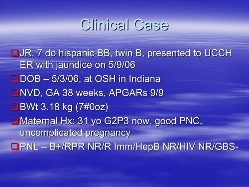
Clinical Case<br />
JR, JR, 7 do hispanic BB, twin B, presented to UCCH<br />
ER with jaundice on 5/9/06<br />
DOB DOB – 5/3/06, at OSH in Indiana<br />
NVD, NVD, GA 38 weeks, APGARs 9/9<br />
BWt BWt 3.18 kg (7#0oz)<br />
Maternal Maternal Hx: Hx:<br />
31 yo G2P3 now, good PNC,<br />
uncomplicated pregnancy<br />
PNL PNL – B+/RPR NR/R Imm/HepB NR/HIV NR/GBS-<br />
NR/GBS
Perinatal course<br />
Initial Initial PE unremarkable; + some facial bruising<br />
noted<br />
Wt Wt 2.9 kg<br />
Feeding Feeding BM/E20 – well per GCN records<br />
Voiding Voiding and stooling well<br />
Discharged Discharged at 48 hrs<br />
D/C D/C Wt – 2.03 kg<br />
Mom Mom told that baby had “borderline borderline” jaundice<br />
To call Pediatrician if she sees baby gets yellow
Baby Baby was OK at home, eating well – 10-15 10 15<br />
min each breast<br />
Stooling Stooling well – per mother<br />
On On DOL 5 mother thought babies look<br />
yellow and called the PMD<br />
She She was told to go to the hospital next day<br />
for Bili check
Baby Baby B+/Coombs-<br />
B+/Coombs<br />
Mom Mom B+<br />
Labs
Work Up<br />
CBC CBC – WBC 13.6, H/H 18.9/54, Plt 301<br />
Diff Diff N26, Bn4<br />
Retic Retic count 0.7%
Bilirubin<br />
5/5/06 5/5/06 - @48 hrs (OSH) – 13.0/0.4<br />
5/9/06 5/9/06 – DOL 7 – (OSH) – 30.9/0.6<br />
5/9/04 5/9/04 – UCCH – 31.5/0.8<br />
Sibling Sibling’s s Bili was 21 – admitted to Peds<br />
Floor for phothotherapy
ER course<br />
VSS, VSS, baby was jaundiced and “sleepy sleepy”<br />
Placed Placed under phothotherapy<br />
Given Given IVF<br />
Admitted Admitted to PICU for exchange transfusion
Here comes the NICU<br />
0230 0230 – NICU fellow received a call from<br />
upstairs to help with the exchange transfusion<br />
By By 0530 baby was in the PICU<br />
Blood Blood ordered<br />
UVC UVC placed<br />
Double Double volume exchange transfusion was<br />
done<br />
Rpt Rpt Bili – 23.8/0.7 half way through the<br />
exchange
<strong>Neonatal</strong> <strong>Hyperbilirubinemia</strong>
WHAT IS HYPERBILIRUBINEMIA AND WHY<br />
DO WE WORRY ABOUT IT?
Jaundice<br />
A A visible manifestation in the skin and sclera<br />
of elevated bilirubin concentrations<br />
Adults Adults are usually jaundiced when bilirubin<br />
levels exceed 2 mg/dL<br />
Neonates Neonates appear jaundiced when serum total<br />
bilirubin (STB) levels reach 5-7 5 7 mg/dL
Some Some degree of jaundice develops in 60-70% 60 70% of<br />
all neonates born on the United States<br />
More than 2.7 million neonates born each year in the<br />
United States will develop jaundice<br />
Chemical Chemical hyperbilirubinemia, a STB > 2.0 mg/dL,<br />
is virtually universal<br />
Although Although most jaundice is benign, there is a<br />
potential for neurological devastation and death<br />
and consequently all newborns must be assessed
WHY DO INFANTS DEVELOP<br />
HYPERBILIRUBINEMIA?
Increased Increased Bilirubin Production<br />
Decreased Decreased Binding and Transport Capacity<br />
Limited Limited Conjugation and Excretion Capacity<br />
Increased Increased Enterohepatic Circulation of<br />
Bilirubin
Bilirubin Bilirubin is the breakdown product of<br />
hemoglobin<br />
Lysis Lysis of red cells releases heme from<br />
hemoglobin<br />
Heme Heme is then converted to bilirubin and<br />
excreted
Bilirubin Synthesis<br />
There There is increased production of bilirubin in the<br />
newborn because of:<br />
Increased rate of degradation<br />
A shortened circulating erythrocyte life span (70-90 (70 90 days<br />
versus 120 days) of an increased mass<br />
A very large pool of hematopoietic tissue that ceases to<br />
function shortly after birth resulting in heme degradation<br />
An increased turnover of cytochromes (nonhemoglobin<br />
heme proteins)<br />
An increase in enterohepatic circulation
Binding and Transport<br />
<strong>Unconjugated</strong> <strong>Unconjugated</strong> bilirubin is quickly bound to<br />
albumin in the serum<br />
Newborns Newborns have reduced albumin<br />
concentrations and consequently a lower<br />
plasma binding capacity for bilirubin<br />
There There is consequently more free bilirubin in<br />
the serum<br />
It It is the free bilirubin that is believed to<br />
cause neurological damage in newborns
Conjugation and Excretion<br />
During During fetal life, removal of bilirubin is<br />
accomplished by the placenta<br />
In In the newborn, bilirubin excretion requires<br />
conversion of the nonpolar unconjugated<br />
bilirubin into a more polar water-soluble<br />
water soluble<br />
substance, conjugated bilirubin
Blood Blood flow through the hepatic artery develops<br />
during the first week of life<br />
The The ductus venosus allows blood to bypass the<br />
liver completely<br />
Conjugation Conjugation depends on the maturity of the liver<br />
cell
UDPGT<br />
UDPGT UDPGT in the newborn liver must be<br />
induced<br />
UDPGT UDPGT activity is extremely low in infants<br />
born at less than 30 weeks, 0.1% of adult<br />
levels<br />
This This activity increases to only 1% at term<br />
The The activity reaches adult levels by 6-12 6 12<br />
weeks of age
Conjugation<br />
Bilirubin Bilirubin dissociates from circulating albumin<br />
before its entry into the liver cell<br />
Bilirubin Bilirubin enters the liver by a process of carrier-<br />
mediated diffusion<br />
It It is carried by hepatic ligandin (Y protein) and Z<br />
protein<br />
Bilirubin Bilirubin is presumed to be transported from the<br />
liver cell membrane to the endoplasmic reticulum,<br />
the site of the conjugating enzyme uridine<br />
diphosphate glucuronyl transferase (UDPGT)<br />
After After conjugation, bilirubin is then excreted into<br />
bile in the intestine
Increased Enterohepatic Circulation<br />
Conjugated Conjugated bilirubin is unstable and can be<br />
easily hydrolyzed back to unconjugated bilirubin<br />
and reabsorbed through the intestinal mucosa<br />
High High mucosal beta-glucuronidase beta glucuronidase activity leads<br />
to increased hydrolysis<br />
An An alkaline environment also facilitates<br />
hydrolysis<br />
In In the newborn, the relative lack of intestinal<br />
bacterial flora to reduce bilirubin to urobilinogen<br />
further increases the bilirubin pool
<strong>Neonatal</strong> <strong>Hyperbilirubinemia</strong><br />
Physiologic Physiologic Jaundice<br />
A A progressive rise in unconjugated bilirubin to a<br />
peak of 5-6 5 6 mg/dL between 60 and 72 hours of<br />
life in white and African-American African American babies and<br />
10-14 10 14 mg/dL between 72-120 72 120 hours of life in<br />
Asian babies<br />
A A rapid decline in TSBs occurs by the 5 th or 7- 7<br />
10 th day respectively
Pathologic <strong>Unconjugated</strong><br />
<strong>Hyperbilirubinemia</strong><br />
Pathologic hyperbilirubinemia is defined as a<br />
prolonged or exaggerated<br />
hyperbilirubinemia<br />
Occurs because of disorders of:<br />
Production Production<br />
Hepatic Hepatic Uptake<br />
Conjugation<br />
Conjugation<br />
Enterohepatic Enterohepatic Circulation
Disorders of Production<br />
Isoimmunization<br />
Isoimmunization<br />
Erythrocyte Erythrocyte Enzymatic Defects<br />
Erythrocyte Erythrocyte Structural Defects<br />
Infection Infection<br />
Sequestration<br />
Sequestration<br />
Polycythemia<br />
Polycythemia
Isoimmunization<br />
Rh Rh Incompatibility<br />
ABO ABO Incompatibility<br />
Other Other Blood Group Incompatibilities
Rh Incompatibility<br />
This is a blood group incompatibility between the mother<br />
and newborn that can cause severe hemolytic anemia in<br />
the fetus and newborn<br />
The Rh antibody is produced by a Rh negative mother after<br />
being exposed to a Rh antigen from fetal blood<br />
The initial response is to make IgM antibodies<br />
Later IgG are produced which cross the placenta and bind<br />
to fetal red blood cells which are consequently destroyed<br />
Infants do not appear jaundiced at birth, but severe anemia<br />
can lead to hydrops and death<br />
After birth, infants may develop hyperbilirubinemia rapidly
The The D antigen may produce sensitization<br />
with a fetomaternal hemorrhage as small as<br />
0.1 mL<br />
At At one time this was the most common<br />
cause of kernicterus; but with the use of<br />
RhoGAM (anti-D (anti D immunoglobulin G) and<br />
careful fetal monitoring, the incidence and<br />
severity have decreased
ABO Incompatibility<br />
This This is a hemolytic disease caused by a<br />
reaction of maternal anti-A anti A or anti-B anti B<br />
antibodies with fetal A or B antigens<br />
Usually Usually milder than Rh<br />
Almost Almost exclusively in type O mothers<br />
Jaundice Jaundice appears at 24-72 24 72 hours<br />
Half Half of infants with a positive Coombs show<br />
hemolysis and some with a negative Coombs<br />
have hemolysis
Minor Blood Groups<br />
Kell, Kell, Kidd, Duffy, Lutheran<br />
< < 2 % of hemolysis from isoimmunization
Erythrocyte Enzymatic Defects<br />
Glucose Glucose-6-Phosphate Phosphate Dehydrogenase<br />
Deficiency<br />
Pyruvate Pyruvate Kinase Deficiency<br />
These These defects may have profound effects on<br />
erythrocyte function and life span
Glucose-6-Phosphate<br />
Glucose Phosphate<br />
Dehydrogenase Deficiency<br />
Glucose Glucose-6-phosphate phosphate dehydrogenase deficiency<br />
(G6PD) is a common disease, especially in people<br />
of Mediterranean; African; and Asian decent<br />
G6PD G6PD deficiency occurs in 11-13% 11 13% of African<br />
Americans<br />
Estimated Estimated 200-400 200 400 million people carry the gene<br />
X-linked linked<br />
Presentation Presentation is heterogeneous<br />
Hemolysis Hemolysis occurs, but can be absent<br />
<strong>Hyperbilirubinemia</strong> <strong>Hyperbilirubinemia</strong> occurs between 24 and 72<br />
hours of life
RBCs RBCs are unable to activate the pentose<br />
phosphate metabolic pathway<br />
And And consequently cannot defend against<br />
oxidative stress<br />
Sepsis and Vitamin K analogues<br />
Severity Severity of disease depends on type and<br />
amount of stress
Pyruvate Kinase Deficiency<br />
This This is the second most common cause of<br />
enzymatic-related enzymatic related hemolytic anemia<br />
Autosomal Autosomal recessive<br />
It It is common in people of Northern<br />
European decent<br />
It It is an enzyme required for production of<br />
ATP in RBCs
Its Its deficiency leads to decreased RBC life<br />
span and hemolysis
Erythrocyte Structural Defects<br />
Hereditary Hereditary Spherocytosis<br />
Hereditary Hereditary Elliptocytosis<br />
These These defects alter RBC structure and cause<br />
sequestration
Hereditary Elliptocytosis<br />
Incidence Incidence of 1:4000<br />
Autosomal Autosomal dominant<br />
Abnormality Abnormality in spectrin or glycophorin C<br />
Hemolysis Hemolysis and hyperbilirubinemia are<br />
unusual in the newborn period
Hereditary Spherocytosis<br />
Incidence Incidence of 1:5000<br />
Autosomal Autosomal dominant<br />
Heterogeneous presentation<br />
Fifty Fifty percent present with hemolytic anemia,<br />
hyperbilirubinemia, reticulocytosis, and<br />
increased erythrocyte osmotic fragility
Infection<br />
<strong>Hyperbilirubinemia</strong> <strong>Hyperbilirubinemia</strong> is believed to be<br />
secondary to hemolysis<br />
Sepsis Sepsis may impair conjugation also leading<br />
to increased bilirubin levels
Sequestration<br />
Sequestration Sequestration of blood in body cavities may<br />
lead to hyperbilirubinemia as the body<br />
metabolizes hemoglobin<br />
Cephalohematomas, Cephalohematomas, subdural hematomas,<br />
subgaleal hematomas<br />
Excessive Excessive bruising
Polycythemia<br />
The The increase in red blood cell mass has the<br />
potential to overload the newborn<br />
hemoglobin metabolism capacities
Disorders of Hepatic Uptake<br />
Gilberts Gilberts Syndrome<br />
This This is a benign disorder producing persistent<br />
unconjugated hyperbilirubinemia<br />
There is defective hepatic uptake and decreased<br />
UDPGT activity<br />
It usually occurs in the second decade of life, but can<br />
present in neonates
Disorders of Conjugation<br />
Crigler Crigler-Najjar Najjar Syndrome<br />
Transient Transient Familial <strong>Neonatal</strong><br />
<strong>Hyperbilirubinemia</strong> (Lucey-Driscoll (Lucey Driscoll Syndrome)<br />
Pyloric Pyloric Stenosis<br />
Hypothyroidism<br />
Hypothyroidism
Type Type I<br />
Crigler-Najjar Crigler Najjar Syndrome<br />
There is absence of UDPGT activity<br />
Autosomal recessive<br />
1:1,000,000<br />
Severe unconjugated hyperbilirubinemia develops and<br />
persists beyond the first week of life<br />
No hemolysis<br />
Lifelong risk of kernicterus<br />
Lifelong phototherapy is needed
Type Type II<br />
There There is various degree of decrease of UDPGT<br />
activity<br />
Typically Typically benign<br />
There There is unconjugated hyperbilirubinemia in the<br />
first few days of life that does not exceed 20<br />
mg/dL<br />
<strong>Hyperbilirubinemia</strong> <strong>Hyperbilirubinemia</strong> persists into adulthood<br />
The The treatment is phenobarbital
Transient Familial <strong>Neonatal</strong><br />
<strong>Hyperbilirubinemia</strong><br />
Neonates Neonates develop severe nonhemolytic<br />
hyperbilirubinemia<br />
Their Their serum contains high concentrations of<br />
glucuronyl transferase inhibitors<br />
This This inhibitor decreases by about 14 days of<br />
life and consequently hyperbilirubinemia<br />
resolves
Pyloric Stenosis<br />
10 10-25% 25% of babies with pyloric stenosis have<br />
hyperbilirubinemia at the time of<br />
presentation<br />
Hepatic Hepatic glucuronyl tranferase activity is<br />
reduced<br />
Surgical Surgical correction improves bilirubin levels
Hypothyroidism<br />
UDPGT UDPGT activity is deficient and remains low<br />
for weeks with hypothyroidism
Disorders of Enterohepatic<br />
Circulation<br />
Breast Breast Feeding Jaundice<br />
Breast Breast Milk Jaundice
Breast Feeding Jaundice<br />
<strong>Unconjugated</strong> <strong>Unconjugated</strong> hyperbilirubinemia is secondary to a<br />
suboptimal establishment of breastfeeding<br />
Newborns Newborns are under-hydrated under hydrated and in a state of<br />
starvation.<br />
They They also have delayed passage of meconium<br />
Enterohepatic Enterohepatic reuptake of bilirubin is consequently<br />
increased, leading to hyperbilirubinemia<br />
Treatment Treatment and prevention include frequent<br />
feedings (8-12/day) (8 12/day)
Breast Milk Jaundice<br />
Occurs Occurs after 3-5 3 5 days of life, typically at 2-3 2 3<br />
weeks of life<br />
The The etiology is unknown, but believed to be<br />
a factor in breast milk or an altered<br />
chemistry in breast milk that enhances<br />
intestinal reabsorption of bilirubin<br />
No No need to stop breastfeeding unless<br />
bilirubin levels are dangerously high
WHY DO WE WORRY ABOUT<br />
HYPERBILIRUBINEMIA?
Sequelae<br />
Bilirubin Bilirubin may penetrate the brain cell and<br />
cause neuronal dysfunction or death if not<br />
carefully managed<br />
Bilirubin Bilirubin causes staining and necrosis of<br />
neurons in the basal ganglia, hippocampal<br />
cortex, subthalamic nuclei, and cerebellum<br />
which is followed by gliosis<br />
50%of 50%of patients with kernicterus die
Acute Bilirubin Encephalopathy<br />
Phase Phase 1 - poor suck, hypotonia, and<br />
depressed sensorium<br />
Phase Phase 2 - fever and hypertonia or<br />
opisthotonos<br />
Phase Phase 3 - less hypertonia, high pitched cry,<br />
hearing and visual abnormalities, poor<br />
feeding, athetosis
Long Long term sequelae:<br />
Kernicterus<br />
Chorioathetoid Chorioathetoid cerebral palsy<br />
Sensorineural Sensorineural hearing loss<br />
Upward Upward gaze palsy<br />
Dental Dental-enamel enamel dysplasia<br />
Mental Mental retardation
SO, WHAT CAN WE DO?
Diagnosis and Management<br />
There There has been evidence that neonatal<br />
jaundice can be treated less aggressively,<br />
but there is not a consensus yet and until<br />
then it should be managed conservatively
Diagnosis<br />
All All neonates are entitled to a thorough<br />
physical examination and evaluation to<br />
determine which neonates are at an<br />
increased risk for becoming abnormally<br />
jaundiced and developing sequelae
Risk Assessment<br />
Every Every newborn should be assessed,<br />
especially if discharged before 72 hours of<br />
life<br />
2 2 options:<br />
TSB or TcB before discharge and plot results on the<br />
nomogram
Nomogram for designation of risk in 2840 well newborns at 36 or more weeks'<br />
gestational age with birth weight of 2000 g or more or 35 or more weeks' gestational<br />
age and birth weight of 2500 g or more based on the hour-specific serum bilirubin<br />
values<br />
Subcommittee on <strong>Hyperbilirubinemia</strong>, Pediatrics 2004;114:297-316<br />
Copyright ©2004 American Academy of Pediatrics
Assessment of risk<br />
– Major<br />
Predischarge TSB or TcB in the high-risk high risk zone<br />
Jaundice in the first 24h<br />
Hemolytic disease<br />
Gestational age 35-36 35 36 weeks<br />
Sibling received phototherapy<br />
Cephalohematoma or bruising<br />
Poor breastfeeding<br />
East Asian descent
Carbon Monoxide<br />
End End Tidal Carbon Monoxide detection<br />
allows rapid noninvasive detection of infants<br />
at risk for hemolytic disease and<br />
consequently at high risk for neurological<br />
sequelae
Carbon Monoxide<br />
The The breakdown of hemoglobin by heme<br />
oxygenase produces free iron and carbon<br />
monoxide in equimolar amounts<br />
The The carbon monoxide formed by heme<br />
degradation is excreted unchanged by the lungs.<br />
Although Although there are other endogenous and<br />
exogenous sources of CO, quantitative estimation<br />
of its excretion or synthesis offers a reasonably<br />
accurate assessment of bilirubin synthesis
Physical Examination<br />
Detection Detection of clinical jaundice requires digital<br />
pressure and the proper lighting, preferably<br />
daylight<br />
If If clinical jaundice is detected, a total and<br />
direct serum bilirubin or transcutaneous<br />
bilirubin (TcB) should be measured and<br />
plotted on the nomogram
If: If:<br />
When should I do more?<br />
Cord Cord bilirubin is greater than 4 mg/dL<br />
A A rate of rise greater than or equal to 0.5<br />
mg/dL/hour over a 4-8 4 8 hour period<br />
An An increase of 5 mg/dL per day<br />
13 13-15 15 mg/dL in a term infant<br />
10 10 mg/dL in a preterm infant<br />
If If jaundice persist greater than 10 days
Then what?<br />
Determination Determination of maternal blood group and Rh<br />
type<br />
Screen Screen for antibodies directed against minor<br />
erythrocyte antigens<br />
Determination Determination of newborns blood type and Rh<br />
type<br />
Direct Direct Coombs test<br />
Hemoglobin Hemoglobin and Hematocrit<br />
Peripheral Peripheral blood smear<br />
Reticulocyte Reticulocyte count<br />
G6PD G6PD level
Algorithm for the management of jaundice in the newborn nursery<br />
Subcommittee on <strong>Hyperbilirubinemia</strong>, Pediatrics 2004;114:297-316<br />
Copyright ©2004 American Academy of Pediatrics
Guidelines for phototherapy in hospitalized infants of 35 or more weeks' gestation<br />
Subcommittee on <strong>Hyperbilirubinemia</strong>, Pediatrics 2004;114:297-316<br />
opyright ©2004 American Academy of Pediatrics
Helpful resources<br />
Bili-Aid Bili Aid
Phototherapy<br />
The The main mechanism of action<br />
Geometric Geometric photoisomerization of unconjugated<br />
bilirubin that can then be excreted without<br />
conjugation
Wavelength<br />
Wavelength<br />
Technique<br />
Bilirubin Bilirubin absorbs light maximally in the blue<br />
range (420-500 (420 500 nm), with a peak at 460 nm for<br />
albumin-bound albumin bound bilirubin and 440 nm for free<br />
bilirubin<br />
Special blue lamps have a spectrum between 420- 420<br />
480
Irradiance Irradiance<br />
The The energy output measured in microwatts per<br />
square centimeter per nanometer<br />
Optimal Optimal level is 11 microwatts per square<br />
centimeter per nanometer<br />
Intensive Intensive phototherapy is 30 microwatts per<br />
square centimeter per nanometer
Positioning Positioning<br />
Within Within 10 cm of the patient for fluorescent tubes<br />
Surface Surface area<br />
The The greater the surface area exposed, the more<br />
effective the phototherapy
Hydration<br />
There There is no evidence that excessive fluid<br />
administration affects the serum bilirubin<br />
concentration<br />
If If admitted with dehydration, babies will<br />
need to be rehydrated and then fed<br />
Feeding Feeding inhibits enterohepatic circulation of<br />
bilirubin<br />
Important Important to watch fluid status for excretion<br />
of bilirubin
The The TSB level for discontinuing<br />
phototherapy depends on the age at which<br />
phototherapy was started and the etiology<br />
For For infants readmitted after birth admission, you<br />
can discontinue usually at 13-14 13 14 mg/dL with a<br />
follow up visit 24 hours after discharge<br />
There There is no need for a rebound bilirubin, unless<br />
there is hemolytic disease
Pharmacological Therapy<br />
Phenobarbital<br />
Phenobarbital<br />
Albumin Albumin<br />
Tin Tin-mesoporphyrin<br />
mesoporphyrin<br />
Inhibits Inhibits heme oxygenase<br />
Intravenous Intravenous gamma-globulin<br />
gamma globulin<br />
Shown Shown to reduce the need for exchange<br />
transfusions in isoimmune hemolytic disease
Guidelines for exchange transfusion in infants 35 or more weeks' gestation<br />
Subcommittee on <strong>Hyperbilirubinemia</strong>, Pediatrics 2004;114:297-316<br />
Copyright ©2004 American Academy of Pediatrics
Exchange Exchange transfusions are recommended if<br />
TSB is greater than or equal to 25 mg/dL in<br />
a healthy full term infant<br />
If If rate of rise is greater than or equal to 0.5<br />
mg/dL/hour<br />
If If there is active hemolysis or other risk<br />
factors, then an exchange transfusion may<br />
be warranted at a lower bilirubin level
Exchange Transfusion<br />
With With a exchange transfusion, approximately<br />
85% of erythrocytes will be replaced<br />
Serum Serum bilirubin levels should decrease by<br />
50%
American Academy of Pediatrics<br />
In In 1994 the AAP established practice<br />
parameters for the management of<br />
hyperbilirubinemia<br />
Revised Revised in 2004
Management Management of <strong>Hyperbilirubinemia</strong> in the<br />
Newborn Infant 35 or More Weeks of<br />
Gestation<br />
Goals: Goals:<br />
Promote and support successful breastfeeding<br />
Establish nursery protocols for the identification and<br />
evaluation of hyperbilirubinemia<br />
Measure the total serum bilirubin or transcutaneous<br />
bilirubin levels on infants jaundiced in the first24<br />
hours
Recognize that visual estimation of the degree of<br />
jaundice can lead to errors, particularly in darkly<br />
pigmented infants<br />
Interpret all bilirubin levels according to the infant’s infant s<br />
age in hours<br />
Recognize that infants less than 38 weeks’ weeks gestation,<br />
particularly those who are breast fed, are at higher risk<br />
of developing hyperbilirubinemai and require closer<br />
surveillance and monitoring<br />
Perform a systematic assessment on all infants before<br />
discharge for the risk of severe hyperbilirubinemia
Provide parents with written and verbal information<br />
about newborn jaundice<br />
Provide appropriate follow-up follow up based on the time of<br />
discharge and the risk assessment<br />
Treat infants, when indicated, with phototherapy or<br />
exchange transfusion
Infant Infant Discharged<br />
< 24 hours<br />
24 to 47.9 hours<br />
48 to 72 hours<br />
Follow Up<br />
Should Be Seen By Age<br />
72 hours<br />
96 hours<br />
120 hours
Fanaroff, Fanaroff, Martin. <strong>Neonatal</strong>-Perinatal<br />
<strong>Neonatal</strong> Perinatal<br />
Medicine, 7 th Edition.<br />
Management Management of <strong>Hyperbilirubinemia</strong> in the<br />
Newborn Infant 35 or More Weeks of<br />
Gestation, Pediatrics 2004;114;297-316<br />
2004;114;297 316<br />
Taeusch, Taeusch, Ballard, Gleason. Avery’s Avery s<br />
Diseases of the Newborn, 8 th Edition