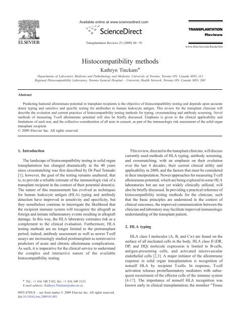
Histocompatibility methods
Histocompatibility methods
Histocompatibility methods
You also want an ePaper? Increase the reach of your titles
YUMPU automatically turns print PDFs into web optimized ePapers that Google loves.
Abstract<br />
<strong>Histocompatibility</strong> <strong>methods</strong><br />
Kathryn Tinckam ⁎<br />
Departments of Laboratory Medicine and Pathobiology and Medicine, University of Toronto, Toronto ON, Canada M5G 1L5<br />
Regional <strong>Histocompatibility</strong> Laboratory, Toronto General Hospital – University Health Network, Toronto ON, Canada M5G 2M1<br />
Predicting humoral alloimmune potential in transplant recipients is the objective of histocompatibility testing and depends upon accurate<br />
donor typing and sensitive and specific testing for antibodies to human leukocyte antigen. This review for the transplant clinician will<br />
describe the evolution and current practices of histocompatibility testing <strong>methods</strong> for typing, crossmatching and antibody screening. Novel<br />
<strong>methods</strong> of measuring T-cell alloimmune potential will also be briefly discussed. Emphasis is given to the clinical applicability and<br />
limitations of each test, and the collective consideration of all tests in concert, as part of the immunologic risk assessment of the solid organ<br />
transplant recipient.<br />
© 2009 Elsevier Inc. All rights reserved.<br />
1. Introduction<br />
The landscape of histocompatibility testing in solid organ<br />
transplantation has changed dramatically in the 40 years<br />
since crossmatching was first described by Dr Paul Terasaki<br />
[1]; however, the goal of the testing remains unaltered, that<br />
is, to provide a reliable estimate of the immunologic risk of a<br />
transplant recipient in the context of their potential donor(s).<br />
The nature of this measurement has evolved as techniques<br />
for human leukocyte antigen (HLA) typing and antibody<br />
detection have improved in sensitivity and specificity, but<br />
they nonetheless continue to interrogate the likelihood that<br />
the recipient immune system will recognize the allograft as<br />
foreign and initiate inflammatory events resulting in allograft<br />
damage. In this way, the HLA laboratory estimates risk as a<br />
complement to the clinical evaluation. Furthermore, HLA<br />
testing <strong>methods</strong> are no longer limited to the pretransplant<br />
period; indeed, antibody assessment as well as newer T-cell<br />
assays are increasingly studied posttransplant as noninvasive<br />
predictors of acute and chronic alloimmune complications.<br />
As such, it is imperative for the clinical service to understand<br />
the complex and interactive nature of the available<br />
histocompatibility testing.<br />
⁎ Tel.: +1 416 340 5142; fax: +1 416 340 3133.<br />
E-mail address: Kathryn.Tinckam@uhn.on.ca.<br />
Available online at www.sciencedirect.com<br />
Transplantation Reviews 23 (2009) 80 –93<br />
0955-470X/$ – see front matter © 2009 Elsevier Inc. All rights reserved.<br />
doi:10.1016/j.trre.2009.01.001<br />
This review, directed to the transplant clinician, will discuss<br />
currently used <strong>methods</strong> of HLA typing, antibody screening,<br />
and crossmatching, with an emphasis on their evolution<br />
over the last 4 decades, their current clinical utility and<br />
applicability in 2008, and the factors that must be considered<br />
in their interpretation. Newer approaches for measuring T-cell<br />
alloimmune potential, which are being explored in some HLA<br />
laboratories but are not yet widely clinically utilized, will<br />
also be briefly discussed. In providing a practical reference of<br />
histocompatibility testing <strong>methods</strong> for the clinician, such<br />
that the basic principles are understood in the context of<br />
clinical outcomes, the improved communication between the<br />
clinician and laboratory may facilitate improved immunologic<br />
understanding of the transplant patient.<br />
2. HLA typing<br />
www.elsevier.com/locate/trre<br />
HLA class I molecules (A, B, and Cw) are found on the<br />
surface of all nucleated cells in the body. HLA class II (DR,<br />
DP, and DQ) molecule expression is limited to B-cells,<br />
antigen-presenting cells, and activated microvascular<br />
endothelial cells [2,3]. A major initiator of the alloimmune<br />
response in solid organ transplantation is recognition of<br />
nonself HLA by recipient T-cells. In response, T-cell<br />
activation releases proinflammatory mediators with subsequent<br />
recruitment of the effector cells of the immune system<br />
[4-17]. The importance of nonself HLA recognition was<br />
known early in clinical transplantation; the moniker “Tissue
Typing Lab” was used for many years for the HLA<br />
laboratory because much of the early quantifiable immune<br />
risk assessment was limited to documenting the number of<br />
HLA mismatches between a given transplant pair. Currently,<br />
both serologic and molecular typing <strong>methods</strong> are routinely<br />
used in a most HLA laboratories.<br />
2.1. Serologic typing<br />
Serologic typing, as the name suggests, requires sera<br />
containing well-characterized antibodies to a wide range of<br />
HLA specificities. Although historically laboratories often<br />
kept large banks of sera from which to make typing reagents,<br />
it is far more common now to use commercially prepared<br />
trays which contain sera to all common, and many of the rare<br />
HLA alleles. Patient lymphocytes are mixed with the various<br />
sera in the tray wells and incubated with complement and a<br />
vital dye. If the cell has antigens on its surface to which<br />
antibody in a particular well are able to bind, then<br />
complement is activated in those well(s), the membrane<br />
attack complex forms and inserts into the cell membrane, cell<br />
death occurs, and the vital dye (often eosin) is taken up into<br />
the cell [18]. Cell death should therefore occur in any well in<br />
which the cell surface antigen and serum antibody are<br />
matched, and those wells are then identified under phasecontrast<br />
microscopy. Comparing and eliminating the serologic<br />
specificities of the positive wells assigns the HLA type.<br />
For example, if 2 wells with sera known to bind (a) A30 and<br />
(b) A30 + A31 are found to have significant cell death, a<br />
negative result in a well containing serum binding A31 + A33<br />
excludes A31 and therefore assigns the type as A30.<br />
Advantages of serologic typing include obtaining rapid<br />
results, which is of particular importance in deceased donor<br />
typing, and also the ability to discriminate “null” HLA<br />
alleles, which have detectable DNA sequences but no<br />
antigen expression on cell surfaces, and less immunologic<br />
relevance. However, finding high-quality sera with sufficient<br />
specificities to reflect the ever-increasing number of HLA<br />
alleles [19,20] is progressively prohibitive especially in light<br />
of increasing clinical interest in HLA-Cw, DP, and DP<br />
contributions to allograft outcomes. In addition, small<br />
differences in HLA molecules at the level of the amino<br />
acid sequence may not be identified using these <strong>methods</strong><br />
despite potential immunologic consequences [21-26].<br />
2.2. Molecular typing<br />
HLA molecules are proteins encoded by DNA regions on<br />
the short arm of chromosome 6. With their sequences well<br />
described, HLA typing is becoming increasingly performed<br />
using molecular <strong>methods</strong> including sequence specific primer<br />
PCR (SSP), sequence specific oligonucleotide probes<br />
(SSOP), and direct DNA sequencing. In SSP, DNA is<br />
isolated from the subject to be typed and amplified in<br />
multiple wells, each containing specific primers complementary<br />
to particular HLA alleles. Only if the DNA is<br />
complementary to the alleles in a given well will an<br />
K. Tinckam / Transplantation Reviews 23 (2009) 80–93<br />
amplification product be formed. The contents of the wells<br />
are then run electrophoretically through an agarose gel: if<br />
amplified product is present, then a band will be detected,<br />
and the HLA typing is assigned by the primers that were<br />
placed in that particular well. The composition of the primers<br />
in each well permits positive identification of the HLA<br />
characteristics. In SSOP, oligonucleotide probes that are<br />
complementary to the unique segments of the DNA of<br />
different alleles are mixed with amplified DNA. Unique<br />
fluorescent tags distinguish those probes that are complimentary<br />
to the DNA, such that the HLA allele is identified.<br />
Sequencing determines the exact order of nucleotides in the<br />
gene of interest, and the HLA type is assigned by comparison<br />
to published HLA allele sequences [20]. Regardless of the<br />
specific method, molecular HLA typing offers more precise<br />
documentation of HLA differences between donor and<br />
recipient, with many alleles discriminated at all loci of<br />
interest, and with resolution at the level of the amino acid.<br />
This may provide a greater understanding of the risk<br />
associated with different mismatched epitopes between<br />
donor and recipient [22-27].<br />
2.3. HLA typing nomenclature and mismatches<br />
The documentation of HLA type varies depending on the<br />
detection method used and is important to review as a direct<br />
consequence of the different <strong>methods</strong> described above.<br />
Historically, because HLA antigens were discovered serologically,<br />
they were numbered in order of that discovery by<br />
gene locus, for example, A1, A2, etc, and B7, B8 etc.<br />
Refinement of serologic <strong>methods</strong> identified even more<br />
antigens, previously thought to represent single allotypes,<br />
which in fact were serologically and genetically unique. For<br />
example, the B40 antigen was ‘split’ into 2 components<br />
named B60 and B61, and both are considered part of the B40<br />
cross reactive group or CREG, which itself is part of the B7<br />
CREG. Public epitopes are those common to all the members<br />
of a CREG, whereas private epitopes delineate the individual<br />
serologically defined antigens.<br />
However, assignment of serological antigens did not<br />
represent the true heterogeneity of the HLA system. Early<br />
studies with mixed lymphocyte culture detected such<br />
heterogeneity in HLA antigens recognition that was not<br />
discerned by serology; for example, HLA A2 was found to<br />
consist of several subtypes stimulating different lymphocyte<br />
reactivity. DNA sequencing confirms that indeed, multiple<br />
alleles of each HLA antigen are known to exist despite the<br />
fact that they may react to a single common typing serum at<br />
the antigen level. A new molecular typing nomenclature was<br />
introduced in 1987, where the locus is followed by an<br />
asterisk, then the first 2 digits describe the type, and then the<br />
next 2 digits represent a unique allele differing by at least 1<br />
amino acid difference.<br />
Frequently, the serological and molecular nomenclature<br />
correspond (eg, HLA-A⁎0201 has the serological equivalent<br />
of HLA-A2), but incongruencies do occur. For the purposes<br />
of solid organ transplantation, it is important for the clinician<br />
81
82 K. Tinckam / Transplantation Reviews 23 (2009) 80–93<br />
to recognize which nomenclature system is used by their<br />
laboratory in the assignment of HLA type as well as in the<br />
assignment of antibody specificities. Failure to recognize that<br />
different nomenclature can be used may miss identifying<br />
donor-specific antibody (DSA). For example, if a donor is<br />
assigned a typing of B⁎1501 and the donor has an antibody<br />
defined to B62, the donor specificity of this antibody may not<br />
be immediately apparent because B62 is a serological<br />
equivalent of B⁎1501. Such concerns may be easily addressed<br />
through communication with the HLA laboratory, and in<br />
addition, several references are readily available [19,20,28].<br />
When the typing of a donor and recipient is considered,<br />
the convention for describing their similarity should reflect<br />
the “immune burden” that a donor presents to a recipient.<br />
Originally, number of HLA matches described recipients and<br />
donors' likeness; however, it is more immunologically<br />
informative to use mismatches to describe their dissimilarity.<br />
For example, if a donor is A1,- B8,39 DR1,3 and the<br />
recipient is A1,24 B39,44 DR1,11, then this is a 0–A,<br />
2–B,1–DR mismatch or 3 HLA mismatch transplant.<br />
However, if the donor and recipient typings were reversed,<br />
then the recipient immune system would see 4 HLA antigens<br />
(1–A, 2–B,1–DR) as nonself.<br />
2.4. Using HLA typing in solid organ transplantation<br />
Before modern era immunosuppression, the impact of<br />
HLA mismatch on transplant was dramatic and clinically<br />
significant [29-32]. With current immunosuppressive regimens,<br />
it is recognized that a most first allografts will have a<br />
substantial number of HLA mismatches and yet have<br />
acceptable graft survival; however, large registries still<br />
show a statistically significant impact of HLA matching on<br />
deceased donor transplants [33-35]. As graft survival rates<br />
improve overall, debate exists as to whether organs should be<br />
allocated according to HLA matching or whether local<br />
allocation to reduce cold ischemia time should prevail.<br />
Results from the Collaborative Transplant Study indicate that<br />
shortening of cold ischemia time does not eliminate the<br />
effect of HLA matching and argue for consideration of HLA<br />
type in deceased donor allocation [34].<br />
In the setting of re-grafts, repeat Class I and/or Class II<br />
antigens with a prior donor may have an independent<br />
deleterious effect on graft survival, underscoring the need for<br />
accurate donor typing such that risk assessment may be<br />
properly estimated [36-39]. Furthermore, even matching of<br />
HLA antigens within a given CREG group may be<br />
associated with better long term allograft survival [40,41].<br />
The development of late antibody mediated outcomes may<br />
require a diagnosis of DSA, which necessitates knowledge of<br />
donor typing.<br />
In addition, for the third of waitlisted patients who have<br />
preformed antibody to HLA antigens (see HLA antibody<br />
screening below), accurate donor typing is paramount in the<br />
identification of lower-risk donors to whom the recipients do<br />
not have alloantibody, as in acceptable mismatch [21] or<br />
paired exchange programs [42-44]. Occasionally, patients<br />
may form antibody to only certain alleles at a given locus, for<br />
example, antibody to B⁎4402 but not B⁎4401. Molecular<br />
typing may be used to ensure only those donors with the<br />
allele of interest are potentially excluded rather than all B44<br />
donors [45-47].<br />
Ongoing work examines whether incompatibilities at<br />
HLA-Cw, DP, DQ and MICA [48,49], MICB and KIR [50]<br />
influence graft outcome. Molecular technologies may be<br />
easily adapted for typing at these loci because the evidence<br />
surrounding their importance is emerging.<br />
3. HLA antibody screening<br />
Transient or persistent sensitization with antibodies to<br />
HLA, or non-HLA, antigens may occur with exposure<br />
during pregnancy, blood transfusion, or prior transplant. Up<br />
to one third of waitlisted patients may have some HLA<br />
antibodies detected when the most sensitive screening<br />
<strong>methods</strong> are used. Preformed antibodies decrease access to<br />
transplantation with higher rates of positive crossmatches<br />
and exclusion of donors with unacceptable antigens on the<br />
basis of antibody screening tests. Indeed, even if the<br />
crossmatch is negative, permitting transplant to proceed in<br />
the short term, low titers of antibody directed to donor HLA<br />
may be associated with high rates of early [51] and late [52]<br />
antibody-mediated outcomes. Therefore, both sensitive and<br />
specific analysis of HLA antibodies is necessary to identify<br />
the risks faced by sensitized recipients and also to explore<br />
novel approaches for successfully transplanting these<br />
patients, such as desensitization [53,54], acceptable mismatching,<br />
and paired exchange [21,42,43]. Repeated<br />
pretransplant antibody screening forms a most solid organ<br />
transplant work in most HLA laboratories.<br />
3.1. Cytotoxic (cell based) antibody screening<br />
Lymphocytes are gathered from “cell donors” (usually<br />
20–40 in number) with variable HLA types including all<br />
common and many rare alleles, and a panel of cells is<br />
formed. These cell donors are not organ donors per se but<br />
rather are meant to be representative the HLA antigen<br />
distribution in the same population from whom deceased<br />
donors may be (randomly) selected. In this way, the<br />
percentage of cell donors to whom a given recipient has<br />
antibodies detected approximates the percentage of potential<br />
organ donors drawn from that same population to whom the<br />
recipient would be expected to have a positive crossmatch.<br />
The test is identical to that of serologic typing except that it is<br />
now the recipient serum that is mixed with cell donor<br />
lymphocytes in individual wells along with complement and<br />
the vital dye. If the serum contains antibodies that bind to the<br />
cell surface with sufficient density, complement will be<br />
activated, the cells killed, and the vital dye with dead cells<br />
may be easily identified (Fig. 1A). If in a panel of 40 cells, 30<br />
of the reaction wells had significant cell death, the panel<br />
reactive antibody (PRA) would be reported as 75%.
An obvious limitation of this method is that the PRA<br />
percentage may numerically change (without a change in<br />
amount or type of antibody) depending on the cell panel that<br />
was used in the screening. Commercially made cell panels<br />
may not necessarily represent the HLA distribution of a<br />
given region where different racial proportions result in<br />
different HLA allele frequencies. Furthermore, substantial<br />
false-positive and false-negative results complicate this test;<br />
the former in the case of non-HLA antibodies and<br />
autoantibodies or nonspecific IgM, and the latter as a result<br />
of its low sensitivity. To activate complement, the antibody<br />
must be of sufficient density to link complement between Fc<br />
receptors, and in the setting of low-titer antibody, this may<br />
not occur and antibody may be missed [55]. Because of<br />
technical challenges, many centers used cytotoxic PRA<br />
screening on T-cells only; indeed, many more B-cell<br />
K. Tinckam / Transplantation Reviews 23 (2009) 80–93<br />
screening tests have false-positive results, which would<br />
limit the amount of reliable class II antibody data. Finally,<br />
detailed specificity analysis is difficult with multiple<br />
antigens per reaction well; so detailed lists of antibody<br />
specificities and unacceptable antigens are frequently not<br />
possible (Table 1).<br />
Cellular or cytotoxic PRA testing may therefore be best<br />
thought of as estimating the risk of a given recipient of having<br />
a positive cytotoxic crossmatch to a potential organ donor<br />
drawn from a comparable population as the cell panel donors.<br />
3.2. Solid phase antibody screening<br />
Clinical syndromes of antibody-mediated damage are<br />
reported in the absence of detectable antibody by cytotoxic<br />
screening <strong>methods</strong> and prompted the development of more<br />
Fig. 1. Schematic representation of cell-based and solid phase (bead-based) antibody screening. A, A panel of cells in individual wells is used: 2 representative<br />
wells are demonstrated. Serum is added to each well in the panel. On the left, the antibody in the serum does not bind to the cells, on the right, DSA are present.<br />
After washing, bound DSA remain and when complement is added, it is activated forming the membrane attack complex and killing the cell with the vital dyeis<br />
taken up. The wells are viewed on a microscope: no antibody (i) leaves live cells, and when DSA are present, the vital dye identifies the dead cells (ii). B, Beads<br />
coated with purified or recombinant HLA antigen are combined with serum. In this case, the antibody in the serum is only specific to the bead on the right. When<br />
fluorescent anti-IgG is added, only the bead with DSA already bound will bind the fluorescent marker. The fluorescence may be measured in comparison to a<br />
negative control (i) or may be multiplexed in a single reaction with all beads' fluorescence ranked to determine those beads that are positive (ii).<br />
83
84 K. Tinckam / Transplantation Reviews 23 (2009) 80–93<br />
Table 1<br />
Differences in antibody screening <strong>methods</strong><br />
Antibody identified Cytotoxic antibody<br />
screening<br />
Solid phase antibody<br />
screening<br />
Class I HLA Ab Yes Yes<br />
Class II HLA Ab If B-cells are used Yes<br />
Non-HLA Ab Yes No<br />
IgM Ab Yes (requires DTT treatment<br />
of serum to exclude)<br />
No<br />
Low-titer Ab No Yes<br />
Specificities of Ab No Yes<br />
(single antigen beads)<br />
Noncomplement<br />
binding Ab<br />
No Yes<br />
Ab indicates antibody.<br />
sensitive assays. The need to precisely distinguish HLA<br />
antibody from non-HLA antibody as well as clearly<br />
discriminate class I and class II antibodies stimulated the<br />
development of currently available solid phase methodologies.<br />
These <strong>methods</strong> use only soluble or recombinant HLA<br />
molecules rather than lymphocyte targets, which present<br />
both HLA and non-HLA molecules. The purified molecules<br />
are adhered to solid phase media, either ELISA [56-58]<br />
platforms or microbeads [59-61], and then bind only HLA<br />
antibody in the added recipient serum. Enzyme conjugated<br />
(ELISA) or fluorescent dye conjugated (microbead) antibodies<br />
to human IgG are added to detect any HLA-bound<br />
IgG antibody in the serum. If antibodies were present and<br />
bound to HLA on the solid phase medium, the optical<br />
density (ELISA) or fluorescence detection (microbead)<br />
reflects this. Microbead may be run on a traditional flow<br />
cytometer (Flow PRA®) or may be multiplexed in a<br />
suspension array on the Luminex® platform allowing for<br />
high throughput detection of multiple analytes in a singlereaction<br />
chamber (Fig. 1B). Both of the microbead-based<br />
assays are up to 10% more sensitive for lower-titer antibody<br />
than the ELISA, which in turn is 10% more sensitive than<br />
antihuman globulin (AHG)-enhanced cytotoxicity–based<br />
assays for the detection of HLA antibody [55]. By virtue<br />
of controlling the antigens placed on the beads, these assays<br />
are specific for HLA antibody only. Furthermore, class I and<br />
class II HLA antibody may be easily distinguished by using<br />
class-specific beads, and by manufacturer design, isotype<br />
detection can be limited to IgG. Finally, precise specificities<br />
may be determined by using beads that each bind only 1<br />
unique HLA antigen (Table 1).<br />
Although the use of these antigen-specific and sensitive<br />
assays has addressed many of the problems associated with<br />
the cellular assays, they have their own limitations including<br />
detection of both noncomplement and complement binding<br />
antibody simultaneously, and detection of antibody below<br />
the level associated with a positive crossmatch. Therefore,<br />
detectable antibody may not always be associated with a<br />
meaningful clinical outcome, potentially limiting transplant<br />
with negligible net benefit. In addition, these assays must not<br />
be considered in isolation as the clinical relevance of non-<br />
HLA antibodies is explored. The number of HLA alleles<br />
identified continues to grow, and the full spectrum of unique<br />
HLA antigens cannot be practically represented on solid<br />
phase assays. Therefore, donor typing must always be<br />
reviewed to ensure that all donor alleles are indeed<br />
represented on the solid phase panel before the absence of<br />
DSA may be concluded.<br />
Fluorescence or optical density readouts are continuous<br />
variables, and considerable controversy exists as to what<br />
threshold values should be considered positive, resulting in<br />
sometimes substantial interlaboratory variability [62].<br />
Approaches including standardized quantification of fluorescence<br />
intensity against molecules of equivalent soluble<br />
fluorochrome may allow for better congruency of antibody<br />
screening result with better crossmatch prediction [63] and a<br />
closer association to clinical outcomes [64]. However,<br />
although ongoing efforts are made to improve standardization,<br />
it remains important for clinical programs to know what<br />
thresholds their own laboratory is using and how such<br />
cutoffs correspond to crossmatch results.<br />
3.3. Clinical application of antibody screening assays<br />
Regardless of method, PRA alone has been repeatedly<br />
associated with poor transplant outcomes [65,66]. Debate<br />
has existed as to what threshold of PRA percentage should<br />
be considered “high risk,” but now that specificities may be<br />
more precisely defined, it is clear that it is the specificity and<br />
not the percent PRA per se, that defines the clinical risk.<br />
With PRA output demonstrable as a continuous variable, the<br />
dichotomous approach of high versus low risk is clearly not<br />
biologically reflective. A PRA of 5% may confer significant<br />
risk if the antibody it represents binds to donor antigens.<br />
Even low-titer antibody may become high-titer antibody if<br />
stimulated by the appropriate antigen from a donor organ and<br />
may explain why pretransplant low-titer antibody to donor is<br />
indeed associated with subsequent posttransplant adverse<br />
outcomes [67]. Conversely, by defining specificities precisely,<br />
high PRA patients can now expect comparable longterm<br />
outcomes as nonsensitized patients provided unsuitable<br />
donors are avoided [21,68], thereby challenging the concerns<br />
that organs transplanted into sensitized individuals do not<br />
achieve their maximum potential survival. Therefore, the<br />
detection of any HLA antibody must initiate the interrogation<br />
of specificities, for it is those specificities in conjunction<br />
with donor typing, rather than a particular PRA percent that<br />
confers the risk assessment.<br />
The PRA percentage does remain relevant, but it may<br />
instead be seen as an estimate of the fraction of potential<br />
donors that may confer posttransplant humoral immunologic<br />
risk, and therefore, it represents the ”risk” of donor specificity<br />
occurring but not necessarily the risk of an immunologic<br />
event. Calculated PRA is a more standardized approach to<br />
determining the likelihood that a recipient will have DSA by<br />
comparing the antibody specificities (determined on solid<br />
phase assay locally) to the defined frequencies of HLA alleles<br />
in the population of interest nationally.
Occasionally, because of the high sensitivity of the solid<br />
phase assays, antibody to donor may still be detected even<br />
with a negative crossmatch. Data on the significance of these<br />
findings are conflicting; some demonstrate no impact on<br />
function [69], whereas more are reporting an impact on<br />
short-term [51,70] and long-term [52] outcomes. However,<br />
an equally significant application of solid phase assays has<br />
been in the determination of which positive crossmatches are<br />
in fact immunologically relevant (see Section 4.3 below).<br />
Regardless of assay type, all antibody-screening tests are<br />
limited by their surrogate status for current or potential<br />
memory responses. The laboratory's ability to predict a<br />
future immunologic event is based only upon the serum<br />
available after patient identification and referral. It is<br />
immediately seen that a recipient's measurable immune<br />
history before the time of transplant referral may be<br />
substantial, but with current laboratory techniques, not<br />
demonstrable where serum is unavailable. Antibodies may<br />
wane over time such that routine pretransplant testing reveals<br />
no antibody, and yet shortly after a repeat stimulus with a<br />
transplant, a memory response may still occur. As such,<br />
negative antibody screening alone will never be 100%<br />
predictive of the absence of humoral alloimmune memory<br />
potential, and the clinical history of sensitizing events<br />
remains a critical consideration in the risk assessment of the<br />
patient without detectable antibodies.<br />
4. Crossmatching<br />
The 1969 landmark article by Patel and Terasaki [1]<br />
unequivocally demonstrated that DSA in the serum of<br />
recipients at the time of transplant was a major risk factor for<br />
hyperacute rejection and primary nonfunction. This finding<br />
was a major driver in the founding of HLA laboratories and<br />
in the establishment of the T-cell cytotoxic crossmatch as the<br />
requisite immunologic test before transplant [71] with a<br />
dramatic subsequent reduction in hyperacute rejection. A<br />
negative test justified proceeding, but a positive test was<br />
considered a contraindication. The original study [1] had a<br />
4% false-negative rate (8/195 negative results had early<br />
failure) and a 20% false-positive rate (6/30) demonstrating it<br />
was neither specific nor sensitive enough to define all<br />
relevant antibodies. In general, the crossmatch identifies<br />
Table 2<br />
Differences between crossmatch <strong>methods</strong><br />
whether a recipient has antibodies to a single donor of<br />
interest, as compared to the PRA, which identifies antibodies<br />
to a pool of potential donors. Over time, assays have been<br />
developed to address these limitations [72-84] (Table 2), and<br />
the improved sensitivity has lead to a critical examination of<br />
which antibodies identified by more sophisticated techniques<br />
are predictive of significant clinical outcomes. The solid<br />
phase antibody screening data must be used in conjunction<br />
with crossmatch results to help classify them as immunologically<br />
irrelevant or relevant (high risk of rejection or graft<br />
loss, or transplant contraindicated) [46] (Table 3).<br />
4.1. Complement-Dependent Cytotoxicity (CDC)<br />
crossmatch <strong>methods</strong><br />
The crossmatch is conceptually a simple reaction between<br />
T lymphocytes and donor serum in the presence of<br />
complement and a vital dye. The T-cell expresses class I<br />
HLA as well as non-HLA antigens and therefore acts as an in<br />
vitro “surrogate” allograft, with the actual allograft expected<br />
to express the same cell surface proteins on its endothelium.<br />
As for cytotoxic antibody screening, the result is considered<br />
positive if a significant proportion of the T lymphocytes<br />
are killed after the addition of complement, inferring that<br />
substantial DSA had been bound to the cell surface (Fig. 2A).<br />
However, similar concerns of low-titer but relevant antibody<br />
going undetected lead to improvements with this technique of<br />
increasing sensitivity, including longer incubation times,<br />
additional wash steps [72], and most commonly, the AHGenhanced<br />
method [74,76,77,79]. Antihuman globulin, a<br />
complement fixing antibody to human immunoglobulins, is<br />
added as a second step and binds any DSA already on the<br />
lymphocyte (both complement binding as well as noncomplement<br />
binding DSA), thereby increasing the antibody<br />
density and improving sensitivity (Fig. 2B). Moreover, the<br />
lower-titer antibodies detected by this method were associated<br />
with 36% 1 year allograft loss compared with 18% loss<br />
in those with a negative test [78]. All these <strong>methods</strong> may also<br />
be applied to B-cells, which may identify classes I and II as<br />
well as non-HLA DSA.<br />
4.2. Flow Cytometry Crossmatch <strong>methods</strong><br />
First described in 1983 [81], flow cytometry crossmatch<br />
(FCXM) detects DSA regardless of the ability for<br />
CDC AHG-CDC Flow<br />
cytometry<br />
Cytotoxicity with<br />
flow cytometry<br />
HLA Ab detected Yes Yes Yes Yes Yes<br />
Non-HLA Ab detected Yes Yes Yes Yes No<br />
IgM detected Yes Yes No No No<br />
Low titer or noncomplement binding Ab detected No Yes but less sensitive<br />
than flow cytometry<br />
Yes Yes Yes<br />
Ab titer detected Moderate to high Low to moderate Low Low Low<br />
Ab indicates antibody.<br />
K. Tinckam / Transplantation Reviews 23 (2009) 80–93<br />
85<br />
Solid<br />
phase
86 K. Tinckam / Transplantation Reviews 23 (2009) 80–93<br />
Fig. 2. Schematic representation of the various types of crossmatches. A, In the CDC crossmatch, when DSA is bound to the cell in sufficient density, complement<br />
is activated, the cell is killed and the vital dye is taken up identifying the dead cells. B, With the AHG modification, antibody may be lower titer and less dense on<br />
the cell surface, but the additional AHG still allows complement to be activated with subsequent cell death as with CDC alone. C, In FCXM, DSA binds the cell but<br />
instead of complement, a second fluorescent antibody to human IgG is added. When run through a flow cytometer, the DSA (which may be complement or<br />
noncomplement binding) may be detected at very low titers, measured as fluorescence on the cells relative to a negative control. D, If complement is added to a<br />
FCXM with a fluorescent vital dye taken up by complement-killed cells, then the degree of complement activating antibody binding (upper right on the scatterplot)<br />
may be distinguished from antibody binding without cell death (lower right). E, Lymphocytes are lysed, and the solubilized donor HLA antigen is bound to the<br />
solid phase. An enzyme-linked secondary antibody that results in a color change detected by a luminometer detects DSA. Only HLA antibodies are detected.<br />
complement fixation. Recipient serum is incubated with<br />
donor lymphocytes, which are then stained with a<br />
fluorochrome-conjugated anti-IgG antibody that remains<br />
bound only if DSA is initially bound to the cell surface.<br />
Additional antibodies with different fluorochromes that<br />
are specific to unique B and T lymphocyte surface<br />
proteins can be added such that when run through a flow<br />
cytometer, the B- and T-cells may be easily distinguished<br />
and individually interrogated for the fluorochrome identifying<br />
any DSA (Fig. 2C). The output of the flow<br />
crossmatch is at least semiquantitative (eg, number of<br />
channel shifts of mean fluorescence above the baseline or<br />
standardized against molecules of equivalent soluble<br />
fluorescence beads), although quantification measures<br />
and thresholds for positivity can vary between individual<br />
laboratories. Nonetheless, it is less subjective than visual<br />
assessment of cell death and more biologically representative<br />
of the continuous nature of antibody amount and<br />
consequent risk than the dichotomous positive/negative<br />
result of cytotoxic crossmatches.<br />
Once again, as for antibody screening, there is considerable<br />
interlaboratory variability in <strong>methods</strong> routinely used for<br />
crossmatching and indeed in concordance of results [85],<br />
necessitating ongoing efforts to develop approaches to<br />
improve reproducibility.<br />
4.3. Crossmatch interpretation<br />
T lymphocytes express class I HLA and non-HLA<br />
antigens, and B lymphocytes express both class I and class<br />
II as well as non-HLA antigens. Therefore, in general, a<br />
positive crossmatch due to HLA class I antibody will be<br />
positive on T- and B-cells and due to HLA class II antibody<br />
will be T-cell negative and B-cell positive. However,<br />
antibody to irrelevant non-HLA targets may confound<br />
these results and must be considered to clarify whether a<br />
crossmatch result is accurately identifying risk (Table 3).
Table 3<br />
Immunologic relevance of positive crossmatches<br />
Positive<br />
crossmatch<br />
Cause Detected by Immunologically<br />
relevant?<br />
T- and B-cell IgG class I HLA<br />
antibody<br />
B-cell Low-titer class I<br />
HLA antibody<br />
B-cell IgG class II<br />
HLA antibody<br />
T and B-cell<br />
or B-cell<br />
IgG class I and<br />
class II HLA<br />
antibody<br />
Solid phase testing<br />
positive for class I<br />
antibody<br />
Solid phase testing<br />
positive for class I<br />
antibody<br />
Solid phase testing<br />
positive for class II<br />
antibody<br />
Solid phase testing<br />
positive for both<br />
class I and class II<br />
antibody<br />
T and/ Autoantibody Autocrossmatch No<br />
or B-cell<br />
positive<br />
T and/ IgG non-HLA Solid phase testing No<br />
or B-cell antibody negative<br />
T and/ IgM non-HLA Negative after DTT No<br />
or B-cell antibody treatment of serum<br />
T and/ IgM class I or Negative after DTT Unknown—general<br />
or B-cell class II HLA treatment of serum practice tends to<br />
antibody<br />
disregard as<br />
evidence is poor<br />
T- and B-cell Thymoglobulin/<br />
Alemtuzumab<br />
Clinical history No<br />
B-cell Rituximab Clinical history No<br />
4.3.1. Non-HLA/Autoantibodies<br />
Historically, a positive crossmatch was considered a<br />
contraindication to transplantation on the assumption that<br />
HLA antibody was causative. Of course, cytotoxic antibodies<br />
to non-HLA antigens on both T- and B-cells may render one<br />
or both crossmatches positive, as will autoreactive IgM or<br />
IgG, none of which are immunologically significant [86-92].<br />
The autocrossmatch is performed by mixing recipient serum<br />
with recipient own cells by the same method as the<br />
allocrossmatch. If the autocrossmatch is positive, the<br />
allocrossmatch against the donor is invalid without further<br />
testing; if negative, the allocrossmatch is interpretable. Other<br />
non-HLA antibodies may be immunologically significant<br />
[49,93-97] but would not otherwise be detected in lymphocyte<br />
crossmatches and will not be discussed further.<br />
4.3.2. Allo-IgM<br />
It is the development of solid phase assays for antibody<br />
screening which has now permitted classification of some<br />
positive crossmatches as immunologically irrelevant when<br />
not due to IgG HLA antibody [56,57,60,98]. This is<br />
particularly important in the interpretation of B-cell crossmatches,<br />
which have a high false-positive rate from non-<br />
HLA antibody corresponding to immunologically irrelevant<br />
positive results [99]. Alloreactive IgM antibodies are not<br />
routinely detected by the manufacturer <strong>methods</strong> for solid<br />
phase assays and indeed are of low incidence with apparently<br />
no impact detected in studies of cytotoxic crossmatch<br />
outcomes [100-103]. Treating serum with dithiothriotol<br />
K. Tinckam / Transplantation Reviews 23 (2009) 80–93<br />
Yes<br />
Yes<br />
Yes<br />
Yes<br />
(DTT) or heat inactivation will break up the pentameric<br />
IgM molecule, often reducing the crossmatch to negative; a<br />
crossmatch that is negative with DTT or heat treatment<br />
should be considered negative in terms of immunologic risk<br />
assessment. There are no definitive studies evaluating IgM<br />
antibody significance in flow crossmatch or solid phase<br />
testing, but general practice is to discount their presence.<br />
4.3.3. Low-titer antibody detected only by FCXM<br />
The practice of FCXM has resulted in the discovery of<br />
low-titer and/or noncomplement binding antibodies not<br />
detected by cytotoxic <strong>methods</strong>. Although not yet adopted<br />
by all transplant centers, use is increasing with recognition<br />
that those antibodies detected do predict risk for posttransplant<br />
rejection and graft loss, not otherwise identified by the<br />
less-sensitive negative cytotoxic crossmatch. It is generally<br />
accepted that up to 15% of primary transplants and 30% of<br />
second transplants may have positive FCXM with negative<br />
CDC/AHG-CDC crossmatches and that such patients had<br />
higher rates of early graft loss (b3 months), rejection, and<br />
worse 1-year allograft survival for both primary [104-109]<br />
and second transplant [104,105,107,110]. In FCXM in<br />
particular, it is important to confirm HLA antibodies on a<br />
solid phase assay [67,98,109,111]; in the absence of HLA<br />
antibody, a positive FCXM has no impact on graft survival<br />
[98]. Conversely, a negative flow crossmatch in the<br />
sensitized patient confirms the absence of low titer DSA<br />
and more importantly predicts the same graft survival as an<br />
nonsensitized recipient [68], further underscoring the<br />
importance of donor specificity rather than PRA as the<br />
main determinant of posttransplant risk.<br />
4.3.4. B-cell crossmatches<br />
B-cell cytotoxic crossmatching became common in the<br />
1980s to ascertain the presence of class II antibody; however,<br />
early studies of isolated positive B-cell crossmatches demonstrate<br />
little impact on outcome [87,112-116]. Later studies<br />
refute this [117-119], and solid phase testing explains this with<br />
greater than 75% of isolated B-cell crossmatches being due<br />
to non-HLA or autoantibodies [99], and in those cases, having<br />
comparably good outcomes to negative crossmatch recipients<br />
[109]. Autocrossmatching to exclude positive B-cell<br />
crossmatch from autoantibody, with solid phase confirming<br />
class II antibody presence or absence, is imperative.<br />
A positive B-cell crossmatch with negative T-cell crossmatch<br />
may also be due to low titer class I antibody because<br />
class I antigen may be expressed with increased density on<br />
B-cells compared with T-cells [120]. This may be confirmed<br />
with solid phase antibody testing and has comparable clinical<br />
impact to isolated positive FCXM due to HLA antibody<br />
[117-119,121,122].<br />
4.3.5. Historic crossmatches<br />
The historical serum stored in the HLA laboratory may be<br />
viewed as a window into immunologic history and memory<br />
of the patient. Indeed, given the dynamic nature of<br />
circulating antibodies, patients may be transplanted with a<br />
87
88 K. Tinckam / Transplantation Reviews 23 (2009) 80–93<br />
negative crossmatch using current serum but with a historic<br />
serum demonstrating a positive crossmatch. Such patients<br />
have higher rates of early graft loss and diminished graft<br />
survival [109,123,124] of comparable magnitude to isolated<br />
positive FCXM recipients, and although not an absolute<br />
contraindication to transplant per se, the results clearly<br />
identify increased posttransplant risk.<br />
4.4. New crossmatch <strong>methods</strong><br />
4.4.1. Cytotoxic antibody identification flow cytometry<br />
Simultaneous measurement of cytotoxicity (by various<br />
cell death markers) over a denominator of total (complement<br />
and noncomplement binding) antibody by flow cytometry<br />
[125-127] has greater sensitivity than standard CDC assays,<br />
but their role in refining immunologic risk assessment has<br />
yet to be demonstrated (Fig. 2D). One cardiac transplant<br />
study of complement fixation by antibody on solid phase<br />
beads showed an incremental increase in allograft loss over<br />
noncomplement fixing antibody [128] (Table 3).<br />
4.4.2. Solid phase crossmatch<br />
In this variant, donor lymphocytes are lysed and their<br />
classes I and II HLA antigens captured on solid phase<br />
platforms, which may then be used to perform sensitive<br />
“HLA antibody only” crossmatches specific to a particular<br />
donor [129,130,131] (Fig. 2E). They have the advantage of<br />
eliminating monoclonal antibody interference [132] and<br />
isolating the impact of HLA antibody without further solid<br />
phase testing, but non-HLA antibody of potential importance<br />
will not be detected, and detection of noncomplement binding<br />
HLA antibody may yield results with less clinical significance<br />
[133]. The overall validity of these relative to<br />
traditional crossmatching is still to be fully interrogated [134].<br />
4.5. Virtual crossmatching<br />
The virtual crossmatch (VXM), despite its name, is not a<br />
true crossmatch but rather an application of solid phase<br />
antibody screening, where the antibody specificities from<br />
solid phase testing are compared to the donor typing as a<br />
prediction of the actual crossmatch. If consistently negative,<br />
this may permit avoiding the preliminary crossmatching,<br />
reducing cold ischemia in nonsensitized recipients, but if<br />
HLA antibody are detected, the virtual crossmatching is not<br />
100% predictive of positive or negative results, and therefore<br />
in 2009, the prospective crossmatch must still be performed<br />
to ensure patient safety [68].<br />
The VXM may be false positive in the case of very lowtiter/noncomplement<br />
binding antibody or where the crossmatch<br />
is less sensitive than the antibody detection method,<br />
and this may unnecessarily exclude donors. Similarly,<br />
patients may demonstrate allele-specific antibodies (eg,<br />
antibody to DRB1⁎0401 but not other DRB1⁎04 alleles)<br />
[46], which may unnecessarily exclude other DR4 donors. In<br />
addition, DNA typing may identify null alleles that are not<br />
expressed as antigens on the cell surface but would be<br />
excluded by VXM based on typing alone. Alternatively, the<br />
VXM may be falsely negative, as the ever-expanding list of<br />
all potential HLA antigens in the population cannot be<br />
represented on solid phase in its entirety [19,28]. Care must<br />
be taken to ensure that the donor alleles are completely<br />
represented on the solid phase panel to report a negative<br />
VXM. Correlation between VXM and actual crossmatch is<br />
highly variable depending on the <strong>methods</strong> used, and the<br />
operating range must be clarified within each transplant<br />
center laboratory, until better standardization is achieved.<br />
5. Immunologic assessment of T-cell<br />
Pre- and posttransplant HLA laboratory testing has<br />
traditionally focused on detecting markers of humoral<br />
pathways; T-cell alloreactivity is as important in the<br />
alloimmune response, yet its testing has been less developed<br />
in the clinical laboratory. Mixed lymphocyte culture and<br />
assays of T-cell precursor frequency are labor intensive and<br />
do not lend themselves to high-throughput and repeated<br />
measurements in a waitlist population. Although not yet<br />
widely used in histocompatibility laboratories, some newer<br />
assays are currently under evaluation for their clinical<br />
relevance with preliminary encouraging results and may<br />
have potential to supplement the current humoral immunologic<br />
assessments commonly available for transplant patients.<br />
5.1. ELISPOT<br />
The ELISPOT assay is analogous to the cytotoxic<br />
crossmatch, which measures donor-specific B-cell reactivity<br />
by quantifying antibody, in that it measures donor-specific Tcell<br />
reactivity (effector and memory cells) by quantifying<br />
IFN-γ and other cytokine production. Recipient T-cells are<br />
mixed with either inactivated donor cells (for direct<br />
presentation of antigen) or peptides of donor HLA (for<br />
indirect presentation of antigen) in a reaction chamber that is<br />
coated with antibodies to capture the cytokine of interest. As<br />
the T-cells recognize the donor HLA epitopes or peptides,<br />
they produce cytokines that are captured on the plate and<br />
with the addition of a detecting dye, a “spot” appears for<br />
every reacting T-cell. Automated programs then count the<br />
spots to quantify the amount of T-cell alloreactivity.<br />
Early studies have shown that patients experiencing<br />
rejection have higher rates of donor-specific IFN-g producing<br />
cells than those without rejection [135] and indeed may<br />
predict posttransplant rejection [136-138]. Analogous to<br />
PRA, the test may be expressed as the percent of panel<br />
T-cells that induce positive ELISPOT (“PRT” assay). Up to<br />
30% of patients with high PRT may be discordant with high<br />
PRA, effectively demonstrating the potential divergence<br />
between T- and B-cell alloreactivity and the potential<br />
importance of measuring both pathways [139]. The ELI-<br />
SPOT may be customized to measure multiple different<br />
cytokines allowing for interrogation of different T-cell<br />
activation pathways (eg, Th0 vs Th1). However, the method<br />
requires long incubation times over several days and is
estricted by moderate reproducibility and lack of commercial<br />
availability, currently limiting its widespread use.<br />
5.2. Measurement of nonspecific ATP production by T-cells<br />
(Cylex Immuknow)<br />
Phytohemagglutinin may be used to stimulate recipient<br />
T-cells, and the amount of adenosine triphosphate produced<br />
is measured in a luminometer. The assay is therefore antigen<br />
and donor nonspecific and is instead a measure of the general<br />
state of activation of the T-cells. It too requires overnight<br />
incubation but is commercially available. A recently<br />
published meta-analysis of 504 organ transplant recipients<br />
showed that those patients with low levels of responsiveness<br />
(ie, overimmunosuppressed) were 12 times more likely to<br />
develop an infection, in comparison to those with a high<br />
response score (under immunosuppressed) who were 30<br />
times more likely to develop cellular rejection [140].<br />
Prospective clinical relevance requires confirmation before<br />
widespread adoption into practice.<br />
5.3. Soluble CD30 measurement<br />
CD30 plays a role in alloimmune responses [141], and<br />
activated T-cells release a soluble form reflecting T-cell<br />
activation state, which can be measured using ELISA. Also<br />
donor nonspecific, it has nonetheless been shown to be a<br />
predictor of graft loss independent of sensitization and with<br />
an additive effect to those who were sensitized, underscoring<br />
the importance of considering T- and B-cell immunity<br />
together [142,143].<br />
6. Posttransplant testing<br />
All of the above testing methodologies are routinely and<br />
systematically applied pretransplant; however, a major<br />
K. Tinckam / Transplantation Reviews 23 (2009) 80–93<br />
immune activating event is the transplant itself with<br />
subsequent alloimmune responses that can be measured<br />
posttransplant. Recently, there has been increased interest in<br />
the posttransplant measurement of alloantibody in particular<br />
with strong associations between the presence of posttransplant<br />
antibodies and acute and chronic pathology and graft<br />
loss in heart [144,145], lung [146-148], and kidney<br />
transplantation [149-153]. However, a major limitation to<br />
all these studies thus far is that measurements of antibody<br />
have not been systematic at serial time points such that the<br />
temporal relationship between markers and outcomes of<br />
interest may be known to guide timely interventions.<br />
Ongoing studies are required to fully investigate these<br />
relationships and guide posttransplant testing protocols<br />
analogous to those currently practiced pretransplant.<br />
7. Conclusions<br />
In summary, HLA laboratory testing in 2008 results in<br />
accurate donor and recipient typing, sensitive and specific<br />
screening for HLA and non-HLA antibodies, and precise<br />
crossmatching methodologies to more closely describe<br />
humoral immunologic risk. Donor-specific antibodies to<br />
HLA and non-HLA may be semiquantitatively ranked by<br />
strength such that risk may be more accurately viewed as a<br />
biological continuum rather than dichotomous (Fig. 3).<br />
Higher titer antibodies may confer immediate risk such that<br />
aggressive therapies or acceptable mismatch strategies are<br />
required to permit safe transplant. Lower-titer antibodies<br />
may identify patients who require altered immunosuppression<br />
or closer follow-up. Solid phase testing determines the<br />
relevance of cell-based assays in clinical practice. Risk<br />
continues to evolve posttransplant, and the utilization of<br />
HLA testing in this period must be systematically evaluated.<br />
Fig. 3. The immunologic risk continuum. Using crossmatch tests of differing sensitivity and confirming relevance of the results with solid phase testing allow for<br />
immunologic risk to be viewed as more biologically continuous. Antibody increases toward the right, with positive tests of decreasing sensitivity, and with<br />
corresponding increased risk. Antibody decreases to the left with increasingly sensitive negative tests and therefore lower immunologic risk. However, immune<br />
memory potential still cannot be fully measured at this time, and consequently, no patient can be truly classified as risk-free. Similarly, even the highest risk<br />
patients may have some opportunity for transplant with modified immunosuppression and monitoring.<br />
89
90 K. Tinckam / Transplantation Reviews 23 (2009) 80–93<br />
Each method outlined in the categories above has inherent<br />
strengths and limitations, and as such, no one test is intended<br />
to function in isolation as the single predictor of transplant<br />
immunologic risk. Human leukocyte antigen typing identifies<br />
potentially appropriate donors for highly sensitized<br />
patients, who in turn must have precise antibody specificities<br />
documented to make the best utilization of modern typing<br />
<strong>methods</strong>. Antibody screening for HLA antibody alone may<br />
miss clinically relevant non-HLA antibodies; therefore,<br />
cellular-based assays continue to have a role, as do novel<br />
solid phase <strong>methods</strong>. Crossmatch results do identify DSAs,<br />
but their interpretation is most accurate and predictive of<br />
clinical outcomes when the results of solid phase antibody<br />
testing is considered concurrently. A complete estimate of<br />
risk must collectively consider donor and recipient HLA<br />
typing, all <strong>methods</strong> of antibody detection, and new <strong>methods</strong><br />
of T-cell alloreactivity to assess risk more accurately on a<br />
biological continuum.<br />
The HLA laboratory has evolved from a “crossmatching<br />
laboratory” to one that provides sophisticated risk assessment<br />
consultation for the clinician. Newer <strong>methods</strong> addressing<br />
measures of T-cell alloimmunity and B-cell memory<br />
must be fully investigated and considered in the context of<br />
clinically relevant outcomes. At the intersection of clinical<br />
patient data with immune testing opportunities, and with<br />
expertise in quality assurance and reproducibility, and<br />
complex genetic and protein platforms, the HLA laboratory<br />
remains the ideal setting for the ongoing translation of<br />
evolving immunologic assays into clinical practice.<br />
The author reports no personal or financial relationship<br />
with any company or entity that might bias her work or is<br />
related to the content of this paper.<br />
References<br />
[1] Patel R, Terasaki PI. Significance of the positive crossmatch test in<br />
kidney transplantation. N Engl J Med 1969;280:735-9.<br />
[2] Arnold ML, Pei R, Spriewald B, Wassmuth R. Anti-HLA class II<br />
antibodies in kidney retransplant patients. Tissue Antigens 2005;65:<br />
370-8.<br />
[3] Muczynski KA, Ekle DM, Coder DM, Anderson SK. Normal human<br />
kidney HLA-DR–expressing renal microvascular endothelial cells:<br />
characterization, isolation, and regulation of MHC class II expression.<br />
J Am Soc Nephrol 2003;14:1336-48.<br />
[4] von Andrian UH, Mackay CR. T-cell function and migration. Two<br />
sides of the same coin. N Engl J Med 2000;343:1020-34.<br />
[5] Ono SJ, Nakamura T, Miyazaki D, Ohbayashi M, Dawson M,<br />
Toda M. Chemokines: roles in leukocyte development, trafficking,<br />
and effector function. J Allergy Clin Immunol 2003;111:1185-99<br />
[quiz 200].<br />
[6] von Andrian UH, Mempel TR. Homing and cellular traffic in lymph<br />
nodes. Nat Rev Immunol 2003;3:867-78.<br />
[7] Cyster JG. Homing of antibody secreting cells. Immunol Rev 2003;<br />
194:48-60.<br />
[8] Zinkernagel RM, Doherty PC. The discovery of MHC restriction.<br />
Immunol Today 1997;18:14-7.<br />
[9] Huppa JB, Davis MM. T-cell-antigen recognition and the immunological<br />
synapse. Nat Rev Immunol 2003;3:973-83.<br />
[10] Jacobelli J, Andres PG, Boisvert J, Krummel MF. New views of the<br />
immunological synapse: variations in assembly and function. Curr<br />
Opin Immunol 2004;16:345-52.<br />
[11] Scholl PR, Geha RS. MHC class II signaling in B-cell activation.<br />
Immunol Today 1994;15:418-22.<br />
[12] Kumanogoh A, Watanabe C, Lee I, et al. Identification of CD72 as a<br />
lymphocyte receptor for the class IV semaphorin CD100: a novel<br />
mechanism for regulating B cell signaling. Immunity 2000;13:621-31.<br />
[13] Sharpe AH, Freeman GJ. The B7-CD28 superfamily. Nat Rev<br />
Immunol 2002;2:116-26.<br />
[14] Bishop GA, Hostager BS. The CD40-CD154 interaction in B cell-T<br />
cell liaisons. Cytokine Growth Factor Rev 2003;14:297-309.<br />
[15] Coyle AJ, Lehar S, Lloyd C, et al. The CD28-related molecule ICOS<br />
is required for effective T cell-dependent immune responses.<br />
Immunity 2000;13:95-105.<br />
[16] Kato H, Kojima H, Ishii N, et al. Essential role of OX40L on B cells in<br />
persistent alloantibody production following repeated alloimmunizations.<br />
J Clin Immunol 2004;24:237-48.<br />
[17] DeBenedette MA, Wen T, Bachmann MF, et al. Analysis of 4-1BB<br />
ligand (4-1BBL)–deficient mice and of mice lacking both 4-1BBL<br />
and CD28 reveals a role for 4-1BBL in skin allograft rejection and in<br />
the cytotoxic T cell response to influenza virus. J Immunol 1999;163:<br />
4833-41.<br />
[18] Prodinger WM, Wurzner R, Stoiber H, Dierich MP. Complement. In:<br />
Paul WE, editor. Fundamentals in immunology. Philadelphia:<br />
Lippincott Williams & Wilkins; 2003. p. 1077-103.<br />
[19] Schreuder GM, Hurley CK, Marsh SG, et al. HLA dictionary 2004:<br />
summary of HLA-A, -B, -C, -DRB1/3/4/5, -DQB1 alleles and their<br />
association with serologically defined HLA-A, -B, -C, -DR, and -DQ<br />
antigens. Hum Immunol 2005;66:170-210.<br />
[20] Robinson J, Waller MJ, Parham P, et al. IMGT/HLA and IMGT/<br />
MHC: sequence databases for the study of the major histocompatibility<br />
complex. Nucleic Acids Res 2003;31:311-4.<br />
[21] Claas FH, Witvliet MD, Duquesnoy RJ, Persijn GG, Doxiadis IIN.<br />
The acceptable mismatch program as a fast tool for highly sensitized<br />
patients awaiting a cadaveric kidney transplantation: short waiting<br />
time and excellent graft outcome. Transplantation 2004;78:190-3.<br />
[22] Duquesnoy RJ, Askar M. HLAMatchmaker: a molecularly based<br />
algorithm for histocompatibility determination. V. Eplet matching for<br />
HLA-DR, HLA-DQ, and HLA-DP. Hum Immunol 2007;68:12-25.<br />
[23] Duquesnoy RJ, Claas FH. Is the application of HLAMatchmaker<br />
relevant in kidney transplantation? Transplantation 2005;79:250-1.<br />
[24] Duquesnoy RJ, Claas FH. 14th International HLA and Immunogenetics<br />
Workshop: report on the structural basis of HLA compatibility.<br />
Tissue Antigens 2007;69(Suppl 1):180-4.<br />
[25] Duquesnoy RJ, Mulder A, Askar M, Fernandez-Vina M, Claas FH.<br />
HLAMatchmaker-based analysis of human monoclonal antibody<br />
reactivity demonstrates the importance of an additional contact site<br />
for specific recognition of triplet-defined epitopes. Hum Immunol<br />
2005;66:749-61.<br />
[26] Claas FH, Roelen DL, Oudshoorn M, Doxiadis IIN. Future HLA<br />
matching strategies in clinical transplantation. Dev Ophthalmol 2003;<br />
36:62-73.<br />
[27] Claas FH. Predictive parameters for in vivo alloreactivity. Transpl<br />
Immunol 2002;10:137-42.<br />
[28] Marsh SG. Nomenclature for factors of the HLA system monthly<br />
updates 2006–2008. http://www.anthonynolan.com/HIG/nomen/<br />
updates/updates.html8 2008.<br />
[29] Cecka JM, Terasaki PI. The UNOS scientific renal transplant registry.<br />
United Network for Organ Sharing. Clin Transpl 1995:1-18.<br />
[30] Takemoto S, Terasaki PI, Cecka JM, Cho YW, Gjertson DW. Survival<br />
of nationally shared, HLA-matched kidney transplants from cadaveric<br />
donors. The UNOS Scientific Renal Transplant Registry. N Engl J<br />
Med 1992;327:834-9.<br />
[31] Opelz G. Influence of HLA matching on survival of second kidney<br />
transplants in cyclosporine-treated recipients. Transplantation 1989;<br />
47:823-7.
[32] Opelz G, Mickey MR, Terasaki PI. HLA matching and cadaver<br />
kidney transplant survival in North America: influence of center<br />
variation and presensitization. Transplantation 1977;23:490-7.<br />
[33] Wissing KM, Fomegne G, Broeders N, et al. HLA mismatches remain<br />
risk factors for acute kidney allograft rejection in patients receiving<br />
quadruple immunosuppression with anti-interleukin-2 receptor antibodies.<br />
Transplantation 2008;85:411-6.<br />
[34] Opelz G, Wujciak T, Dohler B, Scherer S, Mytilineos J. HLA<br />
compatibility and organ transplant survival. Collaborative Transplant<br />
Study. Rev Immunogenet 1999;1:334-42.<br />
[35] Opelz G, Dohler B. Multicenter analysis of kidney preservation.<br />
Transplantation 2007;83:247-53.<br />
[36] Fuller A, Profaizer T, Roberts L, Fuller TC. Repeat donor HLA-DR<br />
mismatches in renal transplantation: is the increased failure rate<br />
caused by noncytotoxic HLA-DR alloantibodies? Transplantation<br />
1999;68:589-91.<br />
[37] House AA, Chang PC, Luke PP, et al. Re-exposure to mismatched<br />
HLA class I is a significant risk factor for graft loss: multivariable<br />
analysis of 259 kidney retransplants. Transplantation 2007;84:722-8.<br />
[38] Lair D, Coupel S, Giral M, et al. The effect of a first kidney transplant<br />
on a subsequent transplant outcome: an experimental and clinical<br />
study. Kidney Int 2005;67:2368-75.<br />
[39] Gjertson DW. A multi-factor analysis of kidney regraft outcomes.<br />
Clin Transpl 2002:335-49.<br />
[40] Crowe DO. The effect of cross-reactive epitope group matching on<br />
allocation and sensitization. Clin Transplant 2003;17(Suppl 9):13-6.<br />
[41] Thompson JS, Thacker 2nd LR, Takemoto S. The influence of<br />
conventional and cross-reactive group HLA matching on cardiac<br />
transplant outcome: an analysis from the United Network of Organ<br />
Sharing Scientific Registry. Transplantation 2000;69:2178-86.<br />
[42] Gentry SE, Segev DL, Simmerling M, Montgomery RA. Expanding<br />
kidney paired donation through participation by compatible pairs.<br />
Am J Transplant 2007;7:2361-70.<br />
[43] Kaplan I, Houp JA, Montgomery RA, Leffell MS, Hart JM,<br />
Zachary AA. A computer match program for paired and<br />
unconventional kidney exchanges. Am J Transplant 2005;5:2306-8.<br />
[44] Segev DL, Gentry SE, Warren DS, Reeb B, Montgomery RA. Kidney<br />
paired donation and optimizing the use of live donor organs. JAMA<br />
2005;293:1883-90.<br />
[45] Bray RA, Gebel HM. Allele specific HLA alloantibodies. Implication<br />
for organ allocation. Am J Transplant 2005;5:488.<br />
[46] Gebel HM, Bray RA, Nickerson P. Pre-transplant assessment of<br />
donor-reactive, HLA-specific antibodies in renal transplantation:<br />
contraindication vs. risk. Am J Transplant 2003;3:1488-500.<br />
[47] Gebel HM, Bray RA. Laboratory assessment of HLA antibodies circa<br />
2006: making sense of sensitivity. Transpl Rev 2006;20:189-94.<br />
[48] Mizutani K, Terasaki P, Rosen A, et al. Serial ten-year follow-up of<br />
HLA and MICA antibody production prior to kidney graft failure.<br />
Am J Transplant 2005;5:2265-72.<br />
[49] Zou Y, Stastny P, Susal C, Dohler B, Opelz G. Antibodies against<br />
MICA antigens and kidney-transplant rejection. N Engl J Med 2007;<br />
357:1293-300.<br />
[50] Tran TH, Mytilineos J, Scherer S, Laux G, Middleton D, Opelz G.<br />
Analysis of KIR ligand incompatibility in human renal transplantation.<br />
Transplantation 2005;80:1121-3.<br />
[51] van den Berg-Loonen EM, Billen EV, Voorter CE, et al. Clinical<br />
relevance of pretransplant donor-directed antibodies detected by<br />
single antigen beads in highly sensitized renal transplant patients.<br />
Transplantation 2008;85:1086-90.<br />
[52] Gupta A, Iveson V, Varagunam M, Bodger S, Sinnott P, Thuraisingham<br />
RC. Pretransplant donor-specific antibodies in cytotoxic<br />
negative crossmatch kidney transplants: are they relevant? Transplantation<br />
2008;85:1200-4.<br />
[53] Stegall MD, Gloor J, Winters JL, Moore SB, Degoey S. A comparison<br />
of plasmapheresis versus high-dose IVIG desensitization in renal<br />
allograft recipients with high levels of donor specific alloantibody.<br />
Am J Transplant 2006;6:346-51.<br />
K. Tinckam / Transplantation Reviews 23 (2009) 80–93<br />
[54] Jordan SC, Vo AA, Nast CC, Tyan D. Use of high-dose human<br />
intravenous immunoglobulin therapy in sensitized patients awaiting<br />
transplantation: the Cedars-Sinai experience. Clin Transpl 2003:<br />
193-8.<br />
[55] Gebel HM, Bray RA. Sensitization and sensitivity: defining the<br />
unsensitized patient. Transplantation 2000;69:1370-4.<br />
[56] Zachary AA, Delaney NL, Lucas DP, Leffell MS. Characterization of<br />
HLA class I specific antibodies by ELISA using solubilized antigen<br />
targets: I. Evaluation of the GTI QuikID assay and analysis of<br />
antibody patterns. Hum Immunol 2001;62:228-35.<br />
[57] Zachary AA, Ratner LE, Graziani JA, Lucas DP, Delaney NL,<br />
Leffell MS. Characterization of HLA class I specific antibodies by<br />
ELISA using solubilized antigen targets: II. Clinical relevance. Hum<br />
Immunol 2001;62:236-46.<br />
[58] Kao KJ, Scornik JC, Small SJ. Enzyme-linked immunoassay for anti-<br />
HLA antibodies—an alternative to panel studies by lymphocytotoxicity.<br />
Transplantation 1993;55:192-6.<br />
[59] Pei R, Lee J, Chen T, Rojo S, Terasaki PI. Flow cytometric detection<br />
of HLA antibodies using a spectrum of microbeads. Hum Immunol<br />
1999;60:1293-302.<br />
[60] Pei R, Wang G, Tarsitani C, et al. Simultaneous HLA class I and class<br />
II antibodies screening with flow cytometry. Hum Immunol 1998;59:<br />
313-22.<br />
[61] Pei R, Lee JH, Shih NJ, Chen M, Terasaki PI. Single human leukocyte<br />
antigen flow cytometry beads for accurate identification of human<br />
leukocyte antigen antibody specificities. Transplantation 2003;75:<br />
43-9.<br />
[62] Campbell P, Bray RA, Eckels D, Gebel HM. Standardization of HLA<br />
antibody identification across multiple laboratories. Is it feasible?<br />
Hum Immunol 2007;68:s117.<br />
[63] Vaidya S. Clinical importance of anti-human leukocyte antigenspecific<br />
antibody concentration in performing calculated panel<br />
reactive antibody and virtual crossmatches. Transplantation 2008;<br />
85:1046-50.<br />
[64] Mizutani K, Terasaki P, Hamdani E, et al. The importance of anti-<br />
HLA-specific antibody strength in monitoring kidney transplant<br />
patients. Am J Transplant 2007;7:1027-31.<br />
[65] Opelz G, Terasaki PI. Recipient selection for renal retransplantation.<br />
Transplantation 1976;21:483-8.<br />
[66] Opelz G. Non-HLA transplantation immunity revealed by lymphocytotoxic<br />
antibodies. Lancet 2005;365:1570-6.<br />
[67] Gebel HM, Bray RA, Ruth JA, et al. Flow PRA to detect<br />
clinically relevant HLA antibodies. Transplant Proc 2001;33:<br />
477.<br />
[68] Bray RA, Nolen JD, Larsen C, et al. Transplanting the highly<br />
sensitized patient: the Emory algorithm. Am J Transplant 2006;6:<br />
2307-15.<br />
[69] Bryan CF, McDonald SB, Luger AM, et al. Successful renal<br />
transplantation despite low levels of donor-specific HLA class I<br />
antibody without IVIg or plasmapheresis. Clin Transplant 2006;20:<br />
563-70.<br />
[70] Patel AM, Pancoska C, Mulgaonkar S, Weng FL. Renal transplantation<br />
in patients with pre-transplant donor-specific antibodies and<br />
negative flow cytometry crossmatches. Am J Transplant 2007;7:<br />
2371-7.<br />
[71] Stiller CR, Sinclair NR, Abrahams S, Ulan RA, Fung M, Wallace AC.<br />
Lymphocyte-dependent antibody and renal graft rejection. Lancet<br />
1975;1:953-4.<br />
[72] Amos DB, Cohen I, Klein Jr WJ. Mechanisms of immunologic<br />
enhancement. Transplant Proc 1970;2:68-75.<br />
[73] Cross DE, Whittier FC, Weaver P, Foxworth J. A comparison of the<br />
antiglobulin versus extended incubation time crossmatch: results in<br />
223 renal transplants. Transplant Proc 1977;9:1803-6.<br />
[74] Johnson AH, Rossen RD, Butler WT. Detection of alloantibodies<br />
using a sensitive antiglobulin microcytotoxicity test: identification of<br />
low levels of pre-formed antibodies in accelerated allograft rejection.<br />
Tissue Antigens 1972;2:215-26.<br />
91
92 K. Tinckam / Transplantation Reviews 23 (2009) 80–93<br />
[75] Ting A, Hasegawa T, Ferrone S, Reisfeld RA. Presensitization<br />
detected by sensitive crossmatch tests. Transplant Proc 1973;5:<br />
813-7.<br />
[76] Fuller TC, Cosimi AB, Russell PS. Use of an antiglobulin-ATG<br />
reagent for detection of low levels of alloantibody-improvement of<br />
allograft survival in presensitized recipients. Transplant Proc 1978;<br />
10:463-6.<br />
[77] Fuller TC, Phelan D, Gebel HM, Rodey GE. Antigenic specificity of<br />
antibody reactive in the antiglobulin-augmented lymphocytotoxicity<br />
test. Transplantation 1982;34:24-9.<br />
[78] Kerman RH, Kimball PM, Van Buren CT, et al. AHG and DTE/AHG<br />
procedure identification of crossmatch-appropriate donor-recipient<br />
pairings that result in improved graft survival. Transplantation 1991;<br />
51:316-20.<br />
[79] Fuller TC, Fuller AA, Golden M, Rodey GE. HLA alloantibodies and<br />
the mechanism of the antiglobulin-augmented lymphocytotoxicity<br />
procedure. Hum Immunol 1997;56:94-105.<br />
[80] Gebel HM, Oldfather JW, Karr RW, Fuller TC, Rodey GE. Antibodies<br />
directed against HLA-DR gene products exhibit the CYNAP<br />
phenomenon. Tissue Antigens 1984;23:135-40.<br />
[81] Garovoy MR, Rheinschmilt MA, Bigos M, et al. Flow Cytometry<br />
Analysis: a high technology crossmatch technique facilitatin<br />
transplantation. Transplant Proc 1983;15:1939-44.<br />
[82] Bray RA, Lebeck LK, Gebel HM. The flow cytometric crossmatch.<br />
Dual-color analysis of T cell and B cell reactivities. Transplantation<br />
1989;48:834-40.<br />
[83] Scornik JC, Brunson ME, Schaub B, Howard RJ, Pfaff WW. The<br />
crossmatch in renal transplantation. Evaluation of flow cytometry as a<br />
replacement for standard cytotoxicity. Transplantation 1994;57:<br />
621-5.<br />
[84] Scornik JC, Clapp W, Patton PR, et al. Outcome of kidney transplants<br />
in patients known to be flow cytometry crossmatch positive.<br />
Transplantation 2001;71:1098-102.<br />
[85] Scornik JC, Bray RA, Pollack MS, et al. Multicenter evaluation of the<br />
flow cytometry T-cell crossmatch: results from the American Society<br />
of <strong>Histocompatibility</strong> and Immunogenetics—College of American<br />
Pathologists proficiency testing program. Transplantation 1997;63:<br />
1440-5.<br />
[86] Cross DE, Greiner R, Whittier FC. Importance of the autocontrol<br />
crossmatch in human renal transplantation. Transplantation 1976;21:<br />
307-11.<br />
[87] Ting A, Morris PJ. Renal transplantation and B-cell cross-matches<br />
with autoantibodies and alloantibodies. Lancet 1977;2:1095-7.<br />
[88] Ettenger RB, Jordan SC, Fine RN. Cadaver renal transplant outcome<br />
in recipients with autolymphocytotoxic antibodies. Transplantation<br />
1983;35:429-31.<br />
[89] Chapman JR, Ting A, Fisher M, Carter NP, Morris PJ. Failure of<br />
platelet transfusion to improve human renal allograft survival.<br />
Transplantation 1986;41:468-73.<br />
[90] Taylor CJ, Chapman JR, Ting A, Morris PJ. Characterization of<br />
lymphocytotoxic antibodies causing a positive crossmatch in renal<br />
transplantation. Relationship to primary and regraft outcome.<br />
Transplantation 1989;48:953-8.<br />
[91] Vaidya S, Ruth J. Contributions and clinical significance of IgM and<br />
autoantibodies in highly sensitized renal allograft recipients.<br />
Transplantation 1989;47:956-8.<br />
[92] Bryan CF, Martinez J, Muruve N, et al. IgM antibodies identified by a<br />
DTT-ameliorated positive crossmatch do not influence renal graft<br />
outcome but the strength of the IgM lymphocytotoxicity is associated<br />
with DR phenotype. Clin Transplant 2001;15(Suppl 6):28-35.<br />
[93] Mauiyyedi S, Crespo M, Collins AB, et al. Acute humoral rejection in<br />
kidney transplantation: II. Morphology, immunopathology, and<br />
pathologic classification. J Am Soc Nephrol 2002;13:779-87.<br />
[94] Jurcevic S, Ainsworth ME, Pomerance A, et al. Antivimentin<br />
antibodies are an independent predictor of transplant-associated<br />
coronary artery disease after cardiac transplantation. Transplantation<br />
2001;71:886-92.<br />
[95] Sumitran-Holgersson S, Wilczek HE, Holgersson J, Soderstrom K.<br />
Identification of the nonclassical HLA molecules, mica, as targets for<br />
humoral immunity associated with irreversible rejection of kidney<br />
allografts. Transplantation 2002;74:268-77.<br />
[96] Ferry BL, Welsh KI, Dunn MJ, et al. Anti-cell surface endothelial<br />
antibodies in sera from cardiac and kidney transplant recipients:<br />
association with chronic rejection. Transpl Immunol 1997;5:17-24.<br />
[97] Ball B, Mousson C, Ratignier C, Guignier F, Glotz D, Rifle G.<br />
Antibodies to vascular endothelial cells in chronic rejection of renal<br />
allografts. Transplant Proc 2000;32:353-4.<br />
[98] Bray RA, Nickerson PW, Kerman RH, Gebel HM. Evolution of HLA<br />
antibody detection: technology emulating biology. Immunol Res<br />
2004;29:41-54.<br />
[99] Le Bas-Bernardet S, Hourmant M, Valentin N, et al. Identification of<br />
the antibodies involved in B-cell crossmatch positivity in renal<br />
transplantation. Transplantation 2003;75:477-82.<br />
[100] Roelen DL, van Bree J, Witvliet MD, et al. IgG antibodies against an<br />
HLA antigen are associated with activated cytotoxic T cells against<br />
this antigen, IgM are not. Transplantation 1994;57:1388-92.<br />
[101] Spees E, McCalmon R. Successful kidney transplantation with a<br />
positive IgM crossmatch. Transplant Proc 1990;22:1887-8.<br />
[102] McCalmon Jr RT, Tardif GN, Sheehan MA, Fitting K, Kortz W,<br />
Kam I. IgM antibodies in renal transplantation. Clin Transplant<br />
1997;11:558-64.<br />
[103] Tardif GN, McCalmon Jr RT. Successful renal transplantation in the<br />
presence of donor specific HLA IgM antibodies. Transplant Proc<br />
1995;27:664-5.<br />
[104] Iwaki Y, Cook DJ, Terasaki PI, et al. Flow cytometry crossmatching<br />
in human cadaver kidney transplantation. Transplant Proc 1987;19:<br />
764-6.<br />
[105] Cook DJ, Terasaki PI, Iwaki Y, et al. The flow cytometry crossmatch<br />
in kidney transplantation. Clin Transpl 1987:409-14.<br />
[106] Mahoney RJ, Ault KA, Given SR, et al. The flow cytometric<br />
crossmatch and early renal transplant loss. Transplantation 1990;49:<br />
527-35.<br />
[107] Kerman RH, Van Buren CT, Lewis RM, et al. Improved graft survival<br />
for flow cytometry and antihuman globulin crossmatch-negative<br />
retransplant recipients. Transplantation 1990;49:52-6.<br />
[108] Ogura K, Terasaki PI, Johnson C, et al. The significance of a positive<br />
flow cytometry crossmatch test in primary kidney transplantation.<br />
Transplantation 1993;56:294-8.<br />
[109] Karpinski M, Rush D, Jeffery J, et al. Flow cytometric crossmatching<br />
in primary renal transplant recipients with a negative anti-human<br />
globulin enhanced cytotoxicity crossmatch. J Am Soc Nephrol 2001;<br />
12:2807-14.<br />
[110] Bryan CF, Baier KA, Nelson PW, et al. Long-term graft survival is<br />
improved in cadaveric renal retransplantation by flow cytometric<br />
crossmatching. Transplantation 1998;66:1827-32.<br />
[111] Kerman RH, Gebel HM, Bray RA. HLA antibody and donor<br />
reactivity define patients at risk for rejection or graft loss. Am J<br />
Transplant 2002:258.<br />
[112] Ettinger RB, Terasaki PI, Opelz G, et al. Successful renal allografts<br />
across a positive cross-match for donor B-lymphocyte alloantigens.<br />
Lancet 1976;2:56-8.<br />
[113] Sirchia G, Mercuriali F, Scalamogna M, et al. Preexistent anti-HLA-<br />
DR antibodies and kidney graft survival. Transplant Proc 1979;11:<br />
950-3.<br />
[114] Ayoub G, Park MS, Terasaki PI, Iwaki Y, Opelz G. B cell antibodies<br />
and crossmatching. Transplantation 1980;29:227-9.<br />
[115] Mohanakumar T, Rhodes C, Mendez-Picon G, Goldman M,<br />
Moncure C, Lee H. Renal allograft rejection associated with<br />
presensitization to HLA-DR antigens. Transplantation 1981;31:93-5.<br />
[116] Jeannet M, Benzonana G, Arni I. Donor-specific B and T lymphocyte<br />
antibodies and kidney graft survival. Transplantation 1981;31:160-3.<br />
[117] Russ GR, Nicholls C, Sheldon A, Hay J. Positive B lymphocyte<br />
crossmatch and glomerular rejection in renal transplant recipients.<br />
Transplant Proc 1987;19:785-8.
[118] Noreen HJ, van der Hagen E, Bach FH, Najarian JS, Fryd DS. Renal<br />
allograft survival in CSA-treated patients with positive donor-specific<br />
B cell crossmatches. Transplant Proc 1989;21:691-2.<br />
[119] Phelan DL, Rodey GE, Flye MW, Hanto DW, Anderson CB,<br />
Mohanakumar T. Positive B cell crossmatches: specificity of antibody<br />
and graft outcome. Transplant Proc 1989;21:687-8.<br />
[120] Pellegrino MA, Belvedere M, Pellegrino AG, Ferrone S. B peripheral<br />
lymphocytes express more HLA antigens than T peripheral<br />
lymphocytes. Transplantation 1978;25:93-5.<br />
[121] Karuppan SS, Lindholm A, Moller E. Characterization and<br />
significance of donor-reactive B cell antibodies in current sera of<br />
kidney transplant patients. Transplantation 1990;49:510-5.<br />
[122] Mahoney RJ, Taranto S, Edwards E. B-Cell crossmatching and<br />
kidney allograft outcome in 9031 United States transplant recipients.<br />
Hum Immunol 2002;63:324-35.<br />
[123] Turka LA, Goguen JE, Gagne JE, Milford EL. Presensitization and<br />
the renal allograft recipient. Transplantation 1989;47:234-40.<br />
[124] Bryan CF, Shield CF, Warady BA, et al. Influence of an historically<br />
positive crossmatch on cadaveric renal transplantation. Transplant<br />
Proc 1999;31:225-7.<br />
[125] Saw CL, Bray RA, Gebel HM. Cytotoxicity and antibody binding by<br />
flow cytometry: a single assay to simultaneously assess two<br />
parameters. Cytometry B Clin Cytom 2008.<br />
[126] Won DI, Jeong HD, Kim YL, Suh JS. Simultaneous detection of<br />
antibody binding and cytotoxicity in flow cytometry crossmatch for<br />
renal transplantation. Cytometry B Clin Cytom 2006;70:82-90.<br />
[127] Schonemann C, Lachmann N, Kiesewetter H, Salama A. Flow<br />
cytometric detection of complement-activating HLA antibodies.<br />
Cytometry B Clin Cytom 2004;62:39-45.<br />
[128] Smith JD, Hamour IM, Banner NR, Rose ML. C4d fixing, luminex<br />
binding antibodies—a new tool for prediction of graft failure after<br />
heart transplantation. Am J Transplant 2007;7:2809-15.<br />
[129] Yang CW, Oh EJ, Lee SB, et al. Detection of donor-specific anti-HLA<br />
class I and II antibodies using antibody monitoring system.<br />
Transplant Proc 2006;38:2803-6.<br />
[130] Fernandez-Fresnedo G, Pastor JM, Lopez-Hoyos M, et al. Relationship<br />
of donor-specific class-I anti-HLA antibodies detected by ELISA<br />
after kidney transplantation on the development of acute rejection and<br />
graft survival. Nephrol Dial Transplant 2003;18:990-5.<br />
[131] Altermann WW, Seliger B, Sel S, Wendt D, Schlaf G. Comparison of<br />
the established standard complement-dependent cytotoxicity and<br />
flow cytometric crossmatch assays with a novel ELISA-based HLA<br />
crossmatch procedure. Histol Histopathol 2006;21:1115-24.<br />
[132] Book BK, Agarwal A, Milgrom AB, et al. New crossmatch technique<br />
eliminates interference by humanized and chimeric monoclonal<br />
antibodies. Transplant Proc 2005;37:640-2.<br />
[133] Wahrmann M, Exner M, Schillinger M, et al. Pivotal role of<br />
complement-fixing HLA alloantibodies in presensitized kidney<br />
allograft recipients. Am J Transplant 2006;6:1033-41.<br />
[134] Billen EV, Voorter CE, Christiaans MH, van den Berg-Loonen EM.<br />
Luminex donor-specific crossmatches. Tissue Antigens 2008;71:<br />
507-13.<br />
[135] Najafian N, Salama AD, Fedoseyeva EV, Benichou G, Sayegh MH.<br />
Enzyme-linked immunosorbent spot assay analysis of peripheral<br />
blood lymphocyte reactivity to donor HLA-DR peptides: potential<br />
novel assay for prediction of outcomes for renal transplant recipients.<br />
J Am Soc Nephrol 2002;13:252-9.<br />
[136] Heeger PS, Greenspan NS, Kuhlenschmidt S, et al. Pretransplant<br />
frequency of donor-specific, IFN-gamma–producing lymphocytes is<br />
K. Tinckam / Transplantation Reviews 23 (2009) 80–93<br />
a manifestation of immunologic memory and correlates with the risk<br />
of posttransplant rejection episodes. J Immunol 1999;163:2267-75.<br />
[137] Nickel P, Presber F, Bold G, et al. Enzyme-linked immunosorbent<br />
spot assay for donor-reactive interferon-gamma–producing cells<br />
identifies T-cell presensitization and correlates with graft function at<br />
6 and 12 months in renal-transplant recipients. Transplantation 2004;<br />
78:1640-6.<br />
[138] Andree H, Nickel P, Nasiadko C, et al. Identification of dialysis<br />
patients with panel-reactive memory T cells before kidney transplantation<br />
using an allogeneic cell bank. J Am Soc Nephrol 2006;17:<br />
573-80.<br />
[139] Poggio ED, Clemente M, Hricik DE, Heeger PS. Panel of reactive T<br />
cells as a measurement of primed cellular alloimmunity in kidney<br />
transplant candidates. J Am Soc Nephrol 2006;17:564-72.<br />
[140] Kowalski RJ, Post DR, Mannon RB, et al. Assessing relative risks of<br />
infection and rejection: a meta-analysis using an immune function<br />
assay. Transplantation 2006;82:663-8.<br />
[141] Chan KW, Hopke CD, Krams SM, Martinez OM. CD30 expression<br />
identifies the predominant proliferating T lymphocyte population in<br />
human alloimmune responses. J Immunol 2002;169:1784-91.<br />
[142] Pelzl S, Opelz G, Wiesel M, et al. Soluble CD30 as a predictor of<br />
kidney graft outcome. Transplantation 2002;73:3-6.<br />
[143] Susal C, Pelzl S, Dohler B, Opelz G. Identification of highly<br />
responsive kidney transplant recipients using pretransplant soluble<br />
CD30. J Am Soc Nephrol 2002;13:1650-6.<br />
[144] Bierl C, Miller B, Prak EL, et al. Antibody-mediated rejection in heart<br />
transplant recipients: potential efficacy of B-cell depletion and<br />
antibody removal. Clin Transpl 2006:489-96.<br />
[145] Xydas S, Yang JK, Burke EM, et al. Utility of post-transplant anti-<br />
HLA antibody measurements in pediatric cardiac transplant recipients.<br />
J Heart Lung Transplant 2005;24:1289-96.<br />
[146] Hadjiliadis D, Chaparro C, Reinsmoen NL, et al. Pre-transplant panel<br />
reactive antibody in lung transplant recipients is associated with<br />
significantly worse post-transplant survival in a multicenter study.<br />
J Heart Lung Transplant 2005;24:S249-54.<br />
[147] Girnita AL, Duquesnoy R, Yousem SA, et al. HLA-specific<br />
antibodies are risk factors for lymphocytic bronchiolitis and chronic<br />
lung allograft dysfunction. Am J Transplant 2005;5:131-8.<br />
[148] Girnita AL, McCurry KR, Iacono AT, et al. HLA-specific antibodies<br />
are associated with high-grade and persistent-recurrent lung allograft<br />
acute rejection. J Heart Lung Transplant 2004;23:1135-41.<br />
[149] Halloran PF, Schlaut J, Solez K, Srinivasa NS. The significance of the<br />
anti-class I response. II. Clinical and pathologic features of renal<br />
transplants with anti-class I-like antibody. Transplantation 1992;53:<br />
550-5.<br />
[150] Halloran PF, Wadgymar A, Ritchie S, Falk J, Solez K, Srinivasa NS.<br />
The significance of the anti-class I antibody response. I. Clinical and<br />
pathologic features of anti-class I-mediated rejection. Transplantation<br />
1990;49:85-91.<br />
[151] Terasaki PI, Ozawa M. Predicting kidney graft failure by HLA<br />
antibodies: a prospective trial. Am J Transplant 2004;4:438-43.<br />
[152] Piazza A, Poggi E, Borrelli L, et al. Impact of donor-specific<br />
antibodies on chronic rejection occurrence and graft loss in renal<br />
transplantation: posttransplant analysis using flow cytometric techniques.<br />
Transplantation 2001;71:1106-12.<br />
[153] Pelletier RP, Hennessy PK, Adams PW, VanBuskirk AM, Ferguson<br />
RM, Orosz CG. Clinical significance of MHC-reactive alloantibodies<br />
that develop after kidney or kidney-pancreas transplantation. Am J<br />
Transplant 2002;2:134-41.<br />
93


